Article

Arabidopsis P4-ATPases ALA1 and ALA7 Enhance Resistance to Verticillium dahliae via Detoxifying Vd-Toxins

1
College of Agronomy and Biotechnology, Southwest University, Beibei, Chongqing 400715, China
2
Chongqing Key Laboratory of Crop Molecular Improvement, Southwest University, Beibei, Chongqing 400715, China
*
Author to whom correspondence should be addressed.
Biology 2025, 14(6), 595; https://doi.org/10.3390/biology14060595
Submission received: 20 April 2025 / Revised: 18 May 2025 / Accepted: 21 May 2025 / Published: 23 May 2025
(This article belongs to the Special Issue The Potential of Genetics and Plant Breeding in Crop Improvement)

Simple Summary

The phytopathogenic fungus Verticillium dahliae secretes diverse mycotoxins that disrupt host cellular processes, including alteration of microtubule cytoskeletons, nucleoli dysfunction, inhibition of sphingolipid synthesis, and disorder of hydrogen peroxide. However, the mechanisms underlying plant cellular detoxification of mycotoxins secreted by V. dahliae remain poorly understood. In this study, we showed that two Arabidopsis P4-ATPases, AtALA1 and AtALA7, are involved in detoxifying distinct Vd-toxins; AtALA1 is responsible for the cell detoxification of indazole, 4MBA, and 3ICD, whereas AtALA7 is essential for CIA, 4MBA, and 3ICD. Transcriptional profiling revealed significant upregulation of reactive oxygen species-associated genes in wild-type Arabidopsis in response to CIA, 3ICD, and indazole exposure. AtALA1- and AtALA7-associated vesicles compartmentalize Vd-toxins and transport them into vacuoles, thereby preventing their binding to intracellular targets and protecting host innate immunity. Notably, the expression of AtALA1 and AtALA7 simultaneously further enhances plant resistance against V. dahliae. This vesicle trafficking-mediated Vd-toxins detoxification mechanism provides a novel strategy to improve plants against various mycotoxin-producing pathogens, offering broad-spectrum applicability in agricultural biotechnology.

Abstract

Background: Verticillium wilt, which is a soil-borne vascular disease, causes serious economic losses worldwide. Various toxins secreted by V. dahliae are key factors that lead to wilt symptoms. Methods: The Vd-toxins CIA, indazole, and 3ICD were labeled with fluorescence groups, respectively, to observe the transport pathway. Transcriptome sequencing and qRT-PCR were employed to assess the expression patterns under Vd-toxin treatment. Results: AtALA1 and AtALA7 were up-regulated by V. dahliae and LC-toxins, and overexpression of either AtALA1 or AtALA7 increased Arabidopsis resistance against LC-toxins. Overexpression of AtALA1 improved the resistance of Arabidopsis to 4MBA, 3ICD, and indazole, while AtALA7 enhanced resistance to 4MBA, 3ICD, and CIA. AtALA7-overexpressing plants showed a stronger capability to transport CIAFITC and 3ICD5-FAM into vacuoles, while AtALA1-overexpressing plants accumulated indazole5-FAM and 3ICD5-FAM. Aggregation of AtALA1 and AtALA7 enhances the resistance of plants to V. dahliae. Conclusions: Arabidopsis P4-ATPase genes AtALA1 and AtALA7 mediated cell detoxification by transporting different Vd-toxins to vacuoles for degradation, thereby increasing resistance to Verticillium wilt.

1. Introduction

Verticillium dahliae is a soil-borne plant fungal pathogen, widely distributed worldwide and well known for causing the destructive wilt disease in many plants [1,2,3,4,5,6]. With a variety of host species, diversity of physiological races, and persistence in soil, Verticillium species are one of the most difficult pathogenic fungi to prevent and control [7,8]. V. dahliae produces various toxic secondary metabolites, also known as phytotoxins, responsible for major symptoms associated with Verticillium wilt (Vw) diseases. Varieties of V. dahliae toxins (Vd-toxins), including high-molecular-weight protein–lipopolysaccharide complexes and low-molecular-weight toxins, were reported [9,10,11,12,13]. It is reported that low-molecular-weight toxins are the key factors resulting in wilt symptoms [9,11,14]. A lipophilic Vd-toxin, cinnamate acetate (CIA), was identified from a V. dahliae strain that is pathogenic to olive trees; CIA induces Vw-like symptoms and thus could be used to select Verticillium-tolerant olive trees [11]. Furthermore, five Vd-toxins containing indole-3-carboxaldehyde (3ICD), 4-hydroxybenzoic acid (4HBA), 2-hydroxyphenylacetic acid (2HPA), 4-methylbenzoic acid (4MBA), and indazole were identified from V. dahliae strain ACCC36009 [15]. The Vd-toxin sulfacetamide (SFA) was reported to have the ability to induce necrosis and wilting symptoms in cotton [16]. The sphingolipid biosynthesis inhibitor fumonisin B1 (FB1) is a metabolite produced in V. dahliae that likely functions as a virulence factor contributing to Vw symptoms in cotton [17]. Nowadays, the molecular mechanisms involved in plant defense responses to Vd-toxins are poorly understood. The endogenous cyclic adenosine monophosphate (cAMP) was reported to defend responses against Vd-toxins by regulating the production of the salicylic acid signal pathway [18]. Vd-toxins also induce alterations in the cytoskeleton of Arabidopsis cell suspension, as well as in hydrogen peroxide (H2O2) and nitric oxide [19,20,21]. Furthermore, they reported that H2B monoubiquitination regulates the NADPH oxidase RbohD-dependent H2O2 production and that the PTP-MPK3/6-WRKY pathway plays an important role in the regulation of RbohD-dependent H2O2 signaling in defense responses to Vd-toxins in Arabidopsis [22]. The detoxification of phytotoxins was used to prevent and control these insurmountable diseases. This strategy avoids hosts from the harm of mycotoxins, thus allowing hosts to maintain their innate immunity against the invasion of pathogenic fungi and increase their resistance to these diseases [23,24,25,26]. However, the detoxifying mechanisms of Vd-toxins are far from understood.
We recently cloned the BbCRPA gene from P4-ATPase of Beauveria bassiana, which is involved in the detoxification of cyclosporine A (CsA) and tacrolimus (FK506) via a vesicle trafficking pathway that targets the compounds into vacuoles for detoxification [24]. The P4-ATPases establish and maintain the asymmetrical distribution of lipids in biological membranes via specifically translocating lipid substrates to the cytoplasmic leaflet [27,28,29], which involves various developmental and physiological processes, including plant growth and development, and the response to biotic and abiotic stress [30,31,32,33,34]. Arabidopsis plants contain twelve P4-ATPase members (ALA1 to ALA12) [28], which have been reported to function in temperature stress tolerance [35,36,37], disease resistance [34,38,39,40], plant development [31,32,41,42,43], and heavy metal detoxification [44]. We previously identified that Arabidopsis P4-ATPase genes AtALA7 and AtALA1 are involved in detoxifying the Vd-toxin CIA and Fusarium graminearum toxin deoxynivalenol (DON). DON/CIA was packaged into AtALA1/AtALA7-mediated vesicles at the plasma membrane, then transported to vacuoles for degradation via the “early endosome–late endosome” vesicle trafficking pathway; the expression of AtALA1 enhances the resistance of Arabidopsis and maize to F. graminearum, and expression of AtALA7 improves the resistance of Arabidopsis and tobacco to V. dahliae [40]. In this study, we further explore the roles of AtALA1 and AtALA7 on Vd-toxins and reveal that AtALA1 is involved in detoxifying 4MBA, indazole, and 3ICD, while AtALA7 is responsible for CIA, 4MBA, and 3ICD. Aggregation of AtALA1 and AtALA7 further enhances the resistance of plants to V. dahliae, demonstrating a key function of P4-ATPases in Vd-toxin detoxification and Vw resistance.

2. Materials and Methods

2.1. Plant Materials and Growth Conditions

The Arabidopsis thaliana T-DNA insertion mutants ala1 (salk_002106), ala7 (salk_063917), and transgenic Arabidopsis plants 35S::AtALA1 (A1-15 and -25), 35S::AtALA7 (A7-9 and -12) used in this study were from our previously published study [40].
Arabidopsis seeds (Col-0) were surface-sterilized in 75% alcohol for 15 min, then rinsed three to five times with sterile water, and sown on an MS medium containing the following: 4.43 g/L MS salt (Murashige and Skoog medium, M519, Phytotech, Lenexa, KS, USA), 15 g/L sucrose, and 2.4 g/L gelrite. Plants were incubated at 4 °C for 2 d in the dark and then transferred to a growth chamber at 21 ± 2 °C with a 16 h light/8 h dark photoperiod, and the light intensity was around 130 μE/m2/s.
Tobacco (Nicotiana tabacum cv. Xanthi) was grown in a greenhouse at 25 °C under an 18 h light/6 h dark photoperiod, and the light intensity was around 130 μE/m2/s. Agrobacterium tumefaciens strain EHA105 containing 35S::AtALA1 or 35S::AtALA7 was used for genetic transformation in plants. Cotton plants (cv. Jimian 14) were grown as previously described [45,46]. Cotton cotyledons were used for Vd-toxin tolerance assays.

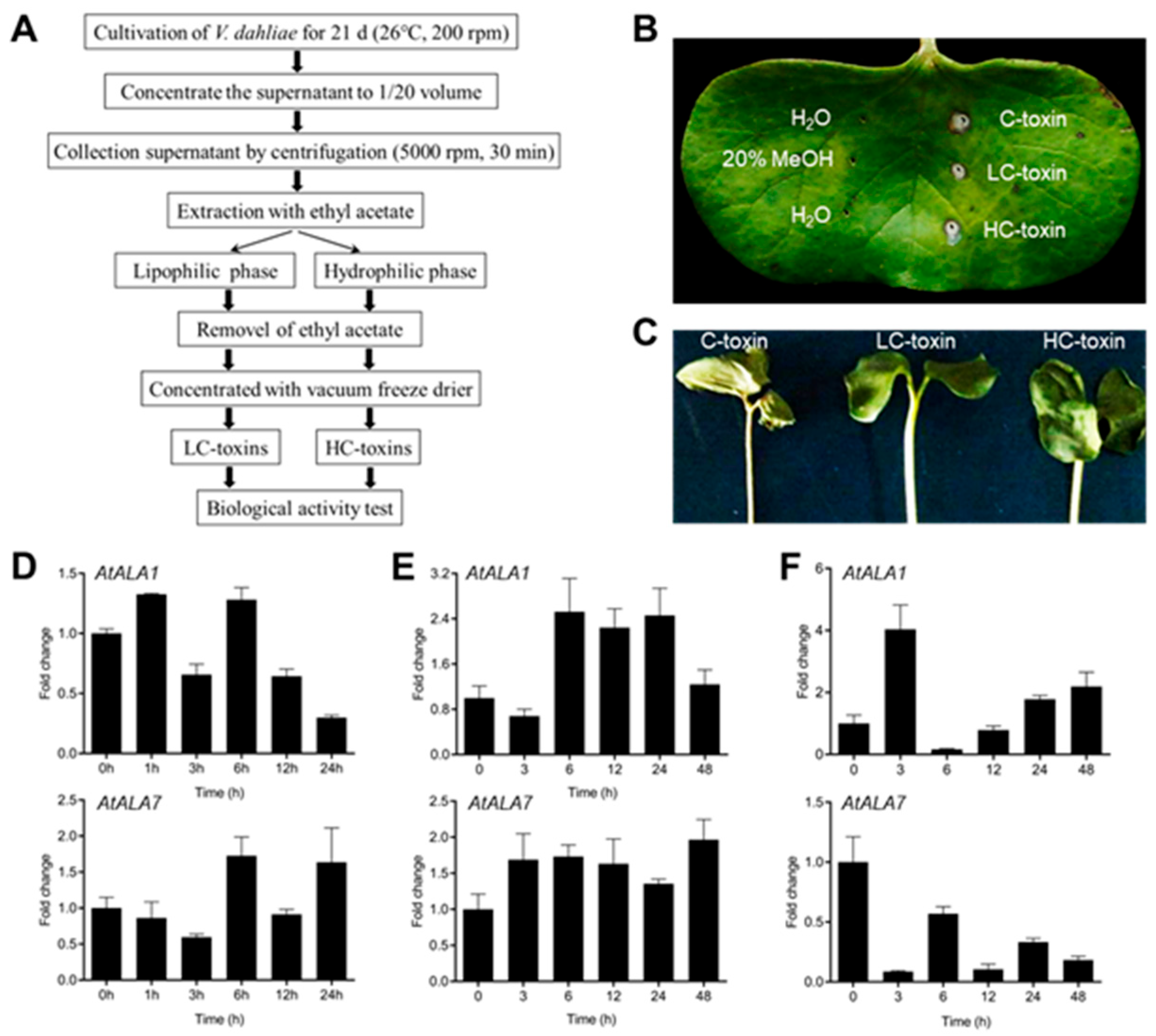
2.2. Vd-Toxins Extraction and Phytotoxicity Assays

The Vd-toxins were extracted from a highly infectious strain of V. dahliae L2-1 according to the method shown in Figure 1A. Briefly, the V. dahliae strain L2-1 was cultivated in CZM medium at 26 °C and 200 rpm for 21 d. The fungus culture was filtered through four layers of gauze and centrifuged at 10,000 rpm for 10 min to remove the spores. The supernatant was concentrated to 1/20 volume with a vacuum rotary evaporator and centrifuged at 5000 rpm for 10 min. The supernatant after centrifugation (Crude-toxins, C-toxins) was used for the extraction of lipophilic phase crude toxins (LC-toxins) from the lipophilic phase using ethyl acetate, and the extraction of hydrophilic phase crude toxins (HC-toxins) from the hydrophilic phase, respectively. Then, ethyl acetate was removed and concentrated with a vacuum freeze-drier to obtain LC-toxins and HC-toxins.
For phytotoxicity assays of Vd-toxins on cotton cotyledons, 14-day-old cotton cotyledons were pierced into holes with a 1 mL syringe needle, then inoculated with 2 μL of C-toxins, LC-toxins, or HC-toxins, respectively. H2O and 20% MeOH were used as controls. After inoculation, these cotyledons were cultured at 26 °C for 3 d with 90% humidity.
For LC-toxin tolerance assays, 2-day-old Arabidopsis plants were transferred to an MS solid medium containing LC-toxins (3 mg·mL−1) for 9 d. For CIA tolerance, 4-day-old Arabidopsis plants were transferred to an MS solid medium containing CIA (80 μg·mL−1) for 9 d. For Vd-toxin assays in root inhibition, four-day-old plants were transferred to an MS solid medium containing 4MBA (3 μg·mL−1), indazole (15 μg·mL−1), 2HPA (2 μg·mL−1), 4HBA (15 μg·mL−1), SFA (20 μg·mL−1), or 3ICD (15 μg·mL−1) for 7 d, respectively.

2.3. Isolation and Quantitative Real-Time PCR (qRT-PCR)

The wild-type Arabidopsis plants grown in MS medium for 14 d were treated with V. dahliae L2-1 (2 × 10⁸ spores/mL), LC-toxins (3 mg·mL−1), or HC-toxins (3 mg·mL−1) for 0, 3, 6, 12, 24, and 48 h. For transcriptome validation, 14-day-old wild-type Arabidopsis plants were treated with CIA (80 μg·mL−1), 3ICD (15 μg·mL−1), and indazole (15 μg·mL−1), respectively. H2O was used as a control.
RNA was extracted using the EASY Spin Plant RNA Kit (Aidlab, Beijing, China) according to the manufacturer’s instructions.
CFX Manager 3.1 software (Bio-Rad, Herculus, CA, USA) was used for data analysis. AtActin2 and NtActin were used as reference genes in Arabidopsis and tobacco, respectively. The primers are listed in Table S1.

2.4. RNA Sequencing

The 14-day-old wild-type Arabidopsis plants grown on an MS solid medium were transferred to an MS liquid medium or MS containing CIA (80 μg·mL−1) for 24 h before RNA isolation. The RNA quality was determined using an Agilent 5300 Bioanalyzer (Agilent, Santa Clara, CA, USA) and quantified using the Nanodrop 2000 (Thermo, Waltham, MA, USA). Only high-quality RNA samples (OD260/280 = 1.8~2.2, OD260/230 ≥ 2.0, RQN ≥ 6.5, 28S:18S ≥ 1) were used to construct sequencing library.
RNA purification, reverse transcription, library construction, and sequencing were performed at Shanghai Majorbio Bio-pharm Biotechnology Co., Ltd. (Shanghai, China) according to the manufacturer’s instructions. The RNA-seq transcriptome library was prepared following Illumina® Stranded mRNA Prep, Ligation (San Diego, CA, USA) using 1 μg of total RNA. Shortly after, mRNA was isolated according to the polyA selection method by oligo (dT) beads and then fragmented with the fragmentation buffer. Double-stranded cDNA was synthesized with random hexamer primers. The synthesized cDNA was subjected to end-repair, phosphorylation, and adapter addition according to the library construction protocol. Libraries were size-selected for cDNA target fragments of 300–400 bp using magnetic beads, followed by PCR amplification for 10–15 PCR cycles. After being quantified using Qubit 4.0, the sequencing library was performed on the NovaSeq X Plus platform (PE150) using the NovaSeq Reagent Kit (Illumina, San Diego, CA, USA).
To identify DEGs (differentially expressed genes) between two different samples, the expression level of each transcript was calculated according to the transcripts per million reads (TPM) method. Differential expression analysis was performed using DESeq2 or DEGseq. DEGs with |log2FC| ≥ 1 and FDR < 0.05(DESeq2) or FDR < 0.001(DEGseq) were considered to be significantly and differentially expressed genes. In addition, functional-enrichment analysis, including GO and KEGG, was performed to identify which DEGs were significantly enriched in GO terms and metabolic pathways at the Bonferroni-corrected p-value < 0.05 compared with the whole-transcriptome background. GO functional enrichment and KEGG pathway analysis were carried out on the online platform of Majorbio Cloud Platform (https://cloud.majorbio.com/, accessed on 10 December 2024).

2.5. Pathogen Inoculation and Disease Scoring

Arabidopsis and tobacco seedlings were inoculated with V. dahliae strains L2-1 (2 × 10⁸ spores/mL) as previously reported [40]. The disease symptoms were classified as level 0–4.0, no visible wilting or chlorosis symptoms; 1, 0–25% (inclusive) of true leaves wilted, chlorosis or dropped off; 2, 25–50% (inclusive) of true leaves wilted, chlorosis or dropped off; 3, 50–75% (inclusive) of true leaves wilted, chlorosis or dropped off; 4, 75–100% (inclusive) of true leaves wilted, chlorosis or dropped off [47,48,49]. The disease index (DI) was obtained according to this formula: DI = [∑ (disease grades × number of infected plants)/(total checked plants × 4)] × 100. A total of 30 Arabidopsis plants and 36 tobacco plants were used for DI analysis.

2.6. Laser Confocal Microscopy Observation

Microscopy images were acquired with a Leica SP-8 (Leica Microsystems, Wetzlar, Germany). Images of the Arabidopsis root were acquired with a 40× objective. The pinhole aperture was 1. GFP, CIAFITC, 3ICD5-FAM, and indazole5-FAM (green channel) were excited at 488 nm, and the emission spectra were 495–540 nm. FM4-64 (red channel) was excited at 552 nm, and the emission spectra were 570–610 nm. The fluorescent intensity in vacuoles was quantified via ImageJ 1.53c (http://imagej.net/ij/, accessed on 16 March 2025). This experiment was independently repeated at least three times.

2.7. Statistical Analysis

Statistical analyses were performed with one-way ANOVA with a Tukey’s multiple comparisons test using SPSS (IBM, version 19, Amonk, NY, USA), or Student’s t-test using Excel (Microsoft, version 16, Redmond, WA, USA).

3. Results

3.1. Both AtALA1 and AtALA7 Contribute to the Resistance of LC-Toxins Secreted by V. dahliae in Arabidopsis

The lipophilic phase crude toxins (LC-toxins) and hydrophilic phase crude toxins (HC-toxins) were obtained from a highly infectious strain of V. dahliae L2-1, respectively (Figure 1A). Both LC-toxins and HC-toxins showed the ability to induce necrosis and wilting symptoms in cotton cotyledon (Figure 1B,C). We previously identified Arabidopsis P4-ATPase genes AtALA1 and AtALA7 as being responsible for the cellular detoxification of mycotoxin DON and CIA, respectively [40]. The transcription level of AtALA1 and AtALA7 was induced by treatment of V. dahliae strain L2-1 and LC-toxins (Figure 1D,E), and AtALA1 was also responsive to HC-toxins treatment (Figure 1F).
To determine whether AtALA1 and AtALA7 can detoxify LC-toxins, we treated wild-type, ala1 mutant, AtALA1 overexpressed Arabidopsis lines (A1-15, A1-25), ala7 mutant, and AtALA7 overexpressed Arabidopsis lines (A7-9, A7-12) with LC-toxins and CIA, respectively. There were no significant differences among these plants in the MS medium (Figure 2A,B). In the presence of LC-toxins, ala1 and ala7 mutants showed serious toxic symptoms (Figure 2C). The root length of ala1 and ala7 mutants was significantly shorter than that of wild-type, while overexpressed lines of AtALA1 and AtALA7 were longer than wild-type (Figure 2D). With CIA treatment, the root length of AtALA7 overexpressed lines was significantly longer than wild-type, and there was no significant difference among wild-type, ala1 mutant, and AtALA1 overexpressed lines (Figure 2E,F). These results indicate that overexpression of either AtALA1 or AtALA7 can increase Arabidopsis resistance against LC-toxins.
It is reported that V. dahliae secreted various secondary metabolites, such as CIA, 3ICD, indazole, 2HPA, 4MBA, and SFA (Table S2). Treated with these Vd-toxins, wild-type Arabidopsis seedlings showed leaf necrosis and death, as well as root growth inhibition (Figure S1). We speculated that these Vd-toxins may play a key role during the colonization of V. dahliae into hosts.

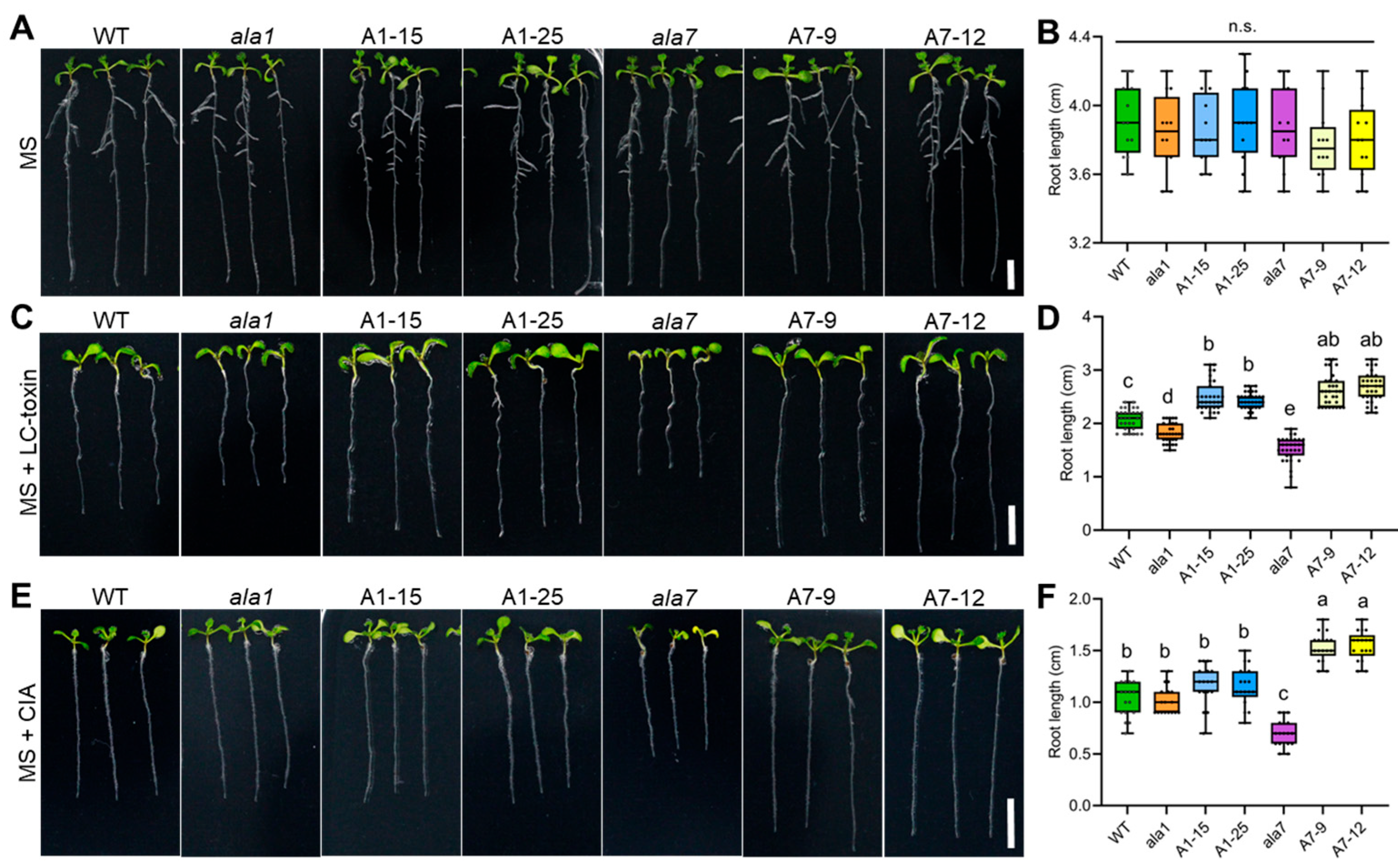
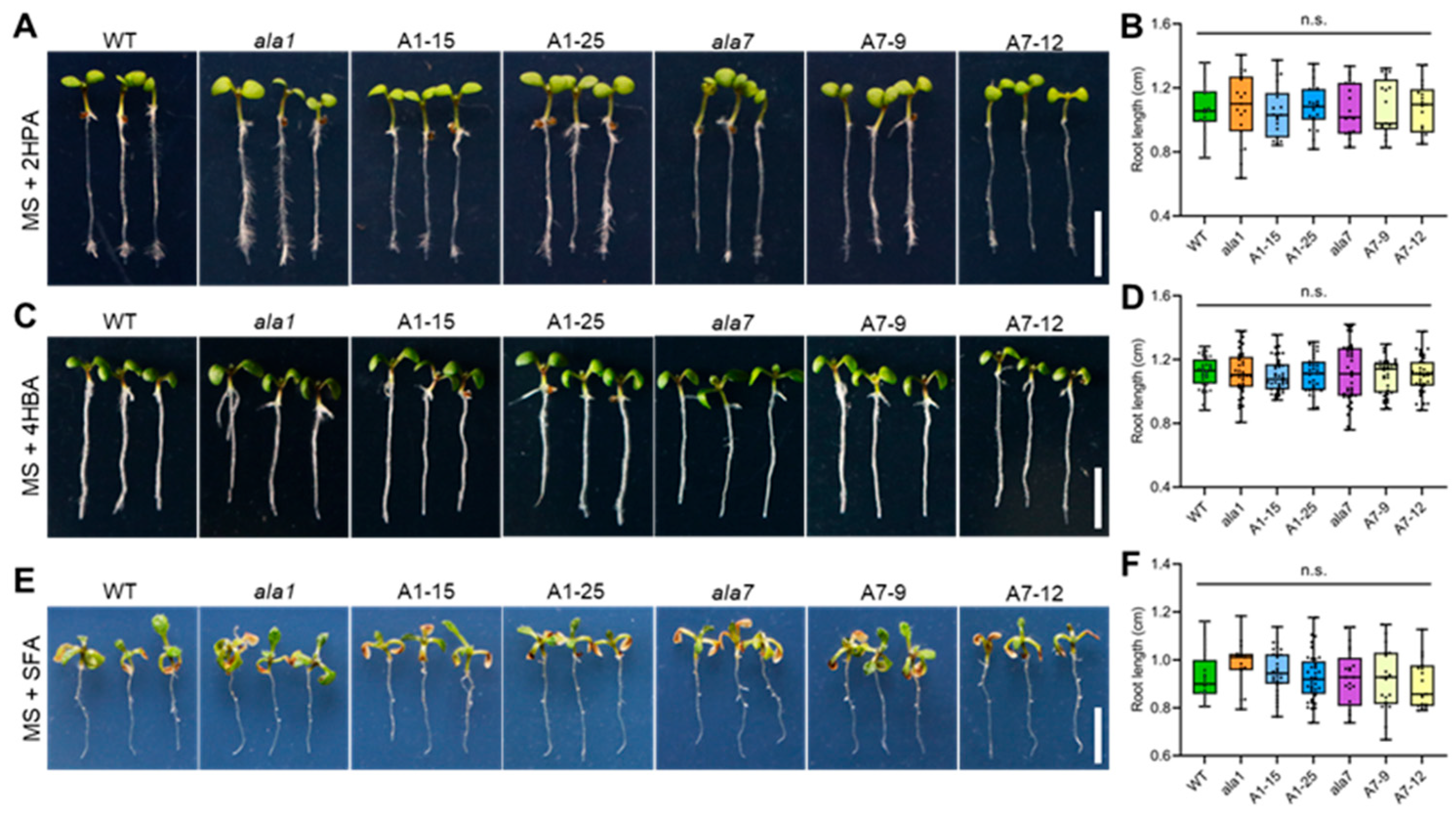
3.2. Overexpression of Either AtALA1 or AtALA7 Enhances the Resistance of Arabidopsis to LC-Toxins Secreted by V. dahliae

To understand whether AtALA1 and AtALA7 can detoxify these Vd-toxins, we treated wild-type, ala1 mutant, ala7 mutant, 35S::AtALA1, and 35S::AtALA7 overexpressed Arabidopsis lines with 4MBA, 3ICD, indazole, 2HPA, 4HBA, and SFA, individually. Treated with 4MBA, the root length of AtALA1 and AtALA7 transgenic plants was significantly longer than that of wild-type, while ala1 and ala7 mutants were shorter than that of wild-type (Figure 3A–D). Similar to the 4MBA treatment, the transgenic plants of AtALA1 and AtALA7 showed less inhibited root growth than wild-type under 3ICD treatment (Figure 3E,F). The root length of AtALA1 transgenic plants was significantly longer than that of wild-type, and there was no difference among ala7 mutants, AtALA7 transgenic plants, and wild-type with indazole treatment (Figure 3G,H). By contrast, overexpression of either AtALA1 or AtALA7 did not improve the resistance of Arabidopsis to 2HPA (Figure 4A,B), 4HBA (Figure 4C,D), and SFA (Figure 4E,F). These results showed that the overexpression of AtALA1 improved the resistance of Arabidopsis to Vd-toxins of 4MBA, 3ICD, and indazole, while overexpressed AtALA7 enhanced resistance to 4MBA, 3ICD, and CIA.

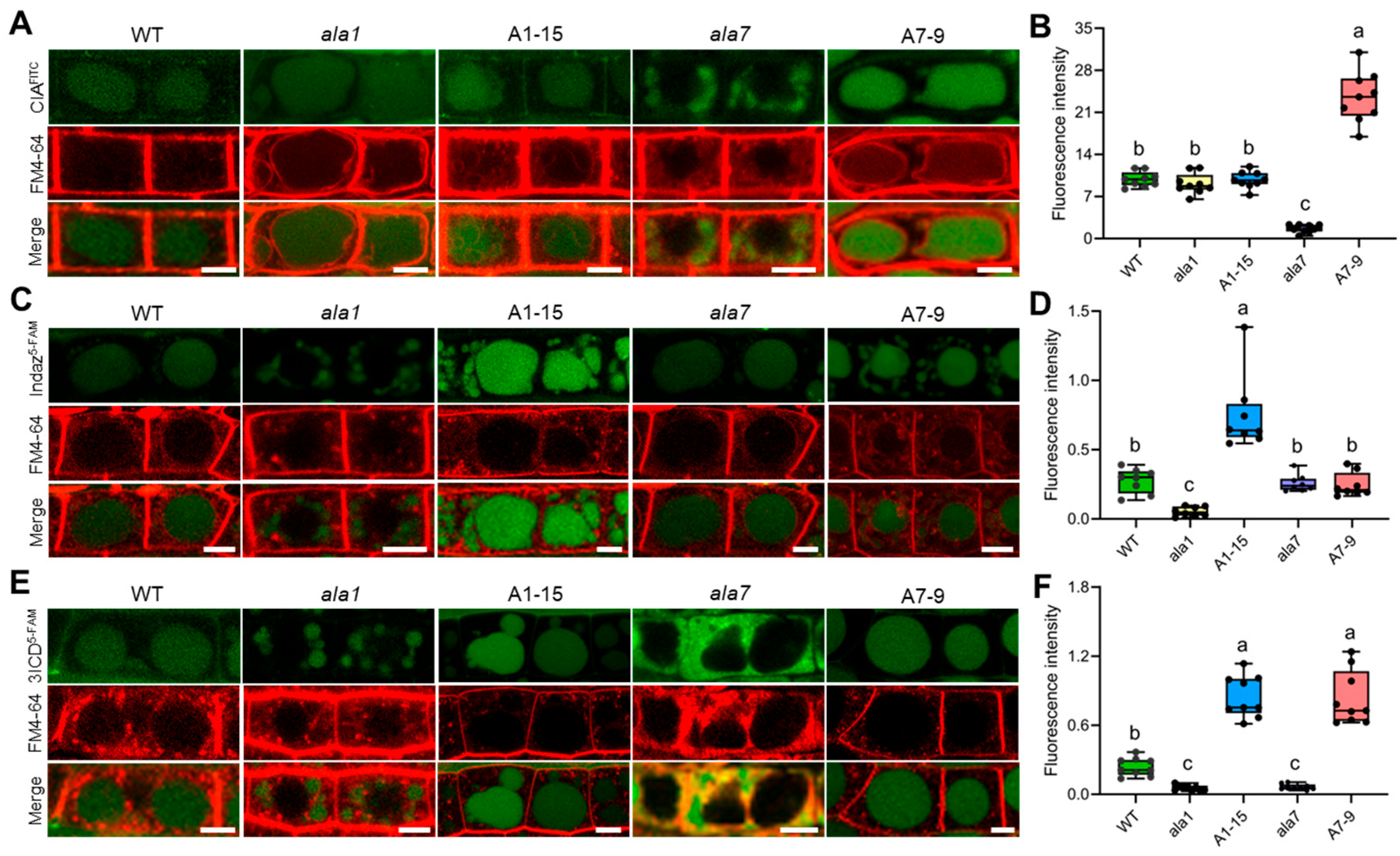
3.3. Overexpression of AtALA1 Promotes Transport Indazole and 3ICD into Vacuoles, While AtALA7 Accumulates CIA and 3ICD to Vacuoles

To investigate the function of AtALA1 and AtALA7 in Vd-toxins detoxification, we treated wild-type, ala1 mutant, ala7 mutant, AtALA1, and AtALA7 overexpressed lines with CIAFITC (CIA was labeled with fluorescein-isothiocyanate-isomer), indazole5-FAM (indazole was labeled with 5-carboxyfluorescein), and 3ICD5-FAM (3ICD was labeled with 5-carboxyfluorescein), respectively, which did not impair the toxicity of either indazole (Figure S2A,B) or 3ICD (Figure S2C,D). Similar to our previous result, the transgenic plants overexpressing AtALA7 exhibited a stronger ability to accumulate CIAFITC into vacuoles than the wild-type, while AtALA1 was invalid (Figure 5A,B). With indazole5-FAM treatment, the transgenic plants overexpressing AtALA1 exhibited a more powerful capacity to accumulate indazole5-FAM into vacuoles than the wild-type, while AtALA7 was ineffective (Figure 5C,D). Interestingly, overexpression of either AtALA1 or AtALA7 promotes 3ICD5-FAM accumulation into vacuoles (Figure 5E,F). These results demonstrated that AtALA1 is involved in detoxifying indazole and 3ICD, while AtALA7 is responsible for detoxifying CIA and 3ICD.

3.4. AtALA1 or AtALA7 Protect Arabidopsis Plants from ROS Toxicity Triggered by Vd-Toxins

To understand the molecular basis of resistance to Vd-toxins in plants, we analyzed the transcriptomic changes in response to CIA. A total of 7104 DEGs were identified (p-adjust ≤ 0.001 and Log2 fold change ≥ 1), including 1247 up-regulated genes and 5857 down-regulated genes (Figure 6A). GO enrichment analysis showed these DEGs are mainly involved in the cellular response to chemical, hypoxia, oxygen levels, stimulus, drug, toxic substance, heat, and hydrogen peroxide (Figure 6B). KNGG analysis showed these DEGs involved in plant–pathogen interactions, MAPK signaling pathways, and peroxisomes (Figure 6C).
We, therefore, analyze the relative expression changes of ROS-related genes (RbohD, PRX33, GSTU1, CAT1, and GAPC1) in wild-type Arabidopsis under CIA, 3ICD, and indazole treatment, respectively. The qRT-PCR results indicated that all of these selected genes were up-regulated under CIA or 3ICD treatment (Figure 6D); the relative expression of GSTU1 and CAT1 was up-regulated, while RbohD and PRX33 were down-regulated under indazole treatment (Figure 6D). These results suggest that Vd-toxins may exert a toxic function by inducing oxidative stress in plant cells.

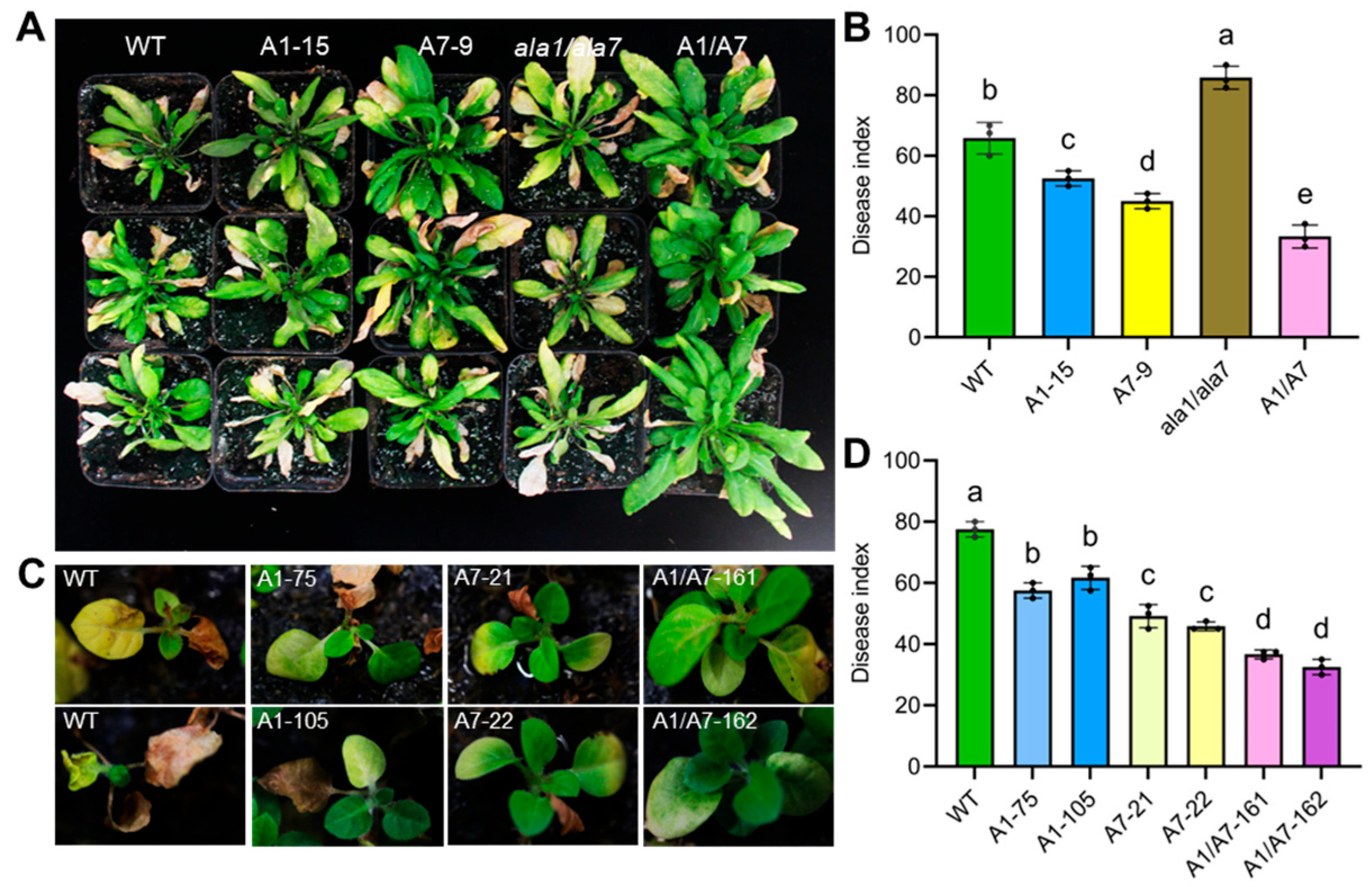
3.5. Aggregation of AtALA1 and AtALA7 Enhances the Plant’s Resistance to V. dahliae

We further analyzed the resistance of AtALA1 and AtALA7 transgenic Arabidopsis to a strong pathogenic strain, V. dahliae L2-1. After inoculation, the ala1/ala7 mutant presented the most serious disease symptoms, while the hybrid progeny of AtALA1 and AtALA7 (A1/A7) showed fewer symptoms (Figure 7A). The DI of AtALA1 and AtALA7 transgenic Arabidopsis was significantly lower than wild-type, while the hybrid progeny (A1/A7) was lower than plants expressing AtALA1 or AtALA7 alone (Figure 7B). Similarly, the DI of hybrid tobacco progeny (A1/A7) was lower than plants expressing AtALA1 or AtALA7 alone (Figure 7C,D). These data suggest that the expression of AtALA1 and AtALA7 simultaneously can further enhance plant resistance against V. dahliae.

4. Discussion

Due to the broad host range, complicated pathogenic mechanisms, high genetic diversity, and the lack of resistance germplasm resources to Verticillium species, the control of Vw is far from successful [50,51,52]. Many low molecular secondary metabolites (also called mycotoxins) secreted by pathogenic fungi play a crucial role during pathogenesis [9,53,54]. V. dahliae belongs to hemibiotrophic pathogens. These pathogens secrete low levels of mycotoxins to suppress the host’s immune response and thus promote their colonization at an early stage of infection, and also generate high levels of mycotoxins to stimulate cell death [55,56]. For example, the trichothecenes family of phytotoxins, mainly produced by necrotrophic fungal phytopathogenic Fusarium species, contains more than 200 members and is divided into four types, and T-2 toxin is the most dangerous mycotoxins [57,58]. The Vd-toxins 3ICD, indazole, 2HPA, 4MBA, and SFA cause leaf necrosis and death, and root growth inhibition in wild-type Arabidopsis seedlings with different concentrations (Figure S1, Figure 3 and Figure 4), implying that Vd-toxins play different roles during the pathogenesis process, which may be related to their structure, sites of action, and targets in plants.
The detoxification of mycotoxins has been developed to control these formidable diseases [59,60,61]. Nowadays, some Vd-toxins have been identified from V. dahliae [1,11,12,17,22,62,63], which are involved in inducing Vw-like symptoms [11,53,64], altering microtubule cytoskeletons and nucleoli [19,21], inhibiting sphingolipid synthesis [17], and causing disorder of hydrogen peroxide [22]. However, the studies on improving the cell detoxification function of plants against Vd-toxins are limited. We recently cloned the BbCRPA gene from P4-ATPase of Beauveria bassiana, which is involved in the detoxification of mycotoxins CsA and FK506 via a vesicle trafficking pathway, and exogenous overexpression of BbCRPA enhances the resistance to V. dahliae via transport CIA to vacuoles in Arabidopsis [24]. We also identified Arabidopsis P4-ATPase genes AtALA1 and AtALA7, which contribute to cellular detoxification of DON and CIA, respectively, using a classical clathrin-associated endocytosis mechanism [40].
In this study, we demonstrate that AtALA1, a membrane of P4-ATPases, mediates the detoxification of 4MBA, indazole, and 3ICD via vesicle trafficking, whereas AtALA7 facilitates CIA, 4MBA, and 3ICD (Figure 3 and Figure 5). Notably, co-expression of AtALA1 and AtALA7 synergistically enhances plant resistance against V. dahliae (Figure 7). These low-molecular-weight Vd-toxins exhibit high lipophilicity, as evidenced by their high LogP values (for CIA, 2.62; indazole, 1.82; 4MBA, 2.36; 3ICD, 1.68; Table S2). Thus, we supposed that the cargo mediated by P4 ATPase-associated vesicle trafficking may be a group of molecules with similar physicochemical or biological properties. It is reported that AtALA1 is also involved in chilling tolerance and antiviral RNA interference [35,38,39]. The lipid substrate of AtALA1 was PS (phosphatidylserine), while with AtALA7 is still unclear [28,35,65]. It seems that the P4-ATPases are unlikely to directly participate in compound recognition but rather act as transporters within broader regulatory networks. For instance, mammal P4-ATPase ATP11C undergoes endocytosis mediated by serotonin or histamine binding to G protein-coupled receptors (GPCRs), followed by Ca2+ influx and PKCa-dependent phosphorylation of its C-terminus [66]. Recently, our work revealed that the DON-stimulated phosphorylation in the C-terminus is responsible for promoting DON trafficking into vacuoles, and the WNK10 kinase could interact with the C-terminus of AtALA1 [66]. Therefore, indazole, 3ICD, and DON may represent a subset of cargo trafficked by AtALA1-mediated vesicle pathways. Elucidating the upstream signaling components of the plasma membrane/intracellular under Vd-toxin induction, and combining with biochemical assays, may provide a basis for recognizing structurally diverse compounds.
Many mycotoxins were reported to induce oxidative stress, thereby inducing apoptosis [67]. For example, both T-2 and DON-induced apoptosis are related to DNA methylation; DON triggers early-stage apoptosis and autophagy [68,69,70,71]. The ROS could be produced in the plasma membrane (RbohD), peroxisome (PRX33), and mitochondria, while the CAT1 and GSTU1 are responsible for ROS scavenging [72]. The cytosolic glycolytic enzymes glyceraldehyde-3-phosphate dehydrogenases (GAPCs) were reported to interact with the plasma membrane-associated phospholipase D to transduce hydrogen peroxide in Arabidopsis [73]. The expression of RbohD, RbohF, and GST1 was upregulated under Vd-toxin treatment [22]. In this study, DEGs under CIA treatment are involved in the cellular response to oxygen levels and hydrogen peroxide (Figure 6B), and ROS-related genes such as RbohD, GSTU1, PRX33, GAPC1, and CAT1 were up-regulated in wild-type Arabidopsis under CIA and 3ICD (Figure 6D), indicating that Vd-toxins, such as CIA and 3ICD, also triggered the response to ROS-related genes in plants. The P4-ATPases AtALA1 and AtALA7 achieve detoxification via transport of Vd-toxins to vacuoles. Once inside cells, Vd-toxins are encapsulated in vesicles, preventing the contact and binding of toxins to their intracellular targets, thus protecting the innate immunity system of hosts from damage and increasing the resistance of hosts against invasion of V. dahliae. Ultimately, the Vd-toxins are transported into vacuoles to be sequestered or digested.

5. Conclusions

In this study, we investigated the damage of Vd-toxins to plants. Both LC-toxins and HC-toxins showed the ability to induce necrosis and wilting symptoms in cotton cotyledons. Vd-toxins such as CIA, 3ICD, indazole, 2HPA, 4MBA, and SFA showed leaf necrosis and root growth inhibition. Overexpressing AtALA1 improved the resistance of Arabidopsis to 4MBA, 3ICD, and indazole, while overexpressing AtALA7 enhanced resistance to 4MBA, 3ICD, and CIA. By observing the distribution of fluorescently labeled Vd-toxins (CIAFITC, indazole5-FAM, and 3ICD5-FAM) in Arabidopsis root cells, we demonstrated that AtALA7 overexpressing plants exhibited a stronger ability to accumulate CIAFITC into vacuoles than wild-type, while AtALA1 was invalid; AtALA1 overexpressing plants showed a stronger accumulate indazole5-FAM into vacuoles than wild-type, while overexpression of either AtALA1 or AtALA7 promotes 3ICD5-FAM accumulation into vacuoles, indicating that AtALA1 is involved in detoxifying indazole and 3ICD, while AtALA7 is responsible for cell detoxification CIA and 3ICD. The ROS-related genes, such as RbohD, PRX33, GAPC1, and CAT1, were up-regulated under Vd-toxins treatment. Furthermore, the expression of AtALA1 and AtALA7 simultaneously further enhances plant resistance against V. dahliae.

Supplementary Materials

The following supporting information can be downloaded at https://www.mdpi.com/article/10.3390/biology14060595/s1. Figure S1: Vd-toxins tolerance assay of wild-type Arabidopsis seedling; Figure S2: 5-FAM labeled do not impair the toxicity of indazole and 3ICD to wild-type Arabidopsis. Table S1: Primers used in this study. Table S2: Summary of reported Vd-toxins of V. dahliae.

Author Contributions

Y.P. conceived and supervised the project. F.W. performed resistance analysis on Vd-toxins and V. dahliae, fluorescence observation of Vd-toxins, and data analysis; M.Q., J.L. and H.R. participated in Arabidopsis transformation and management; X.Y., J.S., C.L. and Q.J. participated in V. dahliae cultivation, Vd-toxins extraction, and qRT-PCR analysis; M.S., Z.Z., Y.L. and J.T. participated in tobacco transformation, transgenic plants identification, and hybridization; X.L. and Y.F. participated in transcriptomic analysis; Y.P. and F.W. wrote the paper with input from all other authors. All authors have read and agreed to the published version of the manuscript.

Funding

This work was funded by “The National Natural Science Foundation of China (32202320 to F.W.)”, “Fundamental Research Funds for the Central Universities (Grant No. SWU-KQ22011 to F.W.)”, “Chongqing Natural Science Foundation Project (Grant No. CSTB2022NSCQ-MSX0625 to F.W.)”, and “National Transgenic New Species Breeding Major Project of China (Grant No. 2016ZX08005-003-004 to Y.P)”.

Institutional Review Board Statement

Not applicable.

Informed Consent Statement

Not applicable.

Data Availability Statement

The data presented in this study are available on request from the corresponding author. The data are not publicly available due to privacy.

Acknowledgments

We are grateful to Zhiying Ma (Hebei Agricultural University, China) for his kind donation of V. dahliae strain L2-1.

Conflicts of Interest

The authors declare no conflicts of interest.

Abbreviations

The following abbreviations are used in this manuscript:
CIACinnamyl acetate
cAMPCyclic adenosine monophosphate
C-toxinsCrude toxin
DGEsDifferentially expressed genes
HC-toxinsHydrophilic phase crude toxin
H2O2Hydrogen peroxide
KNGGKyoto encyclopedia of genes and genomes
LC-toxinsLipophilic phase crude toxin
qRT-PCRQuantitative real-time polymerase chain reaction
rpmRevolutions per minute
SFASulfacetamide
VwVerticillium wilt
2HPA2-hydroxypenylacetic acid
3ICDIndole-3-carboxaldehyde
4HBA4-hydroxybenzoic acid
4MBA4-methylbenzoic acid
5-FAM5-carboxyfluorescein

References

  1. Fradin, E.F.; Thomma, B.P. Physiology and molecular aspects of Verticillium wilt diseases caused by V. dahliae and V. albo-atrum. Mol. Plant Pathol. 2006, 7, 71–86. [Google Scholar] [CrossRef]
  2. Pegg, G.F.; Brady, B.L. Verticillium Wilts; CABI: Wallingford, UK, 2002.
  3. Bhat, R.G.; Subbarao, K.V. Host range specificity in Verticillium dahliae. Phytopathology 1999, 89, 1218–1225. [Google Scholar] [CrossRef] [PubMed]
  4. Jiménez-Díaz, R.M.; Cirulli, M.; Bubici, G.; Del Mar Jiménez-Gasco, M.; Antoniou, P.P.; Tjamos, E.C. Verticillium wilt, a major threat to olive production: Current status and future prospects for its management. Plant Dis. 2012, 96, 304–329. [Google Scholar] [CrossRef] [PubMed]
  5. Baeza-Montañez, L.; Gómez-Cabrera, R.; García-Pedrajas, M. First report of Verticillium wilt caused by Verticillium dahliae on mango trees (Mangifera indica) in southern Spain. Plant Dis. 2010, 94, 380. [Google Scholar] [CrossRef] [PubMed]
  6. Wen, H.G.; Zhao, J.H.; Zhang, B.S.; Gao, F.; Wu, X.M.; Yan, Y.S.; Zhang, J.; Guo, H.S. Microbe-induced gene silencing boosts crop protection against soil-borne fungal pathogens. Nat. Plants 2023, 9, 1409–1418. [Google Scholar] [CrossRef]
  7. Klosterman, S.J.; Atallah, Z.K.; Vallad, G.E.; Subbarao, K.V. Diversity, pathogenicity, and management of Verticillium species. Annu. Rev. Phytopathol. 2009, 47, 39–62. [Google Scholar] [CrossRef]
  8. Zhang, Y.; Zhou, J.; Zhao, L.; Feng, Z.; Wei, F.; Bai, H.; Feng, H.; Zhu, H. A review of the pathogenicity mechanism of Verticillium dahliae in cotton. J. Cotton Res. 2022, 5, 3. [Google Scholar] [CrossRef]
  9. Nachmias, A.; Buchner, V.; Burstein, Y. Biological and immunochemical characterization of a low molecular weight phytotoxin isolated from a protein-lipopolysaccharide complex produced by a potato isolate of Verticillium dahliae Kleb. Physiol. Plant Pathol. 1985, 26, 43–55. [Google Scholar] [CrossRef]
  10. Palmer, C.S.; Saleeba, J.A.; Lyon, B.R. Phytotoxicity on cotton ex-plants of an 18.5kDa protein from culture filtrates of Verticillium dahliae. Physiol. Mol. Plant Pathol. 2005, 67, 308–318. [Google Scholar] [CrossRef]
  11. Laouane, H.; Sedra, M.H.; Lazrek, H.B. Isolation and characterization of a new phytotoxic molecule from culture fluids of Verticillium dahliae. Glob. J. Inc. 2013, 13, 37–42. [Google Scholar]
  12. Jia, Z.Q.; Shi, F.M.; Yuan, H.Y.; Hou, Y.Y.; Li, Y.Z. Comparison of methods of purification and identification of Verticillium dahliae toxins. J. Life Sci. 2007, 1, 67–73. [Google Scholar]
  13. Buchner, V.; Nachmias, A.; Burstein, Y. Isolation and partial characterization of a phytotoxic glycopeptide from a protein-lipopolysaccharide complex produced by a potato isolate of Verticillium dahliae. FEBS Lett. 1982, 138, 261–264. [Google Scholar] [CrossRef]
  14. Mansoori, B.; Milton, J.; Smith, C. Isolation and partial purification of a phytotoxin related to pathogenic Verticillium species. J. Phytopathol. 1995, 143, 33–36. [Google Scholar] [CrossRef]
  15. Cui, X.C.; Luo, D.Q. The study of secondary metabolites of Verticillium dahliae. Asia-Pac. Tradit. Med. 2013, 9, 47–48. [Google Scholar]
  16. Zhang, D.D.; Wang, X.Y.; Chen, J.Y.; Kong, Z.Q.; Gui, Y.J.; Li, N.Y.; Bao, Y.M.; Dai, X.F. Identification and characterization of a pathogenicity-related gene VdCYP1 from Verticillium dahliae. Sci. Rep. 2016, 6, 27979. [Google Scholar] [CrossRef]
  17. Xu, F.; Huang, L.; Wang, J.Y.; Ma, C.X.; Tan, Y.Q.; Wang, F.L.; Fan, Y.H.; Luo, M. Sphingolipid synthesis inhibitor fumonisin B1 causes verticillium wilt in cotton. J. Integr. Plant Biol. 2022, 64, 836–842. [Google Scholar] [CrossRef] [PubMed]
  18. Jiang, J.; Fan, L.W.; Wu, W.H. Evidences for involvement of endogenous cAMP in Arabidopsis defense responses to Verticillium toxins. Cell Res. 2005, 15, 585–592. [Google Scholar] [CrossRef]
  19. Yuan, H.Y.; Yao, L.L.; Jia, Z.Q.; Li, Y.; Li, Y.Z. Verticillium dahliae toxin induced alterations of cytoskeletons and nucleoli in Arabidopsis thaliana suspension cells. Protoplasma 2006, 229, 75–82. [Google Scholar] [CrossRef]
  20. Shi, F.M.; Yao, L.L.; Pei, B.L.; Zhou, Q.; Li, X.L.; Li, Y.; Li, Y.Z. Cortical microtubule as a sensor and target of nitric oxide signal during the defence responses to Verticillium dahliae toxins in Arabidopsis. Plant Cell Environ. 2009, 32, 428–438. [Google Scholar] [CrossRef]
  21. Yao, L.L.; Zhou, Q.; Pei, B.L.; Li, Y.Z. Hydrogen peroxide modulates the dynamic microtubule cytoskeleton during the defence responses to Verticillium dahliae toxins in Arabidopsis. Plant Cell Environ. 2011, 34, 1586–1598. [Google Scholar] [CrossRef]
  22. Zhao, J.; Chen, Q.; Zhou, S.; Sun, Y.; Li, X.; Li, Y. H2Bub1 regulates RbohD-dependent hydrogen peroxide signal pathway in the defense responses to Verticillium dahliae toxins. Plant Physiol. 2020, 182, 640–657. [Google Scholar] [CrossRef] [PubMed]
  23. Wang, H.W.; Sun, S.L.; Ge, W.Y.; Zhao, L.F.; Hou, B.Q.; Wang, K.; Lyu, Z.F.; Chen, L.Y.; Xu, S.S.; Guo, J.; et al. Horizontal gene transfer of Fhb7 from fungus underlies Fusarium head blight resistance in wheat. Science 2020, 368, eaba5435. [Google Scholar] [CrossRef] [PubMed]
  24. Li, Y.J.; Ren, H.; Wang, F.L.; Chen, J.J.; Ma, L.; Chen, Y.; Li, X.B.; Fan, Y.H.; Jin, D.; Hou, L.; et al. Cell detoxification of secondary metabolites by P4-ATPase-mediated vesicle transport. Elife 2023, 12, e79179. [Google Scholar] [CrossRef]
  25. Yang, J.; Liang, K.; Ke, H.; Zhang, Y.B.; Meng, Q.; Gao, L.; Fan, J.P.; Li, G.H.; Zhou, H.; Xiao, J.Y.; et al. Enzymatic degradation of deoxynivalenol with the engineered detoxification enzyme Fhb7. JACS Au 2024, 4, 619–634. [Google Scholar] [CrossRef] [PubMed]
  26. He, W.J.; Yang, P.; Huang, T.; Liu, Y.F.; Zhang, Y.W.; Zhang, W.M.; Zhang, T.T.; Zheng, M.R.; Ma, L.; Zhao, C.X.; et al. Detoxifying bacterial genes for deoxynivalenol epimerization confer durable resistance to Fusarium head blight in wheat. Plant Biotechnol. J. 2024, 22, 619–634. [Google Scholar] [CrossRef]
  27. Sakuragi, T.; Nagata, S. Regulation of phospholipid distribution in the lipid bilayer by flippases and scramblases. Nat. Rev. Mol. Cell Biol. 2023, 24, 576–596. [Google Scholar] [CrossRef]
  28. López-Marqués, R.L. Lipid flippases as key players in plant adaptation to their environment. Nat. Plants 2021, 7, 1188–1199. [Google Scholar] [CrossRef]
  29. Timcenko, M.; Lyons, J.A.; Januliene, D.; Ulstrup, J.J.; Dieudonné, T.; Montigny, C.; Ash, M.R.; Karlsen, J.L.; Boesen, T.; Kühlbrandt, W.; et al. Structure and autoregulation of a P4-ATPase lipid flippase. Nature 2019, 571, 366–370. [Google Scholar] [CrossRef]
  30. Graham, T.R. Flippases and vesicle-mediated protein transport. Trends Cell Biol. 2004, 14, 670–677. [Google Scholar] [CrossRef]
  31. Zhou, Y.L.; Yang, Y.; Niu, Y.; Fan, T.T.; Qian, D.; Luo, C.X.; Shi, Y.M.; Li, S.; An, L.Z.; Xiang, Y. The tip-localized phosphatidylserine established by Arabidopsis ALA3 is crucial for Rab GTPase-mediated vesicle trafficking and pollen tube growth. Plant Cell 2020, 32, 3170–3187. [Google Scholar] [CrossRef]
  32. Zhang, X.X.; Adamowski, M.; Marhava, P.; Tan, S.T.; Zhang, Y.Z.; Rodriguez, L.; Zwiewka, M.; Pukysova, V.; Sanchez, A.S.; Raxwal, V.K.; et al. Arabidopsis flippases cooperate with ARF GTPase exchange factors to regulate the trafficking and polarity of PIN auxin transporters. Plant Cell 2020, 32, 1644–1664. [Google Scholar] [CrossRef] [PubMed]
  33. Yang, Y.; Niu, Y.; Chen, T.; Zhang, H.K.; Zhang, J.; Qian, D.; Bi, M.M.; Fan, Y.M.; An, L.Z.; Xiang, Y. The phospholipid flippase ALA3 regulates pollen tube growth and guidance in Arabidopsis. Plant Cell 2022, 34, 3718–3736. [Google Scholar] [CrossRef]
  34. Underwood, W.; Ryan, A.; Somerville, S.C. An Arabidopsis lipid flippase is required for timely recruitment of defenses to the host-pathogen interface at the plant cell surface. Mol. Plant 2017, 10, 805–820. [Google Scholar] [CrossRef] [PubMed]
  35. Gomès, E.; Jakobsen, M.K.; Axelsen, K.B.; Geisler, M.; Palmgren, M.G. Chilling tolerance in Arabidopsis involves ALA1, a member of a new family of putative aminophospholipid translocases. Plant Cell 2000, 12, 2441–2454. [Google Scholar] [CrossRef]
  36. Niu, Y.; Qian, D.; Liu, B.; Ma, J.; Wan, D.; Wang, X.; He, W.; Xiang, Y. ALA6, a P4-type ATPase, is involved in heat stress responses in Arabidopsis thaliana. Front. Plant Sci. 2017, 8, 1732. [Google Scholar] [CrossRef]
  37. McDowell, S.C.; Lopez-Marques, R.L.; Poulsen, L.R.; Palmgren, M.G.; Harper, J.F. Loss of the Arabidopsis thaliana P4-ATPase ALA3 reduces adaptability to temperature stresses and impairs vegetative, pollen, and ovule development. PLoS ONE 2013, 8, e62577. [Google Scholar] [CrossRef] [PubMed]
  38. Zhu, B.; Gao, H.; Xu, G.; Wu, D.; Song, S.; Jiang, H.; Zhu, S.; Qi, T.; Xie, D. Arabidopsis ALA1 and ALA2 mediate RNAi-based antiviral immunity. Front. Plant Sci. 2017, 8, 422. [Google Scholar] [CrossRef]
  39. Guo, Z.; Lu, J.; Wang, X.; Zhan, B.; Li, W.; Ding, S.W. Lipid flippases promote antiviral silencing and the biogenesis of viral and host siRNAs in Arabidopsis. Proc. Natl. Acad. Sci. USA 2017, 114, 1377–1382. [Google Scholar] [CrossRef]
  40. Wang, F.L.; Li, X.B.; Li, Y.J.; Han, J.; Chen, Y.; Zeng, J.Y.; Su, M.; Zhuo, J.X.; Ren, H.; Liu, H.R.; et al. Arabidopsis P4 ATPase-mediated cell detoxification confers resistance to Fusarium graminearum and Verticillium dahliae. Nat. Commun. 2021, 12, 6426. [Google Scholar] [CrossRef]
  41. Davis, J.A.; Pares, R.B.; Bernstein, T.; McDowell, S.C.; Brown, E.; Stubrich, J.; Rosenberg, A.; Cahoon, E.B.; Cahoon, R.E.; Poulsen, L.R.; et al. The lipid flippases ALA4 and ALA5 play critical roles in cell expansion and plant growth. Plant Physiol. 2020, 182, 2111–2125. [Google Scholar] [CrossRef]
  42. Botella, C.; Sautron, E.; Boudiere, L.; Michaud, M.; Dubots, E.; Yamaryo-Botte, Y.; Albrieux, C.; Marechal, E.; Block, M.A.; Jouhet, J. ALA10, a phospholipid flippase, controls FAD2/FAD3 desaturation of phosphatidylcholine in the ER and affects chloroplast lipid composition in Arabidopsis thaliana. Plant Physiol. 2016, 170, 1300–1314. [Google Scholar] [CrossRef] [PubMed]
  43. Poulsen, L.R.; Lopez-Marques, R.L.; McDowell, S.C.; Okkeri, J.; Licht, D.; Schulz, A.; Pomorski, T.; Harper, J.F.; Palmgren, M.G. The Arabidopsis P4-ATPase ALA3 localizes to the Golgi and requires α-subunit to function in lipid translocation and secretory vesicle formation. Plant Cell 2008, 20, 658–676. [Google Scholar] [CrossRef]
  44. Sanz-Fernández, M.; Rodríguez-Serrano, M.; Sevilla-Perea, A.; Pena, L.; Mingorance, M.D.; Sandalio, L.M.; Romero-Puertas, M.C. Screening Arabidopsis mutants in genes useful for phytoremediation. J. Hazard. Mater. 2017, 335, 143–151. [Google Scholar] [CrossRef] [PubMed]
  45. Li, X.B.; Niu, G.Q.; Fan, Y.H.; Liu, W.Y.; Wu, Q.; Yu, C.; Wang, J.; Xiao, Y.H.; Hou, L.; Jin, D.; et al. Synthetic dual hormone-responsive promoters enable engineering plants with broad-spectrum resistance. Plant Commun. 2023, 4, 100596. [Google Scholar] [CrossRef] [PubMed]
  46. Zeng, J.Y.; Yan, X.Y.; Bai, W.Q.; Zhang, M.; Chen, Y.; Li, X.B.; Hou, L.; Zhao, J.; Ding, X.Y.; Liu, R.C.; et al. Carpel-specific downregulation of GhCKXs in cotton significantly enhances seed and fiber yield. J. Exp. Bot. 2022, 73, 6758–6772. [Google Scholar] [CrossRef]
  47. Xu, L.; Zhang, W.W.; He, X.; Liu, M.; Zhang, K.; Shaban, M.; Sun, L.; Zhu, J.C.; Luo, Y.J.; Yuan, D.J.; et al. Functional characterization of cotton genes responsive to Verticillium dahliae through bioinformatics and reverse genetics strategies. J. Exp. Bot. 2014, 65, 6679–6692. [Google Scholar] [CrossRef]
  48. Cheng, H.Q.; Han, L.B.; Yang, C.L.; Wu, X.M.; Zhong, N.Q.; Wu, J.H.; Wang, F.X.; Wang, H.Y.; Xia, G.X. The cotton MYB108 forms a positive feedback regulation loop with CML11 and participates in the defense response against Verticillium dahliae infection. J. Exp. Bot. 2016, 67, 1935–1950. [Google Scholar] [CrossRef]
  49. Han, L.B.; Li, Y.B.; Wang, F.X.; Wang, W.Y.; Liu, J.; Wu, J.H.; Zhong, N.Q.; Wu, S.J.; Jiao, G.L.; Wang, H.Y.; et al. The cotton apoplastic protein CRR1 stabilizes chitinase 28 to facilitate defense against the fungal pathogen Verticillium dahliae. Plant Cell 2019, 31, 520–536. [Google Scholar] [CrossRef]
  50. Hua, C.; Zhao, J.H.; Guo, H.S. Trans-kingdom RNA silencing in plant–fungal pathogen interactions. Mol. Plant 2018, 11, 235–244. [Google Scholar] [CrossRef]
  51. De Coninck, B.; Timmermans, P.; Vos, C.; Cammue, B.P.; Kazan, K. What lies beneath: Belowground defense strategies in plants. Trends Plant Sci. 2015, 20, 91–101. [Google Scholar] [CrossRef]
  52. Inderbitzin, P.; Subbarao, K.V. Verticillium systematics and evolution: How confusion impedes Verticillium wilt management and how to resolve it. Phytopathology 2014, 104, 564–574. [Google Scholar] [CrossRef]
  53. Mansoori, B.; Smith, J. Verticillium-toxins their role in pathogenesis. J. Agric. Sci. Technol 2005, 7, 103–114. [Google Scholar]
  54. Keller, N.P. Fungal secondary metabolism: Regulation, function and drug discovery. Nat. Rev. Microbiol. 2019, 17, 167–180. [Google Scholar] [CrossRef]
  55. Kemen, E.; Jones, J.D.G. Obligate biotroph parasitism: Can we link genomes to lifestyles? Trends Plant Sci. 2012, 17, 448–457. [Google Scholar] [CrossRef]
  56. Vleeshouwers, V.G.A.A.; Oliver, R.P. Effectors as tools in disease resistance breeding against biotrophic, hemibiotrophic, and necrotrophic plant pathogens. Mol. Plant-Microbe Interact. 2014, 27, 196–206. [Google Scholar] [CrossRef]
  57. Masuda, D.; Ishida, M.; Yamaguchi, K.; Yamaguchi, I.; Kimura, M.; Nishiuchi, T. Phytotoxic effects of trichothecenes on the growth and morphology of Arabidopsis thaliana. J. Exp. Bot. 2007, 58, 1617–1626. [Google Scholar] [CrossRef]
  58. Chen, Y.; Kistler, H.C.; Ma, Z. Fusarium graminearum trichothecene mycotoxins: Biosynthesis, regulation, and management. Annu. Rev. Phytopathol. 2019, 57, 15–39. [Google Scholar] [CrossRef]
  59. Tang, Z.; Wang, H.Q.; Chen, J.; Chang, J.D.; Zhao, F.J. Molecular mechanisms underlying the toxicity and detoxification of trace metals and metalloids in plants. J. Integr. Plant Biol. 2022, 65, 570–593. [Google Scholar] [CrossRef]
  60. Kistler, H.C.; Broz, K. Cellular compartmentalization of secondary metabolism. Front Microbiol 2015, 6, 68. [Google Scholar] [CrossRef]
  61. Hathout, A.S.; Aly, S.E. Biological detoxification of mycotoxins: A review. Ann. Microbiol. 2014, 64, 905–919. [Google Scholar] [CrossRef]
  62. D’Orazio, V.; Stallone, D.; Samer, S.; Loffredo, E.; Cirulli, M.; Bruno, G.L. Phytotoxic metabolites produced by Verticillium dahliae Kleb. in olive wilting: A chemical and spectroscopic approach for their molecular characterisation. Nat. Prod. Res. 2019, 12, 1991–2001. [Google Scholar]
  63. Chen, J.Y.; Xiao, H.L.; Gui, Y.J.; Zhang, D.D.; Li, L.; Bao, Y.M.; Dai, X.F. Characterization of the Verticillium dahliae exoproteome involves in pathogenicity from cotton-containing medium. Front Microbiol. 2016, 7, 1709. [Google Scholar] [CrossRef]
  64. Laouane, H.; Lazrek, H. Synthesis and toxicity evaluation of cinnamyl acetate: A new phytotoxin produced by a strain of Verticillium dahliae pathogenic on olive tree. Int. J. Agric. Biol. 2011, 13, 444–446. [Google Scholar]
  65. Shin, H.W.; Takatsu, H. Phosphatidylserine exposure in living cells. Crit. Rev. Biochem. Mol. Biol. 2020, 55, 166–178. [Google Scholar] [CrossRef]
  66. Wang, F.L.; Qiu, M.L.; Hou, L.; Li, X.B.; Ren, H.; Zhuo, J.X.; Liu, H.R.; Li, Y.J.; Yang, Y.; Yan, X.Y.; et al. N terminus and C terminus of Arabidopsis P4-ATPase AtALA1 facilitate the detoxification of the mycotoxin deoxynivalenol in wheat. Cell Rep. 2025, 44, 115641. [Google Scholar] [CrossRef]
  67. You, L.; Zhao, Y.; Kuca, K.; Wang, X.; Oleksak, P.; Chrienova, Z.; Nepovimova, E.; Jaćević, V.; Wu, Q.; Wu, W. Hypoxia, oxidative stress, and immune evasion: A trinity of the trichothecenes T-2 toxin and deoxynivalenol (DON). Arch. Toxicol. 2021, 95, 1899–1915. [Google Scholar] [CrossRef]
  68. Liu, A.; Sun, Y.; Wang, X.; Ihsan, A.; Tao, Y.; Chen, D.; Peng, D.; Wu, Q.; Wang, X.; Yuan, Z. DNA methylation is involved in pro-inflammatory cytokines expression in T-2 toxin-induced liver injury. Food Chem. Toxicol. 2019, 132, 110661. [Google Scholar] [CrossRef]
  69. Han, J.; Wang, Q.C.; Zhu, C.C.; Liu, J.; Zhang, Y.; Cui, X.S.; Kim, N.H.; Sun, S.C. Deoxynivalenol exposure induces autophagy/apoptosis and epigenetic modification changes during porcine oocyte maturation. Toxicol. Appl. Pharmacol. 2016, 300, 70–76. [Google Scholar] [CrossRef]
  70. Zhang, Z.; Fan, K.; Meng, J.; Nie, D.; Zhao, Z.; Han, Z. Deoxynivalenol hijacks the pathway of Janus kinase 2/signal transducers and activators of transcription 3 (JAK2/STAT-3) to drive caspase-3-mediated apoptosis in intestinal porcine epithelial cells. Sci. Total Environ. 2023, 864, 161058. [Google Scholar] [CrossRef]
  71. Hou, S.; Cheng, Y.; Wang, Z.; Xia, L.; Wang, J.; Wang, H.; Sun, J.; Ma, J.; Yan, Y. DON entry into the nucleus induces DNA damage, apoptosis and cycle arrest in GES-1 cells. Food Chem. Toxicol. 2023, 171, 113531. [Google Scholar] [CrossRef]
  72. Mittler, R. ROS Are Good. Trends Plant Sci. 2017, 22, 11–19. [Google Scholar] [CrossRef] [PubMed]
  73. Guo, L.; Devaiah, S.P.; Narasimhan, R.; Pan, X.; Zhang, Y.; Zhang, W.; Wang, X. Cytosolic glyceraldehyde-3-phosphate dehydrogenases interact with phospholipase Dδ to transduce hydrogen peroxide signals in the Arabidopsis response to stress. Plant Cell 2012, 24, 2200–2212. [Google Scholar] [CrossRef] [PubMed]
Figure 1. Vd-toxin activity and expression of AtALA1 and AtALA7 with different crude toxins of V. dahliae. (A) Purification process of crude toxins of V. dahliae L2-1. (B,C) Phenotype of cotton cotyledon with different V. dahliae toxins (2 μL) for 3 d. C-toxins, crude toxins from V. dahliae L2-1 (Czapek liquid medium, 26 °C, 200 rpm for 21 d). LC-toxins, the upper component of C-toxins extracted with ethyl acetate; HC-toxins, the lower component of C-toxins extracted with ethyl acetate. A 20% MeOH was used as a control. (DF). The transcription levels of AtALA1 and AtALA7 treated with V. dahliae strain L2-1 (D), LC-toxins (E), and HC-toxins (F).
Biology 14 00595 g001
Figure 2. Overexpression of either AtALA1 or AtALA7 enhances the resistance of Arabidopsis to LC-toxins. (A,B) Phenotype and root length of wild-type, ala1 mutant, ala7 mutant, 35S::AtALA1 (A1-15, A1-25) and 35S::AtALA7 (A7-9, A7-12) transgenic Arabidopsis plants. These plants were incubated in MS media for 9 d. ala1, AtALA1 loss-of-function mutant. ala7, AtALA7 loss-of-function mutant. (C,D) LC-toxin tolerance and root length of wild-type, ala1 mutant, ala7 mutant, 35S::AtALA1 and 35S::AtALA7 transgenic Arabidopsis plants. These plants were incubated in MS media containing LC-toxins (3 mg/mL) for 9 d. Scale bar, 0.5 cm. (E,F) CIA tolerance and root length of wild-type, ala1 mutant, ala7 mutant, 35S::AtALA1, and 35S::AtALA7 transgenic Arabidopsis plants. These plants were incubated in MS solid media containing CIA (80 μg/mL) for 9 d. Different letters in (B,D,F) represent significant differences at p < 0.05 by one-way ANOVA with Tukey’s multiple comparisons test. n.s., not significant.
Biology 14 00595 g002
Figure 3. Overexpression of AtALA1 and AtALA7 enhances the resistance of Arabidopsis to different Vd-toxins. (A,B) Phenotype and root length of wild-type, ala1 mutant, ala7 mutant, 35S::AtALA1 (A1-15, A1-25) and 35S::AtALA7 (A7-9, A7-12) transgenic Arabidopsis plants. Four-day-old Arabidopsis seedlings were transferred to MS solid media for 7 d. (C,D) The 4MBA tolerance phenotype and root length of wild-type, ala1 mutant, ala7 mutant, 35S::AtALA1 and 35S::AtALA7 transgenic Arabidopsis plants. (E,F) The 3ICD tolerance phenotype and root length of wild-type, ala1 mutant, ala7 mutant, 35S::AtALA1, and 35S::AtALA7 transgenic Arabidopsis plants. (G,H) Indazole tolerance phenotype and root length of wild-type, ala1 mutant, ala7 mutant, 35S::AtALA1, and 35S::AtALA7 transgenic Arabidopsis plants. Four-day-old seedlings were transferred to an MS solid medium containing 4MBA (3 μg·mL−1), 3ICD (15 μg·mL−1), or indazole (15 μg·mL−1) for 7 d, respectively. Scale bar, 0.5 cm. Different letters in (B,D,F,H) represent significant differences at p < 0.05 by one-way ANOVA with a Tukey’s multiple comparisons test. n.s., not significant.
Biology 14 00595 g003
Figure 4. Overexpression of AtALA1 and AtALA7 fails to enhance the resistance of Arabidopsis to 2HPA, 4HBA, and SFA. (A,B) 2HPA tolerance and root length of wild-type, ala1 mutant, ala7 mutant, 35S::AtALA1 (A1-15, A1-25) and 35S::AtALA7 (A7-9, A7-12) transgenic Arabidopsis plants. (C,D) 4HBA tolerance and root length of wild-type, ala1 mutant, ala7 mutant, 35S::AtALA1 and 35S::AtALA7 transgenic Arabidopsis plants. (E,F) SFA tolerance and root length of wild-type, ala1 mutant, ala7 mutant, 35S::AtALA1, and 35S::AtALA7 transgenic Arabidopsis plants. Four-day-old seedlings were transferred to MS solid media containing 2HPA (2 μg·mL−1), 4HBA (15 μg·mL−1), or SFA (20 μg·mL−1) for 7 d. Scale bar, 0.5 cm. n.s., there was no significant difference at p < 0.05 by one-way ANOVA with a Tukey’s multiple comparisons test.
Biology 14 00595 g004
Figure 5. AtALA7 contributes to the transport of CIA and 3ICD to vacuoles, and AtALA1 enhances the transport of indazole and 3ICD to vacuoles. (A) Distribution of CIAFITC in root cells of wild-type, ala1 mutant, ala7 mutant, 35S::AtALA1 (A1-15, A1-25) and 35S::AtALA7 (A7-9, A7-12) transgenic Arabidopsis plants. Two-week-old Arabidopsis seedlings were simultaneously treated with CIAFITC (5 μg·mL−1) and FM4-64 (8 μM) for 6 h. CIAFITC, CIA was labeled with fluorescein-isothiocyanate-isomer (FITC). Scale bar, 5 μm. (B) Fluorescence intensity of CIAFITC. (C) Distribution of CIAFITC in root cells of wild-type, ala1 mutant, ala7 mutant, 35S::AtALA1 and 35S::AtALA7 transgenic Arabidopsis plants. Two-week-old seedlings were treated with indazole5-FAM (30 μg·mL−1) and FM4-64 (8 μM) for 8 h. Indazole5-FAM, indazole was labeled with 5-carboxyfluorescein (5-FAM). Scale bar, 5 μm. (D) Fluorescence intensity of indazole5-FAM. (E) Distribution of 3ICD5-FAM in root cells of wild-type, ala1 mutant, ala7 mutant, 35S::AtALA1, and 35S::AtALA7 transgenic Arabidopsis plants. Two-week-old seedlings were treated with 3ICD5-FAM (36 μg·mL−1) and FM4-64 (8 μM) for 8 h. 3ICD5-FAM, 3ICD was labeled with 5-carboxyfluorescein (5-FAM). Scale bar, 5 μm. (F) Fluorescence intensity of 3ICD5-FAM. Data is shown as dot plots (n = 8 roots). Different letters in (B,D,F) represent significant differences at p < 0.05 by one-way ANOVA with a Tukey’s multiple comparisons test.
Biology 14 00595 g005
Figure 6. Vd-toxins of CIA, 3ICD, and indazole-induced ROS burst. (A) Volcano plots of differentially expressed genes (DEGs) in Arabidopsis wild-type with water containing 0.2% DMSO (wt_ck) or 100 μg·mL−1 CIA (wt_CIA). (B) GO enrichment upregulated DEGs in wild-type under CIA treatment. (C) KNGG enrichment upregulated DEGs in wild-type under CIA treatment. (D) Transcript levels of ROS-related genes in wild-type treated with CIA, 3ICD, or indazole. Twelve-day-old wild-type seedlings were transferred to MS media containing CIA (80 μg·mL−1), 3ICD (15 μg·mL−1), or indazole (15 μg·mL−1) for 0.5 h. Data are represented as the mean ± SD. * p < 0.05; ** p < 0.01; *** p < 0.001 from Student’s t-test. ns, not significant.
Biology 14 00595 g006
Figure 7. AtALA1 and AtALA7 play key roles in improving Arabidopsis and tobacco resistance to V. dahliae. (A) Vw symptoms of wild-type, mutant (ala1/7), AtALA1-overexpressing Arabidopsis lines (A1-15), AtALA7-overexpressing Arabidopsis lines (A7-9), and hybrid progeny of AtALA1 and AtALA7 (A1/A7). Arabidopsis plants were inoculated with V. dahliae (3 mL per plant, 2 × 10⁸ conidia/mL) for three weeks. Scale bar, 2 cm. (B) Disease index (DI) at 21 dpi. (C) Vw symptoms of wild-type, AtALA1 transgenic tobacco lines (A1-75 and A1-105), AtALA7 transgenic tobacco lines (A7-21 and A7-22), and hybrid progeny of AtALA1 and AtALA7 (A1/A7-161 and A1/A7-162). Tobacco seedlings were inoculated with V. dahliae (10 mL per plant, 2 × 10⁸ conidia/mL) for 3 weeks. Scale bar, 2 cm. (D) DI of tobacco plants inoculated with V. dahliae. Different letters in (B,D) represent significant differences at p < 0.05 by one-way ANOVA with a Tukey’s multiple comparisons test.
Biology 14 00595 g007
Disclaimer/Publisher’s Note: The statements, opinions and data contained in all publications are solely those of the individual author(s) and contributor(s) and not of MDPI and/or the editor(s). MDPI and/or the editor(s) disclaim responsibility for any injury to people or property resulting from any ideas, methods, instructions or products referred to in the content.

