Ultrastructural Evaluation of Mouse Oocytes Exposed In Vitro to Different Concentrations of the Fungicide Mancozeb
by
 , , , ,
Marta Gatti
1,
Manuel Belli
2,3 ,
,
Mariacarla De Rubeis
1,
Mohammad Ali Khalili
4,
Giuseppe Familiari
1, 
Stefania Annarita Nottola
1,*,
Guido Macchiarelli
3,
Edmond Hajderi
5 and
Maria Grazia Palmerini
3,* 1
Department of Anatomy, Histology, Forensic Medicine and Orthopaedics, Sapienza University, 00161 Rome, Italy
2
MEBIC Consortium, IRCCS San Raffaele Roma, 00166 Rome, Italy
3
Department of Life, Health and Environmental Sciences, University of L’Aquila, 67100 L’Aquila, Italy
4
Department of Reproductive Biology, Yazd Reproductive Sciences Institute, Shahid Sadoughi University of Medical Sciences, Yazd 8916877391, Iran
5
Department of Pharmaceutical Sciences, Catholic University Our Lady of Good Counsel, 1000 Tirana, Albania
*
Authors to whom correspondence should be addressed.
Biology 2023, 12(5), 698; https://doi.org/10.3390/biology12050698
Submission received: 24 March 2023
/
Revised: 19 April 2023
/
Accepted: 27 April 2023
/
Published: 10 May 2023
(This article belongs to the Section Toxicology)
Simple Summary
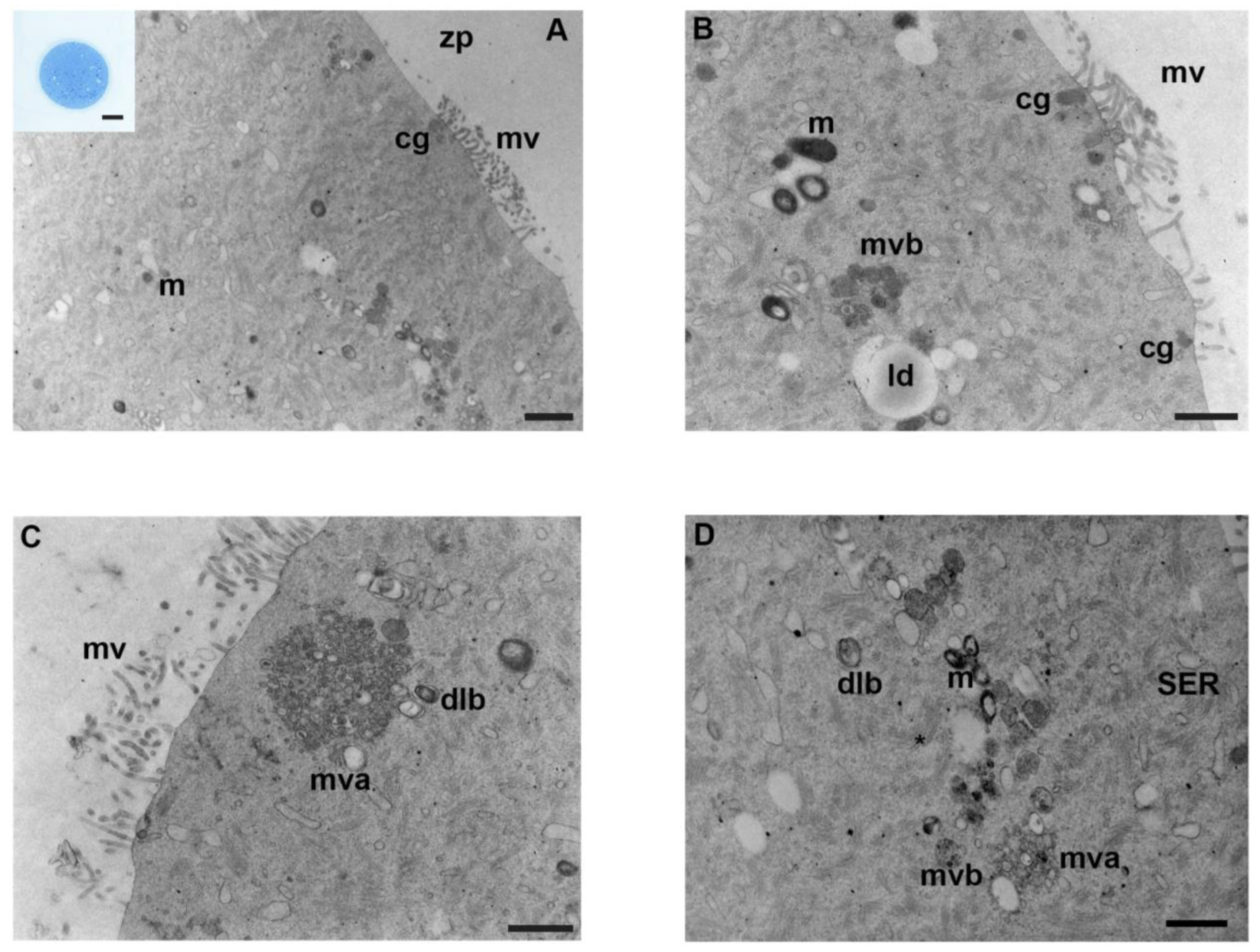
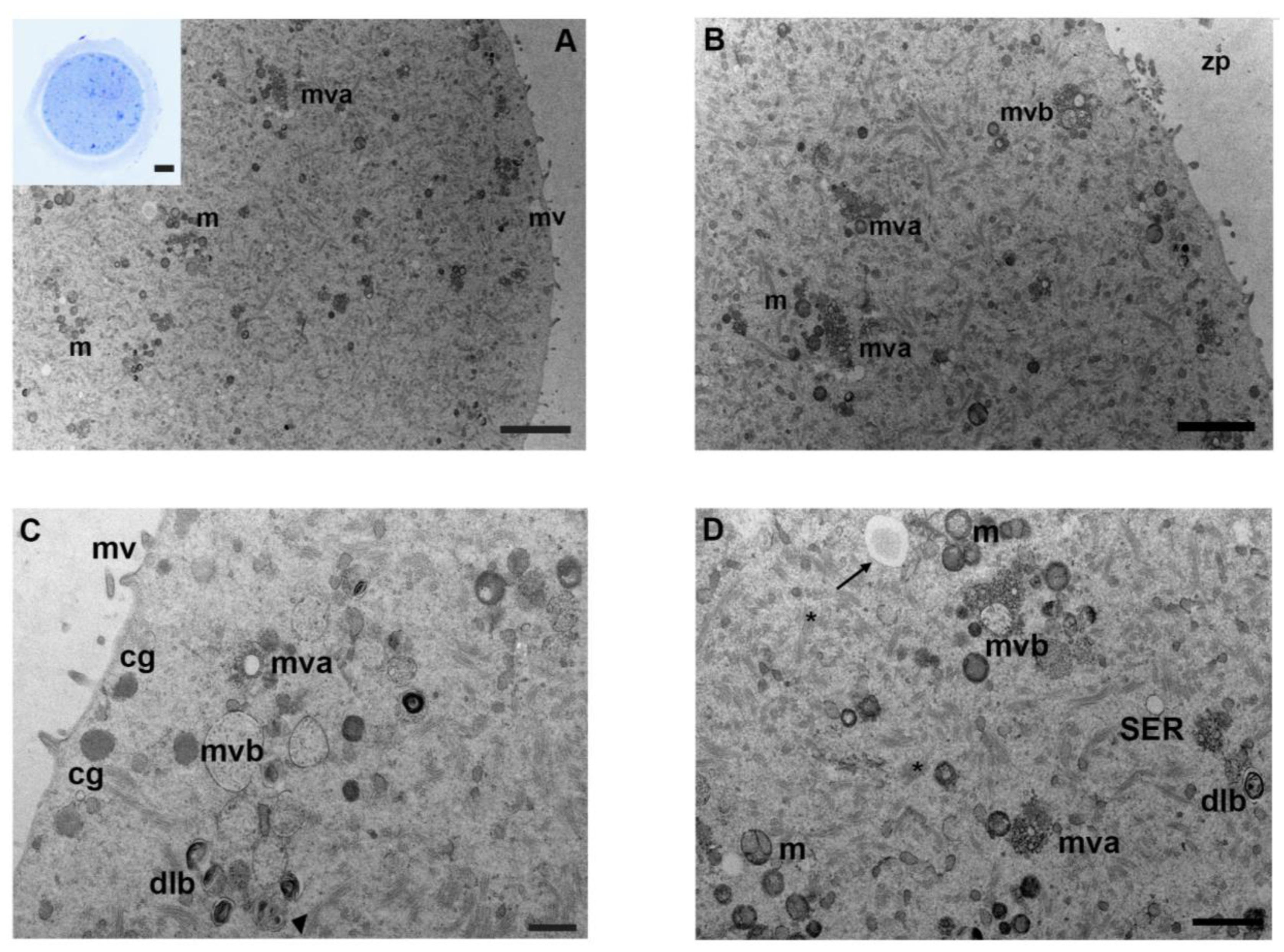
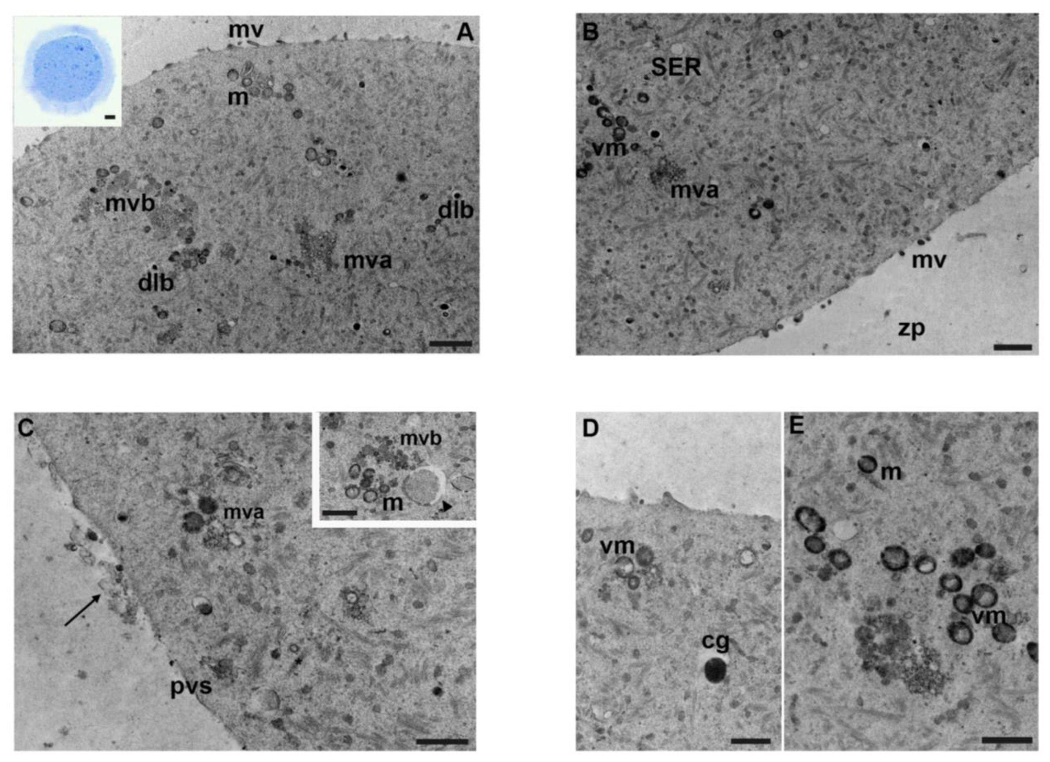
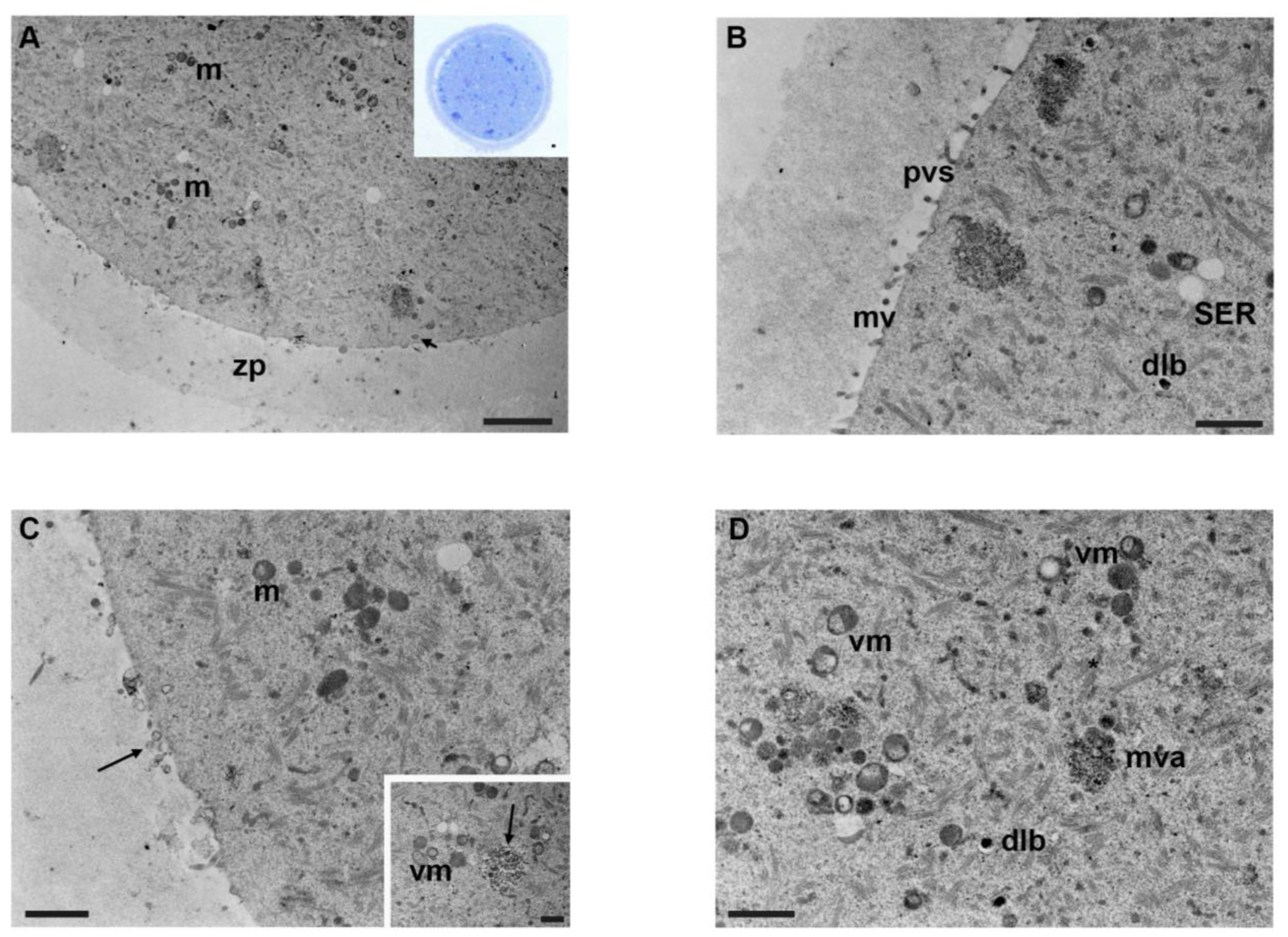
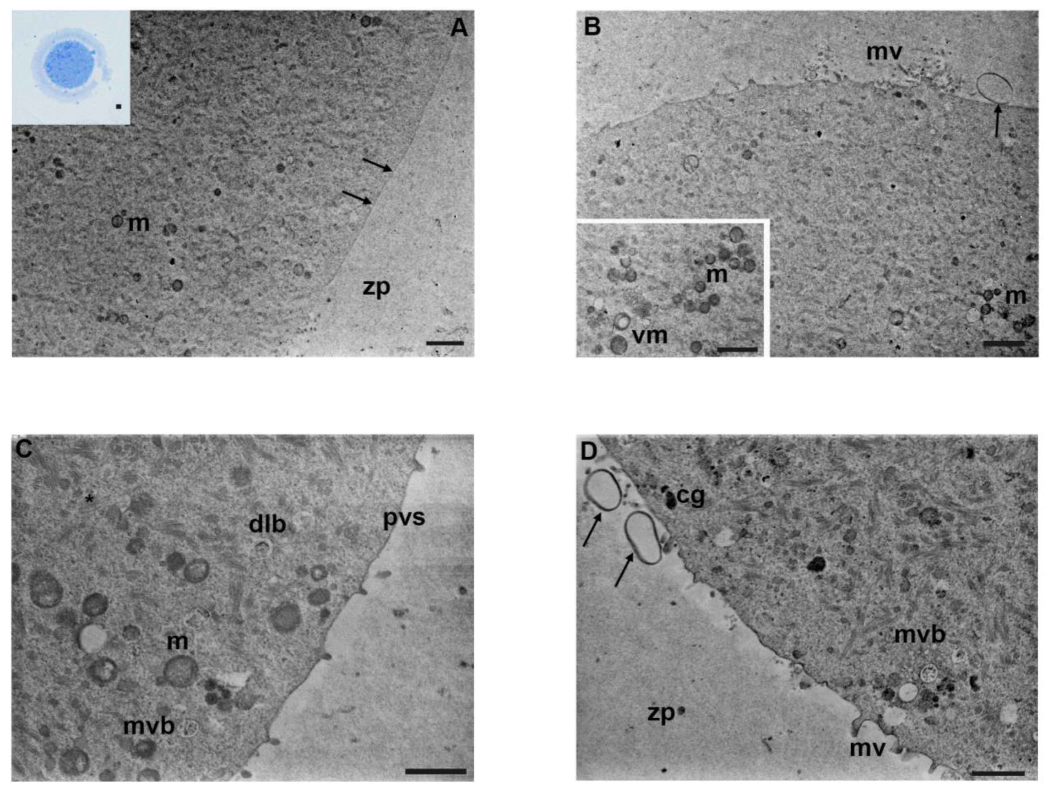
Exposure to endocrine-disrupting pollutants, such as the fungicide mancozeb, is linked to various environmental health hazards, including female fertility. Although the dithiocarbamate mancozeb has low reported toxicity in mammals, it impairs female reproductive functions in exposed animals and humans. The specific mechanism of action of mancozeb and the damage to cell structures in the female reproductive system are still unclear. This study aims to describe the ultrastructure of mouse oocytes exposed in vitro to increasing concentrations of mancozeb (0.001–1 μg/mL) by light and transmission electron microscopy and to perform a morphometric analysis over significant organelles. While from 0.001 to 0.1 μg/mL, oocyte ultrastructure was comparable to controls, at the highest concentration (1 µg/mL), a decrease in the numerical density of mitochondria and cortical granules, an altered organelle distribution, and flattening of microvilli were observed. These results could be responsible for the adverse effect of this fungicide on mammalian reproductive performance.
Abstract
Mancozeb is a widely used fungicide, considered to be an endocrine disruptor. In vivo and in vitro studies evidenced its reproductive toxicity on mouse oocytes by altering spindle morphology, impairing oocyte maturation, fertilization, and embryo implantation. Mancozeb also induces dose-dependent toxicity on the ultrastructure of mouse granulosa cells, including chromatin condensation, membrane blebbing, and vacuolization. We evaluated the effects on the ultrastructure of mouse oocytes isolated from cumulus-oocyte complexes (COCs), exposed in vitro to increasing concentrations of mancozeb. COCs were matured in vitro with or without (control) low fungicide concentrations (0.001–1 μg/mL). All mature oocytes were collected and prepared for light and transmission electron microscopy. Results showed a preserved ultrastructure at the lowest doses (0.001–0.01 μg/mL), with evident clusters of round-to-ovoid mitochondria, visible electron-dense round cortical granules, and thin microvilli. Mancozeb concentration of 1 μg/mL affected organelle density concerning controls, with a reduction of mitochondria, appearing moderately vacuolated, cortical granules, and microvilli, short and less abundant. In summary, ultrastructural data revealed changes mainly at the highest concentration of mancozeb on mouse oocytes. This could be responsible for the previously described impaired capability in oocyte maturation, fertilization, and embryo implantation, demonstrating its impact on the reproductive health and fertility.