Upregulation of Sarcolemmal Hemichannels and Inflammatory Transcripts with Neuromuscular Junction Instability during Lower Limb Unloading in Humans
by
 , , , , , , , ,
Giuseppe Sirago
1,* ,
,
Julián Candia
2 ,
,
Martino V. Franchi
1,3 ,
,
Fabio Sarto
1, 
Elena Monti
1,
Luana Toniolo
1,
Carlo Reggiani
1,4 ,
,
Emiliana Giacomello
5,
Sandra Zampieri
1,3,6 ,
,
Lisa M. Hartnell
2,
Giuseppe De Vito
1,3 ,
,
Marco Sandri
1,3,
Luigi Ferrucci
2 and
Marco V. Narici
1,3,4,*
1
Department of Biomedical Sciences, University of Padova, 35131 Padova, Italy
2
Translational Gerontology Branch Longitudinal Studies Section, National Institute on Aging, Baltimore, MD 21224, USA
3
CIR-MYO Myology Center, University of Padova, 35131 Padova, Italy
4
Institute for Kinesiology Research, Science and Research Center Koper, 6000 Koper, Slovenia
5
Department of Medicine, Surgery and Health Sciences, University of Trieste, 34149 Trieste, Italy
6
Department of Surgery, Oncology and Gastroenterology, University of Padova, 35124 Padova, Italy
*
Authors to whom correspondence should be addressed.
Biology 2023, 12(3), 431; https://doi.org/10.3390/biology12030431
Submission received: 24 January 2023
/
Revised: 23 February 2023
/
Accepted: 5 March 2023
/
Published: 10 March 2023
(This article belongs to the Section Physiology)
Simple Summary
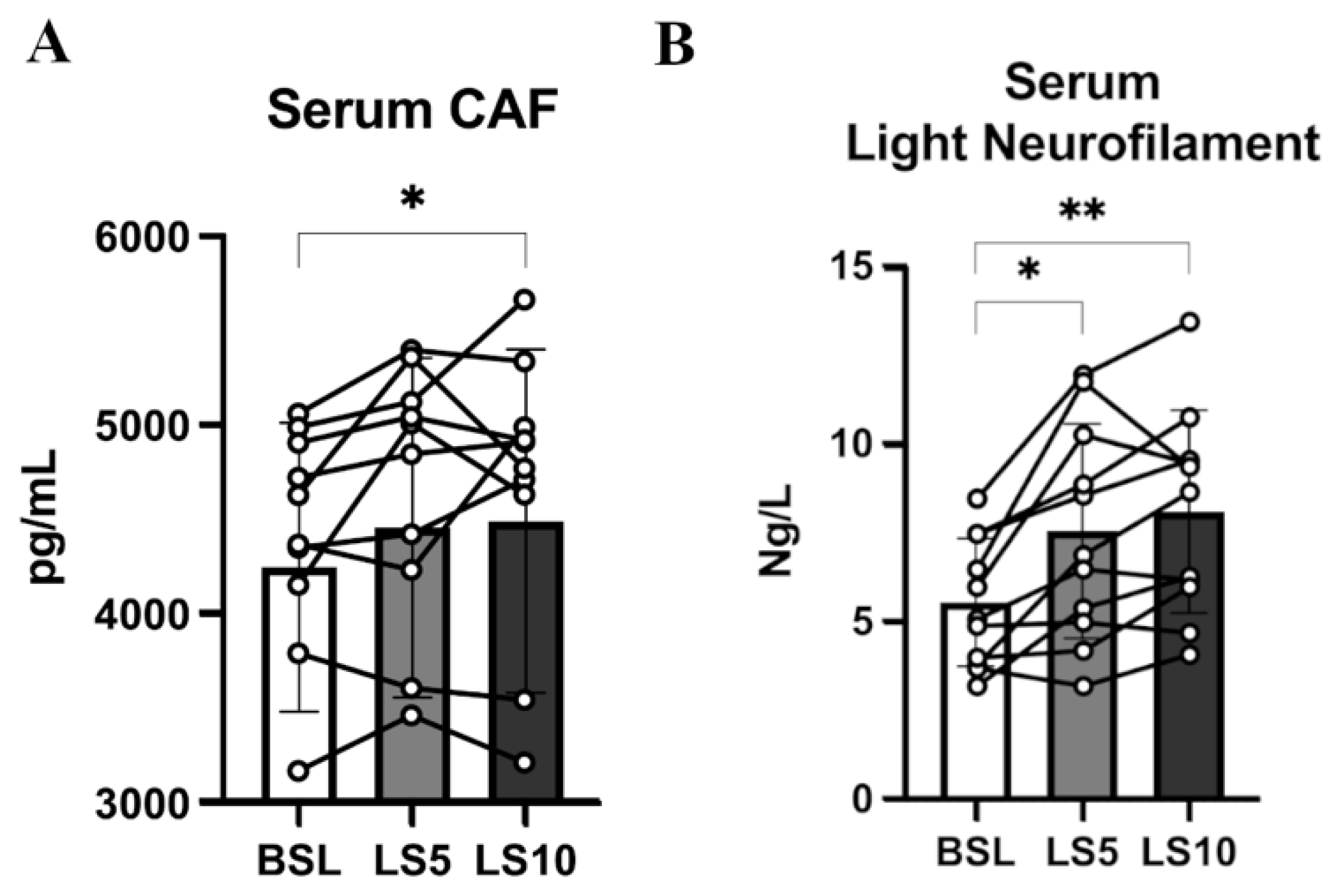
Skeletal muscles need to be continually active. Physical inactivity, reduced gravity, such as when humans are in space, or aging itself can cause neuromuscular frailty and weakness, causing major diseases. Comprehension of the molecular mechanisms that cause skeletal muscle atrophy can be beneficial to limit such processes and improve human health in different conditions: allowing humans to go into space for a long period, to be inactive for a long period or to age in a better way. When skeletal muscles are chronically inactive, within 5 days and more after 10 days, there is an increase of two molecules, called agrin fragments and neurofilaments, which become highly present in the blood, suggesting neuromuscular instability. The process is accompanied by changes in the membranes of single muscle fibres, the unit that forms our skeletal muscles. They could potentially start to lose important electrolytes and molecules and gradually promote a deleterious process. It has been proved in mice that blocking the appearance of these molecules, called hemichannels, can limit the severity of the loss of muscle mass. Interestingly, these hemichannels also appear during inactivity in humans, opening a possible application for human health and space missions.
Abstract
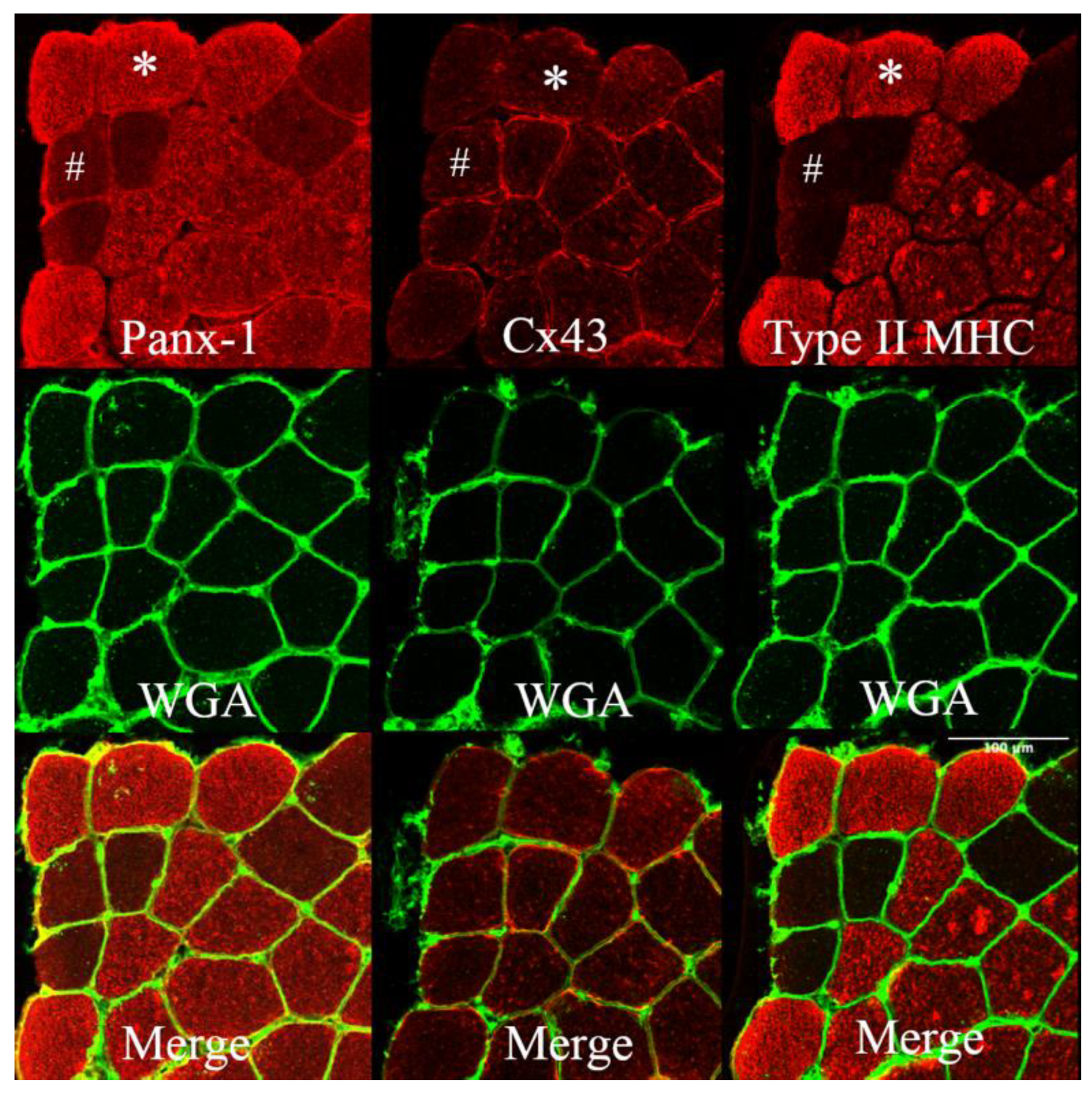
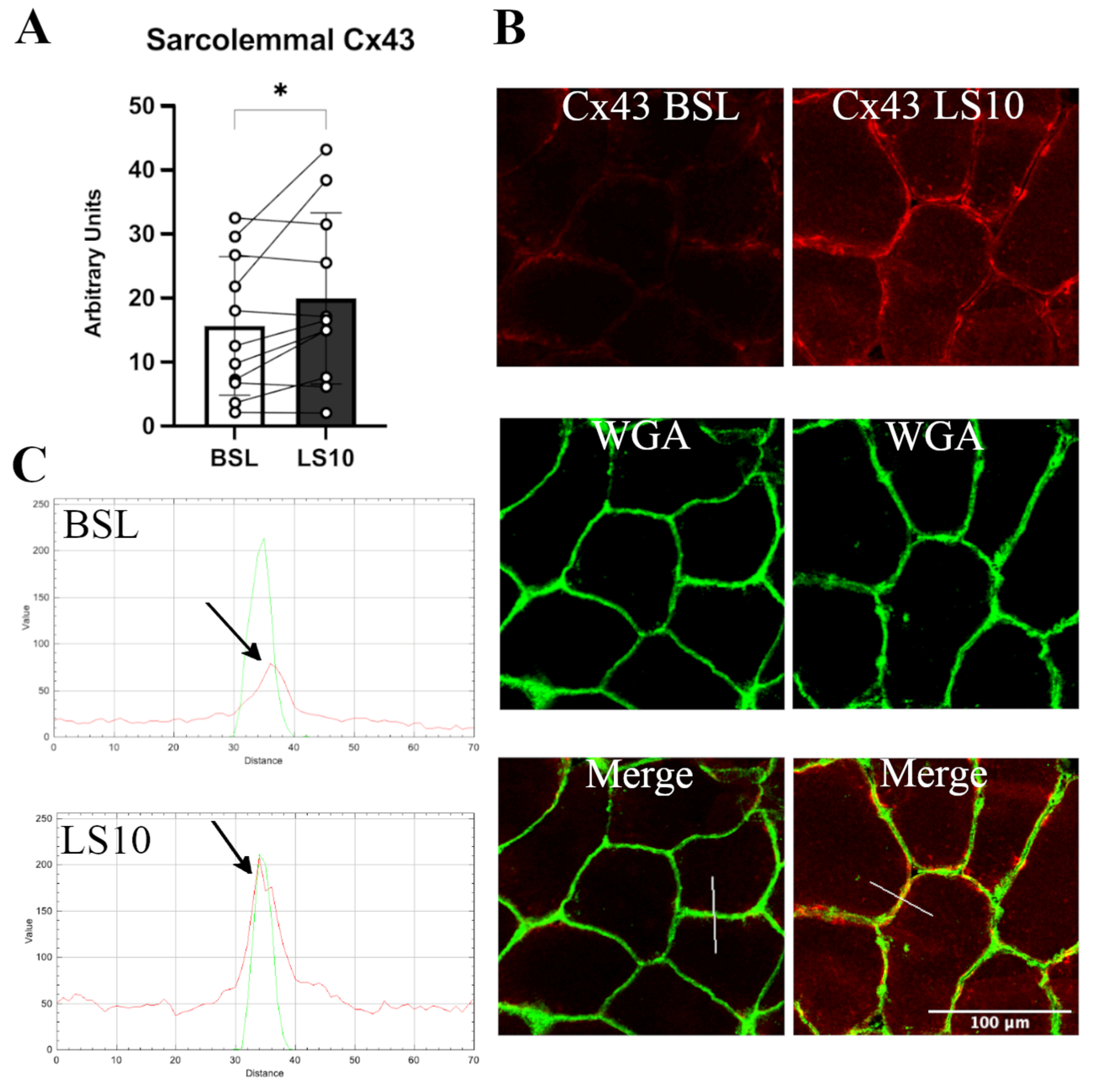
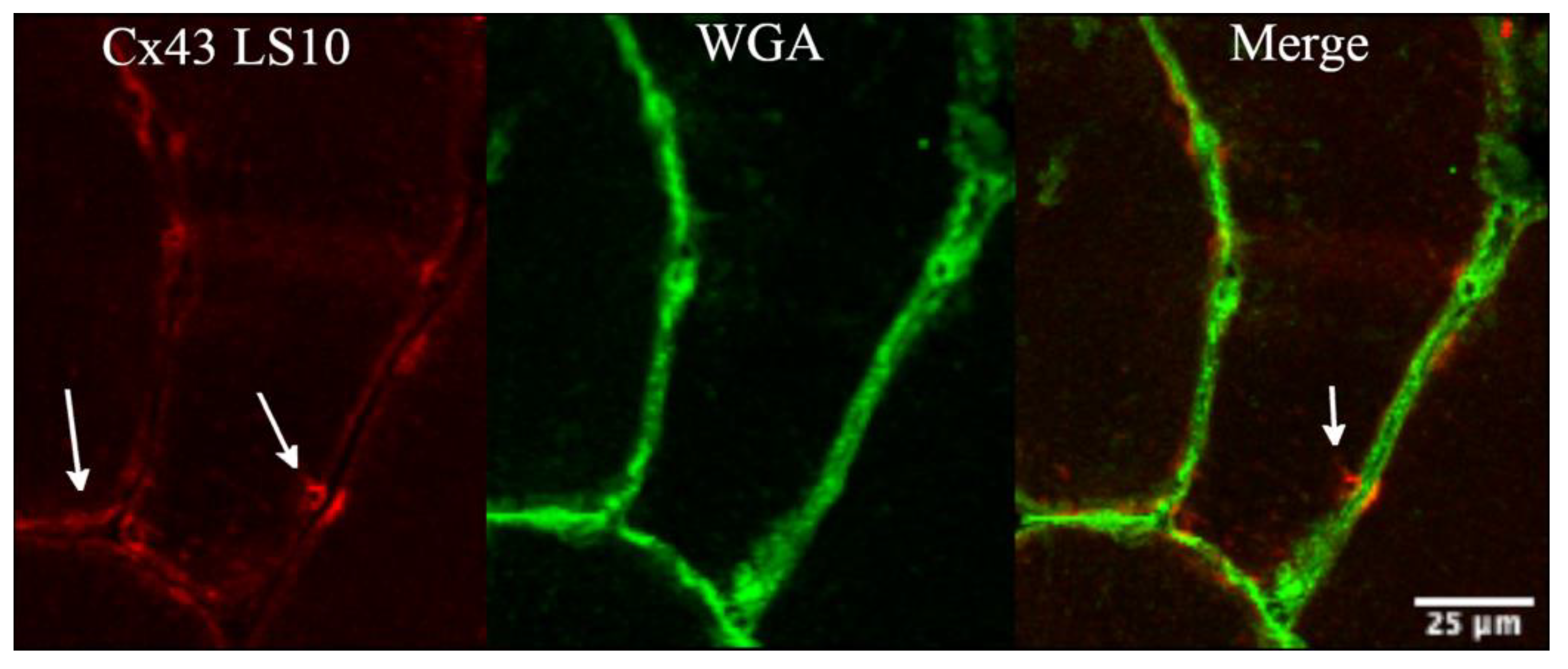
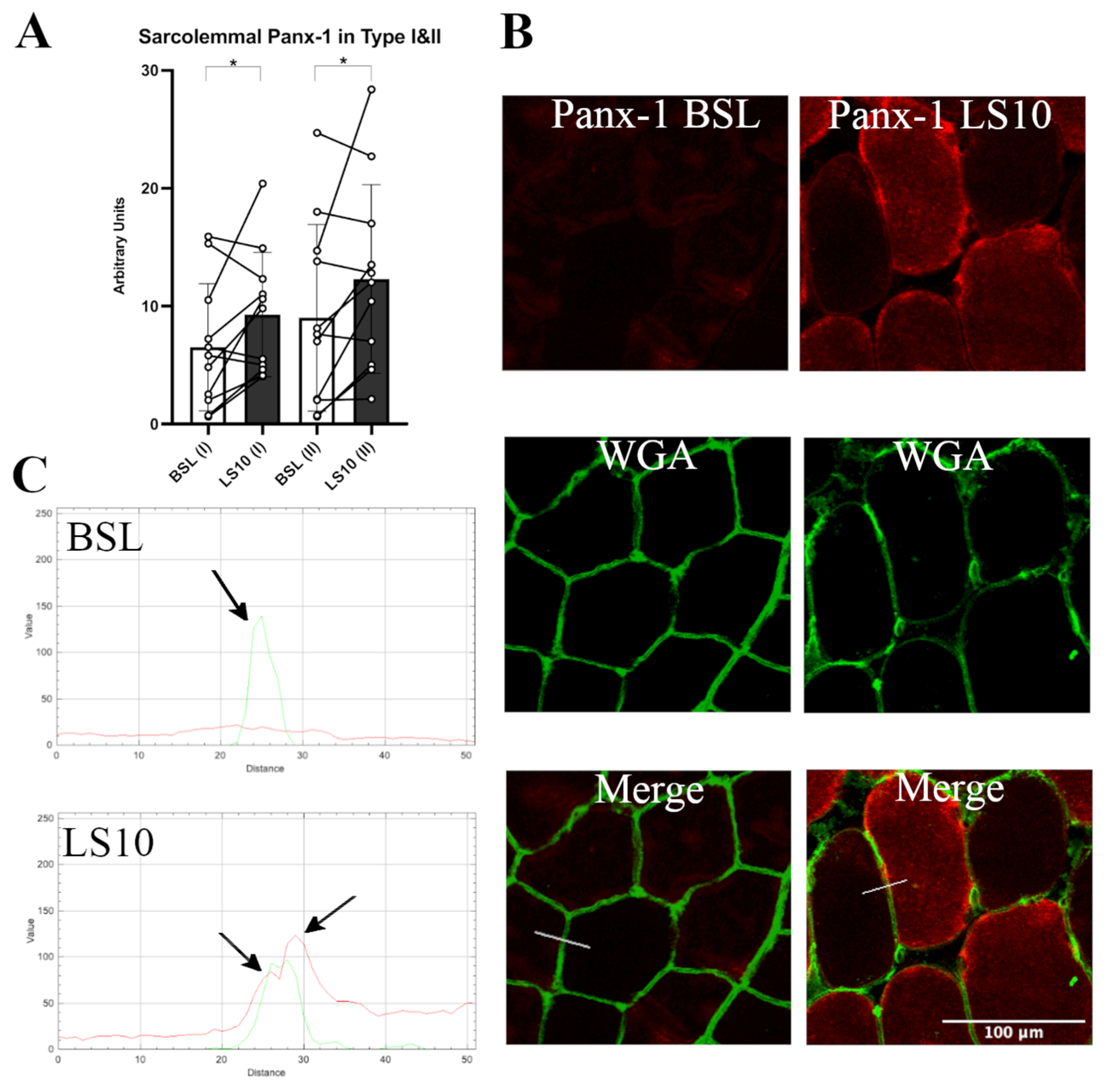
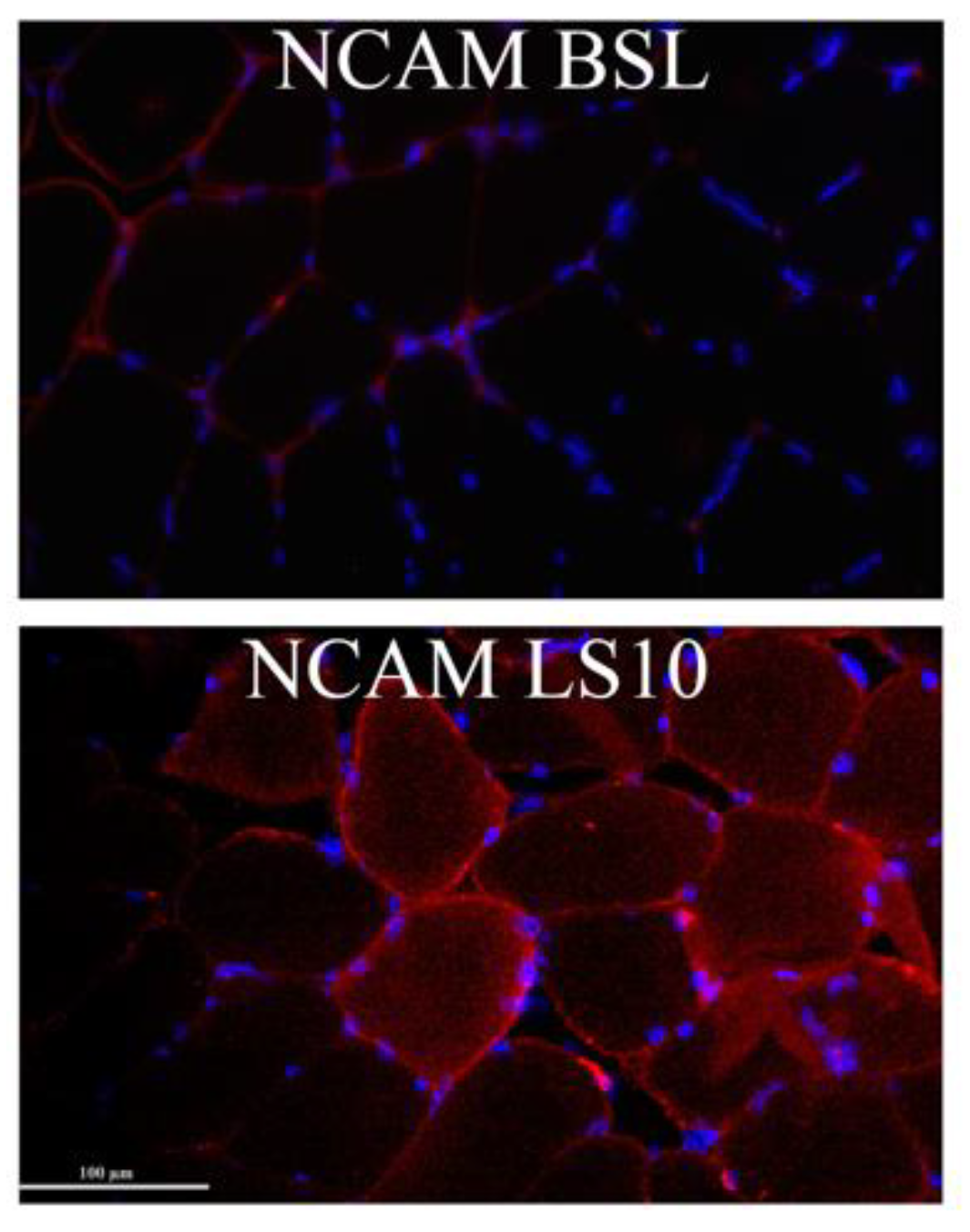
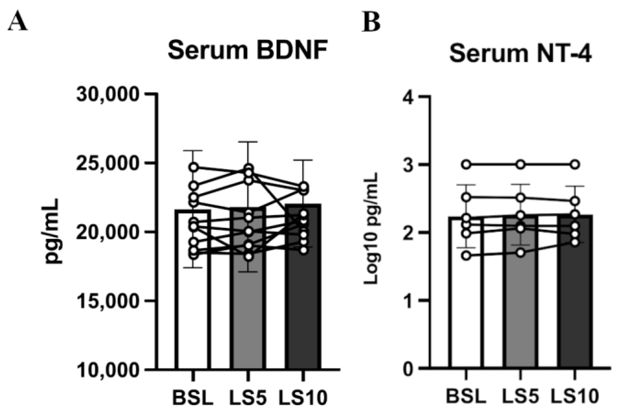
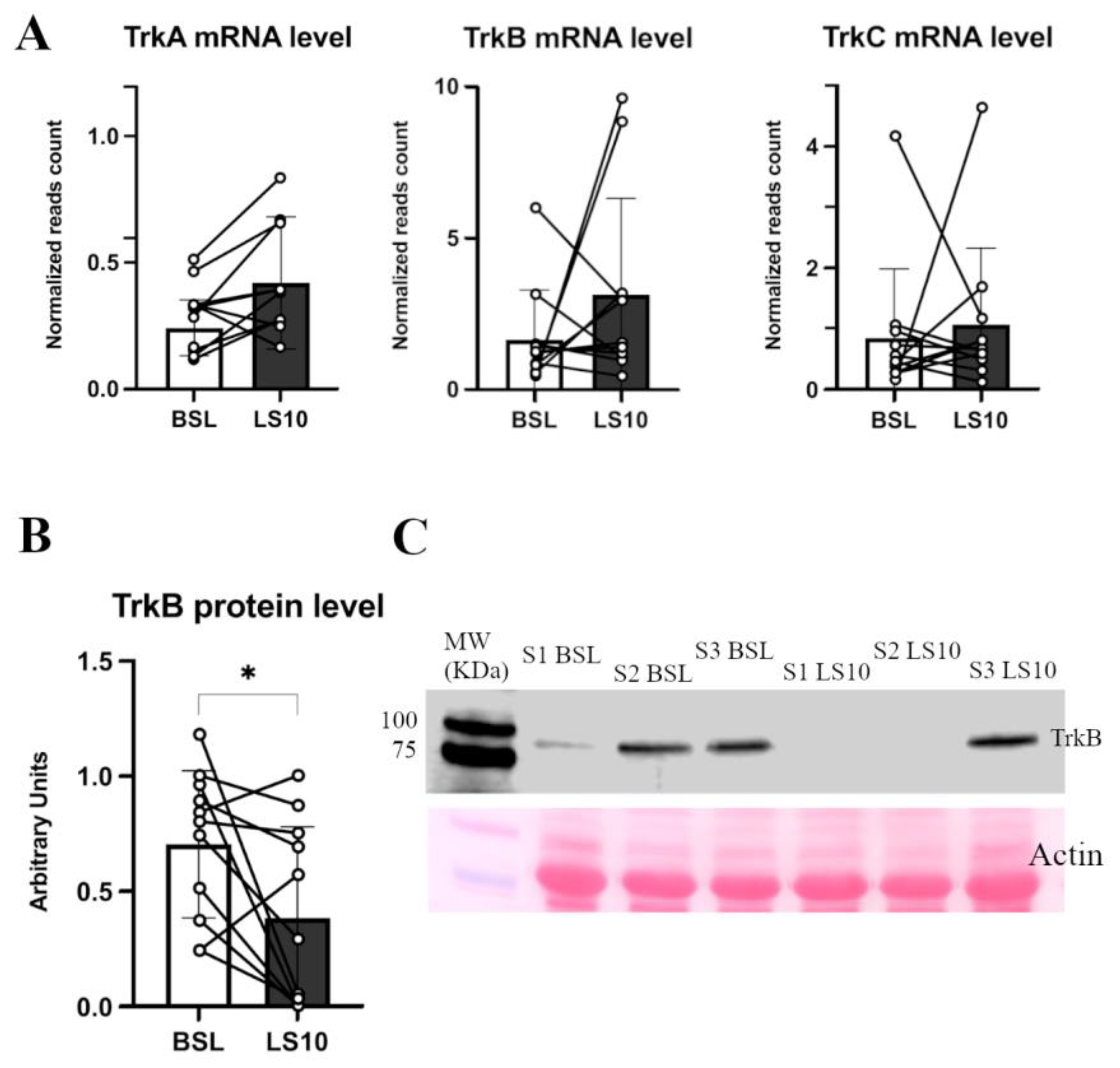
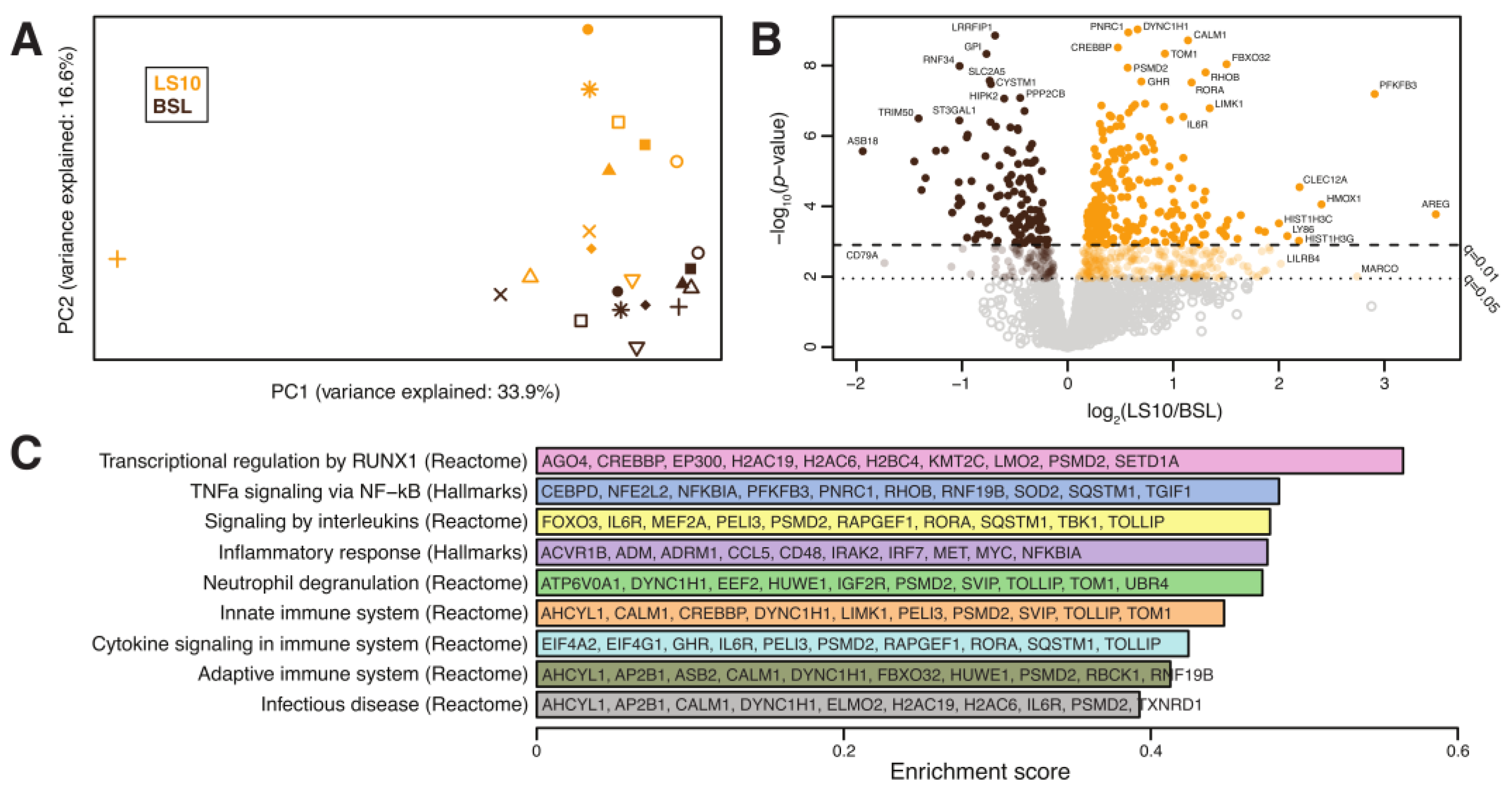
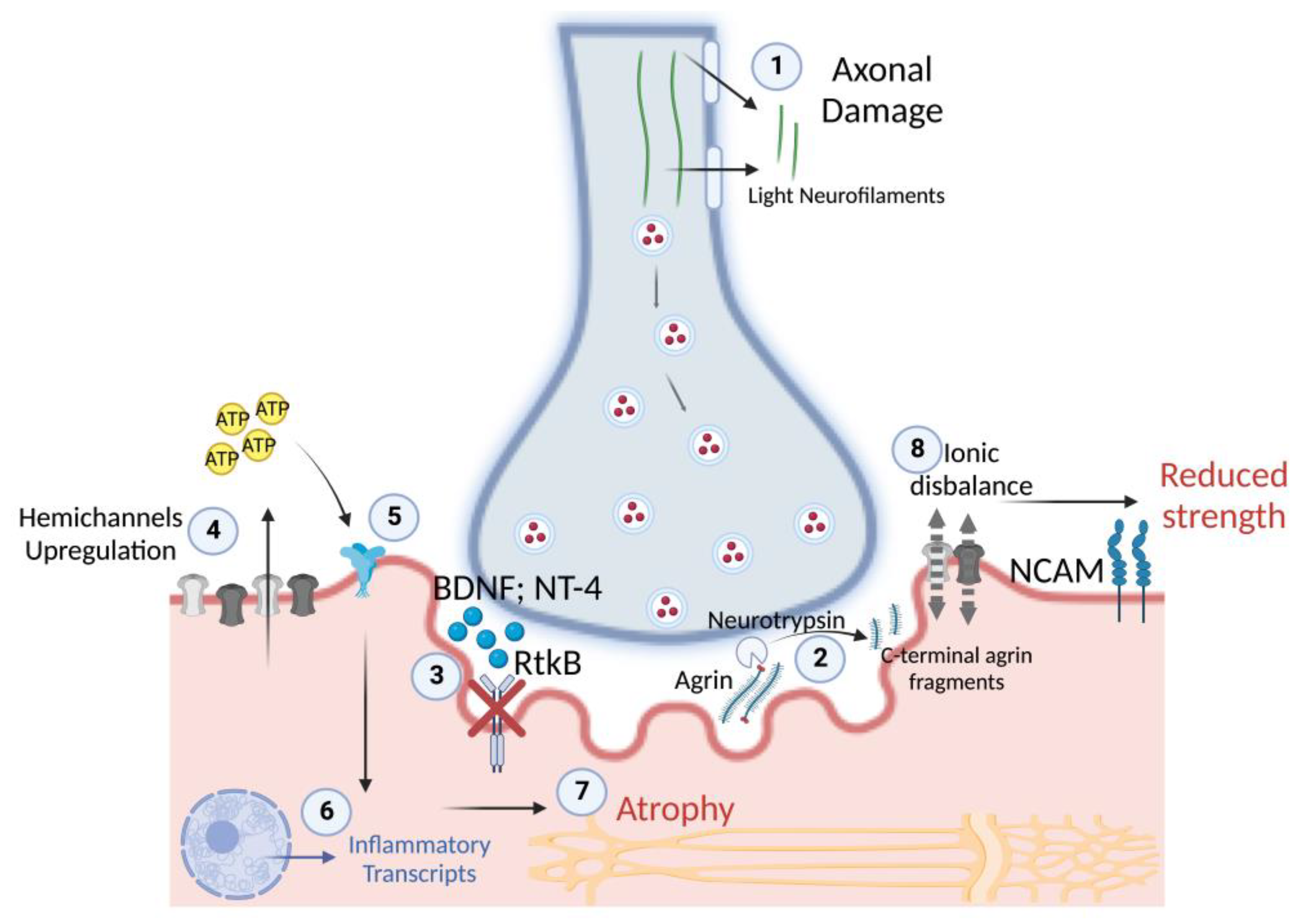
Human skeletal muscle atrophy and a disproportionate force loss occur within a few days of unloading in space and on Earth, but the underlying mechanisms are not fully understood. Disruption of neuromuscular junction homeostasis has been proposed as one of the possible causes. Here, we investigated the potential mechanisms involved in this neuromuscular disruption induced by a 10-day unilateral lower limb suspension (ULLS) in humans. Specifically, we investigated hemichannels’ upregulation, neuromuscular junction and axonal damage, neurotrophins’ receptor downregulation and inflammatory transcriptional signatures. Biomarkers were evaluated at local and systemic levels. At the sarcolemmal level, changes were found to be associated with an increased expression of connexin 43 and pannexin-1. Upregulation of the inflammatory transcripts revealed by deep transcriptomics was found after 10 days of ULLS. The destabilisation of the neuromuscular junction was not accompanied by changes in the secretion of the brain-derived neurotrophic factor and neurotrophin-4, while their receptor, BDNF/NT growth factors receptor (TrkB), decreased. Furthermore, at 5 days of ULLS, there was already a significant upregulation of the serum neurofilament light chain concentration, an established clinical biomarker of axonal injury. At 10 days of ULLS, other biomarkers of early denervation processes appeared. Hence, short periods of muscle unloading induce sarcolemmal hemichannels upregulation, inflammatory transcripts upregulation, neuromuscular junction instability and axonal damage.
Keywords:
disuse; hemichannels; inflammation