Article

Zinc-Dependent Oligomerization of Thermus thermophilus Trigger Factor Chaperone

1
Institute of Advanced Medical Sciences, Tokushima University, Tokushima 770-8503, Japan
2
Graduate School of Chemical Sciences and Engineering, Hokkaido University, Sapporo 060-8628, Japan
3
Department of Chemistry, Faculty of Science, Hokkaido University, Sapporo 060-0810, Japan
*
Authors to whom correspondence should be addressed.
Biology 2021, 10(11), 1106; https://doi.org/10.3390/biology10111106
Submission received: 25 September 2021 / Revised: 20 October 2021 / Accepted: 23 October 2021 / Published: 26 October 2021
(This article belongs to the Special Issue Protein Folding, Aggregation, and Cell Death)

Abstract

:

Simple Summary

Metal ions often play important roles in biological processes. Thermus thermophilus trigger factor (TtTF) is a zinc-dependent molecular chaperone where Zn2+ has been shown to enhance its folding-arrest activity. However, the mechanisms of how Zn2+ binds to TtTF and how Zn2+ affects the activity of TtTF are yet to be elucidated. As a first step in understanding the mechanism, we performed in vitro biophysical experiments on TtTF to investigate the zinc-binding site on TtTF and unveil how Zn2+ alters the physical properties of TtTF, including secondary structure, thermal stability, and oligomeric state. Our results showed that TtTF binds Zn2+ in a 1:1 ratio, and all three domains of TtTF are involved in zinc-binding. We found that Zn2+ does not affect the thermal stability of TtTF, whereas it does induce partial structural change and promote the oligomerization of TtTF. Given that the folding-arrest activity of Escherichia coli TF (EcTF) is regulated by its oligomerization, our results imply that TtTF exploits Zn2+ to modulate its oligomeric state to regulate the activity.

Abstract

Thermus thermophilus trigger factor (TtTF) is a zinc-dependent molecular chaperone whose folding-arrest activity is regulated by Zn2+. However, little is known about the mechanism of zinc-dependent regulation of the TtTF activity. Here we exploit in vitro biophysical experiments to investigate zinc-binding, the oligomeric state, the secondary structure, and the thermal stability of TtTF in the absence and presence of Zn2+. The data show that full-length TtTF binds Zn2+, but the isolated domains and tandem domains of TtTF do not bind to Zn2+. Furthermore, circular dichroism (CD) and nuclear magnetic resonance (NMR) spectra suggested that Zn2+-binding induces the partial structural changes of TtTF, and size exclusion chromatography-multi-angle light scattering (SEC-MALS) showed that Zn2+ promotes TtTF oligomerization. Given the previous work showing that the activity regulation of E. coli trigger factor is accompanied by oligomerization, the data suggest that TtTF exploits zinc ions to induce the structural change coupled with the oligomerization to assemble the client-binding site, thereby effectively preventing proteins from misfolding in the thermal environment.

Graphical Abstract

1. Introduction

In a crowded intracellular environment, newly synthesized polypeptide chains and metastable proteins risk misfolding or aggregation [1,2]. Molecular chaperones play the role of preventing proteins from misfolding or aggregation and removing the denatured proteins, thereby regulating the protein quality in the cell [3,4,5]. Chaperone-mediated protein quality control in bacteria has been extensively studied as a model system. One of the major molecular chaperones in bacterial cytosol, trigger factor (TF) chaperone, plays multiple roles in protein anti-aggregation, folding [6,7], translocation [8,9,10], and degradation [11]. TF binds to the ribosome to prevent the misfolding and aggregation of newly synthesized polypeptide chains [4,7,12,13,14,15]. For these versatile functions, TF has multiple activities, including “foldase activity”, which increases the folding rate and/or yield of the client proteins, and “holdase activity”, which halts or delays the folding of the client protein to prevent the misfolding of client proteins or to promote efficient protein translocation through Sec machinery [16,17]. Thus, TF seemingly has opposing activities in protein folding and switches the foldase/holdase activities depending on the circumstances.
One of the important aspects of TF for activity-switching is oligomerization [17]. It has been shown that Escherichia coli TF (EcTF) forms a relatively weak dimer in the head-to-tail orientation [17,18] and that dimerization enhances holdase activity [17]. The assembly and rearrangement of the client-binding sites on TF induced by dimerization can modulate the binding kinetics with the client proteins, which explains the mechanism of the activity modulation [17].
Another strategy to modulate the activity of molecular chaperones is the binding of metal ions [19,20]. TF from Thermus thermophilus (HB8 strain) (TtTF) is one of those chaperones, and it has been reported that the substrate folding-arrest activity (holdase activity) is activated by Zn2+ [20]. A previous study demonstrated that purified TtTF binds Zn2+ and that zinc-binding can be saturated up to a 1:1 stoichiometric ratio by refolding in the presence of Zn2+. Although the previous study has shown the relationship between zinc-binding and holdase activity modulation [20], the mechanism of how Zn2+ alters the activity of TtTF and whether it is related to oligomerization are unknown. Furthermore, TtTF has no typical zinc-binding motif in its amino acid sequence, and thus the mechanism of how TtTF recognizes Zn2+ ions is unclear.
Here, we focused on the zinc-dependent activity modulation of TtTF and performed biophysical in vitro experiments, including the matrix-assisted laser desorption/ionization time-of-flight mass spectrometry (MALDI-TOF-MS), circular dichroism (CD), size exclusion chomatography-multi-angle light scattering (SEC-MALS), and solution nuclear magnetic resonance (NMR), to investigate the zinc-binding site of TtTF, as well as zinc-dependent changes in the structure, thermal stability, and oligomeric state of TtTF. The data show that all three domains of TtTF are involved in the zinc-binding that induces partial structural changes and the oligomerization of TtTF. Furthermore, given the relationship between the oligomerization and activity shown for EcTF [17], our results suggest the mechanism of activity modulation of TtTF, in which the Zn2+ alters structural properties to induce the oligomerization of TtTF for activity regulation.

2. Materials and Methods

2.1. Plasmid Construction

The plasmids for protein expression in E. coli cells were constructed as follows. The synthetic gene fragments of TtTF (1–404) and Thermotoga maritima TF (TmTF) (1–425) were purchased from Thermo Fisher Scientific (Waltham, MA, USA) and inserted into the pCold vector after His6-tag (Takara, Kusatsu, Japan) to obtain the plasmid named pCold His6-TtTF and pCold His6-TmTF, respectively. The fragments and the vector were amplified by polymerase chain reaction using the primers summarized in Table S1 (Primers 1–8). The plasmid pCold His6-EcTF was constructed in the previous study [13,17]. Given the potential binding of Zn2+ to His6-tag, the His6-tag needed to be removed for the zinc-binding assay. Therefore, pCold His6-TEVCS-TtTF and pCold His6-TEVCS-EcTF were constructed by inserting tobacco etch virus protease cleavage sites (TEVCS) into pCold His6-TtTF and pCold His6-EcTF, respectively. The amino acid residues sequences of TEVCS for TtTF and EcTF are ENLYFQG and ENLYFQ, respectively. The primers used in these constructions are summarized in Table S1 (Primers 9–12).
The constructs of the isolated or tandem domains of TtTF, including pCold His6-TEVCS-TtTFRBD (residues 1–113), pCold His6-TEVCS-TtTFSBD (residues 112–148 and 226–404, linked by GSGSG), pCold His6-TEVCS-TtTFPPD (residues 148–226), pCold His6-TEVCS-TtTFPPD-SBD (residues 112–404), and pCold His6-TEVCS-TtTFRBD-SBD (residues 1–148 and 226–404, linked by GSGSG) (Figure S1), were prepared using the primers and templates summarized in Table S1 (Primers 13–20).
The primers were purchased from Thermo Fisher Scientific and FASMAC (Kanagawa, Japan). The sequences of the constructed plasmids were verified by Eurofins Genomics (Tokyo, Japan).

2.2. Protein Expression and Purification

All protein samples used in this study were overexpressed in E. coli BL21 (DE3) cells and purified as described previously [11,13,20,21]. The sterilized Luria-Bertani (LB) medium (1 L) containing 50 mg/L ampicillin (Amp) was used to culture the cells at 37 °C. A total of 0.5 mM isopropyl β-D-1-thiogalactopyranoside (IPTG) was added to the medium when OD600 reached around 0.6–0.8. The cells were then cultured overnight at 18 °C. Cells were harvested by centrifugation at 4500 rpm for 15 min and resuspended in buffer containing 50 mM Tris-HCl and 0.5 M NaCl (pH 8.0). The cells were disrupted by a sonicator and centrifuged at 18,000 rpm for 45 min. The proteins were purified using Ni-NTA agarose (QIAGEN, Hilden, Germany). A total of 1 mg TEV protease was added to the protein from the 1 L medium to remove the His6-tag of EcTF, followed by the incubation overnight at 4 °C. Similar operations were performed at room temperature to remove the His6-tag of TtTF, but 2 mg TEV protease and longer digestion time (2 days) were required. After TEV protease digestion, the protein sample was incubated with Ni-NTA resin twice. The flowthrough was collected for further purification by gel filtration using a Superdex 200 16/600 column or Superdex 75 16/600 column (Cytiva, Marlborough, MA, USA). His6-tagged proteins (TmTF, TtTFPPD-SBD, and TtTFRBD-SBD) and His6-tag removed proteins including EcTF, TtTF, and isolated domains of TtTF (TtTFPPD, TtTFSBD, and TtTFRBD) were used for the zinc-binding assay. His6-tagged TtTF was used for CD, NMR, and SEC-MALS.
For the preparation of isotopically labeled TtTF, E. coli BL21 (DE3) cells were cultured at 37 °C and in the 1 L sterilized M9 medium containing 6 g Na2HPO4, 3 g KH2PO4, 0.5 g NaCl, 2 g 15NH4Cl, 6 g D-glucose, 1.2 g MgSO4, 0.03 g thiamin (vitamin B1), and 50 mg Amp. [13CH3]-α-ketobutyric acid (50 mg L−1), [13CH3/13CH3]-α-ketoisovaleric acid (85 mg L−1), [13CH3]-methionine (50 mg L−1), and [2-2H, 13CH3]-alanine (50 mg L−1) were used to selectively label the methyl groups of Ile, Val, Leu, Met, and Ala residues, respectively, and these reagents were added to the M9 medium when OD600 reached around 0.5. When OD600 reached around 0.6–0.8, 0.5 mM IPTG was added to the M9 medium. Then, the cells were cultured overnight at 18 °C. The purification protocol of isotopically labeled proteins was the same as described above.

2.3. Preparation of Zinc-Bound and Zinc-Depleted Proteins

All purified proteins were diluted to below 2 μM and divided into two parts for the refolding operations as previously described [20]. One part was unfolded in the buffer containing 2 mM EDTA, 6 M guanidine, and 50 mM HEPES-KOH at pH 7.5 and at room temperature for 12 h. The other part was unfolded in the buffer containing 2 mM Zn(CH3COO)2, 6 M guanidine, and 50 mM HEPES-KOH at pH 7.5 for 12 h at room temperature. Then, proteins were refolded in the buffer containing 2 mM EDTA or 2 mM Zn(CH3COO)2 and 50 mM HEPES-KOH at pH 7.5 for 12 h at 4 °C. The extra EDTA or Zn(CH3COO)2 was removed by dialysis in buffer containing 50 mM HEPES-KOH at pH 7.5 and 4 °C for 6 h (twice). The refolded protein samples were used for in vitro biophysical experiments. Hereafter, TF proteins refolded in the Zn2+-containing and EDTA containing buffers are represented as TF (Zn2+) and TF (EDTA), respectively. The refolded proteins were evaluated by CD and SEC-MALS, which confirmed that the refolded proteins maintained the native structure (see Results).

2.4. MALDI-TOF-MS Spectrometry

For the mass spectrometry analysis, the protein samples, whose concentration was in the 20–100 μM range, were desalted by ZipTip (MILLIPORE, Bedford, MA, USA) and eluted by 1.5 μL saturated sinapinic acid solution (50% acetonitrile, 49.9% H2O, 0.1% TFA) on the target ground steel. After the protein sample was dried, the mass spectra were recorded by Microflex-TK mass spectrometer (Bruker Daltonics, Billerica, MA, USA) and Autoflex speed-DC mass spectrometer (Bruker Daltonics) in linear measurement mode. The protein standard I or II (Bruker Daltonics) were used for calibration.
The theoretical molar masses of TtTF and its isolated domains after His6-tag removal are as below: 46,313 Da [full-length TtTF (EDTA)], 12,706 Da [TtTFRBD (EDTA)], 25,618 Da [TtTFSBD (EDTA)], and 8911 Da [TtTFPPD (EDTA)]. The His6-tag of TmTF (without the TEV cleaved site) was not removed, and the N-terminal of TmTF still carried the MNHKVHHHHHH peptide fragment; thus, the theoretical molar mass of TmTF (EDTA) is 51,330 Da. The theoretical molar mass of EcTF (EDTA) after His6-tag removal is 48,193 Da. The His6-tag of TtTFPPD-SBD and GSGSG linked TtTFRBD-SBD is difficult to remove. Thus, the tandem domains with MNHKVHHHHHHENLYFQG peptide fragments were used in this experiment. The theoretical molar masses of TtTFPPD-SBD (EDTA) and GSGSG linked TtTFRBD-SBD (EDTA) are 36,193 Da and 40,249 Da, respectively.

2.5. Circular Dichroism Measurement

The CD spectra were recorded by a J-1500 CD spectrometer (JASCO, Tokyo, Japan). All protein samples were diluted to around 6 μM in a buffer containing 20 mM MOPS at pH 7.5. The temperature-dependent CD measurements were measured in the fixed wavelength of 222 nm from 20 °C to 95 °C at a 1 °C/min gradient. The CD spectra at the wavelength range from 190 nm to 320 nm were taken at fixed temperatures (20 °C, 35 °C, 50 °C, 65 °C, 80 °C, and 95 °C) 5 min after the cell temperature was stabilized. A quartz cell with 1 mm path length was used in all measurements. Protein concentration was determined based on the absorbance at 280 nm by UV-vis spectrometer JASCO V-730 (JASCO). The neural network program K2D [22] was used to analyze the secondary structure content of TtTF (EDTA) and TtTF (Zn2+) on Dichroweb web service (http://dichroweb.cryst.bbk.ac.uk/html/process.shtml, Accessed date: 14 September 2021 and 8–9 October 2021) [23]. The goodness-of-fit parameters (scaling factor) for the secondary structure analysis of TtTF (EDTA) and TtTF (Zn2+) were set to 0.95–1.05 [24].

2.6. Size Exclusion Chromatography in a Multi-Angle Laser Light Scattering (SEC-MALS)

SEC-MALS was measured using the instrument consisting of a high-performance liquid chromatography (HPLC) pump LC-20AD (Shimadzu, Kyoto, Japan), a refractive index detector RID-20A (Shimadzu), a UV-vis detector SPD-20A (Shimadzu), a light scattering detector DAWN HELEOS8+ (Wyatt Technology Corporation, Santa Barbara, CA, USA), and a TSKgel G3000SWXL column (Tosoh Bioscience, Tokyo, Japan) [for TtTF (EDTA) and TtTF (Zn2+)] or KW-803 column (Shodex, Tokyo, Japan) (for natively purified TtTF). The centrifuged (15,000 rpm for 5 min at 4 °C) TtTF (Zn2+) or TtTF (EDTA) samples at various concentrations in buffer containing 50 mM HEPES-KOH and 100 mM KCl at a pH of 7.5, were injected into the HPLC system at the 1 mL/min flow rate. Data were analyzed with ASTRA version 7 (Wyatt Technology Corporation).

2.7. NMR Spectroscopy

Both TtTF (EDTA) and TtTF (Zn2+) were concentrated to ~0.1 mM and prepared in a buffer containing 50 mM HEPES-KOH (pH 7.5) and 100 mM KCl. 1H-13C heteronuclear multiple quantum coherence (HMQC) spectra were recorded on Bruker Avance III 500 MHz NMR (Bruker Biospin AG, Fällanden, Switzerland) at 65 °C. The spectra were processed with the NMRPipe software [25]. Olivia software (https://github.com/yokochi47/Olivia, Accessed date: 22 December 2017) was used to analyze the spectral data.

3. Results

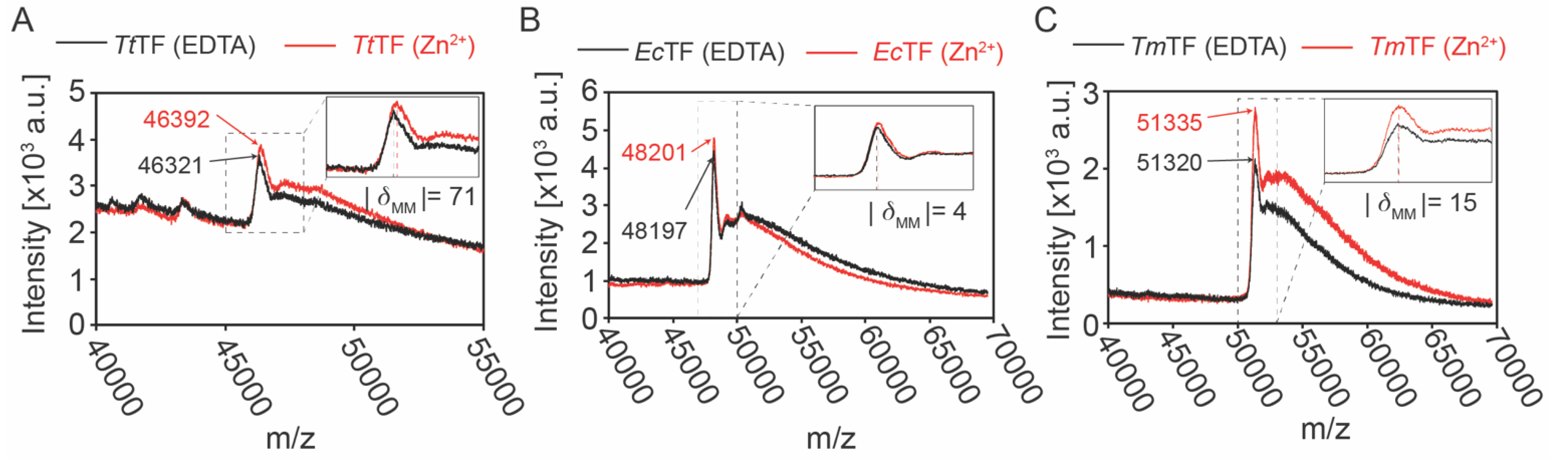
3.1. Zinc-Binding Is Characteristic to TtTF

It has previously been shown that Zn2+ binds to TtTF in a 1:1 stoichiometric ratio [20]. To investigate whether the Zn2+-binding is a unique character of TtTF, TF chaperones from three different organisms, Thermus thermophilus, Thermotoga maritima, and Escherichia coli (Figure S2), were subjected to the zinc-binding assay. Following the procedure described in the previous report [20], the purified TF chaperones, TtTF, TmTF, and EcTF, were refolded in the presence of Zn(CH3COO)2 or EDTA. After buffer exchange by dialysis, the proteins were analyzed by MALDI-TOF-MS, which is widely used in metalloproteomics [26,27]. The molar mass of TtTF refolded in the presence of Zn(CH3COO)2 [TtTF (Zn2+)] was 62 ± 12 Da larger than TtTF refolded in the presence of EDTA [TtTF (EDTA)] (Figure 1A, Table 1). Given the molar mass of Zn2+ (65 Da), the data showed that TtTF binds Zn2+ in a stoichiometric ratio of 1:1, which is consistent with the previous report [20]. Note that the previous study on TtTF exploiting inductively coupled plasma spectroscopy (ICPS) element analysis and spectroscopic titration experiment identified that the purified TtTF from E. coli cells contains a half equimolar Zn2+, which can be saturated at a 1:1 stoichiometric ratio by zinc-saturation by refolding [20]. These data support the idea that the 1:1 zinc-binding seen in the refolded TtTF represents the functional binding. Conversely, EcTF and TmTF showed negligible molar mass differences derived from Zn2+ treatment (Figure 1B,C, Table 1). Therefore, the data showed that TmTF and EcTF bind no Zn2+, and Zn2+-binding is characteristic to TtTF.

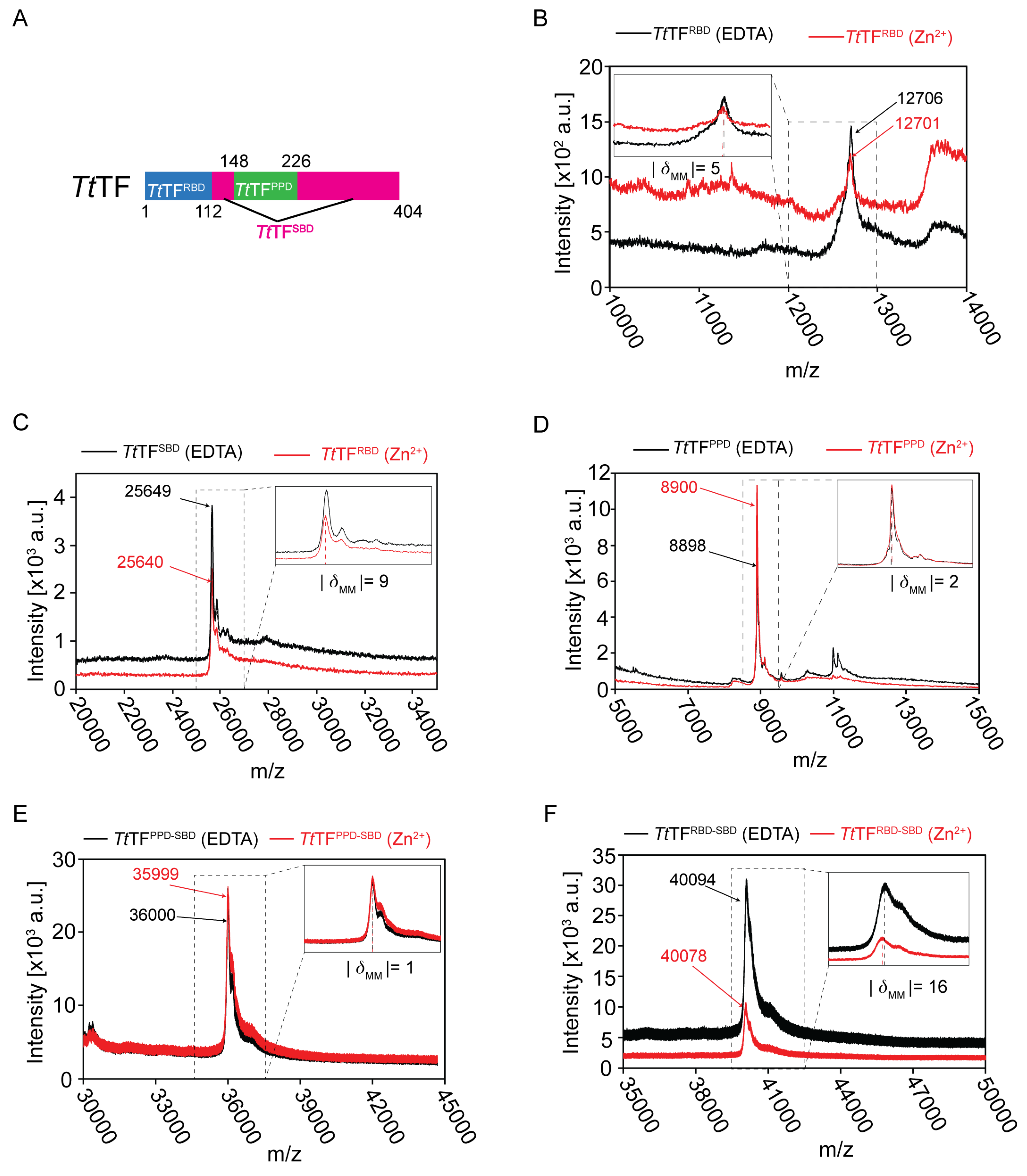
3.2. Full-Length of TtTF Was Required for Zinc Recognition

We next investigated the zinc-binding sites on TtTF. Although TtTF has been shown to bind Zn2+ at a 1:1 stoichiometry (Figure 1A), the amino acid sequence of TtTF does not have a typical zinc-binding motif. To investigate the zinc-binding sites on TtTF, TtTF was divided into the three domains: the ribosome-binding domain (TtTFRBD: Residues 1 to 113), the substrate-binding domain (TtTFSBD: Residues 112 to 148 and 226 to 404, connected by the GSGSG linker), and the peptidyl-prolyl isomerase domain (TtTFPPD: Residues 148 to 226) (Figure 2A and Figure S1). The domain boundaries of TtTF were defined according to the sequence alignment with EcTF, the structures of EcTF [12,13,17], and the Alphafold2 predicted structural model of TtTF [28] (Figures S1 and S2). The zinc-binding assay for the isolated domains was performed by following the same procedure as for the full-length TFs. Each of the domains was refolded in the presence of Zn(CH3COO)2 or EDTA and subjected to MALDI-TOF-MS analysis (Figure 2B–D, Table 1). As summarized in Table 1, all three domains indicated that the zinc-dependent mass difference is negligible compared to the mass of Zn2+ (65 Da). Thus, no zinc-binding was detected for the isolated domains.
Zinc-binding was also tested for the tandem domains of TtTF, TtTFPPD-SBD, and TtTFRBD-SBD (Figure 2E,F). The data showed that the zinc-dependent mass difference is negligible compared to the mass of Zn2+ (65 Da). Thus, no zinc-binding was detected for the tandem domains. Note that the larger differences between the experimental and the theoretical masses for tandem domains (−188 Da for TtTFPPD-SBD and −151 Da for TtTFRBD-SBD) can be explained by the removal of the N-terminal methionine residue (149 Da) by the methionyl-aminopeptidase during protein expression in E. coli [29]. Collectively, our data indicated that neither a single domain nor a tandem domain binds Zn2+. In contrast, the full-length of TtTF is required for zinc recognition, implying that all three domains are involved in zinc recognition.

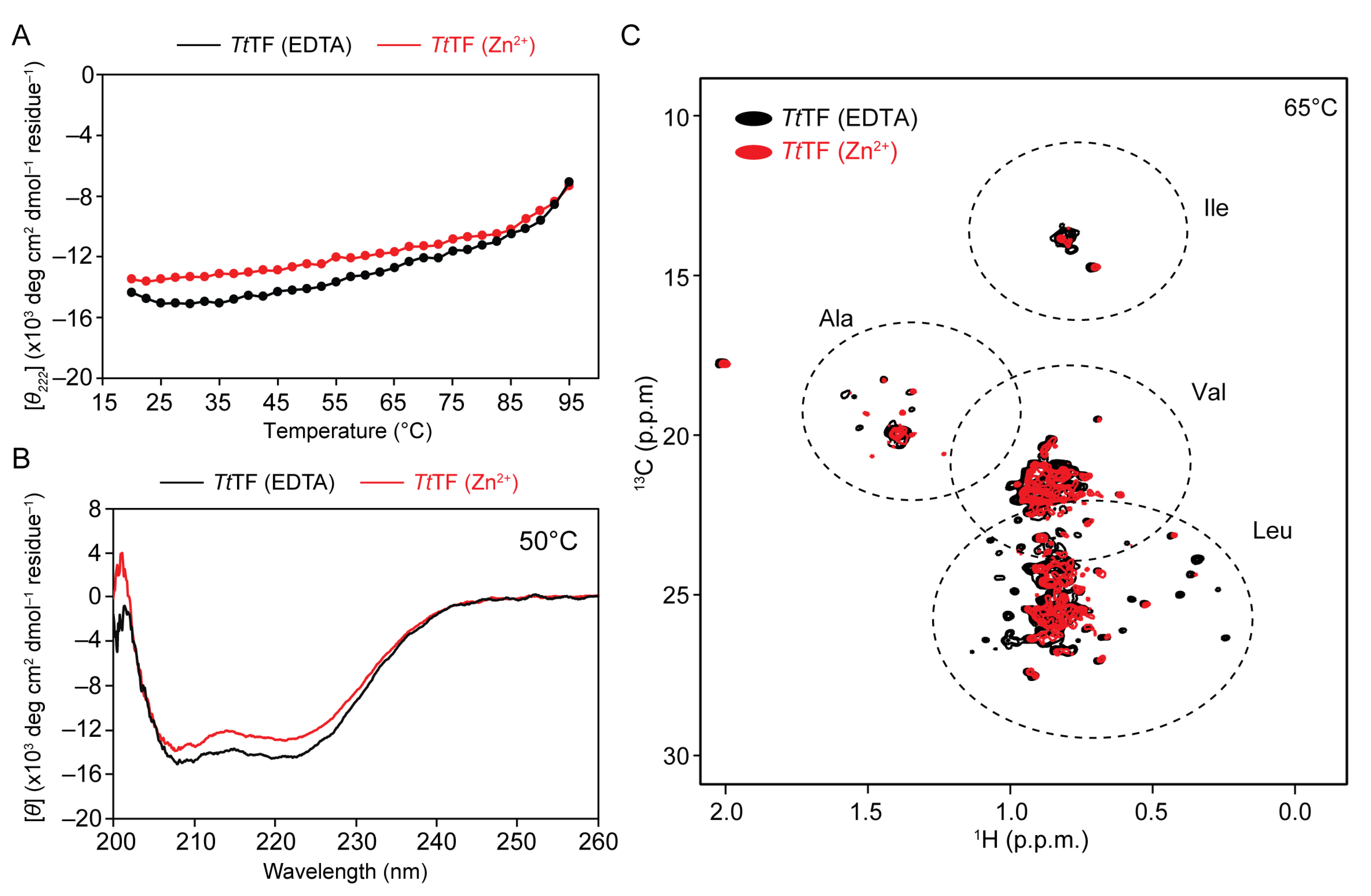
3.3. Zn2+ Induced Little Effect to Thermal Stability of TtTF

One of the possible benefits of binding metal ions is the improvement in thermal stability. To test if Zn2+ influences the thermal stability of TtTF, the mean residue ellipticity at 222 nm, indicative of α-helix, was monitored for TtTF (Zn2+) and TtTF (EDTA) at an increasing temperature from 20 °C to 95 °C (Figure 3A). At the range of 20–80 °C, TtTF (Zn2+) showed a smaller magnitude of the mean residue ellipticities compared to TtTF (EDTA) (Figure 3A). In both cases, the mean residue ellipticity value gradually increased with the temperature in the range of 20–80 °C, whereas the slope became steeper above 85 °C, indicating that the protein started unfolding above 85 °C. This observation is consistent with the fact that the maximum growth temperature of Thermus thermophilus is 85 °C [30]. Due to the limited temperature range of the CD measurement, the unfolding was incomplete, and accordingly, the midpoint of the unfolding transition temperature Tm and thermodynamic parameters [31,32] could not be determined. However, the similar melting profiles of TtTF (Zn2+) and TtTF (EDTA) in the temperature range of 85–95 °C suggested that zinc-binding had little effect on the thermal stability of TtTF in this temperature range.

3.4. Zn2+ Induced Partial Structural Change of TtTF

Although Zn2+ binding has little effect on the thermal stability of TtTF, the data showed that TtTF (Zn2+) has a weaker ellipticity at 222 nm than TtTF (EDTA) (Figure 3A), suggesting the change in the secondary structure of TtTF upon binding to Zn2+. CD spectra were acquired at 50 °C (Figure 3B) to investigate the effect of zinc-binding to the secondary structure of TtTF. Compared to TtTF (EDTA), the CD spectrum of TtTF (Zn2+) showed a smaller magnitude of the mean residue ellipticities for the region from 208 nm to 222 nm (the negative absorption peak of α-helix), indicating that TtTF (Zn2+) contains less α-helix. The secondary structure content predicted from the CD spectra using Dichroweb [23] showed that TtTF (Zn2+) contains a smaller portion of α-helix but contains a larger portion of β-sheet (Figure 3B, Table 2), indicating the zinc-induced structural changes of TtTF. CD measurements at different temperature points, 20 °C, 35 °C, 65 °C, and 80 °C, showed essentially the same trend, that the mean residual ellipticity of TtTF (Zn2+) is less negative than that of TtTF (EDTA) (Figure S3 and Table 2). Note that the purified TtTF before refolding showed essentially the same CD spectrum as TtTF (EDTA), supporting the idea that the TtTF preserves its native structure even after the refolding process (Figure S3C).
The effect of zinc-binding was further investigated by NMR, in which the methyl-HMQC spectra for [U-15N; Ala-13CH3; Met-13CH3; Ile-δ1-13CH3; Leu/Val-13CH3/13CH3]-labeled TtTF (Zn2+), and TtTF (EDTA) were acquired (Figure 3C). The spectrum of TtTF (EDTA) showed well-dispersed methyl resonances, whereas that of TtTF (Zn2+) showed fewer resonances, and several resonances were missing, most probably due to line broadening. The line broadening of TtTF (Zn2+) can be explained by exchange broadening due to the conformational exchange in a μs-ms timescale or by an increase in size due to oligomerization.

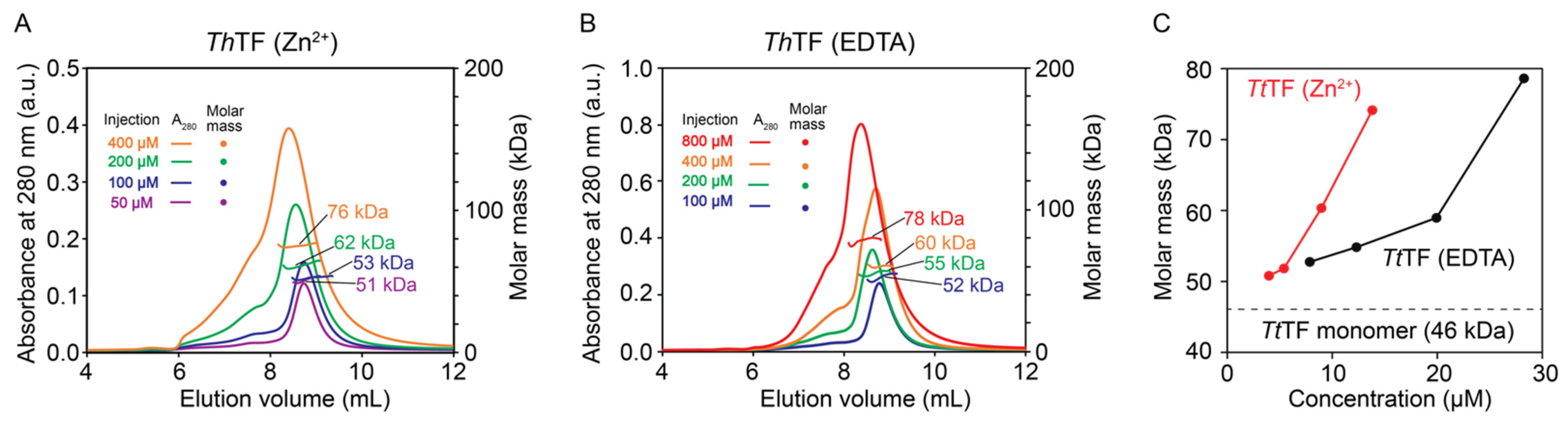
3.5. Zn2+ Promoted Oligomerization of TtTF
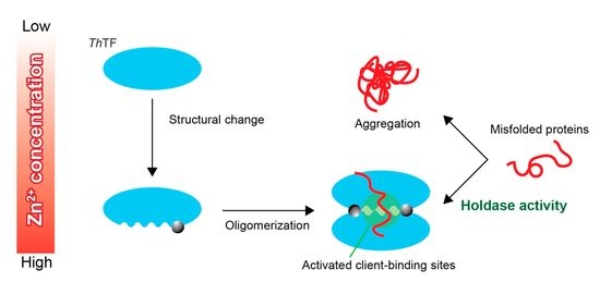
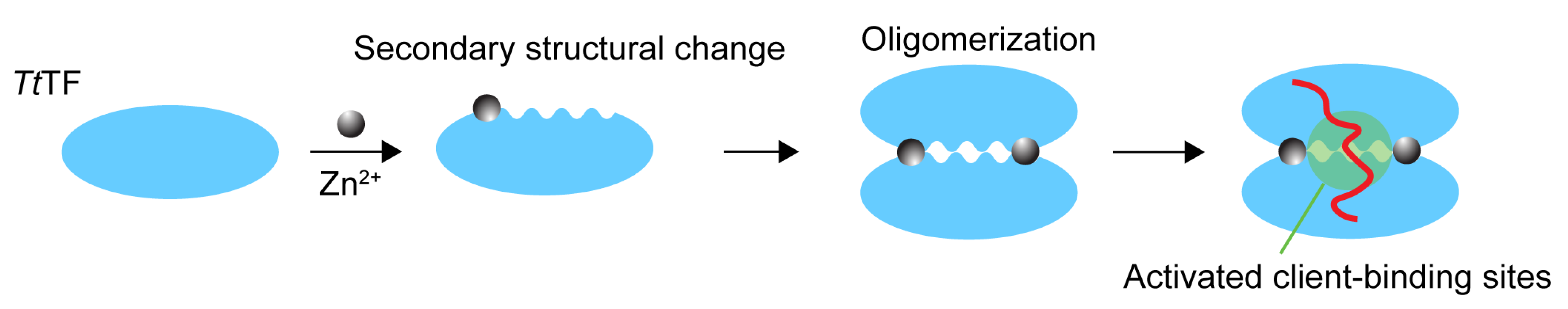
From the above results, we found that Zn2+ induces the partial structural changes of TtTF. SEC-MALS experiments were performed to test if the partial structural change induced by zinc-binding affects the oligomeric state of TtTF, which can be associated with the holdase activity of TtTF. Although the previous study reported that TtTF exists as a monomer at a lower concentration [20], the oligomeric state of TtTF at higher concentrations has not been investigated. Furthermore, TF from other organisms, including E. coli. and V. cholerae, have been shown to form a dimer at higher concentrations [33,34]. The dissociation constant (Kd) of EcTF monomer–dimer equilibrium in solution was estimated as ~2 µM [17], and the crystal structure of V. cholerae TF was solved as a dimer [34]. The SEC-MALS data at lower concentrations showed that TtTF exists as a monomer (Figure 4), which is consistent with the previous gel-filtration analysis [20]. In contrast, the observed molar mass increased at higher concentrations, indicating that TtTF undergoes concentration-dependent oligomerization (Figure 4). Note that the molar mass observed by SEC-MALS reflects the average molar mass of the protein if the protein exists in equilibrium among multiple oligomeric states. Interestingly, TtTF (Zn2+) oligomerized at a lower concentration compared to TtTF (EDTA) and the purified TtTF (Figure 4C and Figure S4). For example, the average molar mass for TtTF (Zn2+) at ~14 μM was estimated as ~76 kDa that is close to the theoretical molar mass for the dimer (92 kDa), whereas the average molar mass for TtTF (EDTA) and the natively purified TtTF at ~20 μM were both estimated as 59 kDa (Figure 4C and Figure S4). Thus, the data indicate that zinc-binding promoted the oligomerization of TtTF.

4. Discussion

TtTF has a unique character among other homologous TFs: it binds the zinc ion with a 1:1 stoichiometry, and zinc-binding regulates the holdase activity of TtTF [20]. However, the mechanism of how Zn2+ regulates TtTF activity has been poorly understood. In general, one of the possible benefits of the metal ion is to improve the thermal stability of proteins [35,36,37], but our data showed no significant thermal stability change between TtTF (Zn2+) and TtTF (EDTA) (Figure 3A), and thus this is not the case with TtTF. Interestingly, our SEC-MALS data show Zn2+ promotes the oligomerization of TtTF (Figure 4), which can be one of the key features in explaining the zinc-dependent activity modulation of TtTF. A previous study on EcTF showed that oligomerization promotes holdase activity, in which the dimer formation of EcTF assembles the client-binding sites and thus can accelerate the association kinetics with the client proteins [17]. Faster association kinetics have been shown to enhance the holdase activity of chaperones [17]. Thus, the zinc-dependent oligomerization of TtTF suggests that TtTF exploits Zn2+ to modulate the binding kinetics and thus activity through oligomerization (Figure 5).
In addition to oligomerization, zinc-binding induces the partial structural changes of TtTF, as shown by CD (Figure 3B, Figure S3, and Table 2) and NMR (Figure 3C). The NMR analysis of TtTF in the absence and presence of Zn2+ indicated that several hydrophobic amino acids, including Leu and Val, are affected by zinc-binding, which thus implies that the hydrophobic region of TtTF is involved in the partial structural changes (Figure 3C). Previous structural studies on EcTF show that both client-binding sites and the dimer-interface consist mainly of hydrophobic residues. The regions undergoing zinc-induced structural change can be a part of the oligomeric interface and/or a part of the client-binding site (Figure 5).
Collectively, our data from biophysical experiments suggest that Zn2+ binds to induce structural change and oligomerization of TtTF, which can be important features to activate the holdase activity of TtTF. Although further structural research is needed, our study provides mechanistic insight into the zinc-dependent activity modulation of TtTF.

5. Conclusions

A series of biophysical experiments were performed to investigate the physical properties of Zn2+-dependent TtTF. Although the specific binding sites of Zn2+ have not been identified in this study, MALDI-TOF-MS experiments show that the full-length of TtTF is involved in zinc coordination. CD, NMR, and SEC-MALS showed that zinc-binding induces the partial structural changes of TtTF and promotes the oligomerization of TtTF. Given the previous report on EcTF showing the relationship between the oligomerization and the activity modulation [17], the zinc-dependent oligomerization of TtTF can be one of the key features to modulate the activity of TtTF.

Supplementary Materials

The following are available online at https://www.mdpi.com/article/10.3390/biology10111106/s1, Table S1: The primers and templates for plasmid construction; Figure S1: The domain architecture of TtTF; Figure S2: The sequence alignment of proteins EcTF, TtTF, and TmTF; Figure S3: The CD spectra of TtTF at varying temperatures; Figure S4: SEC-MALS plot of the molar mass of TtTF before refolding.

Author Contributions

Conceptualization, H.Z., T.S. (Tomohide Saio), and K.I.; CD data collection and analysis, H.Z. and M.M.; NMR data collection and analysis, H.Z.; MS data collection and analysis, H.Z. and T.S. (Taiga Sugawara); MALS data collection and analysis, H.Z. and T.S. (Tomohide Saio); discussion and data interpretation, H.Z., M.M., K.I., and T.S. (Tomohide Saio); writing—original draft preparation, H.Z.; writing—review and editing, H.Z., M.M., T.S. (Taiga Sugawara), K.I., and T.S. (Tomohide Saio); visualization, H.Z. and T.S. (Tomohide Saio); supervision, K.I. and T.S. (Tomohide Saio); project administration, K.I. and T.S. (Tomohide Saio); funding acquisition, H.Z., M.M., K.I., and T.S. (Tomohide Saio). All authors have read and agreed to the published version of the manuscript.

Funding

This work was partially supported by JSPS KAKENHI (20K15969 to M.M., 17H05657, 17H05867, 18H05229, 19K06504, 19H04945, 20KK0156, and 20H03199 to T. Saio and 19H05769 to K.I.), MEXT Grant-in-Aid for Transformative Research Areas (B) JP21H05094 to T. Saio, JST PRESTO Program to T. Saio, JST FOREST Program (JPMJFR204W) to T. Saio, AMED (JP21ek0109437 and JP21wm0425004) to T. Saio, HIRAKU-Global Program, which is funded by MEXT’s “Strategic Professional Development Program for Young Researchers” to M.M., and Hokkaido University through Program for Leading Graduate Schools (Hokkaido University “Ambitious Leader’s Program”) to H.Z. This work was also partially supported by Toyota Riken Scholar, Takeda Science Foundation Grant, The Sumitomo Foundation, Astellas Foundation for Research on Metabolic Disorders, Senri Life Science Foundation, The Nakajima Foundation, The Asahi Glass Foundation, Akiyama Life Science Foundation Grants-in-Aid, and Northern Advancement Center for Science and Technology Grants-in-Aid, The Nakabayashi Trust For ALS Research, and The Kato Memorial Trust for Nambyo Research to T. Saio.

Institutional Review Board Statement

Not applicable. This study does not involve humans or animals.

Informed Consent Statement

Not applicable. This study does not involve humans or animals.

Data Availability Statement

The authors declare that all data supporting the findings of this study are available within the paper. All other information is available from the corresponding authors upon reasonable request.

Acknowledgments

This study was supported by Support Center for Advanced Medical Sciences, Tokushima University Graduate School of Biomedical Sciences.

Conflicts of Interest

The authors declare no conflict of interest.

References

  1. Dobson, C.M. Protein Folding and Misfolding. Nature 2003, 426, 884–890. [Google Scholar] [CrossRef]
  2. Rabbani, G.; Choi, I. Roles of Osmolytes in Protein Folding and Aggregation in Cells and Their Biotechnological Applications. Int. J. Biol. Macromol. 2018, 109, 483–491. [Google Scholar] [CrossRef] [PubMed]
  3. Bukau, B.; Weissman, J.; Horwich, A. Molecular Chaperones and Protein Quality Control. Cell 2006, 125, 443–451. [Google Scholar] [CrossRef] [Green Version]
  4. Hartl, F.U.; Hayer-Hartl, M. Molecular Chaperones in the Cytosol: From Nascent Chain to Folded Protein. Science 2002, 295, 1852–1858. [Google Scholar] [CrossRef] [PubMed] [Green Version]
  5. Saibil, H. Chaperone Machines for Protein Folding, Unfolding and Disaggregation. Nat. Rev. Mol. Cell Biol. 2013, 14, 630–642. [Google Scholar] [CrossRef] [Green Version]
  6. Agashe, V.R.; Guha, S.; Chang, H.; Genevaux, P.; Hayer-hartl, M.; Stemp, M.; Georgopoulos, C.; Hartl, F.U.; Barral, J.M. Function of Trigger Factor and DnaK in Multidomain Protein Folding: Increase in Yield at the Expense of Folding Speed. Cell 2004, 117, 199–209. [Google Scholar] [CrossRef] [Green Version]
  7. Hoffmann, A.; Bukau, B.; Kramer, G. Structure and Function of the Molecular Chaperone Trigger Factor. Biochim. Biophys. Acta Mol. Cell Res. 2010, 1803, 650–661. [Google Scholar] [CrossRef] [Green Version]
  8. Crooke, E.; Wickner, W. Trigger Factor: A Soluble Protein That Folds pro-OmpA into a Membrane-Assembly-Competent Form. Proc. Natl. Acad. Sci. USA 1987, 84, 5216–5220. [Google Scholar] [CrossRef] [Green Version]
  9. De Geyter, J.; Portaliou, A.G.; Srinivasu, B.; Krishnamurthy, S.; Economou, A.; Karamanou, S. Trigger Factor Is a Bona Fide Secretory Pathway Chaperone That Interacts with SecB and the Translocase. EMBO Rep. 2020, 21, 1–17. [Google Scholar] [CrossRef]
  10. Kandror, O.; Sherman, M.; Rhode, M.; Goldberg, A.L. Trigger Factor Is Involved in GroEL-Dependent Protein Degradation in Escherichia coli and Promotes Binding of GroEL to Unfolded Proteins. EMBO J. 1995, 14, 6021–6027. [Google Scholar] [CrossRef] [PubMed]
  11. Rizzolo, K.; Yu, A.Y.H.; Ologbenla, A.; Kim, S.R.; Zhu, H.; Ishimori, K.; Thibault, G.; Leung, E.; Zhang, Y.W.; Teng, M.; et al. Functional Cooperativity between the Trigger Factor Chaperone and the ClpXP Proteolytic Complex. Nat. Commun. 2021, 12, 1–18. [Google Scholar] [CrossRef]
  12. Ferbitz, L.; Maier, T.; Patzelt, H.; Bukau, B.; Deuerling, E.; Ban, N. Trigger Factor in Complex with the Ribosome Forms a Molecular Cradle for Nascent Proteins. Nature 2004, 431, 590–596. [Google Scholar] [CrossRef] [PubMed] [Green Version]
  13. Saio, T.; Guan, X.; Rossi, P.; Economou, A.; Kalodimos, C.G. Structural Basis for Protein Antiaggregation Activity of the Trigger Factor Chaperone. Science 2014, 344, 590–605. [Google Scholar] [CrossRef] [PubMed] [Green Version]
  14. Hoffmann, A.; Becker, A.H.; Zachmann-Brand, B.; Deuerling, E.; Bukau, B.; Kramer, G. Concerted Action of the Ribosome and the Associated Chaperone Trigger Factor Confines Nascent Polypeptide Folding. Mol. Cell 2012, 48, 63–74. [Google Scholar] [CrossRef] [Green Version]
  15. Maier, R.; Eckert, B.; Scholz, C.; Lilie, H.; Schmid, F.X. Interaction of Trigger Factor with the Ribosome. J. Mol. Biol. 2003, 326, 585–592. [Google Scholar] [CrossRef]
  16. Huang, C.; Rossi, P.; Saio, T.; Kalodimos, C.G. Structural Basis for the Antifolding Activity of a Molecular Chaperone. Nature 2016, 537, 202–206. [Google Scholar] [CrossRef] [Green Version]
  17. Saio, T.; Kawagoe, S.; Ishimori, K.; Kalodimos, C.G. Oligomerization of a Molecular Chaperone Modulates Its Activity. Elife 2018, 7, 1–18. [Google Scholar] [CrossRef]
  18. Morgado, L.; Burmann, B.M.; Sharpe, T.; Mazur, A.; Hiller, S. The Dynamic Dimer Structure of the Chaperone Trigger Factor. Nat. Commun. 2017, 8, 1–11. [Google Scholar] [CrossRef] [PubMed] [Green Version]
  19. Linke, K.; Wolfram, T.; Bussemer, J.; Jakob, U. The Roles of the Two Zinc Binding Sites in DnaJ. J. Biol. Chem. 2003, 278, 44457–44466. [Google Scholar] [CrossRef] [Green Version]
  20. Suno, R.; Taguchi, H.; Masui, R.; Odaka, M.; Yoshida, M. Trigger Factor from Thermus Thermophilus Is a Zn2+-Dependent Chaperone. J. Biol. Chem. 2004, 279, 6380–6384. [Google Scholar] [CrossRef] [Green Version]
  21. Kawagoe, S.; Nakagawa, H.; Kumeta, H.; Ishimori, K.; Saio, T. Structural Insight into Proline Cis/Trans Isomerization of Unfolded Proteins Catalyzed by the Trigger Factor Chaperone. J. Biol. Chem. 2018, 293, 15095–15106. [Google Scholar] [CrossRef] [Green Version]
  22. Andrade, M.A.; Chacón, P.; Merelo, J.J.; Morán, F. Evaluation of Secondary Structure of Proteins from UV Circular Dichroism Spectra Using an Unsupervised Learning Neural Network. Protein Eng. Des. Sel. 1993, 6, 383–390. [Google Scholar] [CrossRef] [PubMed]
  23. Miles, A.J.; Ramalli, S.G.; Wallace, B.A. DichroWeb, a Website for Calculating Protein Secondary Structure from Circular Dichroism Spectroscopic Data. Protein Sci. 2021, 1–10. [Google Scholar] [CrossRef]
  24. Miles, A.J.; Whitmore, L.; Wallace, B.A. Spectral Magnitude Effects on the Analyses of Secondary Structure from Circular Dichroism Spectroscopic Data. Protein Sci. 2005, 14, 368–374. [Google Scholar] [CrossRef] [Green Version]
  25. Delaglio, F.; Grzesiek, S.; Vuister, G.W.; Zhu, G.; Pfeifer, J.; Bax, A. NMRPipe: A Multidimensional Spectral Processing System Based on UNIX Pipes. J. Biomol. NMR 1995, 6, 277–293. [Google Scholar] [CrossRef]
  26. Ullah, Z.; Iqbal, A.; Baloch, M.K.; Nishan, U.; Shah, M. New Insights into the Zinc- α2-Glycoprotein (ZAG) Scaffold and Its Metal Ions Binding Abilities Using Spectroscopic Techniques. Life Sci. 2020, 249, 117462. [Google Scholar] [CrossRef] [PubMed]
  27. Gómez-Ariza, J.L.; García-Barrera, T.; Lorenzo, F.; Bernal, V.; Villegas, M.J.; Oliveira, V. Use of Mass Spectrometry Techniques for the Characterization of Metal Bound to Proteins (Metallomics) in Biological Systems. Anal. Chim. Acta 2004, 524, 15–22. [Google Scholar] [CrossRef]
  28. Jumper, J.; Evans, R.; Pritzel, A.; Green, T.; Figurnov, M.; Ronneberger, O.; Tunyasuvunakool, K.; Bates, R.; Žídek, A.; Potapenko, A.; et al. Highly Accurate Protein Structure Prediction with AlphaFold. Nature 2021, 596, 583–589. [Google Scholar] [CrossRef] [PubMed]
  29. Hirel, P.; Schmitter, J.M.; Dessen, P.; Fayat, G.; Blanquet, S. Extent of N-terminal Methionine Excision from Escherichia coli Proteins Is Governed by the Side-chain Length of the Penultimate Amino Acid. Proc. Natl. Acad. Sci. USA 1989, 86, 8247–8251. [Google Scholar] [CrossRef] [Green Version]
  30. Oshima, T.; Imahori, K. Description of Thermus Thermophilus (Yoshida and Oshima) Comb. Nov., a Nonsporulating Thermophilic Bacterium from a Japanese Thermal Spa. Int. J. Syst. Bacteriol. 1974, 24, 102–112. [Google Scholar] [CrossRef] [Green Version]
  31. Benjwal, S.; Verma, S.; Röhm, K.H.; Gursky, O. Monitoring Protein Aggregation during Thermal Unfolding in Circular Dichroism Experiments. Protein Sci. 2006, 15, 635–639. [Google Scholar] [CrossRef]
  32. Greenfield, N.J. Using Circular Dichroism Collected as a Function of Temperature to Determine the Thermodynamics of Protein Unfolding and Binding Interactions. Nat. Protoc. 2007, 1, 2527–2535. [Google Scholar] [CrossRef] [PubMed]
  33. Patzelt, H.; Kramer, G.; Rauch, T.; Schönfeld, H.J.; Bukau, B.; Deuerling, E. Three-State Equilibrium of Escherichia coli Trigger Factor. Biol. Chem. 2002, 383, 1611–1619. [Google Scholar] [CrossRef] [PubMed] [Green Version]
  34. Ludlam, A.V.; Moore, B.A.; Xu, Z. The Crystal Structure of Ribosomal Chaperone Trigger Factor from Vibrio Cholerae. Proc. Natl. Acad. Sci. USA 2004, 101, 13436–13441. [Google Scholar] [CrossRef] [Green Version]
  35. Fujino, Y.; Miyagawa, T.; Torii, M.; Inoue, M.; Fujii, Y.; Okanishi, H.; Kanai, Y.; Masui, R. Structural Changes Induced by Ligand Binding Drastically Increase the Thermostability of the Ser/Thr Protein Kinase TpkD from Thermus Thermophilus HB8. FEBS Lett. 2021, 595, 264–274. [Google Scholar] [CrossRef]
  36. Palm-Espling, M.E.; Niemiec, M.S.; Wittung-Stafshede, P. Role of Metal in Folding and Stability of Copper Proteins in Vitro. Biochim. Biophys. Acta Mol. Cell Res. 2012, 1823, 1594–1603. [Google Scholar] [CrossRef] [PubMed] [Green Version]
  37. Kojoh, K.; Matsuzawa, H.; Wakagi, T. Zinc and an N-Terminal Extra Stretch of the Ferredoxin from a Thermoacidophilic Archaeon Stabilize the Molecule at High Temperature. Eur. J. Biochem. 1999, 264, 85–91. [Google Scholar] [CrossRef] [PubMed] [Green Version]
Figure 1. MALDI-TOF-MS analysis for TtTF, TmTF, and EcTF prepared in the absence and presence of Zn2+. The overlapped mass spectra for TtTF (A), EcTF (B), and TmTF (C) refolded in the buffer containing Zn2+ (red) or EDTA (black). The expanded views are displayed at the top right corner. The theoretical molar masses of TtTF (EDTA), EcTF (EDTA), and TmTF (EDTA) are 46,313 Da, 48,193 Da, and 51,330 Da, respectively. The theoretical molar mass of Zn2+ is 65 Da. The absolute values of the difference between the experimental molar masses (|δMM|) of TFs (Zn2+) and TFs (EDTA) are displayed in the below expanded views. a.u., arbitrary unit.
Biology 10 01106 g001
Figure 2. MALDI-TOF-MS analysis for the TtTF domains prepared in the absence and presence of Zn2+. (A) The domain architecture of TtTF. (BF) The overlaid mass spectra for TtTFRBD (B), TtTFSBD (C), TtTFPPD (D), TtTFPPD-SBD (E), and TtTFRBD-SBD (F) refolded in the presence of Zn2+ (red) and EDTA (black). The expanded views are displayed at the top right or top left. The theoretical molar mass: 12,706 Da for TtTFRBD (EDTA), 25,618 Da for TtTFSBD (EDTA), 8911 Da for TtTFPPD (EDTA), 36,193 Da for TtTFPPD-SBD (EDTA), and 40,249 Da for TtTFRBD-SBD (EDTA). The absolute values of the difference between the experimental molar masses (|δMM|) of TtTFdomains (Zn2+) and TtTFdomains (EDTA) are shown below the expanded views. a.u., arbitrary unit.
Biology 10 01106 g002
Figure 3. Structural characterization of TtTF in the absence and presence of Zn2+. (A) Temperature scan of CD ellipticity at 222 nm performed from 20 °C to 95 °C. The plots for TtTF (Zn2+) and TtTF (EDTA) are shown in red and black, respectively. (B) CD spectra of TtTF (Zn2+) (red) and TtTF (EDTA) (black) at 50 °C. (C) The HMQC spectra of [U-15N; Ala-13CH3; Met-13CH3; Ile-δ1-13CH3; Leu/Val-13CH3/13CH3]-labeled TtTF (Zn2+) (red) and TtTF (EDTA) (black). Typical regions for the methyl resonances from Ala, Ile δ1, Val, and Lue are indicated by dashed eclipses. The NMR experiments were performed at 65 °C.
Biology 10 01106 g003
Figure 4. SEC-MALS for the investigation of the oligomeric state of TtTF. (A,B) SEC-MALS profiles of TtTF (Zn2+) (A) and TtTF (EDTA) (B) injected at varying concentrations. At higher concentration, the larger molar mass was observed for TtTF (Zn2+) and TtTF (EDTA), indicating the increase of the oligomeric fractions at higher concentrations. (C) Plots of the molar masses of TtTF (Zn2+) (red) and TtTF (EDTA) (black) with respect to the concentrations measured by the refractive index at the peak top. Note that TtTF is diluted after injection into the SEC column, and thus, the concentration at the peak top is lower than that at the injection. a.u., arbitrary unit.
Biology 10 01106 g004
Figure 5. Schematic representation of a possible mechanism for the zinc-dependent activity modulation of TtTF. Zn2+ induces the partial structural changes and promotes the oligomerization of TtTF. Oligomerization can assemble the client-binding sites that enables the efficient client binding, as seen in the dimeric EcTF. The client protein is represented as a red line.
Biology 10 01106 g005
Table 1. Summary of MALDI-TOF-MS experimental results for TtTF and its domains in the presence/absence of Zn2+. The values are rounded to integers.
Ave ± SD (Da)
(Refolded in Zn2+ Containing Buffer)
Ave ± SD (Da)
(Refolded in EDTA Containing Buffer)
δ (Da)
TtTF46381 ± 846319 ± 462 ± 12
TmTF51338 ± 1351335 ± 123 ± 25
EcTF48218 ± 1248214 ± 114 ± 22
TtTFRBD12698 ± 212702 ± 3−4 ± 5
TtTFSBD25646 ± 525648 ± 2−2 ± 6
TtTFPPD8899 ± 38899 ± 20 ± 4
TtTFPPD-SBD35990 ± 636005 ± 4−15 ± 10
TtTFRBD-SBD40075 ± 440098 ± 6−23 ± 9
Table 2. Summary of the secondary structure contents of TtTF in the absence and presence of Zn2+ at 20 °C, 35 °C, 50 °C, and 65 °C. The fitting failed for the data of TtTF (Zn2+) at 20 °C and TtTF (EDTA) at 65 °C.
Temperature (°C)State of TtTFα-Helixβ-SheetRandom Coil
20TtTF (EDTA)56%9%35%
TtTF (Zn2+)---
35TtTF (EDTA)59%8%34%
TtTF (Zn2+)47%18%35%
50TtTF (EDTA)54%10%36%
TtTF (Zn2+)46%17%37%
65TtTF (EDTA)---
TtTF (Zn2+)42%20%37%
Publisher’s Note: MDPI stays neutral with regard to jurisdictional claims in published maps and institutional affiliations.

