Article

Antibacterial and Antioxidant Activity of Cotton Fabric Treated with Alginate-Based Microcapsules Containing Nigella sativa Oil as Core Material


1
Department of Textile and Apparel Science, School of Textile and Design (SDT), University of Management & Technology, Lahore 54770, Pakistan
2
Department of Textile Engineering, Daffodil International University, Dhaka 1216, Bangladesh
3
Department of Mechanical Engineering, Gachon University, Seongnam-si 13120, Republic of Korea
4
Department of Textile and Fashion, Punjab Tianjin University of Technology, Lahore 54770, Pakistan
*
Authors to whom correspondence should be addressed.
Fibers 2026, 14(4), 44; https://doi.org/10.3390/fib14040044
Submission received: 3 March 2026 / Revised: 1 April 2026 / Accepted: 9 April 2026 / Published: 10 April 2026

Highlights

What are the main findings?
  • The study presents the successful synthesis of alginate-based microcapsules containing Nigella sativa oil as a core material via the sol–gel technique.
  • The as-synthesized microcapsules were characterized via SEM and optical microscopy and subsequently applied on cotton fabric via a pad–dry–cure method.
What are the implication of the main findings?
  • The developed cotton fabrics revealed excellent antibacterial activity (AATCC TM 147 and AATCC TM 100) and antioxidant (in vitro assay) activity.
  • The development of such functional textiles can serve the medical field by contributing to wound dressings and other skin applications.

Abstract

This study investigates the fabrication of microcapsules using Nigella sativa (N.S.) oil as the core and alginate as the shell material. The N.S. oil microcapsules were prepared using the sol–gel method with different oil concentrations. The microcapsules were applied to the cotton fabric by the pad–dry–cure method, and their attachment was evidenced by scanning electron microscopy (SEM). Air permeability measurements were conducted for all developed samples, revealing that the sample with 8 g loading of N.S. oil and 4.5 g alginate exhibited a 43% reduction compared to the pristine sample. To further investigate the comfort characteristics of the samples, the functionalized cotton samples were subjected to the water vapor permeability index test. The results yielded an index value of 90, indicating that the encapsulation process preserved the comfort characteristics of the samples. Among the samples, the specimen with an oil concentration of 8 mL displayed the maximum antibacterial performance, achieving a 90% reduction in colony-forming units (CFUs) following quantitative testing protocol. However, the qualitative antibacterial assessment indicates no clear zone of inhibition, but no bacterial growth was observed on the samples. Furthermore, the fabric incorporating the maximum loadings of N.S. oil and alginate capsules exhibited the maximum antioxidant activity of 86.5%. These results underscore the critical role of N.S. oil microcapsules in enhancing the antibacterial and antioxidant properties of cotton fabric, while also revealing a harmony between functional performance and comfort characteristics.

1. Introduction

Microbial contamination of textiles poses a serious public health threat, as healthcare-associated infections (HAIs) affect a significant proportion of patients in acute care hospitals worldwide. Healthcare workers serve as significant carriers of pathogen transmission, creating an urgent need for functional medical textiles, such as wound dressings, patient gowns, and hospital bedding, that are capable of providing durable antibacterial protection, particularly in the face of growing antimicrobial resistance [1]. Bio-functional textiles containing bioactive materials have emerged as a promising solution to this challenge, representing a key category of advanced materials for infection control [2]. These bioactive agents can be enclosed in a shell material, which serves as a protective layer for the core material and allows for a controlled release [3]. One of the key applications of microcapsules is the encapsulation of synthetic antibacterial agents [4], and thus, their incorporation into textiles prolongs the functional life of medical fabrics [5]. However, considering synthetic antibacterial materials’ concerns, the researchers are now mainly turning to natural antibacterial agents in textile products [6]. Several studies have explored both synthetic and natural antibacterial agents for textile finishing. Synthetic agents, including metallic nanoparticles of silver, zinc, copper, titanium, and gallium, as well as organic compounds such as triclosan, quaternary ammonium compounds, polyhexamethylene biguanide, and N-halamines, have demonstrated proven antimicrobial activity in textile applications [7,8,9]. More recently, natural antimicrobial agents derived from plant extracts and essential oils have gained significant importance as sustainable alternatives, offering reduced toxic effects on human health and the environment [10,11]. Among many natural antimicrobials, N.S. oil is identified as a profoundly active substance because of the quinone compounds existing in it, which are well known for their antibacterial and antifungal properties [12]. Furthermore, N.S. oil has been clinically reported to be effective against skin infections and in the inhibition of chronic wounds [13]. Owing to its demonstrated bioactivity and clinical validation, N.S. represents a promising natural agent for the development of antibacterial functional textile [14].
The standard structure of a microcapsule consists of a liquid core, containing the active agent, encapsulated within a solid coating material [15]. The encapsulation process creates a protective barrier against degrading environmental factors [16]. Apart from protection, the shell also allows the controlled release of oils embedded inside it [17]. Current encapsulation methodologies include emulsification, sol–gel, coacervation, spray drying, and nanoprecipitation [18]. Among these, the sol–gel process is a versatile approach for the structural stabilization of microcapsules and the controlled release of bioactive oils and compounds [19]. The key to the success of this method is the accurate control of the formulation and the processing conditions, where the shell structure and the release behavior are determined by factors such as precursor selection and solvent choice [20]. From a formulation perspective, hydrophobic agents must first be emulsified prior to encapsulation, requiring ionic or non-ionic surfactants, whose chemistry largely governs the final pore structure [21]. For example, ionic surfactants (e.g., sodium dodecyl sulfate) promote the formation of smaller pores (2–4 nm), whereas nonionic surfactants (e.g., polysorbates) lead to larger pores (~10 nm) and less densely packed shells [22]. In addition to the selection of surfactants, pH is a key factor in determining the final pore morphology [23]. One example is shells with low permeability, resulting from treatment under acidic conditions, while highly porous structures are formed under basic conditions that allow for faster release of the bioactive materials [24]. In this way, the sol–gel process provides a versatile approach to regulate various chemical and physical factors, which results in the production of microcapsules having excellent stability and controlled-release behavior [25].
Another important step in microencapsulation is the selection of shell material, especially when dealing with bioactive oils like N.S. From the available polymers, alginate is a particularly suitable choice, since it is capable of undergoing mild ionic gelation [26]. As a result of exposure to divalent cations such as Ca2+, ionic bridges are created between the parts of guluronic acid, thus forming a cross-linked alginate hydrogel. This network serves as a double layer of protection for the agent, both against the environment and by controlling its release [27,28]. The alginate microcapsules are capable of this dual functionality, i.e., protection and controlled release. This feature makes the alginate-based capsules extremely attractive for use in the field of durable textile finishes [29].
Despite considerable advances in functional textile development, critical research gaps remain. First, synthetic antimicrobial agents raise concerns due to their adverse effects on human health and the environment. Second, while N.S. oil is a potent natural antimicrobial, its practical application is limited by poor water solubility, oxidative instability, and low bioavailability, challenges rarely addressed in textile studies. Third, most prior investigations have focused solely on antibacterial efficacy without evaluating fabric comfort properties such as air permeability and moisture management, despite evidence that polymeric microcapsules can reduce breathability. This dual assessment of antibacterial activity and wearer comfort remains largely unexplored for alginate-based systems incorporating N.S. oil.
Addressing these limitations requires a system that simultaneously delivers: (i) natural biocompatible antibacterial activity; (ii) protection and controlled release of the active agent; and (iii) minimal disruption to the intrinsic comfort properties of the treated fabric. The present study is designed to bridge this gap by developing alginate-based microcapsules loaded with N.S. oil via the sol–gel process, followed by their application onto cotton fabric. Scanning electron microscopy (SEM) and Fourier-transform infrared spectroscopy (FTIR) were used to verify the successful encapsulation. Beyond antibacterial evaluation, this work also focuses on variations introduced by functional microcapsules in comfort properties, like air permeability and moisture management. The findings of this study have broad implications for next-generation functional textiles. The alginate/N.S. oil microcapsule system provides a scalable, sustainable, and biocompatible platform for medical textiles, personal protective equipment, and hygiene apparel. Encapsulation strategies in textile finishing offer a transformative approach for sustained release of therapeutic agents across multiple functionalities. Future work should focus on wash durability, scale-up, and application to other natural oils to advance bio-functional medical textiles.

2. Materials and Methods

2.1. Materials

Laboratory-grade sodium alginate (alginic acid sodium salt from brown algae, medium viscosity, CAS No. 9005-38-3, Cat. No. A2033, ≥98% purity) and calcium chloride (≥97% purity) were purchased from Sigma-Aldrich (St. Louis, MO, USA). Tween-80 (an emulsifier, (≥99% purity)) was obtained from Merck (Darmstadt, Germany). N.S. seeds were obtained from a certified herbal supplier in Valencia Town (Lahore, Pakistan). All the chemicals obtained were used without further purification.

2.2. Methods

2.2.1. Extraction of N.S. Seed Oil

The initial step involved cleaning the N.S. seeds by washing, followed by drying at 40 °C in a vacuum oven to remove the dust and residual moisture. The dried seeds were subjected to cold-press extraction using an oil press (Henan Yishen, Model YS100, Zhengzhou, China) to obtain the oil. The reason for choosing the cold-press method was to avoid the heat degradation of the native bioactive components existing in the seeds, which are usually damaged if a heat-based extraction process is applied.

2.2.2. Preparation of N.S. Oil Microcapsules via Sol–Gel Technique

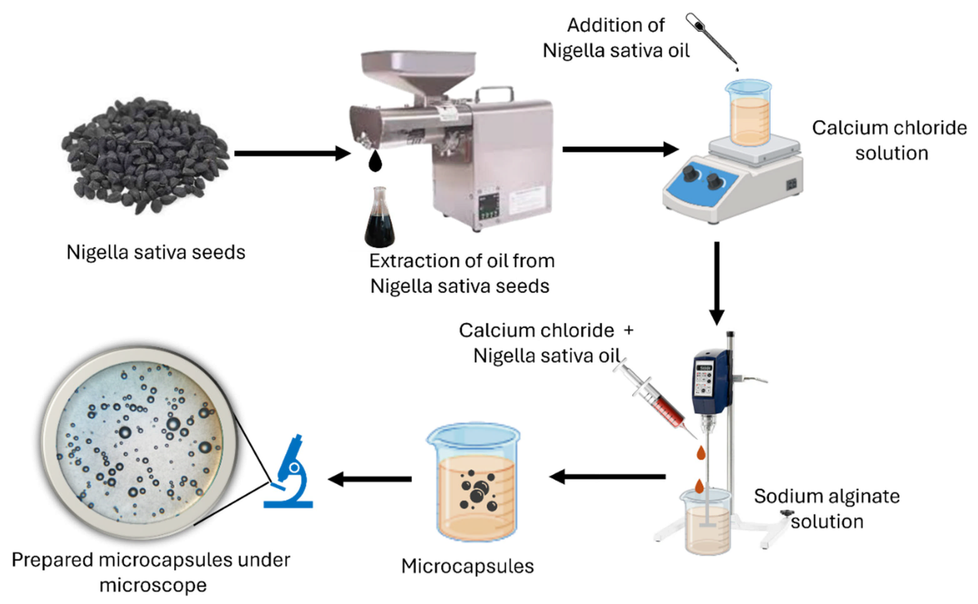
The microcapsules containing N.S. oil were prepared by the sol–gel technique. For this purpose, 1.5 g of sodium alginate was added to 100 mL of distilled water (yielding a final concentration of 1.5% w/v) and heated at 90 °C for a period of 10 min with continuous stirring to prepare Solution I (SI). In Solution II (SII), 4 g of calcium chloride was dissolved in 100 mL of distilled water while stirring at 500 rpm. To this mixture, 2 mL of N.S. oil and three drops of Tween 80 were added, followed by thorough homogenization to ensure effective dispersion. The stirring speed remained the same throughout. Subsequently, the calcium oil mixture (Solution II) was slowly added to Solution I, using a thin gauge syringe with a high-speed homogenizer (10,000 rpm). The droplets were kept in the alginate solution for 15 min to ensure complete ionic cross-linking within the microcapsule shell. A flow diagram showing the procedure for making microcapsules using Solutions I and II is included below as Figure 1.
All other microcapsule formulations were prepared following the same procedure, with only the concentrations of sodium alginate and N.S. oil varied as outlined in Table 1.

2.2.3. Application of N.S. Oil Microcapsules on Cotton Fabric

The microencapsulated formulations were applied to cotton fabric using a conventional pad–dry–cure process. The samples were immersed in the microcapsule dispersions for 5 min to ensure uniform uptake, followed by passage through padding rollers to remove excess liquor and attain a wet pick-up of approximately 70%. After padding, the fabrics were dried at 100 °C for 3 min and then cured at 110 °C for 2 min using a laboratory stenter frame.

2.3. Characterization and Testing

The microcapsules morphologies were characterized via optical microscopy (Infitek, MSC-P3000, Shanghai, China), while the surface microstructure of the treated cotton fabric was examined using field-emission scanning electron microscopy (FE-SEM; Nova NanoSEM 450, Hillsboro, OR, USA). All the fabric samples were separately spread on carbon tape, which was placed on an aluminum stub. Aluminum stub holders, along with the samples, were placed in a gold sputtering unit for gold coating. Once the gold coating was finished, the samples were taken out, and morphological analysis work was carried out. The images were recorded and evaluated. Attenuated total reflectance Fourier transform infrared (ATR-FTIR) spectroscopy (KRÜSS, Hamburg, Germany) was employed to examine and compare the surface chemical functionalities of untreated and treated fabric samples. Air permeability was measured using the Testex FX 3300 Air Permeability Tester (Schwerzenbach, Switzerland) based on the requirements of EN ISO 9237:1995. Water vapor permeability, the main parameter determining thermophysiological comfort, was measured for untreated and functionalized cotton fabrics according to BS 7209:1990. The higher the index value, the better the moisture-regulating capacity. The antimicrobial activity of the functionalized fabrics was determined qualitatively against Staphylococcus aureus (Gram-positive) according to the standard method AATCC TM 147-2011, which corresponds to ISO 20645:2004. Colony-forming units (CFUs) recovered from treated and untreated fabrics were quantitatively determined according to AATCC TM 100–2019, which corresponds to ISO 20743:2021 and ASTM E3160-20. The percent reduction in viable bacteria was calculated using Equation (1) [6].
%   R e d u c t i o n = C o n t r o l   C F U s T r e a t e d   C F U s C o n t r o l   C F U s × 100
The antioxidant activity of the treated samples was also established according to in vitro Antioxidant Activity Assay. A 3.5 mL of DPPH (2,2-diphenyl-1-picrylhydrazyl) methanol solution was used to treat the control and functionalized samples. The samples were placed in a shaking bath for 25 min at 25 °C in a dark environment. The absorbance was measured at 517 nm. Both samples, the treated with microcapsules and the untreated (pristine cotton) samples, contain DPPH. The presence of antioxidants on the microcapsules treated samples initiated the reduction reaction of DPPH radical to a non-radical form (by changing its color to yellow). The following Equation (2) was used to calculate antioxidant efficiency of the treated samples [30].
R a d i c a l   S c a v e n g i n g   E f f i c i e n c y % = [ 1   A b s o r b a n c e   o f   s a m p l e A b s o r b a n c e   o f   b l a n k   c o n t r o l ] × 100

3. Results

3.1. Morphological Analysis

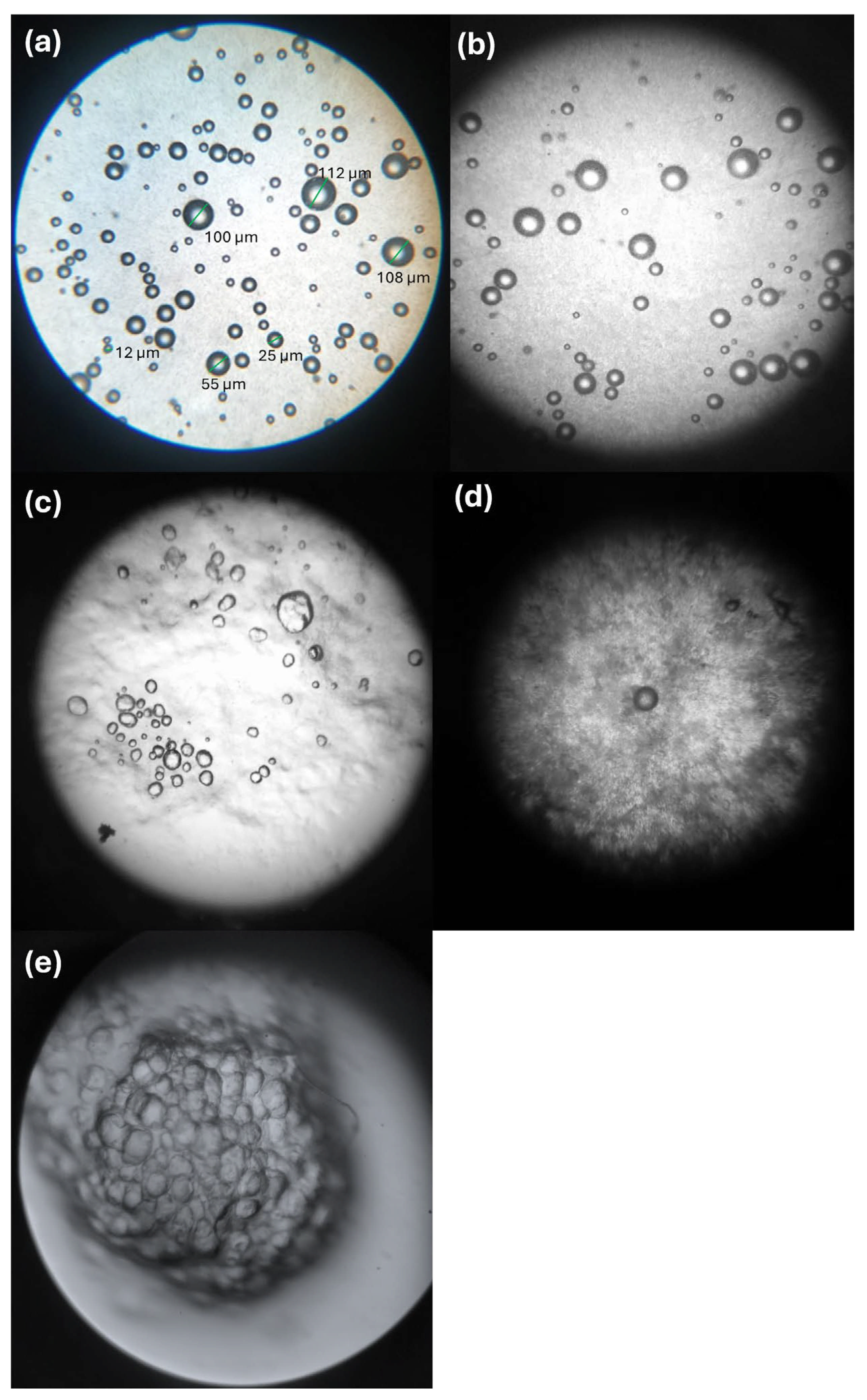
As shown in Figure 2, the micrographs obtained at 180× magnification confirmed that the microcapsules were successfully formed with circular morphologies. The microcapsules exhibited a size distribution ranging from 12 μm to 112 μm. The distribution was polydisperse, which is characteristic of sol–gel microcapsules prepared via conventional emulsification [31]. The structures seen are defined, uniform, and spherical in shape, showing the successful encapsulation of the cores through the applied sol–gel process.
To evaluate the lifecycle of the microcapsule suspension and determine its usability for multiple dip-coating applications, the stability of the formulation was monitored by optical microscopy over a 20-day period. The microcapsule suspension was stored under refrigerated conditions (4 °C) in sealed, dark containers to minimize oil oxidation and microbial growth (Figure 2).
Freshly prepared microcapsules (day 1) exhibited a well-dispersed morphology with no signs of aggregation (Figure 2a). After 5 days of storage, the capsules remained individually dispersed and visibly distinct, indicating good colloidal stability (Figure 2b). By day 10, slight aggregation was observed; however, the majority of capsules remained sufficiently dispersed for uniform application onto fabric (Figure 2c). After 15 days, significant aggregation occurred, with only a few capsules remaining in dispersed form. By day 20, the microcapsules had completely agglomerated, making them unsuitable for further dip-coating applications.
Based on these findings, the microcapsule suspension is recommended for use within 10 days of preparation when stored under refrigerated (4 °C), dark conditions to ensure optimal dispersion, consistent microcapsule loading, and reliable antibacterial performance.
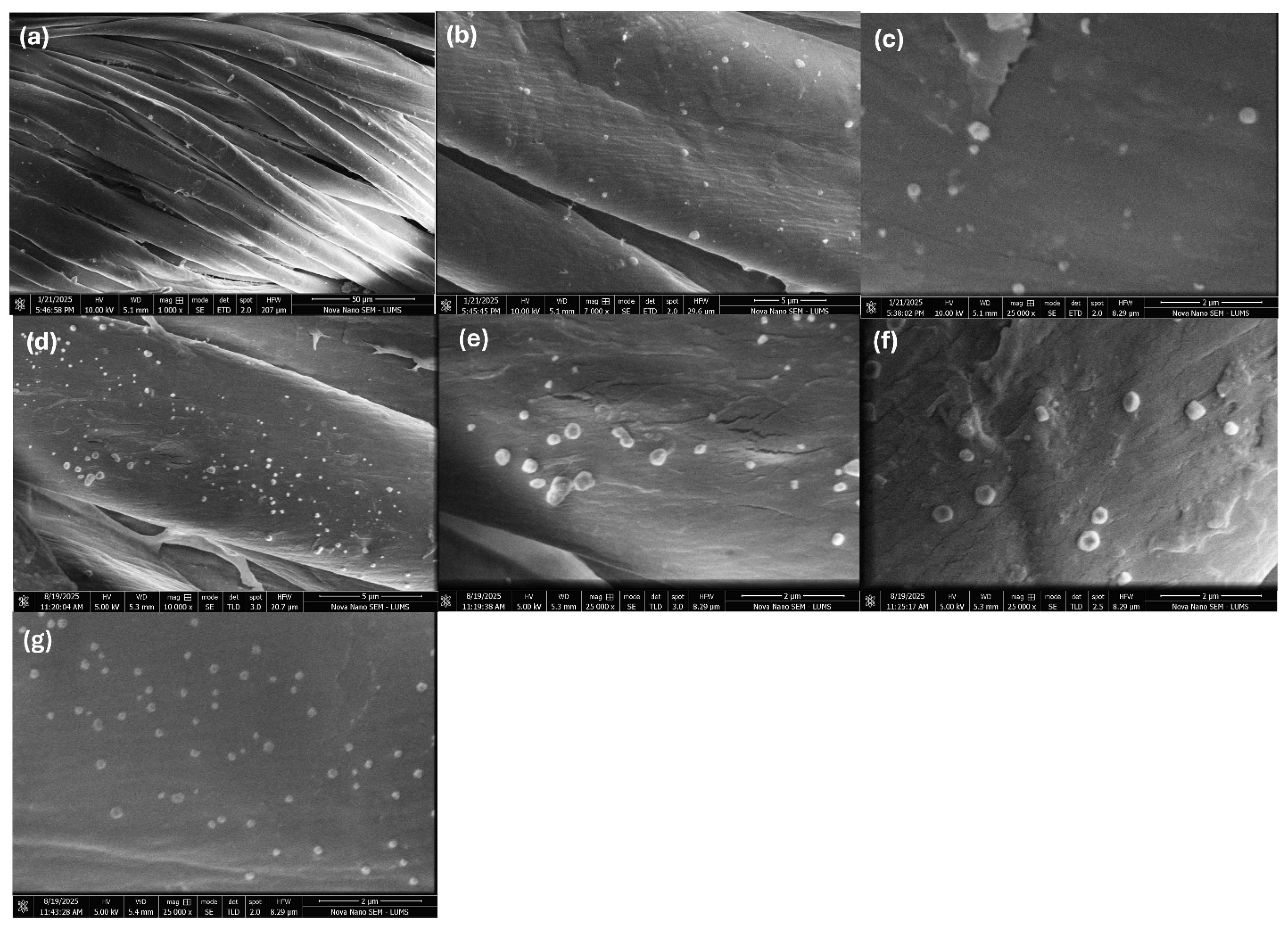
Figure 3a shows an SEM image (1000× magnification) of the surface dispersion of the N.S. oil-based microcapsules within and on the surface of the treated fabric. The morphology confirms the successful anchoring of the microcapsules by the sol–gel and pad–dry–cure deposition method. The higher resolution image (7000× magnification, Figure 3b) shows discrete interparticle gaps between the microcapsules, confirming that the microcapsules are well dispersed and not interconnected to form a continuous film. With a diameter of 0.5–1.0 μm, the size of the microcapsules is reasonably small to adhere onto cotton fibers (~16 μm in diameter) and thus could not occlude fabric porosity. Similar depositions were achieved for all formulations S2 (Figure 3c), S3 (Figure 3d), S4 (Figure 3e), S5 (Figure 3f), and S6 (Figure 3g), all prepared at varying concentrations of N.S. oil. Overall, these SEM analyses confirmed successful preparation and adhesion of the microcapsules to the fabric samples for all formulations used in this work.

3.2. FTIR Analysis

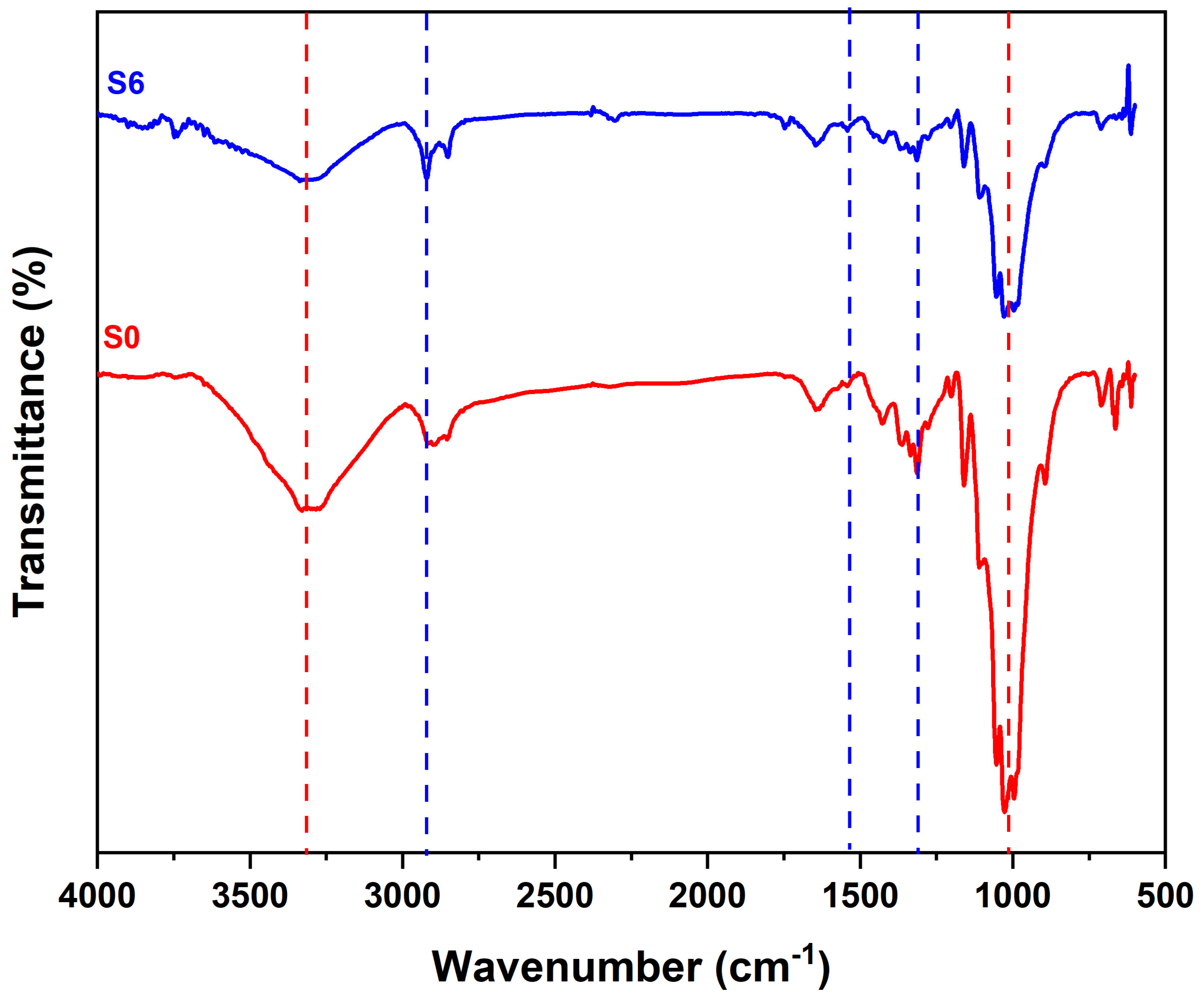
As shown in Figure 4, the ATR-FTIR spectrum of untreated cotton fabric exhibits characteristic cellulose peaks: a broad band at 3300–3400 cm−1 (O–H stretching), a sharp peak at 2900 cm−1 (C–H stretching), a band at 1640 cm−1 (adsorbed water O–H bending), and a strong band at 1030–1060 cm−1 (C–O stretching) [32]. ATR-FTIR analysis of the treated fabric provided direct evidence of calcium alginate formation, indicated by the presence of carboxylate ion vibrations: an asymmetric stretch at 1600–1640 cm−1 and a symmetric stretch at 1400–1430 cm−1, characteristic of ionic cross-linking [33]. The C–H stretching peak at approximately 2900 cm−1 showed contributions from the alkyl chains in cellulose, alginate, and N.S. oil. Furthermore, the broad, intense absorption between 1000 and 1100 cm−1 indicated overlapping C–O and C–O–C stretching vibrations from the polysaccharide backbones of both cellulose and alginate [34,35]. In brief, the sample made from cellulose and functionalized with microcapsules is an indication that the treatment changed the surface of the fabric while the original structure of cotton substrate remained intact.

3.3. Air Permeability Analysis

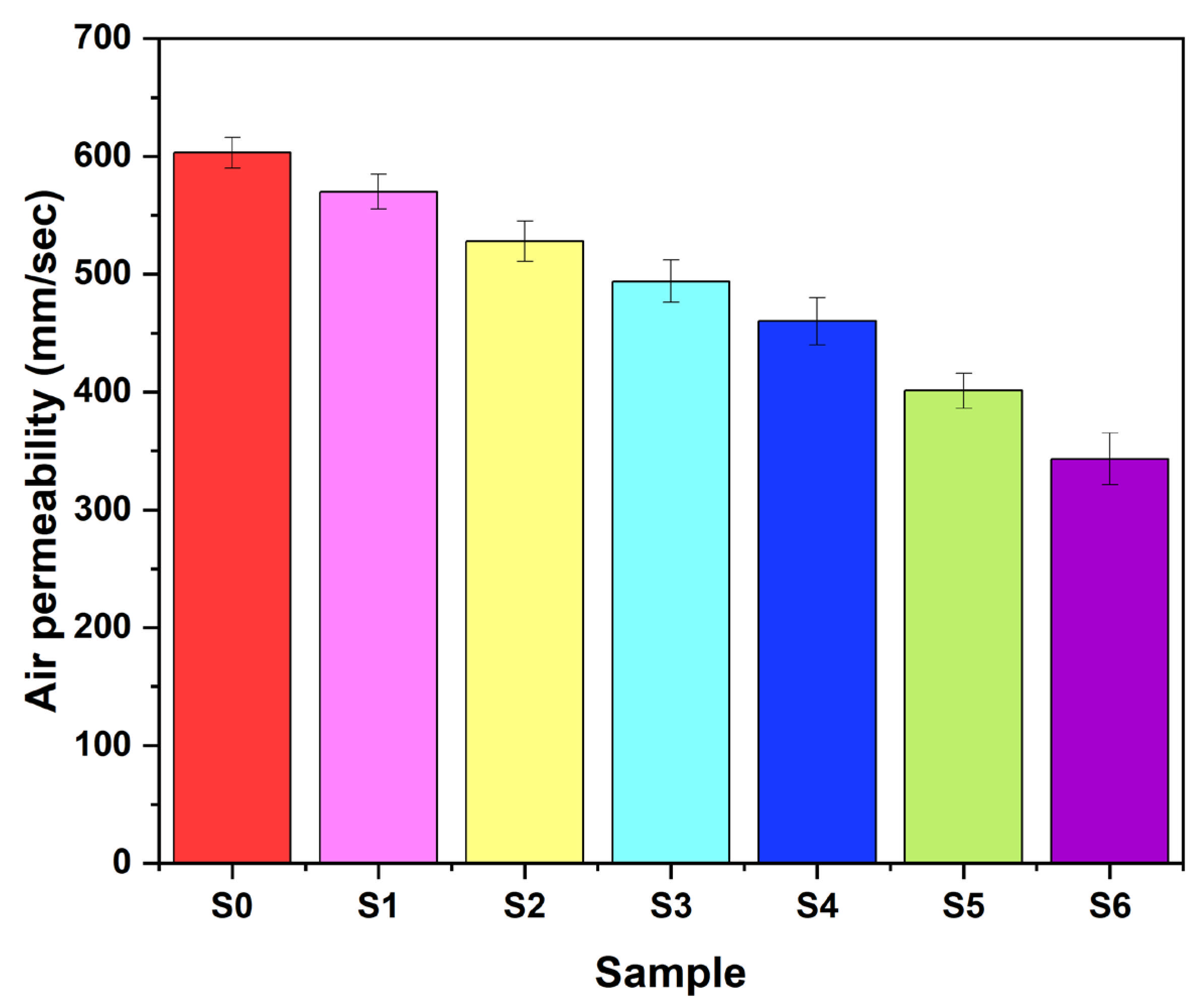
The air permeability of the control and the treated cotton fabrics was measured, and the data are presented in Figure 5. The findings from these measurements reveal that the air permeability of the fabric is inversely proportional to the amount of alginate and oil loaded in the microcapsules, higher loadings progressively obstruct airflow through the textile structure. Air permeability decreased from 603 mm/s (untreated S0) to 570 mm/s (S1: 1.5 g alginate, 2 mL oil) and 528 mm/s (S2). The decrease was gradual, showing that the deposition of microcapsules may have only partially blocked the fabric’s porous structure without completely sealing it. When the concentration of the shell-forming polymer increased to 4.5 g (samples S3 with 2 mL oil and S4 with 4 mL oil), the air permeability dropped to 494 and 460, respectively. The reason for the decrease is that a denser microcapsule cover was formed, which limited the passage of air through the fabric pores. The most significant drop in permeability was noticed for the sample S6 (343 mm/s). According to this finding, combining high levels of the core and shell (4.5 g alginate, 8 mL N.S. oil) makes a microcapsule deposit that is obstructive and thus acts as barrier to the airflow [36]. Overall, the findings reveal that increased microcapsule loading enhances functionalization but progressively reduces air permeability and, consequently, textile breathability.

3.4. Water Vapor Permeability

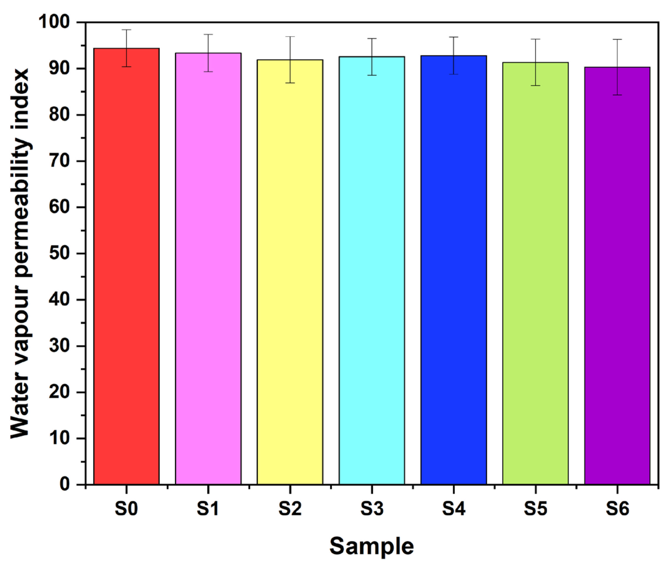
Figure 6 conveys the water vapor permeability index of the samples from S0 to S6. The untreated cotton exhibits an index of 94.4, which corresponds to the highest permeability since no materials were applied to its surface. The water vapor transmission gradually reduced after the addition of microcapsules to the fabric surface from 93.36 g/m2 for S1 to 90.33 g/m2 for S6. These results suggest that the microcapsules of alginate and N.S. oil leave only a slight barrier to water vapor passage. Altogether, the decrease was less than 5%; thus, the moisture transfer was mostly kept intact after functionalization. Vapor permeability retention is possible due to the alginate shell’s hydrophilic nature, whose hydroxyl (–OH) and carboxyl (–COOH) groups enable the passage of moisture even though microcapsules are present on the fabric surface [37].

3.5. Antibacterial Analysis

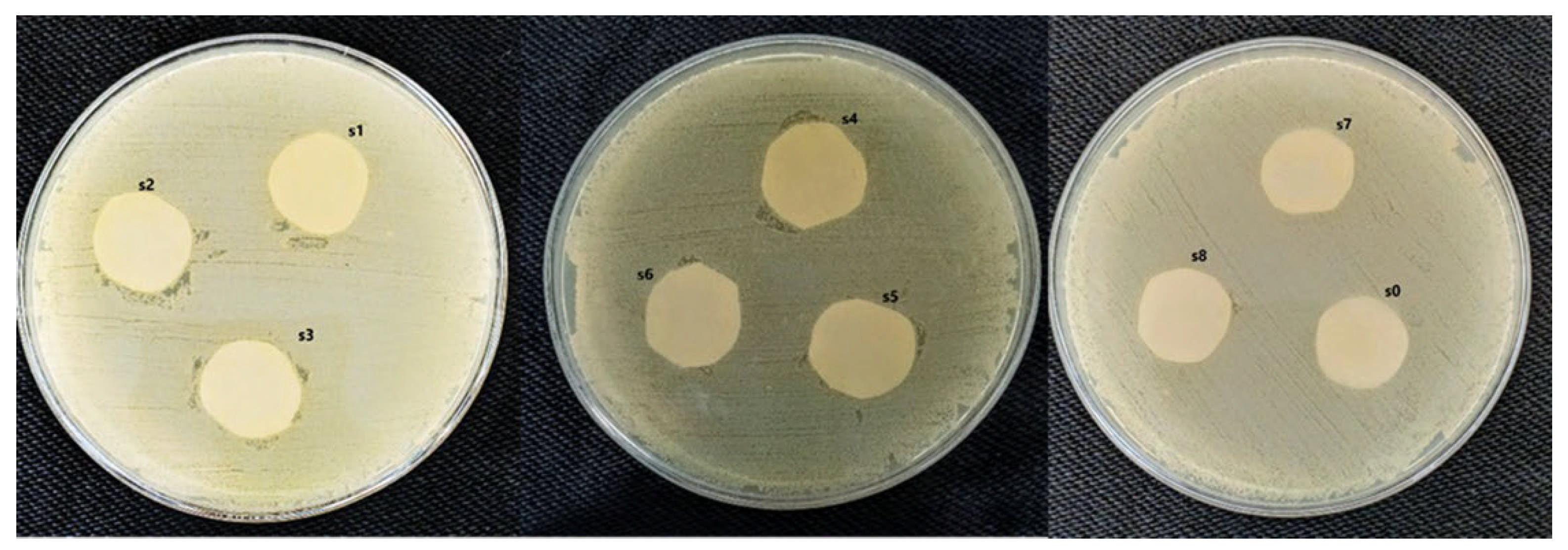
Figure 7 presents the qualitative assessment (AATCC TM 147-2011). While no clear zone of inhibition was observed around the treated samples, no bacterial growth was detected on the fabric surface itself. The absence of a zone of inhibition in the qualitative test (AATCC TM 147-2011), combined with the absence of bacterial growth on the treated fabric surface in the quantitative test (AATCC TM 100–2019), is characteristic of a contact-based (non-leaching) antibacterial mechanism. This indicates that the Nigella sativa oil remains encapsulated within the alginate microcapsules and is not released into the surrounding environment. Instead, antibacterial activity occurs upon direct contact between the bacteria and the functionalized fabric surface, where the active agent exerts its effect through membrane disruption or other contact-dependent mechanisms [38,39]. Since merely visual observation cannot be a reliable measure of bacteriostatic performance, a numerical analysis was accordingly conducted to get a quantitative evaluation; consequently, the AATCC TM 100:2019 standard was used to accurately quantify the antibacterial efficacy.
Table 2 reports the quantitative antibacterial efficacy of the selected samples in accordance with AATCC 100–2019. The untreated cotton (S0) showed no antibacterial activity. After mixing with 1.5 g sodium alginate and 2 mL N.S. oil, sample S2 demonstrated a moderate antibacterial response as S. aureus colony survival was halved. By increasing the shell material to 4.5 g while keeping the oil concentration unchanged (S4), a marked antibacterial activity enhancement was obtained, leading to a 60% reduction in bacterial count. Such a result may be attributed to the formation of a denser microcapsule and better active oil encapsulation. Sample S6, which contained 4.5 g alginate and 8 mL oil, was found to be the most effective in antibacterial action, yielding an almost 90% reduction in CFU. The stronger antibacterial response was due to greater amounts of bioactive oil in conjunction with a thicker encapsulation layer that allows for stronger bacterial growth inhibition. Taken together, the data present a dose-dependent increase in antibacterial effectiveness, indicating that N.S. oil-loaded alginate microcapsules are an attractive option for functional antimicrobial finishing of cotton textiles.
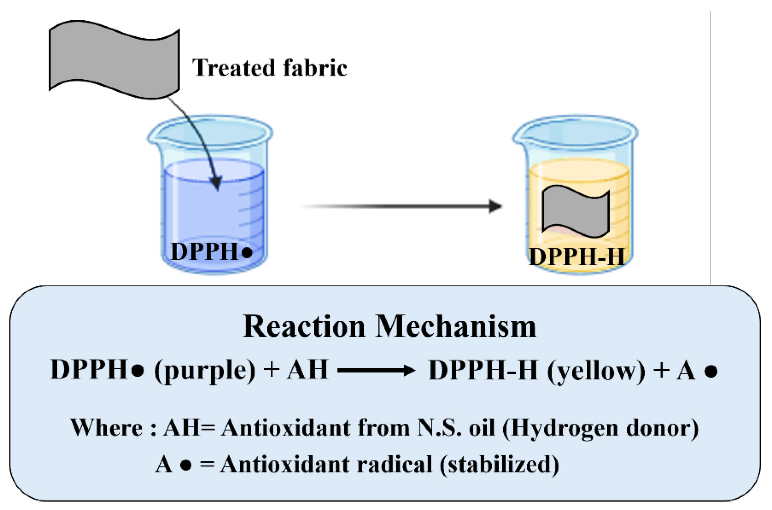
The in vitro antioxidant efficiency (%) observed is summarized in Table 3. The N.S. oil-treated fabrics showed antioxidant efficiency, while the untreated control sample (S0) did not exhibit any. Of the treated samples (S1–S6), the fabric loaded with the maximum amount of oil, 8 mL, and 4.5 g alginate capsules showed a significant increase in antioxidant performance (86.5%). The antioxidant performance of the N.S. oil-treated fabric is due to the phenolic, flavonoids, and other bioactive compounds present in the oil, which can neutralize DPPH free radicals effectively [40,41,42]. As shown in Figure 8, the DPPH assay works through a hydrogen transfer mechanism, where the purple DPPH radical (DPPH•) is reduced when it encounters an antioxidant species (N.S. oil). There are several functional compounds in N.S. oil, such as thymoquinone as a potent antioxidant. In the process, the antioxidant donates a hydrogen atom, resulting in the conversion of DPPH• to its non-radical yellow form (DPPH-H). The antioxidant is converted into a stable radical, the lowering of its reactivity being a consequence of electron delocalization in its aromatic framework; thus, it does not undergo further oxidative reactions [43]. The developed cotton samples revealed strong antioxidant activities which can be applied as potential medical textiles.

4. Limitations and Future Directions

While the present study successfully demonstrates the preparation and antibacterial efficacy of alginate-based microcapsules containing N.S. oil via the sol–gel method, certain limitations should be acknowledged.
The microcapsules exhibited a polydisperse size distribution ranging from 12 μm to 112 μm. The distribution was polydisperse, which is characteristic of sol–gel microcapsules prepared via conventional emulsification. While this polydisperse nature offered practical advantages in terms of surface coverage, adhesion, and fabric hand feel, achieving a more uniform (monodisperse) size distribution could further optimize performance consistency. Techniques such as microfluidic emulsification, membrane emulsification, or electrospray-assisted sol–gel processing represent potential approaches for producing capsules with narrower size distributions in future studies.
The durability of the microcapsules on the fabric surface, particularly their resistance to repeated washing cycles and mechanical abrasion, was not evaluated in this study. For functional textiles intended for practical applications, wash durability and abrasion resistance are critical parameters that influence the longevity and commercial viability of the finish.
The impact of ambient conditions on the stability and performance of microcapsules has not yet been investigated. For medical textile applications such as bandages and wound dressings, these factors are critically important, as products may encounter varying storage temperatures; humidity levels during storage and transportation; pH variations from wound exudate (typically pH 5.5–8.5, with infected wounds often exhibiting elevated pH); and potential contamination from bodily fluids or environmental dirt.

5. Conclusions

This study demonstrates the functionalization of cotton fabrics using sodium alginate-based microcapsules loaded with N.S. oil. The fabrication and textile adhesion of microcapsules were confirmed by SEM analysis. The findings show that while increasing the concentration of microcapsules boosts the functionalization, it also leads to a progressive decrease in air permeability from about 603 to 343 mm/s. Water vapor transmission results suggest that microencapsulation causes very low resistance, with the permeability index being close to 90 even for the most concentrated formulation. Antibacterial tests demonstrated that microencapsulation effectively conferred strong bioactivity to the treated fabrics. Sample S6 with the high oil loading (8 mL) displayed the most effective antimicrobial activity by reducing nearly 90% of CFUs, thus emphasizing the direct correlation between the microcapsule composition and the fabric’s functional efficacy. Sample S6, incorporated with the highest loading of N.S. oil and alginate capsules, exhibited the maximum antioxidant activity of 86.5%. These findings indicate that encapsulating N.S. oil in an alginate shell allows the effective application onto cotton fabrics, conferring antibacterial and antioxidant activities without compromising comfort. Consequently, these results form a tangible basis for manufacturing eco-friendly, functionally durable antimicrobial textiles, thus making them attractive candidates for healthcare textiles, protective wear, and innovative clothing.

Author Contributions

Conceptualization, N.B.; Data curation, M.M., F.H. and M.J.S.; Formal analysis, N.B., I.A.K., T.N., M.M. and M.J.S.; Funding acquisition, K.J., A.K., M.M. and F.H.; Investigation, N.B., I.A.K. and M.J.S.; Methodology, N.B.; Project administration, K.J. and A.K.; Resources, K.J. and T.N.; Software, I.A.K., F.H. and M.J.S.; Supervision, K.J., A.K. and T.N.; Validation, I.A.K.; Visualization, T.N. and M.M.; Writing—original draft, N.B.; Writing—review and editing, K.J., A.K. and F.H. All authors have read and agreed to the published version of the manuscript.

Funding

This research received no external funding.

Data Availability Statement

The original contributions presented in this study are included in the article. Further inquiries can be directed to the corresponding authors.

Acknowledgments

We acknowledge the competent support from the School of Design and Textile UMT in the realization of this research work.

Conflicts of Interest

The authors declare no conflicts of interest.

Correction Statement

This article has been republished with a minor correction to Figure 4. This change does not affect the scientific content of the article.

References

  1. Dixit, S.; Varshney, S.; Gupta, D.; Sharma, S. Textiles as Fomites in the Healthcare System. Appl. Microbiol. Biotechnol. 2023, 107, 3887–3897. [Google Scholar] [CrossRef] [Scilit]
  2. Podgornik, B.B.; Šandric, S.; Kert, M. Microencapsulation for Functional Textile Coatings with Emphasis on Biodegradability—A Systematic Review. Coatings 2021, 11, 1371. [Google Scholar] [CrossRef] [Scilit]
  3. Reisberg, K.; Hõrrak, K.; Tamm, A.; Kõrver, M.; Animägi, L.; Visnapuu, J. Functional Textile Socks in Rheumatoid Arthritis or Psoriatic Arthritis: A Randomized Controlled Study. Textiles 2025, 5, 30. [Google Scholar] [CrossRef] [Scilit]
  4. Solihat, N.N.; Purwanti, T.; Husna, N.; Oktaviani, M.; Zulfiana, D.; Fatriasari, W.; Nawawi, D.S. Capability Lignin from Acacia Crassicarpa Black Liquor as an Environmentally Benign Antibacterial Agent to Produce Antibacterial and Hydrophobic Textiles. Bioresour. Technol. 2024, 413, 131409. [Google Scholar] [CrossRef] [Scilit]
  5. Orasugh, J.T.; Temane, L.T.; Pillai, S.K.; Ray, S.S. Advancements in Antimicrobial Textiles: Fabrication, Mechanisms of Action, and Applications. ACS Omega 2025, 10, 12772–12816. [Google Scholar] [CrossRef] [Scilit]
  6. Khan, I.A.; Khalid, H.; Javed, K.; Fraz, A.; Pasha, K.; Khan, A. Dyeing and Functional Finishing of Cotton Fabric Using Ficus Carica and Eucalyptus Leaf Extracts with Aloe Barbadensis Miller as a Bio-Mordant. Resources 2025, 14, 127. [Google Scholar] [CrossRef] [Scilit]
  7. Karypidis, M.; Karanikas, E.; Papadaki, A.; Andriotis, E.G. A Mini-Review of Synthetic Organic and Nanoparticle Antimicrobial Agents for Coatings in Textile Applications. Coatings 2023, 13, 693. [Google Scholar] [CrossRef] [Scilit]
  8. Fernandes, M.; Padrão, J.; Ribeiro, A.I.; Fernandes, R.D.V.; Melro, L.; Nicolau, T.; Mehravani, B.; Alves, C.; Rodrigues, R.; Zille, A. Polysaccharides and Metal Nanoparticles for Functional Textiles: A Review. Nanomaterials 2022, 12, 1006. [Google Scholar] [CrossRef] [Scilit]
  9. Granados, A.; Pleixats, R.; Vallribera, A. Recent Advances on Antimicrobial and Anti-Inflammatory Cotton Fabrics Containing Nanostructures. Molecules 2021, 26, 3008. [Google Scholar] [CrossRef] [Scilit]
  10. Yarmolinsky, L.; Nakonechny, F.; Haddis, T.; Khalfin, B.; Dahan, A.; Ben-Shabat, S. Natural Antimicrobial Compounds as Promising Preservatives: A Look at an Old Problem from New Perspectives. Molecules 2024, 29, 5830. [Google Scholar] [CrossRef] [Scilit]
  11. Álvarez-Martínez, F.J.; Barrajón-Catalán, E.; Herranz-López, M.; Micol, V. Antibacterial Plant Compounds, Extracts and Essential Oils: An Updated Review on Their Effects and Putative Mechanisms of Action. Phytomedicine 2021, 90, 153626. [Google Scholar] [CrossRef] [Scilit] [PubMed]
  12. Abbas, M.; Gururani, M.A.; Ali, A.; Bajwa, S.; Hassan, R.; Batool, S.W.; Imam, M.; Wei, D. Antimicrobial Properties and Therapeutic Potential of Bioactive Compounds in Nigella sativa: A Review. Molecules 2024, 29, 4914. [Google Scholar] [CrossRef] [Scilit] [PubMed]
  13. Rafati, S.; Niakan, M.; Naseri, M. Anti-Microbial Effect of Nigella sativa Seed Extract against Staphylococcal Skin Infection. Med. J. Islam. Repub. Iran 2014, 28, 42. [Google Scholar] [PubMed]
  14. Momotaz, F.; Repon, M.R.; Prapti, U.S.; Pranta, A.D.; Hasan, M.R. Dyeing Performance and Antimicrobial Activity of Cellulose-Based Biomaterials. Cellulose 2025, 32, 261–273. [Google Scholar] [CrossRef] [Scilit]
  15. Lalarukh; Hussain, S.M.; Ali, S.; Yilmaz, E.; Zahoor, A.F.; Javid, A.; Alshehri, M.A.; Shahzad, M.M.; Naeem, A.; Mahrukh. Microencapsulation: An Innovative Technology in Modern Science. Polym. Adv. Technol. 2025, 36, e70066. [Google Scholar] [CrossRef] [Scilit]
  16. Gvozdeva, Y.; Georgieva, P. Therapeutic Potential of Essential Oils and Their Bioactive Compounds Against Colon Cancer: Focus on Colon-Specific Micro- and Nanocarriers. BioChem 2025, 5, 26. [Google Scholar] [CrossRef] [Scilit]
  17. Naz, S.; Javaid, S.; Rehman, S.U.; Razzaq, H. Recent Advances in Polymer Nanoencapsulation of Essential Oils for Multi-Functional Textile Finishing. Mater. Adv. 2025, 6, 2460–2476. [Google Scholar] [CrossRef] [Scilit]
  18. Nguyen, T.T.T.; Le, T.V.A.; Dang, N.N.; Nguyen, D.C.; Nguyen, P.T.N.; Tran, T.T.; Nguyen, Q.V.; Bach, L.G.; Thuy Nguyen Pham, D. Microencapsulation of Essential Oils by Spray-Drying and Influencing Factors. J. Food Qual. 2021, 2021, 5525879. [Google Scholar] [CrossRef] [Scilit]
  19. Shih, Y.F.; Chen, H.H. Synthesis and Characterization of Phase Change Microcapsules Containing Nano-Graphite. Adv. Technol. Innov. 2024, 9, 1–11. [Google Scholar] [CrossRef] [Scilit]
  20. Beri, D.; Budiman, S.; Oktasendra, F.; Sudiar, N.Y.; Yohandri, Y.; Ganefri, G.; Amran, A. Gel Encapsulation: A Novel Approach to Enhance Material Performances! Results Chem. 2025, 17, 102574. [Google Scholar] [CrossRef] [Scilit]
  21. Mohsin, A.; Zahid, U. SABZ; Developing a Sustainable, All-Natural, Biodegradable, and Zero-Waste Non-Woven Textile Material. J. Des. Text. 2025, 4, 28–62. [Google Scholar] [CrossRef] [Scilit]
  22. Aisha; Batool, I.; Iftekhar, S.; Taj, M.B.; Carabineiro, S.A.C.; Ahmad, F.; Khan, M.I.; Shanableh, A.; Alshater, H. Wetting the Surface: A Deep Dive into Chemistry and Applications of Surfactants. Clean. Chem. Eng. 2025, 11, 100197. [Google Scholar] [CrossRef] [Scilit]
  23. Khawar, M.T.; Razzaq, W.; Farooq, A.; Zubair, Z. Process Optimization for the Production of Chitosan Nanofibers via Electrospinning. J. Des. Text. 2025, 4, 71–93. [Google Scholar] [CrossRef] [Scilit]
  24. Kozlovskaya, V.; Shamaev, A.; Sukhishvili, S.A. Tuning Swelling PH and Permeability of Hydrogel Multilayer Capsules. Soft Matter 2008, 4, 1499–1507. [Google Scholar] [CrossRef] [Scilit]
  25. Solovev, Y.V.; Prilepskii, A.Y.; Krivoshapkina, E.F.; Fakhardo, A.F.; Bryushkova, E.A.; Kalikina, P.A.; Koshel, E.I.; Vinogradov, V.V. Sol-Gel Derived Boehmite Nanostructures Is a Versatile Nanoplatform for Biomedical Applications. Sci. Rep. 2019, 9, 1176. [Google Scholar] [CrossRef] [Scilit]
  26. Kowalski, G.; Witczak, M.; Kuterasiński, Ł. Structure Effects on Swelling Properties of Hydrogels Based on Sodium Alginate and Acrylic Polymers. Molecules 2024, 29, 1937. [Google Scholar] [CrossRef] [Scilit]
  27. Wang, W.; Huang, Y.; Pan, Y.; Dabbour, M.; Dai, C.; Zhou, M.; He, R. Sodium Alginate Modifications: A Critical Review of Current Strategies and Emerging Applications. Foods 2025, 14, 3931. [Google Scholar] [CrossRef] [Scilit]
  28. Ručigaj, A.; Golobič, J.; Kopač, T. The Role of Multivalent Cations in Determining the Cross-Linking Affinity of Alginate Hydrogels: A Combined Experimental and Modeling Study. Chem. Eng. J. Adv. 2024, 20, 100678. [Google Scholar] [CrossRef] [Scilit]
  29. Lai, J.; Azad, A.K.; Sulaiman, W.M.A.W.; Kumarasamy, V.; Subramaniyan, V.; Alshehade, S.A. Alginate-Based Encapsulation Fabrication Technique for Drug Delivery: An Updated Review of Particle Type, Formulation Technique, Pharmaceutical Ingredient, and Targeted Delivery System. Pharmaceutics 2024, 16, 370. [Google Scholar] [CrossRef] [Scilit]
  30. Roy, S.; Banerjee, S.; Joshi, M.; Naik, S.N.; Rajendran, S.; Ali, W. In Vitro Antibacterial and Antioxidant Activities of Cotton Fabrics Treated with Bael Fruit Shell Extract. Indian J. Fibre Text. Res. 2022, 47, 116–124. [Google Scholar] [CrossRef] [Scilit]
  31. Zhu, J.; Tang, Y.; Zhou, J.; Yang, Z.; He, J.; Zhang, J. Preparation and Characterization of Microcapsule Acids via Interfacial Polymerization Based on Mini-Emulsion Templates. Colloids Surf. A Physicochem. Eng. Asp. 2026, 736, 139611. [Google Scholar] [CrossRef] [Scilit]
  32. Hussain, T.; Arain, M.F.; Khan, I.A.; Javed, K.; Khan, H.; Ahmed, A.; Khan, A. Recycling Orange Waste for the Sustainable Dyeing of Polyester Fabric. J. Indian Chem. Soc. 2024, 101, 101293. [Google Scholar] [CrossRef] [Scilit]
  33. Zhao, W.; Qi, Y.; Wang, Y.; Xue, Y.; Xu, P.; Li, Z.; Li, Q. Morphology and Thermal Properties of Calcium Alginate/Reduced Graphene Oxide Composites. Polymers 2018, 10, 990. [Google Scholar] [CrossRef] [Scilit] [PubMed]
  34. Mohammed, S.J.; Amin, H.H.H.; Aziz, S.B.; Sha, A.M.; Hassan, S.; Aziz, J.M.A.; Rahman, H.S. Structural Characterization, Antimicrobial Activity, and In Vitro Cytotoxicity Effect of Black Seed Oil. Evid.-Based Complement. Altern. Med. 2019, 2019, 6515671. [Google Scholar] [CrossRef] [Scilit] [PubMed]
  35. Fajardo, A.R.; Silva, M.B.; Lopes, L.C.; Piai, J.F.; Rubira, A.F.; Muniz, E.C. Hydrogel Based on an Alginate–Ca2+/Chondroitin Sulfate Matrix as a Potential Colon-Specific Drug Delivery System. RSC Adv. 2012, 2, 11095–11103. [Google Scholar] [CrossRef] [Scilit]
  36. Petrulyte, S.; Vankeviciute, D.; Petrulis, D. Characterization of Structure and Air Permeability of Aromatherapic Terry Textile. Int. J. Cloth. Sci. Technol. 2016, 28, 2–17. [Google Scholar] [CrossRef] [Scilit]
  37. Román-Guerrero, A.; Cortés-Camargo, S.; Alpizar-Reyes, E.; Fabela-Morón, M.F.; Cruz-Olivares, J.; Velázquez-Gutiérrez, S.K.; Pérez-Alonso, C. Chemically Modified Alginate-Based Hydrogel-Matrices in Drug Delivery. Macromol 2025, 5, 36. [Google Scholar] [CrossRef] [Scilit]
  38. Rehman, A.; Mukhtar, R.; Naveed, M.; Khan, I.A.; Khan, A.; Javed, K. Green and sustainable development of functional cotton textiles with agro-based bio-colorant. Cellul. Chem. Technol. 2025, 59, 893–904. [Google Scholar] [CrossRef] [Scilit]
  39. Nicoloro, J.M.; Wen, J.; Queiroz, S.; Sun, Y.; Goodyear, N. A Novel Comprehensive Efficacy Test for Textiles Intended for Use in the Healthcare Setting. J. Microbiol. Methods 2020, 173, 105937. [Google Scholar] [CrossRef] [Scilit]
  40. Rabiej-Kozioł, D.; Szydłowska-Czerniak, A. Antioxidant Potential Evaluation at Various Stages of Black Cumin Oil Production. Foods 2024, 13, 3518. [Google Scholar] [CrossRef] [Scilit]
  41. Gueffai, A.; Gonzalez-Serrano, D.J.; Christodoulou, M.C.; Orellana-Palacios, J.C.; Ortega, M.L.S.; Ouldmoumna, A.; Kiari, F.Z.; Ioannou, G.D.; Kapnissi-Christodoulou, C.P.; Moreno, A.; et al. Phenolics from Defatted Black Cumin Seeds (Nigella sativa L.): Ultrasound-Assisted Extraction Optimization, Comparison, and Antioxidant Activity. Biomolecules 2022, 12, 1311. [Google Scholar] [CrossRef] [Scilit]
  42. Raghavendra, V.B.; Sagar, N.; Kusha, L.M.; Krishna, K.L.; Sharma, M.; Sridhar, K.; Puttasiddaiah, R. Recent Advances on Nigella sativa—A Promising Herb: Antihypertensive Properties, Thimoquinone Nanoformulations, and Health Applications. Pharmacol. Res.-Nat. Prod. 2024, 3, 100052. [Google Scholar] [CrossRef] [Scilit]
  43. Gulcin, İ.; Alwasel, S.H. DPPH Radical Scavenging Assay. Processes 2023, 11, 2248. [Google Scholar] [CrossRef] [Scilit]
Figure 1. Schematic presentation of N.S. oil extraction process and its sol–gel microencapsulation.
Fibers 14 00044 g001
Figure 2. Optical microscopy images showing the storage stability of alginate-based microcapsules containing Nigella sativa oil over time. (a) Day 1: freshly prepared, well-dispersed capsules. (b) Day 5: capsules remain individually dispersed without agglomeration. (c) Day 10: slight aggregation observed, but capsules remain suitable for application. (d) Day 15: significant aggregation with few dispersed capsules. (e) Day 20: complete agglomeration.
Fibers 14 00044 g002
Figure 3. SEM micrographs of the prepared microcapsules applied on the cotton fabric surface: (a) S1 at 1000× magnification; (b) S1 at 7000× magnification; (c) S2; (d) S3; (e) S4; (f) S5; and (g) S6.
Fibers 14 00044 g003
Figure 4. FTIR spectra of untreated cotton (S0) and N.S. oil microcapsules-functionalized cotton fabric (S6).
Fibers 14 00044 g004
Figure 5. Air permeability of untreated and N.S. oil microcapsule-treated cotton fabric samples.
Fibers 14 00044 g005
Figure 6. Water vapor permeability of untreated and N.S. oil microcapsule-treated cotton fabric samples.
Fibers 14 00044 g006
Figure 7. Qualitative antibacterial analysis of untreated and N.S. oil microcapsule-functionalized cotton fabric samples.
Fibers 14 00044 g007
Figure 8. Mechanism of DPPH assay for antioxidant analysis of the N.S. oil microcapsule-treated cotton samples.
Fibers 14 00044 g008
Table 1. Formulation for sodium alginate/N.S. oil microcapsules, showing variations in shell and core material concentrations.
SampleSodium Alginate (g)N.S. Oil (mL)
S000
S11.52
S232
S34.52
S44.54
S54.56
S64.58
Table 2. Quantitative antibacterial assessment of some selected N.S. oil microcapsule-functionalized cotton samples.
SampleRecovered CFUs% Reduction vs. S0Log10 ReductionInterpretation
S0 (Control)1.0 × 1040%0.00Baseline
S26.0 × 10350%0.22Moderate Activity
S43.0 × 10360%0.52Moderate Activity
S65.0 × 10290%1.30Strong Activity
Table 3. Antioxidant activity (in vitro assay) of untreated and N.S. oil microcapsule-functionalized cotton samples.
SampleAbsorbance (517 nm)Antioxidant Efficiency (%)
S0 (Control)2.42140.0%
S11.852323.5%
S21.283547.0%
S30.992859.0%
S40.678172.0%
S50.435782.0%
S60.326886.5%
Disclaimer/Publisher’s Note: The statements, opinions and data contained in all publications are solely those of the individual author(s) and contributor(s) and not of MDPI and/or the editor(s). MDPI and/or the editor(s) disclaim responsibility for any injury to people or property resulting from any ideas, methods, instructions or products referred to in the content.