Review

Advanced Peptide Nanofibers in Delivery of Therapeutic Agents: Recent Trends, Limitations, and Critical Properties

by
Razieh Taghizadeh Pirposhteh
1,
Omolbani Kheirkhah
2,
Shamsi Naderi
1,
Fatemeh Borzouee
3,
Masoume Bazaz
4 and
Mazeyar Parvinzadeh Gashti
5,*
1
Department of Genetics and Molecular Biology, School of Medicine, Isfahan University of Medical Sciences, Isfahan 8174673461, Iran
2
Institute of Biochemistry and Biophysics, University of Tehran, Tehran 1417935840, Iran
3
Department of Clinical Biochemistry, School of Pharmacy and Pharmaceutical Sciences, Isfahan University of Medical Sciences, Isfahan 8174673461, Iran
4
Institute for Basic Sciences, Kashan University of Medical Science, Kashan 1617768911, Iran
5
Department of Chemistry, Pittsburg State University, 1701 South Broadway Street, Pittsburg, KS 66762, USA
*
Author to whom correspondence should be addressed.
Fibers 2025, 13(10), 130; https://doi.org/10.3390/fib13100130
Submission received: 9 June 2025 / Revised: 20 August 2025 / Accepted: 3 September 2025 / Published: 25 September 2025
(This article belongs to the Collection Review Papers of Fibers)

Abstract

Peptide nanofibers (PNFs) have emerged as versatile platforms for delivering therapeutic agents due to their biocompatibility, tunable characteristics, and ability to form well-ordered nanostructures. The primary goal of this review is to elaborate on the key features of common PNF fabrication strategies, including both spontaneous and non-spontaneous methods, while exploring how the amino acid sequences of these peptides influence their secondary structure and fiber formation. Additionally, we have compiled studies on PNFs that investigate various delivery approaches, such as systemic delivery, localized delivery, controlled delivery, stimuli-responsive delivery, and targeted delivery. This analysis aims to guide researchers in selecting the most suitable fabrication strategy for specific delivery applications and provide insights into choosing optimal amino acids for rational peptide design. We also focused on the applications of PNFs in delivering various therapeutic agents, including drugs, functional peptides, diagnostic and imaging agents, genes, viral vectors, and vaccines, demonstrating their significant potential in biomedical applications. The synergy between nanofiber fabrication strategies and peptide chemistries offers new avenues for advancing therapeutic products. Overall, this review serves as an important reference for the design and development of advanced PNFs for the effective delivery of various therapeutic agents.

1. Introduction

Nanofibers are a group of nanostructures that have gained significant attention in the field of biomedicine, particularly for the delivery of therapeutic substances [1]. They are typically defined with diameters ranging from 1 nm to 1 μm [2]. In comparison with common spherical nanocarriers [3], nanofibers possess several important properties, including a high surface-area-to-volume ratio, high loading capacity, the ability to form scaffolds for controlled drug release, and enhanced tissue interaction [4]. Their elongated structures support directional release of therapeutic agents and deeper tissue penetration. Additionally, nanofibers act as scaffolds for cell adhesion and exhibit superior loading capacity for biomolecules such as DNA, RNA, and peptides compared to other nanostructures [5].
There are two principal processing methods to generate nanofibers: spontaneous formation and non-spontaneous techniques using external forces. Spontaneous fiber formation arises from the influence of thermodynamic interactions, non-covalent forces, and chemical forces. Diverse strategies are utilized for nanofiber fabrication under these conditions, including self-assembly, phase separation, and template-based synthesis. Non-spontaneous fabrication techniques, dependent on external forces, can be broadly categorized into two main categories based on the fiber preparation strategy and the forces used during their formation, namely, electrospinning and non-electrospinning methods. Electrospinning harnesses high-voltage electrostatic forces to create nanofibers by encompassing both needle and needleless systems. Non-electrospinning methods, including drawing and force spinning, rely on mechanical forces to initiate PNF formation [6,7]. This technique as a most common nanofiber fabrication technique, has received increasing attention for a broad spectrum of applications, especially in healthcare. This method produces various metal, metal oxide, polymer, and carbon nanofibers with a high surface-area-to-volume ratio, an interconnected nano-scaffold, and high-mass-transport properties [8,9]. However, investigating new biocompatible materials for medical applications is essential.
Peptides, which are the basic building blocks of living organisms, have been highlighted in recent studies as promising biomaterials for nanofiber production in therapeutic delivery applications. Given their unique properties, they are stable, biocompatible, biodegradable, bioavailable, and chemically compatible with aqueous solutions and physiological conditions [10,11,12]. Amino acids, the building blocks of peptides and proteins, contain both amino and carboxylic acid functional groups and a distinctive organic R-group that influences their characteristics and chemical behavior. Each amino acid can be positive, negative, polar, or nonpolar, depending on its structure. Although around 500 amino acids exist in nature, only 20 proteogenic amino acids are involved in protein synthesis across biological systems, including plants and microorganisms. Indeed, amino acids can join through covalent amide bonds to form dipeptides, tripeptides, or polypeptides, and thousands of amino acids can also be chemically synthesized for various applications [13].
The main reasons for choosing PNFs for therapeutic delivery are their versatile supramolecular architectures with varying properties, their potential as molecular transporters, and their high efficiency in loading active agents [14,15]. Furthermore, peptides provide high sequence diversity that can be tuned for different biological targets [16]. By altering peptide sequences, various functionalities can be incorporated, such as targeting capabilities, stimuli responsiveness, and antimicrobial and anticancer activities. Peptides can also mimic extracellular matrix (ECM) structures for encapsulating and delivering therapeutic agents. Advances in peptide synthesis methods have led to the production of peptide materials with high purity at reduced costs [5]. These features make peptide-based nanofibers highly promising for targeted delivery of therapeutic agents [14].
Previous reviews have explored fibrous biomaterials derived from proteins and peptides, focusing on natural protein-based fibers for tissue engineering and wound healing [17], specific fabrication techniques such as electrospinning or self-assembly [15,18], or applications in regenerative medicine and therapeutic delivery. However, they lack comprehensive integration of peptide nanofiber systems. This review systematically examines peptide types, sequences, fabrication methods, and delivery applications, while highlighting the advantages and disadvantages of each method. Continuing with this topic, we investigate peptide nanofibers (PNFs) as delivery platforms for therapeutic agents across six categories: drugs, functional peptides, diagnostic and imaging agents, genes, viral vectors, and vaccines, encompassing systemic, local, controlled-release, stimuli-responsive, and targeted delivery approaches. It offers a practical guide for researchers designing peptide-based delivery systems, connecting peptide sequence composition to fabrication strategies and delivery objectives.

1.1. Peptide Nanofiber Fabrication Strategies

1.1.1. Electrospinning Strategy for PNF Fabrication

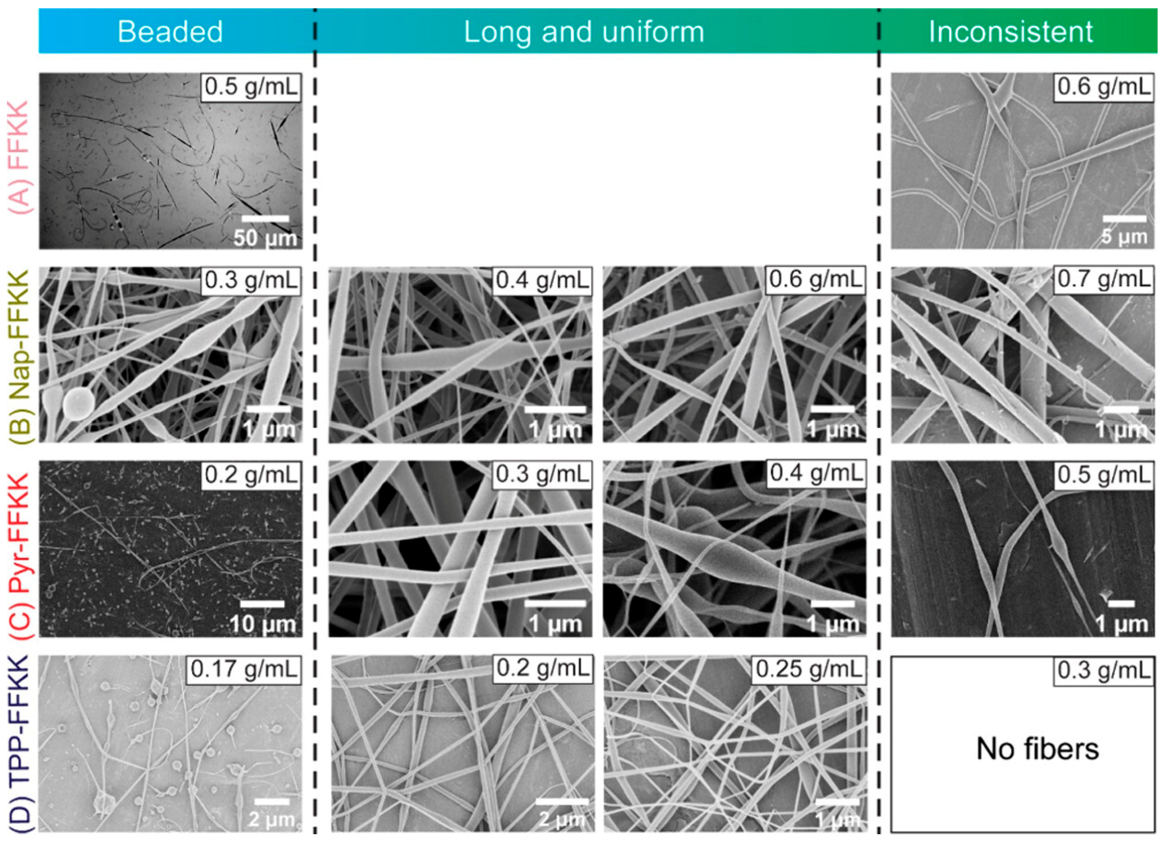
Electrospinning, first introduced by Formhals in 1934 [4], is the most prevalent and effective method for synthesizing uniform nanofibers from biomaterials. This strategy can be categorized into needle-based (Figure 1A1) and needleless electrospinning [7] (Figure 1A2), with needle-based electrospinning being more frequent. The flexibility and broadened capability of this technique have enabled its extensive usage in PNF production [4]. Electrospun fibers typically range from nanometers to micrometers, often thinner than 500 nm in diameter, with a consistent shape and porosity and minimal batch-to-batch variation [19]. The basic setup for needle-based electrospinning consists of a high-voltage power supply, a syringe pump, a spinneret (usually a blunt-tipped hypodermic needle), and a conductive collector. This process involves four key steps: the formation of the Taylor cone (a critical step where the shape of charged fluid droplets changes under an electric field [20]), the straight line extension of the charged jet, thinning and bending instability of the jet, and the solidification and deposition of fibers on the collector [4]. During electrospinning, a high-voltage electric field generates electrically charged jets from a solution or melt of material. The liquid droplet deforms into a Taylor cone, and a charged jet is ejected, stretching into finer diameters under the influence of the electric field. As the jet elongates, it undergoes thinning, experiences electrical bending instability (whipping), and rapidly solidifies. The fibers are deposited on the oppositely charged collector, where the solvent evaporates, forming solid nanofibers [21]. Parameters such as flow rate, needle-to-collector distance, needle inner diameter, solution viscosity, and volatility directly affect the fiber diameter in needle-based electrospinning. On the other hand, conditions such as low conductivity, low solubility, increased humidity, and low temperature promote pore formation on nanofibers [22]. For example, Rathore et al. modified short oligopeptides based on the FFKK sequence with aromatic end-capping groups and dispersed them in fluorinated solvents such as hexafluoro-2-propanol (HFIP). This modification enabled successful needle-based electrospinning, resulting in the formation of continuous, uniform nanofibers with diameters ranging from approximately 7 to 35 nm [23] (Figure 2). Similarly, Tao et al. used emulsion electrospinning to fabricate a bioactive repair layer from a collagen-mimicking peptide (GPO: Gly-Pro-Hyp) conjugated to four-arm polyethylene glycol (4A-PEG). This electrospun layer was designed to enable the controlled release of basic fibroblast growth factor (bFGF) and, with an average fiber diameter of 209.3 nm, was incorporated into a multifunctional patch for abdominal wall defect regeneration [24].
Needleless electrospinning offers higher production efficiency and is more suitable for large-scale fiber production. For instance, Mosayebi et al. utilized this method to produce fast-dissolving antioxidant nanofibers from Spirulina protein concentrate and gelatin, overcoming limitations of traditional needle-based electrospinning, such as low production rates and irregular electric field distribution in multi-needle setups [25]. Its simplicity, high efficiency, and precise control over fiber morphology have established electrospinning as a cornerstone in nanofiber fabrication.
Electrospinning has been widely employed to fabricate various materials. Coaxial and composite electrospinning techniques are highly promising for developing multifunctional systems with broad applications in delivery [26]. In the past two decades, electrospun peptide nanofibers have emerged as prospective materials due to their unique properties and potential for biomedical applications [26,27,28]. Incorporating peptides into polymeric materials enhances their processability for electrospinning and drug delivery vehicles. A huge amount of research has focused on combining peptides with biocompatible high-molecular-weight polymers to improve the mechanical, physical, and biological properties of PNFs. Commonly used natural polymers in mixture with peptides, such as chitosan, gelatin, and collagen, offer high biocompatibility and bioactivity, while synthetic polymers like polylactic acid (PLA), poly-ε-caprolactone (PCL), and polylactic-co-glycolic acid (PLGA) provide tunable mechanical properties and controlled degradation rates [29,30]. These polymers provide structural support and enhance the electrospinnability of peptides [31].
The first electrospun peptide nanofiber was investigated in 2008 by Singh et al., who successfully fabricated nanotubes using pure diphenylalanine, demonstrating self-assembly into tubular structures via π–π interactions [32]. Moreover, short peptides, composed of natural or non-natural amino acids, have been fabricated into solid, continuous, homogeneous, bead-free nanofibers through electrospinning. Their self-assembly behavior and stable conformations promote high-quality electrospun fibers [28]. The fabrication process for pure peptide-based electrospun fibers is still in the early stage, and further studies are needed [21,26,27]. Still, PNFs have been successfully electrospun using very low peptide concentrations in water (<4 wt.%) and peptides with low molecular weights (<1 kDa). Previous reports also show that these electrospun scaffolds keep the bioactive parts of peptides, like bio-epitopes, leveraging their functionality on the fiber surface [33]. Examples include dipeptides like Phe-Phe and Fmoc-Phe-Gly, as well as amphiphilic peptides such as Me(CH2)14-Val3-Ala3-Glu3, which have been successfully electrospun [15]. Antimicrobial peptides (AMPs), which are key players in the innate immune system [34], have been blended into polymer electrospun nanofibers like poly-ε-caprolactone (PCL) or gelatin/chitosan. This approach will give the final nanofibers valuable properties, such as antibacterial, osteogenic, angiogenic, and wound-healing capabilities [34,35,36].
Previous research demonstrated that peptide-based electrospun nanofibers hold great promise for biomedical uses, especially in controlled drug and gene delivery [11,37]. Compared to polymers, peptides provide precise sequence control, biocompatibility, functionality, and consistent results with fewer batch-to-batch differences [21,38]. Additionally, PNFs can form well-defined fibers with customized properties. Their positively charged amino acids make them ideal for enhancing nucleic acid interactions and cell uptake. Moreover, their structural variety and controlled degradation support sustained therapeutic release [29].
For the fabrication of electrospun peptide nanofibers, several parameters can influence the process and the fiber characteristics. They include peptide sequence, peptide solution concentration, molecular weight, conductivity, viscosity, surface tension, flow rate, applied voltage, distance between the needle tip and the collector, collector types, and environmental factors like humidity and temperature [19,22]. In general, increasing the concentrations of polymer solutions and using higher molecular weights result in thicker fibers. Typical electrospinning conditions include voltages of 5–30 kV, peptide concentrations of 3–30 wt.%, and collector distances of 5–20 cm, which produce fibers with diameters of 50 nm to 500 nm. By changing these parameters, the desired nanofibers with specific morphology, length, and features can be produced [22].
Despite its advantages, electrospun peptide nanofiber fabrication has had various challenges. High peptide concentrations of about 10–50 wt.% are one of these challenges that make the peptide fabrication process more challenging than polymer-based systems with 5–10 wt.% concentration [39]. Additionally, solvent choices are restricted to chaotropic fluorinated solvents like HFIP and TFA, which pose environmental risks, while more eco-friendly alternatives like acetic acid and aqueous solutions are currently under investigation [39,40,41]. On the other hand, proteolytic instability of natural peptides presents another challenge, which can be substituted by peptidomimetics to improve their stability [42,43]. The process of electrospinning of PNFs is influenced by several parameters, such as solution concentration, viscosity, voltage, needle–collector distance, and environmental conditions like humidity and temperature. Increased peptide concentrations and molecular weights lead to larger PNF diameters, while factors such as flow rate, needle diameter, and solution volatility affect their size. Solutions with low conductivity or solubility are desirable for the electrospinning of peptides. Applying higher humidity and lower temperature during fabrication encourages pore formation within the PNF matrix [22]. Table 1 lists the previous reported PNFs fabricated by electrospinning strategy and summarizes the process conditions and parameters.
The combination of these intrinsic properties and advances in electrospinning technology continues to position peptide-based nanofibers as highly advantageous materials for a wide range of emerging applications. While challenges such as solvent restrictions, viscosity requirements, and peptide stability persist, advancements in peptidomimetics and greener solvents are paving the way for broader applications. The unique properties of peptides, combined with innovations in electrospinning technology, highlight their substantial potential for diverse biomedical and industrial applications.

1.1.2. Force Spinning Strategy for PNF Fabrication

Force spinning, also known as centrifugal spinning, is a well-developed non-electrospinning strategy for nanofiber fabrication. This method mainly uses centrifugal force as the mechanical energy, instead of the electric field used in the typical electrospinning process [6] (Figure 1B1). Force spinning equipment involves a motor, shaft, spinneret, and collector with polymer solutions or melts that are injected into the spinneret during rapid rotation [2]. The morphology of the resulting nanofibers is influenced by various factors, including temperature, nozzle configuration, rotational speed, and the collection system. In 2020, Mamidi and colleagues utilized force spinning to fabricate PCL incorporating functionalized carbon nano-onion nanofibers (PCL/f-CNOs) capable of encapsulating and releasing the chemotherapeutic drug doxorubicin (DOX) in a specific pH range, thereby demonstrating the potential of this strategy for drug delivery applications [47]. In hybrid protein–polymer nanofibers, increasing protein concentration increases the fiber diameter [48]. The resulting variation in pore size within the matrices can enhance their suitability for being applied in ECM-mimicking and tissue-repair therapies [48]. An illustrative example is the fabrication of hybrid cellulose acetate (CA)/soy protein hydrolysate (SPH) nanofibers via a co-spinning process. These nanofibers successfully emulate the cutaneous microenvironment, exhibiting high water retention capacity. The incorporation of polar groups from SPH improves hydrophilicity, cell adhesion, and surface roughness, thereby promoting cell migration, fibroblast infiltration, and tissue repair while maintaining minimal cytotoxicity [48].
Force spinning offers significant advantages over electrospinning by producing large quantities of nanofibers at higher production rates [48]. Given the versatility, safety, low production costs, and high production rates of force spinning, a high electrical field is not required for producing nanofibers [17]. Additionally, it supports a broad range of conductive and non-conductive materials [2,7,49]. However, there are some limitations regarding the types of proteins or peptides, particularly those with low solubility and low melting points. Furthermore, the quality of nanofibers and the production rate are significantly influenced by the properties of the materials, the spinneret design, and precise control over the process parameters [2,50,51]. Nonetheless, this strategy stands as a practical and scalable alternative to traditional electrospinning for nanofiber production.

1.1.3. Drawing Strategy for PNF Fabrication

The drawing process is another non-electrospinning strategy for fabricating nanofibers using external mechanical forces. In this method, a millimetric droplet of biomaterial solution is deposited on a silicon dioxide (SiO2) surface and allowed to evaporate [6] (Figure 1B2). This technique produces single nanofibers of short length. As the droplet evaporates, capillary flow causes the edges to become more concentrated. A sharp tip or hollow glass micropipette is then dipped into the droplet near the contact line and withdrawn at a speed of approximately 100 mm/s, enabling nanofibers to be drawn out. These fibers are subsequently deposited onto another surface by touching it with the end of the micropipette [7,52].
The process can be repeated multiple times with each droplet, facilitating the extraction of different nanofibers. This technique is particularly effective for viscoelastic materials [7], which can sustain significant deformation while maintaining cohesion to withstand the stresses of pulling. Critical parameters, such as drawing speed and solution viscosity, should be carefully controlled to ensure consistency in fiber dimensions. However, the method is constrained to laboratory-scale applications, as it produces fibers one at a time and is inherently discontinuous, with low productivity. Additionally, only fibers with diameters exceeding 100 nm can be produced, depending on the size of the micropipette [6,7]. For instance, this technique has been used to fabricate PCL nanofibers loaded with ophthalmic drugs such as dexamethasone, highlighting its potential for localized and sustained drug delivery to the eye tissue. Despite its straightforward nature, the approach faces limitations, including batch-to-batch variability, non-uniform fiber thickness, and limited scalability for industrial applications [53].
Although the drawing method is a recognized strategy in nanofiber fabrication, PNFs are rarely studied for this purpose. High concentrations of peptide solutions, with their inherently high viscosity, may lend themselves well to this approach, particularly for experimental and small-scale production; however, limitations in scalability and productivity continue to restrict their broader application.

1.1.4. Self-Assembly Strategy for PNF Fabrication

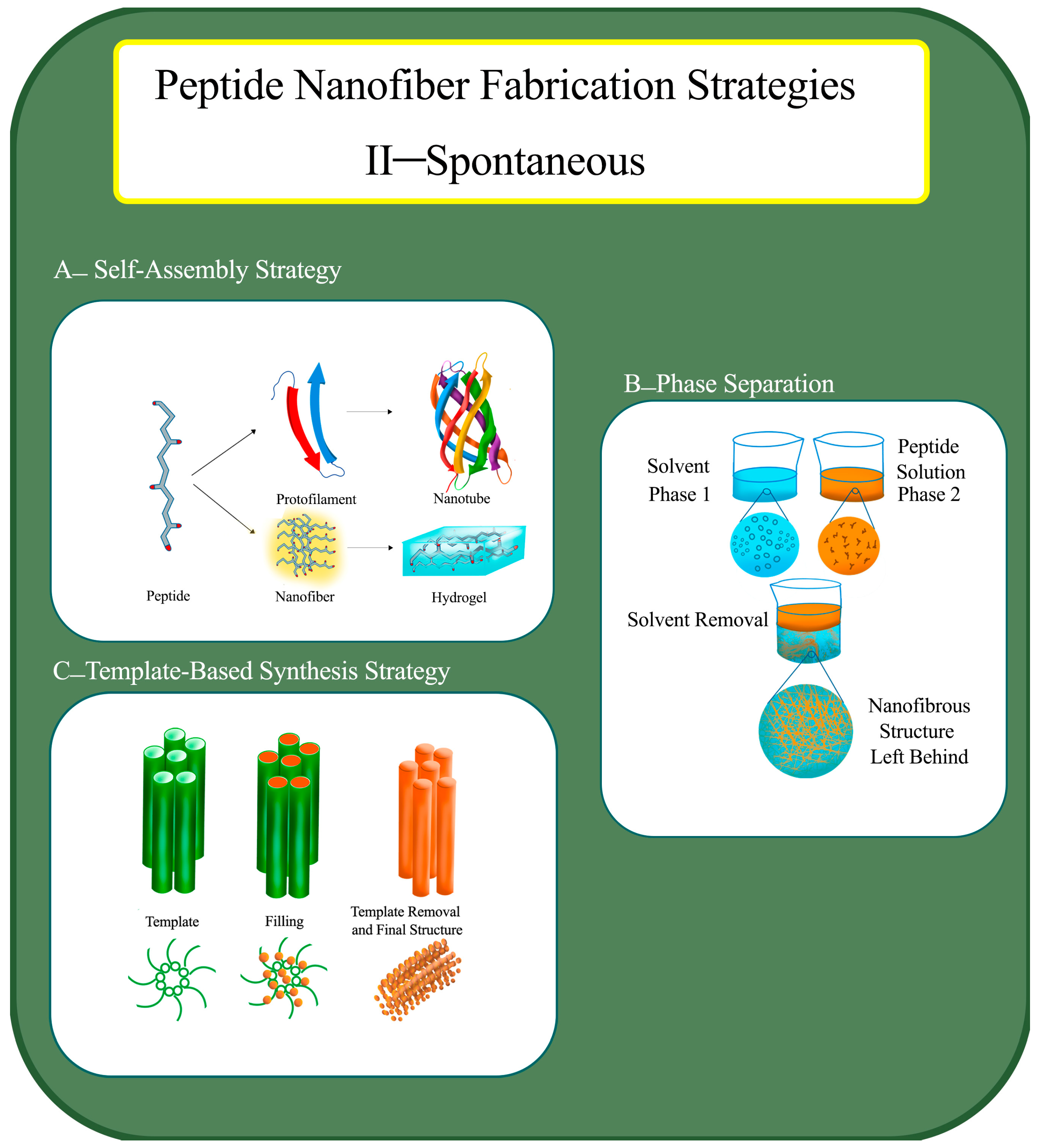
Molecular self-assembly is a spontaneous, thermodynamically favorable process that generates ordered structures in aqueous environments [6] (Figure 3A). This strategy represents a bottom-up approach to nanofiber fabrication, driven by intermolecular forces such as hydrogen bonding, hydrophobic interactions, electrostatic interactions, π-π stacking, and van der Waals forces. The nanofiber properties in this process are determined by the shape of the building block molecules and the intermolecular forces between them [54,55].
Through self-assembly, nanofibers with widths of less than 200 nm and lengths extending to micrometers can be synthesized and form supramolecular structures [7,56]. The fundamental mechanism relies on the interplay of hydrophobic interactions within the peptide backbone and hydrophilic interactions between the peptides and the surrounding aqueous medium, which collectively promote molecular organization [57]. Despite its versatility, this approach is intrinsically dependent on the specific peptide sequence [58,59]. Self-assembly is also a widely used method for fabricating PNF hydrogels. These three-dimensional hydrogels, capable of entrapping solvent molecules, are highly effective for localized drug delivery. Hydrogels with water contents reaching up to 99.9% can act as platforms for site-specific therapeutic release [60,61]. Moreover, these hydrogels could be responsive to specific stimuli, allowing for selective targeting or selective release of therapeutic agents or reversible structural changes [62]. Peptides with α-helical or β-sheet conformations, as well as amphiphilic sequences, are prone to hydrogel formation with this strategy [57,61].
In this fabrication strategy, the solvent type, temperature of the process, and concentration of peptides are key parameters influencing both the assembly process and the final nanofiber length. However, the pH level and the presence or absence of charged amino acids in the peptide sequence do not significantly affect PNF fabrication. Peptides are typically dissolved in aqueous solvents, and temperatures above 25 °C are often required to promote effective self-assembly (Table 2). An example of self-assembly-driven PNFs for therapeutic delivery includes Poly(VPGVG), a simple elastin-like polypeptide (ELP) [63], and K180L20 peptides [64].
While the dependence on peptides with inherent self-assembly behavior can limit the application of this approach, the diversity of peptide motifs available for incorporation into PNF matrices offers considerable opportunity for expanding their applications in drug delivery.

1.1.5. Phase Separation Strategy for PNF Fabrication

Phase separation is an effective strategy for fabricating nanofiber scaffolds without the need for external forces, leveraging the physical incompatibility between different phases in polymer solutions [7] (Figure 3B). Based on thermodynamic principles, this approach allows for the direct formation of nanofiber matrices with mechanical properties that can be tuned according to the polymer type or concentration [69].
Liquid–liquid phase separation (LLPS), which occurs at defined concentration thresholds, has been applied in advanced systems such as the aqueous two-phase system (ATPS) for biomolecular self-assembly and biomolecule purification [70]. Moreover, LLPS plays a critical role in facilitating biomolecular and supramolecular self-assembly in living environments [71]. Typically, a homogeneous polymer or material solution separates into two distinct phases under defined conditions, often a solvent-rich phase and a polymer-rich phase. The solvent phase is then removed, usually through evaporation.
The phase separation strategy generally involves several key steps: dissolving polymers in solvents to create homogeneous solutions at room temperature (RT), inducing gelation by applying a defined gelation temperature, extracting solvents from the formed gels, and finally, freeze-drying the products under vacuum [7]. Among these steps, solvent volatility plays a dominant role in the formation of nanofibrous matrices [21]. Gelation is the most crucial step in this process, as it determines the morphology and properties of the resultant nanofibers. For instance, increasing the polymer concentration can generally reduce the fiber porosity while enhancing the mechanical strength. Other primary factors influencing the process include polymer concentration, gelation time, and temperature [7,72]. Criado-Gonzalez et al. investigated phase separation in the context of PNF hydrogel formation. In their study, silica nanoparticles (NPs) covalently linked to alkaline phosphatase catalyzed the hydrogel formation of Fmoc–FFpY peptides (F: phenylalanine; Y: tyrosine; p: phosphate group), resulting in dense PNF phases surrounded by dilute phases. The dephosphorylation of Fmoc–FFpY peptides further promoted phase separation and nanofiber assembly [73].
Phase separation provides a simple and cost-effective approach for generating porous and continuous nanofiber networks. However, the method is relatively time-consuming, challenging to scale up, and not universally applicable to all polymer systems [74]. Despite these limitations, this fiber fabrication strategy remains a valuable tool for nanofiber fabrication, especially in experimental and small-scale applications.

1.1.6. Template-Based Synthesis Strategy for PNF Fabrication

Template-based synthesis is one of the widely known strategies for fabricating nanofibers, typically utilizing mechanical forces and electrostatic interactions [75], which leads to the production of controlled diameters and diverse material compositions. Generally, nanoporous membranes with cylindrical pores are used as templates for nanofiber formation [7]. The synthesis process is constrained by passing a material solution through pores under water pressure from one side, causing polymer extrusion and subsequent nanofiber formation upon contact with a solidifying solution. The resulting fiber length is generally limited to a few micrometers, constrained by the membrane’s pore size. When the template design changes, nanofibers with varying diameters can be formed [50,72]. This method gives precise control over the fiber diameters and material compositions, making it a great technique for creating advanced materials for specialized biomedical uses. Nanofibers from various polymers, such as polyacrylonitrile (PAN) [76], PCL [77], polyaniline, polypyrrole [78,79,80], and poly(3-methylthiophene) [81], have been successfully fabricated via this fabrication method.
Recently, peptides have been used as templates for nucleation and spontaneous growth of different nanofibers [82]. It should be mentioned that membranes with varying pore sizes are not necessarily required in this method. In a recent study, Ryu et al. [83] illustrated the application of diphenylalanine-derived peptides to form hybrid nanofibers with cobalt oxide, revealing their catalytic potential. Chen et al. [84] utilized peptides with sequence AYSSGAPPMPPF to induce the formation of single-helical gold nanoparticle superstructures. They highlighted the ability of peptides to serve as templates for template-based nanofiber fabrication. The secondary structure of peptides, such as α-helix or β-sheet motifs, can induce specific structural developments and promote the assembly of certain structures. Peptide amphiphiles (PAs), a group of peptides with great potential for nanofiber fabrication, have been applied as templates for the fabrication of nanowires [85]. Furthermore, Li et al. [82] used the tripeptide Fmoc-FFD to direct the formation of well-shaped nanofibers with adjustable photocatalytic properties to reach diameters of 40–70 nm at pH 5. The ability of peptides to control morphology under mild conditions makes them ideal candidates for nanofiber formation.
The template-based synthesis method also enables the construction of hollow fibers by filling template pores with a precursor material, followed by either preserving or removing the template. Biocompatible materials, such as cellulose derivatives, are frequently used in the template synthesis of functional bio-nanofibers (Figure 3C). As a result, they exhibit high water retention after functionalization with hydroxy moieties for therapeutic applications [74]. Despite the restrictions that are inherent in nanofiber length, template-based synthesis is still a generalizable and versatile strategy for PNF synthesis.
The most important features of all these fabrication strategies, including the final nanofiber size, equipment requirements, and their respective advantages and disadvantages, are summarized in Table 3. In addition to these primary strategies for nanofiber formation, less common techniques, such as three-dimensional printing [86], solution blow-spinning [87], and biomimetic approaches [16,88], have also been explored for NF fabrication. However, the application of these methods for PNFs requires further investigation to fully understand their potential.
Table 4 presents peptide sequences and their corresponding amino acids that have been utilized in PNF fabrication across six different strategies, each applied to various delivery applications.

1.2. The Role of Secondary Structure of Peptides in Nanofiber Formation

The peptide secondary structure and side-chain interactions play a very important role in the formation, final stability, functionality, and assembly of PNFs [122]. The β-sheet structure significantly contributes to the mechanical stability and bioactivity of the PNFs. Previous research demonstrated that thermally induced phase transformations can also drive β-sheet arrangement. It is generally postulated that linear dipeptides such as aromatic diphenylalanine (FF) and aliphatic dileucine (LL) undergo cyclization followed by reorganization into β-sheet nanofibers at elevated temperatures up to 120–180 °C. The final formed β-sheet structure is also stabilized by non-covalent hydrogen bonds, which create a stable network that dictates the nanofiber architecture [123]. Hydrogen bonding between peptide segments aligns β-sheets along the fiber axis, stabilizing the cylindrical structure, while charged amino acids provide electrostatic repulsions, dictating peptide size and shape [124,125,126]. Strong hydrogen bonding facilitates the creation of extended nanofibers, whereas a weaker interaction directs the self-assembly process toward spherical micelle formation. Specific sequences, including poly(EA), poly(YE), poly(KF), and repeat motifs GAGAGS, GAGAGY, and GAGAGVGY, display β-sheet conformation in water with periodically repeating polar and nonpolar residues as important determinants [127].
It is important to note that PNFs with the β-sheet arrangement can be elongated into ordered nanofibers, parallel to the fiber’s axis, with a radial packing from the core. Due to professor Stupp and coworkers’ studies, the degree of internal order within these nanofibers depends on the peptide sequence and molecular architecture. Linear peptides typically show greater order than branched peptides, and their internal structure influences both their stability and functionality. A more ordered hydrophobic core promotes mineral templating, whereas a less ordered structure enhances epitope accessibility, facilitating receptor interactions in biological applications. Therefore, the molecular design of PNFs can tune their biomedical applications [128].
In addition to the β-sheet arrangement, α-helices and coiled coils are less common secondary structures found in PNFs. Several studies corroborate the transition of secondary structures in PNFs. In this regard, short peptides adopt β-sheet structures at low temperature but can undergo a transition to α-helical structures upon increasing the temperature. EAK12-d and RADA16-IV peptides undergo transition from β-sheet to α-helix structures in the range of 60–90 °C. Longer peptides with more than 20 residues are expected to form stable hydrogels with α-helical structures [129]. Additionally, alkyl chains in PA stabilize α-helices, influencing their assemblies in PNFs [127]. Maleki et al. demonstrated that self-assembled and co-assembled PNFs transformed from random coil and α-helix conformations in solution to β-sheet arrangements after the electrospinning process. Unlike α-helical peptides, β-sheet-rich PNFs exhibit greater resistance to degradation in aqueous environments. On the other hand, peptide solutions with initial random coil or α-helical arrangements were more appropriate for electrospinning, while β-sheet-rich peptide solutions were less susceptible to this process [29]. These findings highlight the role of peptide secondary structures as the major driving factor influencing the formation, stability, and properties of PNFs.

1.3. Application of Peptide Nanofibers

PNFs have been used to deliver a variety of active agents, including drugs, vaccines, genes, viral vectors, functional peptides, and imaging agents, due to their biocompatibility and tunable physicochemical properties.

1.3.1. Drug Delivery by PNFs

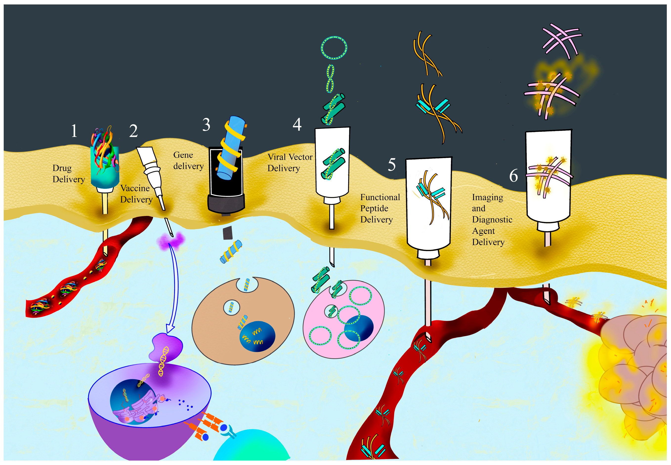
In vivo drug delivery focuses on developing materials and techniques that align well with the body’s physiological environment. These approaches could improve controlled drug release rates, maintain effective therapeutic levels, enable stimuli-responsive delivery [130], improve bioavailability, minimize side effects, and allow precise targeting of agents like small molecules, peptides, and proteins over extended periods [118] (Figure 4, number 1). Peptides, with their unique properties, have emerged as strong candidates for drug delivery systems. Recent work has shown that PNFs can self-assemble into hydrogels or other innovative carriers, efficiently transporting therapeutic agents to specific tissues [131]. PNFs can be engineered into various structural conformations, comprising hollow, porous, nonporous, and core–shell structures, each with distinct potential for drug encapsulation and delivery [7]. PNFs have been investigated in several drug delivery applications, such as cancer therapy. For example, Liu et al. [68] utilized Nap-GFFYG-RGD peptide as a self-assembling peptide (SAP), which spontaneously assembles in aqueous environment to create nanofibers 10–20 nm in diameter and carry curcumin to target cancer cells. They demonstrated tumor-targeting properties of these PNFs in both in vitro and in vivo models and highlighted their potential for precise anticancer drug delivery [68]. Similarly, Song et al. employed the amphiphilic A6K2 peptide to load DOX within its hydrophobic core. Triggered by lysyl oxidase, these nanofibers self-assembled and selectively released DOX in cancer cells, proving effective for targeted therapy [127]. PAs, which spontaneously self-assemble to create nanofibers, have been commonly utilized for drug delivery, as shown by numerous studies. Matson et al. (2012) synthesized the C16-V2A2E2 PA, which self-assembled into nanofiber hydrogels with an average diameter of approximately 10 nm. The nanofibers, with hydrazone-linked Prodan, exhibited prolonged drug release through controlled hydrolysis at physiological pH, thereby illustrating the effect of nanostructural packing density on the release kinetics [132]. Cui et al. developed PA-based nanofibers loaded with cisplatin, incorporating an MMP-2-responsive sequence and RGDS for cell adhesion. These 8–10 nm structures self-assembled spontaneously, providing spatiotemporal control for more accurate anticancer treatment [126]. Furthermore, Soukasene et al. used a PA with a palmitic acid tail and A4G3E3 sequence to encapsulate the hydrophobic drug camptothecin. The resulting nanofibers boosted the drug’s water solubility by over 50 times and increased toxicity to breast cancer cells, highlighting the potential of PAs for the delivery of hydrophobic therapeutics [108].
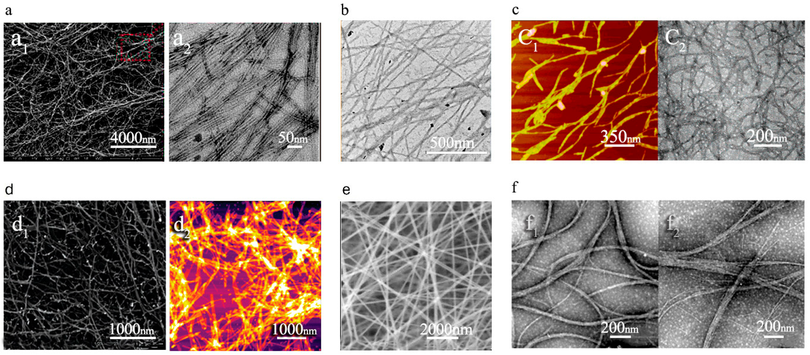
Overall, PNFs offer a versatile platform for delivering unstable, degradation-prone, or poorly soluble drugs, like many anticancer compounds [30,133]. They enable multifunctional designs, such as adding cell-penetrating or tumor-targeting peptides for DOX release in triple-negative breast cancer (TNBC) cells. An MMP-9-cleavable linker ensures site-specific release in enzyme-rich tumor microenvironments, yielding stable systems with high uptake and efficacy at low doses [134]. SEM and TEM analyses further confirm PNFs’ role in controlled DOX release (Figure 5a1,a2) [118], highlighting their promise for precision medicine with lower toxicity.
Most recent advancements in PNF formation have significantly enhanced their potential for drug delivery. Their hydrophobic core and hydrophilic surface enabled higher drug encapsulation and stability [115]. Bellavita et al. (2025) [116] engineered a multifunctional PNF platform for targeted temozolomide delivery across more sensitive barriers and tissues such as the blood–brain barrier (BBB) to treat glioblastoma. For this purpose, they used targeting, penetration-enhancing, and controlled-release peptide moieties and validated it through a 3D dynamic BBB model for physiological relevance. These recent studies underscore the huge application potential of self-assembling PNFs in drug delivery applications [117].

1.3.2. Vaccine Delivery by PNFs

Peptides have emerged as appealing candidates for use as both adjuvants and delivery platforms to enhance overall vaccine efficacy. These systems demonstrated significant advantages over conventional vaccine platforms due to a higher level of precision, modularity, and safety. PNFs, in particular, have drawn considerable interest among these systems because they can co-assemble with epitope peptides to trigger precise immune responses while lowering the chances of side effects like allergic reactions [138]. Therefore, due to their unique structural and functional properties, PNFs demonstrated significant potential in vaccine development. These nanofibers often self-assemble into β-sheet or α-helical configurations, allowing them to integrate various antigens and epitopes for controlled delivery and fine-tuned immune modulation [139].
PNF hydrogels have emerged as effective carriers for DNA- and nucleotide-based therapeutics, including DNA vaccines. For example, Leach and colleagues engineered a nanofibrous hydrogel with K2(SL)6K2 peptide for intratumoral administration of synthetic cyclic dinucleotides (CDNs). This peptide formed antiparallel β-sheet nanofibers that enabled sustained release in experimental settings, and a single injection showed antitumor effects in mice with MOC2-E6E7 tumors [109]. In recent years, SAPs have shown great potential in vaccine development due to their capacity to drive robust and enduring immune responses. SAPs can act as flexible scaffolds for antigen presentation, enhancing the immunogenicity and stability of associated epitopes. Furthermore, their inherent self-adjuvanting properties can activate both humoral and cellular immune responses without the need for additional adjuvants [140,141,142]. For instance, Yang et al. [143] demonstrated that RADA16 peptide nanofibrous hydrogel, which encapsulated bone marrow-derived dendritic cells (DCs), the model antigen ovalbumin (OVA), and anti-PD-1 (Programmed cell death protein-1) antibody, effectively recruited and activated both endogenous and exogenous DCs. This mechanism provokes a stronger antigen-specific immune response and activates CD8+ effector T cells in the EG7-OVA tumor model [143].
Numerous studies highlight how a single SAP type can link up with diverse epitopes to create nanofibers with inherent adjuvant properties [139]. The Q11 peptide (QQKFQFQFEQQ), for instance, pairs well with antigens of varying sources and sizes [139,144]. Huang et al. [107] employed β-turn self-adjuvanting nanofibers comprising the Q11 peptide and the glycosylated MUC1-derived B-cell epitope as the delivery system. TEM images revealed that two peptides, containing full-length MUC1 VNTR domains as an epitope vaccine, were conjugated to the Q11 peptide as an adjuvant, forming nanofibers (Figure 5f1,f2). This combination sparked a strong immune response in mice, effectively homing in on MUC1-expressing MCF-7 breast cancer cells [107]. Similarly, Ding et al. [97] established SAP EAK16-II (AEAEAKAKAEAEAKAK) nanofibers as delivery systems for increasing immunogenicity of their HIV-1 vaccine. These β-sheet-rich nanofibers were conjugated with HIV-1 CTL (cytotoxic T lymphocyte) epitope SL9 and further spontaneously co-assembled with TLR7/8 agonists (R848 or R837) in an aqueous solution. Ex vivo-generated DCs stimulated with this formulation (SL9-EAK16-II/R848 nanofibers) elicited a significantly stronger SL9-specific CTL response compared to the controls [97].
Tian et al. [110] demonstrated that the G-NMe nanovector (comprising a left-handed nanofiber structure and DNA) significantly enhanced both humoral and cellular immunity of HIV Env DNA (DNA encoding the HIV-1 envelope protein gp 145) via various injection methods in murine models, in comparison to G-OMe(p), and G-OH(p). The approach led to higher levels of interferon-gamma (IFN-γ) and interleukin-4 (IL-4) from lymphocytes compared to naked DNA. Furthermore, the nanofibers linked to DNA can protect the DNA from degradation, improving the transfection efficiency in mammalian cells. Importantly, the nanovector exhibited no cytotoxic effects on host immune cell viability [110]. Most studies on nanofiber-based vaccines have focused on β-sheet-forming PNF scaffolds. In contrast, Wu et al. [145] developed a multiepitope nanofiber cancer vaccine and demonstrated that supramolecular α-helical peptide nanofibrils can effectively enhance antitumor immune responses. Specifically, subcutaneous administration of the α-helical SAP Coil29 (QARILEADAEILRAYARILEAHAEILRAD) was shown to simultaneously elicit both humoral and cellular immune responses, leading to a significant reduction in B16vIII tumor growth in mice. Furthermore, the authors reported that unadjuvanted Coil29 nanofibers induced antibody responses with higher titers and superior quality compared to Q11 nanofibers [145].
Recent studies have introduced innovative PNF-based designs that broaden the potential of nanostructured materials in vaccine delivery. Roe et al. [146] developed Coil29, a coiled-coil α-helical PNF platform modified with PAS sequences (proline, alanine, and serine) to enhance epithelial transport and reduce mucosal interaction. Unlike traditional β-sheet-based sublingual formulations, this system enables stable solid-state delivery of peptide antigens via the sublingual route while preserving their immunogenicity [146]. Similarly, Curvino et al. explored PASylated self-assembling nanofibers with D-amino acids for oral vaccination. When paired with mucosal adjuvants, these structures induced both local and systemic immunity against peptide and small-molecule epitopes in mouse models of colitis [147]. In addition, Serdar et al. [119] developed a biocompatible Ac-FFA-NH2-based peptide hydrogel, which significantly enhanced both humoral and cellular immune responses. This platform maintained mechanical stability and structural integrity even when co-assembled with liposomes. Moreover, the liposome-encapsulated antigen exhibited a more controlled release profile compared to the antigen incorporated directly into the hydrogel [119]. Additionally, a multi-epitope PNF vaccine platform that activates both CD4+ and CD8+ T cells, along with functional B cells and other immune cells, has been demonstrated to be effective for pulmonary vaccination. Files et al. demonstrated that β-sheet-forming PNFs, KFE8 (FKFEFKFE-GGAAYFQDAYNAAGGHNAVF), are effective platforms for delivering Ag85B (a highly immunogenic protein secreted by Mycobacterium tuberculosis) and augmenting the lung CD4+ T-cell response in BCG-primed mice [112]. These results highlight the potential of PNF platforms as next-generation vaccine delivery methods that combine antigen delivery with intrinsic adjuvant properties while enhancing both mucosal and systemic immunity.
The size, shape, particular nature, and multivalency of PNFs can all affect the immunological outcomes of vaccine development applications [148,149]. Furthermore, the self-adjuvanting qualities of PNFs can reduce or eliminate the need for traditional adjuvants. Variations in the composition and structure of PNFs can significantly influence immunological responses, making them a highly adaptable and promising platform for vaccine development. As research advances, these supramolecular systems offer an innovative and effective approach for the development of next-generation vaccines in the future (Figure 4, number 6).

1.3.3. Gene Delivery by PNFs

Genes, nucleic acids, and interfering RNAs have been used as valuable therapeutic approaches for the treatment of various genetic disorders [150]. The chemical stability, controlled release, and efficiency of genes are dependent on the type of delivery system. As discussed, PNF carriers such as gel compound networks emerge as the major driving factors for controlled delivery of therapeutic agents with tunable stability. Due to their efficient DNA and RNA encapsulation properties, peptide-based nano- or microcarriers have proven efficacy in delivery applications.
Self-assembling PNF gels for oligonucleotide delivery were reported for the first time by Bulut et al. [136] For this purpose, they used a cationic PA named Lys-PA (C12-VVAGK-Am) that formed β-sheet arrangements through electrostatic interactions to deliver the Bcl-2 antisense ODN, named G3139, into mammalian cells. They observed controlled release of G3139 from the PA structure, its internalization by target cells, and the effects of this nanocarrier system on Bcl-2 mRNA levels. AFM and SEM microscopic images of these PNFs revealed 3D nanofibrous peptide and ODN networks (Figure 5d1,d2) [136]. Peptides are also safe transfection vectors for brain tissue, exhibiting the lowest toxicity. However, PNFs are supramolecular structures that are not able to easily cross the BBB, unless a strategy is used to help them self-assemble after crossing it [151]. In the study by Mazza and coworkers, they designed a surfactant-like peptide (palmitoyl-GGGAAAKRK) capable of self-assembling into PNFs. They explored whether their PNFs could form complexes with biologically active siRNA and facilitate its efficacious intracellular transport inside the deep brain. In their designed peptide structures, repeated alanine and glycine residues worked as sites for β-sheet interactions, facilitating the self-assembly of individual molecules into nanofibers. The presence of positively charged amino acids in the peptide structure enabled electrostatic binding with siRNA, forming PNF and complexes with siRNA [99]. Peptide carriers containing arginine residues have been shown in several studies to have high efficiency in binding and delivering DNA or RNA molecules [152,153,154]. Our recent study demonstrated that the insertion of amino acids such as histidine and phenylalanine can enhance this property, enabling them to form beta-sheet structures and effectively carry nucleic acids into mammalian cells [155]. Therefore, these peptide nano-complexes simplified intracellular uptake and extended the residence time of siRNA in the brain. In vitro experiments showed significant downregulation of BCL2 expression and induction of apoptosis in neuronal cells. This study highlights the potential of PNFs as effective gene delivery vectors for central nervous system (CNS) therapies and offers a potential method for treating neurodegenerative disorders through genetic intervention [99]. There are other studies for PNF hydrogels in neuronal interference. The bioactive IKVAV nanofiber gel has been employed as a scaffold for regenerating descending motor fibers and ascending sensory fibers in spinal cord injuries, as well as for inducing neuronal differentiation from neuronal progenitor cells. PNFs can also interact electrostatically with negatively charged siRNA, facilitating cellular uptake and enhancing cytoplasmic translocation [113].
It has also been reported that peptides are generally employed in the polymeric nanofiber structures for increasing gene delivery efficiency. The study by Mulholland et al. [156] emphasized the innovative combination of polymer and PNFs for localized gene delivery. In this approach, a cell-penetrating CHAT peptide was used for pDNA delivery, which was later incorporated into crosslinked electrospun polyvinyl alcohol (PVA) nanofibers. These PNFs provided increased cellular uptake, high stability and biocompatibility, and sustained-release properties. In vitro studies demonstrated that the pDNAs released from the nanofibers retained their structural integrity and transfection efficacy in fibroblast cells. These PNFs demonstrated a supported, localized, efficient, and targeted gene delivery vehicle with minimized off-target effects that led to improved therapeutic outcomes. This approach highlights the potential of peptide-based nanofiber vehicles to solve challenges in gene therapy delivery [156]. Similarly, the potential of nanofibers for localized siRNA delivery was explored using a scaffold composed of polycaprolactone-co-ethylethylene phosphate combined with cell-penetrating peptides CADY (GLWRALWRLLRSLWRLLWRA) and MPG (GALFLGFLGAAGSTMGAWSQPKSKRKV). These PNF scaffolds (550–650 nm diameter) encapsulated siRNA targeting collagen type I (COL1A1), enabling sustained release of siRNA for at least 28 days. This approach significantly enhanced siRNA uptake and prolonged gene silencing compared to conventional delivery. According to in vivo studies, this method provided a prospective strategy for localized RNA interference therapies [157].
Additionally, PNFs have shown significant ability to serve as gene delivery vehicles due to their ability to self-assemble into stable and functional nanocarriers. In the study by Zhang et al. [101], they designed a novel peptide-based nanofiber system with aggregation-induced emission (AIE) properties for gene delivery applications. These nanofibers were composed of the amphiphilic peptide TR4, with four arginine residues and tetraphenylethene (TPE) functional groups and a hydrophobic tail. The subsequent PNFs efficiently encapsulated and condensed the plasmid DNA (pDNA) and protected it from enzymatic degradation. The pDNA-TR4 complex displayed high stability, strong binding affinity, low cytotoxicity, and excellent transfection efficiency to stem cells [101]. Additionally, PNF scaffolds have recently been highlighted for sustained delivery of siRNA and miRNA [158,159,160]. Other studies have also demonstrated the reliability of using self-assembling PNFs to deliver siRNA therapeutics to triple-negative breast cancer (TNBC), a kind of difficult-to-treat type of cancer. This approach suggests that applying such nanofibers could be an advantageous strategy for developing new therapies [160]. These findings reveal that peptide-based self-assembled nanofibers have shown significant promise as non-viral vectors for the targeted delivery of siRNA and silencing of genes [3,158,160] (Figure 4, number 4).
It is worth noting that peptides inherently possess several advantageous properties, including delivery vectors for genes, DNA, and RNA, making them ideal candidates for further investigations. Their exceptional properties underscore the need for more extensive studies on the use of nanofiber-based peptide carriers for both localized and systemic nucleic acid delivery. Despite the several advantages of PNFs, drawbacks such as limited cellular uptake and inefficient release of nucleic acids at the target site should be considered when employing them for nucleic acid delivery.

1.3.4. Viral Vector Delivery by PNFs

The delivery of viral vectors and viruses to mammalian cells requires well-optimized strategies to enhance transduction efficiency [161]. Recent scientific trends have revealed that PNF scaffolds are a highly effective strategy for targeted and efficient delivery of viral vectors and gene therapy [120,121,162]. Other studies showed the great potential of electrospun nanofibers for local delivery of viral vectors such as AAV (Figure 5e) [137,163]. Moreover, electrospun peptide nanofibrous scaffolds have been shown to function effectively as carriers for viral-based gene delivery systems. Lee et al. [45] blended ELP (consisting of 128 repeating units of Val-Pro-Gly-Val-Gly) with PCL to create electrospun nanofibrous scaffolds for delivering AAV and effectively transducing fibroblasts adhered to the scaffolds. They observed that the presence of ELP resulted in narrower fiber diameters, ranging from approximately 350 to 500 nm. Additionally, they reported that the interactions between ELP and PCL chains significantly modified the mechanical properties, wettability, and elasticity of the nanofibers, leading to controlled release of AAV vectors within the scaffold. They demonstrated that this approach facilitated efficient cellular transduction and highlighted the potential of PNF scaffolds to enhance AAV vector gene delivery [45] (Figure 4, number 5).
One of the pioneering works on PNFs as viral vector delivery systems is related to Münch and coworkers’ research, who discovered that semen-derived amyloid fibrils drastically boost HIV-1 infectivity by enhancing its binding and facilitating cell attachment [120,121,164]. By engineering semen-derived amyloids, they synthesized a 12-mer EF-C peptide (QCKIKQIINMWQ, residues 417–428 of HIV gp120), which self-assembles into β-sheet-rich nanofibrils with a positive zeta potential and polycationic surface that outperform natural amyloids in retroviral transduction efficiency, proving safe in ex vivo gene transfer studies. They reported that the EF-C PNFs induce plasma membrane vesicle formation to increase the viral binding surface, and enter cells via macropinocytosis and degrade lysosomally to avoid amyloid accumulation, while cryo-EM structures reveal polymorphic maturation from thin to thick fibrils, with cationic surfaces driving infectivity enhancement [120]. These findings have advanced applications in chimeric antigen receptor T (CAR-T) and natural killer (NK) cell therapies by improving transduction rates with negligible cytotoxicity.
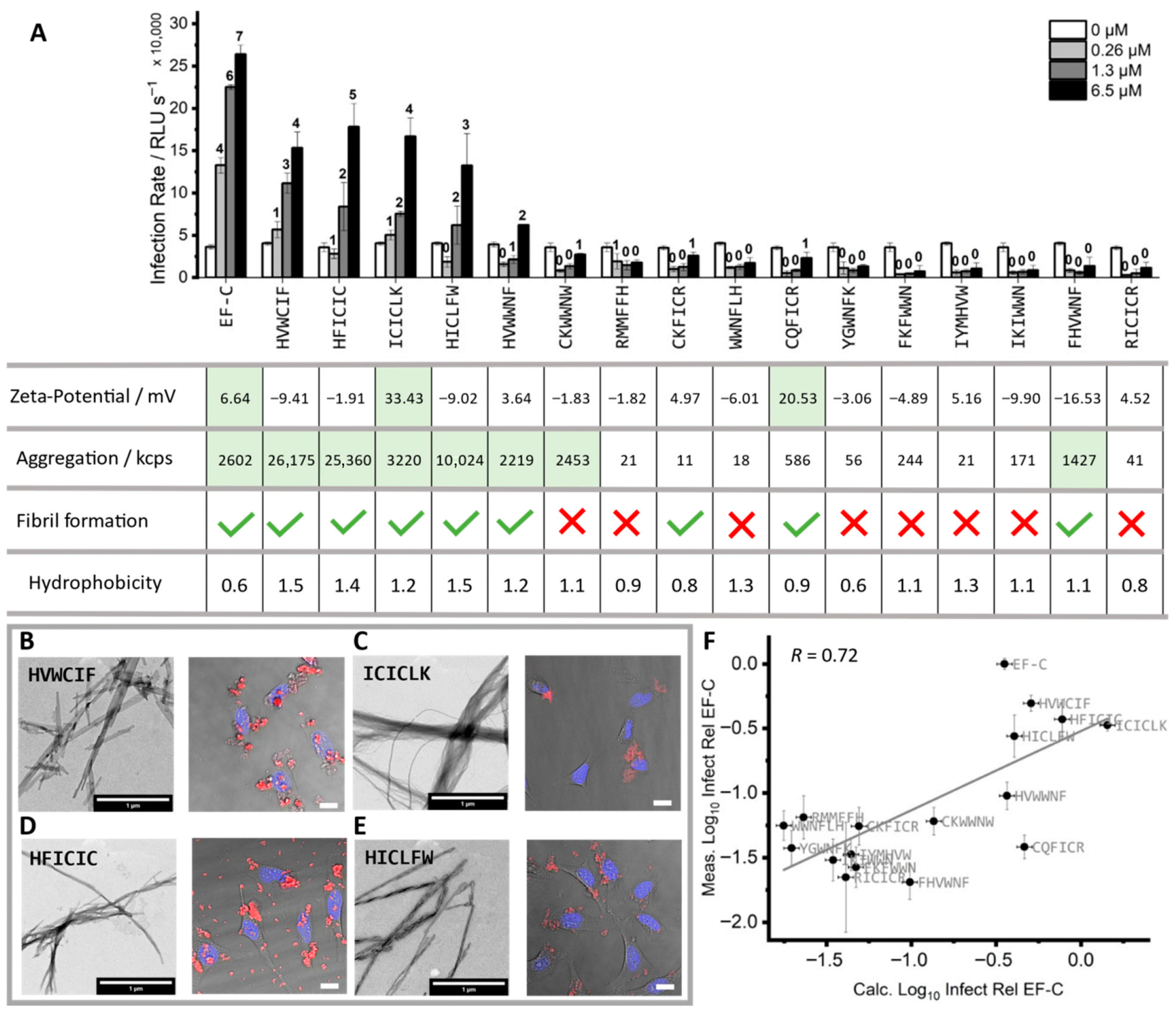
Weil and Synatschke’s group extended these efforts through in silico screening and structure–activity relationship studies to obtain shorter, non-immunogenic PNFs like the 8-mer peptide, which forms stable, smaller fibrils that enhance retroviral and lentiviral transduction in primary T and NKcells up to 65% efficiency, greater than EF-C and commercial enhancers [120,162,165]. They used a library of 163 peptides, derived from the EF-C peptide sequence, as a set for de novo sequence prediction using a machine learning (ML) approach. The resulting 16 new 6-mer peptides were tested, and these short de novo peptides demonstrated active infectivity enhancement. Moreover, they discovered the first hydrophobic peptide fibrils with a slightly negative surface charge, including three peptides with negative zeta potential (HVWCIF, HICLFW, HFICIC) that form fibrils able to enhance the infectivity of retroviruses, alongside a positively charged variant (ICICLK) showing similar activity at over 10% relative to EF-C [121] (Figure 6). Additionally, Münch and colleagues converted short, non-self-assembling peptides into peptide amphiphile nanofibers by attaching fatty acids, triggering β-sheet nanofiber formation. Additionally, these nanofibers were tested for their ability to enhance viral and retroviral vector delivery. Their PNF systems matched commercial enhancers in transduction efficiency while remaining biodegradable and biocompatible [162].
Collectively, these studies demonstrate that rationally designed PNFs are powerful enhancers for retroviral vectors or virus delivery, supporting their broader application in gene and cancer therapies.

1.3.5. Functional Peptides’ Delivery by PNFs

PNFs have emerged as highly versatile platforms for delivering functional peptides, including antimicrobial, immunomodulatory, anticancer, and any other functional peptides [30,166,167,168]. PNFs, due to their high surface area and flexibility, can efficiently entrap various bioactive peptides and provide precise control of their release, either in a sequential manner, at a sustained level, or in response to biological stimuli. PNFs also stabilize functional peptides, minimize cytotoxicity, and improve therapeutic efficacy [30] (Figure 4, number 2).
AMPs possess membrane-interacting and non-membrane-interacting modes of action, driven by their cationic and amphipathic properties. Membrane-interacting AMPs disrupt bacterial membranes, leading to cell death, while non-membrane-interacting AMPs can traverse membranes without compromising structural integrity and inhibit intracellular processes such as protein synthesis or DNA replication. Structural differences between bacterial and mammalian membranes, particularly the higher negative charge on bacterial membranes due to anionic lipids, ensure the selective activity of AMPs toward bacteria over mammalian cells [169,170]. Recent advancements highlight growing interest in microgels and hydrogels as notable carriers for AMPs. These peptide carriers not only enhance AMP stability against proteolytic degradation but also minimize toxicity. An example is the microgel-encapsulated AMP that controls peptide release while maintaining bioactivity, making it an effective candidate for infection-related treatments. Similarly, hydrogels offer responsive delivery systems, particularly in targeted areas [171]. Jeong et al. developed an injectable supramolecular hydrogel designed for diabetic wound healing that responds to high levels of matrix metalloproteinases (MMPs) and reactive oxygen species (ROS), releasing AMPs selectively in the presence of these biomarkers to ensure targeted antimicrobial action with reduced side effects [172].
More than antimicrobial applications, PNF hydrogels have also shown potential in the controlled delivery of anticancer peptides (ACPs). ACPs are emerging candidates for targeting cancer cells through multiple mechanisms. These include activating immune responses by stimulating NK cells and dendritic cells (DCs) with enhanced recognition and destruction of cancer cells. Furthermore, they can directly penetrate cancer cell membranes, nuclear membranes, and mitochondria to induce apoptosis. In conclusion, the membrane’s integrity is disrupted by inhibiting processes such as DNA synthesis and cell division [173,174]. For instance, Chen et al. [98] designed a fibrillar hydrogel with an MMP-2-responsive cleavage site, enabling targeted degradation and peptide release in tumor environments. This system selectively delivered the anticancer peptide G(IIKK)3I-NH2 (G3) to MMP-2-overexpressing HeLa cells, effectively inhibited tumor growth, and demonstrated a valuable strategy for controlled anticancer therapy [98]. Moreover, SEM images showed that PA-based PNFs with neurite-promoting laminin epitope (IKVAV) were produced as nanofibers with diameters ranging from 5 to 8 nm and lengths of hundreds of nanometers to a few micrometers [175]. Advancements in the development of functional peptide nanofibers and hydrogels have led to innovative therapeutic strategies, offering controlled, responsive, and highly effective functional peptide delivery for a wide range of medical applications.
Peptide nanofibers (PNFs) represent a valuable therapeutic approach for wound healing by inducing skin regeneration through mechanisms such as collagen synthesis, angiogenesis, and inflammation modulation. These PNF systems effectively deliver functional therapeutic peptides, promoting cell migration and proliferation while exhibiting a valuable impact on treating chronic wounds, such as diabetic ulcers. The clinical application of single therapeutic peptides is significantly hindered by limitations, including short plasma half-life and poor oral bioavailability. Therefore, developing suitable PNF systems for the effective delivery of these peptides is crucial for overcoming these challenges and unlocking their full therapeutic potential. PNF scaffolds have shown significant results in controlled release, enhanced stability, and improved healing outcomes of functional peptides [176,177]. For example, Jankoski et al. [66] demonstrated that PA nanofibers of the C16V3A3E3G4REGRT peptide, which carried the bioactive REGRT sequence, could affect skin regeneration and enhance wound healing in deep dermal burn injuries. As a result, the nanofiber scaffold significantly accelerated early wound healing in a deep dermal murine burn injury model (Figure 5b) [66]. In accordance with the tissue regenerative capabilities of PNFs, it has been reported that nanofiber-based scaffolds combined with biomolecular cues like peptides have significantly improved the compatibility and efficacy of scaffolds for cardiac tissue regeneration [178]. In conclusion, these advancements reveal that PNFs can serve as effective platforms for the delivery of functional peptides, addressing key challenges in stability, release control, and therapeutic efficacy across diverse biomedical applications, and potentially revolutionizing treatments for infections, cancer, and tissue regeneration.

1.3.6. Diagnostic and Imaging Agents’ Delivery by PNFs

PNFs, through targeted and stimuli-responsive self-assembly, enable the targeted delivery and accumulation of imaging agents in pathological regions to enhance the imaging specificity and sensitivity. Their biocompatibility and ability to improve agent retention make them highly effective for both in vitro and in vivo applications. Their advancements have significantly contributed to precise tumor and disease imaging and are a promising step forward in biomedical diagnostics [179].
For example, protease-sensitive PNFs have shown significant potential in cancer imaging by specifically responding to overexpressed enzymes in tumor microenvironments. Recent studies have revealed that these nanomaterials accumulate selectively in protease-rich regions, enhancing imaging contrast and enabling precise tumor visualization. By integrating imaging agents into the nanofiber structure, these systems improve the pharmacokinetics and biodistribution of probes, offering a promising approach by combining diagnostic and therapeutic functionalities in a single platform [180]. PNFs have also been effectively utilized for delivering and enhancing MRI contrast agents. For instance, PA nanofibers conjugated to a contrast agent chelator, DOTA, a carrier molecule for Gd3+ ions (contrast agent), can slow down the rotation of the contrast agent in the body, thereby enhancing the MRI signal strength and prolonging its duration. These PNFs, with lengths beyond 100 nm and widths of 22 nm, can be used for targeting specific cellular receptors and exhibit strong functionality in precise imaging and potential nontoxic therapeutic applications. Therefore, they are promising tools for advancing diagnostic imaging techniques [135] (Figure 5c1,c2). It should be highlighted that PNFs have garnered substantial interest for carrying various imaging agents, offering smart and precise solutions for tumor and disease imaging (Figure 4, number 3). On the other hand, beyond oncology applications, Li et al. (2023) [181] developed a peptide–nanoparticle hybrid with the sequence GCD-PEG-QK for cardiovascular MRI. The system links the VEGF-mimetic QK peptide (KKLTWQELYQL[K(Ac)]Y[K(Ac)]GI, which promotes angiogenesis and targets VEGFR) to gadolinium-doped carbon dots (providing fluorescence + MRI contrast) via a PEG spacer. This design enables infarct-specific imaging and therapy, showing how peptide-based nanoplatforms, parallel to peptide nanofibers, can advance next-generation cardiovascular imaging and treatment [181]. Furthermore, due to the safe nature of peptides, they are increasingly being applied in imaging of sensitive tissues, such as brain tissue for neurological diseases [116,182]. Moreover, PNFs can be engineered to establish regenerative microenvironments that are simultaneously trackable by noninvasive imaging modalities. For example, injectable self-assembling short peptides forming nanofibrous scaffolds within the myocardium generate biocompatible niches that support endothelial cell adhesion and growth. These nanofibers can be further functionalized with growth factors or bioactive signaling molecules to accelerate tissue repair and promote neovascularization. Importantly, the same regenerative microenvironment can be dynamically monitored using advanced imaging techniques such as MRI, particularly when PNFs are conjugated with contrast agents. This integration underscores the dual functionality of PNFs in driving tissue regeneration while enabling image-guided therapeutic evaluation [183]. Nevertheless, challenges such as improving in vivo stability and reducing off-target accumulation remain, yet the tunable self-assembly of PNFs positions them as versatile and potentially transformative tools for precision medicine.

1.4. Delivery Strategies for Peptide Nanofibers:

There are various approaches for delivering therapeutic agents using PNFs, including systemic and local delivery, controlled release, stimuli-responsive delivery, and targeted delivery. These diverse delivery systems allow scientists to design specific strategies according to their research goals. Moreover, these strategies can be applied separately or in combination to enhance the comprehensive efficiency of the designed delivery systems, reducing off-target effects and minimizing systemic toxicity. In this section, we provide an overview of each delivery strategy, regardless of its delivered cargo, and evaluate how PNFs contribute to these platforms by summarizing findings from previous studies.

1.4.1. Systemic Delivery Strategy

Peptide nanofibers have been used for systemic delivery of various therapeutic agents and their effective distribution throughout the body, making them particularly beneficial for conditions requiring widespread treatment [130,184,185,186]. PNFs have been studied for administration via intravenous (IV), subcutaneous, and sublingual routes to achieve systemic circulation. Barlek et al. [67] investigated the systemic uptake of PA nanofibers with subcutaneous injections in an animal model. They designed four different PA molecules to study the influence of nanofiber consistency and charge on their uptake and systemic distribution. Their findings revealed that negatively charged PA nanofibers exhibited high systemic absorption, highlighting the importance of their molecular structure and charge in improving systemic circulation [67] (Figure 7). Similarly, Coulter et al. [186] developed an enzyme-responsive ultrashort peptide hydrogel, NapFFKY[p]-OH, covalently conjugated to the HIV drug zidovudine (AZT). Following subcutaneous injection, the peptide self-assembled in situ into a hydrogel with 2 nm diameter nanofibers, triggered by phosphatase activity, and enabled sustained AZT release for 28–35 days. In rats, administration of NapFFK(AZT)Y[p]G-OH maintained zidovudine plasma concentrations within the therapeutic IC50 range (30–130 ng mL−1) throughout the study period. This study highlights enzyme-responsive peptide hydrogels as a long-acting injectable platform for the systemic delivery of AIDS drugs [186]. Additionally, another study reported the use of peptides for protein delivery via sublingual injection, enabling efficient diffusion into the bloodstream and targeted cellular uptake. These novel peptides were shown to induce pore formation on the cell surface, thereby enhancing intracellular protein delivery and significantly improving overall uptake efficiency [187].
In general, these findings indicate that nanofibers’ physicochemical characteristics can strongly influence systemic absorption. This represents a critical knowledge gap that must be addressed before their systemic delivery potential can be fully realized. In parallel, biodegradable PA nanofiber hydrogels have enabled prolonged intravitreal delivery of anti-VEGF proteins without loss of bioactivity [188]. While these examples underscore the versatility of peptide nanofiber systems for systemic delivery, they will require comprehensive in vivo studies evaluating their biodistribution, stability, and potential off-target effects to guide rational design for controlled, body-wide delivery.

1.4.2. Local Delivery Strategy

Recent studies postulated that PNFs can be used as efficient local drug delivery systems [118,189]. Their nanofibers can be designed to form ECM-like structures and are well-suited for delivering AMPs, chemical drugs, vaccines, genes, or diagnostic agents [167,190]. Due to their inherent fluidity, biocompatibility, and stability, they have demonstrated substantial potential in local treatments, wound healing, and regenerative medicine [167,191].
Moreover, as we discussed before, PNFs can naturally self-assemble into three-dimensional (3D), crosslinked fibrous networks, which can form mechanically stable hydrogels with high water absorption capacity [192,193]. These hydrogels could serve as drug carriers, relying on the diffusion of small molecules, peptides, or proteins for controlled drug release. Studies have claimed that different drugs can be incorporated into PNF-based hydrogels through physical entrapment or covalent immobilization [64,104,185,186]. In physical entrapment, the drug is non-covalently retained within the nanofiber network, and its release is regulated by the hydrogel’s mesh size, which controls diffusion rates. However, by covalent immobilization, drugs chemically attach to the nanofiber scaffold, allowing for hydrogel degradation to have more controlled and sustained release. Both approaches allow precise tuning of drug release profiles and also make PNFs highly adaptable biomaterials for local and targeted therapies [192]. For instance, Cinar et al. [118] investigated the potential of two designed PA nanofiber hydrogels, E3PA (Lauryl-VVAGEEE) and K3PA (Lauryl-VVAGKKK-Am), as a local and controlled-release drug delivery system for the DOX molecule. They developed injectable supramolecular PA nanofiber gels with high water holding capacity and drug loading properties for both hydrophobic and hydrophilic drugs. In vivo experiments further revealed that local injection of Dox/PA hydrogels at tumor sites significantly slowed tumor growth and increased apoptosis within the tumor tissue [118]. Moreover, self-assembling peptide hydrogels, such as ac-(RADA)4-CONH2, have been used for local chemotherapy in glioblastoma for efficient sustained release of agents with diverse solubility profiles [182]. The structural resemblance of these PNFs to ECM architectures makes them highly suitable for local drug delivery and also provides a suitable platform for targeted and sustained therapeutic delivery applications [61]

1.4.3. Controlled-Release Strategy

Peptide-based nanofibers represent a versatile platform for achieving controlled drug release profiles. The release profile of such nanofibers can be controlled by adjusting key parameters, including fiber diameter, porosity, and the mechanism of drug adsorption. This flexibility has introduced PNFs as an effective solution for accurate and prolonged drug delivery over specific timeframes [192]. Nisbet and colleagues developed a complex scaffold combining DIKVAV peptide hydrogel with electrospun PLA nanofibers to control growth factor release to promote stem cell integration in the injured brain. The hydrogel nanofibers with an average diameter of 10 nm, compared to PLA nanofibers with 100–2000 nm diameter, provided structural support and delayed release. The study demonstrated that this system enabled an initial burst release within 12 h, followed by sustained release, which started after six days. In vitro studies confirmed the therapeutic potential of this ECM-mimicking scaffold due to its ability to stabilize neurotrophic growth factors and enhance neuronal metabolic activity [44,114].
Electrospun peptide nanofibers have demonstrated high efficacy as platforms for controlled drug release over a defined period. Modifications in fiber diameter, porosity, and drug-binding mechanisms enable tuning of release profiles. For instance, incorporating bovine serum albumin (BSA) blended with polyethylene glycol (PEG) into PCL nanofibers, using coaxial electrospinning, revealed the controlled-release capabilities of multi-layer structures and composite peptide and polymer nanofibers. In vitro release studies showed that core–shell nanofibers reduced burst release and provided sustained protein delivery, compared to blend electrospun nanofibers [192,194]. Additionally, electrospun composite nanofibers have been used effectively for multi-drug release without altering the kinetics of individual agents. Studies proved that the combination of chitosan microspheres with mesoporous silica nanoparticles enables the simultaneous and sustained delivery of both hydrophilic and hydrophobic drugs [22]. In another study, Chronopoulou and coworkers demonstrated that peptide nanofiber hydrogels could effectively play a role in controlled drug delivery [102]. Their research focused on the self-assembly of an Fmoc-tripeptide (FmocFFF) in water, forming nanofiber hydrogels under mild conditions. The hydrogel effectively incorporated naproxen and exhibited controlled-release kinetics in aqueous media. Rheological and SEM analyses confirmed that the hydrogel had robust mechanical and morphological properties, whereas cytotoxicity tests highlighted its biocompatibility by validating its potential in advanced drug delivery applications [102]. Additionally, Richard T. Lee and his team at Harvard Medical School designed SAP nanofibers for controlled peptide delivery to the myocardium tissue. In physiological conditions, their peptide sequence (AcN-RARADADARARADADA-CNH2) self-assembled into nanofibers with an approximately 10 nm diameter. These nanofibers create a 3D microenvironment after being injected into the myocardium. In another study, this group used the same nanofibers to deliver IGF-1, a polypeptide that promotes cardiomyocyte growth and differentiation. These PNFs provided sustained IGF-1 release, enhanced Akt activation in the myocardium, and enhanced the efficacy of cell therapy for myocardial infarction [65]. Subsequently, this group utilized the same peptide nanofibers for the controlled delivery of platelet-derived growth factor BB (PDGF-BB) for myocardial protection via PI3K/Akt signaling. These injectable nanofibers are bound to PDGF-BB, leading to sustained release and delivery for 14 days. For their in vivo study in rats, they observed that PDGF-BB delivered via nanofibers can reduce cardiomyocyte apoptosis, preserve heart function, and lower infarct size after ischemia/reperfusion. Their findings also revealed that peptide nanofibers are precise and effective biomaterials for sustained protein delivery in heart disease therapy [195].
These studies demonstrated that PNFs are powerful tools for controlled drug delivery, offering tunable release profiles and sustained therapeutic effects. Further advancements in PNFs’ design will enhance their precision, efficacy, and clinical applicability as the next generation of controlled delivery systems.

1.4.4. Stimulus-Responsive Delivery Strategy

Adding stimulus-responsive behavior to PNF systems is one of the most recent and highly attractive strategies in smart delivery approaches. Previous studies have shown that PNFs with β-sheet arrangements can quickly respond to various stimuli [127]. This approach in PNF platforms can function as a self-regulating strategy for the delivery of therapeutic agents. With dual- or multi-responsive delivery designs, the release of therapeutic agents can be either enhanced or suppressed in response to different signals or conditions [196]. One of the main strategies for designing dual-responsive systems involves using two stimuli, one for carrier loading and the other for triggering cargo release [22]. The major triggering factors for drug release from PNFs include pH, ionic strength, salt concentration, temperature, specific enzyme availability, oxidative stress, magnetic or electric fields, light, ultrasound, and heat [22].
One of the most extensively studied stimuli in drug delivery design is the pH of the targeted environment. In several studies, peptide-based nanocarriers have been developed to respond to changes in the pH microenvironment, enabling the release of cargo molecules such as drugs or genes [197,198]. Dehsorkhi and coworkers (2013) studied the effect of pH changes on the assembly process of a PA. They found that different morphologies of peptides were produced under different pH values, including flat and twisted structures. Their study demonstrated the potential of pH-sensitive stimuli in the fabrication of peptide nanofibers with distinct structures [199]. In another study, pH-responsive thixotropic silk hydrogels were introduced for local and sustained DOX delivery in acidic tumor microenvironments to treat breast cancer cells [197]. Temperature, as another stimulus, has also been shown to influence the self-assembly structure of PNFs. At low temperatures, hydrogen bonding increases the stability of β-sheet arrangements, whereas at higher temperatures, hydrogen bonding is disrupted [57].
Therefore, peptides, with their inherent ability to respond to varying physicochemical conditions, are highly suitable for constructing stimuli-responsive nanofibers. Their dynamic molecular structures enable the development of advanced materials for drug delivery, tissue engineering, and biosensing applications. As versatile smart materials, PNFs hold significant potential for next-generation stimuli-responsive drug delivery systems [200].

1.4.5. Targeted Delivery Strategy

PNFs can be engineered with different motifs to target particular cells and tissues. Their inherent properties allow them to serve as ideal carriers for an effective targeted delivery strategy [61]. For instance, the chondrocyte-affinity peptide (CAP), with the sequence DWRVIIPPRPSA, has been used in the design of a targeted delivery system for cartilage tissue [105]. Additionally, the study of Liu et al. investigated the targeting activity of curcumin-encapsulated Nap-GFFYG-RGD peptide nanofibers and demonstrated significantly higher cellular uptake of these nanofibers in αvβ3 integrin-positive HepG2 liver carcinoma cells, highlighting their potential for targeted delivery of hydrophobic molecules for cancer therapy [68].
PNFs can be designed for specific targeting by including adhesion ligands, receptor recognition ligands, or peptide-based antigens in their structure [201]. In some studies, peptides have been incorporated into polymeric nanofibers to add targeting capabilities. For instance, Man et al. [202] developed coaxial electrospun nanofiber scaffolds incorporating a BMSC-affinity peptide with the sequence EPLQLKM, added to the surface of the electrospun scaffold, to specifically enhance the adhesion and chondrogenic differentiation of bone marrow-derived stem cells (BMSCs). They observed that the peptide-modified scaffolds not only improved cell attachment but also facilitated the sustained release of rhTGF-β1, thus promoting cartilage tissue engineering. Their study further demonstrated the potential of peptide-functionalized nanofibers for enhancing targeted cellular interactions and promoting tissue regeneration [202].
In general, we can say that PNFs offer an effective platform for enhancing stability, solubility, local delivery, controlled release, sustained release, stimuli responsiveness, and targeted delivery.

2. Drawbacks, Limitations, and Critical Properties

PNFs have emerged as versatile platforms for the delivery of therapeutic agents. They are increasingly used in various procedures to develop bio-nanocomposites for a wide range of biomedical applications. Experimental studies have demonstrated their potential in local therapeutic agent delivery, in ECM mimicking, as hydrogel platforms, and as safe and effective adjuvants in vaccine development [74].
Fiber fabrication driven by non-covalent interactions, such as π-π stacking, enables formation of β-sheet or α-helical structures, which influence mechanical strength (typically 1–10 MPa) and porosity. Surface properties such as zeta potential facilitate interactions with negatively charged cell membranes or nucleic acids, enhancing cellular uptake and gene delivery. Additionally, stimuli responsiveness allows on-demand release, with fiber diameters (1–500 nm) and lengths (up to micrometers) determining diffusion rates and tissue penetration. These properties are specified based on the fabrication strategy. For instance, electrospun PNFs exhibit higher mechanical robustness compared to self-assembled ones, but require careful control of viscosity and conductivity to avoid bead formation.
However, several challenges and limitations remain. The fabrication strategies, particularly methods requiring external forces, need further refinement to improve the efficiency and scalability of the desired PNFs. In specific fabrication strategies, such as phase separation and drawing, the fabrication of peptides into nanofiber materials is a relatively recent advancement and remains an underexplored area of research. Additionally, many fabrication techniques involve conditions that may compromise the stability and integrity of peptides, such as elevated temperatures or degrading solvents. Researchers must ensure that chemical modifications of peptides do not induce cytotoxicity or compromise their therapeutic efficacy. Moreover, the reproducibility of PNFs with the same physical and chemical properties, including fiber length and surface characteristics, remains a critical factor, particularly in tissue engineering applications. Therefore, any inconsistency in the fabrication process can directly affect cellular responses and their regeneration.
Furthermore, despite considerable progress in research studies, scaling up the production of PNFs for clinical translation remains a significant challenge. Current fabrication techniques often suffer from limitations in cost-effectiveness and throughput, posing challenges for transitioning from laboratory-scale to large-scale manufacturing. So far, two peptide nanofibers have advanced to clinical trials for potential therapeutic use [203]. This demonstrates both the considerable promise of PNFs as innovative delivery platforms and the significant technical and translational challenges that still hinder their broader development into biomedical products. Therefore, addressing these challenges through optimized fabrication strategies, robust quality control measures, and scalable production techniques is essential for advancing PNFs toward widespread biomedical applications. Additionally, the development of new peptide mimetics or peptide/polymer mixtures for the development of PNFs could enhance their stability and physicochemical properties, making them more desirable for melt fabrication strategies. It is worth noting that surface modification of polymers with plasma discharge methods will open new strategies for their integration into biomedical applications [204]. Various functional domains can be introduced on the peptide nanofiber surface without changing the bulk properties. We have demonstrated that the plasma process facilitates cell adhesion and targeted drug loading, which is critical for tissue regeneration and drug delivery [205,206]. These strategies can be considered to improve the surface functionality of PNFs and their compatibility with cells.

3. Conclusions and Future Perspectives of Peptide Nanofibers in Delivery

In this review, we focused on previous investigations regarding PNFs as delivery systems. These studies established that PNFs have emerged as highly promising platforms for the delivery of therapeutic agents, including drugs, genes, vaccines, viral vectors, functional peptides, and imaging agents. Their ability to self-assemble into well-defined nanostructures provides several distinct advantages, including biocompatibility, tunable degradation rates, and the potential for controlled and sustained release of therapeutic agents. These characteristics make them highly attractive candidates for a wide range of biomedical applications. For example, PNFs offer a significant advantage in their relatively high gene-loading capacity compared to other conventional nanocarriers, such as peptide microspheres, which often require complex and innovative design strategies to achieve efficient molecule encapsulation and delivery. Supported by promising outcomes of different research groups, it can be concluded that PNFs have been widely explored in both local and systemic drug delivery, gene therapy, and vaccine development. Their structural versatility enables their integration into hydrogels, scaffolds, and composite materials, providing finely tuned release profiles for various therapeutic applications. Despite these advantages, several challenges remain unresolved in the practical application of PNFs. A key limitation is that their stability under physiological conditions must be improved, particularly for long-term therapeutic applications. Moreover, their scalability and mechanical properties must be optimized to ensure reproducibility, which is a crucial factor in nanofiber fabrication techniques such as electrospinning and self-assembly. In this respect, advanced developments in peptide chemistry, bio-functionalization, surface activation, and hybrid material engineering are critical for overcoming these challenges. The combination of PNFs with polymeric carriers and the development of dual-responsive or multifunctional systems can further improve their performance in targeted and sustained delivery. With continued research and refinement, PNFs possess significant potential to revolutionize sustained, targeted, and controlled drug, gene, and vaccine release strategies, offering safer, more effective, and patient-specific therapies in regenerative medicine, oncology, and infectious disease management.

Author Contributions

Conceptualization, R.T.P.; investigation, writing—original draft preparation, visualization idea, validation and data curation, review and final editing, O.K.; visualization, S.N.; investigation, writing—original draft preparation, F.B.; writing—original draft preparation, M.B.; writing—original draft preparation, M.P.G.; review and editing, supervision. All authors have read and agreed to the published version of the manuscript.

Funding

This review article received no external funding.

Data Availability Statement

No new data were created or analyzed in this study.

Conflicts of Interest

The authors declare no conflicts of interest.

Abbreviations

The following abbreviations are used in this manuscript:
AIEAggregation-induced emission
AFMAtomic force microscopy
APCsAnticancer peptides
ATPSAqueous two-phase system
AMPsAntimicrobial peptides
BMSCsBone marrow-derived stem cells
BSABovine serum albumin
CACellulose acetate
CAPChondrocyte-affinity peptide
CNSCentral nervous system
CTLCytotoxic T lymphocyte
DOTADerivative of 1,4,7,10-tetraazacyclododecane-1,4,7,10 tetraacetic acid
DOXDoxorubicin
ECMExtracellular matrix
ELPsElastin-like polypeptides
FITCFluorescein isothiocyanate
FRAPFluorescence recovery after photobleaching
f-CNOsFunctionalized carbon nano-onions
HFIP1,1,1,3,3,3-Hexafluoro-2-propanol
IFN-γInterferon gamma
IGF-1Insulin-like growth factor 1
LLPSLiquid–liquid phase separation
MMPMatrix metalloproteinases
MRIMagnetic resonance imaging
ODNOligo deoxy nucleotide
PANPolyacrylonitrile
PAsPeptide amphiphiles
PBSPhosphate-buffered saline
PLAPolylactic acid
PCLPoly-ε-caprolactone
PLGAPoly lactic-co-glycolic acid
PNFsPeptide nanofibers
PTAPhosphotungstic acid
pDNAPlasmid DNA
PVAPolyvinyl alcohol
RTRoom temperature
SAPSelf-assembling peptide
SEMScanning electron microscope
SiO2Silicon dioxide
SPHSoy protein hydrolysate
TEMTransmission electron microscopy
TFATrifluoroacetic acid
TFE2,2,2-Trifluoroethanol
TNBCTriple-negative breast cancer
TPETetraphenylethene
VNTRsVariable-number tandem repeats

References

  1. Yıldız, A.; Kara, A.A.; Acartürk, F. Peptide-Protein Based Nanofibers in Pharmaceutical and Biomedical Applications. Int. J. Biol. Macromol. 2020, 148, 1084–1097. [Google Scholar] [CrossRef]
  2. Kalayil, N.; Budar, A.A.; Dave, R.K. Nanofibers for Drug Delivery: Design and Fabrication Strategies. BIO Integr. 2024, 5, 978. [Google Scholar] [CrossRef]
  3. Taghizadeh Pirposhteh, R.; Arefian, E.; Arashkia, A.; Mohajel, N. Nona-Arginine Mediated Anti-E6 ShRNA Delivery Suppresses the Growth of Hela Cells in Vitro. Iran. Biomed. J. 2023, 27, 349–356. [Google Scholar] [CrossRef]
  4. Xue, J.; Wu, T.; Dai, Y.; Xia, Y. Electrospinning and Electrospun Nanofibers: Methods, Materials, and Applications. Chem. Rev. 2019, 119, 5298–5415. [Google Scholar] [CrossRef]
  5. Pennington, M.W.; Zell, B.; Bai, C.J. Commercial manufacturing of current good manufacturing practice peptides spanning the gamut from neoantigen to commercial large-scale products. Med. Drug Discov. 2021, 9, 100071. Available online: https://www.sciencedirect.com/science/article/pii/S2590098620300580 (accessed on 1 October 2024). [CrossRef]
  6. Nayak, R.; Padhye, R.; Kyratzis, I.L.; Truong, Y.B.; Arnold, L. Recent Advances in Nanofibre Fabrication Techniques. Text. Res. J. 2012, 82, 129–147. [Google Scholar] [CrossRef]
  7. Alghoraibi, I.; Alomari, S. Different Methods for Nanofiber Design and Fabrication. In Handbook of Nanofibers; Springer: Cham, Switzerland, 2018. [Google Scholar] [CrossRef]
  8. Jalili, M.; Mozaffari, A.; Gashti, M.; Parsania, M. Electrospinning Nanofibers Gelatin Scaffolds: Nanoanalysis of Properties and Optimizing the Process for Tissue Engineering Functional. J. Nanoanal. 2019, 6, 289–298. [Google Scholar]
  9. Mozaffari, A.; Gashti, M.P.; Mirjalili, M.; Parsania, M. Argon and Argon–Oxygen Plasma Surface Modification of Gelatin Nanofibers for Tissue Engineering Applications. Membranes 2021, 11, 31. [Google Scholar] [CrossRef]
  10. McMurtrey, R.J. Patterned and Functionalized Nanofiber Scaffolds in Three-Dimensional Hydrogel Constructs Enhance Neurite Outgrowth and Directional Control. J. Neural Eng. 2014, 11, 066009. [Google Scholar] [CrossRef]
  11. Gu, X.; Ding, F.; Williams, D.F. Neural tissue engineering options for peripheral nerve regeneration. Biomaterials 2014, 35, 6143–6156. Available online: https://www.sciencedirect.com/science/article/pii/S0142961214004621 (accessed on 1 October 2024). [CrossRef]
  12. Yuan, Y.; Chen, L.; Kong, L.; Qiu, L.; Fu, Z.; Sun, M.; Liu, Y.; Cheng, M.; Ma, S.; Wang, X.; et al. Histidine Modulates Amyloid-like Assembly of Peptide Nanomaterials and Confers Enzyme-like Activity. Nat. Commun. 2023, 14, 5808. [Google Scholar] [CrossRef] [PubMed]
  13. Lopez, M.J.; Mohiuddin, S.S. Biochemistry, Essential Amino Acids; StatPearls: Treasure Island, IL, USA, 2024. Available online: https://www.ncbi.nlm.nih.gov/books/NBK557845/ (accessed on 22 March 2025).
  14. Mazza, M.; Patel, A.; Pons, R.; Bussy, C.; Kostarelos, K. Peptide Nanofibres as Molecular Transporters: From Self-Assembly to in Vivo Degradation. Faraday Discuss. 2013, 166, 181–194. [Google Scholar] [CrossRef] [PubMed]
  15. Bucci, R.; Georgilis, E.; Bittner, A.M.; Gelmi, M.L.; Clerici, F. Peptide-Based Electrospun Fibers: Current Status and Emerging Developments. Nanomaterials 2021, 11, 1262. [Google Scholar] [CrossRef] [PubMed]
  16. Zhang, W.; Yu, X.; Li, Y.; Su, Z.; Jandt, K.D.; Wei, G. Protein-mimetic peptide nanofibers: Motif design, self-assembly synthesis, and sequence-specific biomedical applications. Prog. Polym. Sci. 2018, 80, 94–124. Available online: https://www.sciencedirect.com/science/article/pii/S007967001730254X (accessed on 2 October 2024). [CrossRef]
  17. DeFrates, K.G.; Moore, R.; Borgesi, J.; Lin, G.; Mulderig, T.; Beachley, V.; Hu, X. Protein-Based Fiber Materials in Medicine: A Review. Nanomaterials 2018, 8, 457. [Google Scholar] [CrossRef]
  18. Moore, A.N.; Hartgerink, J.D. Self-Assembling Multidomain Peptide Nanofibers for Delivery of Bioactive Molecules and Tissue Regeneration. Acc. Chem. Res. 2017, 50, 714–722. [Google Scholar] [CrossRef]
  19. Li, S.; Duan, G.; Zhang, G.; Yang, H.; Hou, H.; Dai, Y.; Sun, Y.; Jiang, S. Electrospun Nanofiber Nonwovens and Sponges towards Practical Applications of Waterproofing, Thermal Insulation, and Electromagnetic Shielding/Absorption. Mater. Today Nano 2024, 25, 100452. [Google Scholar] [CrossRef]
  20. He, J.H. On the Height of Taylor Cone in Electrospinning. Results Phys. 2020, 17, 103096. [Google Scholar] [CrossRef]
  21. Subbiah, T.; Bhat, G.S.; Tock, R.W.; Parameswaran, S.; Ramkumar, S.S. Electrospinning of Nanofibers. J. Appl. Polym. Sci. 2005, 96, 557–569. [Google Scholar] [CrossRef]
  22. Agrahari, V.; Agrahari, V.; Meng, J.; Mitra, A.K. Electrospun nanofibers in drug delivery: Fabrication, advances, and biomedical applications. In Emerging Nanotechnologies for Diagnostics, Drug Delivery and Medical Devices; Elsevier: Amsterdam, The Netherlands, 2017; Available online: https://www.sciencedirect.com/science/article/pii/B9780323429788000097 (accessed on 21 November 2024).
  23. Rathore, P.; Montz, B.; Hung, S.H.; Pandey, P.K.; Perry, S.L.; Emrick, T.; Schiffman, J.D. Electrospinning of Self-Assembling Oligopeptides into Nanofiber Mats: The Impact of Peptide Composition and End Groups. Biomacromolecules 2025, 26, 1604–1613. [Google Scholar] [CrossRef]
  24. Tao, Y.; Luo, P.; Jing, F.; Liu, T.; Tan, X.; Lyu, Z.; VeerleBernaerts, K.; Zhang, T.; Jia, R. Collagen-Inspired 3D Printing Electrospinning Biomimetic Patch for Abdominal Wall Defect Regeneration. Adv. Fiber Mater. 2025, 7, 1177–1194. [Google Scholar] [CrossRef]
  25. Mosayebi, V.; Fathi, M.; Shahedi, M.; Soltanizadeh, N.; Emam-Djomeh, Z. Fast-Dissolving Antioxidant Nanofibers Based on Spirulina Protein Concentrate and Gelatin Developed Using Needleless Electrospinning. Food Biosci. 2022, 47, 101759. [Google Scholar] [CrossRef]
  26. Xue, J.; Xie, J.; Liu, W.; Xia, Y. Electrospun Nanofibers: New Concepts, Materials, and Applications. Acc. Chem. Res. 2019, 50, 1976–1987. [Google Scholar] [CrossRef] [PubMed]
  27. Locarno, S.; Eleta-Lopez, A.; Lupo, M.G.; Gelmi, M.L.; Clerici, F.; Bittner, A.M. Electrospinning of Pyrazole-Isothiazole Derivatives: Nanofibers from Small Molecules. RSC Adv. 2019, 9, 20565. [Google Scholar] [CrossRef]
  28. Nuansing, W.; Frauchiger, D.; Huth, F.; Rebollo, A.; Hillenbrand, R.; Bittner, A.M. Electrospinning of peptide and protein fibres: Approaching the molecular scale. Faraday Discuss. 2013, 166, 209–221. Available online: https://pubs.rsc.org/en/content/articlehtml/2013/fd/c3fd00069a (accessed on 3 October 2024). [CrossRef]
  29. Maleki, M.; Natalello, A.; Pugliese, R.; Gelain, F. Fabrication of Nanofibrous Electrospun Scaffolds from a Heterogeneous Library of Co- and Self-Assembling Peptides. Acta Biomater. 2017, 51, 268–278. [Google Scholar] [CrossRef]
  30. Sousa, M.G.C.; Rezende, T.M.B.; Franco, O.L. Nanofibers as Drug-Delivery Systems for Antimicrobial Peptides. Drug Discov. Today 2021, 26, 2064–2074. [Google Scholar] [CrossRef]
  31. Mu, B.; Xu, H.; Li, W.; Xu, L.; Yang, Y. Spinnability and Rheological Properties of Globular Soy Protein Solution. Food Hydrocoll. 2019, 90, 443–451. [Google Scholar] [CrossRef]
  32. Singh, G.; Bittner, A.M.; Loscher, S.; Malinowski, N.; Kern, K. Electrospinning of Diphenylalanine Nanotubes. Adv. Mater. 2008, 20, 2332–2336. [Google Scholar] [CrossRef]
  33. Tayi, A.S.; Pashuck, E.T.; Newcomb, C.J.; McClendon, M.T.; Stupp, S.I. Electrospinning Bioactive Supramolecular Polymers from Water. Biomacromolecules 2014, 15, 1323–1327. [Google Scholar] [CrossRef]
  34. Huan, Y.; Kong, Q.; Mou, H.; Yi, H. Antimicrobial Peptides: Classification, Design, Application and Research Progress in Multiple Fields. Front. Microbiol. 2020, 11, 582779. [Google Scholar] [CrossRef] [PubMed]
  35. Eriksen, T.H.B.; Skovsen, E.; Fojan, P. Release of antimicrobial peptides from electrospun nanofibres as a drug delivery system. J. Biomed. Nanotechnol. 2013, 9, 492–498. Available online: https://www.ingentaconnect.com/contentone/asp/jbn/2013/00000009/00000003/art00020 (accessed on 21 November 2024). [CrossRef] [PubMed]
  36. He, Y.; Jin, Y.; Wang, X.; Yao, S.; Li, Y.; Wu, Q.; Ma, G.; Cui, F.; Liu, H. An antimicrobial peptide-loaded gelatin/chitosan nanofibrous membrane fabricated by sequential layer-by-layer electrospinning and electrospraying Techniques. Nanomaterials 2018, 8, 327. Available online: https://www.mdpi.com/2079-4991/8/5/327 (accessed on 21 November 2024). [CrossRef] [PubMed]
  37. Yadav, N.; Chauhan, M.K.; Chauhan, V.S. Short to ultrashort peptide-based hydrogels as a platform for biomedical applications. Biomater. Sci. 2020, 8, 84–100. Available online: https://pubs.rsc.org/en/content/articlehtml/2019/bm/c9bm01304k (accessed on 1 October 2024). [CrossRef]
  38. Wu, S. Chain Structure and Entanglement. J. Polym. Sci. Part B Polym. Phys. 1989, 27, 723–741. [Google Scholar] [CrossRef]
  39. Vigneswari, S.; Murugaiyah, V.; Kaur, G.; Khalil, H.P.S.A.; Amirul, A. Simultaneous dual syringe electrospinning system using benign solvent to fabricate nanofibrous P (3HB-co-4HB)/collagen peptides construct as potential leave-on wound dressing. Mater. Sci. Eng. C 2016, 66, 147–155. Available online: https://www.sciencedirect.com/science/article/pii/S0928493116302909 (accessed on 3 October 2024). [CrossRef]
  40. Unalan, I.; Endlein, S.J.; Slavik, B.; Buettner, A.; Goldmann, W.H.; Detsch, R.; Boccaccini, A.R. Evaluation of electrospun poly(ε-caprolactone)/gelatin nanofiber mats containing clove essential oil for antibacterial wound dressing. Pharmaceutics 2019, 11, 570. [Google Scholar] [CrossRef]
  41. Vogt, L.; Rivera, L.R.; Liverani, L.; Piegat, A.; El Fray, M.; Boccaccini, A.R. Poly(ε-caprolactone)/poly(glycerol sebacate) electrospun scaffolds for cardiac tissue engineering using benign solvents. Mater. Sci. Eng. C 2019, 103, 109712. Available online: https://www.sciencedirect.com/science/article/pii/S0928493119305429 (accessed on 2 October 2024). [CrossRef]
  42. Lenci, E.; Trabocchi, A. Peptidomimetic toolbox for drug discovery. Chem. Soc. Rev. 2020, 49, 3262–3277. Available online: https://pubs.rsc.org/en/content/articlehtml/2020/cs/d0cs00102c (accessed on 3 October 2024). [CrossRef]
  43. Bucci, R.; Contini, A.; Clerici, F.; Beccalli, E.M.; Formaggio, F.; Maffucci, I.; Pellegrino, S.; Gelmi, M.L. Fluoro-Aryl Substituted α,β 2,3 -Peptides in the Development of Foldameric Antiparallel β-Sheets: A Conformational Study. Front. Chem. 2019, 7. [Google Scholar] [CrossRef]
  44. Bruggeman, K.F.; Wang, Y.; Maclean, F.L.; Parish, C.L.; Williams, R.J.; Nisbet, D.R. Temporally Controlled Growth Factor Delivery from a Self-Assembling Peptide Hydrogel and Electrospun Nanofibre Composite Scaffold. Nanoscale 2017, 9, 13661–13669. [Google Scholar] [CrossRef]
  45. Lee, S.; Kim, J.S.; Chu, H.S.; Kim, G.W.; Won, J.I.; Jang, J.H. Electrospun Nanofibrous Scaffolds for Controlled Release of Adeno-Associated Viral Vectors. Acta Biomater. 2011, 7, 3868–3876. [Google Scholar] [CrossRef] [PubMed]
  46. John, J.V.; Choksi, M.; Chen, S.; Boda, S.K.; Su, Y.; McCarthy, A.; Teusink, M.J.; Reinhardt, R.A.; Xie, J. Tethering Peptides onto Biomimetic and Injectable Nanofiber Microspheres to Direct Cellular Response. Nanomed. Nanotechnol. Biol. Med. 2019, 22, 102081. [Google Scholar] [CrossRef] [PubMed]
  47. Mamidi, N.; Zuníga, A.E.; Villela-Castrejón, J. Engineering and Evaluation of Forcespun Functionalized Carbon Nano-Onions Reinforced Poly (ε-Caprolactone) Composite Nanofibers for PH-Responsive Drug Release. Mater. Sci. Eng. C. Mater. Biol. Appl. 2020, 112, 110928. [Google Scholar] [CrossRef] [PubMed]
  48. Marjuban, S.M.H.; Rahman, M.; Duza, S.S.; Ahmed, M.B.; Patel, D.K.; Rahman, M.S.; Lozano, K. Recent Advances in Centrifugal Spinning and Their Applications in Tissue Engineering. Polymers 2023, 15, 1253. [Google Scholar] [CrossRef] [PubMed]
  49. Luo, C.J.; Stoyanov, S.D.; Stride, E.; Pelan, E.; Edirisinghe, M. Electrospinning versus Fibre Production Methods: From Specifics to Technological Convergence. Chem. Soc. Rev. 2012, 41, 4708–4735. [Google Scholar] [CrossRef]
  50. Zhang, X.; Lu, Y. Centrifugal Spinning: An Alternative Approach to Fabricate Nanofibers at High Speed and Low Cost. Polym. Rev. 2014, 54, 677–701. [Google Scholar] [CrossRef]
  51. Weitz, R.T.; Harnau, L.; Rauschenbach, S.; Burghard, M.; Kern, K. Polymer Nanofibers via Nozzle-Free Centrifugal Spinning. Nano Lett. 2008, 8, 1187–1191. [Google Scholar] [CrossRef]
  52. Ondarçuhu, T.; Joachim, C. Drawing a Single Nanofibre over Hundreds of Microns. Europhys. Lett. 1998, 42, 215. [Google Scholar] [CrossRef]
  53. Sakpal, D.; Gharat, S.; Momin, M. Recent Advancements in Polymeric Nanofibers for Ophthalmic Drug Delivery and Ophthalmic Tissue Engineering. Biomater. Adv. 2022, 141, 213124. [Google Scholar] [CrossRef]
  54. Nuraje, N.; Bai, H.; Su, K. Bolaamphiphilic Molecules: Assembly and Applications. Prog. Polym. Sci. 2013, 38, 302–343. [Google Scholar] [CrossRef]
  55. Sorrenti, A.; Illa, O.; Ortuño, R.M. Amphiphiles in Aqueous Solution: Well beyond a Soap Bubble. Chem. Soc. Rev. 2013, 42, 8200–8219. [Google Scholar] [CrossRef] [PubMed]
  56. Fujino, K.; Yamamoto, N.; Yoshimura, Y.; Yokota, A.; Hirano, Y.; Neo, M. Repair Potential of Self-assembling Peptide Hydrogel in a Mouse Model of Anterior Cruciate Ligament Reconstruction. J. Exp. Orthop. 2024, 11, e12061. [Google Scholar] [CrossRef]
  57. Pentlavalli, S.; Coulter, S.; Laverty, G. Peptide Nanomaterials for Drug Delivery Applications. Curr. Protein Pept. Sci. 2020, 21, 401–412. [Google Scholar] [CrossRef]
  58. Beachley, V.; Wen, X. Polymer Nanofibrous Structures: Fabrication, Biofunctionalization, and Cell Interactions. Prog. Polym. Sci. 2010, 35, 868–892. [Google Scholar] [CrossRef]
  59. Li, W.; Shanti, R.M.; Tuan, R.S. Electrospinning Technology for Nanofibrous Scaffolds in Tissue Engineering. Nanotechnol. Life Sci. 2007, 125–144. [Google Scholar] [CrossRef]
  60. Zhang, S.; Holmes, T.; Lockshin, C.; Rich, A. Spontaneous Assembly of a Self-Complementary Oligopeptide to Form a Stable Macroscopic Membrane. Proc. Natl. Acad. Sci. USA 1993, 90, 3334–3338. [Google Scholar] [CrossRef]
  61. Habibi, N.; Kamaly, N.; Memic, A.; Shafiee, H. Self-Assembled Peptide-Based Nanostructures: Smart Nanomaterials toward Targeted Drug Delivery. Nano Today 2016, 11, 41–60. [Google Scholar] [CrossRef]
  62. Arslan, E.; Garip, I.C.; Gulseren, G.; Tekinay, A.B.; Guler, M.O. Bioactive Supramolecular Peptide Nanofibers for Regenerative Medicine. Adv. Healthc. Mater. 2014, 3, 1357–1376. [Google Scholar] [CrossRef]
  63. Arias, F.J.; Reboto, V.; Martín, S.; López, I.; Rodríguez-Cabello, J.C. Tailored Recombinant Elastin-like Polymers for Advanced Biomedical and Nano(Bio)Technological Applications. Biotechnol. Lett. 2006, 28, 687–695. [Google Scholar] [CrossRef]
  64. Yang, C.Y.; Song, B.; Ao, Y.; Nowak, A.P.; Abelowitz, R.B.; Korsak, R.A.; Havton, L.A.; Deming, T.J.; Sofroniew, M.V. Biocompatibility of Amphiphilic Diblock Copolypeptide Hydrogels in the Central Nervous System. Biomaterials 2009, 30, 2881–2898. [Google Scholar] [CrossRef]
  65. Davis, M.E.; Hsieh, P.C.H.; Takahashi, T.; Song, Q.; Zhang, S.; Kamm, R.D.; Grodzinsky, A.J.; Anversa, P.; Lee, R.T. Local Myocardial Insulin-like Growth Factor 1 (IGF-1) Delivery with Biotinylated Peptide Nanofibers Improves Cell Therapy for Myocardial Infarction. Proc. Natl. Acad. Sci. USA 2006, 103, 8155–8160. [Google Scholar] [CrossRef]
  66. Jankoski, P.E.; Masoud, A.-R.; Dennis, J.; Trinh, S.; DiMartino, L.R.; Shrestha, J.; Marrero, L.; Hobden, J.; Carter, J.; Schoen, J.; et al. Bioactive Supramolecular Polymers for Skin Regeneration Following Burn Injury. Biomacromolecules 2025, 26, 5471–5482. [Google Scholar] [CrossRef]
  67. Barlek, M.H.; Gillis, D.C.; Egner, S.A.; Maragos, S.L.; Karver, M.R.; Stupp, S.I.; Tsihlis, N.D.; Kibbe, M.R. Systemic Peptide Amphiphile Nanofiber Delivery Following Subcutaneous Injection. Biomaterials 2023, 303, 122401. [Google Scholar] [CrossRef] [PubMed]
  68. Liu, J.; Liu, J.; Xu, H.; Zhang, Y.; Chu, L.; Liu, Q.; Song, N.; Yang, C. Novel Tumor-Targeting, Self-Assembling Peptide Nanofiber as a Carrier for Effective Curcumin Delivery. Int. J. Nanomed. 2014, 9, 197–207. [Google Scholar] [CrossRef] [PubMed]
  69. Ramakrishna, S.; Fujihara, K.; Teo, W.E.; Lim, T.C.; Ma, Z. An Introduction to Electrospinning and Nanofibers. In An Introduction to Electrospinning and Nanofibers; World Scientific: Singapore, 2005; pp. 1–382. [Google Scholar] [CrossRef]
  70. Titus, A.R.; Madeira, P.P.; Ferreira, L.A.; Chernyak, V.Y.; Uversky, V.N.; Zaslavsky, B.Y. Mechanism of Phase Separation in Aqueous Two-Phase Systems. Int. J. Mol. Sci. 2022, 23, 14366. [Google Scholar] [CrossRef]
  71. Yuan, C.; Li, Q.; Xing, R.; Li, J.; Yan, X. Peptide Self-Assembly through Liquid-Liquid Phase Separation. Chem 2023, 9, 2425–2445. [Google Scholar] [CrossRef]
  72. Kumar, P. Effect of Colletor on Electrospinning to Fabricate Aligned Nano Fiber. Bachelor’s Thesis, National Institute of Technology, Rourkela, India, May 2012. [Google Scholar]
  73. Criado-Gonzalez, M.; Fores, J.R.; Carvalho, A.; Blanck, C.; Schmutz, M.; Kocgozlu, L.; Schaaf, P.; Jierry, L.; Boulmedais, F. Phase Separation in Supramolecular Hydrogels Based on Peptide Self-Assembly from Enzyme-Coated Nanoparticles. Langmuir 2019, 35, 10838–10845. [Google Scholar] [CrossRef]
  74. Jarak, I.; Silva, I.; Domingues, C.; Santos, A.I.; Veiga, F.; Figueiras, A. Nanofiber Carriers of Therapeutic Load: Current Trends. Int. J. Mol. Sci. 2022, 23, 8581. [Google Scholar] [CrossRef]
  75. Pérez-Page, M.; Yu, E.; Li, J.; Rahman, M.; Dryden, D.M.; Vidu, R.; Stroeve, P. Template-Based Syntheses for Shape Controlled Nanostructures. Adv. Colloid Interface Sci. 2016, 234, 51–79. [Google Scholar] [CrossRef]
  76. Kim, Y.M.; Ahn, K.R.; Sung, Y.B.; Rai, S.J. Manufacturing Device and the Method of Preparing for the Nanofibers via Electro-Blown Spinning Process. U.S. Patent US7618579B2, 20 November 2009. Available online: https://lens.org/152-379-730-698-289 (accessed on 7 June 2025).
  77. Dalton, P.D.; Grafahrend, D.; Klinkhammer, K.; Klee, D.; Möller, M. Electrospinning of Polymer Melts: Phenomenological Observations. Polymer 2007, 48, 6823–6833. [Google Scholar] [CrossRef]
  78. Ebrahimi, I.; Gashti, M.P. Chemically Reduced versus Photo-Reduced Clay-Ag-Polypyrrole Ternary Nanocomposites: Comparing Thermal, Optical, Electrical and Electromagnetic Shielding Properties. Mater. Res. Bull. 2016, 83, 96–107. [Google Scholar] [CrossRef]
  79. Ebrahimi, I.; Gashti, M.P. Polypyrrole-MWCNT-Ag Composites for Electromagnetic Shielding: Comparison between Chemical Deposition and UV-Reduction Approaches. J. Phys. Chem. Solids 2018, 118, 80–87. [Google Scholar] [CrossRef]
  80. Ikegame, M.; Tajima, K.; Aida, T. Template Synthesis of Polypyrrole Nanofibers Insulated within One-Dimensional Silicate Channels: Hexagonal versus Lamellar for Recombination of Polarons into Bipolarons. Angew. Chem. Int. Ed. 2003, 42, 2154–2157. [Google Scholar] [CrossRef] [PubMed]
  81. Tao, S.L.; Desai, T.A. Aligned Arrays of Biodegradable Poly(Epsilon-Caprolactone) Nanowires and Nanofibers by Template Synthesis. Nano Lett. 2007, 7, 1463–1468. [Google Scholar] [CrossRef]
  82. Li, Q.; Zhang, J.; Wang, Y.; Qi, W.; Su, R.; He, Z. Peptide-Templated Synthesis of TiO2 Nanofibers with Tunable Photocatalytic Activity. Chemistry 2018, 24, 18123–18129. [Google Scholar] [CrossRef]
  83. Ryu, J.; Kim, S.W.; Kang, K.; Park, C.B. Synthesis of Diphenylalanine/Cobalt Oxide Hybrid Nanowires and Their Application to Energy Storage. ACS Nano 2010, 4, 159–164. [Google Scholar] [CrossRef]
  84. Chen, C.L.; Zhang, P.; Rosi, N.L. A New Peptide-Based Method for the Design and Synthesis of Nanoparticle Superstructures: Construction of Highly Ordered Gold Nanoparticle Double Helices. J. Am. Chem. Soc. 2008, 130, 13555–13557. [Google Scholar] [CrossRef]
  85. Zhao, X.; Pan, F.; Xu, H.; Yaseen, M.; Shan, H.; Hauser, C.A.E.; Zhang, S.; Lu, J.R. Molecular Self-Assembly and Applications of Designer Peptide Amphiphiles. Chem. Soc. Rev. 2010, 39, 3480–3498. [Google Scholar] [CrossRef]
  86. Farsheed, A.C.; Thomas, A.J.; Pogostin, B.H.; Hartgerink, J.D. 3D Printing of Self-Assembling Nanofibrous Multidomain Peptide Hydrogels. Adv. Mater. 2023, 35, e2210378. [Google Scholar] [CrossRef]
  87. Sinha-Ray, S.; Zhang, Y.; Yarin, A.L.; Davis, S.C.; Pourdeyhimi, B. Solution Blowing of Soy Protein Fibers. Biomacromolecules 2011, 12, 2357–2363. [Google Scholar] [CrossRef]
  88. Jun, H.W.; Paramonov, S.E.; Hartgerink, J.D. Biomimetic Self-Assembled Nanofibers. Soft Matter 2006, 2, 177–181. [Google Scholar] [CrossRef]
  89. Ahmed, J.; Gultekinoglu, M.; Edirisinghe, M. Recent developments in the use of centrifugal spinning and pressurized gyration for biomedical applications. Wiley Interdiscip. Rev. Nanomed. Nanobiotechnol. 2024, 16, e1916. [Google Scholar] [CrossRef]
  90. Mehta, P.P.; Pawar, V.S. Electrospun nanofiber scaffolds: Technology and applications. In Applications of Nanocomposite Materials in Drug Delivery; Woodhead Publishing: London, UK, 2018; pp. 509–573. [Google Scholar] [CrossRef]
  91. Jeong, H.E.; Lee, S.H.; Kim, P.; Suh, K.Y. Stretched Polymer Nanohairs by Nanodrawing. Nano Lett. 2006, 6, 1508–1513. [Google Scholar] [CrossRef] [PubMed]
  92. Dobry, A.; Boyer-Kawenoki, F. Phase Separation in Polymer Solution. J. Polym. Sci. 1947, 2, 90–100. [Google Scholar] [CrossRef]
  93. Hulteen, J.C.; Martin, C.R. A General Template-Based Method for the Preparation Ofnanomaterials. J. Mater. Chem. 1997, 7, 1075–1087. [Google Scholar] [CrossRef]
  94. Martin, C.R.; Van Dyke, L.S.; Cai, Z. Template-Synthesis—a Method for Enhancing the Ionic and Electronic Conductivity in Electronically Conductive Polymers. Electrochim. Acta 1992, 37, 1611–1613. [Google Scholar] [CrossRef]
  95. Holmes, T.C. Novel Peptide-Based Biomaterial Scaffolds for Tissue Engineering. Trends Biotechnol. 2002, 20, 16–21. [Google Scholar] [CrossRef]
  96. Zhang, S. Emerging Biological Materials through Molecular Self-Assembly. Biotechnol. Adv. 2002, 20, 321–339. [Google Scholar] [CrossRef]
  97. Ding, Y.; Liu, J.; Lu, S.; Igweze, J.; Xu, W.; Kuang, D.; Zealey, C.; Liu, D.; Gregor, A.; Bozorgzad, A.; et al. Self-Assembling Peptide for Co-Delivery of HIV-1 CD8+ T Cells Epitope and Toll-like Receptor 7/8 Agonists R848 to Induce Maturation of Monocyte Derived Dendritic Cell and Augment Polyfunctional Cytotoxic T Lymphocyte (CTL) Response. J. Control. Release 2016, 236, 22–30. [Google Scholar] [CrossRef]
  98. Chen, C.; Zhang, Y.; Hou, Z.; Cui, X.; Zhao, Y.; Xu, H. Rational Design of Short Peptide-Based Hydrogels with MMP-2 Responsiveness for Controlled Anticancer Peptide Delivery. Biomacromolecules 2017, 18, 3563–3571. [Google Scholar] [CrossRef]
  99. Mazza, M.; Hadjidemetriou, M.; De Lázaro, I.; Bussy, C.; Kostarelos, K. Peptide Nanofiber Complexes with SiRNA for Deep Brain Gene Silencing by Stereotactic Neurosurgery. ACS Nano 2015, 9, 1137–1149. [Google Scholar] [CrossRef]
  100. Briuglia, M.L.; Urquhart, A.J.; Lamprou, D.A. Sustained and Controlled Release of Lipophilic Drugs from a Self-Assembling Amphiphilic Peptide Hydrogel. Int. J. Pharm. 2014, 474, 103–111. [Google Scholar] [CrossRef] [PubMed]
  101. Zhang, C.; Zhang, T.; Jin, S.; Xue, X.; Yang, X.; Gong, N.; Zhang, J.; Wang, P.C.; Tian, J.H.; Xing, J.; et al. Virus-Inspired Self-Assembled Nanofibers with Aggregation-Induced Emission for Highly Efficient and Visible Gene Delivery. ACS Appl. Mater. Interfaces 2017, 9, 4425–4432. [Google Scholar] [CrossRef] [PubMed]
  102. Chronopoulou, L.; Toumia, Y.; Cerroni, B.; Pandolfi, D.; Paradossi, G.; Palocci, C. Biofabrication of Genipin-Crosslinked Peptide Hydrogels and Their Use in the Controlled Delivery of Naproxen. N. Biotechnol. 2017, 37, 138–143. [Google Scholar] [CrossRef] [PubMed]
  103. Li, M.; Ehlers, M.; Schlesiger, S.; Zellermann, E.; Knauer, S.K.; Schmuck, C. Incorporation of a Non-Natural Arginine Analogue into a Cyclic Peptide Leads to Formation of Positively Charged Nanofibers Capable of Gene Transfection. Angew. Chem. Int. Ed. Engl. 2016, 55, 598–601. [Google Scholar] [CrossRef]
  104. Kim, J.K.; Anderson, J.; Jun, H.W.; Repka, M.A.; Jo, S. Self-Assembling Peptide Amphiphile-Based Nanofiber Gel for Bioresponsive Cisplatin Delivery. Mol. Pharm. 2009, 6, 978–985. [Google Scholar] [CrossRef]
  105. Pi, Y.; Zhang, X.; Shi, J.; Zhu, J.; Chen, W.; Zhang, C.; Gao, W.; Zhou, C.; Ao, Y. Targeted Delivery of Non-Viral Vectors to Cartilage in Vivo Using a Chondrocyte-Homing Peptide Identified by Phage Display. Biomaterials 2011, 32, 6324–6332. [Google Scholar] [CrossRef]
  106. Cinar, G.; Ceylan, H.; Urel, M.; Erkal, T.S.; Deniz Tekin, E.; Tekinay, A.B.; Dâna, A.; Guler, M.O. Amyloid Inspired Self-Assembled Peptide Nanofibers. Biomacromolecules 2012, 13, 3377–3387. [Google Scholar] [CrossRef]
  107. Huang, Z.H.; Shi, L.; Ma, J.W.; Sun, Z.Y.; Cai, H.; Chen, Y.X.; Zhao, Y.F.; Li, Y.M. A Totally Synthetic, Self-Assembling, Adjuvant-Free MUC1 Glycopeptide Vaccine for Cancer Therapy. J. Am. Chem. Soc. 2012, 134, 8730–8733. [Google Scholar] [CrossRef]
  108. Soukasene, S.; Toft, D.J.; Moyer, T.J.; Lu, H.; Lee, H.K.; Standley, S.M.; Cryns, V.L.; Stupp, S.I. Antitumor Activity of Peptide Amphiphile Nanofiber-Encapsulated Camptothecin. ACS Nano 2011, 5, 9113–9121. [Google Scholar] [CrossRef]
  109. Leach, D.G.; Dharmaraj, N.; Piotrowski, S.L.; Lopez-Silva, T.L.; Lei, Y.L.; Sikora, A.G.; Young, S.; Hartgerink, J.D. STINGel: Controlled Release of a Cyclic Dinucleotide for Enhanced Cancer Immunotherapy. Biomaterials 2018, 163, 67–75. [Google Scholar] [CrossRef]
  110. Tian, Y.; Wang, H.; Liu, Y.; Mao, L.; Chen, W.; Zhu, Z.; Liu, W.; Zheng, W.; Zhao, Y.; Kong, D.; et al. A Peptide-Based Nanofibrous Hydrogel as a Promising DNA Nanovector for Optimizing the Efficacy of HIV Vaccine. Nano Lett. 2014, 14, 1439–1445. [Google Scholar] [CrossRef] [PubMed]
  111. Wu, Y.; Wen, H.; Bernstein, Z.J.; Hainline, K.M.; Blakney, T.S.; Congdon, K.L.; Snyder, D.J.; Sampson, J.H.; Sanchez-Perez, L.; Collier, J.H. Multiepitope Supramolecular Peptide Nanofibers Eliciting Coordinated Humoral and Cellular Antitumor Immune Responses. Sci. Adv. 2022, 8, eabm7833. [Google Scholar] [CrossRef]
  112. Files, M.A.; Naqvi, K.F.; Saito, T.B.; Clover, T.M.; Rudra, J.S.; Endsley, J.J. Self-Adjuvanting Nanovaccines Boost Lung-Resident CD4+ T Cell Immune Responses in BCG-Primed Mice. NPJ Vaccines 2022, 7, 48. [Google Scholar] [CrossRef] [PubMed]
  113. Hartgerink, J.D.; Beniash, E.; Stupp, S.I. Peptide-Amphiphile Nanofibers: A Versatile Scaffold for the Preparation of Self-Assembling Materials. Proc. Natl. Acad. Sci. USA 2002, 99, 5133–5138. [Google Scholar] [CrossRef] [PubMed]
  114. Rodriguez, A.L.; Bruggeman, K.F.; Wang, Y.; Wang, T.Y.; Williams, R.J.; Parish, C.L.; Nisbet, D.R. Using Minimalist Self-Assembling Peptides as Hierarchical Scaffolds to Stabilise Growth Factors and Promote Stem Cell Integration in the Injured Brain. J. Tissue Eng. Regen. Med. 2018, 12, e1571–e1579. [Google Scholar] [CrossRef]
  115. Mendanha, K.; Colherinhas, G. Molecular Dynamics Simulations of Self-Assembled E2(SW)6E2 Peptide Nanofibers: Implications for Drug Delivery and Biomimetic Material Design. ACS Phys. Chem. Au 2025, 5, 302–315. [Google Scholar] [CrossRef]
  116. Bellavita, R.; Barra, T.; Braccia, S.; Prisco, M.; Valiante, S.; Lombardi, A.; Leone, L.; Pisano, J.; Esposito, R.; Nastri, F.; et al. Engineering Multifunctional Peptide-Decorated Nanofibers for Targeted Delivery of Temozolomide across the Blood-Brain Barrier. Mol. Pharm. 2025, 22, 1920–1938. [Google Scholar] [CrossRef]
  117. Li, S.; Guo, C.; Zhang, X.; Liu, X.; Mu, J.; Liu, C.; Peng, Y.; Chang, M. Self-Assembling Modified Neuropeptide S Enhances Nose-to-Brain Penetration and Exerts a Prolonged Anxiolytic-like Effect. Biomater. Sci. 2021, 9, 4765–4777. [Google Scholar] [CrossRef]
  118. Cinar, G.; Ozdemir, A.; Hamsici, S.; Gunay, G.; Dana, A.; Tekinay, A.B.; Guler, M.O. Local Delivery of Doxorubicin through Supramolecular Peptide Amphiphile Nanofiber Gels. Biomater. Sci. 2016, 5, 67–76. [Google Scholar] [CrossRef]
  119. Serdar, N.G.; Pospišil, T.; Šišić, M.; Crnolatac, I.; Maleš, P.; Frkanec, R.; Frkanec, L. Self-Assembled Ac-FFA-NH2 Based Hydrogels with Strong Immunostimulating Activity for Vaccine Delivery. Nanoscale Adv. 2025, 7, 4660–4672. [Google Scholar] [CrossRef]
  120. Rauch-Wirth, L.; Renner, A.; Kaygisiz, K.; Weil, T.; Zimmermann, L.; Rodriguez-Alfonso, A.A.; Schütz, D.; Wiese, S.; Ständker, L.; Weil, T.; et al. Optimized Peptide Nanofibrils as Efficient Transduction Enhancers for in Vitro and Ex Vivo Gene Transfer. Front. Immunol. 2023, 14, 1270243. [Google Scholar] [CrossRef] [PubMed]
  121. Kaygisiz, K.; Dutta, A.; Rauch-Wirth, L.; Synatschke, C.V.; Münch, J.; Bereau, T.; Weil, T. Inverse Design of Viral Infectivity-Enhancing Peptide Fibrils from Continuous Protein-Vector Embeddings. Biomater. Sci. 2023, 11, 5251–5261. [Google Scholar] [CrossRef] [PubMed]
  122. Tarvirdipour, S.; Huang, X.; Mihali, V.; Schoenenberger, C.A.; Palivan, C.G. Peptide-Based Nanoassemblies in Gene Therapy and Diagnosis: Paving the Way for Clinical Application. Molecules 2020, 25, 3482. [Google Scholar] [CrossRef] [PubMed]
  123. Handelman, A.; Natan, A.; Rosenman, G. Structural and Optical Properties of Short Peptides: Nanotubes-to-Nanofibers Phase Transformation. J. Pept. Sci. 2014, 20, 487–493. [Google Scholar] [CrossRef]
  124. Paramonov, S.E.; Jun, H.W.; Hartgerink, J.D. Self-Assembly of Peptide-Amphiphile Nanofibers: The Roles of Hydrogen Bonding and Amphiphilic Packing. J. Am. Chem. Soc. 2006, 128, 7291–7298. [Google Scholar] [CrossRef]
  125. Velichko, Y.S.; Stupp, S.I.; De La Cruz, M.O. Molecular Simulation Study of Peptide Amphiphile Self-Assembly. J. Phys. Chem. B 2008, 112, 2326–2334. [Google Scholar] [CrossRef]
  126. Cui, H.; Webber, M.J.; Stupp, S.I. Self-Assembly of Peptide Amphiphiles: From Molecules to Nanostructures to Biomaterials. Pept. Sci. 2010, 94, 1–18. [Google Scholar] [CrossRef]
  127. Song, Z.; Chen, X.; You, X.; Huang, K.; Dhinakar, A.; Gu, Z.; Wu, J. Self-Assembly of Peptide Amphiphiles for Drug Delivery: The Role of Peptide Primary and Secondary Structures. Biomater. Sci. 2017, 5, 2369–2380. [Google Scholar] [CrossRef]
  128. Jiang, H.; Guler, M.O.; Stupp, S.I. The Internal Structure of Self-Assembled Peptide Amphiphiles Nanofibers. Soft Matter 2007, 3, 454–462. [Google Scholar] [CrossRef]
  129. Gelain, F.; Luo, Z.; Zhang, S. Self-Assembling Peptide EAK16 and RADA16 Nanofiber Scaffold Hydrogel. Chem. Rev. 2020, 120, 13434–13460. [Google Scholar] [CrossRef] [PubMed]
  130. Coulter, S.M.; Pentlavalli, S.; An, Y.; Vora, L.K.; Cross, E.R.; Moore, J.V.; Sun, H.; Schweins, R.; McCarthy, H.O.; Laverty, G. In Situ Forming, Enzyme-Responsive Peptoid-Peptide Hydrogels: An Advanced Long-Acting Injectable Drug Delivery System. J. Am. Chem. Soc. 2024, 146, 21401–21416. [Google Scholar] [CrossRef] [PubMed]
  131. Bond, C.W.; Angeloni, N.L.; Harrington, D.A.; Stupp, S.I.; McKenna, K.E.; Podlasek, C.A. Peptide Amphiphile Nanofiber Delivery of Sonic Hedgehog Protein to Reduce Smooth Muscle Apoptosis in the Penis After Cavernous Nerve Resection. J. Sex. Med. 2011, 8, 78–89. [Google Scholar] [CrossRef] [PubMed]
  132. Matson, J.B.; Newcomb, C.J.; Bitton, R.; Stupp, S.I. Nanostructure-Templated Control of Drug Release from Peptide Amphiphile Nanofiber Gels. Soft Matter 2012, 8, 3586–3595. [Google Scholar] [CrossRef]
  133. Xie, C.; Li, X.; Luo, X.; Yang, Y.; Cui, W.; Zou, J.; Zhou, S. Release Modulation and Cytotoxicity of Hydroxycamptothecin-Loaded Electrospun Fibers with 2-Hydroxypropyl-Beta-Cyclodextrin Inoculations. Int. J. Pharm. 2010, 391, 55–64. [Google Scholar] [CrossRef]
  134. Bellavita, R.; Piccolo, M.; Leone, L.; Ferraro, M.G.; Dardano, P.; De Stefano, L.; Nastri, F.; Irace, C.; Falanga, A.; Galdiero, S. Tuning Peptide-Based Nanofibers for Achieving Selective Doxorubicin Delivery in Triple-Negative Breast Cancer. Int. J. Nanomed. 2024, 19, 6057–6084. [Google Scholar] [CrossRef]
  135. Bull, S.R.; Guler, M.O.; Bras, R.E.; Meade, T.J.; Stupp, S.I. Self-Assembled Peptide Amphiphile Nanofibers Conjugated to MRI Contrast Agents. Nano Lett. 2005, 5, 1–4. [Google Scholar] [CrossRef]
  136. Bulut, S.; Erkal, T.S.; Toksoz, S.; Tekinay, A.B.; Tekinay, T.; Guler, M.O. Slow Release and Delivery of Antisense Oligonucleotide Drug by Self-Assembled Peptide Amphiphile Nanofibers. Biomacromolecules 2011, 12, 3007–3014. [Google Scholar] [CrossRef]
  137. Furuno, K.; Elvitigala, K.C.M.L.; Suzuki, K.; Sakai, S. Local Delivery of Adeno-Associated Viral Vectors with Electrospun Gelatin Nanofiber Mats. J. Biomed. Mater. Res. Part B Appl. Biomater. 2024, 112, e35345. [Google Scholar] [CrossRef]
  138. Li, W.; Joshi, M.D.; Singhania, S.; Ramsey, K.H.; Murthy, A.K. Peptide Vaccine: Progress and Challenges. Vaccines 2014, 2, 515–536. [Google Scholar] [CrossRef] [PubMed]
  139. Rudra, J.S.; Tian, Y.F.; Jung, J.P.; Collier, J.H. A Self-Assembling Peptide Acting as an Immune Adjuvant. Proc. Natl. Acad. Sci. USA 2010, 107, 622–627. [Google Scholar] [CrossRef]
  140. Zhang, S. Discovery and Design of Self-Assembling Peptides. Interface Focus 2017, 7, 20170028. [Google Scholar] [CrossRef] [PubMed]
  141. Lee, S.; Trinh, T.H.T.; Yoo, M.; Shin, J.; Lee, H.; Kim, J.; Hwang, E.; Lim, Y.B.; Ryou, C. Self-Assembling Peptides and Their Application in the Treatment of Diseases. Int. J. Mol. Sci. 2019, 20, 5850. [Google Scholar] [CrossRef] [PubMed]
  142. Zhang, C.; Zhou, Y.-C.; Song, W.-Y.; Liu, X.-X.; Peng, H.-H.; Sun, Y. Rationally Designed Self-Assembled Peptide Nanofibers Provoke Robust Humoral Immunity against Nervous Necrosis Virus. J. Virol. 2025, 99, e0031925. [Google Scholar] [CrossRef]
  143. Yang, P.; Song, H.; Qin, Y.; Huang, P.; Zhang, C.; Kong, D.; Wang, W. Engineering Dendritic-Cell-Based Vaccines and PD-1 Blockade in Self-Assembled Peptide Nanofibrous Hydrogel to Amplify Antitumor T-Cell Immunity. Nano Lett. 2018, 18, 4377–4385. [Google Scholar] [CrossRef]
  144. Rudra, J.S.; Mishra, S.; Chong, A.S.; Mitchell, R.A.; Nardin, E.H.; Nussenzweig, V.; Collier, J.H. Self-Assembled Peptide Nanofibers Raising Durable Antibody Responses against a Malaria Epitope. Biomaterials 2012, 33, 6476–6484. [Google Scholar] [CrossRef]
  145. Wu, Y.; Kelly, S.H.; Sanchez-Perez, L.; Sampson, J.H.; Collier, J.H. Comparative Study of α-Helical and β-Sheet Self-Assembled Peptide Nanofiber Vaccine Platforms: Influence of Integrated T-Cell Epitopes. Biomater. Sci. 2020, 8, 3522–3535. [Google Scholar] [CrossRef]
  146. Roe, E.F.; Freire Haddad, H.; Lazar, K.M.; Liu, P.; Collier, J.H. Tuning Helical Peptide Nanofibers as a Sublingual Vaccine Platform for a Variety of Peptide Epitopes. Adv. Healthc. Mater. 2025, 14, e2402055. [Google Scholar] [CrossRef]
  147. Curvino, E.J.; Woodruff, M.E.; Roe, E.F.; Freire Haddad, H.; Cordero Alvarado, P.; Collier, J.H. Supramolecular Peptide Self-Assemblies Facilitate Oral Immunization. ACS Biomater. Sci. Eng. 2024, 10, 3041–3056. [Google Scholar] [CrossRef]
  148. Hudalla, G.A.; Sun, T.; Gasiorowski, J.Z.; Han, H.; Tian, Y.F.; Chong, A.S.; Collier, J.H. Gradated Assembly of Multiple Proteins into Supramolecular Nanomaterials. Nat. Mater. 2014, 13, 829–836. [Google Scholar] [CrossRef]
  149. Pompano, R.R.; Chen, J.; Verbus, E.A.; Han, H.; Fridman, A.; Mcneely, T.; Collier, J.H.; Chong, A.S. Titrating T-Cell Epitopes within Self-Assembled Vaccines Optimizes CD4+ Helper T Cell and Antibody Outputs. Adv. Healthc. Mater. 2014, 3, 1898–1908. [Google Scholar] [CrossRef]
  150. Myers, K.J.; Dean, N.M. Sensible Use of Antisense: How to Use Oligonucleotides as Research Tools. Trends Pharmacol. Sci. 2000, 21, 19–23. [Google Scholar] [CrossRef]
  151. Hao, Y.; Hou, D.Y.; Zhou, L.; Fan, Y.L.; Wu, X.H.; Liu, Y.X.; Xu, Y.S.; Song, B.L.; Yi, L.; Qiao, Z.Y.; et al. Tumor-Specific Protein Induced in Situ Self-Assembly of Peptide Drugs for Synergistic Mitochondria Disruption. Adv. Mater. 2025, 37, e2413069. [Google Scholar] [CrossRef] [PubMed]
  152. Mitchell, D.J.; Steinman, L.; Kim, D.T.; Fathman, C.G.; Rothbard, J.B. Polyarginine Enters Cells More Efficiently than Other Polycationic Homopolymers. J. Pept. Res. 2000, 56. [Google Scholar] [CrossRef] [PubMed]
  153. Ando, Y.; Nakazawa, H.; Miura, D.; Umetsu, M. Enzymatic Ligation of Antibody and Cell-Penetrating Peptide for Ecient and Cell-Specic SiRNA Delivery. Res. Sq. 2021, 11, 1–22. [Google Scholar] [CrossRef]
  154. Oba, M.; Demizu, Y.; Yamashita, H.; Kurihara, M.; Tanaka, M. Plasmid DNA Delivery Using Fluorescein-Labeled Arginine-Rich Peptides. Bioorg. Med. Chem. 2015, 23, 4911–4918. [Google Scholar] [CrossRef]
  155. Taghizadeh Pirposhteh, R.; Mohajel, N.; Arashkia, A.; Azadmanesh, K.; Masoumi, M. Central Position of Histidine in the Sequence of Designed Alternating Polarity Peptides Enhances PH-Responsive Assembly with DNA. BMC Biotechnol. 2025, 25, 54. [Google Scholar] [CrossRef]
  156. Mulholland, E.J.; McErlean, E.M.; Dunne, N.; McCarthy, H.O. Design of a Novel Electrospun PVA Platform for Gene Therapy Applications Using the CHAT Peptide. Int. J. Pharm. 2021, 598, 120366. [Google Scholar] [CrossRef]
  157. Rujitanaroj, P.O.; Jao, B.; Yang, J.; Wang, F.; Anderson, J.M.; Wang, J.; Chew, S.Y. Controlling Fibrous Capsule Formation through Long-Term down-Regulation of Collagen Type I (COL1A1) Expression by Nanofiber-Mediated SiRNA Gene Silencing. Acta Biomater. 2013, 9, 4513–4524. [Google Scholar] [CrossRef]
  158. Bombin, A.D.J.; Dunne, N.; McCarthy, H.O. Delivery of a Peptide/MicroRNA Blend via Electrospun Antimicrobial Nanofibres for Wound Repair. Acta Biomater. 2023, 155, 304–322. [Google Scholar] [CrossRef]
  159. Pinese, C.; Lin, J.; Milbreta, U.; Li, M.; Wang, Y.; Leong, K.W.; Chew, S.Y. Sustained Delivery of SiRNA/Mesoporous Silica Nanoparticle Complexes from Nanofiber Scaffolds for Long-Term Gene Silencing. Acta Biomater. 2018, 76, 164–177. [Google Scholar] [CrossRef]
  160. Bellavita, R.; Braccia, S.; Piccolo, M.; Bialecki, P.; Ferraro, M.G.; Graziano, S.F.; Esposito, E.; Donadio, F.; Bryszewska, M.; Irace, C.; et al. Shielding SiRNA by Peptide-Based Nanofibers: An Efficient Approach for Turning off EGFR Gene in Breast Cancer. Int. J. Biol. Macromol. 2025, 292, 139219. [Google Scholar] [CrossRef]
  161. Pourzadegan, F.; Shariati, L.; Taghizadeh, R.; Khanahmad, H.; Mohammadi, Z.; Tabatabaiefar, M.A. Using Intron Splicing Trick for Preferential Gene Expression in Transduced Cells: An Approach for Suicide Gene Therapy. Cancer Gene Ther. 2016, 23, 7–12. [Google Scholar] [CrossRef]
  162. Kaygisiz, K.; Rauch-Wirth, L.; Iscen, A.; Hartenfels, J.; Kremer, K.; Münch, J.; Synatschke, C.V.; Weil, T. Peptide Amphiphiles as Biodegradable Adjuvants for Efficient Retroviral Gene Delivery. Adv. Healthc. Mater. 2024, 13, 2301364. [Google Scholar] [CrossRef] [PubMed]
  163. Puhl, D.L.; Mohanraj, D.; Nelson, D.W.; Gilbert, R.J. Designing Electrospun Fiber Platforms for Efficient Delivery of Genetic Material and Genome Editing Tools. Adv. Drug Deliv. Rev. 2022, 183, 114161. [Google Scholar] [CrossRef] [PubMed]
  164. Münch, J.; Rücker, E.; Ständker, L.; Adermann, K.; Goffinet, C.; Schindler, M.; Wildum, S.; Chinnadurai, R.; Rajan, D.; Specht, A.; et al. Semen-Derived Amyloid Fibrils Drastically Enhance HIV Infection. Cell 2007, 131, 1059–1071. [Google Scholar] [CrossRef] [PubMed]
  165. Kaygisiz, K.; Rauch-Wirth, L.; Dutta, A.; Yu, X.; Nagata, Y.; Bereau, T.; Münch, J.; Synatschke, C.V.; Weil, T. Data-Mining Unveils Structure–Property–Activity Correlation of Viral Infectivity Enhancing Self-Assembling Peptides. Nat. Commun. 2023, 14, 5121. [Google Scholar] [CrossRef]
  166. Fan, T.; Yu, X.; Shen, B.; Sun, L. Peptide Self-Assembled Nanostructures for Drug Delivery Applications. J. Nanomater. 2017, 2017, 4562474. [Google Scholar] [CrossRef]
  167. Dart, A.; Bhave, M.; Kingshott, P. Antimicrobial Peptide-Based Electrospun Fibers for Wound Healing Applications. Macromol. Biosci. 2019, 19, e1800488. [Google Scholar] [CrossRef]
  168. Kirbas, Z.; Altay, F. Uniaxial Electrospinning Encapsulation of Bioactive Peptides into Green Nanofibers Containing Pullulan-Alginate-CaCl2. Int. J. Pept. Res. Ther. 2025, 31, 19. [Google Scholar] [CrossRef]
  169. Asif, F.; Zaman, S.U.; Arnab, M.K.H.; Hasan, M.; Islam, M.M. Antimicrobial Peptides as Therapeutics: Confronting Delivery Challenges to Optimize Efficacy. The Microbe 2024, 2, 100051. [Google Scholar] [CrossRef]
  170. Zhang, Q.Y.; Yan, Z.B.; Meng, Y.M.; Hong, X.Y.; Shao, G.; Ma, J.J.; Cheng, X.R.; Liu, J.; Kang, J.; Fu, C.Y. Antimicrobial Peptides: Mechanism of Action, Activity and Clinical Potential. Mil. Med. Res. 2021, 8, 48. [Google Scholar] [CrossRef] [PubMed]
  171. Borro, B.C.; Nordström, R.; Malmsten, M. Microgels and Hydrogels as Delivery Systems for Antimicrobial Peptides. Colloids Surfaces B Biointerfaces 2020, 187, 110835. [Google Scholar] [CrossRef] [PubMed]
  172. Jeong, S.H.; Cheong, S.; Kim, T.Y.; Choi, H.; Hahn, S.K. Supramolecular Hydrogels for Precisely Controlled Antimicrobial Peptide Delivery for Diabetic Wound Healing. ACS Appl. Mater. Interfaces 2023, 15, 16471–16481. [Google Scholar] [CrossRef]
  173. Chinnadurai, R.K.; Khan, N.; Meghwanshi, G.K.; Ponne, S.; Althobiti, M.; Kumar, R. Current Research Status of Anti-Cancer Peptides: Mechanism of Action, Production, and Clinical Applications. Biomed. Pharmacother. 2023, 164, 114996. [Google Scholar] [CrossRef]
  174. Chiangjong, W.; Chutipongtanate, S.; Hongeng, S. Anticancer Peptide: Physicochemical Property, Functional Aspect and Trend in Clinical Application (Review). Int. J. Oncol. 2020, 57, 678–696. [Google Scholar] [CrossRef]
  175. Silva, G.A.; Czeisler, C.; Niece, K.L.; Beniash, E.; Harrington, D.A.; Kessler, J.A.; Stupp, S.I. Selective Differentiation of Neural Progenitor Cells by High-Epitope Density Nanofibers. Science 2004, 303, 1352–1355. [Google Scholar] [CrossRef]
  176. Song, Y.; Wu, C.; Zhang, X.; Bian, W.; Liu, N.; Yin, S.; Yang, M.F.; Luo, M.; Tang, J.; Yang, X. A Short Peptide Potentially Promotes the Healing of Skin Wound. Biosci. Rep. 2019, 39, BSR20181734. [Google Scholar] [CrossRef]
  177. Md Fadilah, N.I.; Shahabudin, N.A.; Mohd Razif, R.A.; Sanyal, A.; Ghosh, A.; Baharin, K.I.; Ahmad, H.; Maarof, M.; Motta, A.; Fauzi, M.B. Discovery of Bioactive Peptides as Therapeutic Agents for Skin Wound Repair. J. Tissue Eng. 2024, 15. [Google Scholar] [CrossRef]
  178. Shenoy, D.; Chivukula, S.; Erdogan, N.; Chiesa, E.; Pellegrino, S.; Reches, M.; Genta, I. Self-Assembled Peptide-Based Nanofibers for Cardiovascular Tissue Regeneration. J. Mater. Chem. B 2025, 13, 844–857. [Google Scholar] [CrossRef]
  179. Jiang, Q.; Liu, X.; Liang, G.; Sun, X. Self-Assembly of Peptide Nanofibers for Imaging Applications. Nanoscale 2021, 13, 15142–15150. [Google Scholar] [CrossRef] [PubMed]
  180. Anderson, C.F.; Cui, H. Protease-Sensitive Nanomaterials for Cancer Therapeutics and Imaging. Ind. Eng. Chem. Res. 2017, 56, 5761–5777. [Google Scholar] [CrossRef] [PubMed]
  181. Li, B.; Li, Y.; Chen, S.; Wang, Y.; Zheng, Y. VEGF Mimetic Peptide-Conjugated Nanoparticles for Magnetic Resonance Imaging and Therapy of Myocardial Infarction. J. Control. Release 2023, 360, 44–56. [Google Scholar] [CrossRef] [PubMed]
  182. Karavasili, C.; Panteris, E.; Vizirianakis, I.S.; Koutsopoulos, S.; Fatouros, D.G. Chemotherapeutic Delivery from a Self-Assembling Peptide Nanofiber Hydrogel for the Management of Glioblastoma. Pharm. Res. 2018, 35, 166. [Google Scholar] [CrossRef]
  183. Sharma, R.; Kwon, S. New Applications of Nanoparticles in Cardiovascular Imaging. J. Exp. Nanosci. 2007, 2, 115–126. [Google Scholar] [CrossRef]
  184. Lou, P.; Liu, S.; Wang, Y.; Pan, C.; Xu, X.; Zhao, M.; Liao, G.; Yang, G.; Yuan, Y.; Li, L.; et al. Injectable Self-Assembling Peptide Nanofiber Hydrogel as a Bioactive 3D Platform to Promote Chronic Wound Tissue Regeneration. Acta Biomater. 2021, 135, 100–112. [Google Scholar] [CrossRef]
  185. Xu, W.; Wu, Y.; Lu, H.; Zhang, X.; Zhu, Y.; Liu, S.; Zhang, Z.; Ye, J.; Yang, W. Injectable Hydrogel Encapsulated with VEGF-Mimetic Peptide-Loaded Nanoliposomes Promotes Peripheral Nerve Repair in Vivo. Acta Biomater. 2023, 160, 225–238. [Google Scholar] [CrossRef]
  186. Coulter, S.M.; Pentlavalli, S.; Vora, L.K.; An, Y.; Cross, E.R.; Peng, K.; McAulay, K.; Schweins, R.; Donnelly, R.F.; McCarthy, H.O.; et al. Enzyme-Triggered l-α/d-Peptide Hydrogels as a Long-Acting Injectable Platform for Systemic Delivery of HIV/AIDS Drugs. Adv. Healthc. Mater. 2023, 12, 2203198. [Google Scholar] [CrossRef]
  187. Wu, J.; Jones, N.; Hohenwarter, L.; Zhao, F.; Chan, V.; Tan, Z.; Carlaw, T.; Morin, T.; Li, J.; Kaur, T.; et al. Systemic Delivery of Proteins Using Novel Peptides via the Sublingual Route. J. Control. Release 2024, 368, 290–302. [Google Scholar] [CrossRef]
  188. Yaylaci, S.; Dinç, E.; Aydın, B.; Tekinay, A.B.; Guler, M.O. Peptide Nanofiber System for Sustained Delivery of Anti-VEGF Proteins to the Eye Vitreous. Pharmaceutics 2023, 15, 1264. [Google Scholar] [CrossRef] [PubMed]
  189. Karavasili, C.; Fatouros, D.G. Self-Assembling Peptides as Vectors for Local Drug Delivery and Tissue Engineering Applications. Adv. Drug Deliv. Rev. 2021, 174, 387–405. [Google Scholar] [CrossRef] [PubMed]
  190. Khosravimelal, S.; Chizari, M.; Farhadihosseinabadi, B.; Moosazadeh Moghaddam, M.; Gholipourmalekabadi, M. Fabrication and Characterization of an Antibacterial Chitosan/Silk Fibroin Electrospun Nanofiber Loaded with a Cationic Peptide for Wound-Dressing Application. J. Mater. Sci. Mater. Med. 2021, 32, 114. [Google Scholar] [CrossRef] [PubMed]
  191. Kyser, A.J.; Fotouh, B.; Harris, V.; Patel, R.; Maners, C.; Frieboes, H.B. Electrospun Nanofibers: Focus on Local Therapeutic Delivery Targeting Infectious Disease. J. Drug Deliv. Sci. Technol. 2025, 104, 106520. [Google Scholar] [CrossRef]
  192. Altunbas, A.; Pochan, D.J. Peptide-Based and Polypeptide-Based Hydrogels for Drug Delivery and Tissue Engineering. Top. Curr. Chem. 2012, 310, 135–167. [Google Scholar] [CrossRef]
  193. Nguyen, L.H.; Gao, M.; Lin, J.; Wu, W.; Wang, J.; Chew, S.Y. Three-Dimensional Aligned Nanofibers-Hydrogel Scaffold for Controlled Non-Viral Drug/Gene Delivery to Direct Axon Regeneration in Spinal Cord Injury Treatment. Sci. Reports 2017, 7, 42212. [Google Scholar] [CrossRef]
  194. Zhang, Y.Z.; Wang, X.; Feng, Y.; Li, J.; Lim, C.T.; Ramakrishna, S. Coaxial Electrospinning of (Fluorescein Isothiocyanate-Conjugated Bovine Serum Albumin)-Encapsulated Poly(Epsilon-Caprolactone) Nanofibers for Sustained Release. Biomacromolecules 2006, 7, 1049–1057. [Google Scholar] [CrossRef]
  195. Hsieh, P.C.H.; Davis, M.E.; Gannon, J.; MacGillivray, C.; Lee, R.T. Controlled Delivery of PDGF-BB for Myocardial Protection Using Injectable Self-Assembling Peptide Nanofibers. J. Clin. Investig. 2006, 116, 237–248. [Google Scholar] [CrossRef]
  196. Alam, A.; Karmakar, R.; Rengan, A.K.; Khandelwal, M. Nanofiber-Based Systems for Stimuli-Responsive and Dual Drug Delivery: Present Scenario and the Way Forward. ACS Biomater. Sci. Eng. 2023, 9, 3160–3184. [Google Scholar] [CrossRef]
  197. Wu, H.; Liu, S.; Xiao, L.; Dong, X.; Lu, Q.; Kaplan, D.L. Injectable and PH-Responsive Silk Nanofiber Hydrogels for Sustained Anticancer Drug Delivery. ACS Appl. Mater. Interfaces 2016, 8, 17118–17126. [Google Scholar] [CrossRef]
  198. Jiang, J.; Xie, J.; Ma, B.; Bartlett, D.E.; Xu, A.; Wang, C.H. Mussel-Inspired Protein-Mediated Surface Functionalization of Electrospun Nanofibers for PH-Responsive Drug Delivery. Acta Biomater. 2014, 10, 1324–1332. [Google Scholar] [CrossRef]
  199. Dehsorkhi, A.; Castelletto, V.; Hamley, I.W. Self-Assembling Amphiphilic Peptides. J. Pept. Sci. 2014, 20, 453–467. [Google Scholar] [CrossRef]
  200. Lowik, D.W.P.M.; Leunissen, E.H.P.; Van Den Heuvel, M.; Hansen, M.B.; Hest, J.C.M.V. Stimulus Responsive Peptide Based Materials. Chem. Soc. Rev. 2010, 39, 3394–3412. [Google Scholar] [CrossRef]
  201. Eskandari, S.; Guerin, T.; Toth, I.; Stephenson, R.J. Recent Advances in Self-Assembled Peptides: Implications for Targeted Drug Delivery and Vaccine Engineering. Adv. Drug Deliv. Rev. 2017, 110–111, 169–187. [Google Scholar] [CrossRef]
  202. Man, Z.; Yin, L.; Shao, Z.; Zhang, X.; Hu, X.; Zhu, J.; Dai, L.; Huang, H.; Yuan, L.; Zhou, C.; et al. The Effects of Co-Delivery of BMSC-Affinity Peptide and RhTGF-Β1 from Coaxial Electrospun Scaffolds on Chondrogenic Differentiation. Biomaterials 2014, 35, 5250–5260. [Google Scholar] [CrossRef] [PubMed]
  203. ClinicalTrials.gov Expert Search: Peptide Nanofibers. Available online: https://clinicaltrials.gov/expert-search?term=peptidenanofiber (accessed on 3 April 2025).
  204. Parvinzadeh Gashti, M.; Hegemann, D.; Stir, M.; Hulliger, J. Thin Film Plasma Functionalization of Polyethylene Terephthalate to Induce Bone-Like Hydroxyapatite Nanocrystals. Plasma Process. Polym. 2014, 11, 37–43. [Google Scholar] [CrossRef]
  205. Parvinzadeh Gashti, M.; Farch, S.; Parvinzadeh Gashti, M.; Pousti, M.; Pakdel, E.; Francisco Martins, A.; Siam, K. Plasma-Assisted Hydroxyapatite/Chitosan Bionanocomposite Films with Improved Thermal Stability, Biomineralization and Optical Absorption Properties. ChemNanoMat 2025, 11, e202400577. [Google Scholar] [CrossRef]
  206. Mozaffari, A.; Parvinzadeh Gashti, M.; Alimohammadi, F.; Pousti, M. The Impact of Helium and Nitrogen Plasmas on Electrospun Gelatin Nanofiber Scaffolds for Skin Tissue Engineering Applications. J. Funct. Biomater. 2024, 15, 326. [Google Scholar] [CrossRef]
Figure 1. Non-spontaneous strategies for the fabrication of PNFs, including (A1) needle-based electrospinning, (A2) needleless electrospinning, (B1) force spinning, and (B2) drawing strategies.
Fibers 13 00130 g001
Figure 2. Microscopy image of FFKK peptide with different capping groups, showing long and uniform fibers formed within a specific concentration range in HFIP (g/mL) using a needle-based electrospinning strategy [23]. (A) FFKK peptide without cap, (B) FFKK with NAP cap, (C) FFKK with Pyr cap, (D) FFKK with TPP cap.
Fibers 13 00130 g002
Figure 3. Spontaneous strategies for the fabrication of PNFs, including (A) self-assembly, (B) phase separation, and (C) template-based synthesis strategies.
Fibers 13 00130 g003
Figure 4. PNFs and their applications in the delivery of therapeutic and diagnostic agents.
Fibers 13 00130 g004
Figure 5. Microscopic images of PNFs’ morphology in various delivery applications: (a) Drug delivery: (a1) SEM and (a2) TEM images of PNF nanofibers used for DOX delivery [118]. (b) Functional peptide delivery: TEM image of bioactive PA nanofibers with REGRT motif [66]. (c) Imaging agent delivery: (c1) AFM image of PNFs with Gd3+ (imaging agent); (c2) TEM image of the same molecule without Gd3+, negatively stained with PTA (Phosphotungstic acid) [135]. (d) Gene delivery: (d1) SEM and (d2) AFM images of the 3D network of PNF and oligo deoxy nucleotide (ODN) complexes [136]. (e) Viral vector delivery: SEM image of gelatin nanofibers delivering AAV vectors [137]. (f) Vaccine delivery: (f1) VNTR domain M3 and (f2) VNTR domain M4, as epitope vaccines, conjugated to SAP nanofibers Q11 [107].
Fibers 13 00130 g005
Figure 6. This figure summarizes the viral vector infectivity-enhancing and physicochemical properties of de novo-predicted peptides from a machine learning (ML) model. Panel (A) compares HIV-1 infection rates for the peptide EF-C as a control and selected MLpredicted peptides at different concentrations, along with their aggregation behavior, fibril formation (TEM), and hydrophobicity. Panels (BE) show TEM images of nanofibril morphology and confocal microscopy images illustrating fibril and cell co-localization for four top-hit peptides (HVWCIF, ICICLK, HFICIC, HICLFW). Panel (F) presents the property–activity correlation model applied to these peptides, linking structural features to infectivity enhancement [121].
Fibers 13 00130 g006
Figure 7. Ex vivo fluorescence imaging of PA nanofibers in major organs at 72 h after subcutaneous injection, showing successful systemic circulation and organ distribution. PAs included KKAAVVKC12, KKGGAAKC12, EEAAVVKC12, and EEGGAAKC12. Among these, the negatively charged PAs with lower internal order, particularly EEGGAAKC12, exhibited the highest fluorescence in the liver and kidney, indicating enhanced systemic absorption [67].
Fibers 13 00130 g007
Table 1. Process conditions and parameters for PNFs fabricated via electrospinning strategy.
ParametersSolvent Voltage (kV)Flow Rate (mL/h)Collector Distance (cm)Fiber Diameter (nm)Collector Spinning (rpm)Scalability (%)Mechanical StrengthReference
DIKVAVPBS20 212 10–3001000100-[44]
ELP/PCLAqueous 13–20113350–500- -[45]
LDLK, LKLK, CDLK, LDLDHFIP, TFE, and TFA0–500.01–0.15–15~200-100-[29]
VEGF-mimickingPBS4–6210--10010 MPa[46]
FFKKWater200.515~7–3520100-[23]
GPO (GP-Hydroxyproline)Dichloromethane
(DCM)
18 kV1 mL/h15 cm209.3 -1003.5–4 MPa[24]
Table 2. Process conditions and parameters for PNFs via self-assembly strategy.
ParametersTemperatureSolventChargewt.%Encapsulation Efficiency YieldsScalability DegradabilityReference
Fmoc–F–F23°CWater Positive -->90%Laboratory scaleAfter 180 days[12]
Palmitoyl-GGGAAAR-WaterPositive-80–95%>90%Laboratory scale After 21 days[14]
Palmitoyl-GGGAAAKRK-WaterPositive-80–95%>90%Laboratory scaleAfter 21 days[14]
(AEAEAKAK)2-PBS/waterPositive--70–90Laboratory scaleAfter 4 h[60]
KLTWQELYQLKYKGI-NH 2-WaterPositive-->90Laboratory scale-[62]
RAD16-II-Water Positive1% (wt/vol)->90Laboratory scale [65]
Poly(VPGVG) (simplest form of ELPs)40 °C<WaterNeutral -->90Laboratory scale [63]
K180L2040 °CNaHCO3 Positive33 wt.%->90Laboratory scale [64]
C16V3A3E3G WaterNegative-20–30%>90Laboratory scale-[66]
KKAAVVKC12,RTWaterPositive 5%>90Laboratory scale-[67]
KKGGAAKC12RTWater Positive 5%>90Laboratory scaleAfter 72 h[67]
EEAAVVKC12RTWater Negative 5%>90Laboratory scale [67]
EEGGAAKC12RTWater Negative 5%>90Laboratory scale [67]
Nap-GFFYG-RGE40 °CPBSNeutral 32%>90Laboratory scale [68]
Table 3. Comparison of six major fabrication strategies for PNF formation.
Fabrication StrategyNanofiber SizeProcess InitiationYear of EmergingEncapsulation EfficiencyYields of Fabrication ProcessScalabilityAdvantagesDisadvantages
Electrospinning strategy2–500 nmNon-Spontaneous193460–90%60–90%High scalability
-
Precise morphology control;
-
Reproducibility;
-
Industrial scalability;
-
Biocompatibility;
-
Customizable degradation and therapeutic release;
-
Tunable fiber length.
-
Requires high peptide concentration;
-
Limited solvent choices;
-
Complex setup of parameters;
-
Sensitive to environmental conditions (e.g., humidity, temperature);
-
Requires parameter optimization.
Force spinning strategy200 nm
Non-Spontaneous2001 [89,90]50–60%40–50%High scalability
-
Scale-up potential;
-
Without high voltage;
-
Avoids high pressure and voltage requirements;
-
Avoids toxic solvents;
-
Cost-effective;
-
Supports diverse materials.
-
Restricted materials;
-
Fiber quality depends on material and spinneret design;
-
Requires precise control of process parameters (e.g., speed, nozzle, temperature).
Drawing strategy100 nm–1 μmNon-Spontaneous2006 [91]No dataNo dataLaboratory scale
-
Effective for viscoelastic materials;
-
Simple process;
-
Precise control over fiber diameter;
-
Produces long, individual nanofibers.
-
Low productivity;
-
Limited scalability;
-
Inconsistent fiber thickness and morphology;
-
High peptide concentration required;
-
Produces discontinuous fibers with batch-to-batch variations.
Self-assembly strategy<200 nmSpontaneous1993 [60]50–85%60–90%Laboratory scale
-
Biocompatible;
-
Simple setup process;
-
Easily combines with other functional proteins and peptides;
-
Capable of stimulus-responsive assembly and disassembly;
-
Spontaneously formed without external forces;
-
Cost-effective.
-
Limited scalability;
-
Requires aqueous conditions;
-
Relies on peptide (self-assembly) properties and environmental conditions.
Phase separation strategy50–500 nmSpontaneous1947 [92]<60%>70%Laboratory scale
-
Cost-effective;
-
Simple setup process;
-
Ability to produce porous nanostructures;
-
Controlled pore size and structure;
-
Spontaneously formed without external forces.
-
Limited scalability;
-
Material limitations (restricted to specific polymers or peptides);
-
Solvent dependency;
-
Environmental concerns;
-
Cannot produce long continuous fibers.
Template-based synthesis1 nm–1 μm
Spontaneous1990s [75,93,94]60–80%>70%Laboratory scale
-
Precise control over fiber diameter and morphology;
-
Adaptable to a wide range of materials;
-
Biocompatible;
-
Produces hybrid and hollow nanofibers.
-
Labor-intensive process;
-
Relies on costly nanoporous templates;
-
Limited scalability;
-
Fiber length limited by template dimensions.
Table 4. Various peptide sequences and their delivery applications.
Peptide SequenceApplicationFabrication MethodAmino AcidsReference
RGDTargeted deliverySelf-assemblyR, G, D[95,96]
Nap-GFFYG-RGDDrug deliverySelf-assemblyG, F, Y, R, D[68]
EAK16-II (AEAEAKAKAEAEAKAK)Vaccine deliverySelf-assemblyE, A, K[97]
K180L20Therapeutic deliverySelf-assemblyK, L[64]
Ac-I3SLKG-NH2Anticancer peptide deliverySelf-assemblyI, S, L, K, G[98]
Palmitoyl-GGGAAAR
Palmitoyl-GGGAAAKRK
Therapeutic deliverySelf-assemblyG, A, R, K[14]
Palmitoyl-GGGAAAKRKsiRNA deliverySelf-assemblyG, A, K, R[99]
RADA-16Tissue engineering/vaccine deliverySelf-assemblyR, A, D[100]
TR4Gene deliverySelf-assemblyR[101]
RAD16-II peptide (AcN-RARA-DADARARADADA-CNH2).Growth factor deliverySelf-assemblyR, A, D[65]
FmocFFFDrug deliverySelf-assemblyF[102]
(KA)4-GCPGene deliverySelf-assemblyK, A[103]
GTAGLIGQRGDSTargeted drug deliverySelf-assemblyG, T, A, L, I, Q, R, G, D[104]
DWRVIIPPRPSATargeted deliverySelf-assemblyD, W, R, V, I, P, R, S, A[105]
Ac-KFFAAK-Am-Self-assemblyK, F, A[106]
Ac-EFFAAE-Am-Self-assemblyE, F, A[106]
Q11 (QQKFQFQFEQQ)Vaccine deliverySelf-assemblyQ, K, F, E[107]
palmitoyl-A4G3E3Drug deliverySelf-assemblyA, G, E[108]
K2 (SL)6K2Vaccine delivery-K, S, L[109]
G-NMeVaccine delivery-G[110]
QARILEADAEILRAYARILEAHAEILRADVaccine delivery-Q, A, R, I, L, E, D, Y, H[111]
KFE8 (FKFEFKFE-GGAAYFQDAYNAAGGHNAVF)Vaccine delivery-K, F, E, G, A, Y, Q, D, H, N, V, F[112]
IKVAVsiRNA deliverySelf-assemblyI, K, V, A[113]
VEGF-mimicking peptidesGrowth factor deliverySelf-assembly-[46]
DIKVAVGrowth factor controlled releaseElectrospinningD, I, K, V, A[44,114]
ELPViral vector deliveryElectrospinningV, P, G[45]
LDLKPeptide-based nanofiber scaffoldElectrospinningL, D, K[29]
LKLKPeptide-based nanofiber scaffoldElectrospinningL, K[29]
CDLKPeptide-based nanofiber scaffoldElectrospinningC, D, L, K[29]
LDLDPeptide-based nanofiber scaffoldElectrospinningL, D[29]
Fmoc-FFDBiomedicineTemplate
synthesis
F, F, D[82]
Fmoc–FFpYPotential therapeutic applicationsPhase separation and enzyme catalysisF, Y[73]
KKAAVVKC12Fluorescent dye deliverySelf-assemblyK, A, V[67]
KKGGAAKC12Fluorescent dye deliverySelf-assemblyK, G, A[67]
EEAAVVKC12Fluorescent dye deliverySelf-assemblyE, A, V[67]
EEGGAAKC12Fluorescent dye deliverySelf-assemblyE, G, A[67]
GPOFunctional peptide delivery Electrospinning G, P, Hydroxyproline[24]
EESWSWSWSWSWSWEEDrug deliverySelf-assemblyE, S, W[115]
gH625Drug deliverySelf-assemblyG, H[116]
mNPS (SFRNGVGTGMKKTSFQRAKS)Drug deliverySelf-assemblyS, F, R, N, G, V, T, M, K, Q, A[117]
Lauryl-VVAGEEE (E3PA)Drug deliverySelf-assemblyV, A, G, E[118]
Lauryl-VVAGKKK(K3PA)Drug deliverySelf-assemblyV, A, G, K[118]
Ac-FFA-NHVaccine deliverySelf-assemblyF, A[119]
QCKIKQIINMWQViral vector deliverySelf-assemblyQ, C, K, I, N, M, W[120]
HVWCIFViral vector deliverySelf-assemblyH, I, V, W, C, F[121]
HICLFWViral vector deliverySelf-assemblyH, I, C, L, F, W[121]
HFICICViral vector deliverySelf-assemblyH, I, C, F[121]
C16V3A3E3G4REGRTFunctional peptide deliverySelf-assemblyV, A, E, G, R, T[66]
NapFFKYDrug deliverySelf-assemblyF, K, Y[23]
Disclaimer/Publisher’s Note: The statements, opinions and data contained in all publications are solely those of the individual author(s) and contributor(s) and not of MDPI and/or the editor(s). MDPI and/or the editor(s) disclaim responsibility for any injury to people or property resulting from any ideas, methods, instructions or products referred to in the content.