Article

Phragmites Communis Leaves with Anisotropy, Superhydrophobicity and Self-Cleaning Effect and Biomimetic Polydimethylsiloxane (PDMS) Replicas

1
College of Mechanical and Electrical Engineering, Jilin Institute of Chemical Technology, Jilin 132022, China
2
Key Laboratory of Bionic Engineering, Ministry of Education, Jilin University, Changchun 130025, China
3
School of Mechanical Engineering, Jiangsu University of Science and Technology, Zhenjiang 212003, China
*
Authors to whom correspondence should be addressed.
Coatings 2019, 9(9), 541; https://doi.org/10.3390/coatings9090541
Submission received: 24 June 2019 / Revised: 12 August 2019 / Accepted: 20 August 2019 / Published: 24 August 2019

Abstract

:
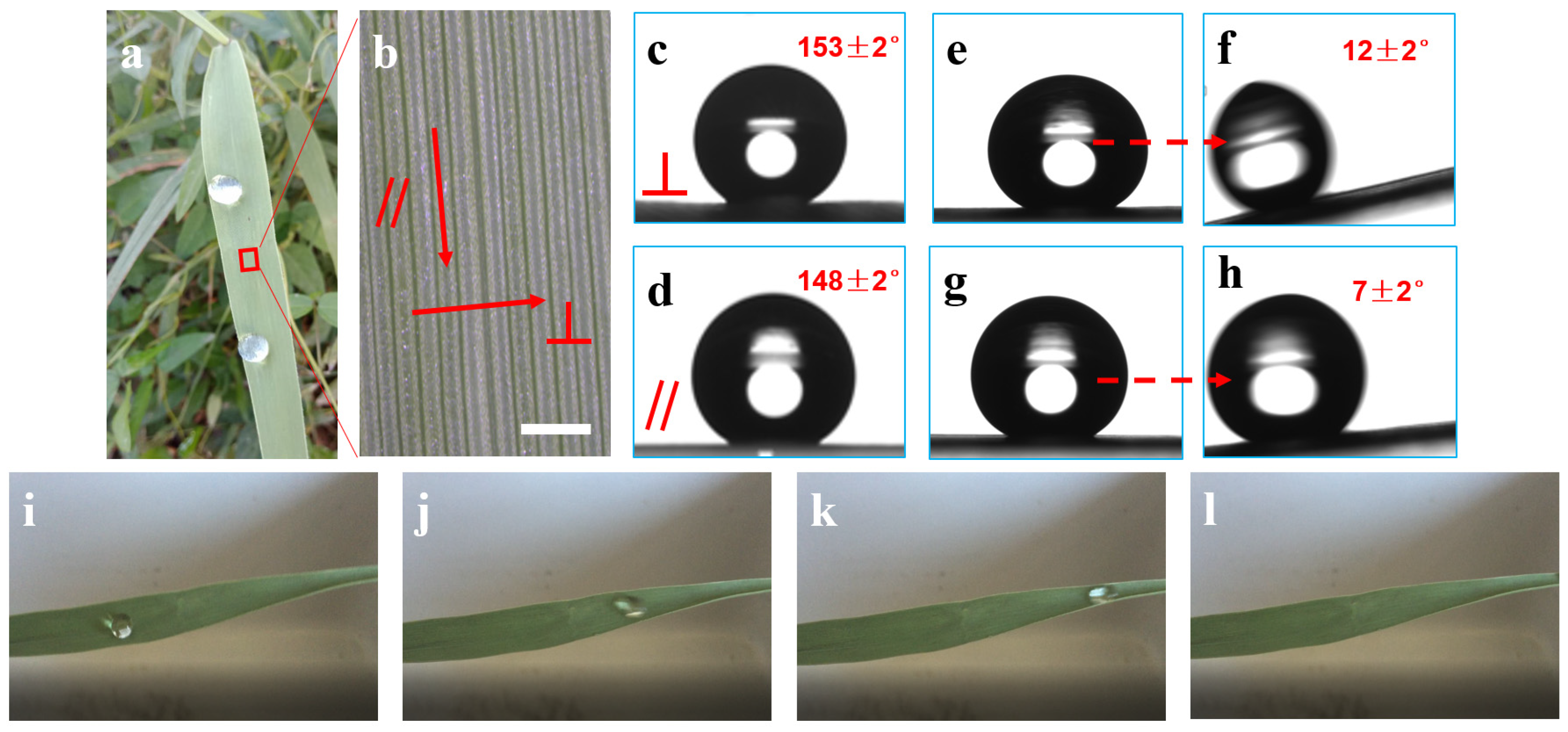
Phragmites communis leaf (PCL) is anisotropic, superhydrophobic and shows a self-cleaning effect. The water contact angle (WCA) values along the vertical and parallel vein directions on PCL are 153° ± 2° and 148° ± 2°, respectively. In contrast, the water sliding angle (WSA) values along the vertical and parallel vein directions for PCL are 12° ± 2° and 7° ± 2°, respectively. The epidermal wax makes the leaves intrinsically hydrophobic. The microstructure of the PCL surface shows sub-millimetre-, micron- and nanometre-scale structures. The sub-millimetre ridge structure is the main reason for the anisotropy of the leaves. The micron-scale papillae structure has a strong hydrophobic enhancement effect, and the nanoscale sheet structure is the key factor in achieving a stable Cassie state, as well as superhydrophobicity and self-cleaning activities. PCL-like polydimethylsiloxane (PDMS) samples fabricated by template transfer technology exhibited the sub-millimetre ridge structure and micron-scale papillae from the natural PCL; they also show obvious anisotropy and strong hydrophobicity and have a certain self-cleaning effect. The WCA and WSA values along the vertical and parallel vein directions on PCL are 146° ± 2°, 23° ± 2°, 142° ± 2° and 19° ± 2°, respectively. The preparation of a biomimetic PCL surface has broad application prospects in micro-fluidic control and the non-destructive transmission of liquids.

1. Introduction

When the water contact angle (WCA) value exceeds 150° on a surface, the surface is generally considered to be superhydrophobic [1,2,3]. Superhydrophobicity is an interesting phenomenon, but more importantly, it has inspired our thinking regarding wettability. More and more possible applications, such as self-cleaning, anti-fogging, and anti-corrosion properties, printing, sensors and water-oil separation, are being proposed and attempted [4,5,6,7,8]. The wide application prospect will inevitably prompt more research and exploration on the mechanism and preparation of superhydrophobicity.
Learning from nature is an important method of innovation. Everything is at ease while watching. There are many super wetting phenomena in nature. In addition to the famous ‘lotus effect’ [9,10], there are many other superhydrophobic plant and animal surfaces, such as anisotropic rice leaves [11], high-adhesion rose petals leaves [12], the ‘salvinia effect’ surface [13], low-adhesion legs of water striders [14], butterfly wings and peacock feathers with structural colours [15,16], anti-fogging mosquito eyes and fly eyes [17,18], and antireflective moth eyes [19]. Lately, the wettability of five plant leaves, including Tendril peanut, Endive, Setose thistle, Hamistepta and Artemisia umbrosa, has been studied. It has been proven that unitary micro-line structures can result in superhydrophobic states. If follow-up studies confirm both theoretically and experimentally that the micro-line structures possess superior mechanical stability in comparison to micro- and nanostructures, then these studies will be very important breakthroughs [20].
There are two main factors affecting the wettability of a surface: the composition of the material itself and the other the surface structure. So far, the limiting surface energy that can be obtained is 6.7 mJ/m2, which corresponds to a limiting WCA value on a smooth surface of 120°. However, the WCA value can reach 160° or higher by constructing surface microstructures [21]. Therefore, the key problem of fabricating superhydrophobic surfaces has been transformed into the construction of microstructures on a material’s surface. At present, the preparation methods of micro-nanostructures are mainly top-down and bottom-up. These methods include lithography [22], the template method [23], etching [24,25], electrochemical deposition [26], spray [27], electrospinning [28], the layer-by-layer assembly method [29], the sol-gel method [30], and the hydrothermal process [31]. Using biological surfaces directly as a template and mimicking their structures into polymers is still a preferred and concise method.
Phragmites communis is a tall perennial aquatic or wet grass. Its leaves are long, linear or lanceolate and are arranged in two rows. After rain, water droplets on the leaves are almost spherical, extremely unstable and easily roll off the surface, indicating its strong hydrophobic character. Although Phragmites australis leaves are very similar to those of rice, their microstructures and hydrophobic mechanisms are different. Therefore, we conducted a series of experiments and theoretical analyses to deepen the understanding of the superhydrophobic mechanism of the surface of plant leaves in nature.
First, the wettability of PCL was investigated from static and dynamic aspects. Then, the macro- and micro-morphologies of PCL were characterised by scanning electron microscopy (SEM) and confocal laser scanning microscopy (CLSM). Based on the obtained data, the reasons for the special wettability of the leaves were discussed. Finally, we prepared PCL-like samples using the template transfer technique and further characterised its structures and wettability.

2. Experimental

2.1. Materials

The leaf samples were natural green young PCLs, which were collected from the banks of the Songhua river in the city of Jilin (China). When the leaves were washed with running water, contaminants were carried away by rolling droplets, showing self-cleaning effects similar to those of lotus leaves. To preserve freshness, the leaves were placed in a refrigerator with a temperature of 4 °C. The material used in the template transfer technique was polydimethylsiloxane (PDMS, Dow Corning, Midland, MI, USA). The fluorinating reagent for low surface energy treatment was fluoroalkylsilane (1H, 1H, 2H, 2H–perfluorooctyl) (Aldrich, Saint Louis, MO, USA).

2.2. Methods

Photographs of the water droplets on the surface of the leaves were captured by a digital camera (EOS 7D, Canon, Tokyo, Japan). The morphology of the leaves was observed preliminarily by a stereoscopic microscope (Stereo Discovery V20, ZEISS, Oberkochen, Germany). The WCAs and water sliding angles (WSAs) of the samples surfaces were measured by a JC2000A Contact Angle System (Shanghai Zhongchen Ltd, Shanghai, China). The WCA and WSA measurements were performed using a droplet with a volume of 0.006 mL and a volumetric flow rate of 0.001 mL/s. When the droplets would fall, the inclination of the sample table was the WSA value of the sample surface.
The micro-morphologies of the samples were characterised by SEM (JSM-6700LF, JEOL, Tokyo, Japan) and CLSM (LEXT OLS3000, Olympus, Tokyo, Japan). The sample preparation process for SEM was as follows. The leaf was washed by running water to get rid of impurities on the surface and fixed for 24 h with glutaraldehyde, followed by a series of dehydration operations. Finally, the dry samples were cold sputtered with gold (SBC-12 sputtering apparatus, Beijing, China). Measurements were captured by Perfect Screen Ruler software (version 3.0). The time of optical observations should be shortened as much as possible to avoid the deformation of the leaf surface under laser light irradiation.
The process of preparing biomimetic PDMS PCL samples by the template transfer technique was as follows. First, by avoiding the main vein and selecting the middle segment of the leaf, the leaf was cut to 5 cm × 3 cm. Then, it was laid flat and pasted onto a slide using double-sided glue. The PDMS solution with a 10:1 weight ratio of Sylgard 184 silicone elastomer and a cross-linking agent was poured onto the leaf template with a thickness of approximately 3 mm. The PDMS solution was then degassed in a vacuum chamber with a pressure of 0.06 MPa to remove the trapped air bubbles. This was followed by curing at 80 °C for 2 h in a vacuum oven. The PDMS film was peeled off the leaf template after it had cooled. After this process, the reverse structure of the leaf sample was prepared. The reverse structure was treated with fluoroalkylsilane in thermal evaporation at 80 °C to facilitate the next peeling process. Finally, the positive structure of the biomimetic PCL sample was prepared by repeating the previous pouring steps with the reverse structure of the leaf sample. The process is shown in Figure 1.

3. Results and Discussion

3.1. The Wettability of the PCL Surface

Figure 2a shows a growing PCL. Water droplets are almost spherical on the surface, suggesting that the leaf may be superhydrophobic. Microscopic observations show that the middle vein of the PCL is almost parallel to the lateral veins (Figure 2b), so the vein sequence of PCLs is parallel, which indicates that the wettability of PCLs may be anisotropic. Further research showed that the WCA values along the vertical and parallel vein directions on the PCL are 153° ± 2° and 148° ± 2°, respectively, as shown in Figure 2c,d. In contrast, the WSA value along the vertical direction is approximately 12° (Figure 2e,f), which is 5° larger than that along the parallel direction (approximately 7°) (Figure 2g,h). Further, when a drop of water with a volume of 0.5 mL was titrated onto the PCL surface, the drop quickly rolled off the leave surface with only a slight quiver (Figure 2i–l). These phenomena show that the PCL surface is not only superhydrophobic but also anisotropic and self-cleaning.

3.2. The Structures of the PCL Surface

Figure 3 shows SEM photographs of the PCL surface. As Figure 3a shows, the leaf surface is a sub-millimetre ridge structure with a period of approximately 180 μm. The top, side and bottom of the ridge are shown by the dotted lines. This undulating ridge shape is caused by the presence of veins. As shown in the magnified images (Figure 3b,c), abundant micron-scale papillae is distributed on the sides of the ridge surface. The width and space of these micron-scale papillae are approximately 14–18 μm and 36–40 μm, respectively, and there is a sharp burr at the top of each papilla. Interestingly, structures that are akin to dense eye hairs, with a height and width of approximately 10 and 1 μm, respectively, grow around the stomata. The surface of the PCL has abundant, three-dimensional and nanoscale structures, namely, epidermal wax, which is a mixture of large hydrocarbon molecules that consists of a large number of cross-linked sheet structures with a thickness of approximately 50 nm and a height of approximately 1 μm (Figure 3d). Figure 3e,f shows SEM photographs of PCLs cooked in boiling water. It can be seen that the nanoscale structure and the eye hair structure near the stomata disappeared, while the other structures did not significantly change. This is because the main composition of the plant cuticular waxes are aliphatic compounds, cyclic compounds and sterols with melting points lower than 80 °C. Therefore, we inferred that the eye hair structure near the stomata is also a type of wax. In summary, the composite of the sub-millimetre ridge structure, the micron-scale papillae and the nanoscale sheet structure are the main structural characteristics of the PCL surface.
Figure 4 shows CLSM photographs of the PCL surface. The 3D morphology of the periodic sub-millimetre ridge structure is shown in Figure 4a. The period length and height of the ridge structure are approximately 180 and 30 μm, respectively. The position of the line perpendicular to the vein direction indicated by the arrow in Figure 4a is the position of the cross-section to be studied next. The section topography and dimension information of the ridge structure, papillae and eye hair structure near the stomata were obtained, especially in terms of height data, as shown in Figure 4b. This kind of convex feature is basically continuous along the direction perpendicular to the veins. Combined with the SEM results, the morphology of this cross section indicates that the width and height of the eye hair structure near the stomata and papillae are basically consistent. The statistical results show that the average width and height of these convexes are 18 and 9 μm, respectively.

3.3. The Analysis of the Wetting Mechanism

Macroscopically, the surface of the PCL exhibits surface curvature along the vertical and parallel vein directions, while microscopically, it presents a complex of the sub-millimetre ridge structure, the micron-scale papillae structure and the nanoscale sheet structure.

3.3.1. The Effect of Surface Curvature on Wettability

The existence of surface curvature in the direction parallel to the veins makes water droplets easily roll off the surface. According to the WSA test, a water droplet with a volume of 0.006 mL can roll off the PCL surface when it tilts at an angle of approximately 7°. Combined with Figure 5a, the main vein curve of the PCL is approximately in a two-dimensional plane and can be simplified to a part of a parabola [32]. We can conclude that as long as the angle θ 1 between the tangent lines and the horizontal lines at the tip of the leaf is greater than or equal to 7°, 0.006 mL water droplets will roll off the surface. The angles, such as θ 2 and θ 3 , between the tangent and horizontal lines in the other positions of the leaf are greater than θ 1 . In addition, according to Figure 2i–l, relatively large water droplets (0.5 mL) more easily roll off the PCL surface and have better self-cleaning effect. In the case of Figure 2a, water droplets can roll off the surface by increasing their volume or increasing the inclination angle of the leaf [33].
At the same time, there exists curvature on the surface of the leaf along the direction perpendicular to the veins, which has a certain influence on the rolling performance of water droplets. First, because of the increase of resistance caused by the curvature, water droplets tend to roll along the direction parallel to the veins rather than perpendicular to the veins. In addition, as shown in Figure 5b, because of the curvature, the force (indicated by the arrow) exerted by the leaf on the water droplets helps to reduce the surface area of the water droplets, making it easier for the water droplets to become spheres and roll. The existence of the curvature causes water droplets to roll approximately in a straight line.

3.3.2. The Effect of the Sub-Millimeter Ridges Structure on Wettability

Next, we discuss the effect of the ridge structure on hydrophobic performance. The ridge structure was considered to be periodic, and the shape of the ridge section element is approximately an arc. The period length and height of the ridge structure are expressed by W and H, respectively. Where n and R are defined as the centre angle (°) and radius of the arc, respectively, θ represents the angle between the tangent at the lowest point on the arc and the bottom.
According to the function relation in Figure 6a:
n / 2 = 180 ° θ
( R H ) 2 + ( W / 2 ) 2 = R 2
sin ( n / 2 ) = W / 2 R
According to the SEM and CLSM data, W and H are approximately 180 and 30 μm, respectively.
Therefore:
θ 143 °
Given that the leaf surface is covered with a wax film, the intrinsic contact angle ( θ Y ) in balance is approximately 105° [34]. It is impossible to find a point to keep the system at the equilibrium state, and water droplets will completely fill the valleys of the ridge structure when they contact. Thus, the contribution of the ridge structure to the superhydrophobicity of PCLs is not significant.
However, we noticed that only the sub-millimetre ridge structure on the PCL surface shows anisotropy. Therefore, we thought that the main reason for the anisotropy of the wettability of the leaves is the sub-millimetre ridge structure. The ridge structure, which is parallel to the vein direction, functions in drainage, and water droplets are more prone to roll. When the ridges are vertical to the vein direction, the adhesion force to be overcome is larger, so the WSA is larger. That is, the influence of the sub-millimetre ridge structure on the dynamic wettability of PCLs is greater than the influence of the ridge structure on the static wettability.

3.3.3. The Effect of the Micro-Nano Structures on Wettability

According to the SEM and CLSM data, the micron-scale structure of the leaf surface is mainly composed of the papillae structures with burrs and eye hairs structures around the stomata. For quantitative analysis, we simplified the papilla structure into a hemisphere without considering the burr structure on the papilla. The leaf surface is considered to consist of these regularly arranged papillae structures (Figure 6b). A suitable point in the three-phase line can always be found to keep the system at the equilibrium state, and water droplets are in the Cassie state, as shown in Figure 6c. The apparent contact angle ( θ c ) according to the Cassie model [2,3] can be described as follows:
cos   θ c = f 1 cos   θ Y f 2
where f 1 and f 2 are the ratios between the actual leaf-water and air-water contact areas over the apparent leaf-water interface area, respectively; H1 represents the height of the dome covered by water when the water droplets are in equilibrium; and R 1 and d represent the hemispheric radius and unit spacing, respectively.
According to the function relation in Figure 6c:
H 1 = R 1 ( 1 + cos   θ Y )
Then, the actual contact area of the leaf–water interface area ( S 1 ) and the actual contact area of the air–water interface area ( S 2 ) can be calculated by the following formula:
S 1 = 4 π R 1 2 ( 1 + cos   θ Y )
S 2 = d 2 2 π R 1 2 sin 2   θ Y
Thus, f 1 and f 2 can be calculated as:
f 1 = 4 π ( R 1 d ) 2 ( 1 + cos   θ Y )
f 2 = 1 2 π ( R 1 d ) 2 sin 2   θ Y
Substituting formulae (8) and (9) into formula (4) gives:
cos   θ C = 2 π ( R 1 d ) 2 ( 1 + cos   θ Y ) 2 1
Based on the SEM and CLSM data, R 1 and d were set to approximately 8 ± 1 μm and 38 ± 2 μm, respectively. The apparent CA value of 141.6°–153.4° was calculated by the Cassie model. This indicates that the micron-scale papillae structure contributes greatly to the hydrophobic performance. In other words, the micron-scale papillae structure has a strong hydrophobic enhancement effect. The superhydrophobic surface can be obtained theoretically through modulation parameters R 1 and d. Next, we consider the role of burrs. As shown in Figure 6d,e, when a burr is present on the top of the papilla, the reaction force (indicated by the arrow) of the papilla structure to the water droplet increases, which further prevents the water droplet from penetrating the PCL surface. That is, the existence of burrs increases the gas-liquid contact area and further improves the hydrophobic performance of the PCL surface.
Next, we discuss the role of stomata. The stomata on the leaf surface play an important role in draining water vapor from inside to outside of the leaf during transpiration. However, the pit structure of the stomata can easily collect liquid. The existence of structures akin to dense eye hairs around the stomata can solve this problem well. The maximum ratio of depth to width of a single hair can reach 10:1, which can significantly improve its intrinsic wettability [19].
The cross-linked sheet structure on the PCL surface is a rough structure. This special structure makes the contact between the water droplets and the PCL surface close to a line or even a point. Therefore, this structure plays an important role in achieving a stable Cassie state and increasing the rolling performance. In addition, the surface of the cooked PCL can be completely wetted, indicating that the waxy layer is the hydrophobic basis of the PCL surface.
In conclusion, the sub-millimetre ridge structure is the main reason for the anisotropy of the leaves. The waxy layer lays the foundation for the hydrophobic performance. The hierarchical structure strengthens the hydrophobic performance and ultimately realises superhydrophobicity and self-cleaning effect.

3.4. Biomimetic Materials and Their Wettability

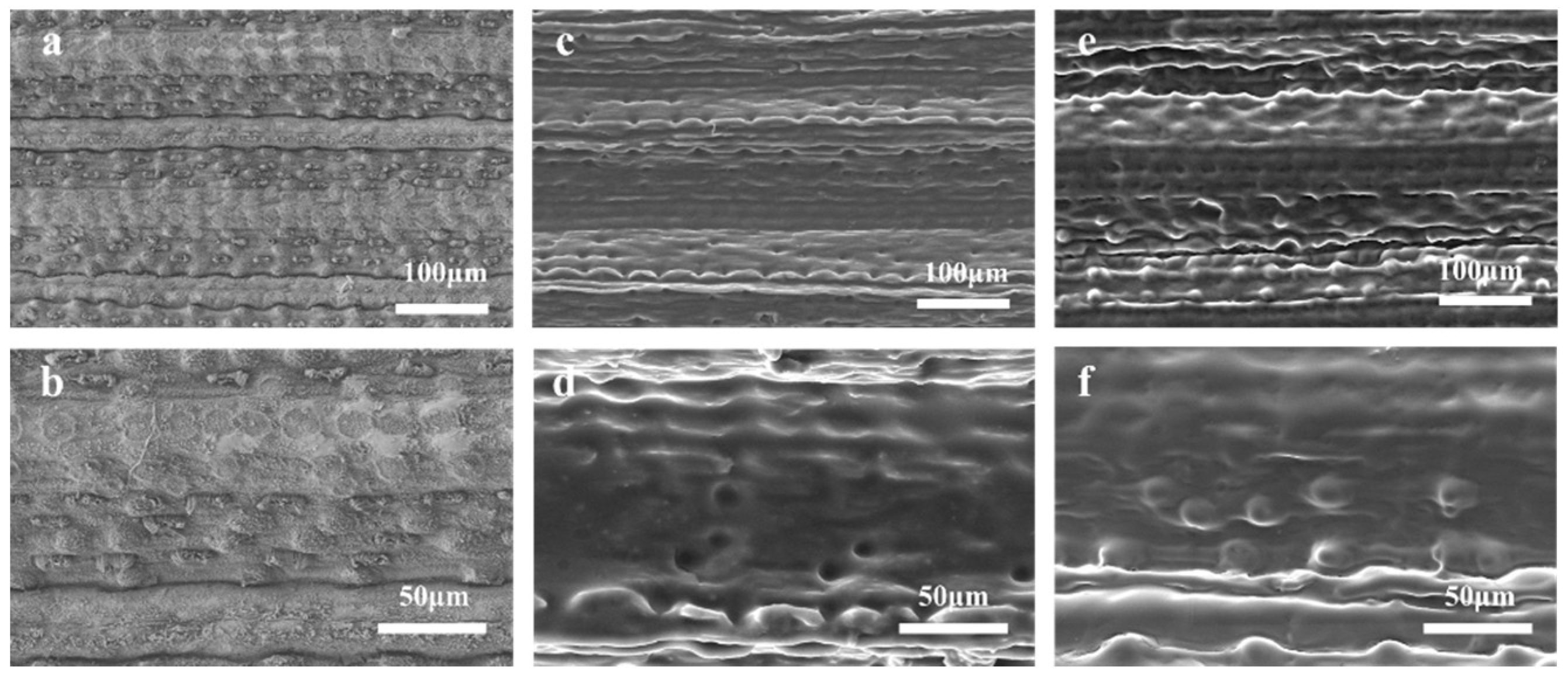
Figure 7 compares the surface morphology of the original PCL, the PDMS inverse structure after the first transcription and the positive PDMS replica. Figure 7a,b shows the surface features of the original PCL. Figure 7a shows the periodic sub-millimetre ridge structure, whereas Figure 7b magnifies the micron-scale papillae on the side surface of the ridges. Figure 7c,d shows the images of the inverse PDMS replicas. This structure includes sub-millimetre ridge structures with the width and the height of 180 and 30 μm, respectively, (Figure 7c) and porous microstructures with the width of approximately 17 μm. (Figure 7d). Burrs in the original leaves could not be accurately reproduced due to their flexibility and fragility. These results show that the sub-millimetre structures and micron-scale structures of the PCL surface were basically retained after the first transcription. Figure 7e,f shows the surface morphology of the positive PDMS replica. As observed in Figure 7e, the papillae structure was shown in a large area (5 cm × 3 cm). According to the smaller view in Figure 7f, the width and height of the papillae structure were approximately 16 and 8 μm, respectively (Figure 7f). Figure 7e,f indicates that the 3D morphology of the papillae structure was almost exhibited by its dimensional features. The reason why wax cannot be copied is that the main composition of plant cuticular waxes are aliphatic compounds, cyclic compounds and sterols with melting points lower than 80 °C, and the wax melts in the curing process of the PDMS material.
The wettability of the artificial PCLs was measured. As shown in Figure 8a,b, the WCA values along the vertical and parallel vein directions on the artificial PCLs are 146° ± 2° and 142° ± 2°, respectively. In contrast, the WSA value along the vertical direction is approximately 23° (Figure 8c,d), which is 4° larger than that along the parallel direction (approximately 19°) (Figure 8e,f). The WCA value of the PDMS was measured to be approximately 106°. The complex of the sub-millimetre ridge structure and the micron-scale papillae structure significantly improves the hydrophobic properties of the PDMS materials, which also further validates the obvious hydrophobic enhancement effect of the PCL structure surface. According to the previous wetting mechanism analysis, the apparent WCA values of the micron-scale papillae structure can be achieved in the range of 141.6°–153.4°. The measured WCA values of the artificial PCL are within the calculated range. The WCA values of the water droplets on the PDMS samples are less than the values for those on the natural PCL surface; thus, the PDMS samples did not attain superhydrophobic properties. The most likely reasons for this are the different materials, a lack of part of the structure and size errors. From the material perspective, the modulation of water on the PCL and PDMS surfaces is almost the same. Therefore, the main difference between the artificial surface and the natural surface is the absence of nanostructures. In addition, the shrinkage of the PDMS material made the depth smaller. The smaller depth–width ratio leads to less air being trapped. Therefore, the apparent WCA value of the artificial leaf is less than that of the natural leaf. At the same time, we also investigated the WCA value of the inverse structure. The WCA values along the vertical and parallel vein directions of the negative PDMS replicas are 135° ± 2° and 130° ± 2°, respectively. The WCA value of the reverse structure is less than that of the positive PDMS replica. Research has shown that the hydrophobic properties of micropillar surfaces and microgroove surfaces with complementary structures and the same parameters are "complementary". That is, when the hydrophobicity of the micro-pillar surfaces is relatively strong, the hydrophobicity of the microgroove surfaces is not ideal, and vice versa [35]. The artificial PCL is similar to the micropillar surface, and it has better hydrophobic properties, so the hydrophobicity of the negative PDMS replicas is relatively poor.

4. Conclusions

PCL is anisotropic and superhydrophobic and shows self-cleaning effect. The WCA values along the vertical and parallel vein directions on the PCL are 153° ± 2° and 148° ± 2°, respectively. In contrast, the WSA values along the vertical and parallel vein directions on the PCL are 12° ± 2° and 7° ± 2°, respectively. The leaf surface was covered with a layer of epidermal wax, which laid the hydrophobic foundation of the PCL surface. The complex of the sub-millimetre ridge structure, the micron-scale papillae structure and the nanoscale sheet structure is the main structural characteristic of the leaf surface. The sub-millimetre ridge structure is the main reason for its anisotropy. The micron-scale papillae structure has a strong hydrophobic enhancement effect, and the nanoscale sheet structure is the key factor in achieving a stable Cassie state as well as superhydrophobic and self-cleaning activities. PCL-like PDMS samples fabricated by template transfer technology exhibited the sub-millimetre ridge structure and the micron-scale papillae from the natural PCL, and also show obvious anisotropy and strong hydrophobicity and have a certain self-cleaning effect. The WCA and WSA values along vertical and parallel veins directions on PCL are 146° ± 2°, 23° ± 2°, 142° ± 2° and 19° ± 2°, respectively. The measured WCA is consistent with the calculated WCA (141.6°–153.4°). The absence of nanostructures is the main reason why the artificial PCL fails to achieve better hydrophobic properties. The construction of stable nanostructures on the surface of artificial PCLs obtained by template transfer method is of great interest.

Supplementary Materials

Supplementary File 1

Author Contributions

Conceptualisation, S.N. and J.Z.; Methodology, S.N.; Software, X.F.; Validation, X.F., H.G. and J.Z.; Formal Analysis, H.G.; Investigation, X.F.; Resources, Z.H.; Data Curation, S.N.; Writing—Original Draft Preparation, H.G.; Writing—Review and Editing, S.N., J.Z.; Visualization, X.F., J.Z.; Supervision, Z.H.; Project Administration, Z.H.; Funding Acquisition, H.G., S.N., J.Z. and Z.H.

Funding

This work is financially supported by the National Natural Science Foundation of China (Grant No. 51805207), the 13th Five-Year Science and Technology Project of Jilin Provincial Department of Education (Grant No. JJKH20170223KJ), Scientific and Technological Development Program of Changchun City (Double Ten Project-19SS001), Science and Technology Development Program of Jilin Province (Technology R&D Project-20190302021GX) and Industrial Technology Research and Development Funds of Jilin Province (project No. 2019C041-1).

Conflicts of Interest

The authors declare no conflict of interest.

References

  1. Wang, S.; Jiang, L. Definition of superhydrophobic states. Adv. Mater. 2007, 19, 3423–3424. [Google Scholar] [CrossRef]
  2. Liu, M.; Wang, S.; Jiang, L. Nature-inspired superwettability systems. Nat. Rev. Mater. 2017, 2, 17036. [Google Scholar] [CrossRef]
  3. Fang, R.C.; Liu, M.J.; Liu, H.L.; Jiang, L. Bioinspired interfacial materials: From binary cooperative complementary interfaces to superwettability systems. Adv. Mater. Interfaces 2018, 5, 1701176. [Google Scholar] [CrossRef]
  4. Shirtcliffe, N.J.; Roach, P. Superhydrophilic surfaces for antifogging and antifouling microfluidic devices. Methods Mol. Biol. 2013, 949, 269–281. [Google Scholar] [PubMed]
  5. Ramachandran, R.; Nosonovsky, M. Coupling of surface energy with electric potential makes superhydrophobic surfaces corrosion-resistant. Phys. Chem. Chem. Phys. 2015, 17, 24988–24997. [Google Scholar] [CrossRef] [PubMed] [Green Version]
  6. Tian, D.; Song, Y.; Jiang, L. Patterning of controllable surface wettability for printing techniques. Chem. Soc. Rev. 2013, 42, 5184–5209. [Google Scholar] [CrossRef] [PubMed]
  7. Lei, Y.; Sun, R.; Zhang, X.; Feng, X.; Jiang, L. Oxygen-rich enzyme biosensor based on superhydrophobic electrode. Adv. Mater. 2016, 28, 1477–1481. [Google Scholar] [CrossRef]
  8. Xue, Z.X.; Wang, S.T.; Lin, L.; Chen, L.; Liu, M.J.; Feng, L.; Jiang, L. A novel superhydrophilic and underwater superoleophobic hydrogel-coated mesh for oil/water separation. Adv. Mater. 2011, 23, 4270–4273. [Google Scholar] [CrossRef]
  9. Barthlott, W.; Neinhuis, C. Purity of the sacred lotus, or escape from contamination in biological surfaces. Planta 1997, 202, 8. [Google Scholar] [CrossRef]
  10. Feng, L.; Li, S.; Li, Y.; Li, H.; Zhang, L.; Zhai, J.; Song, Y.; Liu, B.; Jiang, L.; Zhu, D. Super-hydrophobic surfaces: From natural to artificial. Adv. Mater. 2002, 14, 1857–1860. [Google Scholar] [CrossRef]
  11. Yao, J.; Wang, J.N.; Yu, Y.H.; Yang, H.; Xu, Y. Biomimetic fabrication and characterization of an artificial rice leaf surface with anisotropic wetting. Chin. Sci. Bull. 2012, 57, 2631–2634. [Google Scholar] [CrossRef] [Green Version]
  12. Feng, L.; Zhang, Y.A.; Xi, J.M.; Zhu, Y.; Wang, N.; Xia, F.; Jiang, L. Petal effect: A superhydrophobic state with high adhesive force. Langmuir 2008, 24, 4114–4119. [Google Scholar] [CrossRef]
  13. Barthlott, W.; Schimmel, T.; Wiersch, S.; Koch, K.; Brede, M.; Barczewski, M.; Walheim, S.; Weis, A.; Kaltenmaier, A.; Leder, A.; et al. The Salvinia paradox: Superhydrophobic surfaces with hydrophilic pins for air retention under water. Adv. Mater. 2010, 22, 2325–2328. [Google Scholar] [CrossRef]
  14. Gao, X.; Jiang, L. Biophysics: Water-repellent legs of water striders. Nature 2004, 432, 36. [Google Scholar] [CrossRef]
  15. Kinoshita, S.; Yoshioka, S.; Kawagoe, K. Mechanisms of structural colour in the Morpho butterfly: Cooperation of regularity and irregularity in an iridescent scale. Proc. Biol. Sci. 2002, 269, 1417–1421. [Google Scholar] [CrossRef]
  16. Zi, J.; Yu, X.D.; Li, Y.Z.; Hu, X.H.; Xu, C.; Wang, X.J.; Liu, X.H.; Fu, R.T. Coloration strategies in peacock feathers. Proc. Natl. Acad. Sci. USA 2003, 100, 12576–12578. [Google Scholar] [CrossRef] [Green Version]
  17. Gao, X.F.; Yan, X.; Yao, X.; Xu, L.; Zhang, K.; Zhang, J.H.; Yang, B.; Jiang, L. The dry-style antifogging properties of Mosquito compound eyes and artificial analogues prepared by soft lithography. Adv. Mater. 2007, 19, 2213–2217. [Google Scholar] [CrossRef]
  18. Sun, Z.Q.; Liao, T.; Liu, K.S.; Jiang, L.; Kim, J.H.; Dou, S.X. Fly-eye inspired superhydrophobic anti-fogging inorganic nanostructures. Small 2014, 10, 3001–3006. [Google Scholar] [CrossRef]
  19. Parker, A.R.; Townley, H.E. Biomimetics of photonic nanostructures. Nat. Nanotech. 2007, 2, 347–353. [Google Scholar] [CrossRef]
  20. Gou, X.L.; Guo, Z.G. Superhydrophobic plant leaves with micro-line structures: An optimal biomimetic objective in bionic engineering. J. Bionic. Eng. 2018, 15, 851–858. [Google Scholar] [CrossRef]
  21. Nishino, T.; Meguro, M.; Nakamae, K.; Matsushita, M.; Ueda, Y. The lowest surface free energy based on-CF3 alignment. Langmuir 1999, 15, 4321–4323. [Google Scholar] [CrossRef]
  22. Dumanli, A.G.; Savin, T. Recent advances in the biomimicry of structural colours. Chem. Soc. Rev. 2016, 45, 6698–6724. [Google Scholar] [CrossRef] [Green Version]
  23. Liu, X.J.; Xu, Y.; Ben, K.Y.; Chen, Z.; Wang, Y.; Guan, Z.S. Transparent, durable and thermally stable PDMS-derived superhydrophobic surfaces. Appl. Surf. Sci. 2015, 339, 94–101. [Google Scholar] [CrossRef]
  24. Cho, S.W.; Kim, J.H.; Lee, H.M.; Chae, H.Y.; Kim, C.K. Superhydrophobic Si surfaces having microscale rod structures prepared in a plasma etching system. Surf. Coat. Tech. 2016, 306, 82–86. [Google Scholar] [CrossRef]
  25. Li, L.J.; Huang, T.; Lei, J.L.; He, J.X.; Qu, L.F.; Huang, P.L.; Zhou, W.; Li, N.B.; Pan, F.S. Robust biomimetic-structural superhydrophobic surface on aluminum alloy. ACS Appl. Mater. Inter. 2015, 7, 1449–1457. [Google Scholar] [CrossRef]
  26. Xu, L.Y.; Tong, F.Q.; Lu, X.M.; Lu, K. Multifunctional polypyrene/silica hybrid coatings with stable excimer fluorescence and robust superhydrophobicity derived from electrodeposited polypyrene films. J. Mater. Chem. C 2015, 3, 2086–2092. [Google Scholar] [CrossRef]
  27. Qinga, Y.Q.; Yanga, C.N.; Hub, C.B.; Zheng, Y.S.; Liua, C.S. A facile method to prepare superhydrophobic fluorinatedpolysiloxane/ZnO nanocomposite coatings with corrosion resistance. Appl. Surf. Sci. 2015, 326, 48–54. [Google Scholar] [CrossRef]
  28. Huang, W.H.; Lin, C.S. Robust superhydrophobic transparent coatings fabricated by a low-temperature sol-gel process. Appl. Surf. Sci. 2014, 305, 702–709. [Google Scholar] [CrossRef]
  29. Shang, Q.; Zhou, Y. Fabrication of transparent superhydrophobic porous silica coating for self-cleaning and anti-fogging. Ceram. Int. 2016, 42, 8706–8712. [Google Scholar] [CrossRef]
  30. Wei, W.; Lü, X.M.; Jiang, D.L.; Yan, Z.X.; Chen, M.; Xie, J.M. A novel route for synthesis of UV-resistant hydrophobic titania-containing silica aerogels by using potassiumtitanate as precursor. Dalton. Trans. 2014, 43, 9456–9467. [Google Scholar] [CrossRef]
  31. Liu, M.; Qing, Y.; Wu, Y.Q.; Liang, J.; Luo, S. Facile fabrication of superhydrophobic surfaces on wood substratesvia a one-step hydrothermal process. Appl. Surf. Sci. 2015, 330, 332–338. [Google Scholar] [CrossRef]
  32. Allen, M.; Prusnk, I.P.; Dejon, T. Using L-systems for modeling source–sink interactions, architecture and physiology of growing trees: the L-PEACH model. New Phytol. 2005, 166, 870–880. [Google Scholar] [CrossRef]
  33. Butt, H.J.; Roisman, I.V.; Brinkmann, M.; Papadopoulos, P.; Vollmer, D.; Semprebon, C. Characterization of super liquid-repellent surfaces. Curr. Opin. Colloid Interface Sci. 2014, 19, 343–354. [Google Scholar] [CrossRef] [Green Version]
  34. Barthlott, W.; Neinhuis, C.; Cutler, D.; Ditsch, F.; Meusel, I.; Theisen, I.; Wilhelmi, H. Classification and terminology of plant epicuticular waxes. Botan. J. Linn. Soc. 1998, 126, 237–260. [Google Scholar] [CrossRef]
  35. Ye, X.; Zhou, M.; Li, J.; Liu, H.X.; Yuan, R.; Yang, H.F.; Li, B.J.; Cai, L. Microstructure of superhydrophobic surfaces from natural to artificial. NPE 2009, 7, 381–386. (In Chinese) [Google Scholar]
Figure 1. The schematic illustrating the template transfer technique for the fabrication of biomimetic Phragmites communis leaves (PCLs).
Coatings 09 00541 g001
Figure 2. Photographs of wettability on the PCL surface. (a) A growing PCL; (b) a stereomicroscopic photograph of a leaf, where the bar in the figure represents 1 mm; (c,d) the water contact angles (WCAs) along the perpendicular and parallel vein directions; (e,f) the water sliding angle (WSA) along the perpendicular vein direction; (g,h) the WSA along the parallel vein direction; a drop of water with a volume of 0.5 mL rolls off the PCL surface after the slightest vibration (i) 0 s; (j) 0.5 s; (k) 1 s; (l) 2 s.
Coatings 09 00541 g002
Figure 3. SEM photographs of the PCL surface. (a) The periodic sub-millimetre ridge structure; (b,c) the micron-scale structures on the top, side and bottom of the ridge structure; (d) the nanoscale sheet structure covering the PCL surface; (e,f) PCL surface morphology after cooking in boiling water.
Coatings 09 00541 g003
Figure 4. Confocal laser scanning microscopy (CLSM) photographs of the PCL surface. (a) The 3D morphology of the periodic sub-millimetre ridge structure and the selected cross section position to be studied; (b) the section topography and dimension information of the ridge structure, papillae and eye hair structure near the stomata.
Coatings 09 00541 g004
Figure 5. The schematic diagram of the influence of surface curvature on wettability. (a) In the direction parallel to the veins; (b) in the direction perpendicular to the veins.
Coatings 09 00541 g005
Figure 6. A schematic diagram of the influence of the microstructure on wettability. (a) The effect of the sub-millimetre structure on wettability; (b) the effect of the papillae structure on wettability; (c) the simplified 2D unit model of the papillae structure; (d) the force analysis sketch of the papillae structure without burrs; and (e) the force analysis sketch of the papillae structure with burrs.
Coatings 09 00541 g006
Figure 7. SEM photographs of (a,b) original PCL, (c,d) negative polydimethylsiloxane (PDMS) replica; (e,f) positive PDMS replica.
Coatings 09 00541 g007
Figure 8. Photographs of the wettability of the artificial PCL surface. (a,b) The WCAs along the perpendicular and parallel vein directions; (c,d) the WSA along the perpendicular vein direction; and (e,f) the WSA along the parallel vein direction.
Coatings 09 00541 g008