Article

Carvacrol-Containing Hemicellulose/Methylcellulose Plant-Based Coatings and Films: Evaluation of Antimicrobial Activity, Biodegradability, and Cytocompatibility


Eastern Regional Research Center, Agricultural Research Service, U. S. Department of Agriculture, 600 E. Mermaid Lane, Wyndmoor, PA 19038, USA
*
Author to whom correspondence should be addressed.
Coatings 2026, 16(8), 911; https://doi.org/10.3390/coatings16080911
Submission received: 12 June 2026 / Revised: 27 July 2026 / Accepted: 28 July 2026 / Published: 1 August 2026

Abstract

This study investigated the antimicrobial efficacy, biodegradability, and cytotoxicity of carvacrol-infused hemicellulose/methylcellulose (HB/MC)-based coating solutions and films for active food packaging applications. Micro-emulsified (M) and coarse (C) HB/MC solutions with 1 or 2% carvacrol (Car) were investigated. Their antimicrobial properties were evaluated by applying the solutions directly to eggshell surfaces (coating) or headspace (film). Against Salmonella enterica, the 2% carvacrol micro-emulsified coating (HB/MC+Car-2%/M) produced the greatest bacterial reduction, reaching 8.35 log10 units relative to the untreated control by Day 7. Under the same conditions, the corresponding coarse-emulsified coating (HB/MC+Car-2%/C) achieved a 2.10-log10 reduction. When evaluated as a headspace-active film, HB/MC+Car-2%/M achieved a 6.28-log10 reduction, demonstrating the superior antimicrobial performance of the direct-contact micro-emulsified coating. Biodegradability, assessed via biochemical oxygen demand (BOD), evidenced 100% biodegradation for all formulations under aerobic conditions. The highest BOD value was observed in HB/MC+Car-2%\M, indicating elevated microbial activity. Cytotoxicity evaluation using the MTT assay confirmed that all coating solutions were non-toxic, with HB/MC+Car-2%\M promoting the highest cell viability across tested concentrations. These results demonstrated that industrial crops and by-products could be used as carriers for bioactive agents via a micro-emulsification step, thereby significantly enhancing the functional performance of plant-based materials, offering a promising route for developing safe, biodegradable, and plant-based antimicrobial coatings and packaging materials.

1. Introduction

Foodborne illnesses and food spoilage continue to pose major global challenges to food security, public health, and environmental sustainability. In the United States, foodborne pathogens, including Salmonella spp., Listeria monocytogenes, and Escherichia coli O157:H7, are responsible for millions of illnesses annually and impose substantial economic burdens [1,2,3]. Among these, Salmonella enterica is one of the leading causes of foodborne disease and is frequently associated with eggs and egg products. A recent outbreak of Salmonella enteritidis infections linked to eggs was reported across ten U.S. states, resulting in 134 confirmed cases, 38 hospitalizations, and one death (FDA, 2025) [4]. Food contamination by Salmonella and other foodborne pathogens often results in product recalls, contributing substantially to food loss, economic costs, and environmental impacts [5]. It has been estimated that approximately 1.3 billion tons of food are wasted annually worldwide, representing 30–40% of total food production and contributing more than 4 gigatons of CO2 emissions each year [6,7].
Conventional food packaging materials, primarily petroleum-based plastics, have long been used to preserve food quality and prevent microbial contamination. However, their resistance to degradation and accumulation in ecosystems has raised serious environmental concerns [8,9]. In response, biopolymers derived from renewable resources, such as hemicellulose, a plant-based heteropolysaccharide, have emerged as promising alternatives due to their biodegradability, renewability, and film-forming capabilities [10,11,12]. Despite these advantages, hemicellulose-based films often exhibit poor mechanical strength, limited barrier properties, and insufficient antimicrobial activity, which severely constrain their broader application in active packaging systems [13,14,15].
To overcome these limitations, researchers have explored incorporating natural antimicrobial agents into biopolymer matrices. Carvacrol, a phenolic compound known as 5-isopropyl-2-methylphenol, is produced by a wide variety of plants, such as Thymus, Coridothymus, Oreganum, Satureja, Lippia, and Thymbra. It exists in a liquid state, is insoluble in water, and has demonstrated potent antimicrobial activity against various types of spoilage microorganisms and foodborne pathogens [16,17,18,19]. Carvacrol is certified as Generally Recognized as Safe (GRAS) and approved for use in food-contact applications [20,21,22]. It has been successfully incorporated into various biopolymer films to enhance antimicrobial activity, inhibit microbial growth, and prolong food shelf life [23,24,25]. However, its effectiveness depends heavily on the dispersion of an active agent within the polymer matrix. Conventional mixing methods often result in an uneven distribution, compromising antimicrobial performance. In contrast, micro-emulsification and high-pressure homogenization techniques have shown promise in achieving uniform dispersion of hydrophobic compounds such as carvacrol, thereby improving the film’s antibacterial functionality [26,27,28].
Our recent interventions [29,30,31] demonstrated that hemicellulose-based films incorporating carvacrol via micro-emulsification exhibited superior antimicrobial activity, reduced gas and moisture transmission, and improved mechanical properties compared to those prepared with coarse emulsions. Beyond antimicrobial efficacy, the biodegradability of packaging materials is essential for reducing environmental impact. Hemicellulose-based films have shown favorable degradation behavior under aerobic soil conditions [32,33]. Additionally, ensuring cytocompatibility is critical for materials intended for direct or indirect contact with biological surfaces. Cytotoxicity assessments provide valuable insights into the safety of bioactive formulations [34,35,36].
Recent innovations in biopolymer coatings [37,38,39] have also explored multifunctional enhancements, such as antioxidant and antifogging properties, to improve food preservation and consumer appeal. For example, p-coumaric acid-grafted chitosan coatings [40] have demonstrated transparency, antimicrobial efficacy, and antioxidant activity, highlighting the potential of integrating natural compounds into polymer matrices.
Building on these advancements [41,42] and our recent work [29,30,31], the present study was to investigate the use of industrial crops to carry nano/micro scale of antimicrobials and their applications, specifically, the formulation of hemicellulose-based coatings and films incorporating carvacrol, followed by their application in antimicrobial egg coating and in the production of biodegradable films. These formulations were evaluated for their antimicrobial efficacy, cytotoxicity, and biodegradation performance in the soil. By integrating natural antimicrobial agents into biodegradable matrices, this work contributes to the utilization of industrial crops and the development of eco-functional packaging materials that support both food safety and environmental sustainability.

2. Materials and Methods

2.1. Materials

Grade A chicken eggs, each weighing ~60 g, were purchased from a local grocery store. Only eggs without cracks were selected. Eggs were stored in a refrigerator at 4 °C and allowed to reach room temperature (22 °C) before use. Eggs were first cleaned with tap water, then sanitized with 70% ethanol, and finally artificially inoculated with Salmonella enterica. Carvacrol (Car, 95%), glycerol (G), methylcellulose (MC), high methoxyl pectin (HMP), Hemicellulose B (HB), and pea protein isolate (PPI) were used in this study, and their resources and production information are detailed in our previous publications [29,30].

2.2. Preparation of HB/MC-Based Emulsions with Carvacrol

The blended formulations were prepared as described in our recent publications [29,30]. The formulations were used for both coating solutions and film fabrication Table 1. Each component was individually dispersed in deionized (DI) water at 20 °C, followed by the addition of carvacrol at the final stage of mixing. Coarse emulsions (average particle sizes of 11.59 μm) were prepared after mechanical stirring and degassing using a FlackTek mixer (FlackTek Manufacturing, Landrum, SC, USA) operating at 1500–2000 rpm. These emulsions were then processed by high-pressure homogenization (HPH) using an EmulsiFlex-B3 homogenizer (Avestin Inc., Ottawa, ON, Canada) at 138 MPa (20,000 psi) for 2 cycles, yielding micro-emulsions (average particle sizes of 1.23 μm). The completed coating emulsions were not subjected to heat sterilization because heating could alter the polymer structure and promote carvacrol loss; instead, they were prepared aseptically using sterile deionized water and sterilized equipment. The resulting emulsions were used to coat eggs or cast into thin films to assess their antimicrobial activity, cytocompatibility, and biodegradability, as described below.

2.3. Film Casting and Drying

For film preparation, each emulsion (coarse and micro, 30 g) was poured into 100 mm Teflon Petri dishes. The samples were dried for approximately one week, or until the final film weights ranged from 1.05 to 1.10 g, as described in our previous publications [29,30]. Once dried, the films were carefully peeled off and stored in Ziploc bags within desiccators for further analysis. Films derived from coarse emulsions were labeled as C-films, while those from micro-emulsions were designated as M-films.

2.4. Preparation of Bacterial Cultures

Two strains of Salmonella enterica serotype Typhimurium (ATCC 53647 and ATCC 53648) were obtained from the culture collection of our research center (Wyndmoor, PA, USA). To create working stocks for experiments, the cultures were revived and incubated at 37 °C for 18 h. Bacterial cells were harvested by centrifugation, washed with sterile water, and pooled to create a uniform suspension. The final bacterial concentration was adjusted to approximately 8 log10 CFU/mL.

2.5. Egg Inoculation Procedure Using Salmonella Cocktail

A bacterial cocktail was prepared by combining equal volumes of two Salmonella enterica serotype Typhimurium strains, which had been previously cultured and preserved as described in Section 2.4. A two-strain cocktail was used to better represent strain-to-strain variability and to provide a more robust evaluation of the antimicrobial efficacy of the coating formulations under conditions representative of potential food contamination. The use of multi-strain inocula is a common practice in food microbiology to minimize strain-specific responses and improve the robustness of antimicrobial efficacy assessments. Each egg was inoculated with 100 µL of the prepared bacterial suspension using a spot inoculation technique. The inoculum was distributed across ten evenly spaced spots within a defined area of approximately 7.25 cm2 on the eggshell surface to promote uniform drying. Inoculated eggs were then air-dried in a biohood at room temperature for 3 h to facilitate bacterial adhesion.

2.6. Coating and Film Application and Antimicrobial Testing

To assess the antimicrobial activity of the coatings and films, two complementary methods were employed: direct coating and headspace release assays, adapted from previously established protocols [29,30,31,42].
For direct coating, each inoculated egg was coated with 1 mL of the test emulsion (as listed in Table 1) using a sterile fine-bristle brush to ensure even and consistent coverage across the shell surface of inoculated spots. Eggs without any coating served as untreated controls. All samples were air-dried in a biohood at room temperature (~22 °C) for 3 h prior to microbiological analysis or storage tests.
In the headspace release assay, films cast in 100 mm Petri dishes (total area ≈ 78.5 cm2; dry mass 1.05–1.10 g) were cut into two equal halves. One half-film (≈39.3 cm2; 0.52–0.55 g) was attached to the inner wall of each sterile egg carton housing three inoculated eggs, ensuring the film did not make direct contact with the eggs. This configuration enabled volatile antimicrobial compounds released from the films to diffuse through the container headspace and interact with bacteria present on the eggshell surface. All egg samples were stored in commercial egg cartons at 7 °C until microbiological analysis.

2.7. Microbiological Analysis

Microbial testing was performed at three time points: Days 1, 3, and 7 post-treatment. At each sampling time, eggs were aseptically cracked, and their contents were discarded. A representative portion of the eggshell was transferred into a sterile 50 mL centrifuge tube. The shell was crushed using a sterile glass rod, and 30 mL of sterile 0.1% peptone water was added. Samples were vortexed for 2 min to release bacteria from the shell surface. From each homogenized sample, 1 mL was withdrawn and serially diluted 10-fold. Aliquots (100 µL) of appropriate dilutions were surface-plated on Xylose Lysine Tergitol 4 agar (XLT4), a selective medium for Salmonella spp. Plates were incubated at 37 °C for 24 h, and colony-forming units (CFU) were counted to assess bacterial survival. Based on plating 100 µL of the undiluted eggshell suspension, the theoretical detection limit of the enumeration method was 10 CFU/mL (1.0 Log CFU/mL).
Microbial reductions were calculated relative to the corresponding untreated control at each sampling time using the following equation:
Log reduction = log10(N-control) − log10(N-treatment)
where N-control is the bacterial population recovered from the untreated control, and N-treatment is the bacterial population recovered from the treated sample, both expressed as CFU/mL. A positive value indicates a reduction in the bacterial population relative to the untreated control.
Peptone water without an eggshell sample was used as a negative control to confirm the absence of background contamination.

2.8. Biodegradability Evaluation of Hemicellulose-Based Films

The biodegradation potential of hemicellulose-based films was assessed using a respirometric method in accordance with ISO 17556:2019 standards. OxiTop® Control S6 system (WTW-Xylem, Rye Brook, NY, USA) was used to monitor oxygen consumption during aerobic degradation in soil, measuring biochemical oxygen demand (BOD) as an indicator of microbial activity and material breakdown. The test system consisted of twelve 510 mL glass bottles, each fitted with rubber quivers and pressure-sensitive measuring heads capable of detecting changes between 500 and 1350 hPa with 1% accuracy over a temperature range of 5 to 50 °C. An integrated OC 110 controller facilitated data communication and monitoring throughout the experiment. The biodegradation medium consisted of locally sourced garden soil with high humus content from Wyndmoor, PA. The soil was sifted and dried, with properties standardized to 5% moisture content (ISO 11274), pH 6.0 (ISO 10390), and particle size below 2 mm. Each bottle was loaded with hemicellulose-based film (200 mg), prepared soil (200 g), and distilled water (50 g). The containers were hermetically sealed and placed at 25 °C. Oxygen consumption was recorded over 153 days to capture long-term biodegradation dynamics. Although the respirometric incubation was conducted for 180 days, all formulations reached a biodegradation plateau (Dt ≈ 100%) by Day 153; therefore, Day 153 was used as the reporting endpoint for BOD/TOD and Dt calculations.
The BOD for each bottle was determined using the following formula:
BOD = (BODx − BODg)/c
where BOD is the biochemical oxygen demand of the test material (mg O2 g−1 film), BODx represents the oxygen demand of the complete system (soil + film) (mg O2), BODg accounts for the baseline oxygen demand of the soil (mg O2), without the sample, and c is the concentration of the hemicellulose film in the test system (g).
The degree of biodegradation (Dt) of the hemicellulose-based film was then determined using the equation:
Dt = BODS/TOD × 100
where Dt is the degree of biodegradation (%), BODS is the biochemical oxygen demand of the sample (mg O2 g−1 film), and TOD is the theoretical oxygen demand (mg O2 g−1 film).
Biodegradation percentages were calculated following the principles of ISO 17556:2019. Because complete mineralization cannot exceed the theoretical oxygen demand (TOD) of the test material, calculated biodegradation values exceeding 100% were normalized to 100% for reporting purposes. Slightly elevated BOD values relative to TOD may result from microbial priming effects, background soil organic matter mineralization, or normal experimental variability rather than biodegradation of the test material alone.
The theoretical oxygen demand (TOD) for each formulation was calculated from the elemental composition of the films, determined based on their known chemical constituents (hemicellulose, methylcellulose, pectin, pea protein isolate, glycerol, and carvacrol). TOD was then calculated using the following equation:
TOD = 16[2C + 0.5H − O]/Mn
where TOD is the theoretical oxygen demand (mg O2 g−1 film), C, H, and O are the mass fractions of carbon, hydrogen, and oxygen (g g−1, dimensionless), respectively, and Mn is the molecular weight (g mol−1).
This methodology enabled a quantitative assessment of the film’s biodegradability, providing insight into its environmental decomposition behavior under simulated soil conditions.

2.9. Cytotoxicity Assessment

To evaluate the potential cytotoxicity of the coating materials, mouse embryonic fibroblast cells (NIH 3T3) were used in a standard 3-[4,5-dimethylthiazol-2-yl]-2,5-diphenyl tetrazolium bromide (MTT) assay [40]. NIH 3T3 cells obtained from ATCC were maintained in Dulbecco’s Modified Eagle Medium (DMEM) (Fisher Scientific, Pittsburgh, PA, USA) supplemented with 10% fetal bovine serum (FBS) (Fisher Scientific) and 1% Penicillin-Streptomycin at 37 °C in a humidified incubator with 5% CO2 and passaged at a 1:5 ratio according to the handling procedure for NIH 3T3 cells from ATCC. The MTT assay quantifies metabolically active cells by determining mitochondrial reduction of MTT to the insoluble purple formazan, which can be read spectrophotometrically at 492 nm. Higher absorbance indicates lower cytotoxicity. Cytotoxicity testing was performed using the CyQUANT™ MTT and XTT Cell Viability Assay kit (Invitrogen, Inc., Carlsbad, CA, USA) according to the manufacturer’s instructions. Briefly, NIH 3T3 cells were seeded at 5000 cells/well in 96-well plates and allowed to adhere overnight. Then, the cells were exposed to the various coating solutions listed in Table 1 for 24 h at 37 °C. After incubation, 100 μL of MTT reagent was added to each well to achieve a concentration of 0.5 mg/mL, and the plates were incubated for an additional 4 h to allow the formation of formazan crystals. The supernatant was then carefully removed, and 100 μL of dimethyl sulfoxide (DMSO) was added to solubilize the formazan crystals. Absorbance was measured at 492 nm using a microplate reader (Anthos 2010, Biochrom Ltd., Cambridge, Cambridgeshire, UK). Cell viability was determined using the following equation:
Cell Viability (%) = [ODsODb)/(ODcODb)] × 100
where ODs represent the absorbance of the sample (cells + sample), ODc represents the negative control consisting of cells cultured in complete medium only (DMEM supplemented with 10% FBS and 1% penicillin–streptomycin), and ODb represents the absorbance of the blank (medium without cells). All assays were performed in triplicate.

2.10. Statistical Analysis

Statistical significance was determined using one-way analysis of variance (ANOVA) in GraphPad Prism version 7.0 (GraphPad Software, Boston, MA, USA). Experimental results were expressed as mean ± standard deviation (SD). Statistically significant difference is defined as a p-value ≤ 0.05.

3. Results and Discussion

Building upon the initial screening, the selected formulations were further evaluated for their functional performance with respect to antimicrobial activity, biodegradability, and cytotoxicity. These assessments aimed to determine the suitability of hemicellulose-based systems, particularly those incorporating carvacrol and micro-emulsification techniques, for active food packaging applications.

3.1. Antimicrobial Properties

This study assessed the antimicrobial performance of carvacrol-infused hemicellulose formulations against a two-strain cocktail of Salmonella enterica serovar Typhimurium. Two distinct application strategies were employed: (1) direct coating, where carvacrol-containing hemicellulose solutions were brushed onto the eggshell surface, and (2) headspace release, where carvacrol-infused films were placed inside sealed egg containers without direct contact. Both approaches led to significant reductions in bacterial populations relative to the corresponding untreated controls.
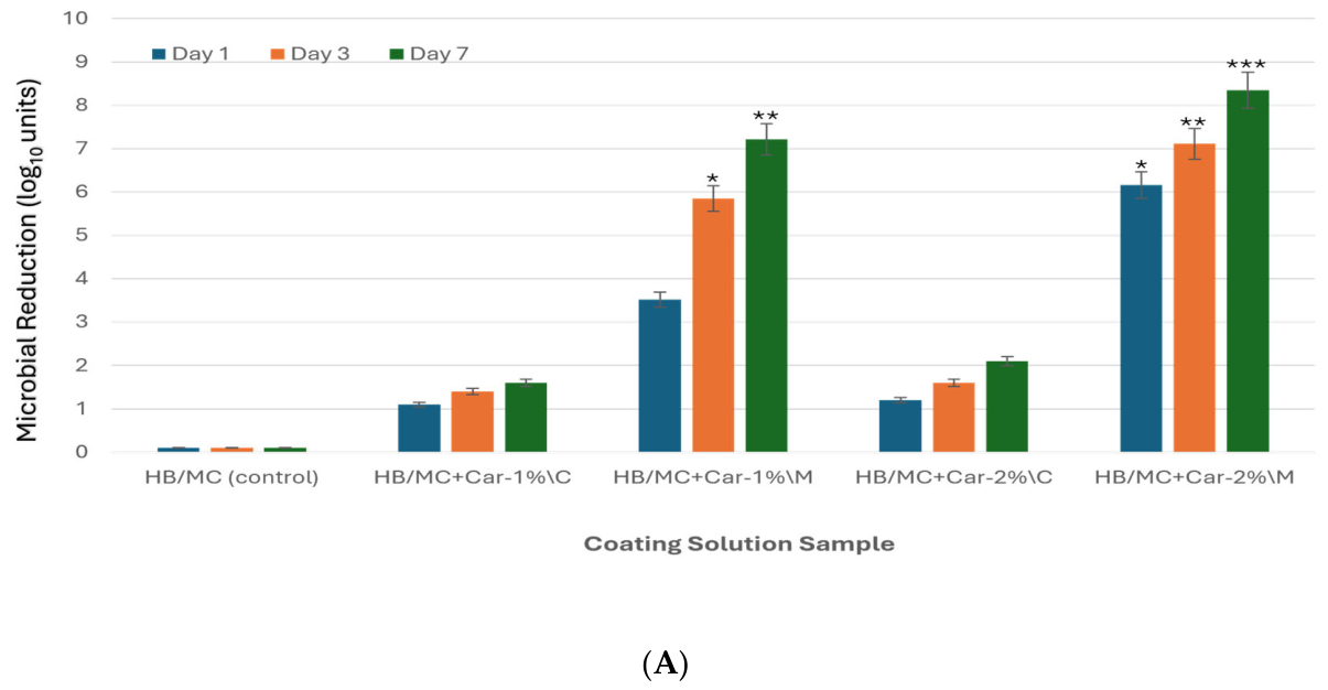
The direct-coating method yielded the most pronounced antimicrobial effects. Micro-emulsified coatings containing 1% carvacrol reduced Salmonella populations by 3.52 log10 units on Day 1, with the reduction increasing to 7.21 log10 units by Day 7 relative to the corresponding untreated control. At 2% carvacrol, the micro-emulsified coating achieved a reduction of 8.35 log10 units by Day 7. In contrast, the coarse-emulsified coatings were less effective, achieving reductions of only 1.60 and 2.10 log10 units at carvacrol concentrations of 1% and 2%, respectively, by Day 7 (Figure 1A).
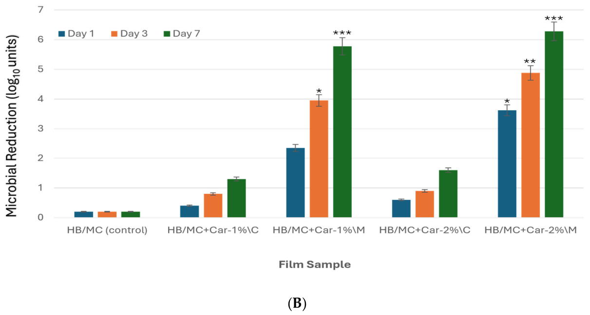
In the headspace release setup, M-films outperformed their coarse-emulsion counterparts, though overall reductions were lower than those observed with direct coating. At 2% carvacrol, M-films achieved reductions of 3.62 log10 units on Day 1 and 6.28 log10 units by Day 7. C-films under the same conditions showed limited efficacy, with a maximum reduction of only 1.6 log10 units by Day 7 (Figure 1B).
Under the tested application conditions (1 mL coating solution per egg versus approximately 13.1 cm2 film per egg in the headspace system), direct coating resulted in greater bacterial reductions than headspace exposure; however, these outcomes reflect antimicrobial performance under the specified application formats rather than a dose-normalized comparison.
M-films showed superior antimicrobial activity, which could be attributed to the homogeneous distribution of carvacrol within the hemicellulose matrix. The dispersion of the active compound was significantly enhanced by micro-emulsification, increasing its availability for interaction with microbial cells [43,44]. This mechanism is supported by previous investigations, demonstrating that micro-emulsified systems enable controlled release and prolonged antimicrobial action [44,45].
It has been established [13,46,47] that the antibacterial properties of carvacrol depend on its hydrophobicity, which enables it to penetrate bacterial cell walls. Research indicates that carvacrol can traverse the lipid layers of bacterial cell walls, disrupt cytoplasmic membrane integrity, inhibit the activity of specific enzymes, and affect cellular functions. The hydroxyl group in carvacrol, along with its delocalized electrons, plays a crucial role in its antimicrobial properties. Carvacrol functions as a transmembrane transporter for monovalent cations by substituting its hydroxyl proton with another ion. The carvacrol-ion complex traverses the cell membrane into the cytoplasm, where it releases its proton into the intracellular environment [48]. Carvacrol disrupts and disorganizes the plasma membrane’s molecular architecture, inducing proton exchange that lowers intracellular pH. These actions lead to structural damage to proteins, DNA, and membranes, ultimately causing cell death [45,49,50]. Similar effects have been observed in fungal pathogens, further supporting carvacrol’s broad-spectrum antimicrobial function [51]. Beyond direct chemical activity, based on our previous characterization, the compact microstructure of micro-emulsified (M) films markedly enhances their antimicrobial performance. SEM observations reported in our previous study demonstrated that the micro-emulsified films formed a dense, cohesive network that has previously been reported to reduce oxygen and moisture permeation, thereby stabilizing the embedded antimicrobial compound [29,30,52]. Recent research [23,31,45,53] has demonstrated that uniform dispersion of bioactive agents within polysaccharide–protein matrices significantly improve barrier integrity and mechanical strength, thereby leading to greater microbial inactivation. In line with this, earlier studies on allyl isothiocyanate-loaded biopolymer films reported [23,43,44,45] microbial reductions exceeding 4–5 log10 units under similar conditions. Carvacrol-loaded micro-emulsified formulations achieved reductions of up to 8.35 log10 units under direct-coating conditions and 6.28 log10 units under headspace-release conditions by Day 7, while offering a more moderate sensory profile, underscoring their suitability for active food packaging applications. Although antimicrobial activity was maintained throughout the 7-day evaluation period, the persistence of carvacrol beyond this period was not investigated. Based on our previous characterization of these formulations [29,30], one possible explanation for the greater microbial reduction observed on Day 7 is the gradual release of carvacrol from the hemicellulose/methylcellulose matrix, which may have maintained inhibitory concentrations during storage. Additionally, prolonged exposure of bacterial cells to carvacrol throughout the storage period may have resulted in cumulative antimicrobial effects, thereby contributing to the greater reduction observed at Day 7 compared with Days 1 and 3. However, because carvacrol release kinetics, residual carvacrol concentrations, and migration behavior were not measured in the present study, these proposed explanations remain speculative and require further experimental validation.
High-pressure homogenization has been shown to reduce emulsion droplet sizes from approximately 12 µm to below 3 µm, which correlates with 20–30% lower oxygen and water vapor transmission rates and greater antimicrobial activity against multiple pathogens in multi-strain assays [29]. Moreover, controlled-release evaluations indicated that micro-emulsified films could sustain carvacrol release for 48–72 h, maintaining over 90% antimicrobial efficacy against Salmonella under ambient storage conditions [54]. In the present study, sustained antimicrobial activity was observed over 3–7 days under refrigerated conditions; however, carvacrol release was not directly measured. This behavior suggests strong potential to enhance short-term storage stability, improve microbial safety, and reduce spoilage risks for coated or packaged produce during cold storage, transportation, and retail display. In summary, both application methods demonstrated antimicrobial potential. Each method offers distinct advantages and limitations. Direct egg surface coating provides a simple and effective operating method, but some coating constituents may migrate to the egg surface, warranting further evaluation. For the film/headspace method, films were not in direct contact with eggs, so there is less concern regarding direct material transfer to the eggshell. For real-world applications, emulsion solutions can be directly sprayed onto egg carton surfaces to form antimicrobial films in situ, thereby eliminating the intermediate processing step from solution to film to carton. Our results also showed that micro-emulsified carvacrol solutions consistently achieved greater reductions in bacterial counts. These results reinforce micro-emulsification as a highly effective strategy for plant-based packaging materials with bioactive compounds. Future studies will investigate additional natural antimicrobials and optimize emulsification conditions to expand pathogen coverage and support commercial-scale implementation.

3.2. Biodegradability Evaluation of Hemicellulose-Based Films

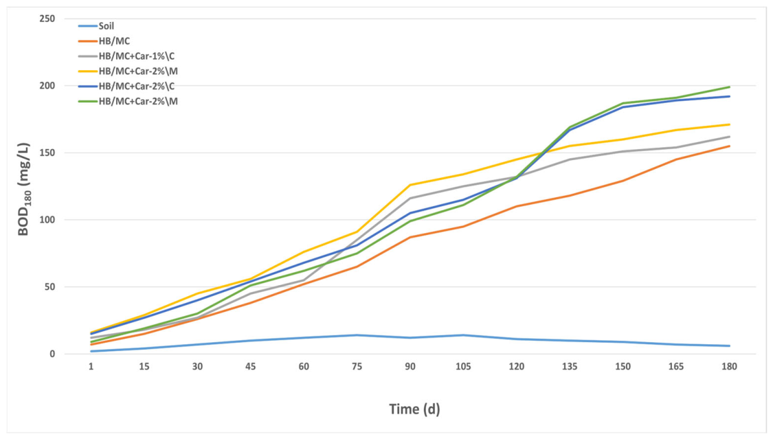
The biodegradability of hemicellulose-based films was assessed over an 180-day incubation period; however, all formulations reached the biodegradation plateau (Dt ≈ 100%) by Day 153, and therefore Day 153 was used as the reporting endpoint (Figure 2, Table 2).
Biodegradation percentages were calculated according to ISO 17556. Values exceeding the theoretical maximum were normalized to 100%, as biodegradation cannot exceed complete mineralization of the test material.
As shown in Table 2, during the 153-day aerobic incubation period, all formulations exhibited BOD values exceeding their corresponding TOD values, resulting in BOD/TOD ratios greater than 1.0 and confirming complete biodegradation under the test conditions. BOD values higher than TOD have been reported in long-term soil respirometric assays and can arise from stimulation of microbial growth, co-metabolism of native soil organic matter (priming effects), and microbial biomass formation, which collectively increase measured oxygen consumption beyond the theoretical demand calculated solely from the test material. Similar observations have been discussed in the context of soil biodegradation standards, including ISO-based respirometric methods [55].
Among the samples, HB/MC+Car-2%\M exhibited the highest BOD value (187.2 mg/L), followed closely by HB/MC+Car-2%\C (184.4 mg/L), suggesting elevated microbial activity and oxygen utilization at higher carvacrol concentrations. Interestingly, despite identical TOD values (181.5 mg/L), the micro-emulsified film (M-film) showed slightly higher oxygen demand, suggesting enhanced microbial accessibility due to its compact, cohesive structure. The HB/MC+Car-1%\M sample also exhibited strong biodegradability, with a BOD of 160.2 mg/L and a TOD of 147.1 mg/L. This formulation benefited from micro-emulsification, which likely improved the dispersion of bioactive compounds and facilitated microbial colonization. In contrast, the control film (HB/MC) showed the lowest BOD value (129.1 mg/L), while still achieving complete biodegradation, confirming the inherent degradability of the hemicellulose matrix. The lower BOD observed for the control relative to carvacrol-containing films likely reflects differences in soil microbial responses and co-metabolic activity, as antimicrobial efficacy against target pathogens does not directly translate to suppressed respiration in complex soil microbial communities.
Across all samples, BOD values were comparable to or slightly exceeded the corresponding TOD values, indicating extensive microbial utilization of the hemicellulose-based formulations under aerobic soil conditions. According to the principles of ISO 17556, biodegradation cannot exceed the theoretical oxygen demand (TOD) of the test material. Therefore, biodegradation values exceeding 100% were normalized to 100% for reporting purposes, as complete mineralization represents the theoretical maximum. Slightly elevated BOD values may reflect microbial priming effects, background soil organic matter mineralization, or normal experimental variability rather than biodegradation of the test material alone. The observed variations in oxygen demand highlight the influence of film composition and emulsification technique on biodegradation dynamics. In addition to chemical composition, physicochemical characteristics such as film thickness, microstructure, density, and structural stability may also influence biodegradation by affecting moisture penetration, oxygen diffusion, microbial colonization, and enzymatic degradation. Therefore, the biodegradation behavior observed in the present study should not be attributed solely to the presence of carvacrol but rather to the combined effects of material composition and film structure. Although these physicochemical properties were not systematically investigated in this study, previous studies have shown that they can substantially influence degradation kinetics and should be evaluated in future work. While increased carvacrol content appears to enhance microbial activity, the structural characteristics imparted by micro-emulsification may further support oxygen transfer and enzymatic breakdown. Our findings underscore the importance of optimizing film formulations to balance functional performance with environmental sustainability. Overall, these results demonstrate that all hemicellulose-based formulations are readily biodegradable under aerobic conditions.

3.3. Cytotoxicity Assessment

Ensuring the biological safety of packaging materials is critical, particularly for applications involving direct contact with food surfaces. The biocompatibility of hemicellulose-based coating solutions (Table 1) was evaluated via the MTT assay, which measures mitochondrial activity as an indicator of cell viability.
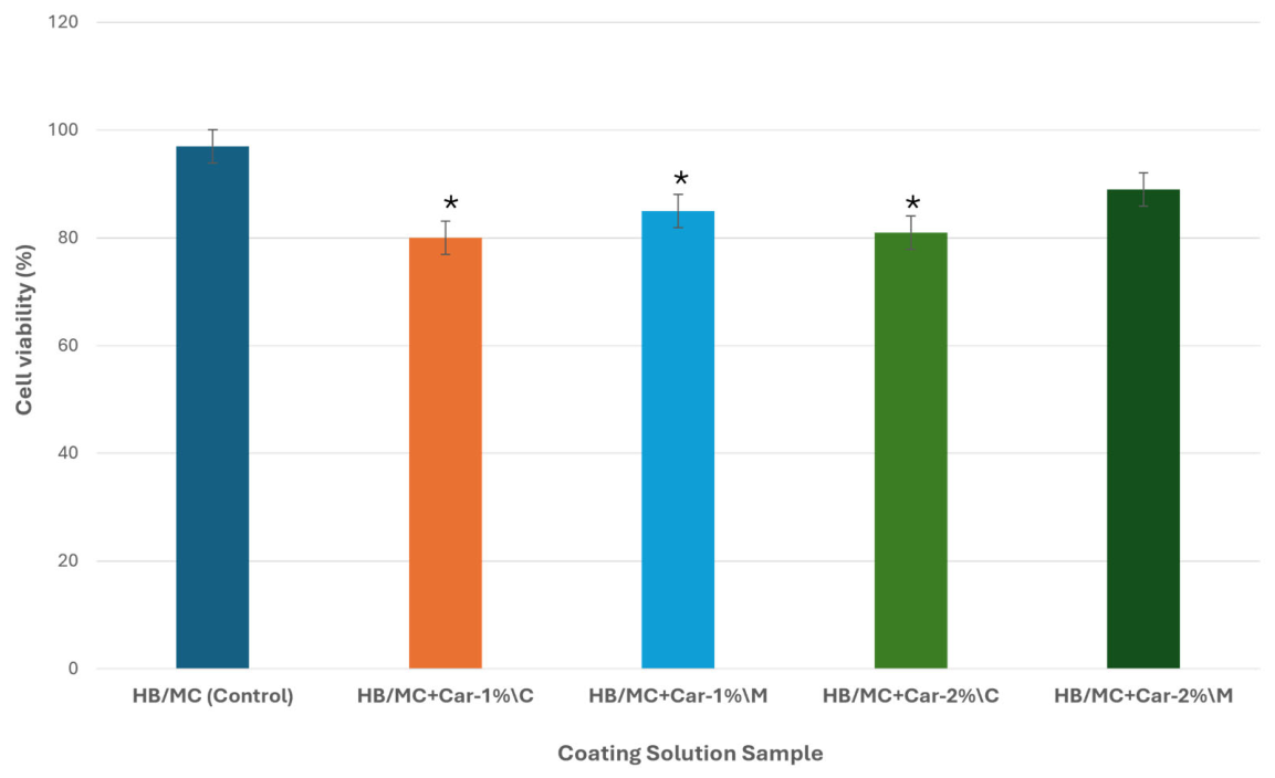
All tested coating solutions exhibited high cell viability (>80%), confirming minimal cytotoxicity across the formulations (Figure 3). The HB/MC control showed the highest cell viability (~97%), indicating excellent baseline cytocompatibility of the hemicellulose/methylcellulose matrix. Carvacrol incorporation resulted in a moderate but acceptable reduction in cell viability, with coarse-emulsion formulations (C) showing slightly lower viability than their corresponding micro-emulsified (M) counterparts. Specifically, HB/MC+Car-1%/C exhibited the lowest viability (~80%), whereas micro-emulsified formulations consistently improved cellular tolerance at the same carvacrol concentration.
Among the carvacrol-containing systems, HB/MC+Car-2%/M demonstrated the highest cell viability (~90%), followed by HB/MC+Car-1%/M (~85%), indicating that micro-emulsification mitigates potential cytotoxic effects associated with higher carvacrol loading. These results suggest that micro-emulsification improves the dispersion of carvacrol within the polymer matrix, reducing localized high concentrations that could otherwise compromise cell integrity. These observations are consistent with prior investigations [56,57].
Notably, all micro-emulsified coatings (M-formulations) maintained higher cell viability than their corresponding coarse-emulsion counterparts, while coarse-emulsion formulations showed slightly reduced yet still acceptable viability levels, consistent with established cytotoxicity benchmarks [58]. The enhanced biocompatibility of micro-emulsified systems is likely attributable to a more homogeneous distribution of carvacrol and controlled exposure at the cell interface, thereby minimizing membrane disruption. Similar trends have been reported for other phenolic-loaded biopolymer systems [59].
Although this assessment focused on coating solutions, the formulations are identical to those used for film preparation; therefore, comparable biological responses are expected for the corresponding films. Collectively, these results confirm that hemicellulose-based coatings, particularly micro-emulsified carvacrol formulations, are non-toxic and suitable for active food-packaging applications that require antimicrobial performance and cytocompatibility.
Taken together, the findings of this study demonstrate the multifunctional potential of carvacrol-infused hemicellulose-based formulations for active food-packaging applications. Antimicrobial evaluation showed that micro-emulsified coatings (M-formulations) consistently outperformed coarse emulsions in reducing Salmonella enterica populations, with direct surface application providing greater efficacy than headspace release. Biodegradability analysis confirmed that all formulations were fully degradable under aerobic conditions, with observed differences in oxygen demand attributable to formulation composition and emulsification strategy. Furthermore, cytotoxicity assessment demonstrated that all coating solutions were non-toxic and biocompatible, with carvacrol-containing formulations maintaining acceptable cell viability while achieving enhanced antimicrobial performance.
Despite these promising results, the intervention was limited to in vitro evaluations under controlled laboratory conditions. Real-world factors such as interactions between packaging materials, food matrix effects, and environmental variability were not addressed. Additionally, the scope was restricted to a single pathogen and one type of bioactive compound. Future research should explore broader microbial targets, assess long-term stability and migration behavior of active compounds, and validate performance under simulated storage and handling conditions. Expanding the range of natural antimicrobials and refining formulation strategies will be essential to optimize functionality while maintaining environmental and biological safety for commercial-scale implementation.

4. Conclusions

This study presents a comprehensive evaluation of hemicellulose-based formulations enriched with carvacrol, highlighting their potential as multifunctional materials for active food packaging. Through systematic investigation, the micro-emulsified coating solutions demonstrated superior antimicrobial efficacy against Salmonella enterica, achieving reductions that meet industrial benchmarks. The enhanced performance of these formulations is attributed to the uniform dispersion of carvacrol and the structural integrity imparted by micro-emulsification. Biodegradability assessments confirmed that all tested samples were fully degradable under aerobic conditions, with variations in oxygen demand reflecting compositional influences. Importantly, cytotoxicity analysis showed no adverse effects on cell viability, confirming the biocompatibility of the coating solutions and supporting their safe application in food-contact applications.
Collectively, these findings underscore the value of micro-emulsification as a formulation strategy that balances antimicrobial functionality, environmental sustainability, and biological safety and demonstrate that plant by-products can be used as carriers for nano- and microscale bioagents. However, the current investigation was limited to controlled laboratory conditions and a single microbial target. Future research should expand the scope to include broader pathogen profiles, assess long-term material stability, egg quality, and simulate real-world packaging environments. Advancing these formulations toward commercial viability will require interdisciplinary efforts to optimize performance, scalability, and regulatory compliance.

Author Contributions

Conceptualization, S.A.H., Y.L., B.K.S., M.I.S. and T.Z.J.; Methodology, S.A.H., M.I.S. and B.K.S.; Validation, Y.L., P.X.Q. and B.K.S.; Formal analysis, P.X.Q., M.P.Y. and T.Z.J.; Investigation, S.A.H.; Resources, Y.L., M.P.Y., B.K.S. and T.Z.J.; Data curation, M.I.S. and P.X.Q.; Writing—original draft, S.A.H.; Writing—review and editing, Y.L., P.X.Q., M.P.Y., B.K.S., M.I.S. and T.Z.J.; Visualization, M.I.S., P.X.Q. and S.A.H.; Supervision, T.Z.J.; Funding acquisition, T.Z.J. All authors have read and agreed to the published version of the manuscript.

Funding

The U.S. Department of Agriculture-Agriculture Research Services provided funding for this research.

Data Availability Statement

Data is contained within the article.

Acknowledgments

The authors would like to thank Anita Parameswaran, Amy Ream, and Stefanie Simon for providing excellent technical support.

Conflicts of Interest

The authors declare no conflicts of interest. Mention of trade names or commercial products in this publication is solely for the purpose of providing specific information and does not imply recommendation or endorsement by the U.S. Department of Agriculture (USDA). The USDA is an equal opportunity provider and employer.

References

  1. Batz, M.B.; Richardson, L.C.; Bazaco, M.C.; Parker, C.C.; Chirtel, S.J.; Cole, D.; Golden, N.J.; Griffin, P.M.; Gu, W.; Schmitt, S.K. Recency-weighted statistical modeling approach to attribute illnesses caused by 4 pathogens to food sources using outbreak data, United States. Emerg. Infect. Dis. 2021, 27, 214. [Google Scholar] [CrossRef] [Scilit] [PubMed]
  2. Carstens, C.K.; Salazar, J.K.; Darkoh, C. Multistate outbreaks of foodborne illness in the United States associated with fresh produce from 2010 to 2017. Front. Microbiol. 2019, 10, 2667. [Google Scholar] [CrossRef] [Scilit] [PubMed]
  3. Chanamé Pinedo, L.; Mughini-Gras, L.; Franz, E.; Hald, T.; Pires, S.M. Sources and trends of human salmonellosis in Europe, 2015–2019: An analysis of outbreak data. Int. J. Food Microbiol. 2022, 379, 109850. [Google Scholar] [CrossRef] [Scilit] [PubMed]
  4. FDA. Outbreak Investigation of Salmonella: Eggs. 2025. Available online: https://www.fda.gov/food/outbreaks-foodborne-illness/outbreak-investigation-salmonella-eggs-june-2025 (accessed on 10 January 2026).
  5. Olanya, O.M.; Hoshide, A.K.; Ukuku, D.O.; Mukhopadhyay, S.; He, Z.; Jin, T.Z.; Sharma, B.K.; Kohli, K.; Ijabadeniyi, O.A. Food Loss Reduction: The Pathway Towards Sustainability. In Antimicrobial Strategies in the Food System: Updates, Opportunities, Challenges; Li, D., Ed.; Springer Nature: Cham, Switzerland, 2025; pp. 43–78. [Google Scholar]
  6. Morassi, L.L.P.; Bernardi, A.O.; Amaral, A.L.P.M.; Chaves, R.D.; Santos, J.L.P.; Copetti, M.V.; Sant’Ana, A.S. Fungi in cake production chain: Occurrence and evaluation of growth potential in different cake formulations during storage. Food Res. Int. 2018, 106, 141–148. [Google Scholar] [CrossRef] [Scilit] [PubMed]
  7. Yang, X.; Scharff, R. Foodborne illnesses from leafy greens in the United States: Attribution, burden, and cost. J. Food Prot. 2024, 87, 100275. [Google Scholar] [CrossRef] [Scilit] [PubMed]
  8. Mohanty, A.K.; Vivekanandhan, S.; Pin, J.-M.; Misra, M. Composites from renewable and sustainable resources: Challenges and innovations. Science 2018, 362, 536–542. [Google Scholar] [CrossRef] [Scilit] [PubMed]
  9. Baghaei, B.; Skrifvars, M. All-cellulose composites: A review of recent studies on structure, properties and applications. Molecules 2020, 25, 2836. [Google Scholar] [CrossRef] [Scilit] [PubMed]
  10. Hansen, N.M.L.; Plackett, D. Sustainable films and coatings from hemicelluloses: A review. Biomacromolecules 2008, 9, 1493–1505. [Google Scholar] [CrossRef] [Scilit] [PubMed]
  11. He, Y.; Liu, Y.; Zhang, M. Hemicellulose and unlocking potential for sustainable applications in biomedical, packaging, and material sciences: A narrative review. Int. J. Biol. Macromol. 2024, 280, 135657. [Google Scholar] [CrossRef] [Scilit] [PubMed]
  12. Liu, G.; Shi, K.; Sun, H. Research progress in hemicellulose-based nanocomposite film as food packaging. Polymers 2023, 15, 979. [Google Scholar] [CrossRef] [Scilit] [PubMed]
  13. Burt, S. Essential oils: Their antibacterial properties and potential applications in foods—A review. Int. J. Food Microbiol. 2004, 94, 223–253. [Google Scholar] [CrossRef] [Scilit] [PubMed]
  14. Motelica, L.; Ficai, D.; Ficai, A.; Oprea, O.C.; Kaya, D.A.; Andronescu, E. Biodegradable antimicrobial food packaging: Trends and perspectives. Foods 2020, 9, 1438. [Google Scholar] [CrossRef] [Scilit] [PubMed]
  15. Rao, J.; Lv, Z.; Chen, G.; Peng, F. Hemicellulose: Structure, chemical modification, and application. Prog. Polym. Sci. 2023, 140, 101675. [Google Scholar] [CrossRef] [Scilit]
  16. Hajibonabi, A.; Yekani, M.; Sharifi, S.; Nahad, J.S.; Dizaj, S.M.; Memar, M.Y. Antimicrobial activity of nanoformulations of carvacrol and thymol: New trend and applications. OpenNano 2023, 13, 100170. [Google Scholar] [CrossRef] [Scilit]
  17. Krepker, M.; Prinz-Setter, O.; Shemesh, R.; Vaxman, A.; Alperstein, D.; Segal, E. Antimicrobial carvacrol-containing polypropylene films: Composition, structure and function. Polymers 2018, 10, 79. [Google Scholar] [CrossRef] [Scilit] [PubMed]
  18. Persico, P.; Ambrogi, V.; Carfagna, C.; Cerruti, P.; Ferrocino, I.; Mauriello, G. Nanocomposite polymer films containing carvacrol for antimicrobial active packaging. Polym. Eng. Sci. 2009, 49, 1447–1455. [Google Scholar] [CrossRef] [Scilit]
  19. Sun, X.; Cameron, R.G.; Plotto, A.; Zhong, T.; Ference, C.M.; Bai, J. The effect of controlled-release carvacrol on safety and quality of blueberries stored in perforated packaging. Foods 2021, 10, 1487. [Google Scholar] [CrossRef] [Scilit] [PubMed]
  20. Hyldgaard, M.; Mygind, T.; Meyer, R.L. Essential oils in food preservation: Mode of action, synergies, and interactions with food matrix components. Front. Microbiol. 2012, 3, 12. [Google Scholar] [CrossRef] [Scilit] [PubMed]
  21. Imran, M.; Aslam, M.; Alsagaby, S.A.; Saeed, F.; Ahmad, I.; Afzaal, M.; Arshad, M.U.; Abdelgawad, M.A.; El-Ghorab, A.H.; Khames, A. Therapeutic application of carvacrol: A comprehensive review. Food Sci. Nutr. 2022, 10, 3544–3561. [Google Scholar] [CrossRef] [Scilit] [PubMed]
  22. Nostro, A.; Papalia, T. Antimicrobial activity of carvacrol: Current progress and future prospectives. Recent Pat. Anti-Infect. Drug Discov. 2012, 7, 28–35. [Google Scholar] [CrossRef] [Scilit] [PubMed]
  23. Krümmel, A.; Pagno, C.H.; Malheiros, P.S. Active films of cassava starch incorporated with carvacrol nanocapsules. Foods 2024, 13, 1141. [Google Scholar] [CrossRef] [Scilit] [PubMed]
  24. Salabat, A.; Mirhoseini, F. Polymer-based nanocomposites fabricated by microemulsion method. Polym. Compos. 2022, 43, 1282–1294. [Google Scholar] [CrossRef] [Scilit]
  25. Sharifi-Rad, M.; Varoni, E.M.; Iriti, M.; Martorell, M.; Setzer, W.N.; del Mar Contreras, M.; Salehi, B.; Soltani-Nejad, A.; Rajabi, S.; Tajbakhsh, M. Carvacrol and human health: A comprehensive review. Phytother. Res. 2018, 32, 1675–1687. [Google Scholar] [CrossRef] [Scilit] [PubMed]
  26. Mitra, D. Microemulsion and its application: An inside story. Mater. Today Proc. 2023, 83, 75–82. [Google Scholar] [CrossRef] [Scilit]
  27. Mushtaq, A.; Mohd Wani, S.; Malik, A.R.; Gull, A.; Ramniwas, S.; Ahmad Nayik, G.; Ercisli, S.; Alina Marc, R.; Ullah, R.; Bari, A. Recent insights into Nanoemulsions: Their preparation, properties and applications. Food Chem. X 2023, 18, 100684. [Google Scholar] [CrossRef] [Scilit] [PubMed]
  28. Ramos, M.; Jiménez, A.; Garrigós, M.C. Ch. 26. Carvacrol-Based Films: Usage and Potential in Antimicrobial Packaging, Antimicrobial Food Packaging. In Antimicrobial Food Packaging; Barros-Velázquez, J., Ed.; Elsevier Ltd., Academic Press: Oxford, UK, 2016; pp. 329–338. [Google Scholar]
  29. Hussain, S.A.; Sharma, B.K.; Qi, P.X.; Yadav, M.P.; Jin, T.Z. Antimicrobial and physicochemical properties of hemicellulose-based films incorporating carvacrol. Polymers 2025, 17, 2073. [Google Scholar] [CrossRef] [Scilit] [PubMed]
  30. Hussain, S.A.; Yadav, M.P.; Sharma, B.K.; Qi, P.X.; Jin, T.Z. Biodegradable food packaging films using a combination of hemicellulose and cellulose derivatives. Polymers 2024, 16, 3171. [Google Scholar] [CrossRef] [Scilit] [PubMed]
  31. Jin, T.Z.; Yadav, M.P.; Qi, P.X. Antimicrobial and physiochemical properties of films and coatings prepared from bio-fiber gum and whey protein isolate conjugates. Food Control 2023, 148, 109666. [Google Scholar] [CrossRef] [Scilit]
  32. Abe, M.M.; Branciforti, M.C.; Brienzo, M. Biodegradation of hemicellulose-cellulose-starch-based bioplastics and microbial polyesters. Recycling 2021, 6, 22. [Google Scholar] [CrossRef] [Scilit]
  33. Zhao, Y.; Sun, H.; Yang, B.; Weng, Y. Hemicellulose-based film: Potential green films for food packaging. Polymers 2020, 12, 1775. [Google Scholar] [CrossRef] [Scilit] [PubMed]
  34. De Luca, A.; Ruggiero, R.; Cordaro, A.; Marrelli, B.; Raimondi, L.; Costa, V.; Bellavia, D.; Aiello, E.; Pavarini, M.; Piccininni, A.; et al. Towards accurate biocompatibility: Rethinking cytotoxicity evaluation for biodegradable magnesium alloys in biomedical applications. J. Funct. Biomater. 2024, 15, 382. [Google Scholar] [CrossRef] [Scilit] [PubMed]
  35. Murata, K.; Washio, A.; Morotomi, T.; Rojasawasthien, T.; Kokabu, S.; Kitamura, C. Physicochemical properties, cytocompatibility, and biocompatibility of a bioactive glass based retrograde filling material. Nanomaterials 2021, 11, 1828. [Google Scholar] [CrossRef] [Scilit] [PubMed]
  36. Vrchovecká, K.; Pávková-Goldbergová, M.; Engqvist, H.; Pujari-Palmer, M. Cytocompatibility and bioactive ion release profiles of phosphoserine bone adhesive: Bridge from in vitro to in vivo. Biomedicines 2022, 10, 736. [Google Scholar] [CrossRef] [Scilit] [PubMed]
  37. Ghaani, M.; Soltanzadeh, M.; Carullo, D.; Farris, S. Development of a biopolymer-based anti-fog coating with sealing properties for applications in the food packaging sector. Polymers 2024, 16, 1745. [Google Scholar] [CrossRef] [Scilit] [PubMed]
  38. Perera, K.Y.; Jaiswal, A.K.; Jaiswal, S. Biopolymer-based sustainable food packaging materials: Challenges, solutions, and applications. Foods 2023, 12, 2422. [Google Scholar] [CrossRef] [Scilit] [PubMed]
  39. Periyasamy, T.; Asrafali, S.P.; Lee, J. Recent advances in functional biopolymer films with antimicrobial and antioxidant properties for enhanced food packaging. Polymers 2025, 17, 1257. [Google Scholar] [CrossRef] [Scilit] [PubMed]
  40. Liu, X.; Sun, X.; Du, H.; Li, Y.; Wen, Y.; Zhu, Z. A transparent p-coumaric acid-grafted-chitosan coating with antimicrobial, antioxidant and antifogging properties for fruit packaging applications. Carbohydr. Polym. 2024, 339, 122238. [Google Scholar] [CrossRef] [Scilit] [PubMed]
  41. Hou, T.; Ma, S.; Wang, F.; Wang, L. A comprehensive review of intelligent controlled release antimicrobial packaging in food preservation. Food Sci. Biotechnol. 2023, 32, 1459–1478. [Google Scholar] [CrossRef] [Scilit] [PubMed]
  42. Mkhari, T.; Adeyemi, J.O.; Fawole, O.A. Recent advances in the fabrication of intelligent packaging for food preservation: A review. Processes 2025, 13, 539. [Google Scholar] [CrossRef] [Scilit]
  43. Guo, M.; Jin, T.Z.; Yadav, M.P.; Yang, R. Antimicrobial property and microstructure of micro-emulsion edible composite films against Listeria. Int. J. Food Microbiol. 2015, 208, 58–64. [Google Scholar] [CrossRef] [Scilit] [PubMed]
  44. Guo, M.; Yadav, M.P.; Jin, T.Z. Antimicrobial edible coatings and films from micro-emulsions and their food applications. Int. J. Food Microbiol. 2017, 263, 9–16. [Google Scholar] [CrossRef] [Scilit]
  45. Jin, T.Z.; Fan, X.; Mukhopadhyay, S. Antimicrobial coating with organic acids and essential oil for the enhancement of safety and shelf life of grape tomatoes. Int. J. Food Microbiol. 2022, 378, 109827. [Google Scholar] [CrossRef] [Scilit] [PubMed]
  46. Lambert, R.J.W.; Skandamis, P.N.; Coote, P.J.; Nychas, G.J.E. A study of the minimum inhibitory concentration and mode of action of oregano essential oil, thymol and carvacrol. J. Appl. Microbiol. 2001, 91, 453–462. [Google Scholar] [CrossRef] [Scilit] [PubMed]
  47. Memar, M.Y.; Raei, P.; Alizadeh, N.; Akbari Aghdam, M.; Kafil, H.S. Carvacrol and thymol: Strong antimicrobial agents against resistant isolates. Rev. Res. Med. Microbiol. 2017, 28, 63–68. [Google Scholar] [CrossRef] [Scilit]
  48. Xu, J.; Zhou, F.; Ji, B.P.; Pei, R.S.; Xu, N. The antibacterial mechanism of carvacrol and thymol against Escherichia coli. Lett. Appl. Microbiol. 2008, 47, 174–179. [Google Scholar] [CrossRef] [Scilit] [PubMed]
  49. Ben Arfa, A.; Combes, S.; Preziosi-Belloy, L.; Gontard, N.; Chalier, P. Antimicrobial activity of carvacrol related to its chemical structure. Lett. Appl. Microbiol. 2006, 43, 149–154. [Google Scholar] [CrossRef] [Scilit]
  50. Kachur, K.; Suntres, Z. The antibacterial properties of phenolic isomers, carvacrol and thymol. Crit. Rev. Food Sci. Nutr. 2020, 60, 3042–3053. [Google Scholar] [CrossRef] [Scilit] [PubMed]
  51. Vylkova, S. Environmental pH modulation by pathogenic fungi as a strategy to conquer the host. PLoS Path. 2017, 13, e1006149. [Google Scholar] [CrossRef] [Scilit] [PubMed]
  52. Hussain, S.A.; Qi, P.X.; Sharma, B.K.; Yadav, M.P.; Mainali, K.; Jin, T.Z. Valorization of corn bran-derived carbohydrate polymers for developing biodegradable packaging films. J. Polym. Environ. 2025, 33, 2552–2566. [Google Scholar] [CrossRef] [Scilit]
  53. Jin, T.Z.; Zhang, H. Biodegradable polylactic acid polymer with nisin for use in antimicrobial food packaging. J. Food Sci. 2008, 73, M127–M134. [Google Scholar] [CrossRef] [Scilit] [PubMed]
  54. de Castro, D.O.; Tabary, N.; Martel, B.; Gandini, A.; Belgacem, N.; Bras, J. Controlled release of carvacrol and curcumin: Bio-based food packaging by synergism action of TEMPO-oxidized cellulose nanocrystals and cyclodextrin. Cellulose 2018, 25, 1249–1263. [Google Scholar] [CrossRef] [Scilit]
  55. Pires, J.R.; Souza, V.G.; Fuciños, P.; Pastrana, L.; Fernando, A.L. Methodologies to assess the biodegradability of bio-based polymers—Current knowledge and existing gaps. Polymers 2022, 14, 1359. [Google Scholar] [CrossRef] [Scilit] [PubMed]
  56. Sun, C.; Zeng, X.; Zheng, S.; Wang, Y.; Li, Z.; Zhang, H.; Nie, L.; Zhang, Y.; Zhao, Y.; Yang, X. Bio-adhesive catechol-modified chitosan wound healing hydrogel dressings through glow discharge plasma technique. Chem. Eng. J. 2022, 427, 130843. [Google Scholar] [CrossRef] [Scilit]
  57. Zhou, C.; Ao, H.-Y.; Han, X.; Jiang, W.-W.; Yang, Z.-F.; Ma, L.; Deng, X.-Y.; Wan, Y.-Z. Engineering a novel antibacterial agent with multifunction: Protocatechuic acid-grafted-quaternized chitosan. Carbohydr. Polym. 2021, 258, 117683. [Google Scholar] [CrossRef] [Scilit] [PubMed]
  58. Präbst, K.; Engelhardt, H.; Ringgeler, S.; Hübner, H. Basic Colorimetric Proliferation Assays: MTT, WST, and Resazurin. In Cell Viability Assays: Methods and Protocols; Gilbert, D.F., Friedrich, O., Eds.; Springer: New York, NY, USA, 2017; pp. 1–17. [Google Scholar]
  59. Saadi, S.; Nacer, N.E.; Chenaker, H.; Ariffin, A.A.; Ghazali, H.M.; Saari, N.; Mohammed, A.S.; Anwar, F.; Hamid, A.A. A review on trends in microencapsulation of bioactive compounds: Coating materials, design, and applications. Eur. Food Res. Technol. 2023, 249, 3123–3139. [Google Scholar] [CrossRef] [Scilit]
Figure 1. (A) Antimicrobial efficacy of carvacrol-infused HB/MC coating formulations applied directly to eggshell surfaces against a two-strain cocktail of Salmonella enterica during 7 days of storage at 7 °C. Microbial reductions were calculated relative to untreated control at each sampling time. Error bars represent the standard deviation. Asterisks indicate statistically significant differences among treatments: * p < 0.05; ** p < 0.01; *** p < 0.001. (B) Antimicrobial efficacy of carvacrol-infused hemicellulose films sample (headspace release) against a two-strain cocktail of Salmonella enterica over 7 days at 7 °C. Microbial reductions were calculated relative to the corresponding untreated control at each sampling time. Asterisks indicate statistically significant differences among treatments: * p < 0.05; ** p < 0.01; *** p < 0.001.
Coatings 16 00911 g001aCoatings 16 00911 g001b
Figure 2. Biochemical oxygen demand (BOD) profiles of hemicellulose/methylcellulose-based film formulations during aerobic soil incubation for 180 days at 25 °C. BOD was monitored using the OxiTop® respirometric system in accordance with the principles of ISO 17556:2019.
Coatings 16 00911 g002
Figure 3. Assessment of NIH 3T3 cell viability following exposure to hemicellulose-derived coating solutions, * p < 0.05 represents statistically significant differences between treatments.
Coatings 16 00911 g003
Table 1. Composition of the hemicellulose/methylcellulose-based formulations used for coating-solution preparation and film fabrication. Data were adapted from Hussain et al. [29]. All components are expressed as % (w/w), except carvacrol, which is expressed as % (v/v).
Sample CodeHBMCPectin (HMP)Pea Protein Isolate (PPI)Glycerol (G)Carvacrol (%, v/v)
HB/MC (control)88.4100.10.51.00.0
HB/MC+Car-1%\C88.4100.10.51.01.0
HB/MC+Car-1%\M88.4100.10.51.01.0
HB/MC+Car-2%\C88.4100.10.51.02.0
HB/MC+Car-2%\M88.4100.10.51.02.0
Abbreviations: C, coarse-emulsified formulation; M, micro-emulsified formulation.
Table 2. Biodegradability performance of hemicellulose-based film formulations based on biochemical oxygen demand (BOD), theoretical oxygen demand (TOD), and calculated biodegradation degree (Dt%). All samples were tested under controlled aerobic conditions for 153 days at 25 °C.
Sample AttributesHB/MC
(Control)
HB/MC+Car-1%\CHB/MC+Car-1%\MHB/MC+Car-2%\CHB/MC+Car-2%\M
BOD153 (mg/L)129.1151.5160.2184.4187.2
TOD (mg/L)124.5147.1147.1181.5181.5
Dt (%)100100100100100
Disclaimer/Publisher’s Note: The statements, opinions and data contained in all publications are solely those of the individual author(s) and contributor(s) and not of MDPI and/or the editor(s). MDPI and/or the editor(s) disclaim responsibility for any injury to people or property resulting from any ideas, methods, instructions or products referred to in the content.