Article

Spin-Coated PCL/PVP Biofilms with Amniotic Membrane Matrix Enhance Proliferation and Migration of BM-MSC


by
Juan de Dios Mendez Quezada
1,
Antonio Rojas Murillo
1,
Mario Simental-Mendía
2,
Rodolfo Franco Marquez
3,
Paulina Delgado Gonzalez
1,
Jose F. Islas
1,
Jorge Lara Arias
2,
Celia N. Sanchez Dominguez
2,
Hector Leija Gutierrez
4,* and
Elsa N. Garza Treviño
1,*
1
Department of Biochemistry and Molecular Medicine, Universidad Autonoma de Nuevo Leon, Monterrey 64460, Mexico
2
Orthopedic Trauma Service, University Hospital Dr. José Eleuterio González, School of Medicine, Universidad Autonoma de Nuevo Leon, Monterrey 64460, Mexico
3
Department of Anatomic Pathology and Cytopathology, University Hospital Dr. José Eleuterio González, School of Medicine, Universidad Autonoma de Nuevo Leon, Monterrey 64460, Mexico
4
Center for Research in Physical and Matematical Sciences, Universidad Autonoma de Nuevo Leon, San Nicolas de los Garza 66455, Mexico
*
Authors to whom correspondence should be addressed.
Coatings 2026, 16(6), 719; https://doi.org/10.3390/coatings16060719
Submission received: 5 May 2026 / Revised: 3 June 2026 / Accepted: 10 June 2026 / Published: 16 June 2026

Highlights

What are the main findings?
  • Integration of dHAM into PVP biofilms enhances hydrophobicity.
  • Soluble dHAM extracts stimulate BM-MSC proliferation without cytotoxicity.
  • PCL/PVP biofilms functionalized with dHAM improve cell viability and accelerate wound healing.
What are the implications of the main findings?
  • Functionalization with dHAM preserves biological properties while improving structural stability.
  • dHAM integration provides a strategy for developing biofunctional polymeric biomaterials.
  • PCL/PVP biofilms incorporating dHAM represent promising candidates for tissue regeneration.

Abstract
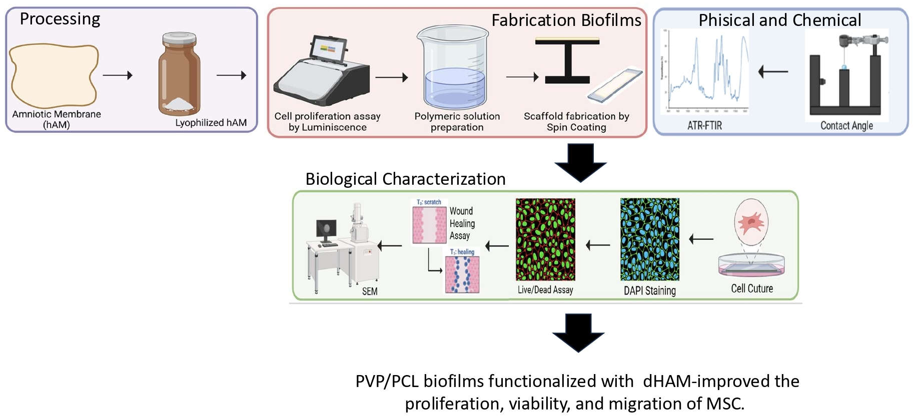
The amniotic membrane is widely recognized in regenerative medicine due to its rich content of extracellular matrix proteins and growth factors that confer anti-inflammatory and pro-regenerative properties. However, its rapid degradation restricts its standalone clinical use. To overcome these limitations, we developed biofilms by incorporating decellularized human amniotic membrane matrix (dHAM) into polycaprolactone (PCL) and polyvinylpyrrolidone (PVP) matrices using spin-coating. Bone marrow-derived mesenchymal stem cells (BM-MSCs) were used to evaluate film biocompatibility through cell viability, proliferation, and wound healing migration assays. Surface characterization was performed using contact angle measurements, Attenuated Total Reflectance-Fourier Transform Infrared (ATR-FTIR) spectroscopy, and scanning electron microscopy. Soluble dHAM extracts (4–6 mg/mL) significantly enhanced BM-MSC proliferation at 48 h compared to controls (p ≤ 0.01 and p ≤ 0.0001). Both PCL-dHAM and PVP-dHAM biofilms exhibited high cell viability (>90%) and improved initial adhesion. Notably, dHAM incorporation significantly increased wound closure rates at 24 h, reaching 98.47% for PCL-dHAM and 93.13% for PVP-dHAM, compared to 76.56% and 64.20% for pure polymers (p = 0.0001). All scaffolds maintained hydrophilic surfaces (<90°), favorable for cell interaction. The integration of dHAM into PCL and PVP by spin-coating produces biofilms biocompatible with enhanced regenerative potential, representing promising candidates for wound healing applications. In conclusion, these coatings support BM-MSC adhesion, proliferation, and migration, while significantly accelerating wound closure, underscoring their value as advanced bioactive coatings for regenerative medicine.

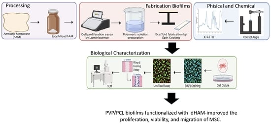
Graphical Abstract

1. Introduction

Regenerative medicine has been a promising therapeutic strategy to overcome the limitations of conventional wound healing treatments, particularly in chronic wounds, extensive tissue damage, and conditions associated with impaired regenerative capacity [1]. Tissue engineering uses cells, biomaterials, biochemical factors, and their combination to produce scaffolds capable of supporting cellular adhesion, proliferation, migration, and tissue remodeling while providing adequate mechanical stability [2,3].
Functionalized coatings for regenerative purposes have been extensively explored through strategies such as embedding biomolecules, nanoparticles, or matrices to enhance bioactivity [4]. However, these approaches often require complex chemical modifications that may compromise biocompatibility or reduce the biological activity of the incorporated molecules. In this context, the amniotic membrane emerges as a compelling alternative, as it constitutes a naturally derived extracellular matrix inherently enriched in growth factors, structural proteins, and bioactive molecules that confer intrinsic anti-inflammatory, antimicrobial, and pro-regenerative properties—without the need for complex chemical modifications [5].
The amniotic membrane is a tissue with high potential in regenerative medicine due to its more than 226 bioactive molecules, which provide anti-inflammatory, antimicrobial, and wound-healing properties, among others [6]. This natural biomaterial is mainly composed of extracellular matrix (ECM) proteins such as collagen, laminin, fibronectin, and elastin, as well as growth factors and other molecules, including EGF, bFGF, VEGF, Alpha-2-macroglobulin (A2M), and hyaluronic acid (HA). These provide a bioactive microenvironment that promotes cell proliferation, migration, and differentiation [7,8].
Its potential as a skin dressing, surgical patch, and tissue engineering scaffold has been extensively investigated, with clinically approved presentations supporting its safety, efficacy, and feasibility as a regenerative biomaterial [7,9]. Additional processing methods, such as decellularization and lyophilization, that aim to overcome limitations commonly associated with biological biomaterials, including immune rejection, microbial contamination, and limited shelf life, have been demonstrated to improve key cellular processes in tissue regeneration over time [10,11,12,13,14,15]. Nevertheless, the amniotic membrane still presents certain drawbacks, such as low mechanical strength and a high degradation rate, which restrict its extended application [6,10].
To address these limitations, recent studies have explored hybrid scaffolds combining amniotic membrane derivatives with other biomaterials such as polymers [16,17,18]. Polycaprolactone (PCL) and polyvinylpyrrolidone (PVP) are particularly attractive for scaffold fabrication. PCL, a non-toxic synthetic polymer, provides mechanical robustness, controlled degradation, and excellent processability [19,20,21]. A major flaw in this polymer is its hydrophobicity, which can hinder cell-material interactions; therefore, its evaluation in combination with bioactive components such as dHAM is relevant. In contrast, PVP is FDA-approved, hydrophilic, biocompatible, and facilitates water absorption, favoring cell adhesion and proliferation [17,22,23,24]. Each polymer was therefore considered individually to explore how its distinct physicochemical properties influence scaffold performance when combined with dHAM through spin coating.
Lotfi et al. (2023) reported improved hydrophilicity, mechanical performance, and stem cell proliferation in electrospun PCL–amniotic membrane scaffolds [25]. Majidnia et al. (2022) showed that incorporation of amniotic membrane powder into PCL/collagen matrices enhanced surface wettability and cell viability (>90%) in retinal epithelial cells [26]. In burn wound models, Kanaujiya et al. (2024) demonstrated that collagen-enriched PVP-based scaffolds improved hydrophilicity, antimicrobial activity, and in vivo wound healing outcomes [27]. Collectively, these findings support the biological relevance of amniotic membrane incorporation into synthetic matrices.
Furthermore, previous studies combining PCL/PVP in an electrospinning system have demonstrated tunable physicochemical properties and cytocompatibility, particularly in drug delivery and regenerative applications [28]. Despite these advances, the most reported systems are based on electrospinning, which, while effective, presents limitations in terms of process complexity, possible residual solvent, and cost [29]. Alternative techniques such as spin coating provide a more accessible and reproducible platform, with advantages including controlled film thickness and uniform surface morphology [30]. Nevertheless, studies exploring the integration of dHAM into polymer matrices using these methods are still scarce.
To evaluate the performance of scaffolds or films, mesenchymal stem cells (MSCs) represent an ideal cellular model given their pivotal role in tissue repair through multipotent differentiation, paracrine signaling, and immunomodulation [31,32,33]. Among MSC sources, bone marrow-derived MSCs (BM-MSCs) are particularly relevant in wound healing contexts due to their well-documented regenerative and immunomodulatory capacity [29,34]. Therefore, understanding the interaction between MSCs and biomaterials is essential for rational scaffold design and improved regenerative outcomes.
The present study aims to develop and characterize spin-coated PCL and PVP scaffolds incorporating dHAM and to evaluate their physicochemical properties, cytocompatibility with BM-MSCs, and functional performance in wound healing assays. We hypothesize that dHAM incorporation will enhance the biological activity of synthetic polymer scaffolds while preserving their mechanical stability, thereby generating hybrid biomaterials with improved regenerative potential.

2. Materials and Methods

2.1. Decellularized Amniotic Membrane Matrix

Human amniotic membranes (hAMs) were obtained from placentas donated by patients undergoing obstetric surgery after written informed consent and approval from the Research Ethics Committee (approval number: BI23-001). The amniotic membrane was separated from the chorion under sterile conditions as previously described by De la Garza-Kalife et al. [35]. Decellularization to obtain decellularized amniotic membrane matrix (dAMM) was performed using a combination of physical (liquid nitrogen freezing) and chemical treatments, following a protocol modified from Villamil-Ballesteros et al. [36]. Briefly, membranes were rinsed three times in cold phosphate-buffered saline (PBS) supplemented with antibiotic–antimycotic, cut into ~1 cm2 fragments, and frozen at −80 °C. A total of 125 g of tissue underwent five freeze–thaw cycles (liquid nitrogen for 5 min, followed by thawing in PBS for 10 min). Samples were then incubated in 0.1% Tween 80 (SIGMA-ALDRICH, USA) for 4 h, treated with 0.1 M NaOH (97%, JALMEX, Guadalajara, México) for 1 h, and subjected to two sequential 1 h treatments with 0.15% peracetic acid (PAA, 15%, Cetik, Cualiacán, México) in 96% ethanol (CTR, Monterrey, México), with intermediate NaOH exposure. The tissue was subsequently rinsed in 70% ethanol (CTR, Monterrey, México) for 1 h and washed three times in PBS. Finally, membranes were frozen, lyophilized for 24 h, and stored at −80 °C until use, as previously described by Rojas-Murillo et al. [37].

2.2. Extracts of dAMM

A vial of previously decellularized and lyophilized amniotic membrane matrix (dAMM), stored at −80 °C and exhibiting particle sizes between 0.3 and 1700 µm, was used as the starting material [37]. Only the fraction with particle sizes <179 µm was selected, sterilized by ethylene oxide gas, and stored at −80 °C until use. For soluble extract preparation dilute 12 mg and 18 mg of dAMM were diluted in 3 mL of serum-free Dulbecco’s Modified Eagle Medium (DMEM, Gibco, USA) under sterile conditions to obtain final concentrations of 4 mg/mL and 6 mg/mL, respectively. Suspensions were incubated at 37 °C for 24 h under continuous agitation. The supernatants were then collected and centrifuged at 4000 rpm for 20 min to separate soluble fractions from insoluble residues. The clarified extracts were subsequently used for cell culture assays.

2.3. Culture Bone Marrow-Derived MSC

A cryopreserved vial of murine bone marrow-derived mesenchymal stem cells (BM-MSCs) previously isolated from BALB/c mice [38] was thawed and expanded under standard culture conditions. Cells were characterized by immunohistochemistry with markers CD105, CD90, and CD34, with the primary monoclonal antibodies anti-CD90, anti-CD105, and anti-CD34 (United States Biologicals, Salem, MA, USA), and PBS was diluted to 1:25, 1:200, and 1:100, respectively, as the International Society for Cellular Therapy specifies. A mouse- and rabbit-specific HPR/DAB (ABC) detection IHC kit (ab64264 Abcam, Burlingame, CA, USA) and Harris hematoxylin as a counterstain were used.

2.4. Cell Viability Assay

To evaluate the effect of soluble factors of dHAM on BM-MSCs proliferation, the CellTiter-Glo® Luminescent Cell Viability Assay (G7572Promega®, Madison, WI, USA) was used. BM-MSCs were seeded in 96-well plates at a density of 2 × 10³ cells/well; 100 µL of DMEM supplemented with 10% FBS, 2.5 µg/mL of amphotericin B, and 100 µg/mL of gentamicin was added to each well. After 24 h incubation, the medium was carefully removed from each well, and 100 µL of the dHAM soluble extracts at concentrations of 4 and 6 mg/mL were added without dilution to the wells as experimental treatments. The indirect cytocompatibility assessment was performed in accordance with ISO 10993-5 guidelines [39]. MSCs cultured in DMEM without FBS were used as the negative control, while MSCs treated with 40% DMSO served as the death control. The plates with dHAM soluble extracts were incubated for 24, 48 and 72 h. CellTiter-Glo reagent (100 µL) was added to each well, agitated for 2 min at 300–500 rpm, and incubated for 10 min at room temperature. Luminescence was quantified using a Cytation 3 plate reader (BioTek, Winooski, VT, USA). Cell death percentage was calculated with the formula: [1 − (treatment mean luminescence/control mean luminescence)] × 100.

2.5. Preparation of PCL and PVP Solutions With or Without dHAM

Polymeric solutions of 9% (w/v) polycaprolactone (PCL; Sigma-Aldrich®, 440744, Steinheim, Germany) were prepared using glacial acetic acid (99%; J.T.Baker, 9508-05, USA) as solvent, while 12.5% (w/v) polyvinylpyrrolidone (PVP; Sigma-Aldrich®, 437190, St. Louis, MO, USA) solutions were prepared in absolute ethanol (>99.5%; CTR01164®, Monterrey, Mexico). Polymers were added gradually to their respective solvents to prevent agglomeration and promote complete solubilization. The solutions were maintained under continuous stirring at 300 rpm at room temperature overnight to ensure complete dissolution.
For dHAM-containing formulations, dHAM was incorporated into the solvent at a concentration of 4 mg/mL (40 mg per 10 mL of solvent), the minimum concentration with demonstrated biological activity as determined in soluble extracts prior to polymer addition. This sequential order was adopted to prevent aggregation, promote homogeneous distribution of dHAM bioactive components, and preserve the biological integrity of the matrix. The polymer–biological mixture was homogenized under continuous magnetic stirring for at least 12 h before biofilm fabrication to ensure uniform dispersion.

2.6. Biofilms With or Without dHAM

Prior to fabrication, the equipment was calibrated, and a 25 × 75 mm glass slide (Corning®, Mexico) was mounted onto the rotating holder. Once a homogeneous mixture was obtained, polymeric films with or without dHAM were fabricated by spin coating using a Spin Coater (L2001A3, Software Ossila®, V4.2.1). Biofilms were generated by depositing a 0.5 mL aliquot of the polymeric solution onto the glass substrate. Coating was performed using different combinations of rotational speed and time to obtain smooth biofilms with uniform thickness, complete surface adhesion, absence of roughness, and no visible defects. The optimized parameters were as follows: PCL (3000 rpm for 60 s), PCL-dHAM (4000 rpm for 90 s), PVP (5000 rpm for 60 s), and PVP-dHAM (6000 rpm for 90 s). The scaffolds were stored in rigid containers for 48 h after fabrication to allow for solvent residue evaporation. They were air-dried inside a chemical extraction hood for approximately 2–4 h at room temperature (23–27 °C), followed by 44–46 h under ambient laboratory conditions prior to sterilization.

2.7. Sterilization of Biofilms

Slides containing the biofilms were sterilized by exposure to ethylene oxide (EtO) gas. As a complementary step, the biofilms were subsequently subjected to ultraviolet (UV) irradiation for 30 min at room temperature inside a biosafety cabinet equipped with a UV function, providing an additional reduction in surface microbial load prior to use in cell culture experiments.

2.8. Determination of Hydrophobicity

Surface hydrophobicity was evaluated by water contact angle measurements using a goniometer (L2004A1, Ossila® V4.2.1). A droplet of ultrapure water (~8 µL) was deposited onto the surface of each sample using a 20 µL chromatography microsyringe (Hamilton®) coupled to the instrument. Images of the droplet were captured using the integrated camera, and the contact angle was determined using the manufacturer’s analysis software (Ossila Contact Angle). Each sample was measured at least three times at different surface locations, and the mean value was calculated to obtain a representative contact angle.

2.9. Characterization by Attenuated Total Reflectance Fourier-Transform Infrared Spectroscopy (ATR-FTIR)

Polymer samples (PCL and PVP) and the biofilms were removed from the glass slides, pulverized, and analyzed by FTIR spectroscopy (PerkinElmer-Frontier®, Thermo Scientific, Ncolet iS50 Software OMNIC 9.15.119). Spectra were recorded in the range of 1000 to 4000 cm−1 to detect the polymers’ characteristic bands and analyze potential variations caused by the solvents, the incorporation of amniotic membrane, or from the EtO and UV light sterilization processes.

2.10. Live/Dead Cell Viability Assay

A central area of approximately 3 cm on a slide of 25 × 75 mm was delimited, and a total of 50,000 BM-MSCs were seeded onto each slice and incubated for 24, 48, and 72 h, as described in the previous section. At the end of each time point, the culture medium (300 µL) was removed and replaced with 200 µL of Live/Dead assay solution (Invitrogen™, L3224, Eugene, OR, USA) prepared by mixing 2.5 µL of Calcein-AM and 2 µL of ethidium homodimer-1 per 5 mL of PBS to assess cell viability. Samples were incubated at room temperature for 15 min and then observed under a fluorescence microscope (Olympus AX70), capturing representative images with a minimum of 6 random fields per sample. Live cells emitted green fluorescence (Calcein AM, L3224A, Life Technologies, Eugene, OR, USA), whereas dead cells emitted red fluorescence (EthD-1, L3224B, Life Technologies, Eugene, OR, USA).

2.11. Cell Proliferation Assay

A total of 5 × 10⁴ BM-MSCs were seeded in 300 µL of DMEM supplemented with 10% FBS onto the films of PVP and PCL with or without dHAM and incubated at 37 °C with 5% CO2 for 24, 48, and 72 h. Afterwards, the medium was removed, and the samples were washed with 1× PBS. Cells were fixed with 300 µL of a methanol–acetone (1:1) solution for 10 min at 4 °C. After a second wash with PBS, the samples were air-dried for 15 min. Then, 7 µL of DAPI solution (4′,6-diamidino-2-phenylindole; Vector Laboratories©, Newark, CA, USA, H-2000 1:1000 dilution in PBS) was applied on a coverslip and incubated in the dark for 10 min. Stained nuclei were observed under a fluorescence microscope (Olympus IX73) at 10× magnification, capturing representative images with a minimum of 6 random fields per sample. Data were analyzed using ImageJ software (version 1.54p, Fiji distribution) and normalized against positive controls treated with Poly-L-lysine.

2.12. Wound Healing/Migration Assay

A total of 5 × 10⁴ BM-MSCs were seeded onto pure PCL and PVP films, as well as onto dHAM-containing biofilms. Upon reaching approximately 80% confluence, a linear scratch was created using a sterile 200 µL pipette tip, taking care not to damage the underlying film. The samples were gently washed once with 1× PBS to remove detached cells, and fresh culture medium was added. Cells were then incubated for 0, 6, 12, and 24 h. At each time point, cells were fixed with a methanol–acetone solution (1:1) and examined under an optical microscope. A minimum of 6 random fields per sample were captured. Cell migration was quantified by measuring the distance between the wound edges using ImageJ software (version 1.54p, Fiji distribution), and the percentage of wound closure was calculated.

2.13. Scanning Electron Microscopy

PCL and PVP scaffolds with dHAM were seeded with dHAM 5 × 10⁴ BM-MSC for 24 h, and at the end of that time, the media were taken out, and the scaffolds were washed with PBS. For the fixation of the cell samples, a 2.5% glutaraldehyde solution in cacodylate buffer was added for 2 h at room temperature. Then the cells were washed with PBS, and samples were dehydrated using serial dehydration with ethanol at 10%, 35%, 50%, 70%, 90%, and 100% for 5 min each. The samples were sputter-coated with gold and transferred to a scanning electron microscope (SEM) (JSM-6390LV, JEOL, Tokyo, Japan). SEM analyses were performed exclusively on cell-seeded scaffolds.

2.14. Statistical Analysis

All experiments were performed twice in duplicate or triplicate. Statistical significance was determined using one-way ANOVA followed by Tukey’s post hoc test, performed with GraphPad Prism 8 software. Data are presented as mean ± standard deviation (SD). Statistical significances were set at * p < 0.05; ** p < 0.01; *** p < 0.001; and *** p < 0.0001.

3. Results

3.1. Effect of Biocompatibility and Proliferation of dHAM Soluble Extracts

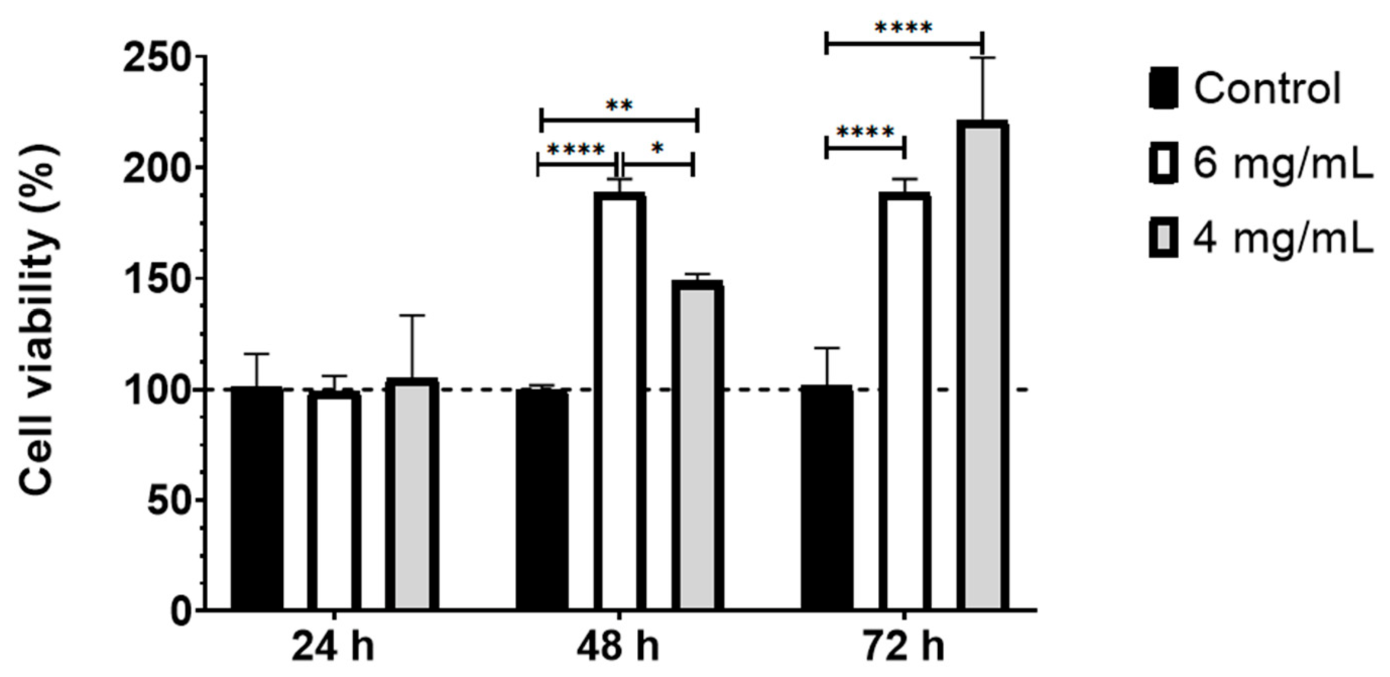
As shown in Figure 1, no cytotoxic effects were observed at any of the evaluated concentrations of dHAM soluble extracts on BM-MSCs. At 24 h, cell viability did not differ significantly between experimental groups and the control. However, at 48 h, both experimental groups exhibited a statistically significant increase in cell proliferation compared to the control (p ≤ 0.01 for 4 mg/mL and p ≤ 0.0001 for 6 mg/mL). Notably, no significant differences were observed between the two concentrations (4 and 6 mg/mL) at 72 h.

3.2. Scaffold Fabrication by Spin-Coating

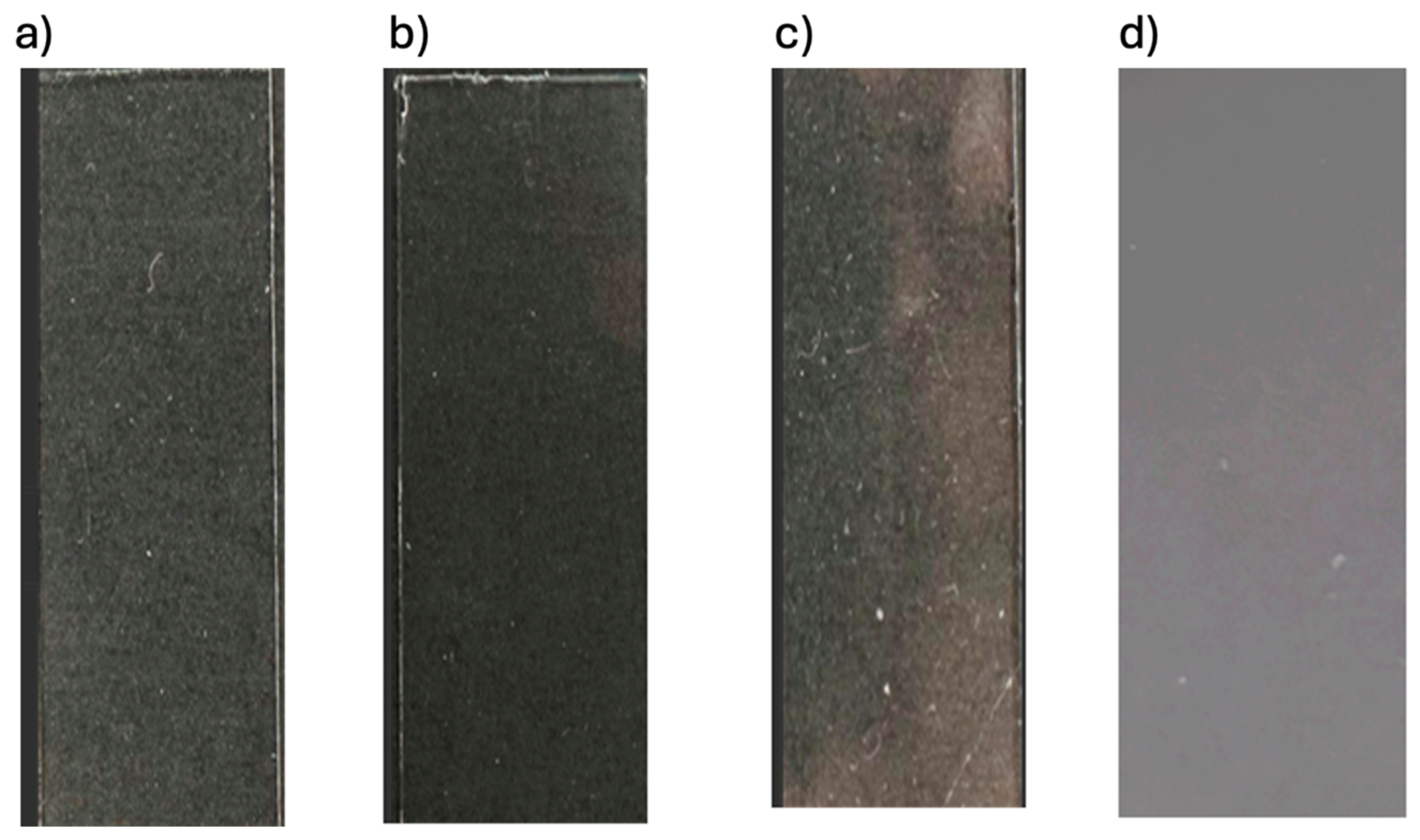
Different rotational speeds and spin-coating durations were evaluated to optimize scaffold fabrication. The resulting polymeric films exhibited smooth, homogeneous, and translucent surfaces, with complete adhesion to the glass substrate and no visible defects. As shown in Figure 2, dHAM-functionalized biofilms displayed distinct macroscopic characteristics compared to their non-functionalized counterparts. PVP-based biofilms demonstrated greater transparency than those fabricated from PCL. Nevertheless, irrespective of the polymer employed, all optimized scaffolds retained homogeneous, defect-free surfaces following dHAM incorporation.

3.3. Determination of Hydrophobicity

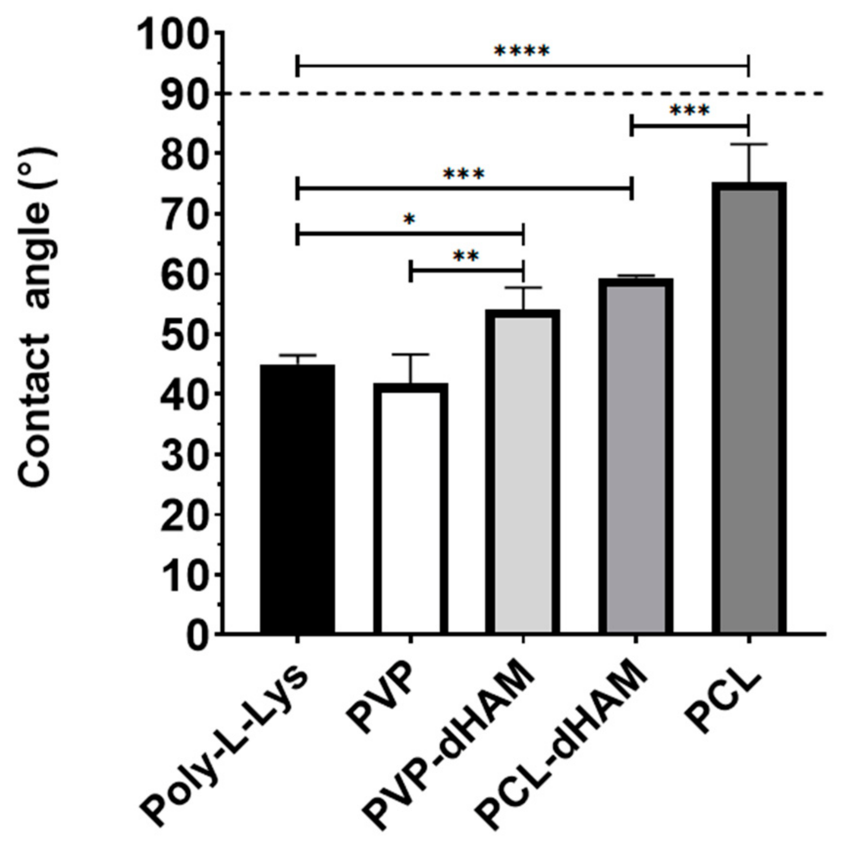
Surface hydrophobicity was evaluated by contact angle measurements. Results are displayed in Figure 3. PVP scaffolds exhibited the lowest contact angle values, whereas PCL scaffolds showed the highest. Incorporation of dHAM significantly modified scaffold wettability. PCL-dHAM demonstrated a significant decrease in contact angle compared to pure PCL (p ≤ 0.001), while PVP-dHAM showed a significant increase relative to pure PVP (p ≤ 0.01). No significant differences were observed between PCL-dHAM and PVP-dHAM scaffolds. Importantly, none of the evaluated scaffolds exceeded the 90° threshold, indicating that all materials can be described as hydrophilic.

3.4. ATR-FTIR Spectroscopy

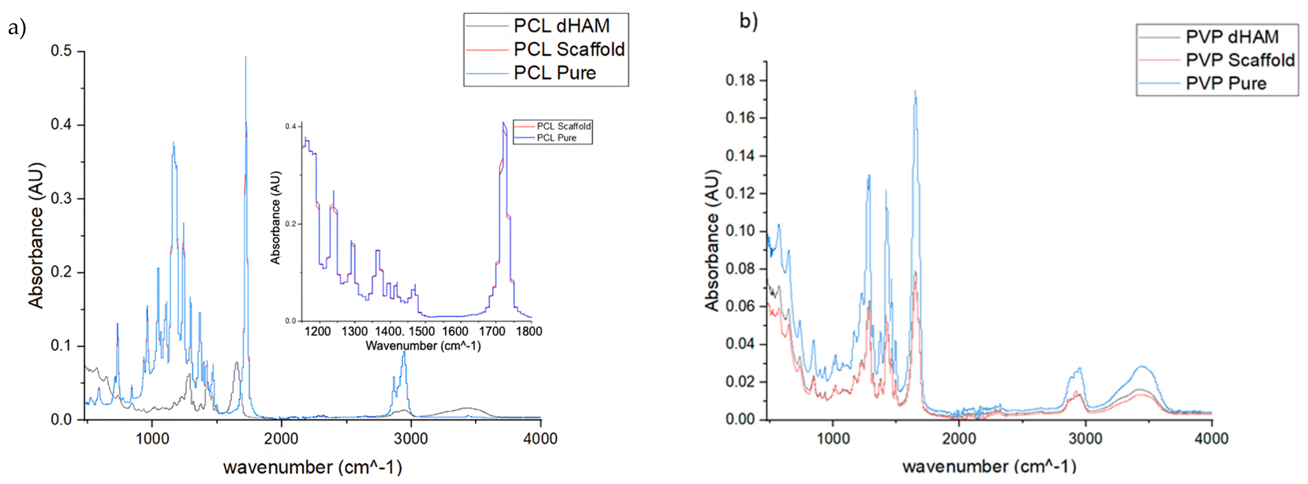
ATR-FTIR analysis was performed to confirm polymer presence, structural integrity, and potential chemical variations induced by dHAM incorporation or sterilization processes. The ATR-FTIR showed in Figure 4a that the spectra of pure PCL and PCL scaffold exhibited overlapping, suggesting that the scaffold fabrication process did not significantly alter the characteristic functional groups of the polymer. Furthermore, the spectra of pure PCL and PCL-dHAM scaffolds (Figure 4a) showed characteristic absorption bands of PCL, including prominent peaks at 2940 cm−1 and 2860 cm−1, and a sharp peak at 1722 cm−1 corresponding to ester carbonyl (C=O) stretching vibrations. Although PCL-dHAM exhibited similar absorption bands to pure PCL, slight variations in peak intensity were observed, particularly in the 1000–1500 cm−1 region, suggesting possible interactions or overlapping signals from amniotic membrane components. These findings confirm successful dHAM incorporation without significant alteration of the polymer’s chemical structure.
Similarly, ATR-FTIR spectra of pure PVP and PVP-dHAM scaffolds (Figure 4b) showed comparable absorption profiles, with minor reductions in intensity at 1662, 2961, and 3300 cm−1. The absence of new peaks and similarity to the commercial reference spectrum confirm preservation of the characteristic chemical structure of PVP following solubilization, dHAM incorporation, and exposure to EtO and UV sterilization.

3.5. Cell Quantification by Fluorescence Microscopy

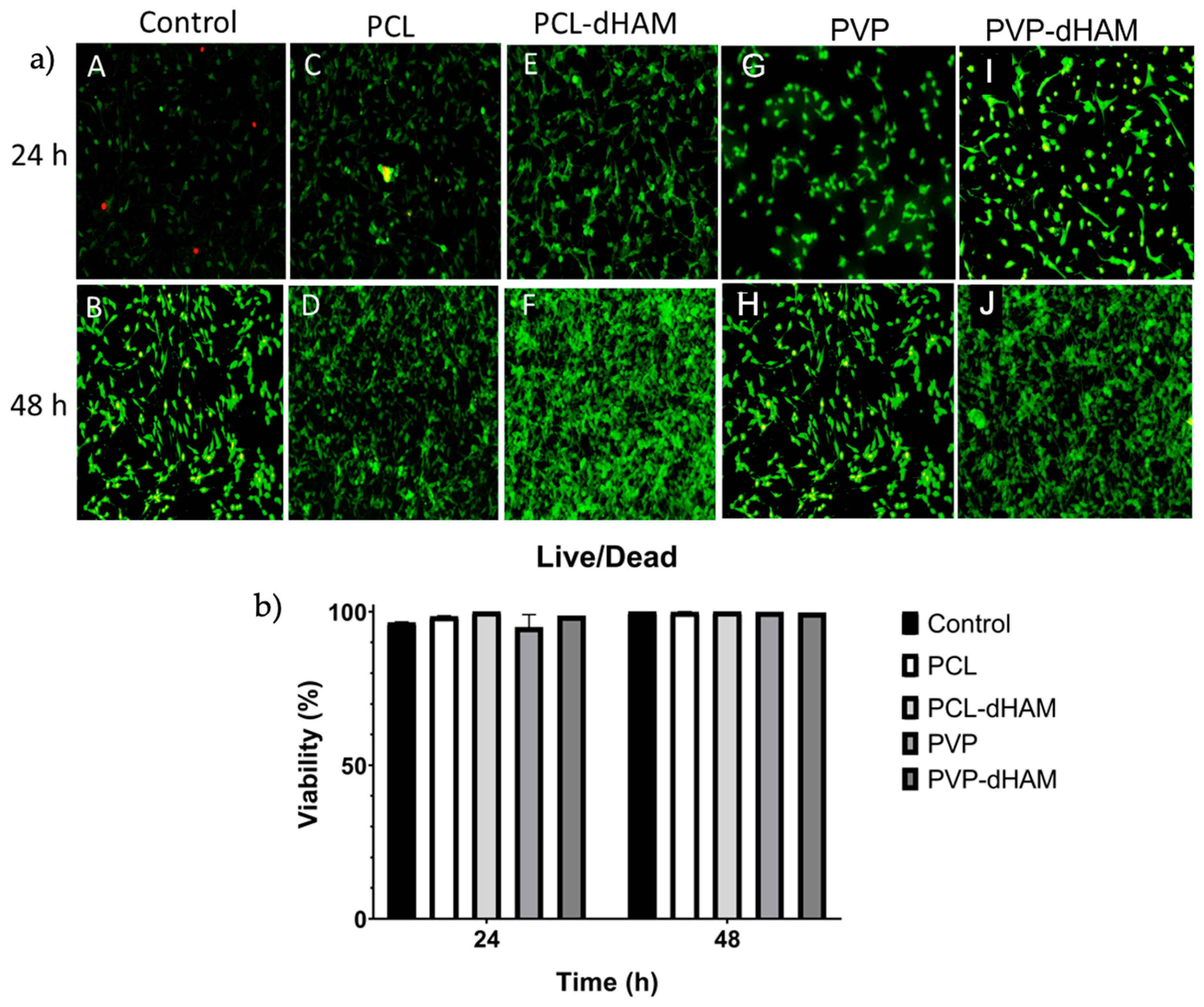
Cell viability was confirmed using the LIVE/DEAD™ cytotoxicity assay. Fluorescence microscopy (Figure 5a) revealed minimal red fluorescence across all biofilm types, indicating low cytotoxicity. Quantitative analysis (Figure 5b) demonstrated that cell viability remained above 90% at 24 and 48 h for all evaluated films and biofilms. Consistent with the proliferation analysis, dHAM-functionalized biofilms exhibited increased cell density at 48 h compared to non-functionalized materials, without compromising cell viability.
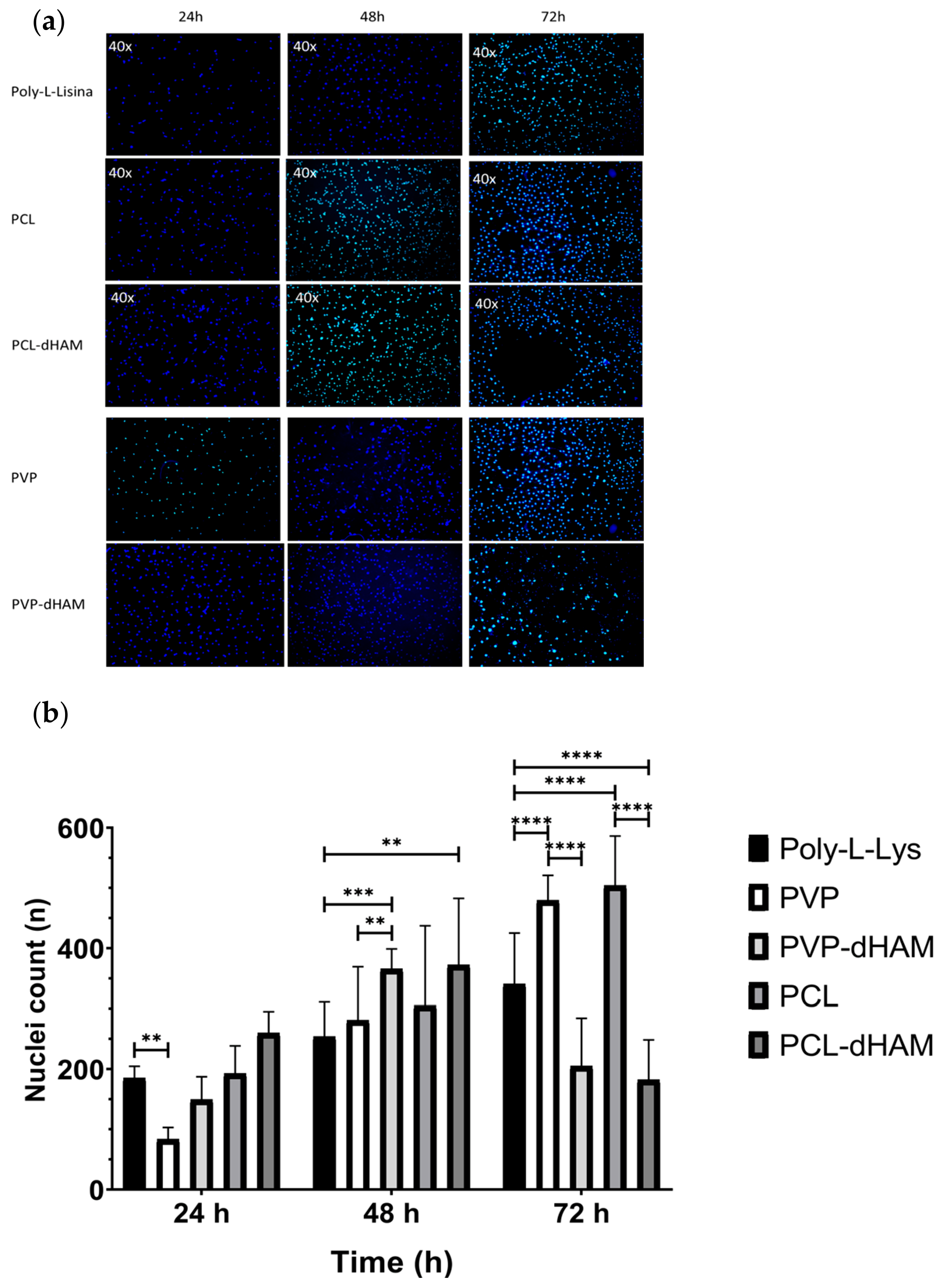
BM-MSCs cultured on polymeric films and dHAM-functionalized biofilms were evaluated over a 72 h period by fluorescence microscopy following DAPI staining Figure 6a. Across all experimental conditions, cells exhibited typical oval-shaped nuclei with well-defined borders and homogeneous chromatin distribution, with no evident signs of nuclear fragmentation. Importantly, no detachment of the biofilms from the glass substrate was observed during the experimental period, confirming structural stability under cell culture conditions. At 24 h, an increased number of adherent cells was observed on dHAM-functionalized scaffolds compared to their non-functionalized counterparts. This effect was particularly evident in PCL-dHAM scaffolds, which displayed a greater initial cell density.
A semi-quantitative analysis shown in Figure 6b showed that PVP-based scaffolds, PVP-dHAM, demonstrated a significant increase in viable cell numbers at 24 h compared to pure PVP and the poly-L-lysine control (p ≤ 0.01). At 48 h, overall cell numbers increased in both groups; however, no significant differences were detected between PVP and PVP-dHAM, although both remained significantly different from the control. At 72 h, a marked reduction in cell number was observed in PVP-dHAM scaffolds, showing significantly lower values compared to pure PVP (p ≤ 0.0001) and the control (p ≤ 0.001). A similar pattern was observed for PCL-based scaffolds. At 48 h, PCL-dHAM exhibited significantly higher proliferation than pure PCL (p ≤ 0.01) and the control group (p ≤ 0.001). However, at 72 h, viable cell numbers decreased notably in PCL-dHAM scaffolds, paralleling the trend observed in PVP-based materials. dHAM-functionalized biofilms exhibited increased cell density at 48 h compared to non-functionalized materials, without compromising cell viability.

3.6. In Vitro Wound Healing Assay

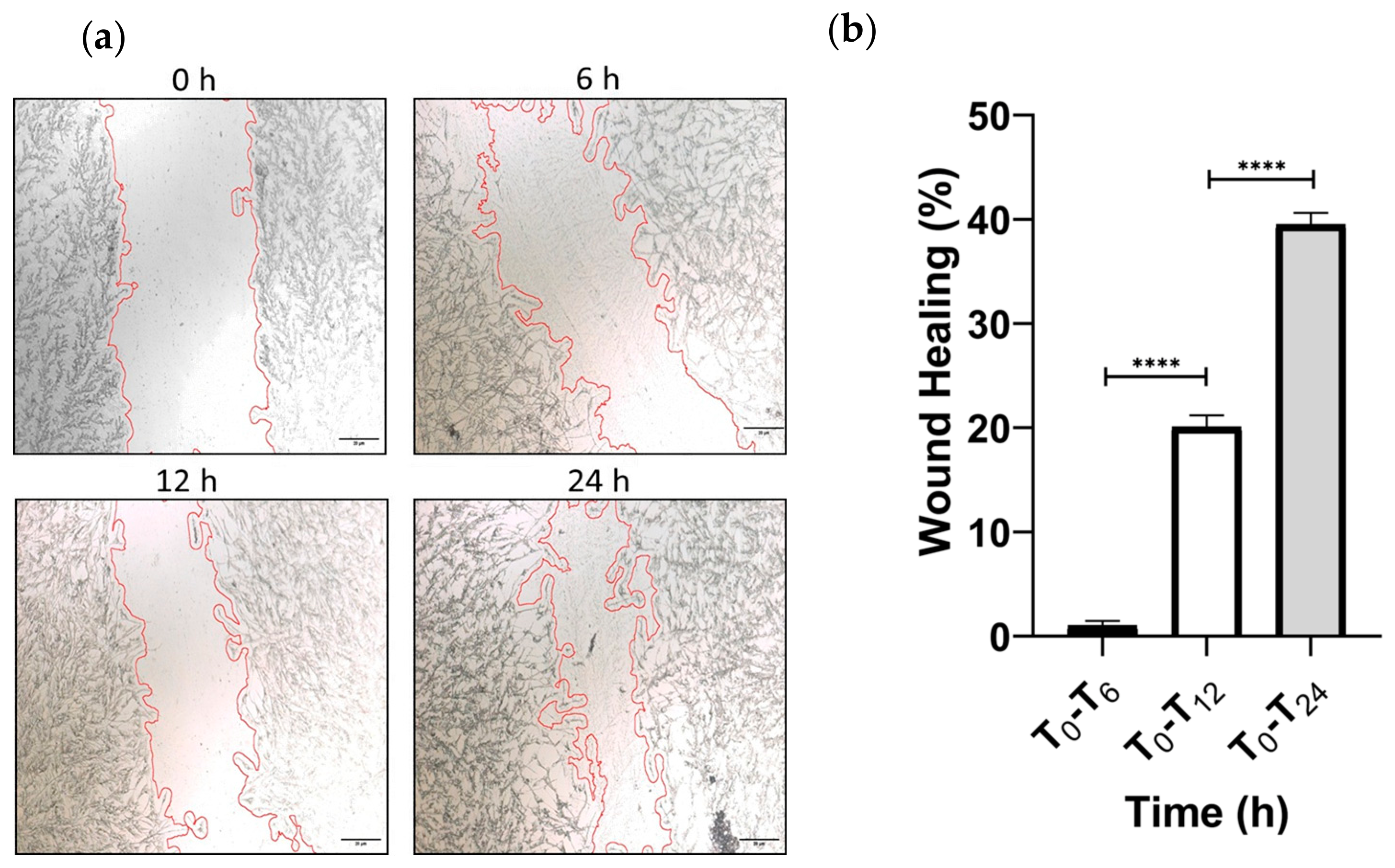
The regenerative capacity of the biofilms was investigated using an in vitro wound healing assay. In the poly-L-lysine control group, wound closure progressed in a time-dependent manner, reaching 1.04% at 6 h, 20.08% at 12 h, and 39.51% at 24 h. Based on this temporal profile, 24 h was established as the reference time point for comparative evaluation of scaffold performance (Figure 7).
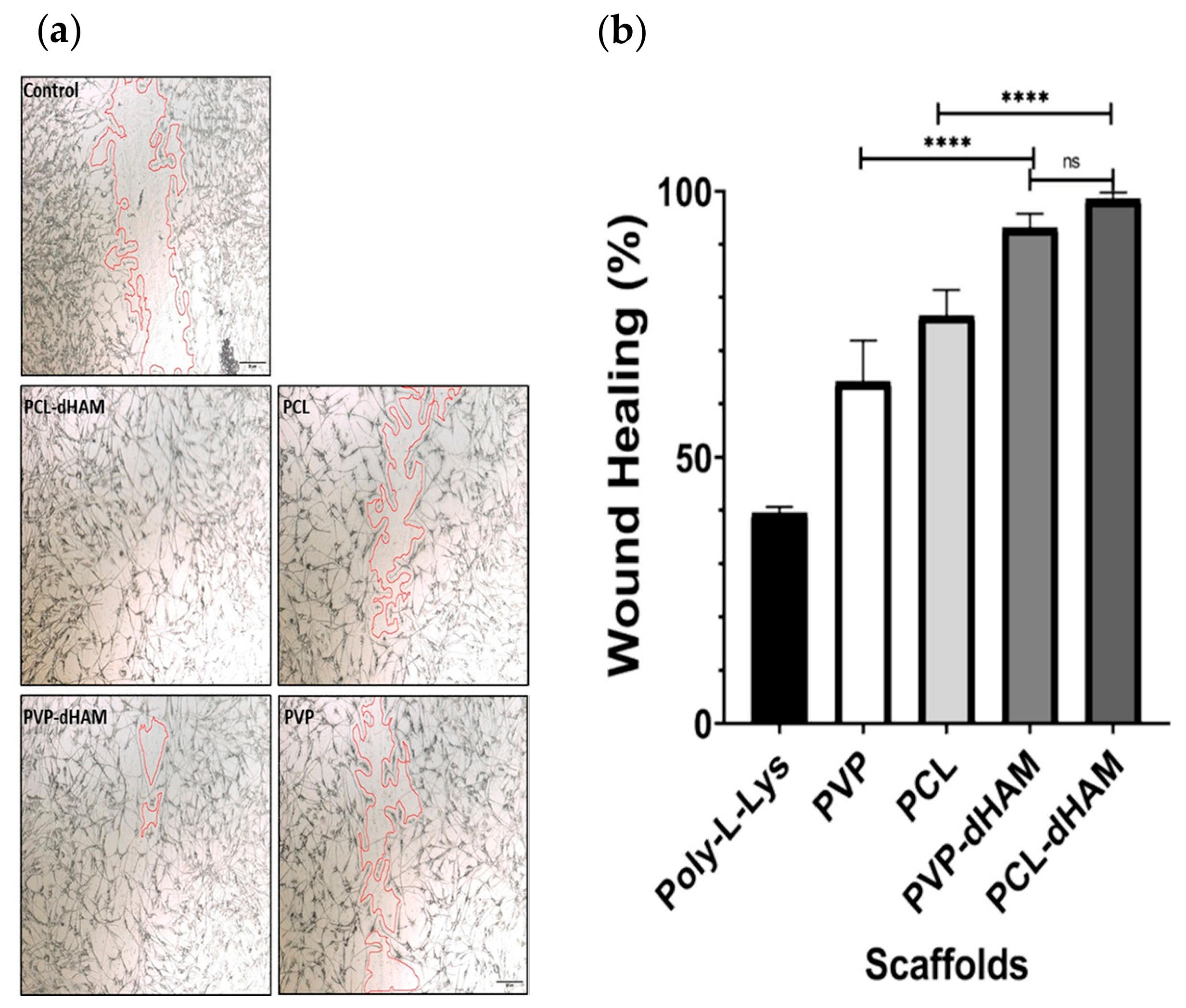
As shown in Figure 8, at 24 h, pure PVP scaffolds achieved 64.20% wound closure, whereas PCL scaffolds reached 76.56%, demonstrating enhanced migratory activity relative to the control surface. Importantly, functionalization with dHAM significantly augmented cellular migration. PVP-dHAM scaffolds exhibited 93.13% wound closure, while PCL-dHAM achieved the highest closure rate at 98.47%.
Statistical analysis confirmed significant differences between non-functionalized scaffolds and their respective dHAM-functionalized counterparts (PCL vs. PCL-dHAM, p = 0.0001; PVP vs. PVP-dHAM, p = 0.0001). No significant differences were observed between PCL-dHAM and PVP-dHAM groups, indicating that the incorporation of the biological matrix is the principal factor driving the enhanced migratory response.

3.7. Scanning Electron Microscopy

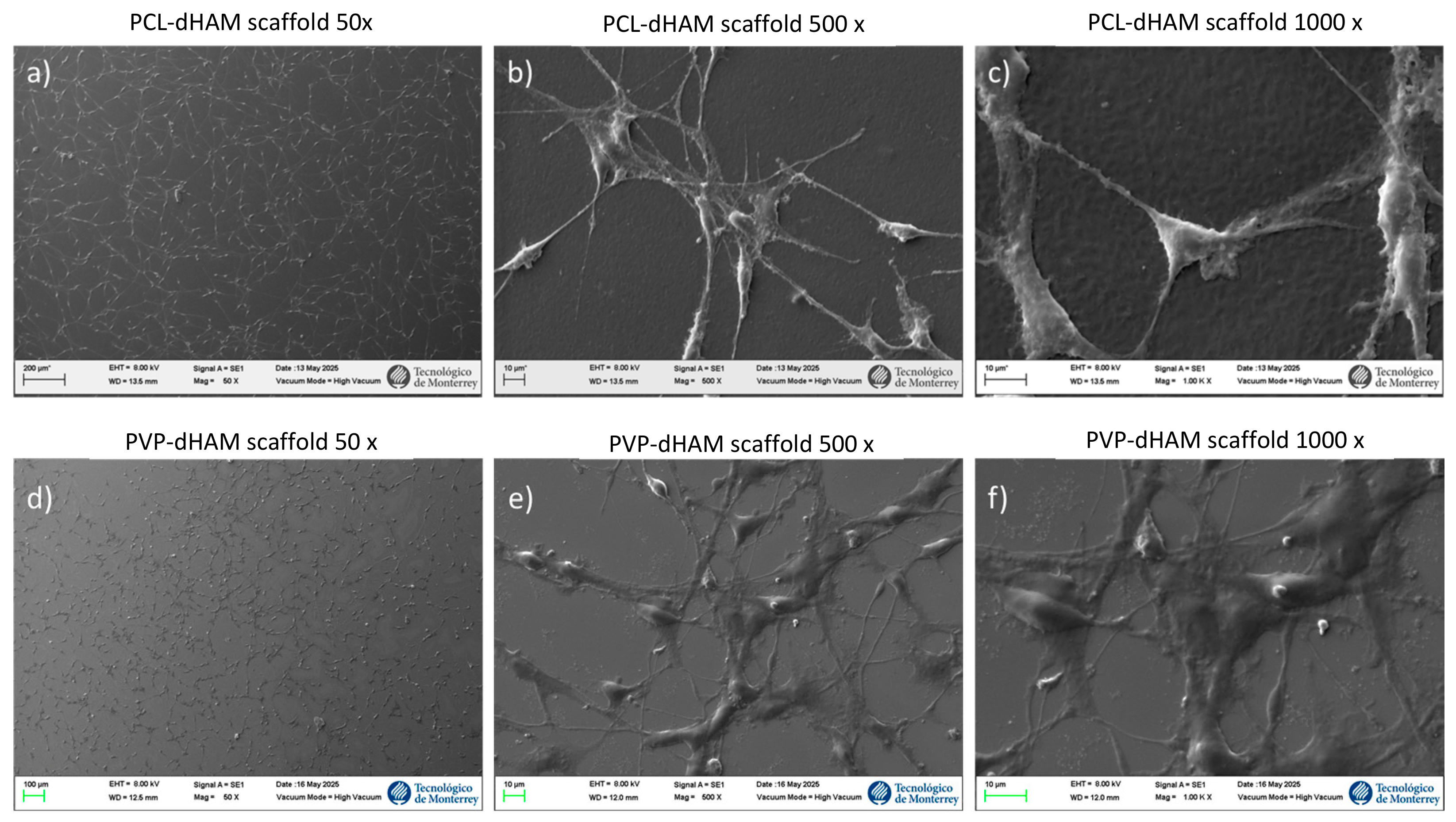
Figure 9 shows the adhesion of BM-MSC at 24 h to different polymeric scaffold surfaces coated with dHAM. SEM analysis revealed successful adhesion of BM-MSCs on both PCL-dHAM and PVP-dHAM scaffolds after 24 h of culture. On PCL-based substrates, cells exhibited a well-spread, uniform, and elongated morphology with evident cytoplasmic extensions and filopodia, indicating good cell–substrate interaction. In contrast, cells cultured on PVP-dHAM scaffolds displayed a comparatively less extended morphology, with fewer visible protrusions and a more localized attachment pattern. These observations suggest that while both matrices support early cell adhesion, the PCL-dHAM scaffold promotes better cell spreading and surface interaction, potentially due to differences in surface topography and physicochemical properties.

4. Discussion

The human amniotic membrane has been extensively recognized as a bioactive platform in regenerative medicine due to its availability, low immunogenicity, and rich extracellular matrix (ECM) composition. Structural proteins such as collagen, laminin, and fibronectin, together with growth factors including EGF, bFGF, and VEGF, contribute to its well-documented pro-regenerative and anti-inflammatory properties. However, its rapid biodegradation and limited mechanical resistance restrict its standalone clinical application, particularly in load-bearing or long-term tissue repair contexts. Therefore, hybridization with synthetic polymers has been proposed as a rational strategy to enhance structural stability while preserving biological functionality.
In this study, spin-coated PCL and PVP biofilms incorporating decellularized human amniotic membrane matrix (dHAM) were successfully fabricated. Unlike electrospinning, spin coating enabled the deposition of thin films with homogeneous distribution of biological components at the surface, favoring direct cell–material interactions. Although macroscopic uniformity was confirmed, high-resolution analyses (AFM and XPS) would be valuable to further characterize surface-micro-topographical surfaces that may influence cell adhesion and migration. Moreover, a limitation of this work is the absence of cytoskeletal focal adhesion staining, which would have provided a more direct assessment of cell adhesion on the films.
Soluble dHAM extracts significantly promoted BM-MSC proliferation at 48 h without evidence of cytotoxic effects, suggesting that retained growth factors and ECM-derived peptides preserved their bioactivity and may be associated with activation of mitogenic pathways such as PI3K/AKT and ERK1/2 [41]. SEM analysis at higher magnifications revealed that PCL films appear noticeably rougher than PVP ones. Such rough surfaces provide additional anchoring points for integrins and extracellular matrix proteins, thereby promoting the formation of stable focal adhesions and stronger attachment of MSCs [42]. Furthermore, micro- and nanoscale roughness has been reported to stimulate the elongation of fine filopodia, which actively explore the substrate and facilitate a more homogeneous cell distribution, as observed in our PCL-ECM-dHAM films [43]. This phenomenon, commonly referred to as contact guidance, describes how cells follow the topographical irregularities of the ECM, thereby regulating cell behavior and function [42]. Our 24 h SEM images revealed extensive adhesion and propagation of BM-MSCs on the scaffolds, suggesting early confluence. This may explain the lack of significant differences between 4 and 6 mg/mL at later time points, as the cells had already reached a proliferative plateau. Such stabilization (threshold) [44] could also be attributed to receptor saturation or signaling plateau effects [45]; once integrin binding sites and downstream pathways are maximally activated, additional ligand availability or higher concentrations of bioactive factors fail to elicit further responses. However, this remains a hypothetical interpretation, as these mechanisms were not directly assessed. In addition, the lack of quantification of specific factors limits identification of the principal mediators, underscoring the need for proteomic profiling or ELISA-based assays in future studies.
Although dHAM-functionalized scaffolds significantly enhanced early BM-MSC proliferation (24–48 h), a reduction in cell numbers was observed at 72 h in dHAM-functionalized scaffolds, despite viability remaining above 90%. This pattern may suggest that early stimulation may accelerate proliferation, leading to premature confluence and contact inhibition. Similar trends have been reported in growth factor-enriched systems, where proliferative responses occur within an optimal range but decline at later stages due to feedback regulation or density-dependent effects [37]. Alternatively, rapid release of bioactive components may create a transient stimulatory microenvironment followed by depletion. Evaluation of release kinetics will be essential to determine whether sustained presentation of ECM-derived signals could support long-term proliferation.
Surface wettability analysis revealed polymer-dependent modulation of hydrophilicity following dHAM incorporation. The decrease in contact angle observed in PCL-dHAM scaffolds may be attributed to the exposure of hydrophilic protein domains, particularly collagen, enhancing integrin-mediated adhesion, consistent with previous reports [24,25]. In contrast, the modest increase observed in PVP-dHAM scaffolds may reflect surface rearrangement of polar domains during solvent evaporation [38,40]. Despite these differences, all scaffolds remained within the hydrophilic range (approximately 70–90°), which is generally favorable for mammalian cell attachment [41].
ATR-FTIR analysis confirmed preservation of the characteristic chemical structures of PCL and PVP following dHAM incorporation and sterilization. The presence of amide-related bands supports successful integration of proteinaceous components without evidence of polymer degradation. Importantly, the spin-coating process did not induce significant chemical alterations, supporting its suitability for generating biofunctional coatings. Minor spectral variations suggest non-covalent interactions, likely mediated by hydrogen bonding. However, residual DNA quantification and direct confirmation of complete decellularization were not performed. Although established protocols were followed, additional biochemical validation would strengthen translational and regulatory robustness.
The wound healing assay provided functional evidence of the regenerative capacity of the scaffolds. Both PCL-dHAM and PVP-dHAM significantly enhanced cell migration compared to pure polymers and poly-L-lysine controls. The high wound closure rates observed (≈98% for PCL-dHAM and ≈93% for PVP-dHAM at 24 h) indicate a strong pro-migratory effect driven by the biological matrix. ECM components and associated growth factors, including collagen, laminin, fibronectin, EGF, bFGF, and VEGF, are known to regulate cytoskeletal organization and directional migration [42]. In addition to biochemical cues, physicochemical properties such as substrate stiffness and surface composition also influence migration dynamics [43]. The superior performance of dHAM-functionalized scaffolds highlights the importance of integrating biological signals within structurally stable matrices.
The present study has several limitations. Experiments were conducted exclusively in vitro using murine BM-MSCs, which limits generalizability to human cell populations. The release kinetics of bioactive components were not characterized, and high-resolution surface analyses (AFM and XPS) as well as cytoskeletal staining were not performed, limiting mechanistic interpretation of cell-material interactions. Additionally, residual DNA quantification was not included to formally confirm complete decellularization. Moreover, mechanical characterization and degradation behavior of the biofilms were not evaluated, which restricts results regarding substrate stiffness and long-term stability, both of which are critical for wound healing applications. Future studies should incorporate human multi-donor cell populations, proteomic profiling of dHAM components, release kinetics characterization, mechanical and degradation analyses, and preclinical in vivo wound healing models to strengthen the translational potential of these hybrid biomaterials.

5. Conclusions

Overall, this study demonstrates that the incorporation of decellularized human amniotic membrane into synthetic polymer matrices via spin coating generates hybrid biomaterials that combine biological activity with the processing advantages of synthetic polymer matrices. dHAM-functionalized scaffolds enhanced mesenchymal stem cell proliferation, viability, and migration, supporting their potential role in promoting tissue repair. Importantly, the use of spin coating provides a simple, reproducible, and scalable alternative to more complex fabrication techniques, enabling controlled surface biofunctionalization. These findings position PCL-dHAM and PVP-dHAM scaffolds as promising candidates for applications in wound healing and regenerative medicine. Future work should focus on characterizing the release kinetics of bioactive components, confirming complete decellularization at the molecular level, and evaluating in vivo performance. Such studies will be essential to further define the translational potential and clinical applicability of these hybrid biomaterials.

Author Contributions

J.d.D.M.Q. conducted the experiments and drafted the original manuscript; A.R.M. contributed to the design and execution of experiments as well as the analysis and discussion of results; R.F.M. supervised staining procedures and image analyses; P.D.G. and J.F.I. designed experiments and carried out literature analysis and discussion of results; J.L.A. and M.S.-M. contributed to writing, analysis, and manuscript revision; C.N.S.D. edited the text, analyzed results, and reviewed the final manuscript; H.L.G. and E.N.G.T. designed experiments, managed the project, and contributed to writing, analysis, and manuscript revision; J.d.D.M.Q. and E.N.G.T. confirmed the authenticity of all raw data. All authors have read and agreed to the published version of the manuscript.

Funding

This work was partially supported by the Secretaría de Ciencia, Tecnología e Innovación (SECITHI), grant number CBF-2025-I-2328.

Institutional Review Board Statement

The study was conducted in accordance with the Declaration of Helsinki and approved by the Ethics Committee of the School of Medicine, Universidad Autónoma de Nuevo Leon (BI23-001, approval on 1 March 2023).

Informed Consent Statement

Written informed consent has been obtained from the patients.

Data Availability Statement

Data is contained within the article.

Acknowledgments

I would like to express my gratitude to the Laboratory of Nanoscience and Nanotechnology of the Center for Research in Physical-Mathematical Sciences, affiliated with the Universidad Autónoma de Nuevo León, Monterrey, México, and also to the Center for Innovation in Digital Technologies, affiliated with the School of Engineering and Sciences, Tecnológico de Monterrey.

Conflicts of Interest

The authors declare no conflicts of interest.

References

  1. Altyar, A.E.; El-Sayed, A.; Abdeen, A.; Piscopo, M.; Mousa, S.A.; Najda, A.; Abdel-Daim, M.M. Future regenerative medicine developments and their therapeutic applications. Biomed. Pharmacother. 2023, 158, 114131. [Google Scholar] [CrossRef] [Scilit] [PubMed]
  2. Echeverria Molina, M.I.; Malollari, K.G.; Komvopoulos, K. Design Challenges in Polymeric Scaffolds for Tissue Engineering. Front. Bioeng. Biotechnol. 2021, 9, 617141. [Google Scholar] [CrossRef] [Scilit] [PubMed]
  3. Hoang, V.T.; Nguyen, Q.T.; Phan, T.T.K.; Pham, T.H.; Dinh, N.T.H.; Anh, L.P.H.; Dao, L.T.M.; Bui, V.D.; Dao, H.; Le, D.S.; et al. Tissue Engineering and Regenerative Medicine: Perspectives and Challenges. MedComm 2025, 6, e70192. [Google Scholar] [CrossRef] [Scilit] [PubMed]
  4. Phutane, P.; Telange, D.; Agrawal, S.; Gunde, M.; Kotkar, K.; Pethe, A. Biofunctionalization and Applications of Polymeric Nanofibers in Tissue Engineering and Regenerative Medicine. Polymers 2023, 15, 1202. [Google Scholar] [CrossRef] [Scilit] [PubMed]
  5. Kafili, G.; Niknejad, H.; Tamjid, E.; Simchi, A. Amnion-derived hydrogels as a versatile platform for regenerative therapy: From lab to market. Front. Bioeng. Biotechnol. 2024, 12, 1358977. [Google Scholar] [CrossRef] [Scilit] [PubMed]
  6. Fenelon, M.; Galvez, P.; Kalbermatten, D.; Scolozzi, P.; Madduri, S. Emerging Strategies for the Biofabrication of Multilayer Composite Amniotic Membranes for Biomedical Applications. Int. J. Mol. Sci. 2023, 24, 14424. [Google Scholar] [CrossRef] [Scilit]
  7. Jahanafrooz, Z.; Bakhshandeh, B.; Behnam Abdollahi, S.; Seyedjafari, E. Human amniotic membrane as a multifunctional biomaterial: Recent advances and applications. J. Biomater. Appl. 2023, 37, 1341–1354. [Google Scholar] [CrossRef] [Scilit] [PubMed]
  8. Arki, M.K.; Moeinabadi-Bidgoli, K.; Hossein-Khannazer, N.; Gramignoli, R.; Najimi, M.; Vosough, M. Amniotic Membrane and Its Derivatives: Novel Therapeutic Modalities in Liver Disorders. Cells 2023, 12, 2114. [Google Scholar] [CrossRef] [Scilit]
  9. Li, Y.; An, S.; Deng, C.; Xiao, S. Human Acellular Amniotic Membrane as Skin Substitute and Biological Scaffold: A Review of Its Preparation, Preclinical Research, and Clinical Application. Pharmaceutics 2023, 15, 2249. [Google Scholar] [CrossRef] [Scilit] [PubMed]
  10. Hofmann, N.; Rennekampff, H.-O.; Salz, A.K.; Börgel, M. Preparation of human amniotic membrane for transplantation in different application areas. Front. Transplant. 2023, 2, 1152068. [Google Scholar] [CrossRef] [Scilit] [PubMed]
  11. Nazari, H.; Heirani-Tabasi, A.; Esmaeili, E.; Kajbafzadeh, A.-M.; Hassannejad, Z.; Boroomand, S.; Alavijeh, M.H.S.; Mishan, M.A.; Tafti, S.H.A.; Warkiani, M.E.; et al. Decellularized human amniotic membrane reinforced by MoS2-Polycaprolactone nanofibers, a novel conductive scaffold for cardiac tissue engineering. J. Biomater. Appl. 2022, 36, 1527–1539. [Google Scholar] [CrossRef] [Scilit] [PubMed]
  12. Hadipour, A.; Bayati, V.; Rashno, M.; Orazizadeh, M. Aligned Poly(ε-caprolactone) Nanofibers Superimposed on Decellularized Human Amniotic Membrane Promoted Myogenic Differentiation of Adipose Derived Stem Cells. Cell J. 2021, 23, 603–611. [Google Scholar] [CrossRef] [Scilit]
  13. Hasmad, H.; Yusof, M.R.; Mohd Razi, Z.R.; Hj Idrus, R.B.; Chowdhury, S.R. Human Amniotic Membrane with Aligned Electrospun Fiber as Scaffold for Aligned Tissue Regeneration. Tissue Eng. Part C Methods 2018, 24, 368–378. [Google Scholar] [CrossRef] [Scilit] [PubMed]
  14. Zhang, Z.; Lu, B.; Zou, L.; Huang, X.; Yang, F.; Lv, H. Decellularized human amniotic membrane scaffolds: Influence on the biological behavior of dental pulp stem cells. BMC Oral Health 2024, 24, 394. [Google Scholar] [CrossRef] [Scilit] [PubMed]
  15. Arasteh, S.; Kazemnejad, S.; Khanjani, S.; Heidari-Vala, H.; Akhondi, M.M.; Mobini, S. Fabrication and characterization of nano-fibrous bilayer composite for skin regeneration application. Methods 2016, 99, 3–12. [Google Scholar] [CrossRef] [Scilit] [PubMed]
  16. Agrawal, R.; Kumar, A.; Mohammed, M.K.A.; Singh, S. Biomaterial types, properties, medical applications, and other factors: A recent review. J. Zhejiang Univ.-Sci. A 2023, 24, 1027–1042. [Google Scholar] [CrossRef] [Scilit]
  17. Bhaskar, B.; Sreenivasa Rao, P.; Kasoju, N.; Nagarjuna, V.; Baadhe, R.R. Biomaterials in Tissue Engineering and Regenerative Medicine: From Basic Concepts to State of the Art Approaches; Springer: Singapore, 2021. [Google Scholar] [CrossRef] [Scilit]
  18. Mozafari, M.; Sefat, F.; Atala, A. Handbook of Tissue Engineering Scaffolds, 1st ed.; Elsevier Science: Amsterdam, The Netherlands, 2019; Volume 1. [Google Scholar]
  19. Robles, K.N.; tuz Zahra, F.; Mu, R.; Giorgio, T. Advances in Electrospun Poly(ε-caprolactone)-Based Nanofibrous Scaffolds for Tissue Engineering. Polymers 2024, 16, 2853. [Google Scholar] [CrossRef] [Scilit] [PubMed]
  20. Siddiqui, N.; Asawa, S.; Birru, B.; Baadhe, R.; Rao, S. PCL-Based Composite Scaffold Matrices for Tissue Engineering Applications. Mol. Biotechnol. 2018, 60, 506–532. [Google Scholar] [CrossRef] [Scilit]
  21. Mandal, P.; Shunmugam, R. Polycaprolactone: A biodegradable polymer with its application in the field of self-assembly study. J. Macromol. Sci. Part A 2021, 58, 111–129. [Google Scholar] [CrossRef] [Scilit]
  22. de Boer, J.; van Blitterswijk, C.A.; Uquillas, J.A.; Malik, N. (Eds.) Tissue Engineering, 3rd ed.; Agon Elsevier: Amsterdam, The Netherlands, 2022. [Google Scholar] [CrossRef] [Scilit]
  23. Xu, R.; Fang, Y.; Zhang, Z.; Cao, Y.; Yan, Y.; Gan, L.; Xu, J.; Zhou, G. Recent Advances in Biodegradable and Biocompatible Synthetic Polymers Used in Skin Wound Healing. Materials 2023, 16, 5459. [Google Scholar] [CrossRef] [Scilit] [PubMed]
  24. Kesharwani, R.K.; Keservani, R.K.; Sharma, A.K. Tissue Engineering: Applications and Advancements; Apple Academic Press: Oakville, ON, USA, 2022. [Google Scholar]
  25. Lotfi, Z.; Khakbiz, M.; Davari, N.; Bonakdar, S.; Mohammadi, J.; Shokrgozar, M.A.; Derhambakhsh, S. Fabrication and multiscale modeling of polycaprolactone/amniotic membrane electrospun nanofiber scaffolds for wound healing. Artif. Organs 2023, 47, 1267–1284. [Google Scholar] [CrossRef] [Scilit] [PubMed]
  26. Majidnia, E.; Ahmadian, M.; Salehi, H.; Amirpour, N. Development of an electrospun poly(ε-caprolactone)/collagen-based human amniotic membrane powder scaffold for culturing retinal pigment epithelial cells. Sci. Rep. 2022, 12, 6469. [Google Scholar] [CrossRef] [Scilit] [PubMed]
  27. Kanaujiya, S.; Arya, D.; Pandey, P.; Singh, S.; Pandey, G.; Anjum, S.; Anjum, M.; Ali, D.; Alarifi, S.; Vijayakumar; et al. Resveratrol-Ampicillin Dual-Drug Loaded Polyvinylpyrrolidone/Polyvinyl Alcohol Biomimic Electrospun Nanofiber Enriched with Collagen for Efficient Burn Wound Repair. Int. J. Nanomed. 2024, 19, 5397–5418. [Google Scholar] [CrossRef] [Scilit]
  28. Sruthi, R.; Balagangadharan, K.; Selvamurugan, N. Polycaprolactone/polyvinylpyrrolidone coaxial electrospun fibers containing veratric acid-loaded chitosan nanoparticles for bone regeneration. Colloids Surf. B Biointerfaces 2020, 193, 111110. [Google Scholar] [CrossRef] [Scilit] [PubMed]
  29. Adel, I.M.; ElMeligy, M.F.; Elkasabgy, N.A. Conventional and Recent Trends of Scaffolds Fabrication: A Superior Mode for Tissue Engineering. Pharmaceutics 2022, 14, 306. [Google Scholar] [CrossRef] [Scilit] [PubMed]
  30. Moreira, J.; Vale, A.C.; Alves, N.M. Spin-coated freestanding films for biomedical applications. J. Mater. Chem. B 2021, 9, 3778–3799. [Google Scholar] [CrossRef] [Scilit] [PubMed]
  31. Jo, H.; Brito, S.; Kwak, B.M.; Park, S.; Lee, M.-G.; Bin, B.-H. Applications of Mesenchymal Stem Cells in Skin Regeneration and Rejuvenation. Int. J. Mol. Sci. 2021, 22, 2410. [Google Scholar] [CrossRef] [Scilit] [PubMed]
  32. Rodríguez-Fuentes, D.E.; Fernández-Garza, L.E.; Samia-Meza, J.A.; Barrera-Barrera, S.A.; Caplan, A.I.; Barrera-Saldaña, H.A. Mesenchymal Stem Cells Current Clinical Applications: A Systematic Review. Arch. Med. Res. 2021, 52, 93–101. [Google Scholar] [CrossRef] [Scilit] [PubMed]
  33. Guimarães-Camboa, N.; Cattaneo, P.; Sun, Y.; Moore-Morris, T.; Gu, Y.; Dalton, N.D.; Rockenstein, E.; Masliah, E.; Peterson, K.L.; Stallcup, W.B.; et al. Pericytes of Multiple Organs Do Not Behave as Mesenchymal Stem Cells In Vivo. Cell Stem Cell 2017, 20, 345–359.e5. [Google Scholar] [CrossRef] [Scilit] [PubMed]
  34. Ullah, S.; Chen, X. Fabrication, applications and challenges of natural biomaterials in tissue engineering. Appl. Mater. Today 2020, 20, 100656. [Google Scholar] [CrossRef] [Scilit]
  35. de la Garza Kalife, D.A.; Rojas Murillo, A.; Franco Marquez, R.; Morales Wong, D.L.; Lara Arias, J.; Vilchez Cavazos, J.F.; Gutierrez, H.L.; Mendía, M.A.S.; Treviño, E.N.G. Human Amniotic Membrane Procurement Protocol and Evaluation of a Simplified Alkaline Decellularization Method. Methods Protoc. 2026, 9, 5. [Google Scholar] [CrossRef] [Scilit] [PubMed]
  36. Ballesteros, A.C.V.; Puello, H.R.S.; Lopez-Garcia, J.A.; Bernal-Ballen, A.; Mosquera, D.L.N.; Forero, D.M.M.; Charry, J.S.S.; Bejarano, Y.A.N. Bovine decellularized amniotic membrane: Extracellular matrix as scaffold for mammalian skin. Polymers 2020, 12, 590. [Google Scholar] [CrossRef] [Scilit] [PubMed]
  37. Rojas-Murillo, A.; Lara-Arias, J.; Leija-Gutiérrez, H.; Franco-Márquez, R.; Moncada-Saucedo, N.K.; Guzmán-López, A.; Vilchez-Cavazos, F.; Garza-Treviño, E.N.; Simental-Mendía, M. The Combination of Decellularized Cartilage and Amniotic Membrane Matrix Enhances the Production of Extracellular Matrix Elements in Human Chondrocytes. Coatings 2024, 14, 1083. [Google Scholar] [CrossRef] [Scilit]
  38. Quiroz-Reyes, A.G.; Gonzalez-Villarreal, C.A.; Limon-Flores, A.Y.; Delgado-Gonzalez, P.; Martinez-Rodriguez, H.G.; Said-Fernandez, S.L.; Soto-Dominguez, A.; Rivas-Estilla, A.M.; Islas, J.F.; Molina-De la Garza, J.F.; et al. Mesenchymal Stem Cells Genetically Modified by Lentivirus-Express Soluble TRAIL and Interleukin-12 Inhibit Growth and Reduced Metastasis-Relate Changes in Lymphoma Mice Model. Biomedicines 2023, 11, 595. [Google Scholar] [CrossRef] [Scilit] [PubMed]
  39. ISO 10993-5:2009; Biological Evaluation of Medical Devices—Part 5: Tests for In Vitro Cytotoxicity. International Organ-ization for Standardization (ISO): Geneva, Switzerland, 2009.
  40. Mendez-Quezada, J.D.D. Evaluation, Characterization, and Cytocompatibility of PCL/PVP Biocomposite Matrices with Decellularized Amniotic Membrane for Use in Regenerative Medicine; Universidad Autónoma de Nuevo León: San Nicolás de los Garza, Mexico, 2025. [Google Scholar]
  41. Hu, Z.; Luo, Y.; Ni, R.; Hu, Y.; Yang, F.; Du, T.; Zhu, Y. Biological importance of human amniotic membrane in tissue engineering and regenerative medicine. Mater. Today Bio 2023, 22, 100790. [Google Scholar] [CrossRef] [Scilit] [PubMed]
  42. bin Anwar Fadzil, A.F.; Pramanik, A.; Basak, A.K.; Prakash, C.; Shankar, S. Role of surface quality on biocompatibility of implants—A review. Ann. 3D Print. Med. 2022, 8, 100082. [Google Scholar] [CrossRef] [Scilit]
  43. Anderson, H.J.; Sahoo, J.K.; Ulijn, R.V.; Dalby, M.J. Mesenchymal Stem Cell Fate: Applying Biomaterials for Control of Stem Cell Behavior. Front. Bioeng. Biotechnol. 2016, 4, 38. [Google Scholar] [CrossRef] [Scilit] [PubMed]
  44. Ravindran, S.; Gao, Q.; Kotecha, M.; Magin, R.L.; Karol, S.; Bedran-Russo, A.; George, A. Biomimetic Extracellular Matrix-Incorporated Scaffold Induces Osteogenic Gene Expression in Human Marrow Stromal Cells. Tissue Eng. Part A 2012, 18, 295–309. [Google Scholar] [CrossRef] [Scilit] [PubMed]
  45. Borisov, N.M.; Markevich, N.I.; Hoek, J.B.; Kholodenko, B.N. Signaling through Receptors and Scaffolds: Independent Interactions Reduce Combinatorial Complexity. Biophys. J. 2005, 89, 951–966. [Google Scholar] [CrossRef] [Scilit] [PubMed]
Figure 1. Percentage of cell viability of BM-MSC in the presence of soluble dHAM extracts. * = ANOVA p < 0.05; ** = ANOVA p < 0.01; **** = ANOVA p < 0.0001. Cell proliferation assay was performed three times in triplicate. Figure reproduced from a Master’s thesis [40], with permission.
Coatings 16 00719 g001
Figure 2. Formation of polymeric films of PCL and PVP with and without dHAM on microscope slides using the spin-coating technique. (a) PVP, (b) PCL, (c) PVP-dHAM, (d) PCL-dHAM.
Coatings 16 00719 g002
Figure 3. Measurement of the contact angle on different polymer surfaces. * = ANOVA p ≤ 0.05; ** = ANOVA p ≤ 0.01; *** = ANOVA p ≤ 0.001; **** = ANOVA p ≤ 0.0001. Wettability assays were performed three times in triplicate. Figure reproduced from a Master’s thesis [40], with permission. The dashed line represents the threshold used to differentiate hydrophobic (≥90°) and hydrophilic (≤90°) surfaces.
Coatings 16 00719 g003
Figure 4. ATR-FTIR spectrum of (a) polycaprolactone (PCL) and (b) Polyvinylpyrrolidone (PVP) films. ATR-FTIR assay was performed in duplicate.
Coatings 16 00719 g004
Figure 5. Viability assay on PCL and PVP scaffolds with and without dHAM using Live/Dead test at different time points. Comparison of cell viability observed against the Poly-L-Lysine group. (a) Live/Dead staining of cells at 24 and 48 h. Control (A,B), PCL (C,D), PCL-dHAM (E,F), PVP (G,H), PVP-dHAM (I,J). (b) Semi-quantitative analysis of the number of cells per scaffold. Cell counting was performed in 6 random fields of view per condition. Live cells—green fluorescence; dead cells—red fluorescence.
Coatings 16 00719 g005
Figure 6. BM-MSCs cultured on PCL and PVP scaffolds with and without dHAM at different time points compared to a poly-L-lysine control. (a) DAPI staining of cells. (b) Semi-quantitative analysis of the number of cells per scaffold. Cell counting was performed in 10 random fields of view per condition; (n) represents the average number of nuclei counted per field. DAPI 40×. ** = ANOVA p ≤ 0.01; *** = ANOVA p ≤ 0.001. **** = ANOVA p ≤ 0.0001. Nuclei stained with DAPI appear blue. Assays were performed twice, with a minimum of 6 fields of view, in duplicate.
Coatings 16 00719 g006
Figure 7. (a) Wound healing test on Poly-L-Lysine at 0, 6, 12, and 24 h. (b) Graph of the percentage of healing observed over time. **** = ANOVA p ≤ 0.0001. Wound healing assay at different times with Poly-L-Lysin was performed two times, with a minimum of 6 fields of view, in triplicate. Red lines delineate the perimeter of the cell-free gap.
Coatings 16 00719 g007
Figure 8. (a) Wound healing assay on PCL-dHAM, PVP-dHAM, PCL, and PVP 24 h. (b) Comparative graph of the percentage of healing at 24 h. **** = ANOVA p ≤ 0.0001. Wound healing assays were performed 2 times, with a minimum of 6 fields of view, in triplicate. Figure reproduced from Master’s thesis [40], with permission. Red lines delineate the perimeter of the cell-free gap. ns; not significant.
Coatings 16 00719 g008
Figure 9. Scanning Electron Micrography (SEM) of PCL and PVP with dHAM scaffolds seeded with BM-MSC after 24 h. SEM image of PCL-dHAM scaffold at 50, 500 and 1000× ((a–c), respectively). SEM image of PVP-dHAM scaffold at 50, 500 and 1000× ((d–f), respectively).
Coatings 16 00719 g009
Disclaimer/Publisher’s Note: The statements, opinions and data contained in all publications are solely those of the individual author(s) and contributor(s) and not of MDPI and/or the editor(s). MDPI and/or the editor(s) disclaim responsibility for any injury to people or property resulting from any ideas, methods, instructions or products referred to in the content.