3.1. Microstructural Morphology Before and After Heat Treatment
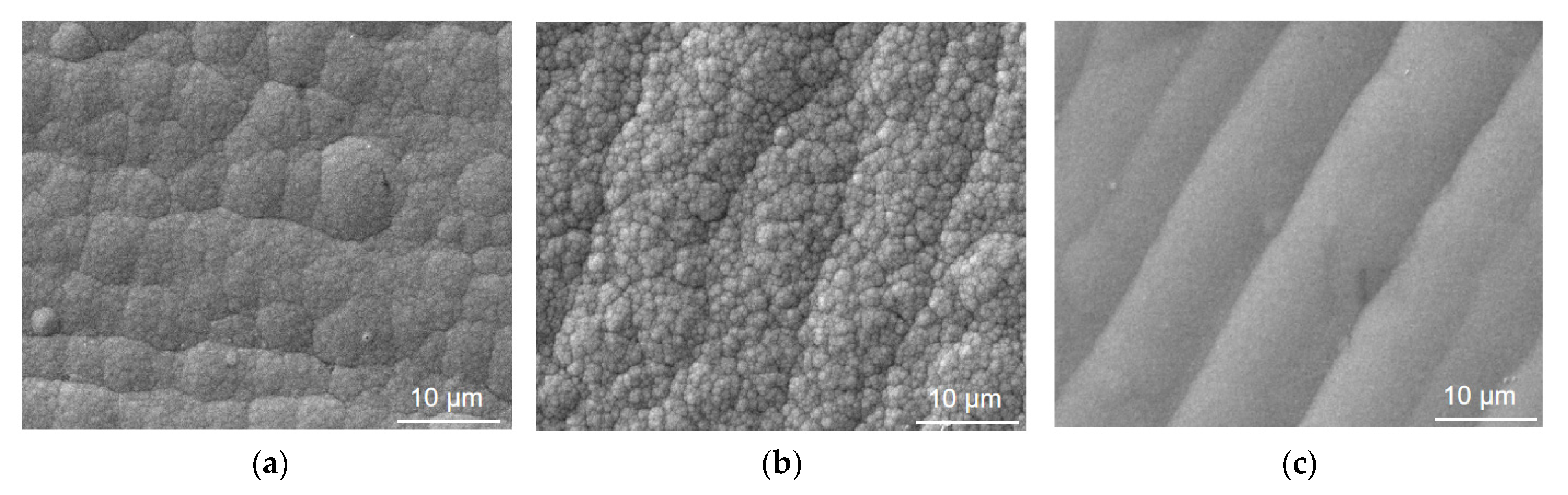
Figure 1 exhibits the typical SEM surface morphologies of the three Ni-based coatings on 6061 aluminum alloy.
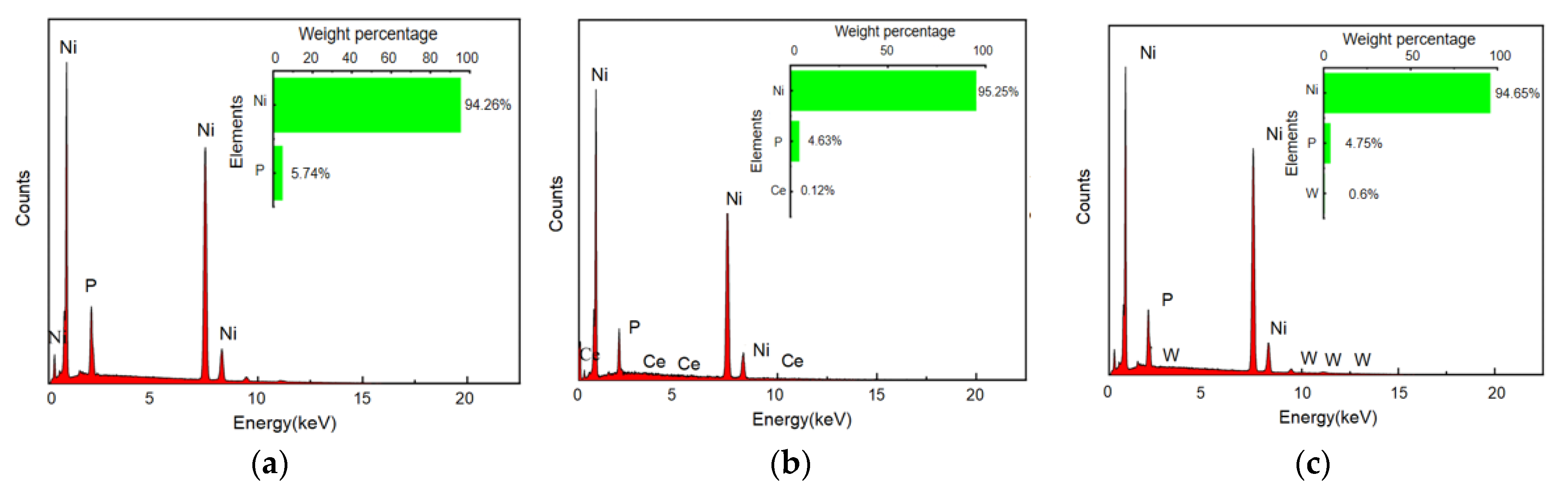
Figure 2 shows the EDS results of three Ni-based coatings without heat treatment. In the non-heat-treated state, the Ni-P coating presents a continuous, dense, cellular grain accumulation structure. The grain size is approximately 5–8 μm, arranged uniformly and tightly, with almost no visible macroscopic pores or cracks. This “cellular” structure is a typical characteristic of high-phosphorus Ni-P electroless coatings, formed by the nucleation of multiple fine crystallites in the initial deposition stage, followed by outward growth and mutual coverage of these cells to form quasi-spherical clusters. Additionally, the co-deposition of phosphorus hinders Ni grain growth, resulting in a partially amorphous structure that enhances overall density.
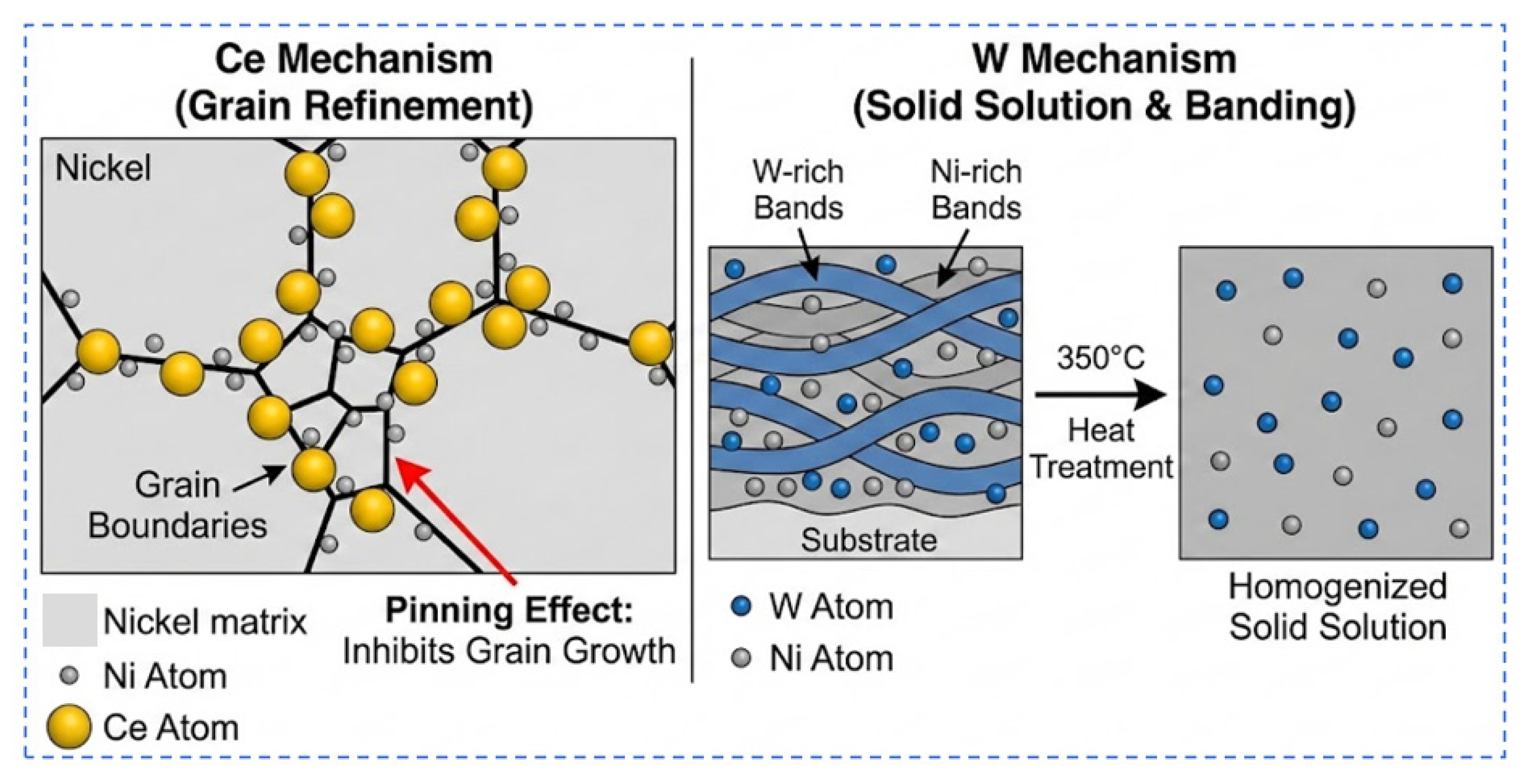
The Ni-Ce-P coating surface consists of a dense accumulation of numerous fine “nodular” (bread-loaf-like) particles. The grain size is significantly smaller than that of the Ni-P coating, making the coating finer and more uniform. This indicates that trace amounts of Ce element significantly refined the coating grains and improved deposition uniformity.
In contrast, the Ni-W-P coating surface presents an ordered “banded” (strip-like) structure. Fine grains are arranged along the direction of the substrate’s polishing texture, forming parallel “queues,” with clear interfaces visible between the bands. This preferred orientation strip morphology may be related to the distribution mode of tungsten in the plating solution: under specific conditions, the addition of W alters the nucleation and growth process of Ni-P, causing the deposit to periodically form lamellar or layered structures; furthermore, pre-treatment scratches on the substrate surface serve as “templates” for the ordered arrangement of nuclei. Overall, the addition of W and Ce did not introduce obvious defects but rather increased the coating density and refined the microstructure.
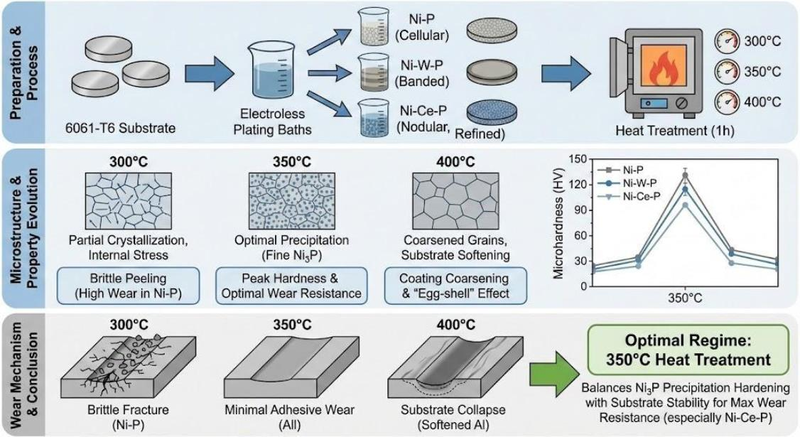
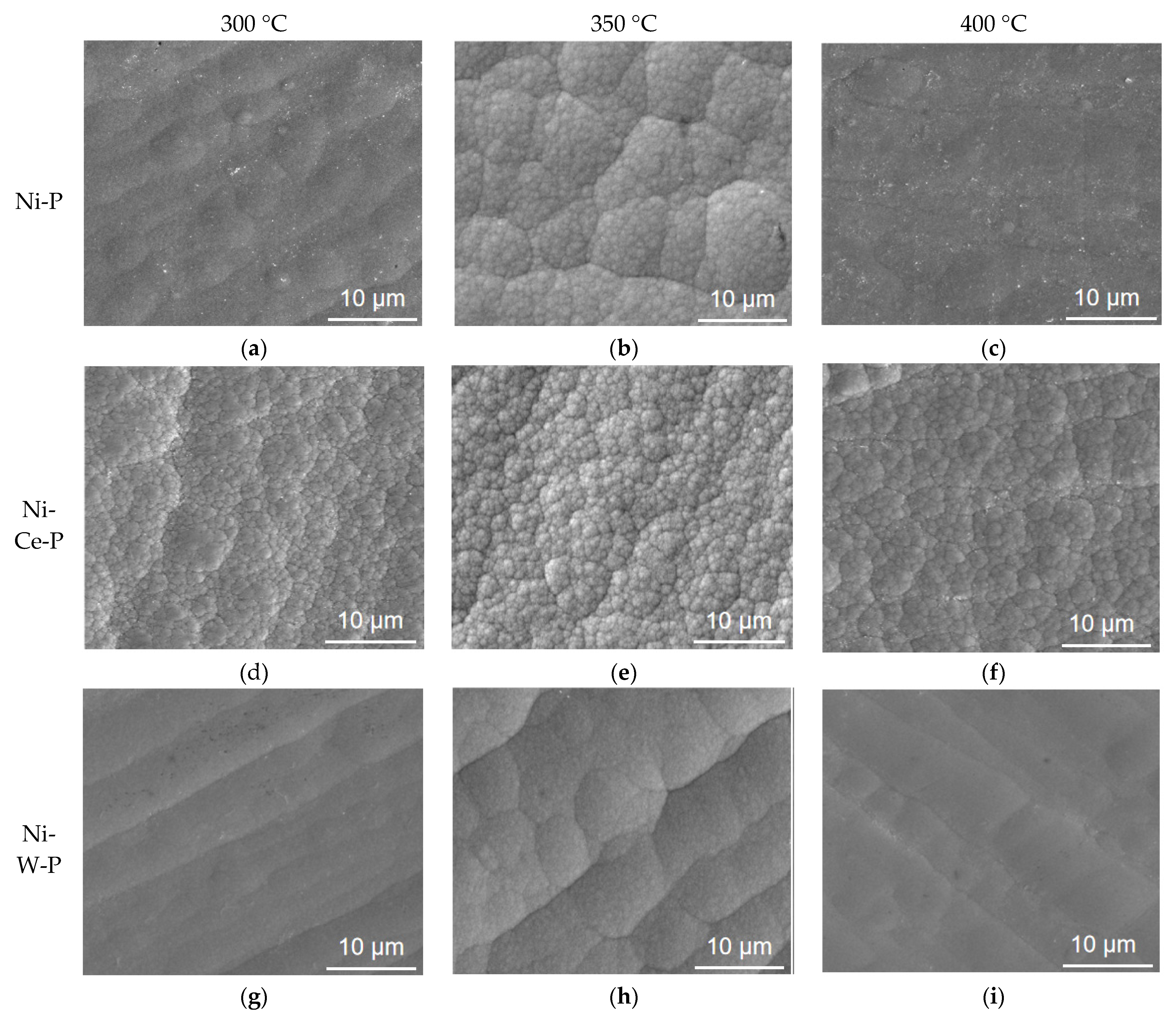
Figure 3 compares the SEM surface morphologies of the coatings under different heat treatment temperatures. For the Ni-P coating, heat treatment at 300 °C caused slight expansion of the original cellular grains and thinning of grain boundaries. While local porosity increased slightly, the overall surface became flatter and denser, maintaining continuous coverage. This suggests that low-temperature treatment promoted stress relaxation and atomic diffusion in the Ni matrix, causing the originally distinct cell boundaries to blur and fuse. Upon increasing the temperature to 350 °C, Ni-P grains continued to grow, with some fine grains merging into larger aggregates; however, the surface remained uniform and smooth without obvious cracks or large pores. At this stage, the coating achieved peak density and hardness. Further heating to 400 °C caused the cellular structure to become loose, with a significantly increased number of pores and cracks appearing. This indicates that high temperatures promoted significant grain coarsening and the release of greater internal stress, ultimately leading to the deterioration of the coating’s overall microstructure.
For the Ni-Ce-P coating, at 300 °C, only slight grain growth occurred, with most fine grains retaining their original size while fusing. At 350 °C, grains grew further, tending to form regular polygonal morphologies. The surface became smoother and denser, with no obvious holes or cracks observed. This indicates that moderate heat treatment promoted the transformation of the Ni-Ce-P structure towards a more uniform and dense state, maintaining good compactness while significantly enhancing hardness. However, at 400 °C, obvious cracks and holes began to appear on the surface, and grain size increased significantly: the excessively high temperature counteracted the beneficial grain-refining effect of the Ce element, and thermal stress exacerbated coating cracking.
In contrast, the Ni-W-P coating underwent more drastic structural evolution during heat treatment. At 300 °C, the banded structure began to disperse, and Ni grains showed a tendency to aggregate and grow; the interfaces between bands gradually blurred, and preferred orientation weakened. After 350 °C treatment, the banded structure almost completely disappeared. The surface roughness increased significantly, and visible cracks and holes appeared. This suggests that the Ni-W-P coating underwent significant recrystallization and stress release processes at medium-to-high temperatures, with micro-cracks easily initiating at grain boundaries. This is likely attributable to the high crystallization tendency and internal stress concentration of the initial amorphous structure of the Ni-W-P coating [
17]. Furthermore, the presence of W may lead to thermal expansion coefficient mismatch or the precipitation of brittle phases, resulting in crack defects at relatively lower temperatures. Simultaneously, W solid solution in the Ni matrix significantly alters crystallization kinetics and thermal stability, promoting the precipitation of Ni(W) solid solution and Ni
3P strengthening phases [
18].
The evolution of surface roughness with heat treatment temperature is summarized in
Table 3. For all coatings, the roughness initially decreases at 300 °C due to surface relaxation and then increases at higher temperatures, especially at 400 °C, which is consistent with the observed grain coarsening and defect formation.
In summary, the influence of heat treatment at different temperatures on the microstructure manifests as initially promoting densification and stress release, followed by inducing grain coarsening and increased defects. This pattern aligns with literature reports.
3.2. Effect of Heat Treatment on Phase Transformation
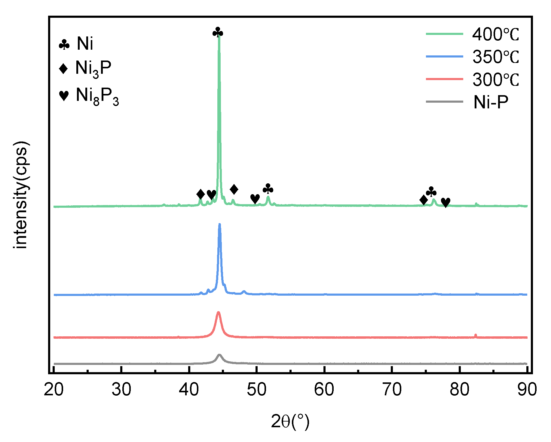
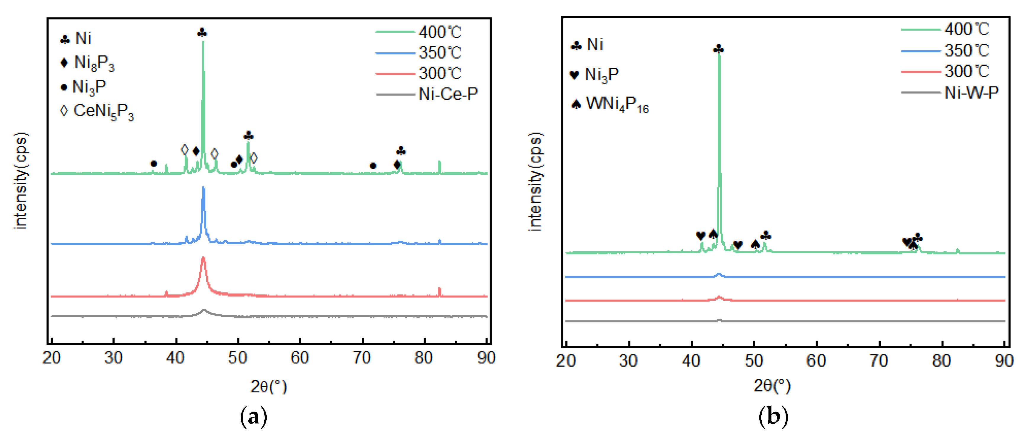
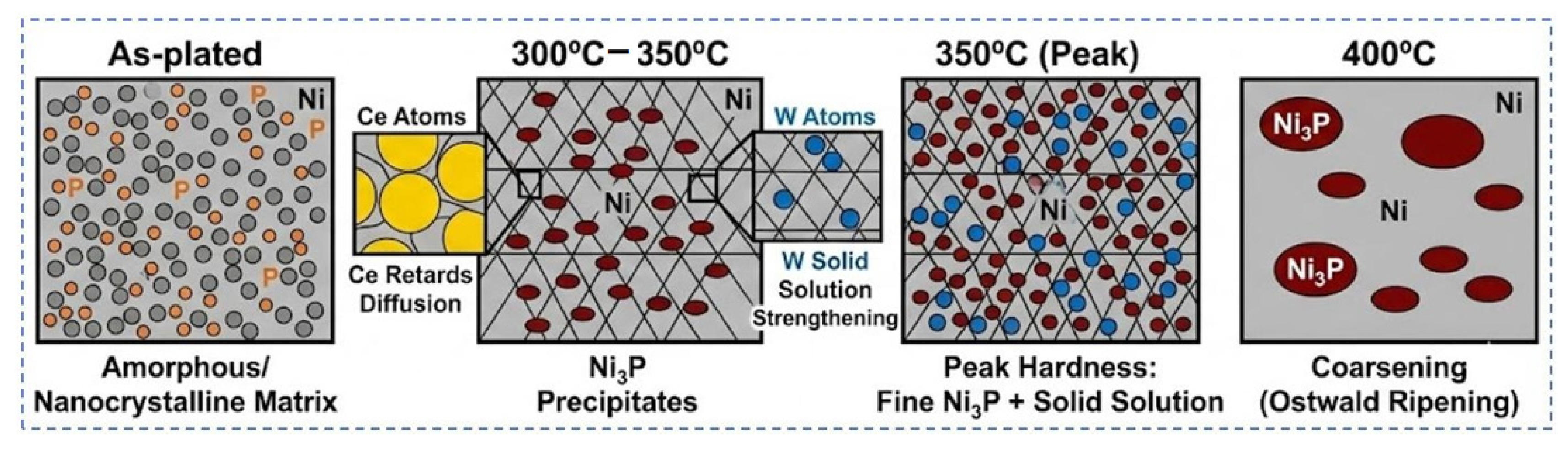
Figure 4 shows the XRD patterns of the coatings under different heat treatment conditions. For the Ni-P coating, the as-deposited XRD spectrum shows only a broadened diffuse peak around 2θ = 44°, with no obvious crystalline peaks, indicating the coating is primarily composed of an amorphous phase and a small amount of nanocrystalline Ni. This is consistent with the amorphous + microcrystalline mixed structure of typical medium-phosphorus Ni-P coatings. After 300 °C treatment, the sharpness of the broad diffraction peak increased slightly, accompanied by the appearance of several weak crystalline peaks, confirming partial crystallization and the initial precipitation of fine crystalline phases; however, diffraction peaks remained broad and low intensity due to limited precipitate content. This stage may involve the formation of metastable nickel phosphides. When the temperature rose to 350 °C, characteristic diffraction peaks for Ni
3P and Ni
8P
3 became clearly visible, and the Ni phase peaks intensified significantly. This indicates the massive precipitation of the stable hard phase Ni
3P, transforming the coating from an amorphous matrix to a crystalline structure of Ni + Ni
3P. At 400 °C, the intensity of Ni
3P peaks continued to increase and sharpen, confirming that the coating was nearly fully crystallized. However, despite the higher Ni
3P content, the hardness did not exceed the peak at 350 °C, suggesting that excessive coarsening of Ni
3P particles and defects generated by high-temperature treatment weakened the precipitation strengthening effect.
The phase transformation behaviors of Ni-Ce-P and Ni-W-P coatings (
Figure 5) were similar to Ni-P but with distinct differences. For Ni-Ce-P, the as-deposited state also presented an amorphous/microcrystalline structure. As the temperature increased from 300 °C to 400 °C, the amorphous diffuse scattering peak gradually weakened, and crystalline peaks strengthened. After treatment at 350 °C and above, Ni and Ni
3P peaks were clearly detected. Notably, the introduction of Ce exhibited an inhibitory effect on Ni
3P precipitation. Literature indicates that rare earth elements can improve the thermal stability of Ni-P coatings, raising the Ni
3P precipitation temperature. In this experiment, the limited hardness increase of Ni-Ce-P at 300 °C confirms Ce’s retarding effect on crystallization; however, at 350 °C, massive Ni
3P precipitation occurred, completing the strengthening transformation.
For the Ni-W-P coating, XRD results showed a phase evolution highly similar to Ni-P: from 300 to 400 °C, the amorphous broad peak sharpened, and Ni/Ni3P peak intensities increased, indicating continuous transformation to a crystalline state. No new W-related diffraction peaks appeared, suggesting W mainly existed as a solid solution in the Ni matrix or Ni3P phase. Literature confirms that small W additions to Ni-P typically do not form Ni-W intermetallics but dissolve in the Ni lattice.
In summary, heat treatment promotes the transformation of all three coatings from amorphous/microcrystalline to crystalline Ni solid solution + Ni3P hard phase. Compared to pure Ni-P, W and Ce incorporation delayed Ni3P precipitation: Ni-Ce-P showed a lag in crystallization peak temperature, while Ni-W-P required higher temperatures for complete crystallization. However, between 350 and 400 °C, all coatings eventually formed Ni3P, differing only slightly in the temperature of peak precipitation. This indicates that Ce atoms increased the undercooling required for Ni3P nucleation, while W influenced the diffusion rates of Ni/P atoms, without altering the final precipitate type.
3.3. Effect of Heat Treatment on Microhardness
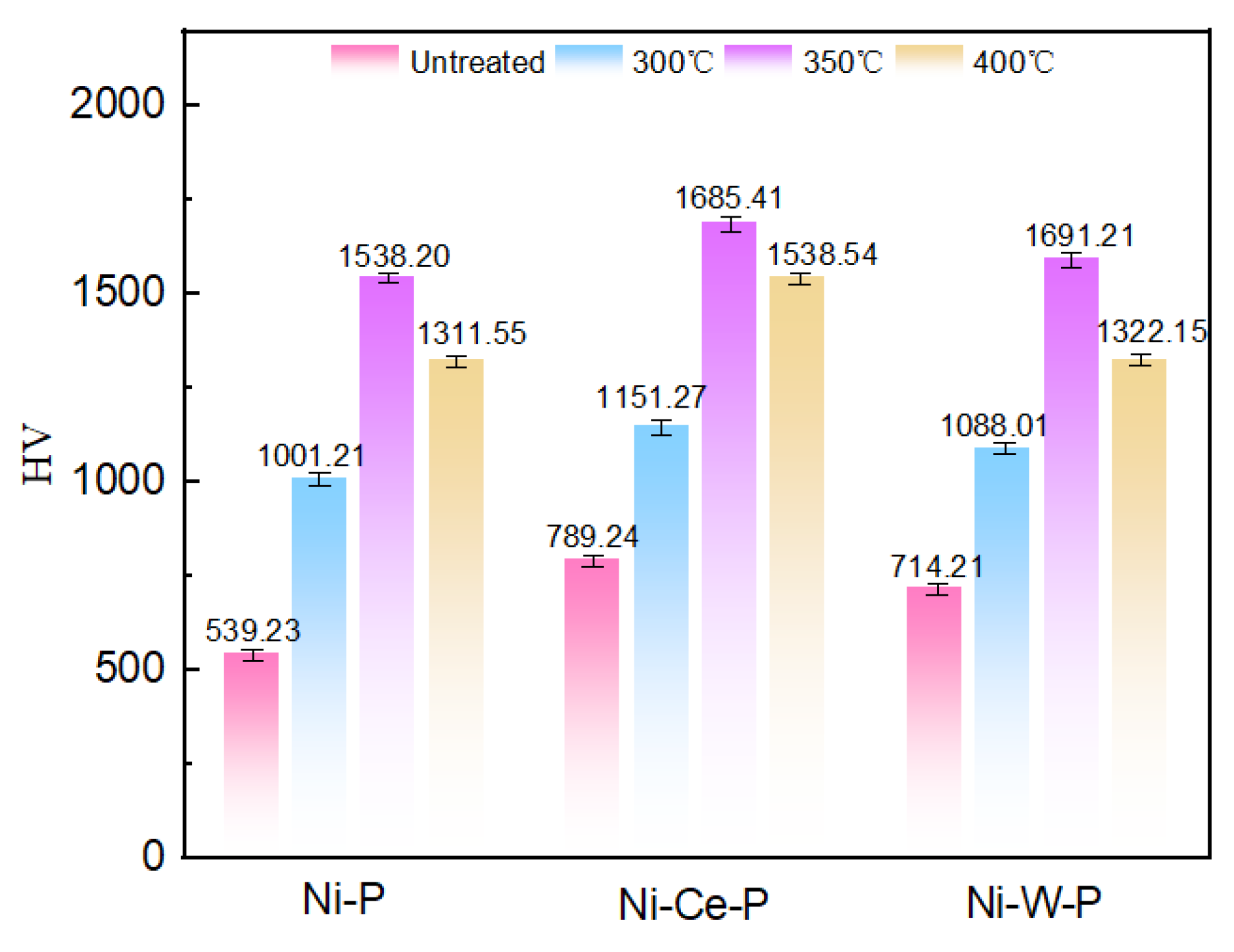
Figure 6 illustrates the Vickers hardness changes of the three coatings under different heat treatment conditions. The hardness of all three coatings in the non-heat-treated state was significantly higher than that of the 6061 aluminum alloy substrate. Specifically, the hardness of the Ni-P coating was approximately 539 HV
0.1, Ni-W-P was approximately 714 HV
0.1, and Ni-Ce-P reached the highest at 789 HV
0.1. This indicates that electroless plating formed a high-hardness Ni-P solid solution coating on the aluminum surface, significantly improving surface hardness compared to the soft aluminum substrate. The hardness of Ni-W-P and Ni-Ce-P coatings was higher than that of Ni-P because the addition of alloying elements introduced extra strengthening effects: W served as a solid solution strengthening element to increase the hardness of the Ni matrix, while Ce enhanced the hardening effect through grain refinement and possible dispersion strengthening.
After heat treatment, the hardness of all three coatings significantly increased. All coatings reached peak hardness at 350 °C: Ni-P hardness increased from 539 HV0.1 to approximately 1538 HV0.1 (an increase of about 1.85 times); Ni-W-P increased from 714 HV0.1 to 1691 HV0.1 (an increase of about 1.37 times); Ni-Ce-P increased from 789 HV0.1 to 1685 HV0.1 (an increase of about 1.13 times). At 350 °C, the hardness of the Ni-W-P coating was slightly higher than that of the Ni-Ce-P coating, with both reaching the highest hardness level of approximately 1690 HV0.1. This may be because the Ni-W-P coating had a higher initial degree of crystallinity, and at 350 °C, the Ni3P hard phase precipitated massively and dispersed uniformly, causing hardness to rise rapidly; while the Ni-Ce-P coating had the highest initial hardness, its Ni3P precipitation slightly lagged, resulting in a relatively smaller hardness increase.
The primary mechanism of hardness strengthening lies in the precipitation of the Ni3P hard phase during heat treatment. At approximately 350 °C, the coating undergoes a transformation process of “amorphous Ni-P matrix → metastable nickel phosphides → stable Ni + Ni3P crystalline phases.” Ni3P precipitates as fine particles from the amorphous matrix and is uniformly distributed within the coating. On one hand, these high-hardness particles effectively hinder dislocation movement, increasing the yield strength of the matrix; on the other hand, the hardness of Ni3P itself is extremely high, and its massive precipitation naturally increases the overall hardness of the coating. Additionally, lattice distortion during crystallization also provides certain solid solution strengthening. Therefore, the heat-treated Ni-based coatings possess both solid solution strengthening and precipitation strengthening mechanisms, making the hardness far superior to the original as-deposited level.
It is worth noting that the effect of different heat treatment temperatures on hardness exhibits an “over-aging” phenomenon. As seen in
Figure 6, after heat treatment at 300 °C, the coating hardness only increased slightly: Ni-P increased by about 0.85 times, Ni-Ce-P by about 0.45 times, and Ni-W-P by about 0.52 times, indicating that only a small amount of Ni
3P and metastable phases precipitated at this time, with limited strengthening effect; however, hardness jumped to the peak value when the temperature rose to 350 °C. When the temperature continued to rise to 400 °C, the hardness actually decreased compared to 350 °C: Ni-P decreased by about 14.3%, Ni-Ce-P decreased by about 8.6%, and Ni-W-P decreased by about 21.8%. Although the hardness after 400 °C treatment was still far higher than the untreated state, it had clearly fallen back from the peak. The reasons for the decrease in hardness may be two-fold: on one hand, high temperature promoted the further growth and aggregation of Ni
3P particles, reducing the precipitation strengthening effect [
19]; on the other hand, defects such as cracks and pores appearing in the coating at 400 °C reduced the actual load-bearing area of the coating, leading to a decrease in macroscopic hardness [
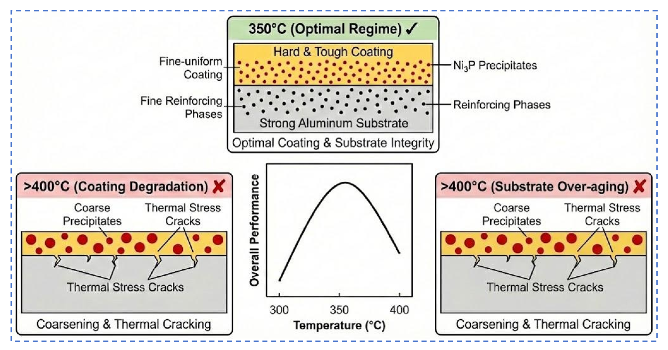
20]. Therefore, medium-temperature heat treatment around 350 °C is the optimal hardening condition for Ni-based coatings on 6061-T6 alloy, consistent with the recommended heat treatment temperature range of 345–400 °C in the literature [
21,
22]. Under this condition, the highest coating hardness and optimal microstructural state can be obtained.
3.4. Effect of Heat Treatment on Wear Resistance and Mechanism
Table 4 summarizes the wear rate test results for the substrate and the as-deposited coatings. The wear rate of the 6061 aluminum alloy substrate was approximately 1.03 × 10
−5 g/(N·m). All coatings significantly improved wear resistance. The wear rate of the Ni-P coating was 1.65 × 10
−6 g/(N·m), a reduction of about 84.0%. The Ni-W-P coating showed a wear rate of 2.66 × 10
−7 g/(N·m), a reduction of 97.4%. The Ni-Ce-P coating exhibited the best performance, with the lowest wear rate of 1.12 × 10
−7 g/(N·m), a reduction of 98.9% compared to the substrate. It is evident that Ni-based electroless coatings greatly improved the wear resistance of the aluminum alloy surface, with Ni-Ce-P coating showing the best effect, followed by Ni-W-P, and Ni-P being relatively weaker. This trend is highly correlated with the initial hardness of each coating; the Ni-Ce-P coating with the highest hardness had the best wear resistance, while the Ni-P coating with the lowest hardness had relatively weaker wear resistance. However, it must be emphasized that even the Ni-P coating with the lowest hardness reduced the wear rate by over 80% compared to the unplated aluminum substrate, indicating that electroless plating significantly improved the wear resistance of the aluminum alloy.
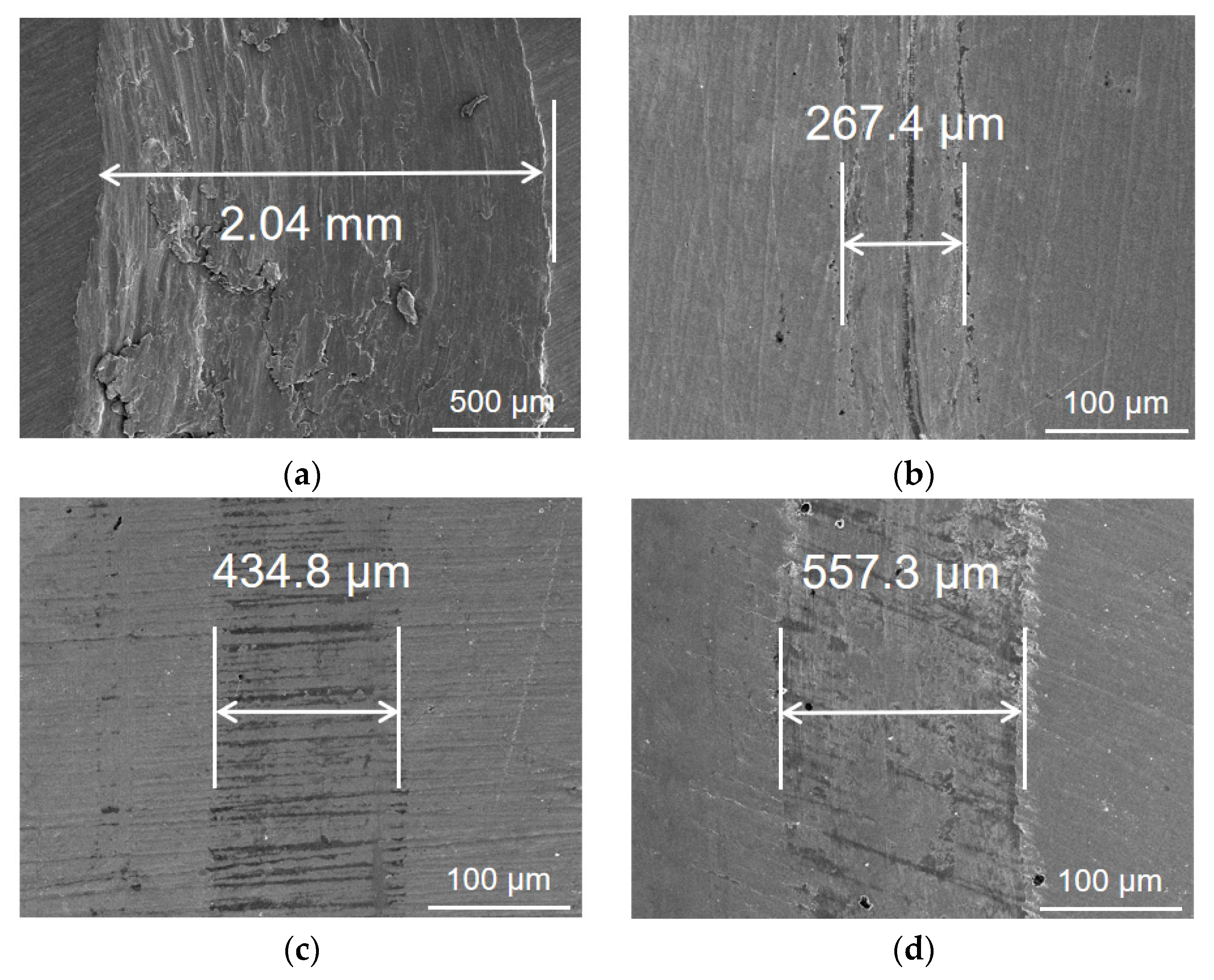
SEM observation of the wear scar morphology further revealed differences in wear mechanisms (
Figure 7). The unplated 6061 aluminum alloy surface showed deep plowing grooves about 2.1 mm wide and a large amount of massive debris after wear, typically characteristic of abrasive wear; while the wear scar width of the Ni-P plated sample was only about 281 μm, with shallower surface scratches and significantly reduced debris size. The wear scar width of the Ni-Ce-P coating was about 436 μm, and Ni-W-P was about 577 μm (wear scar width relates to coating hardness and microstructure, discussed later). Overall, the wear scar width of the coated samples was far smaller than the substrate, and the surface was relatively smooth without wide plowing grooves, producing mainly fine debris rather than large chunks of material. This indicates that the coatings mainly experienced slight surface abrasion during friction, rather than severe cutting on the substrate. The aluminum substrate, due to low hardness, allowed the grinding ball to penetrate deep into the material and grind out a large amount of plastic chips during friction, dominated by abrasive wear; whereas the hardened Ni-based coatings were harder, preventing deep penetration by the grinding ball, causing only microscopic scratches on the surface, accompanied by a small amount of adhesive transfer. Therefore, the wear mechanism of Ni-based coatings manifested as a “abrasive wear + adhesive wear” composite mode. The Ce and W elements added to the coating further improved wear behavior. The addition of Ce and W refined coating grains and increased hardness, reducing the depth of abrasive scratching; simultaneously, the presence of rare earth and transition metals may have altered the oxidation and tribochemical characteristics of the coating surface, reducing the tendency for severe adhesion. Consequently, regarding wear scar morphology, neither Ni-Ce-P nor Ni-W-P coatings showed large-area material peeling, and their debris was mainly in the form of fine particles. This characteristic is directly related to the higher hardness and more uniform, dense microstructure of these two coatings compared to the Ni-P coating.
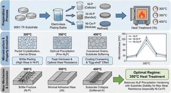
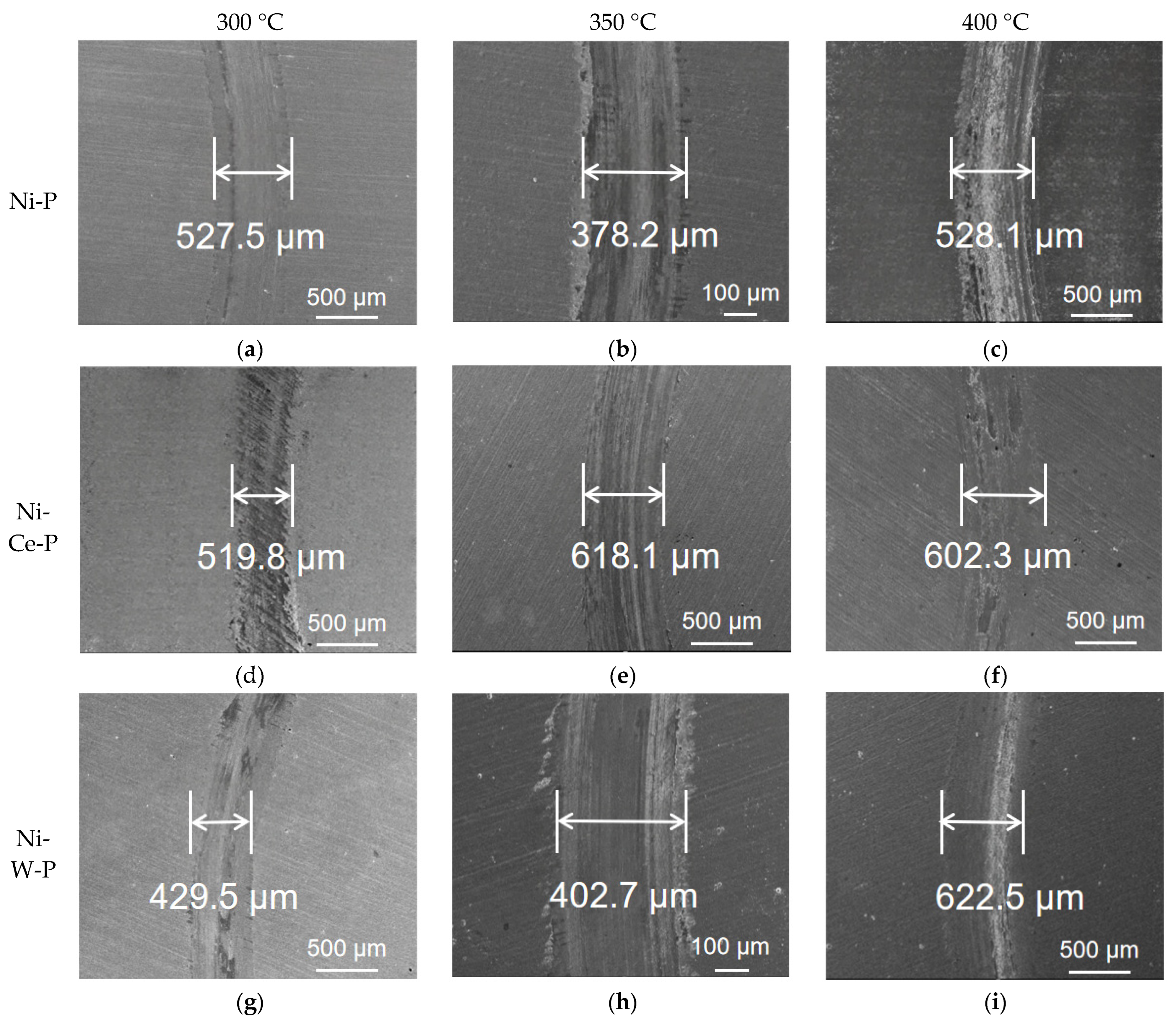
Heat treatment had a very significant effect on the wear resistance of the coatings, but it presented a non-monotonic trend of “deterioration–improvement–deterioration”. The changes in wear rate of the coatings are shown in
Table 5, and the microscopic morphology of wear scars is shown in
Figure 8.
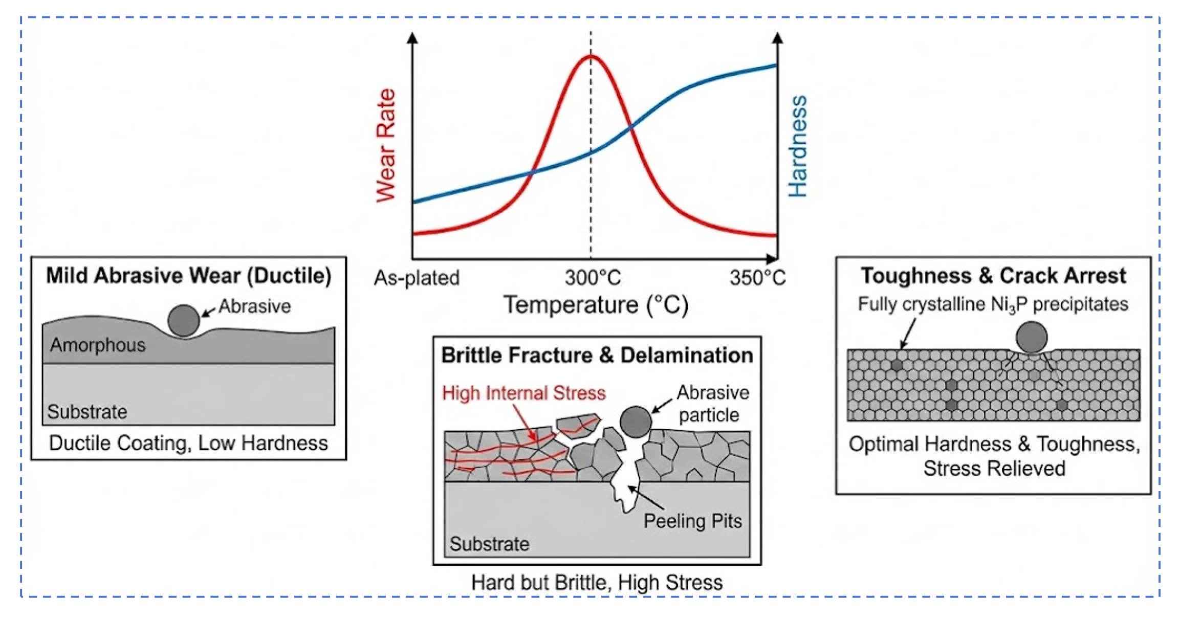
Ni-P: The wear rate was 1.79 × 10⁻⁶ g/(N·m) without heat treatment; after heat treatment at 300 °C, it surged to 9.53 × 10⁻⁵ g/(N·m), an increase of about 53 times, indicating significant deterioration in wear resistance; when the temperature was raised to 350 °C, the wear rate dropped to 5.14 × 10⁻⁶ g/(N·m), a reduction of about 94.6% compared to 300 °C, approaching the level of the untreated sample. Clearly, 350 °C heat treatment drastically improved the poor wear resistance at 300 °C. This is because at 300 °C, the coating only partially crystallized, precipitating some hard and brittle Ni3P and metastable phases while the matrix still retained a large amount of ductile Ni solid solution, causing the coating to have insufficient toughness despite increased hardness. During friction, it was prone to large-area peeling into coarse fragments, leading to a surge in wear volume. SEM observation confirmed that the wear scar of the Ni-P coating heat-treated at 300 °C had obvious peeling pits and large detached fragments, verifying the deteriorated wear phenomenon caused by coating embrittlement. After raising the heat treatment temperature to 350 °C, the wear rate of the Ni-P coating significantly decreased to 5.14 × 10⁻⁶ g/(N·m), almost recovering to the level of the unheated sample; at this time, the coating structure was more uniform and stable, with Ni3P hard phases fully precipitated and uniformly distributed. Hardness increased significantly while retaining certain toughness, enabling the coating to effectively resist abrasive plowing without overall peeling. Wear scar examination showed that the Ni-P coating treated at 350 °C only left fine scratches without obvious peeling pits, indicating a good balance between hardness and toughness. However, when the temperature further rose to 400 °C, the wear rate of the Ni-P coating rose again to 2.23 × 10⁻⁵ g/(N·m), an increase of about 11 times compared to the untreated state. Although the hardness of the coating at 400 °C was slightly lower than at 350 °C, the main problem was that grain coarsening and cracks caused by high temperature reduced the overall strength of the coating. Once subjected to friction load, material was easily peeled off along the crack propagation. Therefore, the wear resistance of the Ni-P coating deteriorated significantly at 400 °C.
Ni-Ce-P: This coating showed a different trend. The wear rate was lowest when untreated (1.10 × 10⁻⁷ g/(N·m)), but rose sharply to 4.87 × 10⁻⁶ g/(N·m) after 300 °C heat treatment (an increase of about 42 times), dropped to 2.68 × 10⁻⁶ g/(N·m)at 350 °C (about 23 times), and further slightly dropped to 2.27 × 10⁻⁶ g/(N·m) at 400 °C (about 20 times). That is, the wear resistance of the Ni-Ce-P coating recovered to some extent at high temperatures, but even with 400 °C heat treatment, its wear rate was still far higher than the untreated state. This may be related to the inhibition of Ni3P precipitation by the Ce element. The Ni-Ce-P coating did not increase much in hardness at 300 °C but had already become brittle, thus wear resistance decreased drastically; whereas at 400 °C, the Ni3P phase fully precipitated, and the coating hardness was close to 2.1 times that of the untreated state, and the structure remained relatively fine (compared to the coarsening degree of Ni-P and Ni-W-P), so the wear rate decreased relative to 350 °C.
Ni-W-P: The wear resistance change of the Ni-W-P coating was similar to Ni-P but with a smaller amplitude. At 300 °C, its wear rate rose from 2.70 × 10⁻⁷ g/(N·m) to 6.49 × 10⁻⁷ g/(N·m), dropped to 5.14 × 10⁻⁷ g/(N·m) at 350 °C, and rose to 9.37 × 10⁻⁷ g/(N·m) at 400 °C. Thus, the optimal wear resistance of the Ni-W-P coating appeared at 350 °C heat treatment. Although the wear rate was slightly higher than the untreated state, it was significantly better than at 300 °C and 400 °C. Compared with Ni-P, the deterioration of wear resistance of the Ni-W-P coating at 300 °C was smaller, because its original structure was more crystalline, and the addition of W improved the thermal stability of the coating, resulting in fewer brittle defects generated at low temperatures. However, at 400 °C, wear resistance declined again due to grain growth and cracks.
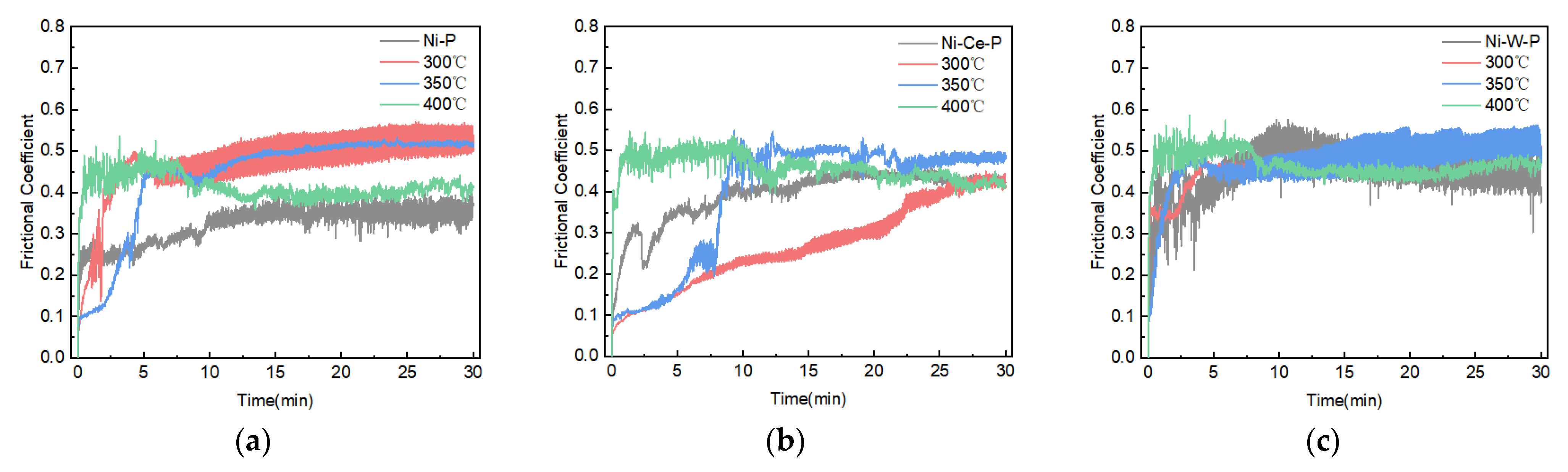
Figure 9 shows the real-time friction coefficient curves for the coatings heat-treated at the optimal temperature of 350 °C. The Ni-Ce-P coating exhibited the lowest and most stable friction coefficient (≈0.45), followed by Ni-W-P (≈0.52) and Ni-P (≈0.56). The lower friction coefficient of the Ni-Ce-P coating can be attributed to its fine-grained, dense structure, which promotes the formation of a stable tribo-layer and reduces adhesive interactions. The higher friction coefficients of the other coatings correspond to their relatively rougher surfaces and different wear mechanisms.
The above results indicate that the relationship between coating hardness improvement and wear resistance is not a simple linear one; wear resistance depends on the synergistic optimization of hardness and toughness. In the initial stage of hardness increase, if the coating toughness is insufficient to withstand the wear load, brittle peeling will primarily occur, leading to increased wear volume; when hardness reaches a specific threshold and the microstructure tends to be stable, the coating can effectively resist abrasive cutting while suppressing macroscopic peeling, at which point the wear volume reaches a minimum; however, continued temperature increase or prolonged holding time induces micro-cracks, after which the coating easily peels along crack propagation, causing the wear volume to rise again. It can be seen that there is an optimized temperature range for the heat treatment process to achieve synergistic control of high hardness and stable structure. In this study, this optimized temperature range is approximately 350 °C. It is worth noting that even under conditions where heat treatment parameters were mismatched leading to decreased wear resistance, the overall wear resistance of the heat-treated coatings was still significantly superior to that of the aluminum alloy substrate.