Article

Atomic Layer Deposition of Zirconia on Cobalt–Chromium Alloys for Dental Prosthetics: Surface Functionalization Under MDR 2017/745

by
Anna Ziębowicz
1,* and
Mirosława Pawlyta
2
1
Department of Biomaterials and Medical Devices Engineering, Silesian University of Technology, 41-800 Zabrze, Poland
2
Department of Engineering Materials and Biomaterials, Silesian University of Technology, 44-100 Gliwice, Poland
*
Author to whom correspondence should be addressed.
Coatings 2025, 15(9), 994; https://doi.org/10.3390/coatings15090994
Submission received: 28 July 2025 / Revised: 12 August 2025 / Accepted: 14 August 2025 / Published: 27 August 2025

Abstract

The primary goal of this study was to assess the suitability of the proposed method for modifying the surface of cobalt alloys in dental prosthetics, taking into account the specific characteristics of the stomatognathic system during long-term use and their impact on physicochemical properties and the adhesion of cariogenic bacteria such as Streptococcus mutans. Technological factors influencing the quality of the product and its final dimensional characteristics were considered, confirming or ruling out the possibility of iatrogenic errors (related to poorly shaped prostheses) occurring during laboratory fabrication. This study demonstrates that atomic layer deposition of ZrO2 on CoCr dental alloys results in a chemically stable, uniform, and protective surface layer, reducing ion release and improving surface quality. These improvements address key safety and performance requirements outlined in MDR 2017/745, supporting the use of ALD as a state-of-the-art technique for functionalizing dental prosthetic devices. Such coating development may influence the final quality of the denture and also verify its suitability for use in the oral environment (reducing the likelihood of denture stomatitis).

Graphical Abstract

1. Introduction

Directive 93/42/EEC from 14 June 1993 concerning medical devices (MDD—Medical Devices Directive) served as the legal basis for regulating both the marketing and use of medical devices in European Union countries for over two decades [1]. One of its main goals was to harmonize regulations across member states and ensure the free movement of products on the market while maintaining the highest possible level of patient health protection. Manufacturers of medical devices were obligated to design and manufacture products in a manner that ensured safety and effectiveness in accordance with the current state of technical and medical knowledge. Before a product could be placed on the market, a conformity assessment had to be conducted, often with the participation of a notified body, whose positive verification was the basis for granting the CE marking.
On 5 May 2017, the Regulation (EU) 2017/745 of the European Parliament and of the Council on medical devices (MDR) was published [2], replacing the previously applicable Directive 93/42/EEC. This regulation aimed to increase the level of safety, primarily for patients and users, and to introduce more stringent requirements for conformity assessment and placing on the market. The MDR applies in all European Union countries; implementation into national law is not necessary. A significant aspect of the new Regulation is the introduction of more precise and detailed provisions regarding the chemical composition of products and the presence of substances considered hazardous. This is the first time that a clear entry can be seen regarding substances that are carcinogenic, mutagenic, and reprotoxic (CMR) and substances that disrupt the functioning of the endocrine system (EDC) [3,4].
The quality of a prosthetic restoration, which influences treatment outcomes and meets the unique aesthetic and biocompatibility requirements, depends on the technologies and materials used. A suitable base material will fulfill the therapeutic and preventive objectives of a prosthetic design that is integrated into the stomatognathic system. This task has taken on a new dimension with the implementation of radical changes to regulations regarding the safety of medical devices in 2020. According to the new standards, cobalt has been classified as a CMR substance (Cat. 1B, Mutagenic 2, Toxic to Reproduction 1B) and limited to a concentration of 0.1% in the final product. With Co concentrations ranging from 31 to 63% in alloys used for partial dentures, this assumption becomes problematic, as it directly impacts the use of Co–Cr dental alloys [5]. For this reason, considering both the physicochemical properties of the materials used to manufacture dentures and the material’s reactivity in the oral cavity has become a priority. This is also true for plaque accumulation, bacterial settlement on denture surfaces, and the allergenic properties of denture materials [6].
Biological reactions to dental alloys depend on the alloys’ susceptibility to corrosion and the release of ions, which can be washed away by saliva [6]. However, prolonged contact of the denture with the oral environment reduces the body’s ability to perform physiological functions. In the case of upper dentures, where the palate is not in contact with saliva, the released metal ions accumulated under the metal frame can cause local irritation, hypersensitivity, or allergic reactions, thus contributing to the development of a complex of pathological changes within the oral mucosa known as denture stomatitis. Importantly, the solubility of cobalt ions increases in the presence of exudates from allergic vesicles containing amino acids. Along with wear particles, their excess can be toxic to the body if the mechanisms for efficient utilization and excretion fail. Therefore, it seems important to distinguish between physiological amounts of these metals and amounts that cause reactive complications [7].
A literature analysis confirmed that various methods and types of surface modifications are a way to improve biocompatibility [8,9,10]. To limit microbial colonization of denture surfaces, ultrathin coatings applied by the Atomic Layer Deposition (ALD) method are increasingly being used. Due to its high antibacterial potential, zinc oxide is of particular interest to researchers, as it increases resistance to the adhesion of microorganisms such as Escherichia coli and Staphylococcus aureus on 316L steel and Ti6Al4V alloy surfaces. Huang et al., in their study [11], deposited a uniform TiO2 layer ~1 μm thick on a Co–Cr alloy. The coating reduced the contact angle and caused virtually complete inhibition of Candida albicans growth compared to an uncoated surface. The increased hydrophilicity of the surface hinders biofilm adhesion and may, therefore, reduce the risk of oral diseases.
The results of the cited studies can be considered a new strategy for preventing bacterial biofilm formation, encompassing intervention in the initial phase of biofilm formation and modification of biomaterials aimed at increasing resistance to microbial adhesion. They also inspired this study, as they provide a beneficial factor in reducing the risk of microbially influenced/induced corrosion (MIC) of metal prosthetic restorations.
The possibility of coating large-scale surfaces of three-dimensional objects with complex shapes, as well as the significantly reduced microbial colonization of zirconium dioxide surfaces confirmed in the literature [12,13], led to the proposal to combine both approaches by applying a ceramic ZrO2 coating using the ALD method onto cobalt–chromium alloy substrates (commercially used for partial dentures) intended for contact with the oral cavity. The primary goal of this research was to assess the suitability of the proposed method for modifying the surface of cobalt alloys in prosthetic dentistry, taking into account the specific characteristics of the stomatognathic system during long-term use and their impact on physicochemical properties and the adhesion of secondary caries bacteria such as Streptococcus mutans (dominant in supragingival bacterial plaque). Technological factors influencing the quality of the product and its final dimensional characteristics were considered, confirming or ruling out the possibility of iatrogenic errors (related to poorly shaped prostheses) occurring during laboratory fabrication. Furthermore, developing conditions for producing coatings that affect the final quality of the prosthesis, along with verifying their suitability for use in the oral environment (reducing the likelihood of prosthetic stomatitis), provides a new approach to these issues.

2. Materials and Methods

In this study, the base biomaterials were three different cobalt-based alloys used to fabricate skeletal prostheses using a specific technology. The following were selected: a casting alloy (Gialloy PA), a milling alloy (CopraBond K), and an incremental DMLS alloy (EOS CoCr RPD) [14,15,16]. The chemical composition of all materials complied with standard recommendations [17]. To ensure the required physicochemical properties, the surfaces of the cast and printed samples (after cutting off the sprue channels and supports) were subjected to grinding and electrolytic polishing. Contaminants from the surfaces of the milled samples were cleaned ultrasonically and with steam after cutting off and smoothing the connector areas. A 50 nm thick ZrO2 coating was applied using Atomic Layer Deposition (ALD). The final stage included a process of cyclical temperature changes (55 °C/5 °C) simulating 6-month use of the prosthesis to achieve a realistic simulation of oral temperature changes during eating (Thermocycler Model 1140, SD Mechatronik GmbH, Feldkirchen-Westerham, Germany).

2.1. Phase Composition of the Surface Layer

Structural studies of ZrO2 thin films deposited on various CoCr biomedical alloy substrates were performed using a WITec CRM alpha 300R confocal Raman microscope (WITec, Ulm, Germany) equipped with an air-cooled solid-state laser (λ = 532 nm, laser power 10 mW) and a CCD camera. Excitation laser radiation was introduced into the microscope through a polarization-maintaining single-mode optical fiber with a diameter of 50 μm. The laser radiation was focused on the sample by a long-range Olympus MPLAN objective (100x/0.90NA), while the scattered light passed through a multimode optical fiber (diameter 50 μm). Due to the high transparency of the film, depth-scanning profiling was performed to find the strongest signal. Raman spectra were then accumulated over the range of 120–4000 cm−1 with 20 scans, an integration time of 20 s, and a spectral resolution of 3 cm−1, using a 600-line/mm grating monochromator. The spectrometer monochromator was calibrated using the emission lines of a Ne lamp, while the silicon wafer signal (520.7 cm−1) was used to check beam alignment. Post-processing analysis, including background correction and cosmic ray removal, was performed using WITecProjectFive Plus software (WITec, version 5.1., Ulm, Germany). Peak fitting was performed using the Lorentz–Gaussian function in the GRAMS 9.2 software package to separate monoclinic (m), tetragonal (t), and cubic (c) ZrO2. Additionally, XPS photoemission studies were performed on PHI5700/660 Physical Electronics spectrometer (Chanhassen, MN, USA); the results for DMLS samples were previously reported in [18].

2.2. Microstructure of the Surface Layer

SEM examinations were performed on a ZEISS SUPRA 35 scanning electron microscope (Carl Zeiss Microscopy, Oberkochen, Germany) equipped with an EDS spectrometer for chemical composition analysis. Samples for TEM examination were prepared using the Focused Ion Beam (FIB) technique using a Thermo Fischer Scientific (formerly FEI) Helios NanoLab™ 600i SEM/Ga-FIB microscope (TFS, Waltham, MA USA). TEM examinations were performed using a Thermo Fischer Scientific (formerly FEI) S/TEM TITAN 80–300 transmission electron microscope (TFS, Waltham, MA USA) equipped with a STEM scanning system, BF, DF, and HAADF scanning-transmission detectors, a CEOS CETCOR Cs condenser spherical aberration corrector, a Gatan Ultrascan camera, an EDAX EDS energy dispersive spectrometer, and a Gatan Tridiem GIF energy filter. CrystalMaker and SingleCrystal software (CrystalMaker Software Limited, version 10.4.1, Oxfordshire, UK) were used to simulate the crystal structure and electron diffraction patterns. This publication focuses mainly on presenting the test results obtained for coatings produced on individual substrates.

2.3. Wettability and Surface Energy

Contact angle and surface energy measurements were performed using the sessile drop method using an OEG Surftens Universal goniometer (OEG GmbH, Frankfurt, Germany) with Surftens 4.3 software (OEG GmbH, Frankfurt, Germany) to analyze the captured drop image. A drop of distilled water and diiodomethane, each with a volume of 1.5 mm3, was applied to the surface of each sample. The measurement began 20 s after the drop landed on the surface. The duration of one measurement was 60 s with a sampling frequency of 1 Hz. Measurements were performed 10 times at room temperature (T = 25 °C ± 1 °C). Based on the obtained results, the average contact angle for water (θwśr) and diiodomethane (θdav) was determined. The Owen–Wendt method was used to determine the surface free energy (SEP) (γs).

3. Results

3.1. Phase Composition of the Surface Layer

Tetragonal and monoclinic zirconia varieties are considered the most interesting for biomedical applications. One non-invasive and relatively rapid, structure-dependent technique for distinguishing these two phases is Raman spectroscopy. Therefore, the structural image of the layers deposited by ALD revealed the coexistence of monoclinic and tetragonal ZrO2. Monoclinic ZrO2 was characterized by a sharp doublet at 174 and 190 cm−1 and weak bands at 335, 380, 476, 558, and 624 cm−1. In turn, well-separated bands around 229, 267, 318, 456, and 640 cm−1 indicated the tetragonal variety.
The Raman results suggest the existence of at least a two-layer system with monoclinic zirconia crystallized in the first stage of ALD deposition. Furthermore, the band positions are only slightly shifted compared to the values available in the literature [19,20,21] and indicate layers with a more extensive structure [22,23]. The slight discrepancies between the coatings on CoCr substrates and the data available in the literature resulted from orientation effects related to crystallization. A closer analysis revealed a band around 285 cm−1 for ZrO2/CoCr (PRINT and CAST), which was not detected on the milled substrate, and which resulted in the crystallization of a single nanometric tetragonal zirconia layer on previously formed structures [21]. The higher peak intensity for the “CAST” suggests a potentially thicker layer than for the printed material and indicates a variable substrate composition favoring a more diverse structure. Other unresolved bands were attributed to zirconium, with non-overlapping bands at 244, 305, 433, and 550 cm−1, and overlapping tetragonal bands at 149 and 607 cm−1 (Figure 1). Throughout the spectroscopic pattern of the ALD coatings, the cubic phase with a disordered oxygen sublattice [21,24,25,26] appeared to recrystallize in the final phase of zirconium deposition. As a result, Raman analysis indicated the presence of a four-layer zirconium dioxide system stabilized at room temperature in the casts and prints. At the same time, coatings grown on milled substrates appeared to be less differentiated monoclinic, tetragonal, and cubic systems. Estimation of monoclinic (xm) and tetragonal (xt) content in bioactive zirconia thin films calculated using a slightly modified empirical Formula (1) given by Clarke and Adar [23]
x m [ % ] = I m 174 + I m 190 0.97 I t 269 + I t 285 + I t 640 + I c 246 + I c 301 + I m 174 + I m 190
and Godlewski et al. [27]
x t [ % ] = I t 269 + I t 285 + I t 640 I t 269 + I t 285 + I t 640 + I c 246 + I c 301 + I m 174 + I m 190
where I represents the integrated intensity of individual bands, while the subscripts m, t, and c denote the monoclinic, tetragonal, and cubic phases, respectively. The phase content determination was based on the non-overlapping band signals. Therefore, the presented modified formula also included cubic and nanosized signals of tetragonal zirconia. Some differences in band positions compared to the literature resulted from varying orientations of the experimental conditions or the crystallites forming the zirconia film [28]. Using empirical formulas, the monoclinic zirconia content was estimated at 4% (PRINT) and 12% (MILL), while the tetragonal content was estimated at 72% (PRINT) and 65% (MILL). The 6% (monoclinic) and 66% (cubic) values for the cast indicate the greatest thickness of zirconia formed during the ALD process.

3.2. Microstructure of the Surface Layer

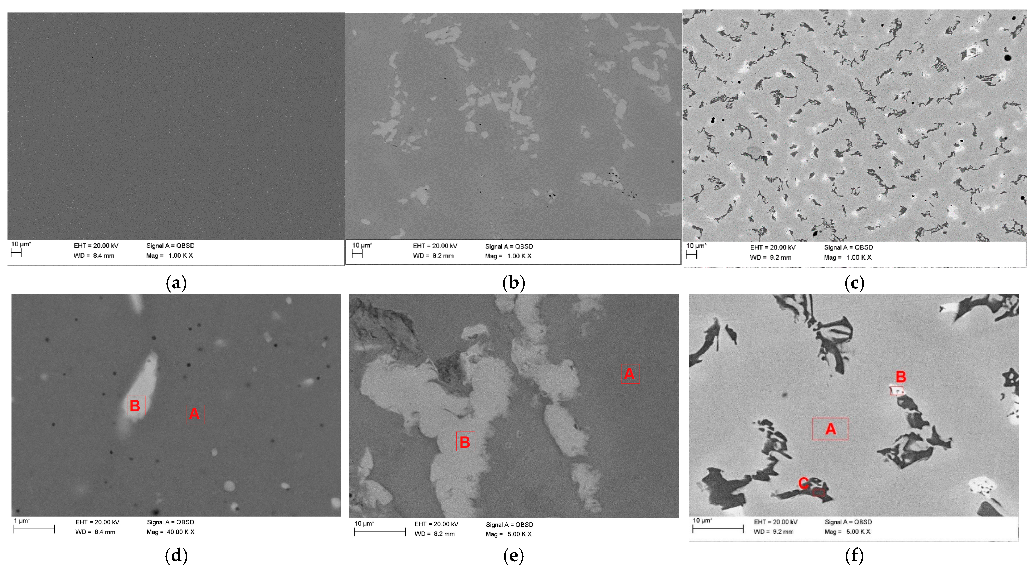
A BSE (Back Scattered Electrons) detector was used to image the microstructures, revealing chemical contrast. Typical microstructures of CoCr casting alloys are visible in Figure 2b,c,e,f. Light precipitates (enriched in elements with a higher atomic number than the matrix) of irregular shape are visible in the images of the MILL sample (Figure 2e). Their size can be estimated at 1 to 100 μm. The CAST sample contains two types of precipitates: light precipitates (enriched in elements with a higher atomic number than the matrix), several micrometers in size, and dark precipitates (containing a higher concentration of light elements). The dark precipitates are in the form of regularly arranged dendrites (Figure 2f). Compared to the matrix, they contain less Co and Cr but more Mo and W (Table 1). In the MILL sample, the bright precipitates also contain a higher concentration of Mo and W. The matrix of the CAST sample does not contain W. This element is also absent in the precipitates. The bright precipitates of the CAST sample have a significantly increased Mo content compared to the matrix, while the dark precipitates have a lower Mo content and a high chromium content (Table 1).
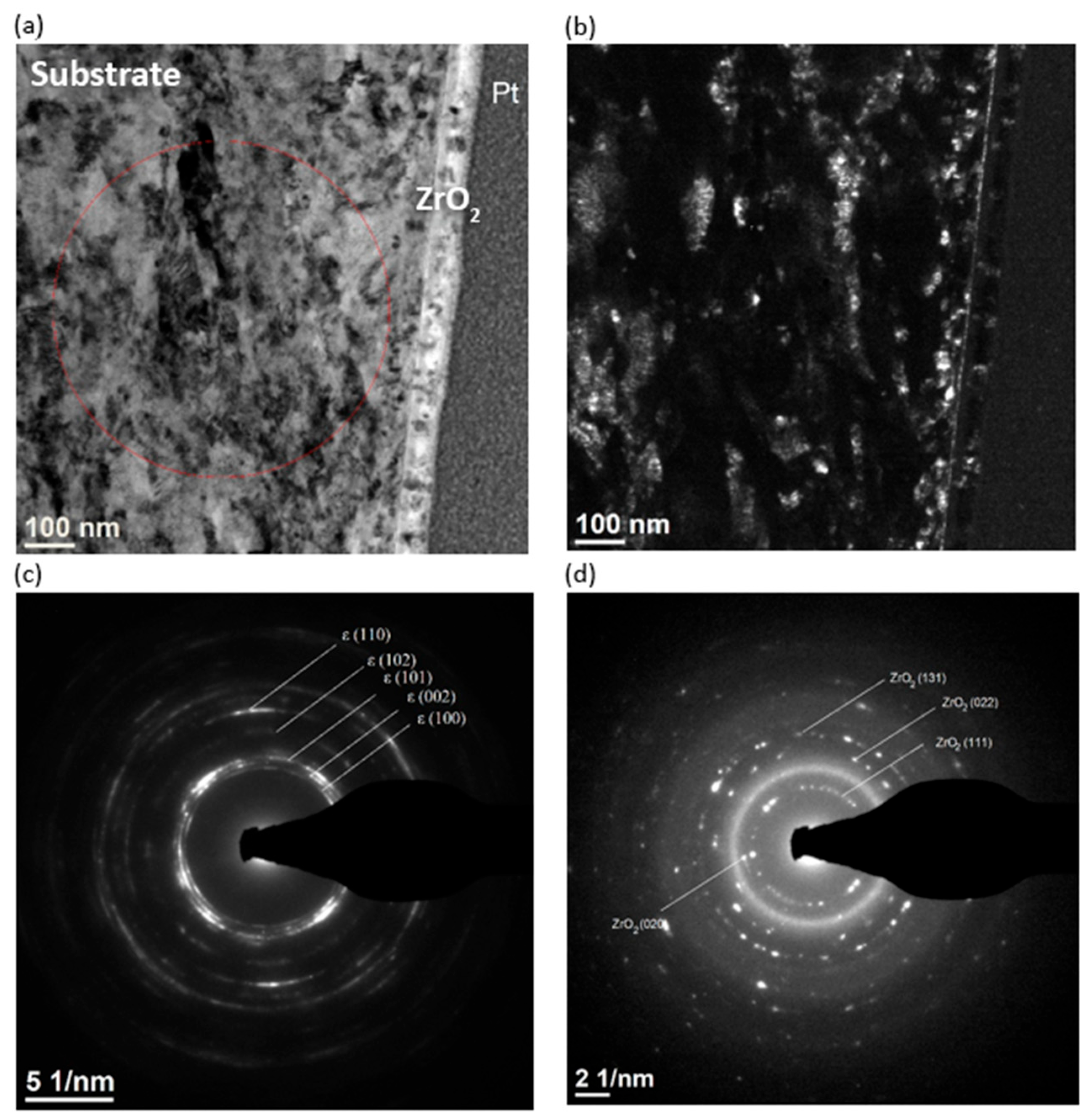
The ZrO2 coating on the MILL substrate is shown in Figure 3. The lamella was cut from the area of the substrate containing the epsilon phase and covered with a ZrO2 coating. The coating has a uniform thickness of 50 nm (Figure 3b) and adheres well to the substrate. Chemical composition analysis using the EDS technique (Figure 3c) showed a weight content of Co 56%, Cr 25%, Si 2%, Mo 7%, and W 10%. The presence of Zr and O was confirmed in the coating. The substrate has a nanocrystalline structure (Figure 4), which was confirmed by electron diffraction (Figure 4c). SAED electron diffraction also confirmed the phase composition of the analyzed fragment—the ε phase (hexagonal close-coupled lattice (hcp) with lattice constants a = 0.25071 nm and c = 0.40686 nm [28]). The dark-field image (Figure 4b) shows crystallites elongated in one direction, with a size of about 100–300 nm. Electron diffraction of the coating (Figure 4d) confirmed the presence of the cubic ZrO2 phase with the lattice constant a = 0.509 nm [29].
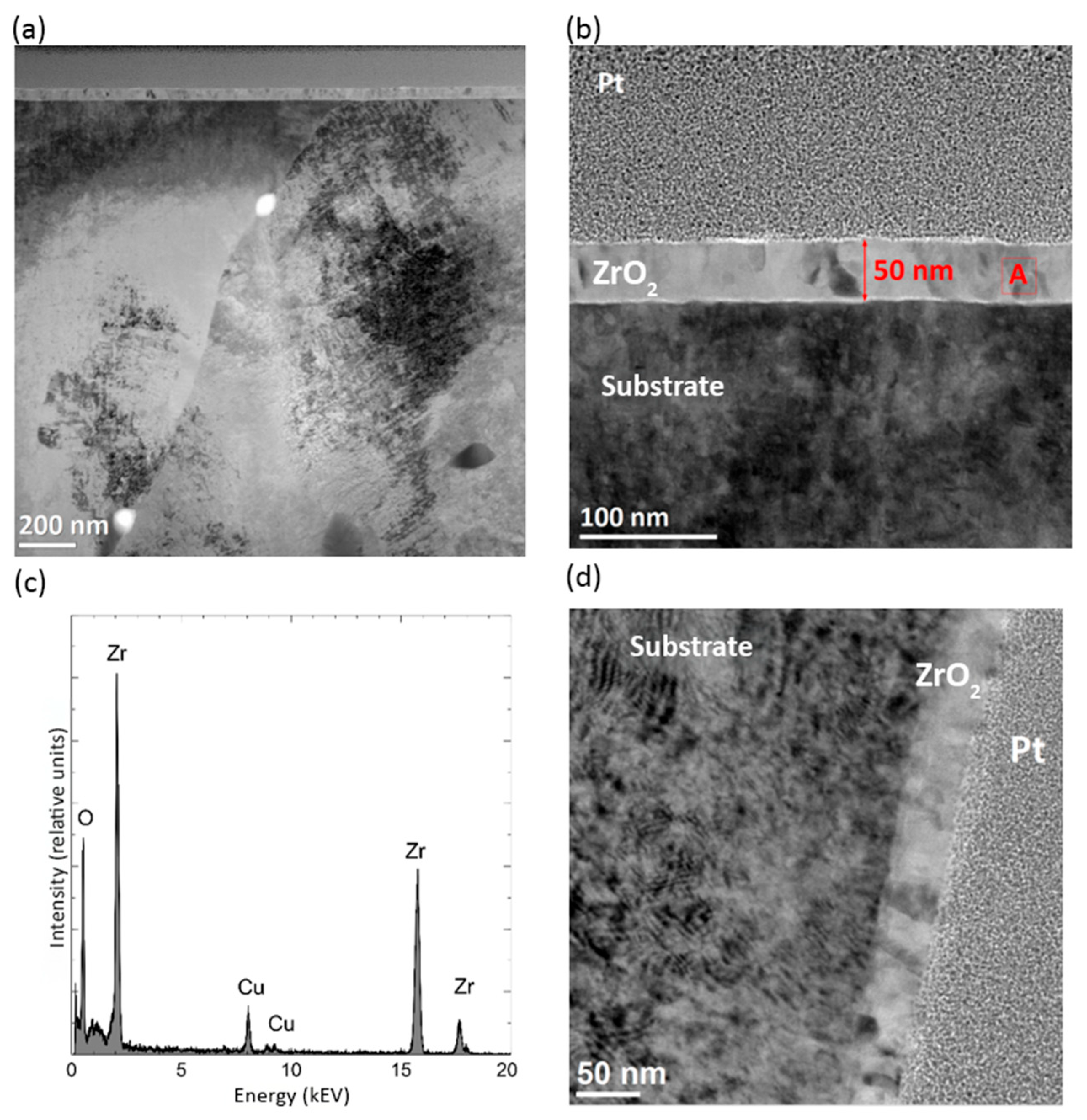
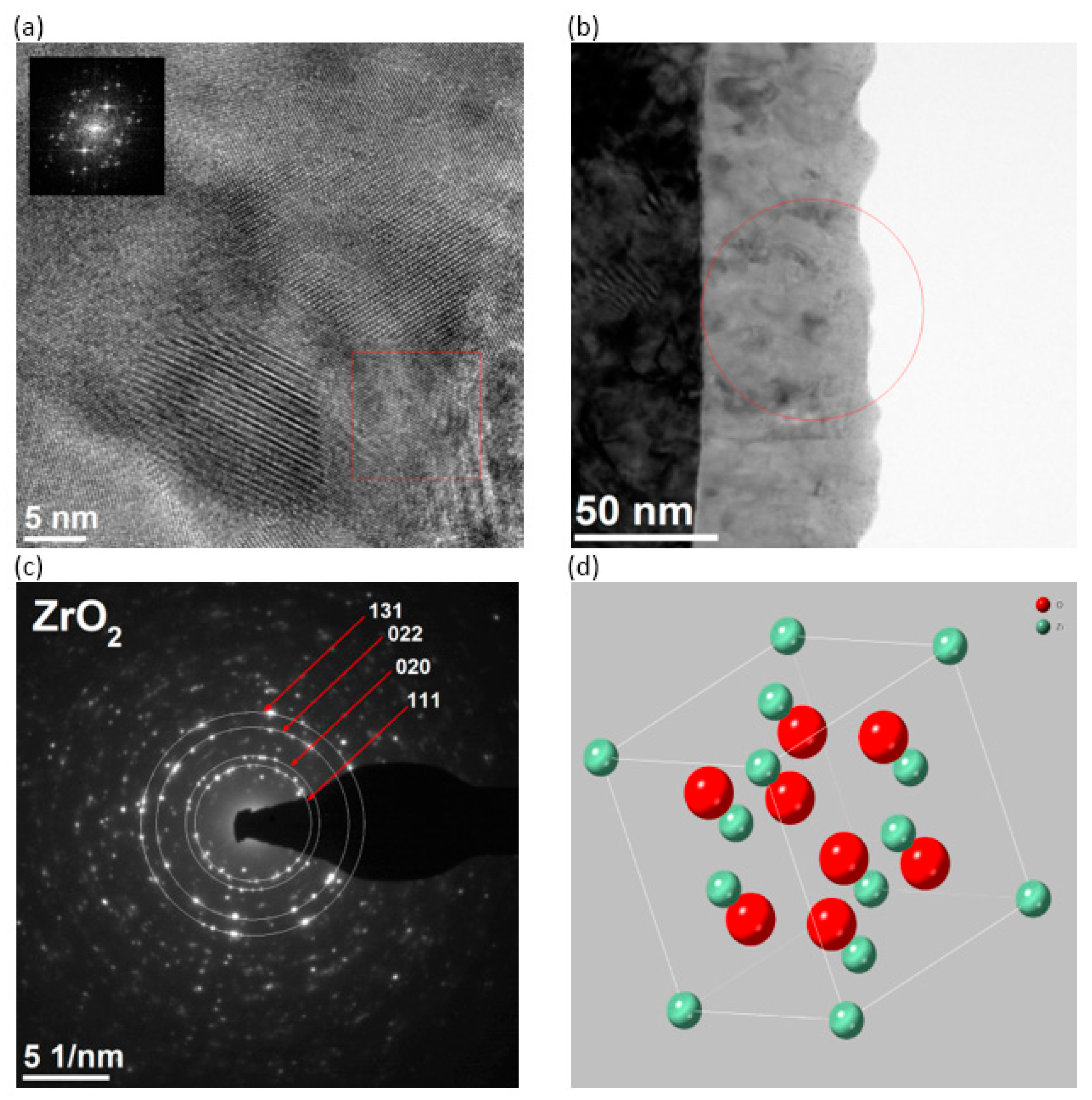
Microscopic observations confirmed the homogeneity of the deposited coating on the PRINT sample and its good adhesion to the substrate (Figure 5a). The coating is 50 nm thick (Figure 6b) and contains Zr and O (Figure 5c). The EDS spectra show a peak indicating Cu content, which can be ignored (due to the presence of this element in the sample holder and microscope pole pieces). Contrast changes visible in the BF images (Figure 5a,b,d) indicate a polycrystalline structure. The polycrystalline structure of the coating is clearly visible in the HRTEM images (Figure 6a). The grain size is approximately 10–30 nm. To determine the crystalline structure, a lamella was used, in which the ion beam removed virtually the entire platinum protective layer. This enabled the selection of an area of the sample containing only the coating using a selective diaphragm (Figure 6b). Based on the obtained electron diffraction, the presence of the cubic ZrO2 phase with lattice constant a = 0.509 nm was confirmed [29]. The unit cell model is shown in Figure 6d.

3.3. Wettability and Surface Energy

The study results are presented in Table 2. The obtained varied values of the θ angles indicate the influence of surface modification and exposure to the oral environment on wettability and surface energy. No significant differences in the θ angle values were observed depending on the treatments applied after a given technological process. In all cases, the surface was hydrophilic. Application of the ZrO2 coating resulted in an increase in the θ contact angle, thus changing the surface’s character to hydrophobic. To assess the degree of interaction between the material and oral tissues, the surface free energy was determined. SEF is considered an indicator providing preliminary information on cell adhesion and a measure of the chemical and biological resistance of the material surface to biodegradation. It is generally believed that surfaces characterized by high surface energy values also demonstrate good cell adhesion. The diverse oral microflora, composed of over 700 species of Gram-positive and Gram-negative bacteria, both aerobic and anaerobic, colonizes tooth surfaces in the form of a multispecies biofilm. This forms dental plaque, which causes caries and periodontal disease. These microorganisms can also be a causative factor in soft tissue inflammation [30]. The weakest affinity of Streptococcus mutans was observed for ceramic surfaces [31,32]. Based on this analysis, it was found that applying a ZrO2 coating reduced the surface free energy of the cast and printed material (electrolytic polishing). However, an increase in SFE values was observed for the milled material (no finishing).
It should be noted that the polar component γsp (in all cases of modified surfaces) constitutes a smaller share of the overall value than the dispersion component γsd. Since the polar component is the sum of components originating from interactions between molecules, there is a high probability of reducing the adhesion of, e.g., bacteria residing in the oral cavity environment, which may be a desirable phenomenon [33].

4. Discussion

The presented research focused on obtaining an answer to whether the applied modification technology allows for obtaining a repeatable thickness of the ZrO2 coating regardless of the manufacturing technology and the CoCr alloy used. The expected verification was successful, in line with the literature reports [34,35], namely that ALD offers nanoscale precision, uniformity, and excellent conformality, which are critical for complex geometries of removable partial dentures. This modification also meets the MDR requirements regarding the safety of surfaces of materials in contact with tissues and enables the achievement of repeatable, validated results, which is important when introducing class IIa/IIb medical devices to the EU market.
These modifications address critical MDR requirements:
Annex I, Chapter II—Risk Management: Reduced microbial adhesion lowers infection risk [16].;
Annex I, Chapter III—Performance Requirements: Coatings maintain structural integrity and do not interfere with the primary function;
Annex I, Chapter VI—Information Supplied: The applied coating process must be traceable and documented for conformity assessment;
Annex XIV, Clinical Evaluation—in that context (future studies required): In vitro test results support clinical data when coatings are used on proven materials (Co–Cr). Furthermore, according to MDCG 2020-6 [4], surface modifications (nano/coating) are considered "significant changes" and require separate technical documentation. This highlights the need for reproducible coating processes in accordance with ISO 13485 [36].
Because the conducted studies were preliminary in nature, certain considerations regarding future research arise. The presented results were conducted to verify the suitability of the proposed modification of the surface layer of cobalt–chromium alloys for dental prosthetic applications and to increase their safety in the oral cavity. Therefore, it will be very interesting to determine the differences in the release of metal ions when prosthetic restorations are exposed to an environment enriched with bacteria, which appear in a persistent biofilm layer after the release of toxins by the pioneering bacteria Streptococcus mutans. Such a complex biofilm on the alloy surface will allow for more reliable results regarding the hypothetical effect of biocorrosion induced by the released enzymes [6]. Measurements of the electrokinetic properties of cobalt alloys in an aqueous electrolyte solution are also planned. This appears to be worthwhile, as they play a significant role in processes that minimize the likelihood of adverse phenomena (local and general reactivity), directly related to the presence of dentures in the oral cavity (physiological pH changes caused by nonphysiological conditions induced by the denture plate, e.g., limited oxygen supply).
The cognitive achievement of this work was the demonstration of the relationship between the surface quality of the cobalt alloy (obtained by a given technological process and appropriate post-processing procedures) and the production of a new generation coating on it.

5. Conclusions

Based on these results, the following generalizations can be formulated:
  • For the selected cobalt–chromium alloys, slight differences in their chemical composition and substrate structure, additionally modified by surface treatment, do not significantly affect the final thickness and quality of the ZrO2 coating;
  • The universal low-temperature process method used allows for obtaining a coating with comparable performance characteristics and biocompatibility and can be promoted for clinical applications in the production of new-generation removable prosthetic restorations in relation to the scenario assuming the implementation of high quality and safety standards for removable partial dentures, in the context of the use of CMR substances in the dental alloy and the applicable package of regulations (MDR, REACH, CLP) [2,5];
  • ZrO2 coatings applied by the ALD method could be a potential method of surface modification for cobalt–chromium alloys, improving corrosion protection and creating an antifungal and bacterial barrier for removable partial dentures.

Author Contributions

Conceptualization, A.Z.; data curation, A.Z.; investigation, A.Z. and M.P.; methodology, A.Z. and M.P.; project administration, A.Z.; resources, A.Z. and M.P.; supervision, A.Z.; visualization, A.Z.; writing—original draft, A.Z.; writing—review and editing, A.Z. All authors have read and agreed to the published version of the manuscript.

Funding

The Article Processing Charge was financed under the European Funds for Silesia 2021–2027 Program co-financed by the Just Transition Fund—project entitled “Development of the Silesian biomedical engineering potential in the face of the challenges of the digital and green economy (BioMeDiG)”. Project number: FESL.10.25-IZ.01-07G5/23.

Institutional Review Board Statement

Not applicable.

Informed Consent Statement

Not applicable.

Data Availability Statement

The data presented in this study are available on request from the corresponding author.

Acknowledgments

Some of the preliminary data included in this article were initially presented (in Polish) in a university-level monograph: “Powłoka ZrO2 na podłożu stopów kobaltowych jako czynnik minimalizujący stomatopatie protetyczne”, Silesian University of Technology, Gliwice, 2023. The current manuscript significantly extends and updates this work for the purpose of publication in an international peer-reviewed journal.

Conflicts of Interest

The authors declare no conflicts of interest.

References

  1. Council Directive 93/42/EEC of 14 June 1993 Concerning Medical Devices. Available online: https://eur-lex.europa.eu/LexUriServ/LexUriServ.do?uri=CONSLEG:1993L0042:20071011:en:PDF (accessed on 12 August 2025).
  2. Regulation (EU) 2017/745 of The European Parliament and of The Council of 5 April 2017. Available online: https://eur-lex.europa.eu/eli/reg/2017/745/oj/eng (accessed on 12 August 2025).
  3. Ziębowicz, A.; Woźniak, A.; Ziębowicz, B. The Influence of Technology on the Physicochemical and Electrochemical Properties of the Prosthetic Materials. In Proceedings of the Innovations in Biomedical Engineering IBE, Zabrze, Poland, 19–20 October 2017; Gzik, M., Tkacz, E., Paszenda, Z., Piętka, E., Eds.; Springer: Cham, Switzerland, 2018; pp. 349–357. [Google Scholar]
  4. MDCG 2020-6 Medical Device Coordination Group Document. Regulation (EU) 2017/745: Clinical Evidence Needed for Medical Devices Previously CE Marked Under Directives 93/42/EEC or 90/385/EEC. Available online: https://health.ec.europa.eu/system/files/2020-09/md_mdcg_2020_6_guidance_sufficient_clinical_evidence_en_0.pdf (accessed on 12 August 2025).
  5. Available online: https://echa.europa.eu/en/home (accessed on 12 August 2025).
  6. Lucchetti, M.C.; Fratto, G.; Valeriani, F.; De Vittori, E.; Giampaoli, S.; Papetti, P.; Spica, V.R.; Manzon, L. Cobalt-chromium alloys in dentistry: An evaluation of metal ion release. J. Prosthet. Dent. 2015, 114, 602–608. [Google Scholar] [CrossRef]
  7. Shapiro, J.A.; Eskildsen, S.M.; Del Gaizo, D.J. Systemic cobaltism manifesting as oral mucosal discoloration and metallic gustation after metal-on-metal hip resurfacing. Arthroplast. Today 2018, 16, 436–440. [Google Scholar] [CrossRef] [PubMed] [PubMed Central]
  8. Woźniak, A.; Adamiak, M. The Electrochemical Impedance Spectroscopy and wettability analysis of Ti6Al4V alloy modified by Atomic Layer Deposition method. Acta Phys. Pol. A 2022, 142, 88–91. [Google Scholar] [CrossRef]
  9. Walke, W. Surface Phenomena on Titanium Substrates with SiO2 Coating in the Circulatory System; Silesian University of Technology Publishing House: Gliwice, Poland, 2017. (In Polish) [Google Scholar]
  10. Liu, Z.; Zhu, Y.; Liu, X.; Yeung, K.W.K.; Wu, S. Construction of poly (vinyl alcohol)/poly (lactide-glycolide acid)/vancomycin nanoparticles on titanium for enhancing the surface self-antibacterial activity and cytocompatibility. Colloids Surf. B Biointerfaces 2017, 151, 165–177. [Google Scholar] [CrossRef]
  11. Huang, L.; Jing, S.; Zhuo, O.; Meng, X.; Wang, X. Surface hydrophilicity and antifungal properties of TiO2 films coated on a Co-Cr substrate. BioMed Res. Int. 2017, 2054723. [Google Scholar] [CrossRef]
  12. Mohseni, P.; Sou, A.; Ramos Chrcanovic, B. Clinical outcomes of zirconia implants: A systematic review and meta-analysis. Clin. Oral Investig. 2024, 28, 15. [Google Scholar] [CrossRef]
  13. Kui, A.; Manziuc, M.; Petruțiu, A.; Buduru, S.; Labuneț, A.; Negucioiu, M.; Chisnoiu, A. Translucent zirconia in fixed prosthodontics—An integrative overview. Biomedicines 2023, 11, 3116. [Google Scholar] [CrossRef]
  14. Uggowitzer, P.J.; Magdowski, R.; Speidel, M.O. Nickel free high nitrogen austenitic steels. ISIJ Int. 1996, 36, 901–908. [Google Scholar] [CrossRef]
  15. EOS. Instruction for Use & Technical Data EOS CobaltChrome RPD; EOS: Seattle, WA, USA, 2018. [Google Scholar]
  16. Ziębowicz, A.; Matus, K.; Pakieła, W.; Matula, G.; Pawlyta, M. Comparison of the crystal structure and wear resistance of Co-Based alloys with low carbon content manufactured by Selective Laser Sintering and Powder Injection Molding. Crystals 2020, 10, 197. [Google Scholar] [CrossRef]
  17. ISO 22674; Dentistry—Metallic Materials for Fixed and Removable Restorations and Appliances. International Organization for Standardization: Geneva, Switzerland, 2016.
  18. Ziębowicz, A.; Sambok-Kiełbowicz, A.; Walke, W.; Mzyk, A.; Kosiel, K.; Kubacki, J.; Bączkowski, B.; Pawlyta, M.; Ziębowicz, B. Evaluation of bacterial adhesion to the ZrO2 atomic layer deposited on the surface of cobalt-chromium dental alloy produced by DMLS method. Materials 2021, 14, 1079. [Google Scholar] [CrossRef]
  19. Xin, Q. Phase Transformation in the Surface Region of Zirconia Detected by UV Raman Spectroscopy. J. Phys. Chem. B 2001, 105, 8107–8111. [Google Scholar] [CrossRef]
  20. Shi, L.; Tin, K.; Wong, N. Tetragonal zirconia (t-ZrO2) begins to change into monoclinic. J. Mater. Sci. 1999, 34, 3367. [Google Scholar] [CrossRef]
  21. Basahel, S.N.; Ali, T.T.; Mokhtar, M.; Narasimharao, K. Influence of crystal structure of nanosized ZrO2 on photocatalytic degradation of methyl orange. Nanoscale Res. Lett. 2015, 10, 73. [Google Scholar] [CrossRef]
  22. Pezzotti, G.; Porporati, A.A. Raman spectroscopic analysis of phase-transformation and stress patterns in zirconia hip joints. J.Biomed. Opt. 2004, 9, 372–384. [Google Scholar] [CrossRef]
  23. Clarke, D.R.; Adar, F. Measurement of the crystallographically transformed zone produced by fracture in ceramics containing tetragonal zirconia. J. Am. Ceram. Soc. 1982, 65, 284–288. [Google Scholar] [CrossRef]
  24. Gazzoli, D.; Mattei, G.; Valigi, M. Raman and X-ray investigations of the incorporation of Ca2+ and Cd2+ in the ZrO2 structure. J.Raman Spectrosc. 2007, 38, 824–831. [Google Scholar] [CrossRef]
  25. Chervin, C.N.; Clapsaddle, B.J.; Chiu, H.W.; Gash, A.E.; Satcher, J.H.; Kauzlarich, S.M. Aerogel synthesis of yttria-stabilized zirconia by a non-alkoxide sol-gel route. Chem. Mater. 2005, 17, 3345–3351. [Google Scholar] [CrossRef]
  26. Kontoyannis, C.G.; Orkoula, M. Quantitative determination of the cubic, tetragonal and monoclinic phases in partially stabilized zirconias by Raman spectroscopy. J. Mater. Sci. 1994, 29, 5316–5320. [Google Scholar] [CrossRef]
  27. Pêcheur, D.; Lefebvre, F.; Motta, A.T.; Lemaignan, C.J.; Wadier, F. Precipitate evolution in the Zircaloy-4 oxide layer. J. Nucl. Mater. 1992, 189, 318–332. [Google Scholar] [CrossRef]
  28. Beltrán, J.; Hafner, J.; Muñoz, M.C. Structural, electronic and magnetic properties of the surfaces of tetragonal and cubic HfO2. New J. Phys. 2008, 10, 2–20. [Google Scholar] [CrossRef]
  29. Katz, G. Crystal Structure of ZrO2. J. Am. Ceram. Soc. 1971, 54, 531. [Google Scholar] [CrossRef]
  30. Lin, Y.; Chen, J.; Zhou, X.; Li, Y. Inhibition of Streptococcus mutans biofilm formation by strategies targeting the metabolism of exopolysaccharides. Crit. Rev. Microbiol. 2021, 47, 667–677. [Google Scholar] [CrossRef]
  31. Pasich, E.; Walczewska, M.; Pasich, A.; Marcinkiewicz, J. Mechanism and risk factors of oral bacterial biofilm formation. Adv. Hyg. Exp. Med. 2013, 67, 736–741. (In Polish) [Google Scholar]
  32. Kozmos, M.; Virant, P.; Rojko, F.; Abram, A.; Rudolf, R.; Raspor, P.; Bohinc, A.; Klemen, Z. Bacterial adhesion of Streptococcus mutans to dental material surfaces. Molecules 2021, 26, 1152. [Google Scholar] [CrossRef] [PubMed]
  33. Hjerppe, J.; Rodas, S.; Korvala, J.; Pesonen, P.; Kaisanlahti, A.; Özcan, M.; Suojanen, J.; Reunanen, J. Surface roughness and Streptococcus mutans adhesion on metallic and ceramic fixed prosthodontic materials after scaling. Materials 2021, 14, 1027. [Google Scholar] [CrossRef]
  34. Boryło, P.; Łukaszkowicz, K. ZnO thin films prepared by Atomic Layer Deposition. Mater. Eng. Mater. Inźinierstvo 2017, 24, 21–25. [Google Scholar]
  35. Basiaga, M.; Walke, W.; Lisoń, J.; Taratuta, A.; Szewczenko, J.; Ziębowicz, B.; Szindler, M.; Dyner, A. Tin dioxide in terms of physical properties on steel AISI 316 LVM. Mater. Werkst. 2022, 54, 517–525. [Google Scholar] [CrossRef]
  36. ISO 13485; Medical Devices—Quality Management Systems—Requirements for Regulatory Purposes. International Organization for Standardization: Geneva, Switzerland, 2016.
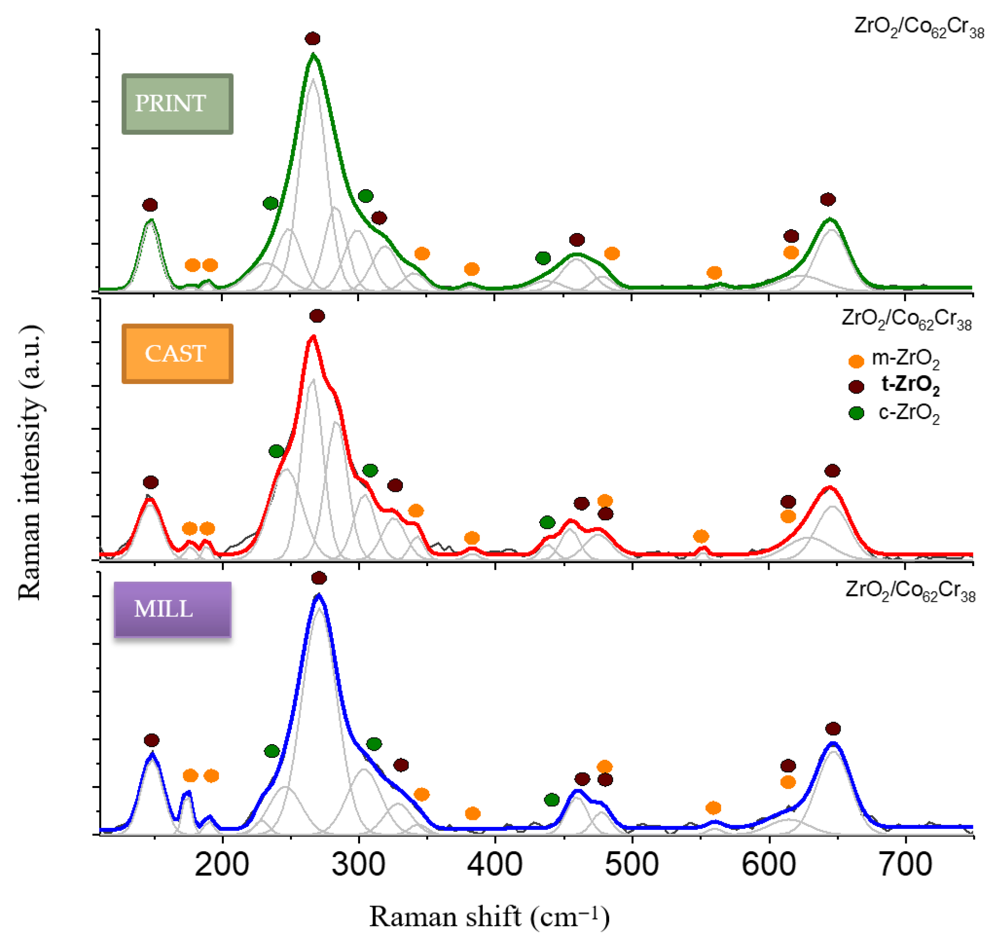
Figure 1. Raman spectra of ZrO2 deposited on variable biomedical implants, summarized in the 110–750 cm−1 range. Spectra were fitted using Gauss–Lorentz function to separate monoclinic (orange dots), tetragonal (brown dots), and cubic (green dots) ZrO2.
Coatings 15 00994 g001
Figure 2. Microstructure of PRINT (a,d), MILL (b,e), and CAST (c,f) samples. BSE images with indication of areas of chemical composition determination using energy dispersion spectroscopy of characteristic X-rays in the matrix (A), bright precipitates (B), and dark precipitates (C).
Coatings 15 00994 g002
Figure 3. Structure of the ZrO2 coating on the MILL substrate. STEM-BF image (a) and STEM-HAADF image (b). EDS spectrum in the region labeled B (c). EDS spectrum in the region marked as A (d).
Coatings 15 00994 g003
Figure 4. Substrate structure MILL with ZrO2 coating. TEM-BF image (a). TEM-DF image (b). SAED electron diffraction of the substrate from the area indicated in Figure 4a. (c). Coating electron diffraction (d).
Coatings 15 00994 g004
Figure 5. Structure of the ZrO2 coating on the PRINT substrate. STEM-BF images (a,b). EDS spectrum in the region labeled A (c). TEM-BF image (d).
Coatings 15 00994 g005
Figure 6. Structure of the ZrO2 coating on the PRINT substrate. HRTEM image (a). TEM image (b). Electron diffraction of the SAED from the area labeled b (c). ZrO2 unit cell model (d).
Coatings 15 00994 g006
Table 1. Results of chemical composition analysis (% wt.) in micro-areas of PRINT, MILL, and CAST samples.
Type of MaterialCoCrMoWSi
CAST substrate66285-1
CAST bright precipitates 502524-1
CAST dark precipitates197110--
MILL substrate5827511-
MILL bright precipitates44261218-
PRINT substrate622657-
PRINT bright precipitates39162025-
Table 2. Wettability and surface energy test results.
Average Contact Angle θ,
[o]
Surface Free Energy and Its Components, [mJ/m2]
Distilled Water θw¬avDiiodomethane θdavγsPolar γspDispersive γsd
CAST71.21±1.2451.76±0.2436.28±0.2013.27±0.3323.01±0.21
CAST + ZrO2100.31±1.8159.38±0.2231.43±0.180.53±0.0430.91±0.26
MILL83.19±1.8258.65±0.2930.99±0.177.49±0.1023.51±0.22
MILL + ZrO299.59±1.4557.45±0.2532.82±0.160.57±0.0432.24±0.31
PRINT62.38±1.5951.71±0.2734.08±0.269.21±0.1725.36±0.28
PRINT + ZrO294.75±1.4160.34±0.2229.23±0.161.88±0.1227.36±0.18
Disclaimer/Publisher’s Note: The statements, opinions and data contained in all publications are solely those of the individual author(s) and contributor(s) and not of MDPI and/or the editor(s). MDPI and/or the editor(s) disclaim responsibility for any injury to people or property resulting from any ideas, methods, instructions or products referred to in the content.