Article

Effects of Ti6Al4V Substrate Roughness on the Surface Morphology, Mechanical Properties, and Cell Proliferation of Diamond-like Carbon Films

1
Department of Mechanical and Materials Engineering, Tatung University, Taipei 104, Taiwan
2
Department of Biomechatronic Engineering, National Chiayi University, Chiayi 600, Taiwan
*
Author to whom correspondence should be addressed.
Coatings 2025, 15(9), 1086; https://doi.org/10.3390/coatings15091086
Submission received: 17 August 2025 / Revised: 12 September 2025 / Accepted: 13 September 2025 / Published: 16 September 2025

Abstract

This study investigated how Ti6Al4V substrate topography affects the performance of diamond-like carbon (DLC) coatings. Substrates with four finishes (unpolished, #100, #400, #800 grit) were coated, and their morphology, wettability, bonding structure, mechanical properties, and biological response were examined. Characterization was performed using AFM, SEM, contact angle tests, Raman spectroscopy, and nanoindentation. Biocompatibility was evaluated with A549 epithelial cells. DLC deposition reduced roughness while partly preserving surface features. Increasing Ra was associated with lower surface free energy and ID/IG ratios. It also correlated with higher hardness and modulus, reflecting greater sp3 bonding. Biological results, however, indicated that surface organization was more decisive than Ra magnitude. The #100-grit surface, with aligned anisotropic grooves, supported uniform wetting, protein adsorption, and sustained proliferation. In contrast, the unpolished and smoother surfaces did not maintain long-term growth. These findings suggest that anisotropy, rather than Ra alone, plays a key role in optimizing DLC-coated Ti6Al4V implants.

1. Introduction

With an aging population, the demand for medical implants continues to rise, particularly for replacements of the pelvis, hips, knees, elbows, shoulders, and orodental structures [1,2,3]. To be clinically successful, implants must satisfy three key requirements: biocompatibility (e.g., cell adhesion, water absorption, and osseointegration), mechanical performance (e.g., hardness, wear resistance, fatigue, and corrosion resistance), and manufacturing feasibility (e.g., precision, material characteristics, and cost effectiveness). Ti6Al4V is widely used in orthopedic and dental implants because of its high strength-to-weight ratio, low density, corrosion resistance, and overall biocompatibility [4]. However, in physiological environments, it still presents challenges, including ion release that may cause cytotoxicity, elastic modulus mismatch that leads to stress shielding and bone resorption, and limited osseointegration [5,6,7].
To address these issues, various surface modification and coating strategies have been explored. Texturing methods such as grit blasting and etching improve roughness and wettability [8]. More advanced approaches—including laser texturing, biomimetic modification, and functionalization with proteins or ceramics—provide enhanced bioactivity [9,10,11,12,13]. For example, microgrooved surfaces guided osteoblast alignment and promoted their proliferation. Hierarchical micro/nanostructures improved fibroblast adhesion and enhanced antibacterial performance. These studies demonstrate that combining micro- and nanoscale features can strengthen both mechanical durability and biological performance of Ti6Al4V. Yet, conventional texturing alone cannot fully overcome problems such as wear, corrosion, and ion release.
Diamond-like carbon (DLC) coatings have therefore attracted attention due to their high hardness, low friction, chemical inertness, and favorable biocompatibility [14,15,16,17,18,19,20]. Recent studies have shown the benefits of DLC coatings on Ti6Al4V implants. Zhang et al. [21] reported that DLC with photochemical grafting lowered surface energy and roughness while improving cytocompatibility, corrosion resistance, and wear behavior. Joska et al. [22] found that chromium interlayers provided the best corrosion resistance, while titanium interlayers were more effective on Ti6Al4V than on pure titanium. Grabarczyk et al. [23] showed that oxidation pretreatment before DLC deposition reduced friction and increased durability. Pinilla-Cienfuegos et al. [24] demonstrated that submicron DLC films in artificial saliva reduced adhesion, friction, and wear. Together, these works confirm that DLC coatings can enhance surface properties, corrosion resistance, and biocompatibility of Ti6Al4V implants.
Prior studies have shown that DLC combined with grafting or interlayers enhances cytocompatibility and anticorrosion performance. In recent years, several advances have expanded DLC applications. In particular, Peng et al. [25] reviewed the broad biomedical potential of DLC coatings, emphasizing their use in vascular stents, prosthetic heart valves, and biosensors. Building on this, Shah et al. [26] reviewed recent advances and challenges. They emphasized the roles of the sp3/sp2 ratio, hydrogenation, and adhesion in biomedical performance. These studies show the potential of DLC coatings and the approaches being developed to improve their use in biomedical implants.
Within this context, the interaction between substrate topography and DLC coatings warrants closer examination. While many studies have focused on roughness amplitude (Ra), the influence of surface organization—particularly anisotropy versus isotropy—has been less thoroughly addressed. In this work, DLC films were deposited on Ti6Al4V substrates with different surface finishes and evaluated for morphology, bonding structure, wettability, surface energy, hardness, and elastic modulus. Biocompatibility was assessed using A549 epithelial cells. By considering anisotropy alongside roughness, this study offers additional insight into how substrate micro-topography influences DLC film properties and cellular responses. These findings may help guide the optimization of DLC-coated Ti6Al4V implants.

2. Materials and Methods

Ti6Al4V substrates were purchased from Gredmann Group, Taipei, Taiwan, and the Ti6Al4V alloy sheet was cut into 20 mm × 20 mm × 5 mm. The substrates were either left bare (unpolished) or mechanically polished using sandpapers with grit sizes of #100, #400, and #800 to produce different levels of surface roughness. After polishing, the substrates were cleaned in an ultrasonic bath for 20 min in acetone. The chemical composition of Ti6Al4V is presented in Table 1.
After substrate roughening, DLC was deposited via CH4 ion-beam deposition (IBD) (Model: Dash 700) at an acceleration energy of 750 eV and a current density of approximately 2.5 mA cm−2, without an interlayer. The deposition chamber was evacuated to 5 × 10⁻⁴ Pa, and Ar and hydrogen gases were introduced until the background pressure reached 0.1 Pa. The substrate temperature was maintained at 200 °C. Surface morphology was analyzed using an atomic force microscope (Solver R47-pro, NT-MDT, Moscow, Russia), and surface roughness parameters were quantified with Nova software (version 1.1.1 R16703). To analyze the hybridization of carbon in the films, Raman spectroscopy (Renishaw, Wotton-under-Edge, UK) was performed using a 514.5 nm laser with a spot size of approximately 2 μm. The bonding structure, particularly the ID/IG ratio, was evaluated from the Raman spectra. Raman spectral analysis was performed using Origin 2019b (OriginLab, Northampton, MA, USA). The spectra were first imported and calibrated against the Si reference at 520.7 cm−1 to ensure accuracy of the Raman shift scale. A light Savitzky–Golay smoothing was applied to reduce noise without distorting the peak profile. Fluorescence background was removed by baseline correction within the 1000–1800 cm−1 region using an asymmetric least squares method. The D (~1350 cm−1) and G (~1580 cm−1) bands were then fitted iteratively with Lorentzian peak functions until convergence was reached with minimal residuals. From the fitted peaks, the integrated areas were extracted, and the intensity ratio ID/IG was calculated as the ratio of the D-band area to the G-band area. This procedure provided a consistent and reproducible evaluation of the relative disorder and sp2/sp3 bonding characteristics in the carbon films.
Surface wettability was assessed via contact angle measurements using distilled water and glycerol as probe liquids, and the surface free energy (SFE) was calculated using the Owens–Wendt method. Mechanical properties, including hardness and reduced modulus, were measured via nanoindentation using a Berkovich tip (Nanoindenter XP, MTS, Eden Prairie, MN, USA). The indentation tests were conducted with a maximum load of 10 mN, loading and unloading times of 30 s each, and a holding time of 10 s at the maximum load.
A549 lung epithelial cells were cultured under sterile conditions in a Class II biosafety cabinet and maintained at 37 °C with 5% CO2 in a humidified incubator. DLC/Ti6Al4V samples were ultrasonically cleaned, immersed in 70% ethanol for 20 min, rinsed three times with sterile PBS, and UV-irradiated for 20 min on each side before use. Cells were maintained in F-12K (Kaighn’s) medium supplemented with 10% fetal bovine serum. For assays, cells were seeded onto each sample at a density of 1–3 × 10⁴ cells cm−2 in complete medium and cultured for 24–48 h. After incubation, adherent cells were labeled with Calcein-AM/EthD-1 for live/dead staining, imaged on an epifluorescence microscope using 10–20× objectives, and quantified by counting nuclei per field to estimate adhesion and proliferation.

3. Results

3.1. Surface Morphology, Wettability, and Surface Free Energy

The Ti6Al4V substrates subjected to mechanical polishing exhibited distinct differences in surface roughness, as shown in Figure 1. AFM shows a clear evolution of Ti6Al4V topography with polishing grit. The unpolished surface (Ra ≈ 548 nm) exhibited long machining furrows. Their orientations were irregular and often discontinuous. The grooves also varied in depth and direction, producing a rough but quasi-isotropic morphology with limited long-range order. Meanwhile, the #100-grit surface (Ra = 654 nm) displayed more uniform, well-defined parallel ridges. These were separated by deeper valleys. The consistently aligned features created a strongly anisotropic topography. With #400 polishing (Ra = 528 nm), the long striations break into shorter segments and clusters of micro-asperities. Groove depth decreases, but occasional pull-out pits remain. The overall texture shifts toward quasi-isotropy. The #800 finish (Ra = 478 nm) is the smoothest surface. It is dominated by fine-scale asperities and modest local slopes. The height fluctuations are nearly isotropic. Overall, coarse polishing increases the amplitude and regularity of grooves compared with the unpolished surface. In contrast, finer grits progressively suppress anisotropy and vertical height. This results in a more homogeneous surface. These AFM images show that finer pre-polishing reduces the depth of machining grooves. However, it cannot completely eliminate topographical peaks and valleys on the metallic alloy.
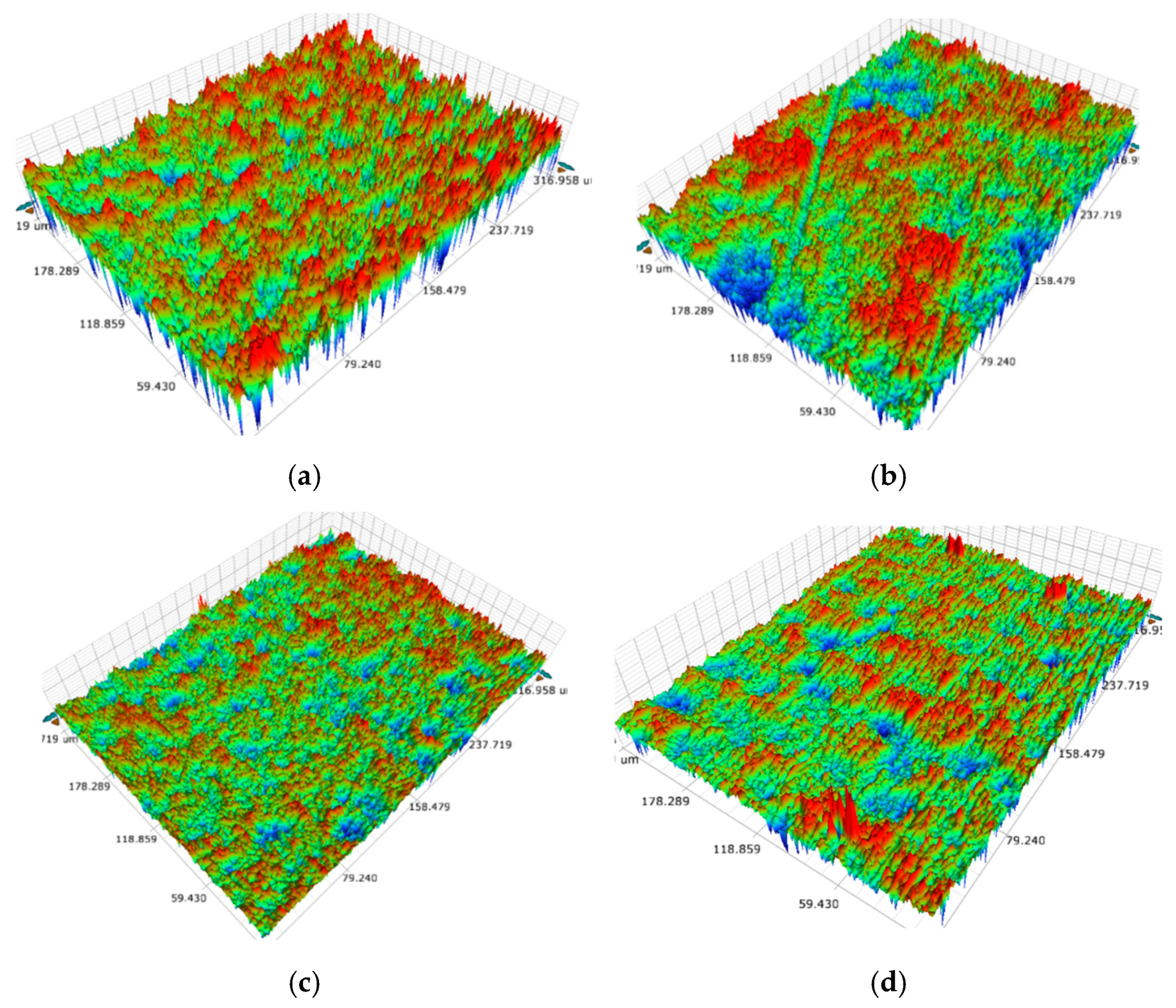
After DLC deposition (Figure 2), smoother surfaces allowed the DLC to form a more conformal layer. This layer filled valleys and reduced peak height. As a result, the smoothing effect was stronger. On the unpolished substrate (Figure 2a), the DLC film reduces the average roughness from 548 nm to about 512 nm by partially filling the deep grooves. For the #100-grit substrate (Figure 2b), Ra decreases from 654 nm to 532 nm despite the initial coarse scratches. On the #400-grit and #800-grit substrates, the DLC films display a relatively uniform granular morphology; the roughness drops from 528 nm to 423 nm and from 478 nm to 386 nm, respectively. The smoother surfaces allowed the DLC to form a more conformal layer. This layer filled valleys and reduced peak height. As a result, the smoothing effect was stronger. These AFM images suggest that the deposition produces a compact amorphous carbon network. The DLC film partly replicates the substrate topography and smooths out high-amplitude asperities.
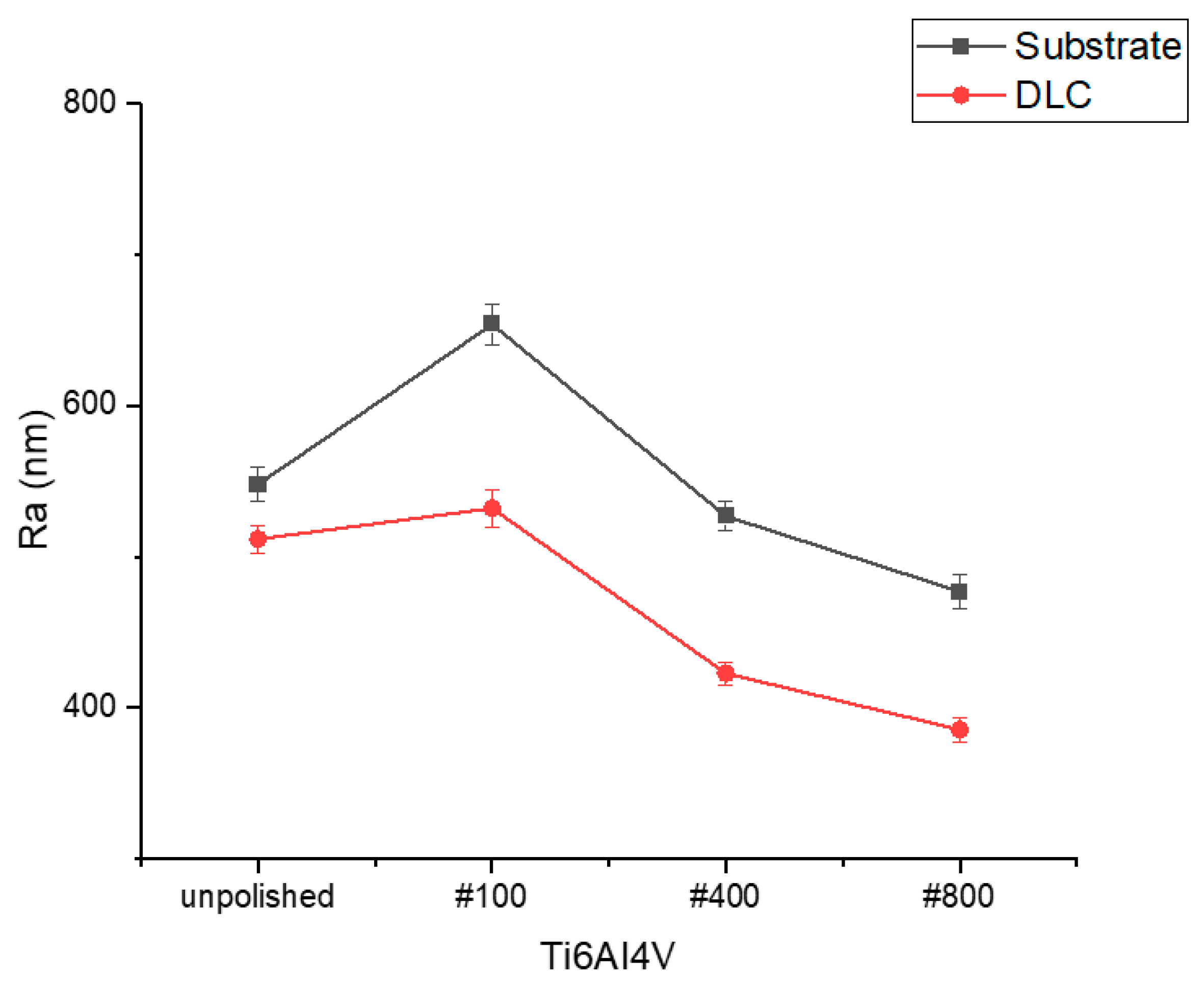
The trend in roughness reduction is summarized in Figure 3. For all pre-treatments, the arithmetic roughness decreases after DLC deposition. The unpolished sample shows a modest reduction of about 36 nm. When the substrate is ground with #100 grit, the roughness decreases by 122 nm, and reductions of 105 nm and 92 nm for the #400 and #800 grit-published surfaces, respectively. This trend indicates that the smoothing effect of DLC is most pronounced when the initial surface contains valleys that are shallower or comparable to the film thickness. Under these conditions, finer polishing reduces asperity amplitude, enabling energetic carbon species to fill valleys and form a more uniform film. The data demonstrate that DLC can significantly smooth Ti6Al4V surfaces [27,28].
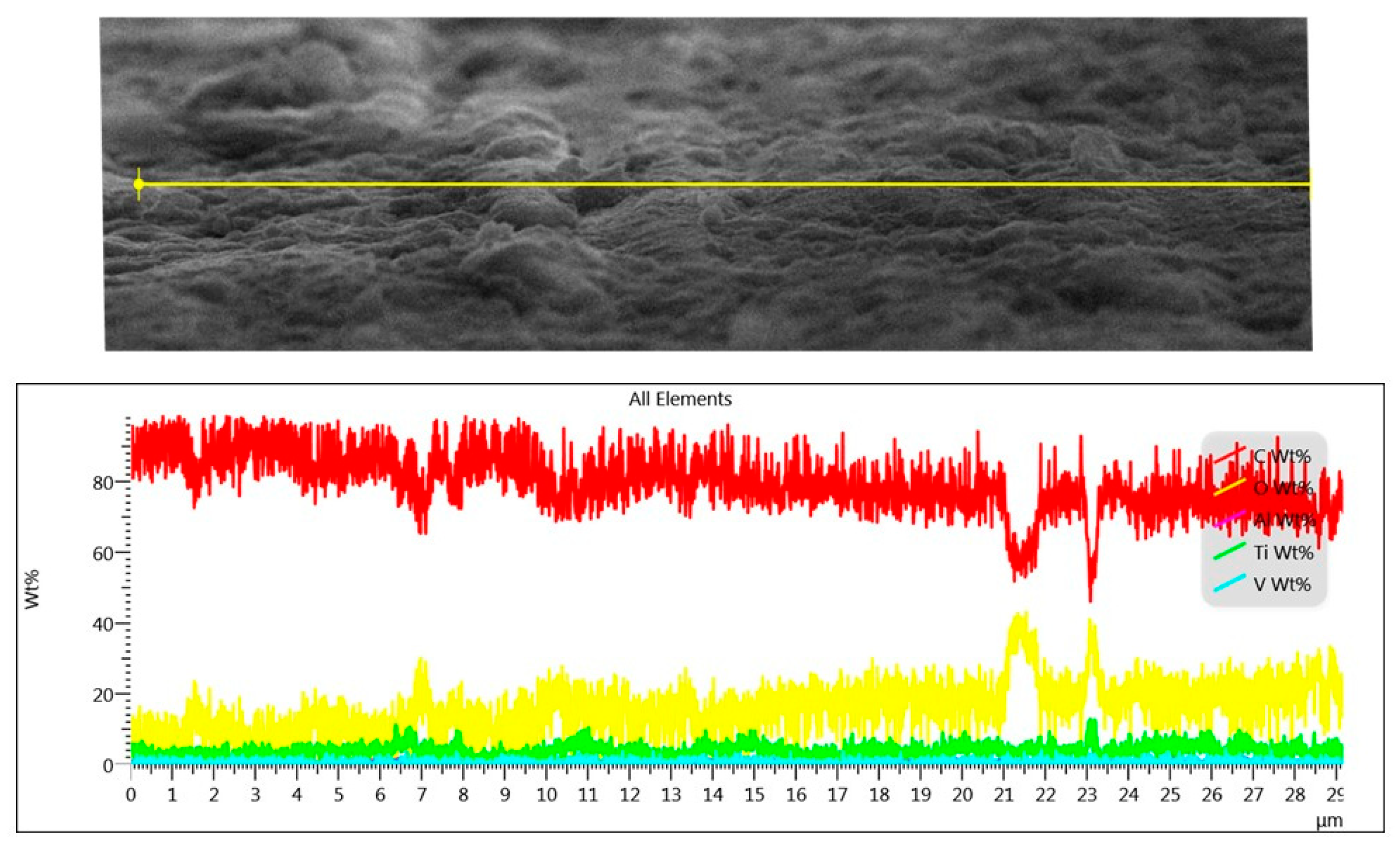
A cross-sectional SEM image of a diamond-like carbon (DLC) film grown on a Ti6Al4V substrate and the corresponding energy-dispersive X-ray spectroscopy (EDS) line scan is shown in Figure 4. The EDS profile is dominated by carbon. The red signal remains at about 80–90 wt% across most of the film thickness, whereas oxygen (yellow line) is confined to about 10 wt%. Titanium and vanadium peaks appear only at the film–substrate interface. This composition confirms that the deposited film is essentially pure carbon and that the underlying titanium alloy is sealed by the DLC layer. Such carbon-rich coatings are characteristic of amorphous DLC and produce dense, pinhole-free barriers [29].
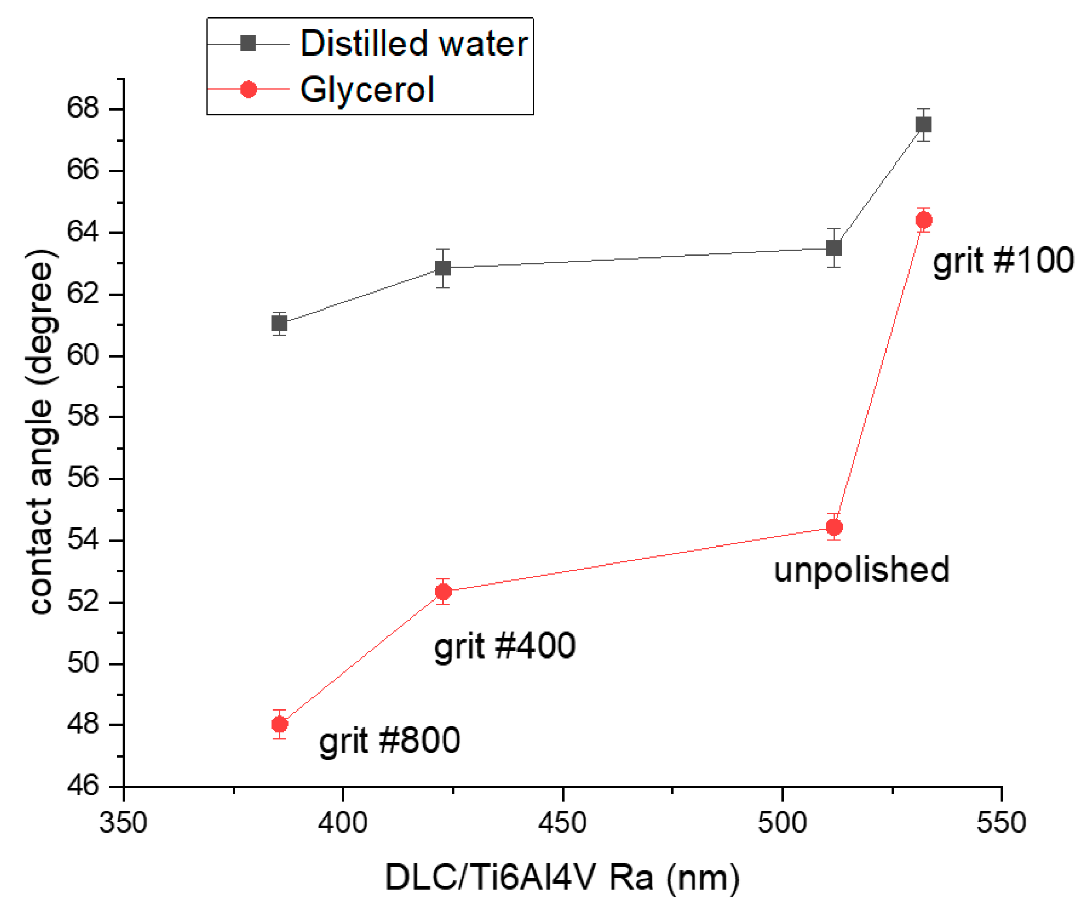
The contact angles of distilled water and glycerol on DLC-coated various roughness Ti6Al4V surfaces are shown in Figure 5. As roughness increases from 386 nm to 532 nm, the water contact angle rises from about 61° to 67°, while the glycerol contact angle increases from about 48° to 64°. Although DLC is moderately hydrophilic, micro-asperities and changes in carbon bonding contribute to a slight increase in hydrophobicity as roughness grows. The Wenzel model predicts that surface roughness amplifies the intrinsic wetting behavior of a material, but in this case, the modest increase in contact angle suggests that chemical effects from DLC bonding states act alongside topography to influence wettability [30,31].
The calculation of surface free energy (SFE) from contact angle measurements is based on the Young equation [32]. This equation comes from the equilibrium of surface tension forces at the meeting point of solid, liquid, and gas. The surface free energy of the DLC films was determined using the Owens–Wendt geometric mean method [33]. In this approach, the contact angle θ of a liquid on a solid surface is related to the dispersive ( γ d ) and polar ( γ p ) components of the solid (S) and liquid (L) surface tensions according to the following:
γ L cos θ = 2 γ S d γ L d + γ S p γ L p
where γ L is the total surface tension of the liquid, θ is the contact angle, γ L d and γ L p are the dispersive and polar components of the liquid, and γ s d and γ s p are the corresponding components of the solid (film) to be determined. Water was chosen for its high polar component, while ethylene glycol, which contains both polar and dispersive contributions, served as the complementary liquid to enable separation of the SFE into its dispersive ( γ d ) and polar ( γ p ) components. The surface tension parameters of the probe liquids were taken from the literature [34]: for water, total surface tension, γ = 72.8   × 10 3 N / m with γ d = 21.8 × 10 3 N / m , γ p = 51 × 10 3 N / m ; for glycerol, γ = 64   × 10 3 N / m with γ d = 34 × 10 3 N / m , γ p = 30 × 10 3 N / m . Contact angle values were substituted into Equation (1) to calculate the polar and dispersive components of the DLC surface free energy, whose sum gave the total SFE.
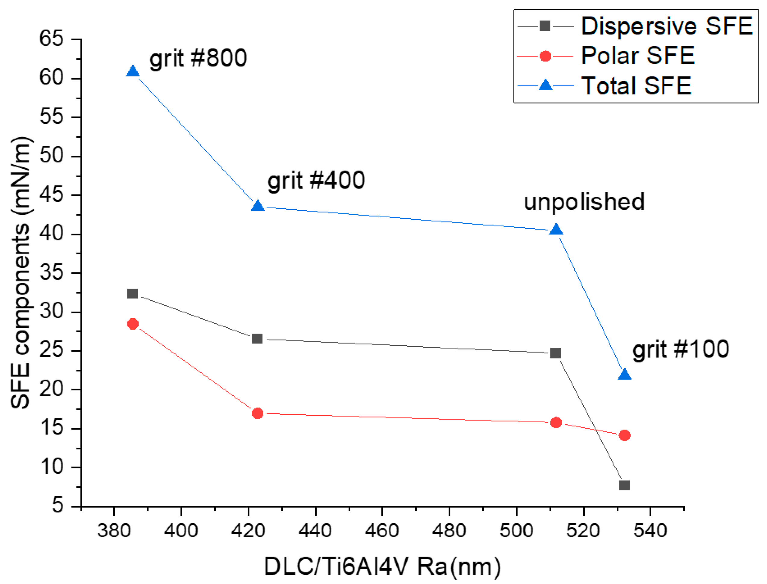
The dispersive, polar, and total components of the surface free energy (SFE) of the DLC films are shown in Figure 6. The total SFE drops markedly from ~60 mN·m−1 at Ra = 386 nm to ~22 mN·m−1 at Ra = 532 nm. This reduction is driven primarily by a decrease in the dispersive component. The concurrent reduction in the polar component may indicate a lower density of polar surface groups. It may also reflect the limited accessibility of these groups caused by roughness-induced heterogeneity. Increasing roughness correlates with a higher sp3 fraction, consistent with our Raman and hardness data. This higher sp3 content lowers electronic polarizability and diminishes dispersive interactions. In contrast, a phosphorus-doped DLC study reported increased polar but decreased dispersive contributions owing to surface functionalization, highlighting that our trend is topology/bonding-driven rather than chemistry-driven [35]. Overall, the reduction in SFE accompanying the rise in contact angle is consistent with prior reports [36,37,38]. However, Owens–Wendt [33] analyses assume smooth and homogeneous surfaces. At high Ra, Wenzel or Cassie–Baxter effects [39] may further depress apparent SFE, reinforcing the observed decrease.
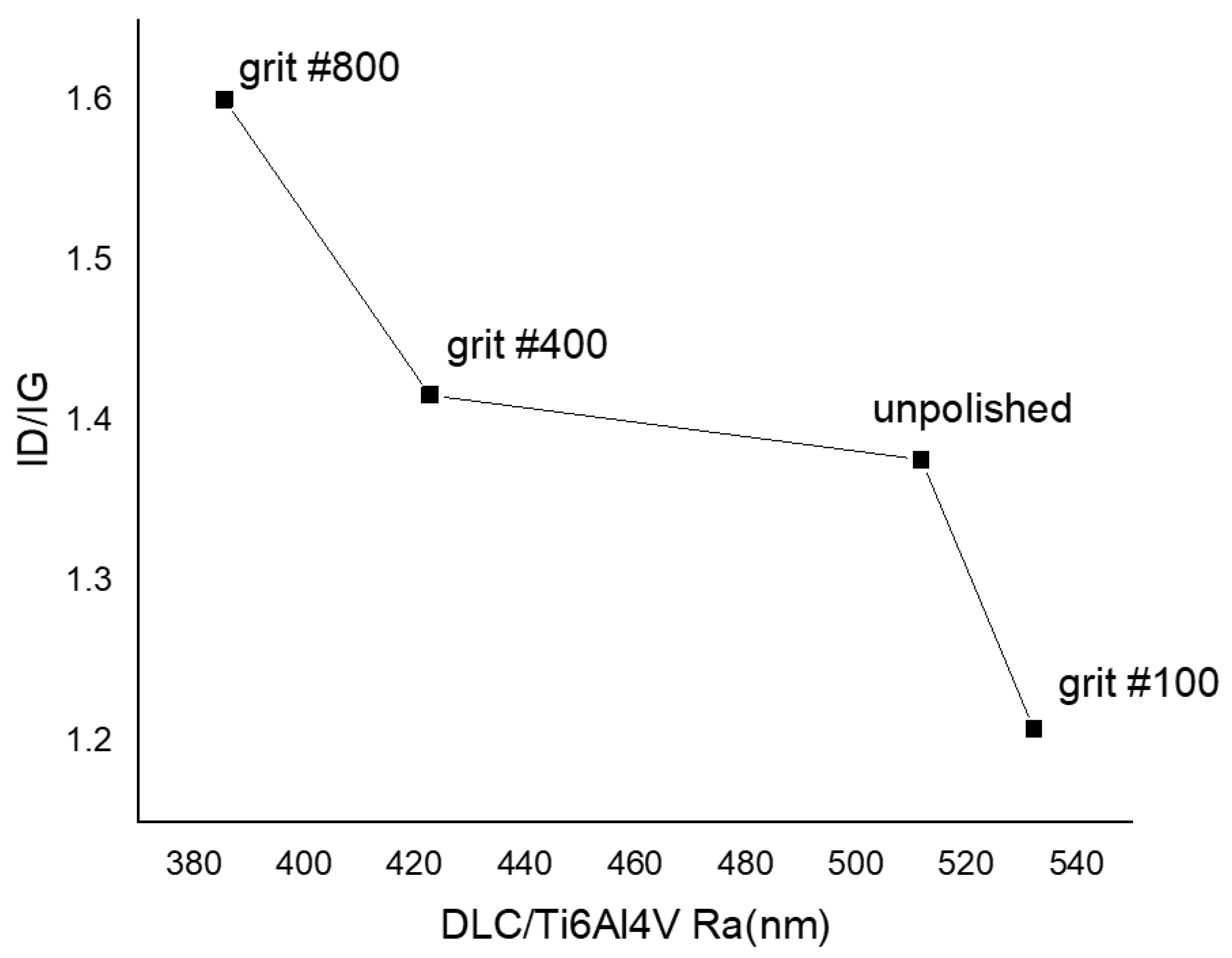
Figure 7 shows that the Raman ID/IG intensity ratio decreases from about 1.6 to 1.2 as the surface roughness increases from roughly 386 nm to 532 nm. The ID/IG ratio measures the relative intensities of the disorder (D) and graphitic (G) bands in Raman spectroscopy; a lower ratio generally indicates fewer sp2 clusters or a smaller sp2 domain size and hence a higher fraction of sp3-bonded carbon. Therefore, the observed decrease implies that films grown on rougher Ti6Al4V substrates develop a more diamond-like structure with a higher sp3 content. This trend is consistent with the contact-angle data: a study on barrier DLC coatings found that higher C–C sp3 content (i.e., lower ID/IG ratio) leads to higher contact angles [40]. Meanwhile, high sp3 content is associated with greater hardness and enhanced chemical inertness of DLC films. These characteristics have direct implications for both mechanical properties and biocompatibility [41]. To sum up, the substrate roughness influences not only the morphology but also the bonding structure of the DLC coatings. These changes, in turn, affect their wettability and surface energy [42,43,44].

3.2. Mechanical Properties

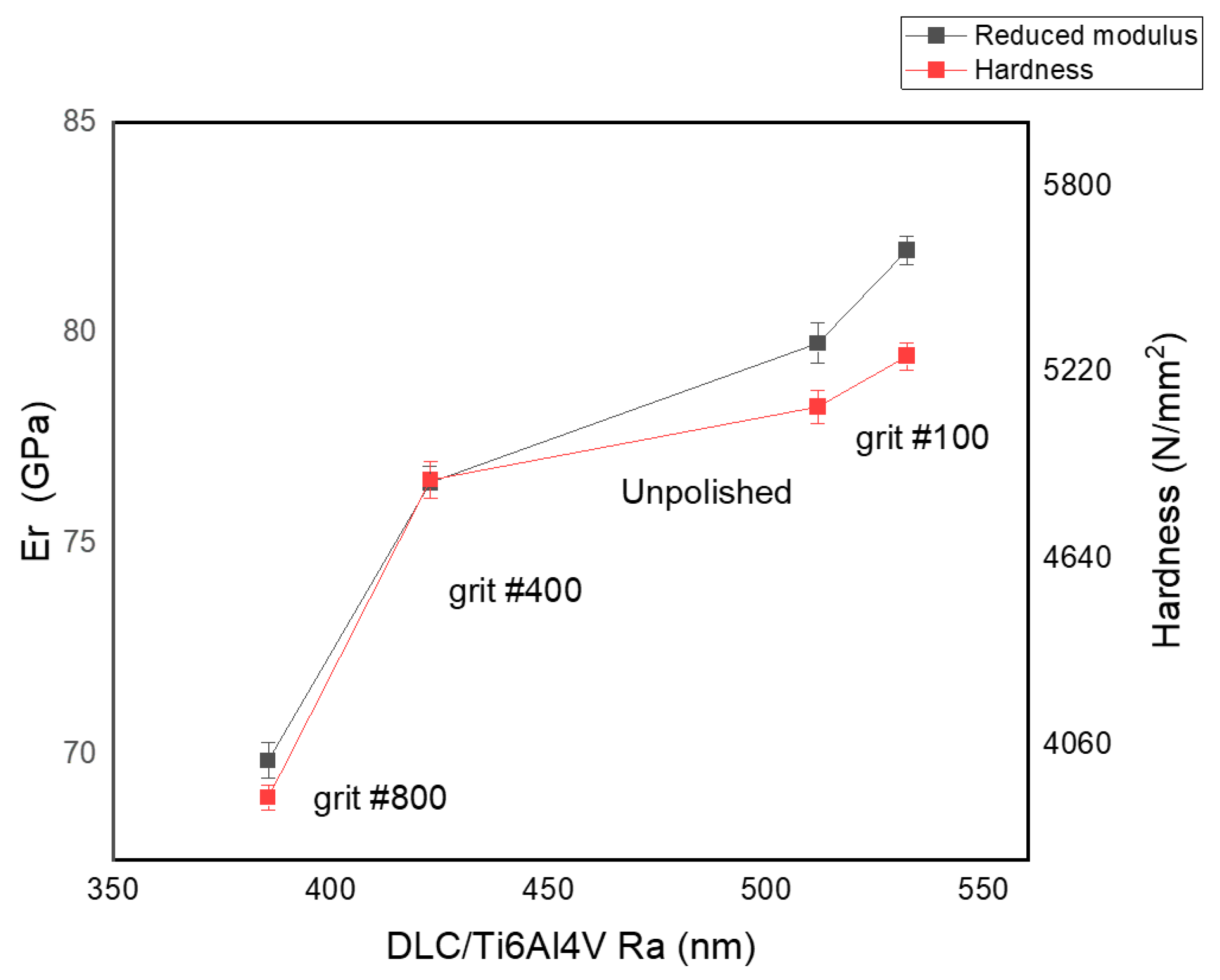
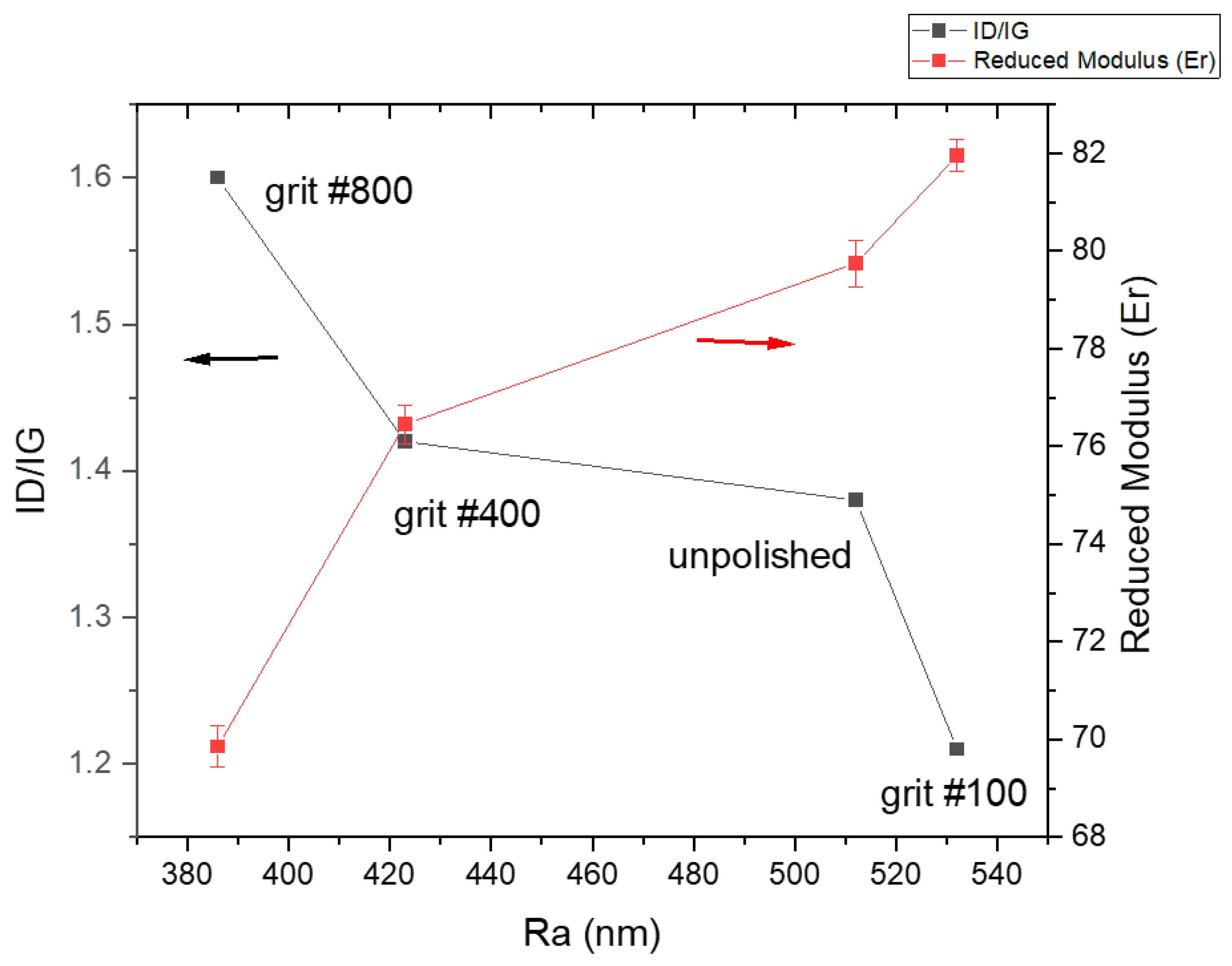
Figure 8 shows that hardness (H) and reduced elastic modulus (Er) increase with higher substrate roughness (Ra). This trend is consistent with the Raman results, where lower ID/IG coincides with higher H and Er. These observations can be interpreted within a residual-stress framework and established Raman–structure–property relationships. Our finite-element analysis indicated that thermally induced residual stress scales with the CTE mismatch. The model further predicted that compressive residual stress increases the apparent hardness and stiffness during indentation. Surface roughness magnified this effect. For example, at Ra = 0.2 μm, the elastic modulus increased by 20.53% and hardness by 5.83% compared with a smooth substrate [45]. Figure 9 and Figure 10 likewise support the mechanical trend. A lower ID/IG generally reflects a more sp3-rich (diamond-like) network. This bonding structure correlates with higher hardness and modulus in both graded and undoped DLC systems [46]. Thus, as the surface roughness (Ra) increases, hardness (H) and reduced modulus (Er) increase, whereas the Raman ID/IG ratio decreases. This behavior suggests that rougher topography concentrates compressive stresses during cooling. These stresses enhance the coating’s apparent load-bearing capacity. At the same time, the roughness favors a more diamond-like (sp3-rich) bonding configuration in the Raman spectra.

3.3. Cell Growth and Proliferation with A549 Cells

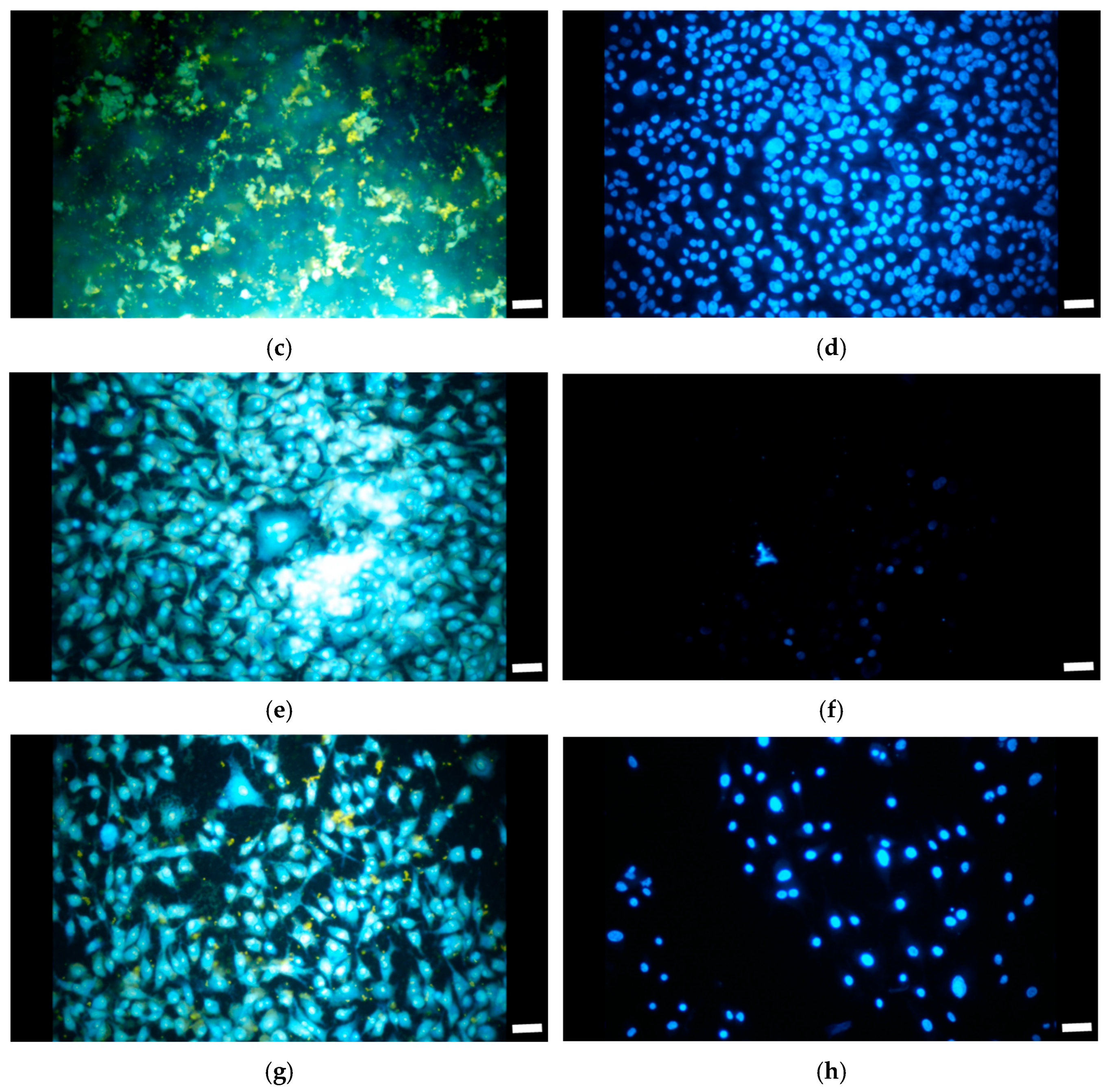
Figure 11 presents representative bright-field (left) and fluorescence (right) micrographs of A549 cells cultured on DLC-coated Ti6Al4V with four substrate finishes (unpolished, #100, #400, and #800 (Ra ≈ 512, 532, 423, and 386 nm, respectively). The unpolished surface (Figure 11a,b) exhibits patchy, rounded cells and sparse nuclei despite high 24 h counts, and its sharp 24→48 h drop suggests unstable adhesion and poor retention. The #100 finish (Figure 11c,d) shows more uniformly distributed, well-spread cells with near-confluent fluorescence, matching the sustained increase in counts to 48 h. The #400 and #800 finishes (Figure 11e–h) display locally well-spread, cobblestone-like monolayers in bright-field. However, their fluorescence fields appear sparse, and total counts drop by 24 h. This decline continues at 48 h, suggesting uneven coverage and poor long-term retention. Overall, the data indicate that the anisotropic groove pattern of the #100 surface appeared to support both early anchoring and sustained proliferation. In contrast to the quasi-isotropic unpolished surface and the smoother #400, #800 finishes appeared less favorable under the same conditions.
Figure 12 quantifies the number of A549 cells on the four DLC-coated Ti6Al4V substrates after 24 and 48 h. At 24 h, both the unpolished and #100-grit surfaces supported higher initial attachment compared with the #400 and #800-grit groups. By 48 h, only the #100-grit surface showed a further increase in cell number. In contrast, the unpolished, #400-, and #800-grit surfaces all declined. These quantitative results are consistent with the fluorescence images (Figure 11). They suggest that the #100-grit condition promotes both initial adhesion and continued proliferation. In contrast, the unpolished and smoother surfaces appear less favorable for long-term cell retention.

4. Discussion

Our results suggest that Ti6Al4V substrate topography influences both DLC coating characteristics and A549 cell behavior. All surfaces exhibited Ra values between 386 and 548 nm. However, biological outcomes were not explained by roughness magnitude alone. They appeared more closely related to surface organization. The #100-grit surface (Ra ≈ 532 nm) contained aligned anisotropic grooves that supported longer-term proliferation, while the unpolished surface (Ra ≈ 548 nm) displayed quasi-isotropic, irregular features that were less favorable for retention despite similar Ra. Thus, while roughness is recognized as important for cell compatibility [47], our results suggest that anisotropy could also contribute to long-term cellular behavior.
Wettability may also have contributed to the observed differences. According to the Wenzel [30,31] and Cassie–Baxter models [38], roughness can either promote complete liquid infiltration or cause air entrapment that limits effective adhesion. The #100-grit anisotropic surface likely showed complete wetting. This promoted protein adsorption and stable cell adhesion. Similar effects of wettability on protein adsorption and cell attachment were reported by Arima and Iwata [48]. In contrast, the quasi-isotropic unpolished surface likely trapped air pockets (Cassie–Baxter state), restricting contact with the medium. The smoother #400 (Ra ≈ 423 nm) and #800 (Ra ≈ 386 nm) surfaces presented another limitation. They provided fewer adhesion sites and a smaller effective surface area. As a result, they supported initial attachment but appeared less able to sustain proliferation at 48 h. Consistent with our observations, Majhy et al. [49] showed that surfaces with moderate surface energy support good cell adhesion, spreading, and proliferation. They also found that extreme wetting or very high roughness suppressed cellular activity. This result suggests that surface energy works together with topography to control protein adsorption and cell anchorage. Teixeira et al. [50] reported that nanoscale grooves can guide epithelial cells. The cells elongated and aligned along the grooves, even when the features were only 70 nm wide. These studies support our conclusion that both roughness and anisotropy are critical factors in determining the cellular response to DLC-coated Ti6Al4V substrates.
In summary, our findings indicate that anisotropy and groove alignment, alongside roughness, influence the cellular response to DLC-coated Ti6Al4V substrates. This interpretation helps explain why the #100-grit surface performed better than the smoother (#400, #800) or quasi-isotropic unpolished conditions, offering a broader framework for understanding how topography may shape biointerface behavior.

5. Conclusions

This study investigated how Ti6Al4V substrate topography influences the morphology, wettability, mechanical properties, and biological performance of DLC coatings.
Surface characteristics: DLC deposition reduced surface roughness while partially replicating the underlying features. Rougher substrates promoted lower ID/IG ratios and higher hardness and modulus, consistent with increased sp3 bonding and compressive stresses.
Biological response: A549 cell assays revealed that anisotropy and surface order were more critical than Ra magnitude. The #100-grit surface, with aligned grooves, promoted complete wetting, uniform protein adsorption, and contact-guided proliferation. In contrast, the quasi-isotropic unpolished surface, despite having a similar Ra, failed to sustain growth due to irregular topography and possible air entrapment. The smoother #400 and #800 surfaces supported initial attachment but offered fewer attachment sites and reduced long-term proliferation.
Previous studies have primarily emphasized roughness amplitude as the main determinant of cell behavior. Our results indicate that anisotropy versus isotropy is equally important in influencing the biological responses of DLC–Ti6Al4V biointerfaces. This distinction helps explain why surfaces with similar Ra values can nevertheless produce divergent cellular outcomes. In conclusion, DLC-coated Ti6Al4V surfaces with anisotropic grooves provided a favorable combination of coating durability, wettability, and biological response. Considering surface order and groove orientation may, therefore, represent a useful strategy for improving DLC-coated Ti6Al4V implants to support epithelial adhesion and proliferation.

Author Contributions

Conceptualization, C.W.; methodology, B.-C.W.; validation, C.W. and M.-S.H.; formal analysis, C.W.; investigation, C.W.; resources, C.W.; data curation, B.-C.W.; writing—original draft preparation, C.W.; writing—review and editing, C.W.; visualization, C.W.; supervision, C.W.; project administration, C.W.; funding acquisition, C.W. All authors have read and agreed to the published version of the manuscript.

Funding

This research was funded by the National Science and Technology Council (NSTC) of Taiwan, grant number MOST 108-2813-C-036-007-E.

Institutional Review Board Statement

Not applicable.

Informed Consent Statement

Not applicable.

Data Availability Statement

Data are contained within this article.

Conflicts of Interest

The authors declare no conflicts of interest.

Abbreviations

The following abbreviations are used in this manuscript:
DLCDiamond-like carbon
SFE Surface free energy
IBDIon-beam deposition
AFMAtomic force microscopy
SEMScanning electron microscopy
EDSEnergy-dispersive spectroscopy
RaSurface roughness (arithmetical mean)
PBSPhosphate-buffered saline
H Hardness
Er Reduced modulus

References

  1. Ramakrishna, S.; Mayer, J.; Wintermantel, E.; Leong, K.W. Biomedical applications of polymer–composite materials: A review. Compos. Sci. Technol. 2001, 61, 1189–1224. [Google Scholar] [CrossRef]
  2. Wise, D.L.; Gresser, J.D.; Trantolo, D.J.; Cattaneo, M.V.; Lewandrowski, K.-U.; Yaszemski, M.J. Biomaterials Engineering and Devices: Human Applications; Volume 2: Orthopedic, Dental, and Bone Graft Applications; Humana Press: Totowa, NJ, USA, 2000. [Google Scholar] [CrossRef]
  3. Park, J.B.; Bronzino, J.D. (Eds.) Biomaterials: Principles and Applications, 1st ed.; CRC Press: Boca Raton, FL, USA, 2002. [Google Scholar] [CrossRef]
  4. Geetha, M.; Singh, A.K.; Asokamani, R.; Gogia, A.K. Ti based biomaterials, the ultimate choice for orthopaedic implants—A review. Prog. Prog. Mater. Sci. 2009, 54, 397–425. [Google Scholar] [CrossRef]
  5. Hoeppner, D.W.; Chandrasekaran, V. Fretting in orthopaedic implants: A review. Wear 1994, 173, 189–197. [Google Scholar] [CrossRef]
  6. Okazaki, Y.; Gotoh, E. Comparison of metal release from various metallic biomaterials in vitro. Biomaterials 2005, 26, 11–21. [Google Scholar] [CrossRef] [PubMed]
  7. Wapner, K.L. Implications of metallic corrosion in total knee arthroplasty. Clin. Orthop. Relat. Res. 1991, 271, 12–20. [Google Scholar] [CrossRef] [PubMed]
  8. Souza, J.C.M.; Sordi, M.B.; Kanazawa, M.; Ravindran, S.; Henriques, B.; Silva, F.S.; Aparicio, C.; Cooper, L.F. Nano-scale modification of titanium implant surfaces to enhance osseointegration. Acta Biomater. 2019, 94, 112–131. [Google Scholar] [CrossRef]
  9. Zheng, Q.; Mao, L.; Shi, Y.; Fu, W.; Hu, Y. Biocompatibility of Ti-6Al-4V titanium alloy implants with laser microgrooved surfaces. Mater. Technol. 2020, 37, 2039–2048. [Google Scholar] [CrossRef]
  10. Ji, M.; Zhang, S.; Qiu, J.; Liu, G.; Guo, F.; Xu, J.; Chen, M. Enhanced tribological properties and cyto-biocompatibility of dental Ti6Al4V alloy via laser surface texturing. J. Mater. Res. Technol. 2024, 33, 4105–4115. [Google Scholar] [CrossRef]
  11. Melo-Fonseca, F.; Gasik, M.; Freitas, P.P.; Carvalho, D.N.; Pinto, I.M.; Silva, F.S.; Miranda, G. Hydrothermal treatment of laser micro-textured Ti6Al4V systems for osseointegration of orthopaedic implants. Opt. Laser Eng. 2024, 183, 108511. [Google Scholar] [CrossRef]
  12. Yan, Y.; Mu, L.; Liu, Y.; Li, Q.; Liu, Y.; Ma, Z.; Xu, J.; Di, K. Enhanced wear resistance, corrosion resistance and biocompatibility on micro-texture Ti6Al4V surfaces by pulse laser remelted. Surf. Coat. Technol. 2025, 513, 132528. [Google Scholar] [CrossRef]
  13. Gupta, R.; Gaddam, A.; Naveena, H.A.; Prajapati, D.; Dimov, S.; Bhatia, D.; Mishra, A.; Sofronov, Y.; Vadali, M. Improving the cell adhesion and antibacterial behaviour on Ti6Al4V through micro and nano hierarchical laser surface texturing. Surf. Interfaces 2025, 58, 105857. [Google Scholar] [CrossRef]
  14. Jelínek, M.; Smetana, K.; Kocourek, T.; Dvořánková, B.; Zemek, J.; Remsa, J.; Luxbacher, T. Biocompatibility and sp3/sp2 Ratio of Laser Created DLC Films. Mater. Sci. Eng. B 2010, 169, 89–93. [Google Scholar] [CrossRef]
  15. Wei, C.; Pan, W.-J.; Hung, M.-S. The Effects of Substrate Roughness and Associated Surface Properties on the Biocompatibility of Diamond-Like Carbon Films. Surf. Coat. Technol. 2013, 224, 8–17. [Google Scholar] [CrossRef]
  16. Wei, C.; Peng, K.-S.; Hung, M.-S. The Effect of Hydrogen and Acetylene Mixing Ratios on the Surface, Mechanical and Biocompatible Properties of Diamond-Like Carbon Films. Diam. Relat. Mater. 2016, 63, 108–114. [Google Scholar] [CrossRef]
  17. Gotzmann, G.; Beckmann, J.; Scholz, B.; Herrmann, U.; Wetzel, C. Low-Energy Electron-Beam Modification of DLC Coatings Reduces Cell Count While Maintaining Biocompatibility. Surf. Coat. Technol. 2018, 336, 34–38. [Google Scholar] [CrossRef]
  18. Wongpanya, P.; Pintitraratibodee, N.; Thumanu, K.; Euaruksakul, C. Improvement of Corrosion Resistance and Biocompatibility of 316L Stainless Steel for Joint Replacement Application by Ti-Doped and Ti-Interlayered DLC Films. Surf. Coat. Technol. 2021, 425, 127734. [Google Scholar] [CrossRef]
  19. Shen, B.; He, Z.; Huang, H.; He, F.; Chen, Y.; Wu, P.; Li, M.; Penkov, O.V.; Wu, H. Improved biocompatibility of durable Si-DLC periodical nanocomposite coatings modified by plasma treatment for medical implants. Appl. Surf. Sci. 2025, 695, 162907. [Google Scholar] [CrossRef]
  20. Xiao, Z.; Ren, L.; Guo, C.; Liang, L.; Dai, Y.; Tang, K.; Lu, L.; Qi, F.; She, J.; Wang, L.; et al. Enhanced Corrosion Resistance, Mechanical Properties, and Biocompatibility of N-DLC Coatings Prepared on WE43 Alloy via FCVA Technology. J. Alloys Compd. 2025, 1010, 178106. [Google Scholar] [CrossRef]
  21. Liu, Y.; Fu, X.; Bu, Y.; Zhang, J.; Zhang, J.; Wu, L. Photochemical grafting of fluorinate alkenes on DLC coated Ti6Al4V to improve in vitro cytocompatibility, friction and corrosion resistance. Surf. Coat. Technol. 2012, 208, 51–56. [Google Scholar] [CrossRef]
  22. Joska, L.; Fojt, J.; Mestek, O.; Cvrcek, L.; Brezina, V. The effect of a DLC coating adhesion layer on the corrosion behavior of titanium and the Ti6Al4V alloy for dental implants. Surf. Coat. Technol. 2012, 206, 4899–4906. [Google Scholar] [CrossRef]
  23. Grabarczyk, J.; Gaj, J.; Pazik, B.; Kaczorowski, W.; Januszewicz, B. Tribocorrosion behavior of Ti6Al4V alloy after thermo-chemical treatment and DLC deposition for biomedical applications. Tribol. Int. 2021, 153, 106560. [Google Scholar] [CrossRef]
  24. Rodríguez-Rojas, F.; Kovylina, M.; Pinilla-Cienfuegos, E.; Borrero-López, Ó.; Bendavid, A.; Martin, P.J.; Hoffman, M. Effect of a DLC film on the sliding-wear behaviour of Ti6Al4V: Implications for dental implants. Surf. Coat. Technol. 2023, 460, 129409. [Google Scholar] [CrossRef]
  25. Peng, Y.; Peng, J.; Wang, Z.; Xiao, Y.; Qiu, X. Diamond-like carbon coatings in the biomedical field: Properties, applications and future development. Coatings 2022, 12, 1088. [Google Scholar] [CrossRef]
  26. Shah, R.; Pai, N.; Khandekar, R.; Aslam, R.; Wang, Q.; Yan, Z.; Rosenkranz, A. DLC coatings in biomedical applications—Review on current advantages, existing challenges, and future directions. Surf. Coat. Technol. 2024, 487, 131006. [Google Scholar] [CrossRef]
  27. Jiang, J.; Arnell, R.D. The Effect of Substrate Surface Roughness on the Wear of DLC Coatings. Wear 2000, 239, 1–9. [Google Scholar] [CrossRef]
  28. Salvadori, M.C.; Martins, D.R.; Cattani, M. DLC Coating Roughness as a Function of Film Thickness. Surf. Coat. Technol. 2006, 200, 5119–5122. [Google Scholar] [CrossRef]
  29. Hoque, M.J.; Li, L.; Ma, J.; Cha, H.; Sett, S.; Yan, X.; Rabbi, K.F.; Ho, J.Y.; Khodakarami, S.; Suwala, J.; et al. Ultra-Resilient Multi-Layer Fluorinated Diamond-Like Carbon Hydrophobic Surfaces. Nat. Commun. 2023, 14, 4902. [Google Scholar] [CrossRef]
  30. Wenzel, R.N. Resistance of Solid Surfaces to Wetting by Water. Ind. Eng. Chem. 1936, 28, 988–994. [Google Scholar] [CrossRef]
  31. Bell, M.S.; Borhan, A. A Volume-Corrected Wenzel Model. ACS Omega 2020, 5, 8875–8884. [Google Scholar] [CrossRef]
  32. Young, T. An Essay on the Cohesion of Fluids. Philos. Trans. R. Soc. Lond. 1805, 95, 65–87. [Google Scholar] [CrossRef]
  33. Owens, D.K.; Wendt, R.C. Estimation of the surface free energy of polymers. J. Appl. Polym. Sci. 1969, 13, 1741–1747. [Google Scholar] [CrossRef]
  34. Good, R.J. Contact Angle, Wetting, and Adhesion: A Critical Review. J. Adhes. Sci. Technol. 1992, 6, 1269–1302. [Google Scholar] [CrossRef]
  35. Wang, L.; Liu, Y.; Chen, H.; Wang, M. Modification Methods of Diamond-Like Carbon Coatings and the Performance in Machining Applications. Coatings 2022, 12, 224. [Google Scholar] [CrossRef]
  36. Fujii, Y.; Nakatani, T.; Ousaka, D.; Oozawa, S.; Sasai, Y.; Kasahara, S. Development of Antimicrobial Surfaces Using Diamond-Like Carbon or Diamond-Like Carbon-Based Coatings. Int. J. Mol. Sci. 2024, 25, 8593. [Google Scholar] [CrossRef] [PubMed]
  37. Wang, J.; Zhang, K.; Wang, F.; Zheng, W. Improving Frictional Properties of DLC Films by Surface Energy Manipulation. RSC Adv. 2018, 8, 11388–11394. [Google Scholar] [CrossRef]
  38. Sharifahmadian, O.; Pakseresht, A.; Mirzaei, S.; Eliáš, M.; Galusek, D. Mechanically Robust Hydrophobic Fluorine-Doped Diamond-Like Carbon Film on Glass Substrate. Diam. Relat. Mater. 2023, 138, 110252. [Google Scholar] [CrossRef]
  39. Cassie, A.B.D.; Baxter, S. Wettability of porous surfaces. Trans. Faraday Soc. 1944, 40, 546–551. [Google Scholar] [CrossRef]
  40. Kaczorowski, W.; Batory, D.; Szymański, W.; Lauk, K.; Stolarczyk, J. Barrier Diamond-Like Carbon Coatings on Polydimethylsiloxane Substrates. Materials 2022, 15, 3883. [Google Scholar] [CrossRef]
  41. Malisz, K.; Świeczko-Żurek, B.; Sionkowska, A. Preparation and Characterization of Diamond-Like Carbon Coatings for Biomedical Applications—A Review. Materials 2023, 16, 3420. [Google Scholar] [CrossRef] [PubMed]
  42. Nelson, N.; Rakowski, R.T.; Franks, J.; Woolliams, P.; Weaver, P.; Jones, B.J. The Effect of Substrate Geometry and Surface Orientation on the Film Structure of DLC Deposited Using PECVD. Surf. Coat. Technol. 2014, 254, 73–78. [Google Scholar] [CrossRef]
  43. Pougoum, F.; Jedrzejczak, A.; Azzi, M.; Martinu, L.; Klemberg-Sapieha, J.-E. Effect of Interface Roughness on the Tribo-Corrosion Behavior of Diamond-Like Carbon Coatings on Titanium Alloy. J. Vac. Sci. Technol. A 2022, 40, 033405. [Google Scholar] [CrossRef]
  44. Laumer, J.; O’Leary, S.K. An Adhesion Analysis of Thin Carbon Films Deposited onto Curved and Flat Ti-6Al-4V Substrates Using RF Magnetron Sputtering and Plasma Enhanced Chemical Vapor Deposition Techniques. J. Mater. Sci. Mater. Electron. 2019, 30, 5185–5193. [Google Scholar] [CrossRef]
  45. Wei, C.; Yang, J.-F. A Finite Element Analysis of the Effects of Residual Stress, Substrate Roughness and Non-Uniform Stress Distribution on the Mechanical Properties of Diamond-Like Carbon Films. Diam. Relat. Mater. 2011, 20, 839–844. [Google Scholar] [CrossRef]
  46. Tai, F.C.; Lee, S.C.; Wei, C.H.; Tyan, S.L. Correlation between ID/IG Ratio from Visible Raman Spectra and sp2/sp3 Ratio from XPS Spectra of Annealed Hydrogenated DLC Film. Mater. Trans. 2006, 47, 1847–1852. [Google Scholar] [CrossRef]
  47. Gentile, F.; Tirinato, L.; Battista, E.; Causa, F.; Liberale, C.; di Fabrizio, E.; Decuzzi, P. Cells Preferentially Grow on Rough Substrates. Biomaterials 2010, 31, 7205–7212. [Google Scholar] [CrossRef] [PubMed]
  48. Arima, Y.; Iwata, H. Effect of Wettability and Surface Functional Groups on Protein Adsorption and Cell Adhesion Using Well-Defined Mixed Self-Assembled Monolayers. Biomaterials 2007, 28, 3074–3082. [Google Scholar] [CrossRef]
  49. Majhy, B.; Priyadarshini, P.; Sen, A.K. Effect of Surface Energy and Roughness on Cell Adhesion for Biosensor Applications. RSC Adv. 2021, 11, 15467–15476. [Google Scholar] [CrossRef]
  50. Teixeira, A.I.; Abrams, G.A.; Bertics, P.J.; Murphy, C.J.; Nealey, P.F. Epithelial Contact Guidance on Well-Defined Micro- and Nanostructured Substrates. J. Cell Sci. 2003, 116, 1881–1892. [Google Scholar] [CrossRef] [PubMed]
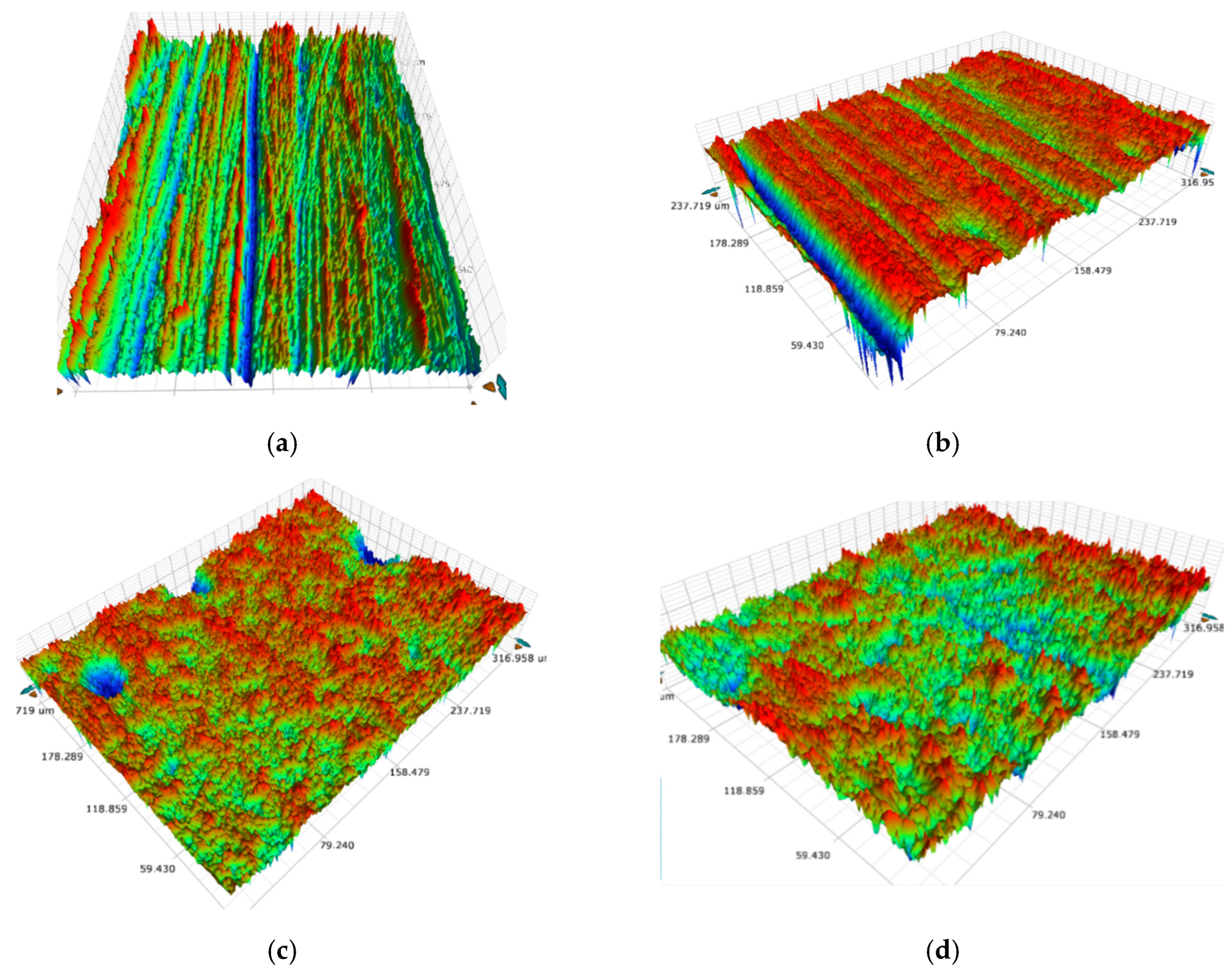
Figure 1. Surface morphology of Ti6Al4V substrates: (a) unpolished (Ra = 548 nm), (b) polished with #100 sandpaper (Ra = 654 nm), (c) polished with #400 sandpaper (Ra = 528 nm), (d) polished with #800 sandpaper (Ra = 478 nm).
Coatings 15 01086 g001
Figure 2. Surface roughness of DLC films deposited on Ti6Al4V substrates: (a) unpolished (Ra = 512 nm), (b) polished with #100 sandpaper (Ra = 532 nm), (c) polished with #400 sandpaper (Ra = 423 nm), (d) polished with #800 sandpaper (Ra = 386 nm).
Coatings 15 01086 g002
Figure 3. Surface roughness reductions after DLC deposition on Ti6Al4V substrates: (a) unpolished substrate (~36 nm decrease), (b) polished with #100-grit sandpaper (~122 nm decrease), (c) polished with #400-grit sandpaper (~105 nm decrease), and (d) polished with #800-grit sandpaper (~92 nm decrease).
Coatings 15 01086 g003
Figure 4. EDS analysis of DLC films deposited on Ti6Al4V substrates polished with #400 sandpaper.
Coatings 15 01086 g004
Figure 5. Contact angles of DLC films on Ti6Al4V substrates with different surface roughness, measured using distilled water and glycerol.
Coatings 15 01086 g005
Figure 6. Dispersive, polar, and total surface free energy (SFE) of DLC films on Ti6Al4V substrates with different surface roughness.
Coatings 15 01086 g006
Figure 7. ID/IG ratio of DLC films on Ti6Al4V substrates with different surface roughness.
Coatings 15 01086 g007
Figure 8. Hardness and reduced elastic modulus of DLC films on Ti6Al4V substrates with different surface roughness.
Coatings 15 01086 g008
Figure 9. ID/IG ratio versus hardness of DLC films on Ti6Al4V substrates with different surface roughness.
Coatings 15 01086 g009
Figure 10. ID/IG ratio versus reduced elastic modulus of DLC films on Ti6Al4V substrates with different surface roughness.
Coatings 15 01086 g010
Figure 11. Bright-field (left) and fluorescence (right) microscopy images of A549 cells cultured on DLC-coated Ti6Al4V substrates with varying surface roughness: (a,b) unpolished (Ra = 512 nm), (c,d) polished with #100 sandpaper (Ra = 532 nm), (e,f) polished with #400 sandpaper (Ra = 423 nm), and (g,h) polished with #800 sandpaper (Ra = 386 nm). Scale bar: 20 µm.
Coatings 15 01086 g011aCoatings 15 01086 g011b
Figure 12. A549 cell numbers on DLC-coated Ti6Al4V substrates with different surface roughness after 24 and 48 h in two separate experiments: (a) first experiment; (b) second experiment.
Coatings 15 01086 g012
Table 1. Chemical composition of Ti6Al4V.
ElementTiAlVFeO
Content (wt%)Balanced6.0%4.0%≤0.25%≤0.2%
Disclaimer/Publisher’s Note: The statements, opinions and data contained in all publications are solely those of the individual author(s) and contributor(s) and not of MDPI and/or the editor(s). MDPI and/or the editor(s) disclaim responsibility for any injury to people or property resulting from any ideas, methods, instructions or products referred to in the content.