Article

Physicochemical, Microstructural and Biological Evaluation of Dressing Materials Made of Chitosan with Different Molecular Weights

by
Zofia Płonkowska
,
Alicja Wójcik
and
Vladyslav Vivcharenko
*
Department of Tissue Engineering and Regenerative Medicine, Medical University of Lublin, Chodzki 1, 20-093 Lublin, Poland
*
Author to whom correspondence should be addressed.
Coatings 2025, 15(10), 1116; https://doi.org/10.3390/coatings15101116
Submission received: 26 August 2025 / Revised: 15 September 2025 / Accepted: 18 September 2025 / Published: 24 September 2025

Abstract

The use of advanced wound dressings can significantly support the skin healing process by maintaining optimal conditions for tissue regeneration. In this study, foam-like dressings composed of agarose and chitosan, enriched with vitamin C, were developed using a simple and cost-effective freeze-drying method. Three types of chitosan with varying molecular weights (low, medium, high) were used to investigate their impact on the biological, physicochemical, and mechanical properties of the resulting foams. All fabricated biomaterials were biocompatible, non-toxic, and did not promote cell adhesion to their surfaces. The foams exhibited highly porous, hydrophilic microstructures with excellent fluid absorption capacity (~20 mL/g) and sustained vitamin C release over the first 24 h. Chitosan molecular weight had no significant effect on biological properties, but influenced samples’ wettability and mechanical parameters. The hydrophilic character of samples was observed in all tested biomaterials, with the strongest enhancement of hydrophilicity noted for the low molecular weight variant. The highest tensile strength was observed in samples prepared with medium molecular weight chitosan. The results indicate that among the analyzed variants, agarose-chitosan foam biomaterials containing medium molecular weight chitosan exhibited the most favorable properties, making them the most promising candidates for the treatment of wounds with excessive exudate.

1. Introduction

Chitosan is one of the most widely used natural aminopolysaccharides for wound dressing production, derived from chitin [1]. Chitosan is easy to process, which allows for the creation of different types of dressings, such as films, foams, or hydrogels. Chitosan-based dressings can significantly improve wound healing due to a range of bioactivities including antibacterial, antioxidant, anti-inflammatory, hemostasis, tissue regeneration, and promotion of scar-free healing. The clinical efficacy of chitosan wound dressings has been confirmed in numerous scientific studies, demonstrating reduced wound healing time, decreased wound pain, and reduced wound infection, as well as improving the condition of scar hyperplasia and high hemostatic properties [2,3,4,5,6]. Wound dressings are typically intended to be changed every few days; therefore, it is important that they do not support cell attachment and growth on their surface, otherwise replacing them could damage newly formed tissue in the wound bed [7]. Simultaneously, it should be non-toxic in direct contact and should not disrupt cell proliferation in the wound bed [8]. Non-toxicity is a fundamental requirement for biomaterials intended for biomedical applications [9].
Chitosan can have various derivatives made by chemical modifications which affect the properties of the polymer [10]. The degree of deacetylation, additional chemical or mechanical modifications, and a purification process affect the final molecular weight of chitosan, which has a crucial impact on the biomaterial’s characteristics. Since there are chitosan-based wound dressings approved by the Food and Drug Administration (FDA), such as the Maxiocel Chitosan Wound Dressing [11], the KA01 Chitosan Wound Dressing [12], and the AQUANOVA Super-Absorbent Dressing [13], differences in its molecular weight are not expected to significantly affect cell viability. Nevertheless, some authors revealed a molecular-weight-dependent negative impact on cell viability in vitro. Wiegand et al. investigated the impact of 0.5% and 1% concentrations of chitosan oligosaccharide (5 kDa) and medium molecular weight chitosan (120 kDa) on human HaCaT keratinocytes [14]. The obtained results showed that both medium molecular weight chitosan and chitosan oligosaccharide dilutions exhibited antiproliferative effects on HaCaT cells in a time- and dose-dependent manner. Higher molecular weight contributed to a more radical decrease in cell viability. It has also been observed that low, medium and high molecular weight chitosan differ in solubility, viscosity, elasticity, bioactivity, and tear strength. It has been shown that the low molecular weight chitosan dressing has lower mechanical strength but exhibits the highest inhibitory effect on phytopathogen [1]. In another study, a high degree of deacetylation, as well as a low molecular weight of chitosan, revealed a strong ability in fibroblast activity stimulation [15]. The successful clinical application of chitosan requires precise adjustment of its physicochemical characteristics [16]. Given that biomaterials exhibit different properties depending on the molecular weight of chitosan, it seems reasonable to investigate the effect of molecular weight in the context of the wound dressing fabrication process.
The aim of this study was to produce a foam dressing made of agarose and chitosan with different molecular weights and to assess the impact of these molecular weights on the properties of the samples. Specifically, the study sought to investigate how variations in chitosan molecular weight affect the biological, mechanical, and physicochemical characteristics of the chitosan/agarose foam dressing materials, including their biological response, surface morphology, wettability, exudate management, tensile strength, elasticity, and the release of bioactive compounds. Vitamin C has a range of activities which enhance the wound healing process. It promotes collagen synthesis, supports fibroblasts proliferation and migration, provides an antioxidant environment, and plays a role in the differentiation of keratinocytes [17]. In view of these properties, it was also decided to immobilize vitamin C within the material microstructure to further improve its functional characteristics.

2. Materials and Methods

2.1. Preparation of the Foams

Chitosan polymer with three different molecular weights was used: low molecular weight (75.8 kDa, 79.0% ± 1.0% deacetylation degree), medium molecular weight (140.5 kDa, 84.2% ± 0.3% deacetylation degree), and high molecular weight (247.8 kDa, 81.8% ± 0.1% deacetylation degree) (Sigma-Aldrich Chemicals, Warsaw, Poland) [18]. To prepare a 3% chitosan solution, the chitosan powder was dissolved in 0.5% (v/v) acetic acid (CH3COOH) (Polskie Odczynniki Chemiczne S.A, Gliwice, Poland) and stirred at room temperature in glass beakers for approximately 30 min to fully dissolve. A 4% agarose suspension was prepared (Sigma Aldrich Chemicals, Warsaw, Poland) in 0.1% (w/v) sodium hydroxide (NaOH, Polskie Odczynniki Chemiczne S.A, Gliwice, Poland). The suspension was then heated on a hotplate stirrer at 95 °C for 10 min until fully dissolved. A vitamin C stock solution (L-Ascorbic acid 2-phosphate sesquimagnesium salt hydrate, Sigma Aldrich Chemicals, Warsaw, Poland) was prepared at a concentration of 40 mg/mL using ultrapure water obtained from a Milli-Q® Water Purification System (Merck, Warsaw, Poland). Chitosan and agarose solutions were mixed in a 1:1 volume ratio, resulting in a final concentration of 1.5% (w/v) chitosan and 2% (w/v) agarose. For materials containing vitamin C, the vitamin C stock solution was added when the mixed polysaccharides solution reached a temperature of approximately 40 °C, yielding a final concentration of 200 µg/mL in the homogeneous mass. The prepared compositions were then poured into a polystyrene Petri dish (Bionovo, Legnica, Poland) to form a uniform layer approximately 2 mm thick. All samples were then frozen at −80 °C for 24 h and lyophilized under a vacuum of 1 mbar (LYO GT2-Basic, SRK Systemtechnik GmbH, Riedstadt, Germany) for 20 h. All stages of production were carried out using sterile reagents under sterile conditions in a laminar flow cabinet (Esco Lifesciencies GmbH, Friedberg, Germany). Figure 1 shows a scheme of production steps of chitosan-based foams. The optimization of sample preparation was developed and described in the previous work [19].
Six different biomaterials were prepared using chitosan and agarose, with or without the addition of vitamin C. Table 1 presents compositions of all produced materials.

2.2. Cell Culture Tests

For all cell-culture assays, normal human skin fibroblasts (BJ cell line, CRL-2522™, American Type Culture Collection, Teddington, UK) were used. Cells were cultured in Eagle’s Minimum Essential Medium (EMEM, American Type Culture Collection, Teddington, UK) supplemented with 10% fetal bovine serum (FBS, Pan-Biotech GmbH, Aidenbach, Bavaria, Germany), streptomycin (100 µg/mL), and penicillin (100 U/mL) (both from Sigma-Aldrich Chemicals, Warsaw, Poland). Cells were kept in conditions recommended by ATCC: 37 °C, 5% CO2, and 95% air humidity.

2.2.1. Cytotoxicity Evaluation

The cytotoxicity of the produced biomaterials was assessed in accordance with ISO 10993 5:2009 [20] by determining BJ cell viability after exposure to 24 h sample extracts prepared in accordance with ISO 10993-12 [21]. Biomaterials were incubated in a BJ culture medium (EMEM) for 24 h at 37 °C to prepare the extracts. The ratio of biomaterial to medium was reduced to 15 mg per 1 mL due to the highly porous microstructure of the samples and their high absorption capacity. To evaluate the cytotoxicity of the produced biomaterials, the Cell Counting Kit-8 (Sigma-Aldrich Chemicals, Warsaw, Poland) was used. This colorimetric assay relies on the reduction in WST-8 tetrazolium salt [2-(2-methoxy-4-nitrophenyl)-3-(4-nitrophenyl)-5-(2,4-disulfophenyl)-2H-tetrazolium, monosodium salt] into formazan, an orange, water-soluble dye, by viable cells. The WST-8 cell assay does not require cell fixation or lysis, allowing measurements to be repeated in multiple time points. The cell suspension at a concentration of 2 × 10⁵ per mL was prepared and seeded into a 96-well plate at 100 µL per well and incubated at 37 °C for 24 h. After incubation, the medium was carefully removed and replaced with 100 µL of either the biomaterial extracts, negative control, or positive control. The positive control group was treated with 1% (w/v) phenol solution (Chimopat S.A., Bucharest, Romania) prepared in cell culture medium. The negative control group was treated with polypropylene extract prepared by immersing polypropylene (nontoxic material) (Bionovo, Legnica, Poland) in the cell culture medium. Cells were then incubated for an additional 48 h under the same conditions. Afterwards, the extracts and controls were removed, cells were rinsed with PBS containing calcium chloride and magnesium chloride (Sigma-Aldrich Chemicals, Warsaw, Poland), and the WST-8 test was performed. Briefly, a mix of WST-8 reagent and EMEM was prepared in 1:10 ratio, respectively, and 100 µL of the mixture was added to each well. The prepared plate was incubated under standard conditions for 2 h. After incubation, absorbance was measured using a microplate reader (Synergy H1, BioTek, Winooski, VT, USA). The results were presented as the mean percentage of absorbance values relative to the viable cells in the negative control group.

2.2.2. Live/Dead Assay

Cell viability in direct contact with the tested biomaterials was assessed using the Live/Dead Double Fluorescent Staining Kit (Sigma-Aldrich Chemicals, Warsaw, Poland), according to the manufacturer’s protocol. Calcein-AM is a non-fluorescent, cell-permeable compound that, upon enzymatic cleavage of the acetoxymethyl (AM) group by intracellular esterases in viable cells, produces a membrane-impermeable, fluorescent-green calcein. Propidium iodide penetrates cells with compromised membranes, binds to nucleic acids, and emits a red fluorescence. Small biomaterial samples (2 mm × 2 mm) were attached to a 48-well plate using 2% (w/v) agarose. Prior to cell seeding, the biomaterials were soaked in 300 µL of medium for 20 min under standard incubation conditions (37 °C, 5% CO2). After soaking, excess liquid was carefully removed and cells were seeded onto the samples at a concentration of 2 × 10⁵ cells/mL, adding 300 µL per well. The plates were then incubated for 3 days under standard conditions. Following incubation, Live/Dead staining was performed according to the manufacturer’s instructions. The samples were visualized using confocal laser scanning microscope (CLSM, Olympus Fluoview equipped with FV1000, Olympus Corporation, Tokyo, Japan).

2.3. Wound Healing Assay

To evaluate the effect of biomaterials on cell migration, a wound healing assay (scratch assay) was performed. Cells were seeded into a 24-well plate at a concentration of 2 × 10⁵ cells/mL and incubated for 3 days until a confluent monolayer was formed. Uniform scratches were created in each well using a BioTek AutoScratch device (Agilent Technologies, Santa Clara, CA, USA). Small samples of biomaterials (2 mm × 2 mm) were placed into the wells using cell culture inserts (Greiner Bio-One, Kremsmünster, Austria). To ensure full submersion of the materials, 1.1 mL of culture medium was added to each well. Wells without inserts served as the control group. Then, the cells were cultured for 48 h under standard conditions. Images of the scratches were made and captured using Celloger® Mini Plus (Curiosis Inc., Seoul, Republic of Korea) at three time points: immediately after scratching (0 h), after 24 h, and after 48 h. Cell confluence within the scratch area was analyzed using dedicated software (CellogerMiniPlus V2.1.2023.0512). Wound closure for each tested biomaterial was measured as a percentage of the scratch area populated with cells after incubation. Cells were cultured at 24 and 48 h with the tested biomaterials or pure culture medium (in the case of the control group) using cell culture inserts and a two-compartment environment. A schematic representation of the experiment conditions is shown in Figure 2.

2.4. Surface Roughness Analysis

To visualize the biomaterial topography and evaluate the surface roughness, samples were scanned using a confocal laser scanning optical profilometer (LEXT OLS5100, Olympus, Tokyo, Japan) with a 5× magnifications lens. Single measurements were performed over areas of 2.6 mm × 2.6 mm. At least fifteen fields of view were scanned per sample. The raw data were processed with the built-in software (OLS5_Analysis 2.2.3), applying an auto noise reduction filter before roughness calculation. Sample roughness was expressed as the areal average roughness (Sa), representing the arithmetic mean height of the surface area.

2.5. Wettability Evaluation

To estimate the wettability of biomaterials, the static contact angle method was applied using a DSA 30 goniometer (Kruss GmbH, Hamburg, Germany) equipped with a high-resolution camera and drop dispenser. Ultrapure water droplets (2 µL) were deposited onto the biomaterials’ surfaces using an automatic syringe. The contact angle was measured after the droplet stabilized, using image analysis software (ADVANCE 1.11). For each sample, measurements were performed at least eight different locations.

2.6. Mechanical Properties Analysis

To determine the mechanical properties, an Autograph AG-X plus universal testing machine (Shimadzu, Kyoto, Japan) was used in tensile mode. Samples were prepared as strips with a width of 10 mm, 2.5 mm thickness, and a gauge length of 40 mm, and presoaked in PBS for 30 min. The test was conducted with a constant crosshead speed of 50 mm/min. The applied force and elongation were continuously recorded to generate stress–strain curves. Based on this data, Young’s modulus, ultimate tensile strength (UTS), and elongation at break were calculated in dedicated software (TRAPEZIUMX-V Ver200). UTS was calculated by dividing the maximum applied force by the original cross-sectional area of the sample, while elongation at break was calculated by dividing the increase in length at the point of fracture by the original gauge length. At least three specimens were tested for each type of biomaterial.

2.7. Absorption Capacity Test

To assess the absorption capacity of the biomaterials, 2 mm × 2 mm samples with an average weight of 6 ± 1 mg were soaked in 2 mL of 0.9% solution of sodium chloride (Sigma-Aldrich Chemicals, Warsaw, Poland) prepared in ultrapure water. At specified time points (1 s, 2 s, 4 s, 8 s, 16 s, 30 s, 1 min, 2 min, 5 min, 10 min, 30 min, 1 h, 2 h, 24 h, 48 h) the samples were immersed and kept in the solution for the given duration, then removed, gently blotted with paper to remove excess fluid, weighted and then re-immersed in the solution. Absorption capacity was expressed as volume of fluid absorbed per 1 g of biomaterial (mL/g), calculated using the following Equation (1):
V = m w   m d d s
where V represents a volume of saline absorbed by 1 g of biomaterial (mL/g), md represents the dry sample weight (mg), mw represents the wet sample weight (mg), and ds represents saline density (mg/µL). Each type of biomaterial was tested using three independent samples.

2.8. Vitamin C Release

To evaluate the amount of vitamin C released from the foams and the dynamics of this process, samples with an average weight of 60 mg ± 2 mg were placed in tubes containing 10 mL of PBS and incubated for 96 h at 37 °C. At defined time points (15 min, 30 min, 1 h, 2 h, 4 h, 6 h, 24 h, 48 h, 72 h, 96 h), 0.5 mL of liquid was collected, and the absorbance at wavelength 252 nm was measured using a UV-spectrophotometer (Genesys 6 UV-Vis, Thermo Fisher Scientific, Waltham, MA, USA). Following absorbance measurement, the solution was returned to the corresponding tube to continue incubation. The concentration of vitamin C was calculated using the calibration curve prepared for L-Ascorbic acid 2-phosphate sesquimagnesium salt hydrate (Sigma Aldrich Chemicals, Warsaw, Poland) at concentrations from 6.25 to 62.5 µg/mL. Subsequently, the total amount of vitamin C released into the 10 mL of liquid was calculated. The final result was expressed as a percentage of the initial amount of vitamin C present in the sample. Each material was tested using three independent samples.

2.9. Statistical Analyses

All statistical analyses were performed using GraphPad Prism version 8.0 (GraphPad Software, San Diego, CA, USA). One-way analysis of variance (ANOVA) followed by Tukey’s or Dunnett’s post hoc tests were performed. Results were expressed as means ± standard deviations (SD). p-values less than 0.05 (p < 0.05) were considered statistically significant.

3. Results and Discussion

In this study, foam wound dressings made of agarose and chitosan with different molecular weights, including variants enriched with vitamin C, were prepared. The method used for foam production is cost-effective and easily accessible, with most of the required instruments available in basic laboratory equipment. Chitosan, agarose, and vitamin C are well known for their biocompatibility, non-toxicity, and low cost [22,23]. After production, the biomaterials exhibited a porous microstructure that was enclosed on both sides by a thin outer layer that can be observed in Figure 1.

3.1. Cell Culture Tests

In this study, the cytotoxicity of the wound dressings produced using chitosan with different molecular weights was assessed in accordance with ISO 10993-5 [20] using normal human skin fibroblasts and biomaterials extracts that were prepared according to ISO 10993-12 [21]. It was demonstrated that all of the biomaterials exhibited no cytotoxic effect towards skin fibroblasts. After 48 h of incubation, the viability of BJ cells treated with biomaterials extracts was comparable to the negative control group, where cells were treated with non-toxic polypropylene extract (Figure 3A).
Moreover, no statistically significant differences were observed between the LOW, MED, and HIGH samples. The viability was high (approximately 100%), indicating non-toxicity according to ISO 10993-5 [20], which defines cytotoxicity as a reduction in cell viability below 70% compared to the negative control. To estimate cytotoxicity in direct contact with the biomaterials and access cell adhesion preference, cells were cultured directly on the LOW, MED, and HIGH samples. After 3 days of cells culturing, BJ cells were visualized with Live/Dead staining. In all tested samples, no dead cells (stained in red) were detected. It should be noted that cells seeded next to the biomaterials on the polystyrene surface possessed normal morphology, which confirms the lack of cytotoxicity (Figure 3B). Furthermore, green, spherical, non-flattened cells were present on the biomaterial surface, suggesting that the BJ cells were viable but not attached to the top surface of the samples.
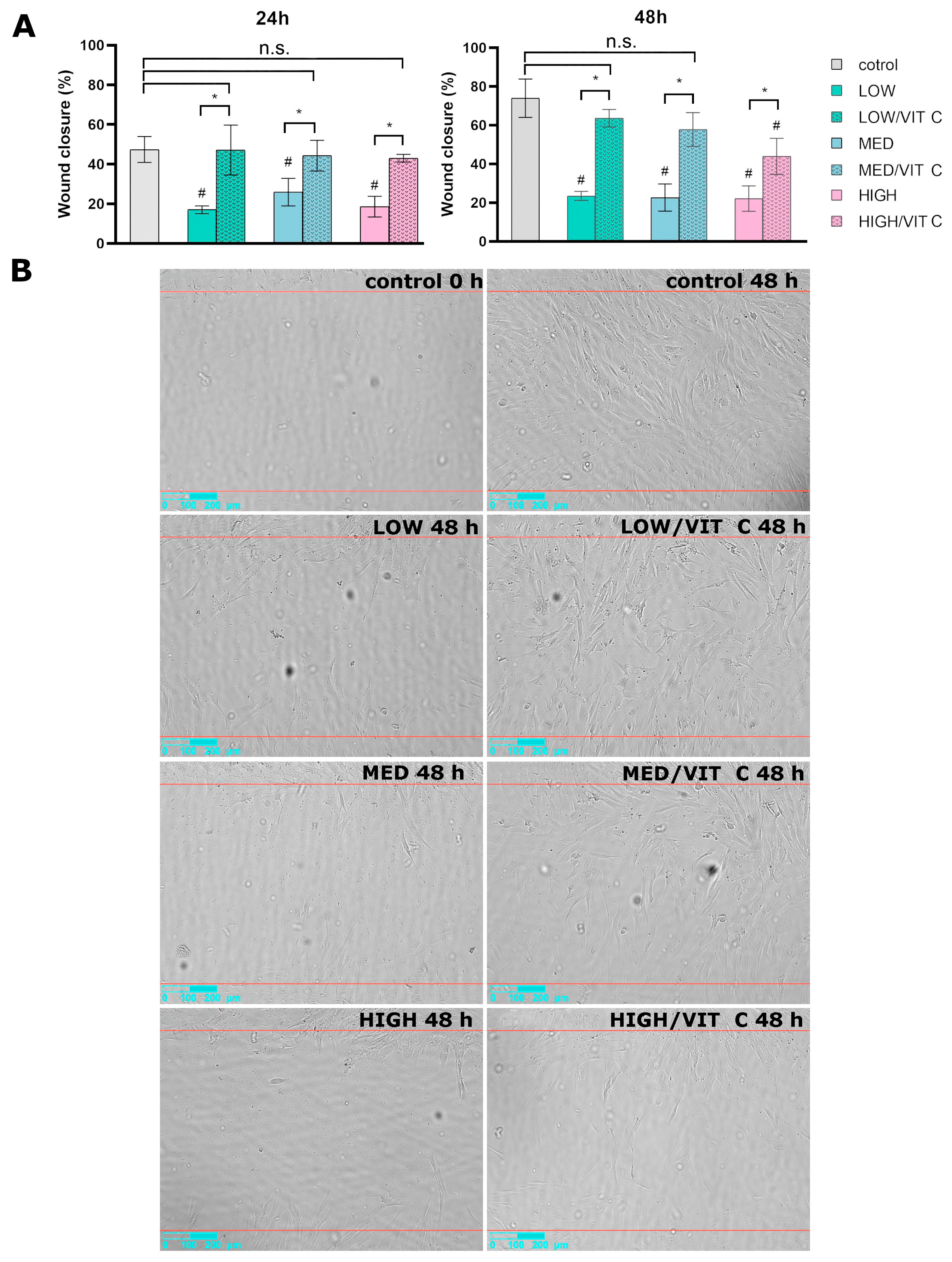
Cell migration plays a vital role in different physiological processes, such as immune response, morphogenesis, angiogenesis, and tissue repair. To investigate and compare the effects of the biomaterials on wound closure, fibroblast migration was assessed using a wound healing assay. After 24 h, BJ cells cultured in control wells achieved a wound closure of 47%. However, cells exposed to LOW, MED, and HIGH samples showed reduced migration in comparison to the control group, exhibiting only 17%, 25%, and 18% of wound closure, respectively (Figure 4A). After 48 h of incubation, wound closure for all tested biomaterials was near 20%, confirming the negative effect of the dressings on fibroblast migration.
The results clearly demonstrated that all tested materials, regardless of molecular weight, significantly hindered cell migration. Upon the addition of vitamin C to the samples, hindered cell migration caused by the biomaterials has been significantly overcome. Comparable results were observed 48 h after scratching. LOW/VIT C and MED/VIT C samples exhibited similar wound closure to the control group (64%, 58%, and 74%, respectively). However, the wound closure in the HIGH/VIT C sample was significantly lower in comparison to the control group (43%). Representative images of wound closure are shown in Figure 4B. The acceleration of cell migration after the addition of vitamin C is consistent with the reports in the literature, where authors have highlighted the positive impact of ascorbic acid on wound closure [25]. Since vitamin C induces collagen synthesis, the acceleration of wound healing is likely due to enhanced collagen deposition, which ultimately promotes improved cell migration and faster wound closure [26]. The wound healing assay demonstrated that cell migration was better in the presence of biomaterials enriched with vitamin C, highlighting the rationale for using this compound to accelerate the healing process. Taking into account both time intervals, the HIGH/VIT C sample exhibited slightly reduced wound closure, making the remaining samples more attractive candidates for potential wound dressing applications.

3.2. Surface Roughness Analysis

Surface roughness can affect various wound dressing characteristics, including cell adhesion and proliferation, handling, exudate absorption, and even protein adsorption and immune response [27]. To evaluate the surface roughness, the LOW, MED, and HIGH biomaterials were imaged using a confocal laser scanning optical profilometer. The mean areal surface roughness of the tested samples (Sa) ranged between 51 and 68 µm and did not differ significantly between materials with different molecular weight chitosan. Thus, molecular weight of chitosan does not significantly affect the roughness of the resultant biomaterial (Figure 5A). Representative 3D images of the samples’ topography are presented in Figure 5B.

3.3. Wettability Evaluation

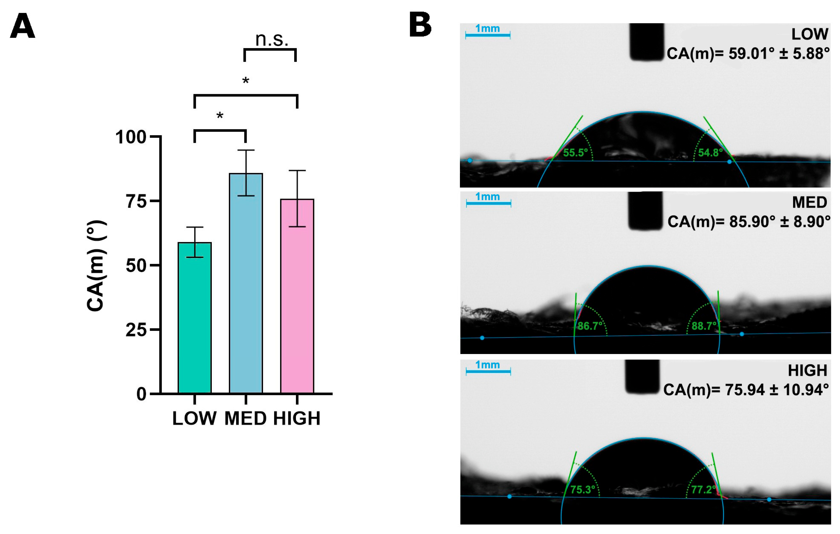
Wettability of the wound dressing directly impacts how the material interacts with wound exudate and healing tissue [28]. An optimal wound dressing should be able to: maintain secure contact with the skin surrounding the wound; absorb excess exudate; keep a moist environment in the wound bed; allow air circulation; and be easily removed without damaging the newly formed tissue [29]. A hydrophilic surface is suitable for exudate-absorbent wound dressings because it attracts and spreads water-based fluids, like wound exudate, while keeping the environment moist. It also prevents excess fluid accumulation, reducing the risk of maceration, infection, or inflammation. Simultaneously, optimal moisture is crucial for re-epithelialization and pain reduction. Improved exudate management also reduces the need for frequent dressing changes, improving patient comfort [29]. To estimate the wettability of produced biomaterials and assess whether the molecular weight of chitosan affects hydrophilicity, the contact angle of ultrapure water droplets on the produced materials was measured using a goniometer. It should be highlighted that the LOW biomaterial possessed a contact angle (59.0°) that was significantly lower in comparison to both the MED and HIGH samples (85.9° and 75.9°, respectively). The observed differences may be explained by the greater roughness of the LOW samples, which translates into a larger specific surface area of the sample and greater contact of the water droplet with the sample surface. Nevertheless, all tested biomaterials exhibited hydrophilic surfaces, as their contact angles were lower than 90°. The mean contact angles of the samples are presented in Figure 6A. Figure 6B presents images of a water droplet on the biomaterial surface.

3.4. Mechanical Test

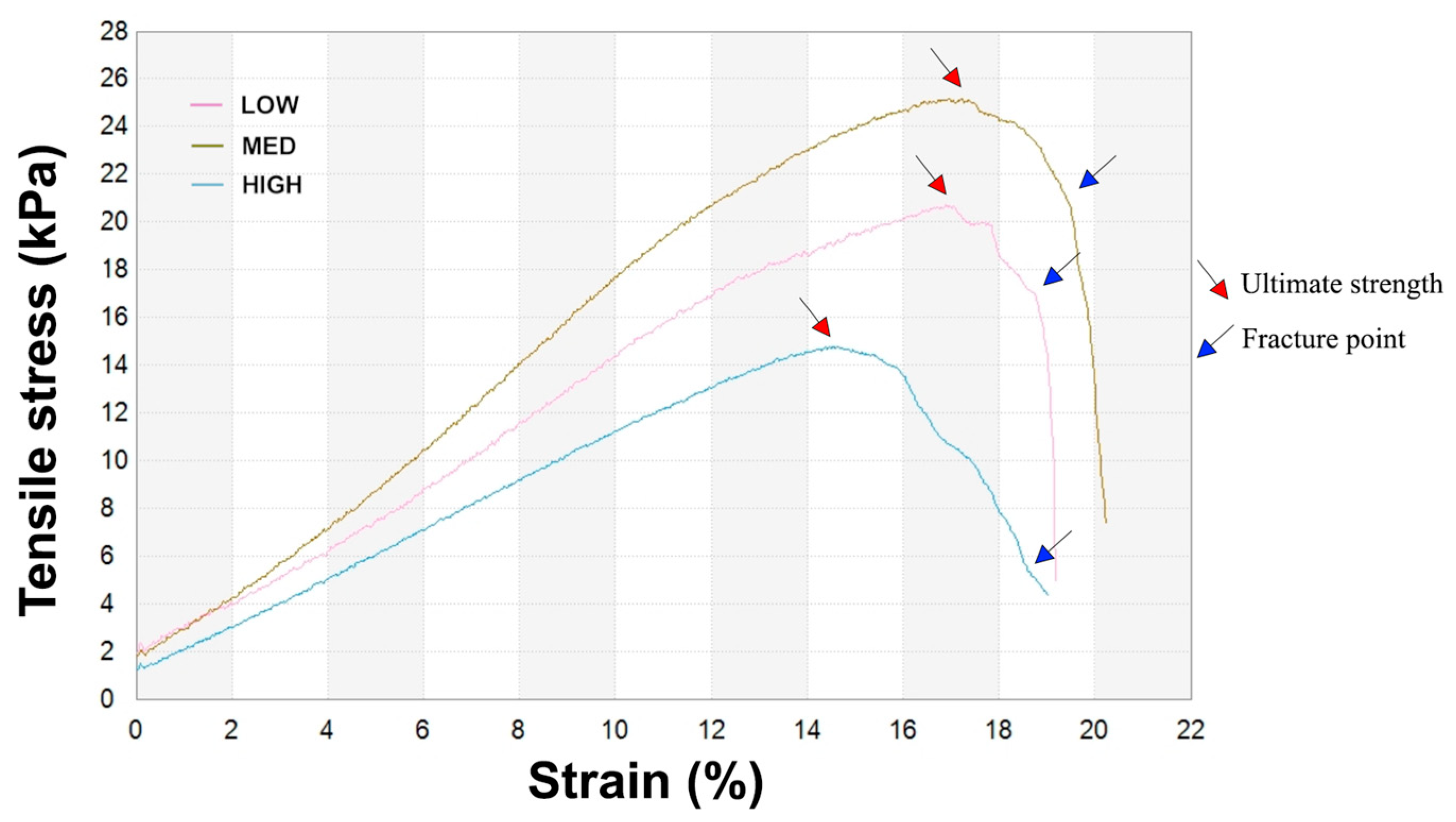
The mechanical properties of biomaterials are crucial for ensuring protective functionality, comfortable handling, application, and resistance to wear [30]. The prepared foams are intended to be used on exudative wounds, therefore the mechanical properties were evaluated in the wet state, as this better reflects the moist environment of a healing wound. Measured parameters included tensile strength, elongation at break, and Young’s modulus to characterize samples’ stiffness and elastic behavior (Table 2). Figure 7 presents the stress–strain curve of three representative samples.
All tested biomaterials reached similar elongation at break values, which ranged from 19.6% to nearly 24%, suggesting that the molecular weight of chitosan does not significantly influence the elongation capacity of the samples. These elongation values are consistent with those reported for chitosan/agarose film, indicating similar stretching behavior for both chitosan foams and films [19]. In contrast to the findings of the present study, Kwon et al. reported that the elongation at break of chitosan films in the dry state decreased with increasing molecular weight [31]. However, water molecules weaken the intramolecular hydrogen bonds by interacting with hydroxyl groups of chitosan. Han et al. demonstrated that chitosan exhibits different mechanical behavior in the wet state compared to the dry state, highlighting the material’s sensitivity to hydration, and changes in the mechanical properties and the material’s behavior [32]. As presented in Table 2, samples produced using chitosan with different molecular weights exhibited varying ultimate tensile strengths, ranging from 15 to 25 kPa. The highest tensile strength was observed in the sample prepared with the medium molecular weight chitosan, reaching 24.1 kPa. Similarly, as shown in Table 2, the sample prepared with the medium molecular weight chitosan exhibited the highest Young’s modulus (128.1 kPa), indicating that chitosan’s molecular weight influences the stiffness of the biomaterial. This value indicates relatively low stiffness and high elasticity, characteristics typical of soft, compliant materials [33]. Notably, it falls within the reported range for native human skin in its hydrated state (approximately 5 kPa to 2 MPa, depending on factors such as measurement technique, skin layer, hydration level, and age), highlighting the potential suitability of the material for biomedical applications such as wound dressings [34]. Foams with the addition of high molecular weight chitosan exhibited the poorest mechanical properties among all tested samples (Table 2). This may be caused by the worse solubility of the higher molecular weight chitosan. When the chitosan molecular weight is higher, hydrogen bonding interactions between molecules are stronger and solubility is worse [35]. In summary, the biomaterial with the medium molecular weight chitosan showed comparable elongation at break value compared to the other samples but reached the best resistance against mechanical stress and an appropriate Young’s modulus, providing an optimal balance between those mechanical properties.

3.5. Absorption Capacity Evaluation

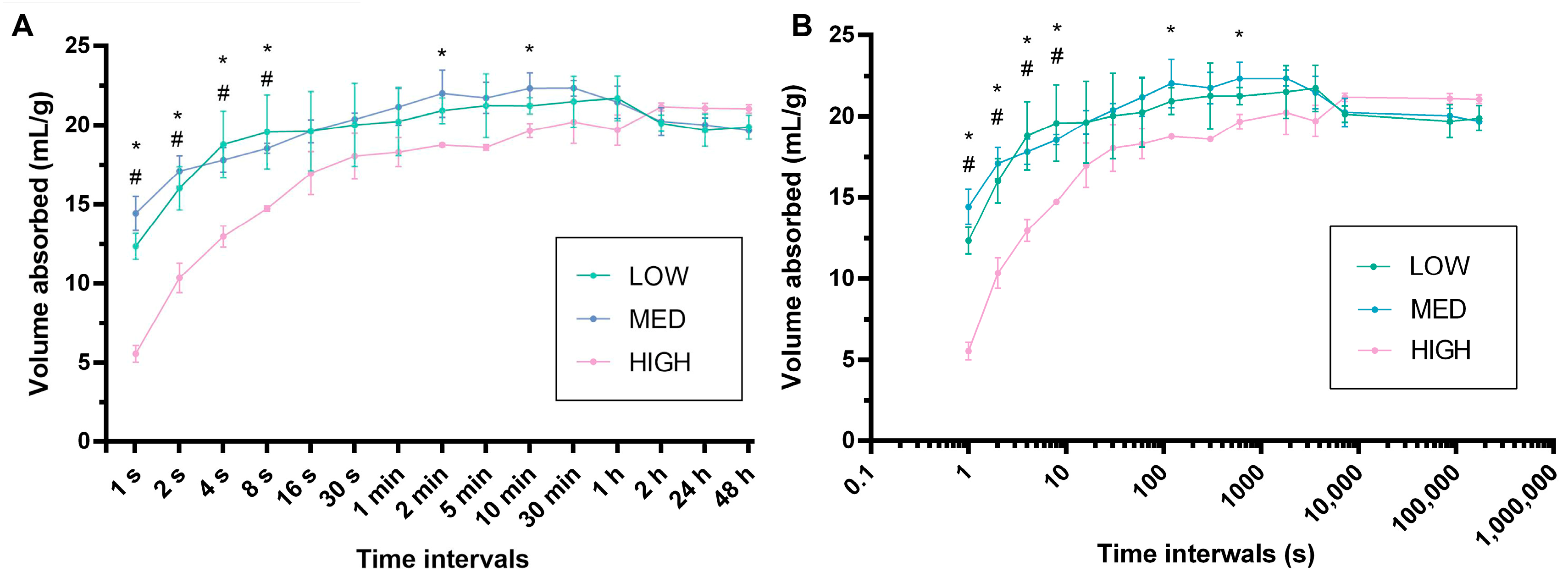
In the process of wound healing, exudate formation is a normal occurrence. It helps to clean the wound and maintain a moist environment, which promotes epithelialization and provides nutrients and growth factors to cells [36]. However, both excessive and insufficient exudate levels can disrupt the normal wound healing process. The amount of exudate can differ depending on the wound type or healing phase, so it is important to choose a wound dressing that will match the amount of fluid secreted from the wound [37]. To assess the ability of the produced biomaterials to manage the wound exudate, the absorption capacity of LOW, MED, and HIGH samples was evaluated over time (Figure 8).
All tested biomaterials reached absorption capacity values close to 20 mL/g, which surpasses most commercial foam and alginate dressings. Fulton et al. evaluated the absorption properties of 61 commonly used wound dressings available on the market. The study revealed that the average absorption capacity of foam wound dressings ranged from 5.4 to 13.4 g/g and the average absorption capacity of alginates reached from 9.4 to 22.5 g/g [38]. For the first 8 s, the HIGH sample absorbed less saline in comparison to the LOW and MED samples. The aforementioned phenomenon may be attributed to the lower roughness of the HIGH sample, which in turn results in a smaller total surface area. It is known that higher surface roughness increases the exposed surface area, enhancing the material’s ability to absorb fluids [39]. Nevertheless, due to the foam-like microstructure of the samples and their high porosity, the lower surface roughness estimated for the HIGH sample impacted absorption, primarily during the initial seconds after immersion in saline. In contrast, the total volume absorbed after 48 h was similar across the LOW, MED, and HIGH samples (approximately 20 mL/g), indicating a high absorption capacity of the produced samples. It can be noted that the MED biomaterial possessed the strongest water absorption capacity at 1 s, while simultaneously demonstrating the highest contact angle value, indicating the weakest hydrophilicity. The obtained phenomenon may be explained by differences in the internal porous microstructure and capillary effects, which can dominate over surface wettability in the early stages of water uptake. It should also be emphasized that contact angle measurements describe only the surface characteristics, whereas water absorption depends on the internal morphology and the pore connectivity within the biomaterial [40].

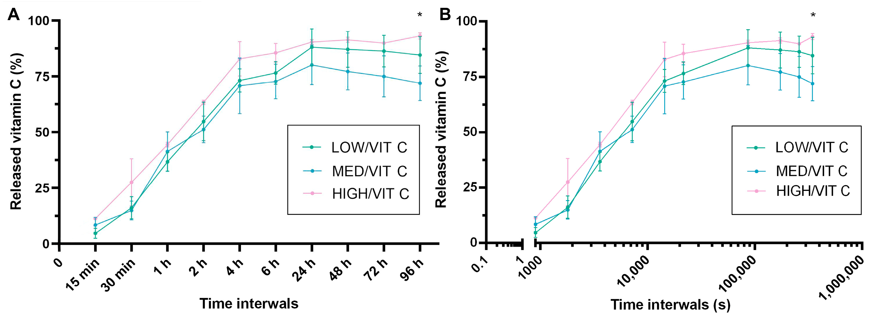
3.6. Vitamin C Release Analysis

Testing the bioactive substances release dynamics from wound dressings is important to evaluate whether the material can deliver the bioactive compound in a controlled and effective manner, which is essential for its therapeutic function in wound healing [41]. Topical application of the wound dressing could be an effective way of administering this molecule directly to the wound bed. To investigate the vitamin C release profiles, the amounts of released bioactive substance were measured over a 96 h period. All samples were enriched with the equal concentration of vitamin C (200 µg per 30 mg of dry sample). The release profiles of the ascorbic acid from the biomaterials are shown in Figure 9.
All samples exhibited an initial burst release within the first 4 h, followed by a gradual release phase. In the conducted test, no significant impact of chitosan molecular weight on vitamin C release was observed. The biomaterials reached a maximum vit C release after 24 h (LOW/VIT C = 88.1%, MED/VIT C = 80.1%, HIGH/VIT C = 90.4%) and a plateau effect was observed afterwards. The obtained results suggest a very effective release of bioactive compound from the biomaterial microstructure. Also, a prolonged compound release between 4 and 24 h can help maintain vitamin C concentrations at therapeutically beneficial levels. Considering the fact that wound dressings should be changed every 1–2 days, observed release dynamics allows for the optimal use of vitamin C contained in the dressing [8]. The negligible decrease observed after 24 h in the tested samples may be attributed to the inherent instability of ascorbic acid and its degradation over time.

4. Conclusions

In this study, agarose-chitosan foams enriched with vitamin C were successfully developed using a cost-effective freeze-drying method, resulting in a biocompatible, flexible, and hydrophilic biomaterials microstructure. The results showed no significant influence of chitosan molecular weight on the biological properties of the samples. All tested foams were non-toxic and did not support cell adhesion or growth on their surfaces. Furthermore, a wound healing assay demonstrated comparable effects between low and medium Mw chitosan-based samples enriched with vitamin C, with no significant differences in wound closure compared to the control. Notably, the high Mw chitosan-based sample exhibited the poorest wound-healing properties, showing the least favorable effect on wound closure among all tested samples. In contrast, chitosan molecular weight significantly influenced the physicochemical and mechanical properties of the foams. Samples containing low Mw chitosan exhibited the highest wettability, making them especially suitable for maintaining a moist wound environment. Due to their highly porous microstructure, all of the biomaterials demonstrated comparable and high fluid absorption capacity (~20 mL/g) as well as a sustained release of vitamin C over 24 h, supporting their potential for prolonged therapeutic applications. A slightly accelerated vitamin C release was observed from the HIGH sample compared to the other biomaterials, but the differences were not statistically significant, except at the final selected time interval when compared to the MED material. The mechanical test confirmed that the medium Mw chitosan-based sample exhibited superior performance, yielding the highest mechanical strength among all analyzed materials. The conducted analysis demonstrated that, among the tested formulations, foam produced with the use of medium Mw chitosan exhibited the most promising characteristics for managing skin wounds, offering the most favorable balance of biological, physicochemical, and mechanical properties.

5. Patents

The method for the production of the biomaterial was claimed in the Polish Patent Office: Medical University of Lublin, Cryogel wound dressing material based on chitosan and agarose and method of its production, Pat. 242473, 27 February 2023.

Author Contributions

Conceptualization, V.V.; methodology, V.V.; software, Z.P.; validation, Z.P. and V.V.; formal analysis, V.V.; investigation, Z.P. and V.V.; resources, A.W.; data curation, Z.P. and V.V.; writing—original draft preparation, Z.P. and V.V.; writing—review and editing, A.W.; visualization, Z.P.; supervision, V.V.; project administration V.V. All authors have read and agreed to the published version of the manuscript.

Funding

The research was funded by the Ministry of Science and Higher Education in Poland within the statutory activity of the Medical University of Lublin (DS 630 project).

Institutional Review Board Statement

Not applicable.

Informed Consent Statement

Not applicable.

Data Availability Statement

The raw/processed data required to reproduce these findings can be obtained from the corresponding author (vladyslav.vivcharenko@umlub.pl) upon reasonable request.

Conflicts of Interest

The authors declare no conflicts of interest.

References

  1. Moeini, A.; Pedram, P.; Makvandi, P.; Malinconico, M.; Gomez d’Ayala, G. Wound healing and antimicrobial effect of active secondary metabolites in chitosan-based wound dressings: A review. Carbohydr. Polym. 2020, 233, 115839. [Google Scholar] [CrossRef] [PubMed]
  2. Hu, J.; Lin, Y.; Cui, C.; Zhang, F.; Su, T.; Guo, K.; Chen, T. Clinical efficacy of wet dressing combined with chitosan wound dressing in the treatment of deep second-degree burn wounds: A prospective, randomised, single-blind, positive control clinical trial. Int. Wound J. 2023, 20, 699–705. [Google Scholar] [CrossRef] [PubMed]
  3. Stricker-Krongrad, A.-H.; Alikhassy, Z.; Matsangos, N.; Sebastian, R.; Marti, G.; Lay, F.; Harmon, J.W. Efficacy of Chitosan-Based Dressing for Control of Bleeding in Excisional Wounds. Eplasty 2018, 18, e14. [Google Scholar]
  4. Cicciù, M.; Fiorillo, L.; Cervino, G. Chitosan use in dentistry: A systematic review of recent clinical studies. Mar. Drugs 2019, 17, 417. [Google Scholar] [CrossRef] [PubMed]
  5. Liu, J.; Shen, H. Clinical efficacy of chitosan-based hydrocolloid dressing in the treatment of chronic refractory wounds. Int. Wound J. 2022, 19, 2012–2018. [Google Scholar] [CrossRef]
  6. Mo, X.; Cen, J.; Gibson, E.; Wang, R.; Percival, S.L. An open multicenter comparative randomized clinical study on chitosan. Wound Repair Regen. 2015, 23, 518–524. [Google Scholar] [CrossRef] [PubMed]
  7. Unnithan, A.R.; Ghavami Nejad, A.; Sasikala, A.R.K.; Thomas, R.G.; Jeong, Y.Y.; Murugesan, P.; Nasseri, S.; Wu, D.; Park, C.H.; Kim, C.S. Electrospun zwitterionic nanofibers with in situ decelerated epithelialization property for non-adherent and easy removable wound dressing application. Chem. Eng. J. 2016, 287, 640–648. [Google Scholar] [CrossRef]
  8. Jayakumar, R.; Prabaharan, M.; Sudheesh Kumar, P.T.; Nair, S.V.; Tamura, H. Biomaterials based on chitin and chitosan in wound dressing applications. Biotechnol. Adv. 2011, 29, 322–337. [Google Scholar] [CrossRef]
  9. Bernard, M.; Jubeli, E.; Pungente, M.D.; Yagoubi, N. Biocompatibility of polymer-based biomaterials and medical devices—regulations, in vitro screening and risk-management. Biomater. Sci. 2018, 6, 2025–2053. [Google Scholar] [CrossRef]
  10. Shen, S.; Chen, X.; Shen, Z.; Chen, H. Marine polysaccharides for wound dressings application: An overview. Pharmaceutics 2021, 13, 1666. [Google Scholar] [CrossRef]
  11. Kumar, A.; Agrawal, A.; Mavely, L.; Devadasu, V. Innovations in Advanced Wound Care from India. Int. J. Health Technol. Innov. 2023, 2, 9–14. [Google Scholar] [CrossRef]
  12. Davidson, E.; Pereira, J.; Gan Giannelli, G.; Murphy, Z.; Anagnostopoulos, V.; Santra, S. Multi-Functional Chitosan Nanovesicles Loaded with Bioactive Manganese for Potential Wound Healing Applications. Molecules 2023, 28, 6098. [Google Scholar] [CrossRef] [PubMed]
  13. Benediktsdóttir, B.E. Chitosan and trimethyl chitosan (TMC) as drug absorption enhancers: Moving forward toward clinical acceptance. In Encyclopedia of Marine Biotechnology; Wiley: Hoboken, NJ, USA, 2020; pp. 2773–2795. [Google Scholar] [CrossRef]
  14. Wiegand, C.; Winter, D.; Hipler, U.C. Molecular-weight-dependent toxic effects of chitosans on the human keratinocyte cell line HaCaT. Skin Pharmacol. Physiol. 2010, 23, 164–170. [Google Scholar] [CrossRef]
  15. Zhao, J.; Qiu, P.; Wang, Y.; Wang, Y.; Zhou, J.; Zhang, B.; Zhang, L.; Gou, D. Chitosan-based hydrogel wound dressing: From mechanism to applications, a review. Int. J. Biol. Macromol. 2023, 244, 125250. [Google Scholar] [CrossRef] [PubMed]
  16. Guariento, S.C.F.; Rodrigues, L.I.; Basilio, L.B.B.; Freitas, L.D.; de Oliveira, R.J.; Cardoso, M.A.; Pereira, M.L.S.; Passos, M.F.; Santos, T.M.; da Cruz Ferraz Dutra, J.; et al. Systematic review on chitosan dressings for diabetic and burn wound healing: Preclinical outcomes and limitations. Acad. Mater. Sci. 2025, 2, 1–26. [Google Scholar] [CrossRef]
  17. Pullar, J.M.; Carr, A.C.; Vissers, M.C.M. The roles of vitamin C in skin health. Nutrients 2017, 9, 866. [Google Scholar] [CrossRef]
  18. Garcia, L.G.S.; Guedes, G.M.d.M.; da Silva, M.L.Q.; Castelo-Branco, D.S.C.M.; Sidrim, J.J.C.; Cordeiro, R.d.A.; Rocha, M.F.G.; Vieira, R.S.; Brilhante, R.S.N. Effect of the molecular weight of chitosan on its antifungal activity against Candida spp. in planktonic cells and biofilm. Carbohydr. Polym. 2018, 195, 662–669. [Google Scholar] [CrossRef]
  19. Vivcharenko, V.; Benko, A.; Palka, K.; Wojcik, M.; Przekora, A. Elastic and biodegradable chitosan/agarose film revealing slightly acidic pH for potential applications in regenerative medicine as artificial skin graft. Int. J. Biol. Macromol. 2020, 164, 172–183. [Google Scholar] [CrossRef]
  20. ISO 10993-5:2009; Biological Evaluation of Medical Devices—Part 5: Tests for In Vitro Cytotoxicity. ISO: Geneva, Switzerland, 2009.
  21. ISO 10993-12:2021; Biological Evaluation of Medical Devices—Part 12: Sample Preparation and Reference Materials. ISO: Geneva, Switzerland, 2021. Available online: https://www.iso.org/standard/75769.html (accessed on 10 July 2023).
  22. Mohammadi, P.; Khounani, Z.; Hosseinzadeh-Bandbafha, H.; Foong, S.Y.; Yusoff, M.A.; Shahbeik, H.; Abdul Razak, N.N.; Lam, S.S.; Sheikh Ahmad Tajuddin, S.A.F.; Aghbashlo, M.; et al. Comparative environmental impact of chitosan extraction methods from shrimp waste. J. Environ. Chem. Eng. 2025, 13, 115124. [Google Scholar] [CrossRef]
  23. Zhang, X.; Liang, Y.; Huang, S.; Guo, B. Chitosan-based self-healing hydrogel dressing for wound healing. Adv. Colloid Interface Sci. 2024, 332, 103267. [Google Scholar] [CrossRef] [PubMed]
  24. Vivcharenko, V.; Wojcik, M.; Palka, K.; Przekora, A. Highly Porous and Superabsorbent Biomaterial Made of Marine-Derived Polysaccharides and Ascorbic Acid as an Optimal Dressing for Exuding Wound Management. Materials 2021, 14, 1211. [Google Scholar] [CrossRef]
  25. Weeks, B.S.; Fu, R.; Zaidi, M. Vitamin C Promotes Wound Healing: The Use of In Vitro Scratch Assays to Assess Re-Epithelialization; IntechOpen: London, UK, 2023. [Google Scholar] [CrossRef]
  26. Murad, S.; Grove, D.; Lindberg, K.A.; Reynolds, G.; Sivarajah, A.; Pinnell, S.R. Regulation of collagen synthesis by ascorbic acid. Proc. Natl. Acad. Sci. USA 1981, 78, 2879–2882. [Google Scholar] [CrossRef]
  27. Jurak, M.; Wiącek, A.E.; Ładniak, A.; Przykaza, K.; Szafran, K. What affects the biocompatibility of polymers? Adv. Colloid Interface Sci. 2021, 294, 102451. [Google Scholar] [CrossRef]
  28. Kayati, F.N.; Purnomo, C.W.; Kusumastuti, Y.; Rochmadi. The optimization of hydrogel strength from cassava starch using oxidized sucrose as a crosslinking agent. Green Process. Synth. 2023, 12, 2855–2879. [Google Scholar] [CrossRef]
  29. Jin, S.G.; Yousaf, A.M.; Kim, K.S.; Kim, D.W.; Kim, D.S.; Kim, J.K.; Yong, C.S.; Youn, Y.S.; Kim, J.O.; Choi, H.G. Influence of hydrophilic polymers on functional properties and wound healing efficacy of hydrocolloid based wound dressings. Int. J. Pharm. 2016, 501, 160–166. [Google Scholar] [CrossRef] [PubMed]
  30. Yan, X.; Huang, H.; Bakry, A.M.; Wu, W.; Liu, X.; Liu, F. Advances in enhancing the mechanical properties of biopolymer hydrogels via multi-strategic approaches. Int. J. Biol. Macromol. 2024, 272, 132583. [Google Scholar] [CrossRef]
  31. Hwang, K.T.; Kim, J.T.; Jung, S.T.; Cho, G.S.; Park, H.J. Properties of chitosan-based biopolymer films with various degrees of deacetylation and molecular weights. J. Appl. Polym. Sci. 2003, 89, 3476–3484. [Google Scholar] [CrossRef]
  32. Han, D.; Yan, L.; Chen, W.; Li, W. Preparation of chitosan/graphene oxide composite film with enhanced mechanical strength in the wet state. Carbohydr. Polym. 2011, 83, 653–658. [Google Scholar] [CrossRef]
  33. Wahlsten, A.; Stracuzzi, A.; Lüchtefeld, I.; Restivo, G.; Lindenblatt, N.; Giampietro, C.; Ehret, A.E.; Mazza, E. Multiscale mechanical analysis of the elastic modulus of skin. Acta Biomater. 2023, 170, 155–168. [Google Scholar] [CrossRef]
  34. Kalra, A.; Lowe, A. An Overview of Factors Affecting the Skins Youngs Modulus. J. Aging Sci. 2016, 4, 1000156. [Google Scholar] [CrossRef]
  35. Ji, M.; Li, J.; Wang, Y.; Li, F.; Man, J.; Li, J.; Zhang, C.; Peng, S.; Wang, S. Advances in chitosan-based wound dressings: Modifications, fabrications, applications and prospects. Carbohydr. Polym. 2022, 297, 120058. [Google Scholar] [CrossRef] [PubMed]
  36. Adderley, U.J. Managing wound exudate and promoting healing. Br. J. Community Nurs. 2013, 15 (Suppl. 3), 15–20. [Google Scholar] [CrossRef]
  37. Nuutila, K.; Eriksson, E. Moist Wound Healing with Commonly Available Dressings. Adv. Wound Care 2020, 10, 685–698. [Google Scholar] [CrossRef]
  38. Fulton, J.A.; Blasiole, K.N.; Cottingham, T.; Tornero, M.; Graves, M.; Smith, L.G.; Mirza, S.; Mostow, E.N. Wound dressing absorption: A comparative study. Adv. Ski. Wound Care 2012, 25, 315–320. [Google Scholar] [CrossRef]
  39. Kumar, S.; Charitatos, V. Influence of surface roughness on droplet evaporation and absorption: Insights into experiments from lubrication-theory-based models. Langmuir 2022, 38, 15889–15904. [Google Scholar] [CrossRef] [PubMed]
  40. Wang, Y.; Li, L.; An, M.; Sun, Y.; Yu, Z.; Huang, H. Factors Influencing the Capillary Water Absorption Characteristics of Concrete and Their Relationship to Pore Structure. Appl. Sci. 2022, 12, 2211. [Google Scholar] [CrossRef]
  41. Pachuau, L. Recent developments in novel drug delivery systems for wound healing. In Expert Opinion on Drug Delivery; Taylor and Francis Ltd.: Oxford, NY, USA, 2015; pp. 1895–1909. [Google Scholar] [CrossRef]
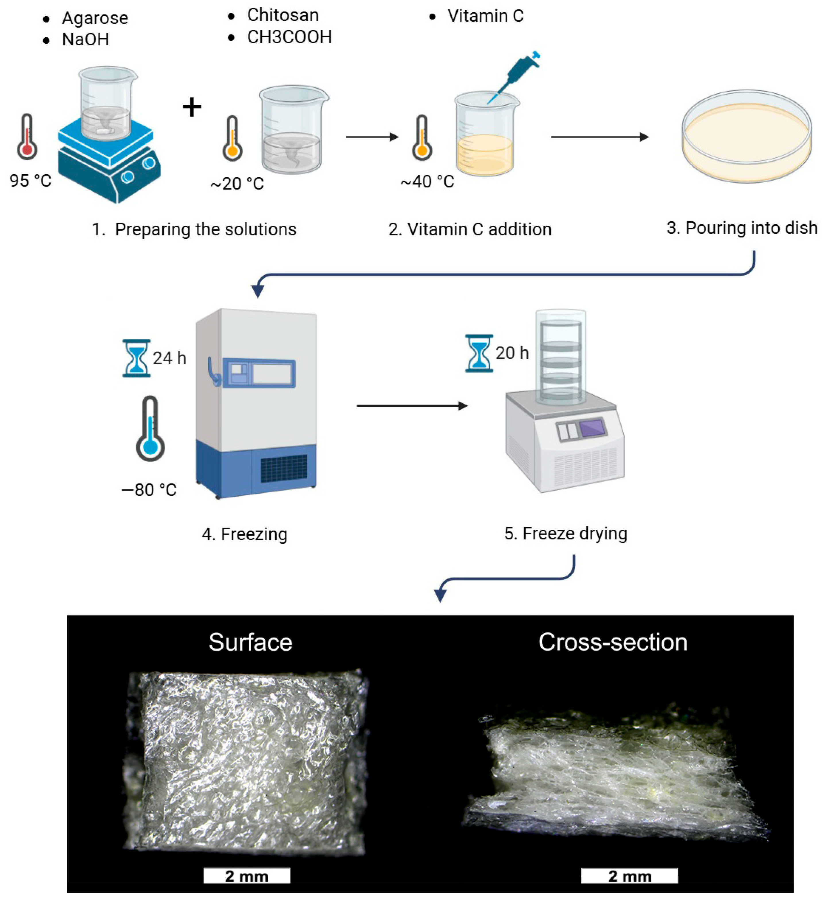
Figure 1. Schematic representation of the process of chitosan-based foams production.
Coatings 15 01116 g001
Figure 2. Schematic representation of the wound healing assay set up.
Coatings 15 01116 g002
Figure 3. Cytotoxicity tests of chitosan-based foam dressing materials: (A) WST-8 cell viability test using biomaterial extracts and human skin fibroblasts conducted according to ISO 10993-5 [20] and ISO 10993-12 [21] (results are presented as the mean percentage of viable cells relative to the negative control group; (#) above the bars indicates statistically significant differences compared to negative control group, p < 0.05, one-way ANOVA followed by Dunnet’s test; (n.s.) indicates statistical non-significance p > 0.05); (B) Live/Dead staining of human skin fibroblasts after 3 days of culture on the biomaterials and adjacent polystyrene surface (green fluorescence represents viable cells; red fluorescence represents dead cells) [24].
Coatings 15 01116 g003
Figure 4. Wound healing assay: (A) percentage of the scratch area covered by migrating cells; (#) above the bars indicates statistically significant differences compared to the control; (*) indicates pairwise comparisons, p < 0.05, one-way ANOVA followed by Tukey’s test; and (n.s.) indicates statistical non-significance, p > 0.05. (B) selected images of the scratched areas captured by Celloger® Mini Plus right after the scratching (0 h) and 48 h post-scratching; red lines indicate the scratch borders.
Coatings 15 01116 g004
Figure 5. Surface roughness analysis: (A) graph showing the mean areal surface roughness (Sa), defined as the arithmetic mean height of the surface area; (n.s.) indicates statistical non-significance p > 0.05. (B) Representative 3D views of the scanned areas measuring 2.6 mm × 2.6 mm.
Coatings 15 01116 g005
Figure 6. Wettability analysis of produced biomaterials: (A) mean values of contact angles between water droplet and biomaterial surface; (*) indicates pairwise comparisons, p < 0.05, one-way ANOVA followed by Tukey’s test; and (n.s.) indicates statistical non-significance p > 0.05. (B) Images of a water droplet on the biomaterial surface, with the measured contact angle marked by green lines.
Coatings 15 01116 g006
Figure 7. Representative tensile stress–strain curves of representative samples.
Coatings 15 01116 g007
Figure 8. Absorption capacity analysis expressed as a volume of saline (mL) absorbed by 1 g of biomaterial: (A) time intervals presented as selected experimental points and (B) time intervals presented on a logarithmic X-axis. (#) indicates statistically significant differences between the HIGH and LOW samples and (*) indicates statistically significant differences between the HIGH and MED samples, p < 0.05, one-way ANOVA followed by Tukey’s test.
Coatings 15 01116 g008
Figure 9. The amount of the released vitamin C presented as a percentage of the initial content, calculated individually for each tested sample: (A) time intervals presented as selected experimental points and (B) time intervals presented on a logarithmic X-axis. (*) indicates statistically significant differences between HIGH/VIT C and MED/VIT C, p < 0.05, one-way ANOVA followed by Tukey’s test.
Coatings 15 01116 g009
Table 1. Composition of the initial solutions used for biomaterial preparation.
DesignationComposition
LOW2% (w/v) agarose, 1.5% (w/v) low molecular weight chitosan
MED2% (w/v) agarose, 1.5% (w/v) medium molecular weight chitosan
HIGH2% (w/v) agarose, 1.5% (w/v) high molecular weight chitosan
LOW/VIT C2% (w/v) agarose, 1.5% (w/v) low molecular weight chitosan,
vitamin C (200 µg per 1 mL of prepared homogeneous mass)
MED/VIT C2% (w/v) agarose, 1.5% (w/v) medium molecular weight chitosan,
vitamin C (200 µg per 1 mL of prepared homogeneous mass)
HIGH/VIT C2% (w/v) agarose, 1.5% (w/v) high molecular weight chitosan,
vitamin C (200 µg per 1 mL of prepared homogeneous mass)
Table 2. Mechanical properties characterization of tested samples.
SampleYoung’s Modulus
(kPa)
Ultimate Tensile Strength (UTS) (kPa)Elongation at Break
(%)
LOW105.4 ± 10.620.3 ± 1.220.9 ± 2.4
MED128.1 ± 21.3 *24.1 ± 4.0 *19.6 ± 0.8
HIGH87.4 ± 7.215.4 ± 1.223.6 ± 6.7
(*) indicates statistically significant differences compared to the HIGH sample; p < 0.05, one-way ANOVA followed by Tukey’s test.
Disclaimer/Publisher’s Note: The statements, opinions and data contained in all publications are solely those of the individual author(s) and contributor(s) and not of MDPI and/or the editor(s). MDPI and/or the editor(s) disclaim responsibility for any injury to people or property resulting from any ideas, methods, instructions or products referred to in the content.