Article

Comparison Between Bond Strengths of a Resin Cement on Traditional Prosthetic Substrates and a 3D-Printed Resin for Permanent Restorations

1
Dental Academy, University of Portsmouth, Portsmouth PO1 2QG, UK
2
Department of Restorative Dentistry, Faculty of Dentistry, King Abdulaziz University, Jeddah 21589, Saudi Arabia
*
Author to whom correspondence should be addressed.
Coatings 2025, 15(8), 896; https://doi.org/10.3390/coatings15080896 (registering DOI)
Submission received: 17 June 2025 / Revised: 13 July 2025 / Accepted: 15 July 2025 / Published: 1 August 2025
(This article belongs to the Special Issue Advanced Polymer Coatings: Materials, Methods, and Applications)

Abstract

Recently, 3D-printed resins have been introduced as materials for definitive indirect restorations. Herein, a comparative assessment of the bond strengths of 3D-printed resins to a resin cement was performed. Methods: four definitive restorative materials were selected, i.e., a Feldspar ceramic (VITA Mark II, VM), a polymer-infiltrated ceramic network (VITA Enamic, VE), a nanohybrid resin composite (Grandio Bloc, GB), and one 3D-printed resin (Crown Permanent, CP). VM and VE were etched and silanized, GB was sandblasted, and CP was glass bead blasted; for one further experimental group, this was followed by sandblasting (CPs). A resin cement (RelyX Unicem) was then used for bonding, and then a notched shear bond strength test (nSBS) was performed. Failure modes were observed and classified as adhesive, cohesive, or mixed, and SEM representative images were taken. Data were statistically analyzed with one-way ANOVA, Tukey, and Chi-square tests. Significant differences were detected in nSBS among materials (p < 0.001). The highest nSBS was found in VM (30.3 ± 1.8 MPa) a, followed by CPb, GBbc, CPbc, and VEc. Failure modes were significantly different (p < 0.001), and with different prevalent failure modes. The bond strength for 3D-printed permanent resin materials was shown to be lower than that of the felspathic ceramic but comparable to that of the resin block and PICN substrates.

1. Introduction

Adhesive technologies have revolutionized restorative and prosthetic dentistry in recent decades [1,2,3]. Accordingly, the performance of more conservative restorations, such as resin and ceramic inlays and onlays, as well as more invasive procedures including crown and bridge applications, has improved [4,5]. Among other tooth-related, patient-related and operator-related factors, the success and longevity of adhesively bonded indirect restorations rely on the efficacy of the adhesive process [4,6,7,8]. Several studies have investigated different restorative materials and pre-cementation conditioning protocols to optimize their bonding to the underlying substrate [9,10].
The advances of additive manufacturing process, i.e., 3D printing, have enhanced its employment for dental applications, thus facilitating the fabrication of monolithic and veneered, single and multi-unit restorations with promising preliminary results, comparable to those fabricated through subtractive milling (CAD/CAM) and conventional processes [11,12,13]. Initially, 3D-printed resin materials were predominantly utilized as interim provisional restorative options; however, with the development of novel reinforced resin formulations, their applications have expanded to include definitive restorations. Currently, evidence on the adhesion of 3D-printed resin materials is limited to provisional restorations [14,15,16,17,18,19,20,21,22,23]; however, studies exploring the adhesion of printed definitive resin restorations are scarce and mainly limited to manufacturer resources [14,15,16,17,18]. Moreover, surface treatments, ageing, printing technology, printing parameters and post-processing conditions have shown to considerably influence the bonding performance of 3D-printed resin restorations [17,18,19,20,21]. Crown Permanent (Formlabs GmbH, Somerville, MA, USA) is a recently introduced 3D-printed resin intended for use in definitive single crowns, inlays, onlays, and veneers [22,23], with specific manufacturer pre-cementation guidelines instructing sandblasting intaglio surfaces with lead-free soda glass bead blasting materials, such as Perlablast® Micro (Bego, Bremen, Germany), followed by cementation with self-adhesive cements or with composite cement with primers. The glass bead blasting procedure promotes the removal of filler deposits on the surface of the polymerized printed resin structure, wherein the metal-free nature combined with the miniature particle size of the glass beads prevents aggressive surface disintegration of the resin substrate. Nevertheless, indirect resin restorations are traditionally sandblasted with 30–50 µm Al2O3 particles prior to their adhesive cementation to tooth structures [24,25]. Notably, to preserve the long-term adhesion of 3D-printed resin restorations to the underlying substrates, it is clinically relevant to investigate suitable surface treatments for 3D-printed resin materials used as the definitive prosthesis. To date, only one study has compared the bond strength of 3D-printed resin to varying luting agents following different intaglio surface blasting regimens; the authors reported the resin cement to have higher bond strength to the 3D-printed composite when a combined glass blasting and sandblasting regimen was employed [26]. Nevertheless, precise recommendations addressing the optimal pre-cementation surface treatment of 3D-printed definitive resin restorations are lacking in the existing literature. Therefore, this study aimed to investigate the adhesion of 3D-printed materials in comparison to traditional prosthetic substrates. In the present study, the notched-edge shear bond strength was used by virtue of its uniform distribution of loading forces to the bonded interface compared to that of the knife-edge shear loading apparatus, thus yielding accurate and clinically relevant test results for the former [27]. The null hypothesis tested was that there is no difference between the shear bond strength of a 3D-printed resin intended for definitive restorations and traditional prosthetic definitive materials in terms of adherence to a resin cement.

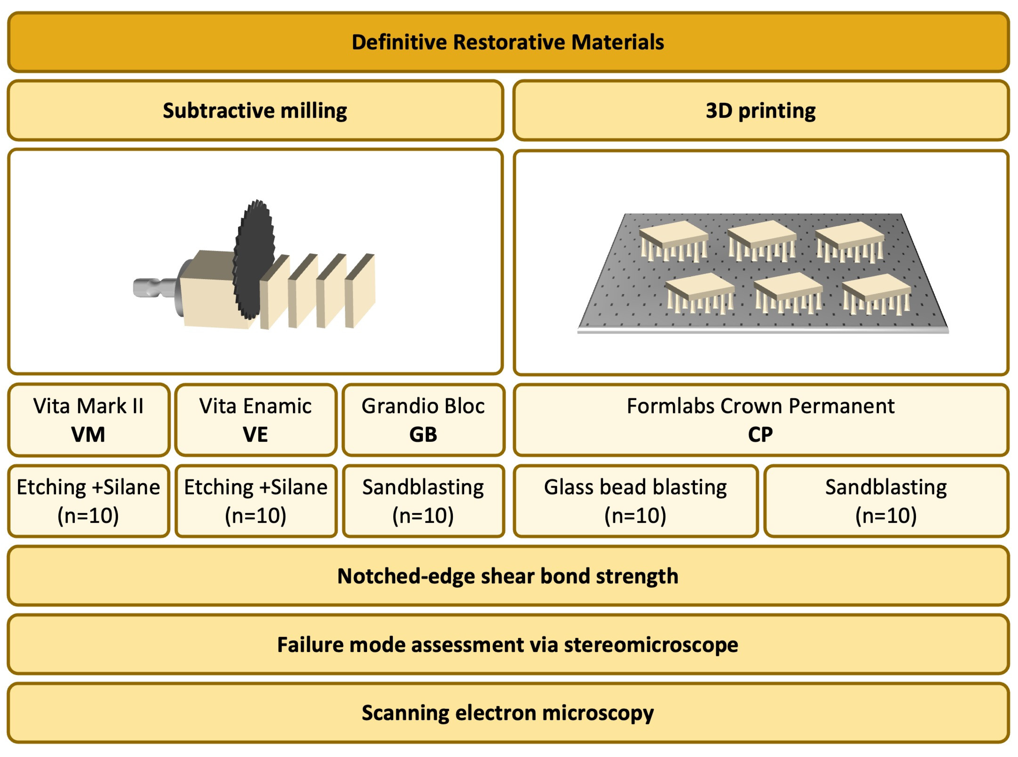
2. Materials and Methods

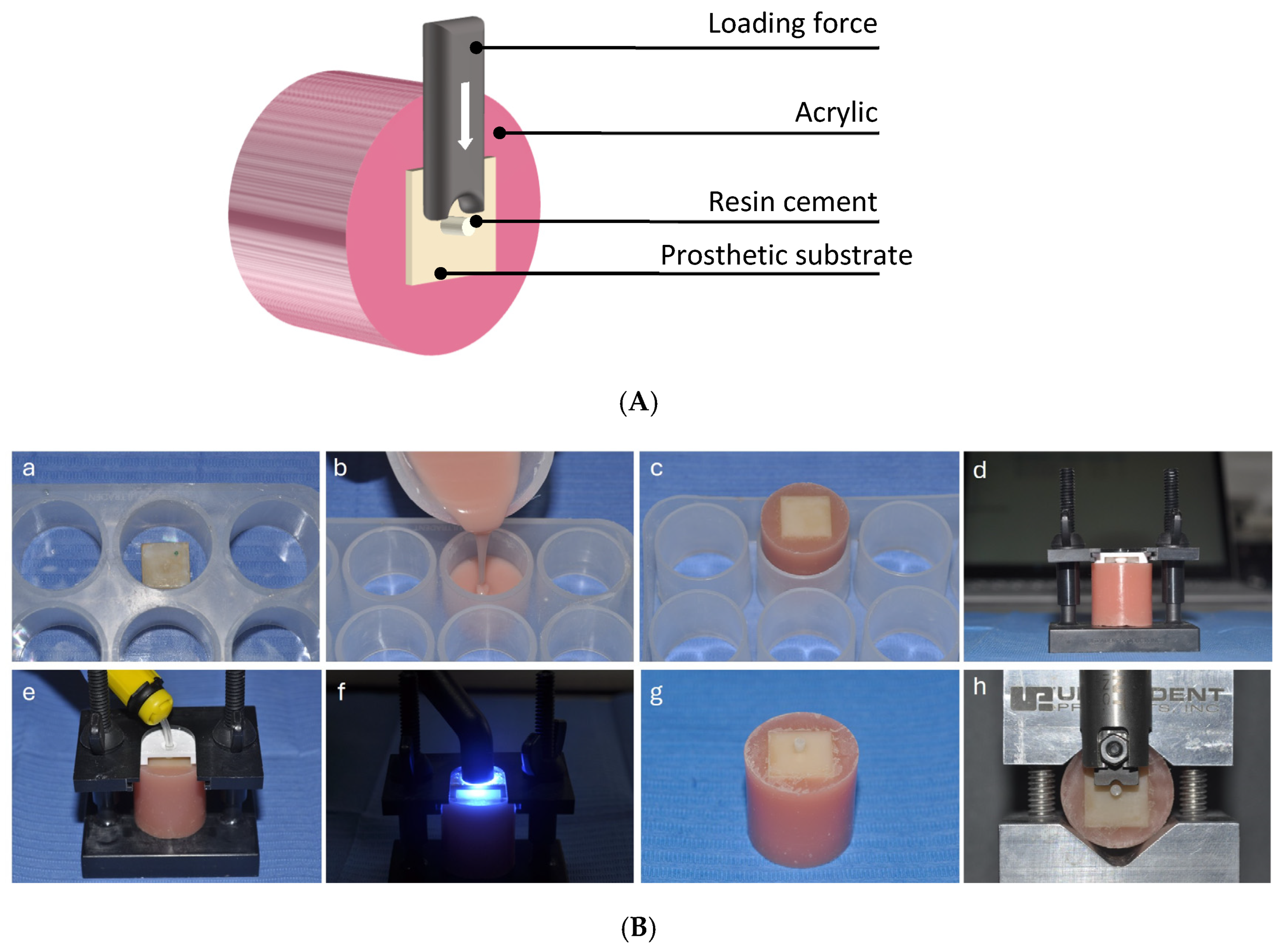
2.1. Specimen Preparation

Four materials indicated for definitive restorations were selected: a feldspar-ceramic-based material (VITABLOC Mark II, VITA Zahnfabrik, Bad Säckingen, Germany, VM), a polymer-infiltrated ceramic network (VITA Enamic, VITA Zahnfabrik, Bad Säckingen, Germany, VE), a nanohybrid resin composite (Grandio Blocs, VOCO GmbH, Cuxhaven, Germany, GB), and a 3D-printed methacrylic acid ester-based resin (Crown Permanent, Formlabs GmbH, Somerville, MA, USA, CP). The number of specimens in each group (n = 10) was selected via a priori power analysis using a statistical software program (G*power, V. 3.1.3; Heinrich Heine University, Düsseldorf, Germany), wherein an effect size of f = 0.6 and a confidence interval of 90% were deemed sufficient to obtain 90% power. The composition of the tested materials is reported in Table 1.
The VM, VE, and GB specimens were obtained by cutting the blocks with a water-cooled low-speed diamond saw (Isomet, Buehler, Lake Bluff, IL, USA). During the cutting process, the alignment of the blocks with respect to the saw blade was maintained through a custom-made setup. The final dimensions of all CAD/CAM specimens were 14 × 14 × 5 mm3. The CP specimens were designed using Thinkercad software (https://www.thinkercad.com accessed on 7 November 2023) (Autodesk, San Rafael, CA, USA) to obtain 14 × 14 × 5 mm3 plates using a horizontal printing orientation in relation to the build platform. Subsequently, the file was exported in stl file format and imported into PreForm software 3.32.0 (Formlabs, Somerville, MA, USA) for automatic support calculation and slicing. The printing parameters were set to 50 µm layer thickness using the exposure time recommended by the software specifically used for CP resin materials. CP specimens were printed (Formlabs 3B+ 3D printer, Formlabs, Sommerville, MA, USA), and then the specimens were manually removed from the build platform and washed for 3 min with the 99% isopropyl alcohol in an automated washing machine to remove uncured resin (FormWash, Formlabs, Somerville, MA, USA). In line with the manufacturer’s recommendations, the CP specimens were subjected to two curing cycles. First, the CP samples were cured for 20 min at 60 °C in an automatic curing machine from the same manufacturer (FormCure, Formlabs, Somerville, MA, USA); this was followed by a 30 min drying period. The supports were then manually detached, and the specimens were blasted to remove surface filler particles using 50 μm glass beads (Perlablast Micro, Bego, Bremen, Germany) for 10 s at a pressure of 1.5 bar and at 10 mm distance. Subsequently, the CP specimens were post-cured in a second curing cycle for 20 min at 60 °C (FormCure, Formlabs, Somerville, MA, USA) [28]. The specimens were embedded in a chemically polymerized methacrylate, exposing the surfaces that were not attached to supports (i.e., surfaces away from the platform); the exposed surfaces were not polished. The study design is shown in Figure 1.

2.2. Bonding Procedure

The bonding procedure was performed according to individual manufacturing guidelines as follows: VM specimens (n = 10) were etched with 5% hydrofluoric acid (HF) (VITA ADIVA CERA-ETCH, VITA Zahnfabrik, Bad Säckingen, Germany) for 60 s, washed and dried, and then coated with silane (VITA ADIVA C-PRIME, VITA Zahnfabrik, Bad Säckingen, Germany). Similarly, the VE (n = 10) specimens were treated with HF for 60 s, washed and dried, and then silane was applied. GB (n = 10) specimens were sandblasted with aluminum oxide 40 µm particles, at a pressure of 2.0 bar at a 10 mm distance for 10 s. CP specimens (n = 20) were pearl blasted with 50 μm glass beads (Perlablast micro, Bego, Bremen, Germany) at a pressure of 1.5 bar at a 10 mm distance for 10 s. Then, they were subdivided into two equal groups (n = 10). One of the two groups was not further processed, while the specimens of the other group were additionally sandblasted with aluminum oxide 40 µm particles at a pressure of 2.0 bar and a 10 mm distance for 10 s.
All specimens were then placed on a dedicated bonding mold (Ultradent, South Jordan, UT, USA), wherein the hole of the insert was filled with resin cement (RelyX Unicem, 3M ESPE, St. Paul, MN, USA) in a single increment and then light cured for 20 s with a high-power LED lamp (Demi Ultra, Kerr, Orange, CA, USA), which was routinely charged after each curing cycle. The bonded assemblies were stored for an hour in an incubator with 37 °C at 100% humidity, and then the bonding jigs were removed. The specimens were then conditioned in 37 °C deionized water for 24 h prior to the notched-edge shear bond strength test.

2.3. Notched-Edge Shear Bond Strength Test

A calibrated custom notched-edge shear bond strength testing device (Ultratester, Ultradent, South Jordan, UT, USA) was employed (Figure 2). The bonded apparatus was loaded at a crosshead speed of 1 mm/min using a 1000 lb load cell. A notched crosshead corresponding to the diameter of the bonded resin cement cylinder was used to apply the testing load following the ISO 29022/2013 standard [27]. The notched-edge bond strength (nSBS, MPa) was computed using the following formula:
n S B S = F / A b
where F is the maximum force prior to failure of the bond (N) and Ab is the bonded area (Ab = 4.45 mm2) [27].
Figure 2. (A) Notched-edge shear bond strength test setup. Schematic representation. (B) Notched-edge shear bond strength test setup. (a) The 3D-printed specimen marked and fixed on adhesive tape, (b) the acrylic resin poured into the system’s mold, (c) the specimens after acrylic curing during removal from the mold (opposite side of the mold), (d) the specimen in the system’s fixing device. (e) The cement applied onto the surface after the adhesive procedure, (f) curing of the cement, (g) specimen with the cement pin ready for the notch shear test, (h) the specimen in the notch shear bonding device ready for testing.
Coatings 15 00896 g002

2.4. Failure Mode Assessment

The failure mode of the debonded specimens was determined using a stereomicroscope (SMZ 800, Nikon, Tokyo, Japan) at 120× magnification. Failures were classified as adhesive, cohesive in the substrate, and mixed.

2.5. Statistical Analysis

nSBS data were tested to fit a normal distribution with the Kolmogorov–Smirnov test, and the homogeneity of variances was verified using Levene’s test. Accordingly, a one-way ANOVA was performed to detect significant differences in nSBS among different materials, followed by the Tukey test for post hoc pairwise comparisons.
The between-group differences in the distribution of failure modes were statistically analyzed using the Chi-square test, followed by a series of Chi-squares or Fisher exact tests with Bonferroni’s correction for post hoc comparisons.
In all the statistical tests, the level of significance was set at α = 0.05. The statistical analyses were processed using SigmaPlot 11.0 (Systat Software, Inc., San Jose, CA, USA) software.

2.6. SEM Observation

Representative images of the debonded surfaces were taken with a Scanning Electron Microscope. Selected specimens were secured onto SEM (Tescan MIRA 3, Brno, Czech Republic) slabs using gold conducting tape and gold coated in a vacuum sputter coater (Quorum Q150R sputter coater, Quorum Technologies, Laughton, UK) at 30× magnification.

3. Results

3.1. Notched-Edge Shear Bond Strength Test

The one-way ANOVA revealed a statistically significant difference in nSBS among the different materials (p < 0.001) (Table 2). The mean and standard deviation of the nSBS and the statistical significance values are reported in Table 3. VM with 30.3 ± 1.8 MPa demonstrated the highest nSBS and the difference between it and the other materials was statistically significant (p ≤ 0.008). CPs yielded an nSBS of 25.2 ± 3.8 MPa, statistically higher than VE (p = 0.021). VE exhibited the lowest nSBS (20.6 ± 3.0 MPa), which was statistically significantly lower than VM and CPs. The nSBS values of GB and CP were not significantly different from each other or from VE or CPs (p > 0.050).

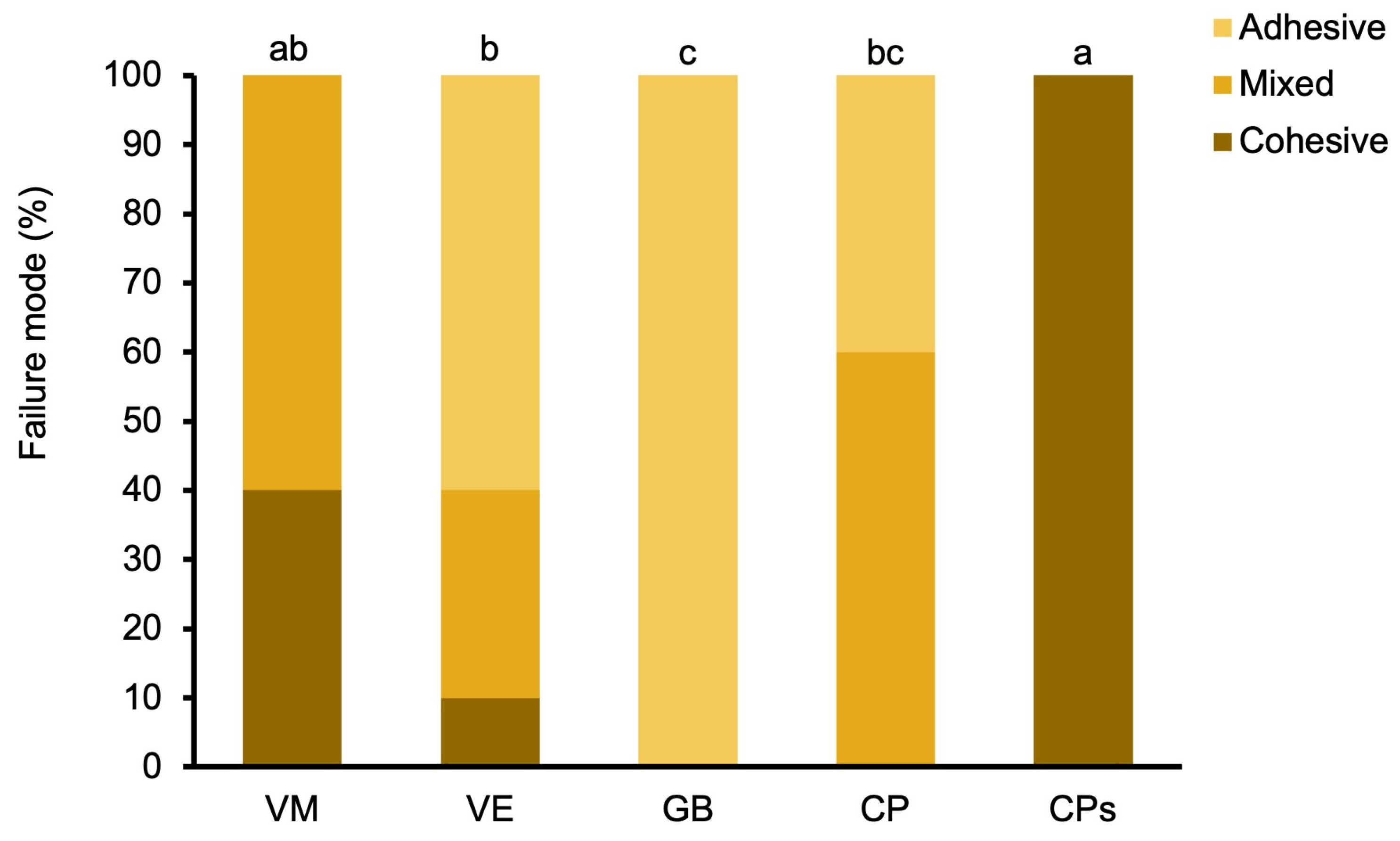
3.2. Failure Mode Assessment

The distribution of failure modes in the bonded prosthetic substrates is presented in Figure 3. Mixed failure was the predominant failure mode detected in VM (60%), with some cohesive failures (40%). VE showed a combination of adhesive (60%), mixed (30%), and cohesive failures (10%). GB showed 100% adhesive failures. For CP, the predominant failure was mixed (60%) with some adhesive (40%). CPs only showed cohesive failure (100%).
The statistical analysis revealed a statistically significant difference in the failure modes among different materials (p < 0.001). The post hoc comparisons revealed statistically significant differences in the failure modes of CPs compared to those of VE, CP, and GB. Statistically significant differences were found between VE and GB; however, GB was not statistically significantly different from CP. No statistically significant difference was found between VM and CPs or between VM, VE, and CP.

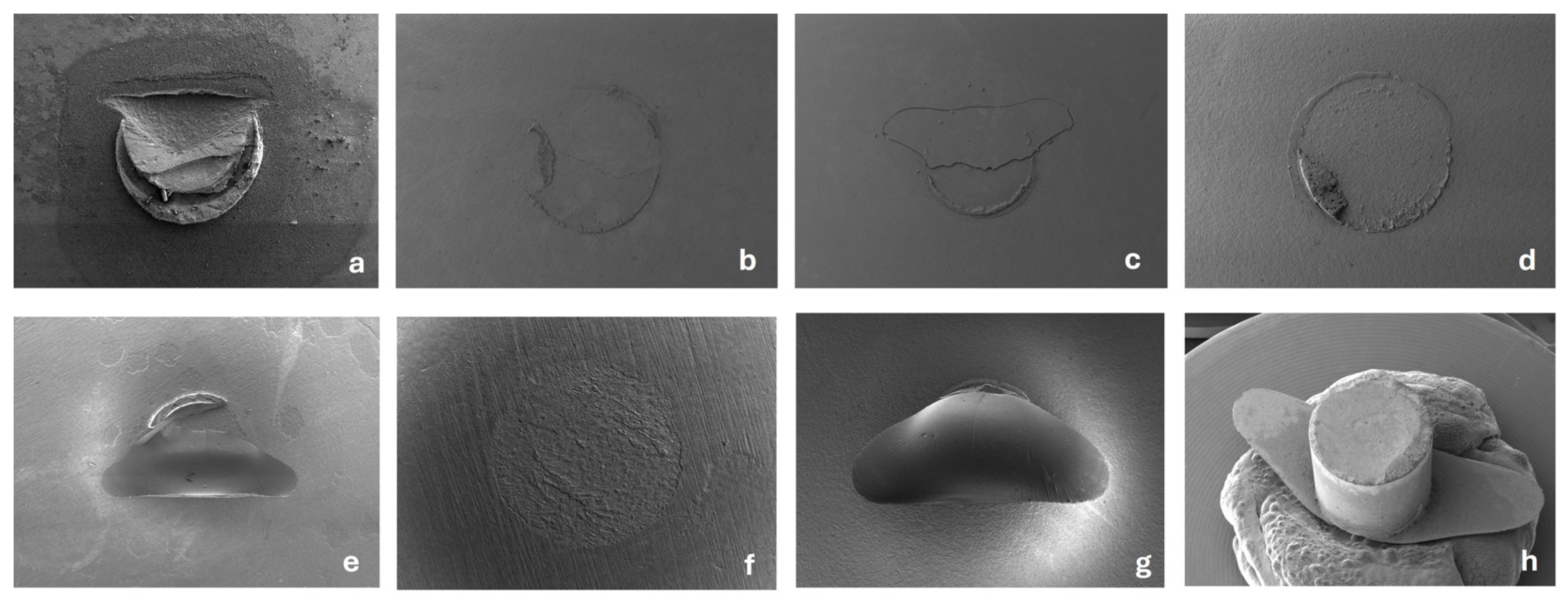
3.3. SEM Observation

Representative SEM images of the debonded surfaces of each material are shown in Figure 4. The failure patterns were quite different between the materials. VM showed predominantly mixed failures (a), but cohesive failures were also observed. VE showed the highest degree of various types of failures, mostly adhesive (b) and mixed (c). GB showed only adhesive failures (d). CP showed mainly mixed failures (e) but also some adhesive failures (f). For CPs, all the failures were cohesive, with remarkable substrate damage (g,h).

4. Discussion

This in vitro study evaluated the differences in notched-edge shear bond strength between a resin cement and a 3D-printed resin indicated for definitive restorations and traditional prosthetic substrates. The null hypothesis was rejected herein, as the adhesion of 3D-printed resins significantly differed among the tested materials. The optimal adhesion of indirect restorations is crucial for achieving high bond strengths to the luting cement and, in turn, to the underlying tooth substrate. Conventionally, adhesive cementation comprises the multi-stage pretreatment of tooth tissues as well as of the restorative material intaglio surface, including etching, priming, and/or bonding [29]. The quality of adhesive cementation is significantly influenced by various factors, including the technique sensitivity, curing mode, operator proficiency, viable dentin substrate, humidity, and contamination [4,30,31,32]. Nevertheless, resin cements with self-adhesive technology such as RelyX Unicem have been reported to be moisture tolerant, less sensitive to technique, and the least influenced by operator experience, by virtue of their relatively straightforward single-step approach; they are thus suitable for a variety of restorative indications with high success rates [33,34,35]. Moreover, RelyX Unicem can chemically bond to the abutment without the need for pre-treatment through its phosphate multifunctional group monomers, as well as to ZrO2 containing ceramics by means of its phosphate ester group [33,35]. Additionally, RelyX Unicem was selected as the resin cement of choice in the present study owing to evidence in the literature reporting its superior degree of conversion [36], radiopacity [37], biofilm formation [38], and adhesion to various prosthetic substrates [18,32,33,34,36,37,38,39,40,41,42,43,44,45].
The bond strength between luting cement and indirect restorative material can be quantified using a variety of macro- and micro-bond shear and tensile strength test apparatuses. However, these test models are not exempt from limitations, which may lead to false estimations, thus impacting the reliability and validity in predicting the clinical performance of bonded dental materials. Moreover, the findings obtained from bond strength tests are contingent on substrate properties, ageing, environmental conditions, and testing parameters [46,47]. The notched-edge shear bond strength device used in the current study consists of a loading blade with a semicircular slot, corresponding in diameter to that of the bonded cylinder [27,48]. This assembly delivers even shear forces simultaneously along 180° of the bonded cement area, thus accurately mimicking shear dislodgment forces with the oral cavity. Conversely, in the conventional shear bond strength test setup, the straight knife-edge chisel has peeling forces limited to the highest point of the bonded area. Furthermore, the notched-edge blade is positioned precisely above the bonded cement button, ensuring the absence of premature contact and thereby eliminating the risk of preload failure [48,49]. Nonetheless, adequate bond strengths are required to resist the contraction stresses generated at the bonded interface due to the polymerization shrinkage [50,51]. In the present investigation, the nSBS results of the indirect restorative materials varied, ranging from 20.6 MPa to 30.3 MPa, exceeding the 20 MPa requirement reported in previously established clinical thresholds [52,53]. The obtained findings are within the scope of the bond strength values reported in the literature for similar materials, ranging from 4.7 MPa to 56.49 MPa [34,40,43,45,54]. The broad scale of reported bond strengths can be rationalized by the heterogeneities among testing methodologies, luting cements, or substrate surface conditioning regimens; therefore, objective comparisons cannot be made. Regardless, there is an overall consensus endorsing the implementation of intaglio surface modifications of prosthetic substrates through mechanical and chemical treatments to increase the bonded surface area, thus improving their bond strength and retention to resin cements [18,32,33,34,36,37,38,39,40,41,42,43,44,45,54]. According to Donmez et al. [40], the shear bond strength of a resin cement to 3D-printed resin for definitive restorations (VarseoSmile Crown Plus) was 8.71–9.70 MPa, whereas the polymer-infiltrated ceramic network displayed bond strength of 11.84–22.46 MPa (VITA Enamic). Likewise, Straface et al. [43], measured the lowest shear bond strengths in unetched feldspathic ceramics of 4.7 MPa (VITABLOC Mark II) and the highest value when etched with 5% hydrofluoric acid equal to 9.1 MPa. Moreover, the authors reported the lowest shear bond strength value of the unetched polymer-infiltrated ceramic network equal to 4.9 MPa, and the highest value when etched with 5% hydrofluoric acid at 8.9 MPa. A similar outcome was observed by Wiedenmann et al. [45], reporting shear bond strength values of 9.81 MPa between a resin cement and feldspar ceramic material (VITABLOC Mark II) prior to artificial ageing, and 11.2 MPa after ageing. On the other hand, Papadopoulos et al. [54] registered higher µSBS values than the present study, with values ranging from 37.98 MPa between the resin cement and untreated polymer-infiltrated ceramic network to 56.49 MPa after etching with hydrofluoric acid and silane application, with the µSBS values significantly decreasing after artificial ageing. The decreased bond strength values observed in the VE group in the present study corroborate the findings reported in similar studies [55,56]. This may be justified as an outcome of prolonged HF etching, in which HF selectively dissolves the superficial layer of inorganic phase therein [57], thereby negatively impacting the chemical bond between silane and SiO2, while simultaneously creating shallow surface irregularities within the remaining organic phase, thus producing VE with reduced micro-mechanical retentive features.
When bonded to a resin cement, the 3D-printed resins tested in the present study demonstrated similar nSBS values to those of the hybrid resin composite and polymer-infiltrated ceramic network, regardless of the blasting method employed on their intaglio surfaces (Table 3). Therefore, it can be concluded, based on this preliminary in vitro study, that horizontally printed definitive resin restorations display comparable bond strength to self-adhesive resin cement, similar to that of traditional prosthetic substrates. This is in contrast with outcomes reported by Donmez et al. [40] who evaluated other 3D-printed resins: Crowntec (Saremco Dental) and VarseoSmile Crown Plus (Bego) and Graf et al. [18] who also assessed VarseoSmile Crown Plus. Both studies observed higher bond strength values in the subtractively milled resins than in the 3D-printed resins, which was justified by the enhanced degree of conversion and controlled polymerization of the subtractively milled resin blocks. Nevertheless, such contrast may be due to dissimilar test setups (shear versus tensile), different 3D resin formulations, or varying luting agents. Moreover, the higher bond strength findings in the present study compared to the aforementioned studies may be explained by the difference in printing parameters and post-processing conditions; the present study employed a horizontal printing orientation followed by two separate curing cycles for 20 min at 60 °C, however, the study by Donmez et al. used vertical printing orientation followed by curing under nitrogen oxide gas atmosphere for 4000 flashes [40], and Graf et al. cured the 3D-printed resins under a nitrogen oxide gas atmosphere for 2 × 1500 flashes [18]. Additionally, a study conducted by Pfeffer et al. [26] assessed the impact of different surface treatments on the microtensile bond strength of 3D-printed resins after aging (10,000 thermocycles) and six-month water storage. Therein, the authors concluded that Al2O3 sandblasting combined with glass bead blasting did not significantly enhance the bond strength to a self-adhesive resin cement to that of glass bead blasting alone, until after six months of water storage. The short-term findings in the present study corroborate those reported by Pfeffer et al. [26], as no significant differences in µSBS were detected between 3D-printed resin groups. Thus, it can be inferred that glass bead blasting fostered surface roughness sufficient for the retention of the resin cement. Nonetheless, the long-term effects of the glass bead blasting and Al2O3 sandblasting are still unknown; thus, further studies with clinically relevant simulated ageing regimens are crucial to generalize this assumption to long-term bond performance.
Bond strength data should be supplemented with an additional assessment of the failure modes to obtain a comprehensive evaluation of bond quality [18,39,40,42,43,44,45,54]. Therein, fractured interfaces are microscopically observed, and failure modes are labelled as adhesive (complete failure along the cement/substrate interface, with no signs of remnants of cement or prosthetic substrate), cohesive (complete fracture of prosthetic substrate), or mixed (a combination of both adhesive and cohesive) failures [58,59]. The notched-edge shear bond strength test is designed to prevent cohesive failures within the luting cement; thus, on occasions when cohesive failures arise in the resin cement button, this could be the result of improper specimen positioning or the inadequate polymerization of the resin cement. In the present study, most failure modes demonstrated in the sandblasted 3D-printed resins were cohesive substrate failure within the 3D resin substrate. The predominant cohesive substrate failure in CPs could suggest that its bond strength to the resin cement exceeded the cohesive strength within the substrate itself. The high prevalence of cohesive failures in 3D-printed definitive resins has been reported in the literature [60] and has also been detected in 3D-printed temporary resin restorations [61,62], as well as 3D-printed denture base resins [63,64]; this is ostensibly because of the chemical compatibility among the resin cement and resin substrate compositions. Moreover, printing parameters such layer thickness, printing angulation, and post-processing conditions may adversely alter the cohesivity between the layers of printed resin specimens and cause greater cohesive failures therein. On the other hand, studies that utilized microtensile bond strength test methods observed a majority of adhesive failures among 3D-printed resin restorations [26,65]. Disparities in failure modes could be the by-product of the chemical pretreatment with dentin bonding adhesives [65] or dissimilar luting agents [26]. It should be noted that, regardless of the failure modes in the present study, the bond strength values were higher than the clinical threshold formerly established [52,53]. Considering the sandblasting procedure, the manufacturer of the 3D-printed resin does not recommend it after pearl blasting due to the possibility of damaging the intaglio surface and compromising accuracy. As the bond strength of sandblasted 3D-printed substrate did not show a statistically significant increase, it indicates the need to avoid sandblasting, contrary to the Rely-X cement recommendation. It should also be considered that an increase in the cohesive strength of the 3D-printed substrate may lead to an increase in the bond strength. Thus, the authors recommend following the manufacturer’s instructions in terms of printing parameters and post-processing curing and washing guidelines, to enhance the cohesivity among printed layers and, in turn, minimize the cohesive failure of the bonded 3D-printed definitive resin restorations. Conversely, adhesive failures were identified in the VE and GB specimens, in line with similar studies [60], by virtue of the higher monomer saturation, which could lead to greater water absorption and increased stresses within the bonded apparatus. Moreover, the lack of silane application to the GB specimens may have contributed to the predominant adhesive failure modes therein.
The limitations of the present study are as follows. First, the lack of a long-term thermodynamic ageing process, since the specimens were evaluated after a 24 h water storage period only. Previous studies have verified the detrimental impact of ageing and thermocycling on the bond strength values [62,63]; therefore, it can be assumed that the findings in the current study are an overestimation of the long-term bond strength values. Furthermore, a single dual-cure resin cement was used, which could be deemed a limitation, considering that the type of resin cement and curing mode significantly influence the overall bond strength [34,39]. Additionally, other types of resin cements have been reported to yield varying results [18,32,36,40,41,43,45]. Moreover, one 3D-printed resin was investigated in the present study; thus, additional studies are needed to explore other 3D-printed resins indicated for definitive restorations to achieve a comprehensive understanding of their adhesion performance. Future studies should also assess the impact of different printers, printing parameters and post-processing conditions on 3D-printed resin bond strength.

5. Conclusions

Based on the findings of this in vitro study, the following conclusions can be drawn:
  • The bond strength between Feldspathic ceramic and Rely X cement was shown to be higher than all the other materials/combinations tested. The CAD/CAM block resin, 3D-printed definitive resin and PICN performed similarly.
  • The bond strength between Rely-X resin cement and 3D-printed definitive resin could be considered clinically sufficient considering the thresholds reported in the literature.
  • The sandblasting procedure after pearl blasting did not improve the short-term bond strength of the 3D-printed definitive resin.

Author Contributions

Conceptualization, A.V.; methodology, A.V.; investigation, A.V. and D.B.; resources, A.V.; data curation, A.V.; writing—original draft preparation, H.A.-J., D.B. and A.V.; writing—review and editing, H.A.-J., A.V. and C.L.; supervision, A.V. and C.L. All authors have read and agreed to the published version of the manuscript.

Funding

This research received no external funding.

Institutional Review Board Statement

Not applicable.

Informed Consent Statement

Not applicable.

Data Availability Statement

The data presented in this study are available on request from the corresponding author due to University public repository access restriction.

Conflicts of Interest

The authors declare no conflicts of interest.

References

  1. Roulet, J.-F.O.; Degrange, M. Adhesion: The Silent Revolution in Dentistry; Quintessence: Chicago, IL, USA, 2000. [Google Scholar]
  2. Perdigão, J.; Araujo, E.; Ramos, R.Q.; Gomes, G.; Pizzolotto, L. Adhesive dentistry: Current concepts and clinical considerations. J. Esthet. Restor. Dent. 2021, 33, 51–68. [Google Scholar] [CrossRef] [PubMed]
  3. Cadenaro, M.; Josic, U.; Maravić, T.; Mazzitelli, C.; Marchesi, G.; Mancuso, E.; Breschi, L.; Mazzoni, A. Progress in Dental Adhesive Materials. J. Dent. Res. 2023, 102, 254–262. [Google Scholar] [CrossRef] [PubMed]
  4. O’Connor, C.; Gavriil, D. Predictable bonding of adhesive indirect restorations: Factors for success. Br. Dent. J. 2021, 231, 287–293. [Google Scholar] [CrossRef] [PubMed]
  5. Skorulska, A.; Piszko, P.; Rybak, Z.; Szymonowicz, M.; Dobrzyński, M. Review on Polymer, Ceramic and Composite Materials for CAD/CAM Indirect Restorations in Dentistry-Application, Mechanical Characteristics and Comparison. Materials 2021, 14, 1592. [Google Scholar] [CrossRef] [PubMed]
  6. Galiatsatos, A.; Galiatsatos, P.; Bergou, D. Clinical Longevity of Indirect Composite Resin Inlays and Onlays: An Up to 9-Year Prospective Study. Eur. J. Dent. 2022, 16, 202–208. [Google Scholar] [CrossRef] [PubMed]
  7. Unlu, N.; Gunal, S.; Ulker, M.; Ozer, F.; Blatz, M.B. Influence of operator experience on in vitro bond strength of dentin adhesives. J. Adhes. Dent. 2012, 14, 223–227. [Google Scholar] [PubMed]
  8. Santos, M.; Zare, E.; McDermott, P.; Santos Junior, G.C. Multifactorial Contributors to the Longevity of Dental Restorations: An Integrated Review of Related Factors. Dent. J. 2024, 12, 291. [Google Scholar] [CrossRef] [PubMed]
  9. Abad-Coronel, C.; Naranjo, B.; Valdiviezo, P. Adhesive Systems Used in Indirect Restorations Cementation: Review of the Literature. Dent. J. 2019, 7, 71. [Google Scholar] [CrossRef] [PubMed]
  10. Dioguardi, M.; Alovisi, M.; Troiano, G.; Caponio, C.V.A.; Baldi, A.; Rocca, G.T.; Comba, A.; Lo Muzio, L.; Scotti, N. Clinical outcome of bonded partial indirect posterior restorations on vital and non-vital teeth: A systematic review and meta-analysis. Clin. Oral Investig. 2021, 25, 6597–6621. [Google Scholar] [CrossRef] [PubMed]
  11. Alammar, A.; Kois, J.C.; Revilla-León, M.; Att, W. Additive Manufacturing Technologies: Current Status and Future Perspectives. J. Prosthodont. 2022, 31, 4–12. [Google Scholar] [CrossRef] [PubMed]
  12. Methani, M.M.; Revilla-León, M.; Zandinejad, A. The potential of additive manufacturing technologies and their processing parameters for the fabrication of all-ceramic crowns: A review. J. Esthet. Restor. Dent. 2020, 32, 182–192. [Google Scholar] [CrossRef] [PubMed]
  13. Revilla-León, M.; Meyers, M.J.; Zandinejad, A.; Özcan, M. A review on chemical composition, mechanical properties, and manufacturing work flow of additively manufactured current polymers for interim dental restorations. J. Esthet. Restor. Dent. 2019, 31, 51–57. [Google Scholar] [CrossRef] [PubMed]
  14. Çokakoğlu, S.; Karaokutan, I.; Buyuk, S.K. Effects of orientation and postprocessing on the bonding of 3-dimensional printed brackets to printable permanent crown resin. Am. J. Orthod. Dentofacial Orthop. 2025, 167, 362–370. [Google Scholar] [CrossRef] [PubMed]
  15. Daher, R.; Ardu, S.; di Bella, E.; Krejci, I.; Duc, O. Efficiency of 3D printed composite resin restorations compared with subtractive materials: Evaluation of fatigue behavior, cost, and time of production. J. Prosthet. Dent. 2024, 131, 943–950. [Google Scholar] [CrossRef] [PubMed]
  16. Kim, S.Y.; Shin, Y.; Kim, I.-H.; Song, J.S. In Vitro Study on the Bond Strength Between 3D-Printed Resin and Resin Cement for Pediatric Crown Restoration. J. Korean Acad. Pediatr. Dent. 2023, 50, 104–112. [Google Scholar] [CrossRef]
  17. Ersöz, B.; Aydın, N.; Ezmek, B.; Karaoğlanoğlu, S.; Çal İ, K. Effect of Surface Treatments Applied to 3D Printed Permanent Resins on Shear Bond Strength. J. Clin. Exp. Dent. 2024, 16, e1059–e1066. [Google Scholar] [CrossRef] [PubMed]
  18. Graf, T.; Erdelt, K.J.; Güth, J.F.; Edelhoff, D.; Schubert, O.; Schweiger, J. Influence of Pre-Treatment and Artificial Aging on the Retention of 3D-Printed Permanent Composite Crowns. Biomedicines 2022, 10, 2186. [Google Scholar] [CrossRef] [PubMed]
  19. Aktas, N.; Bankoglu Güngör, M. Effects of 3D-Printing Technology and Cement Type on the Fracture Resistance of Permanent Resin Crowns for Primary Teeth. Int. J. Prosthodont. 2024, 37, 195–202. [Google Scholar] [CrossRef] [PubMed]
  20. Lim, N.K.; Shin, S.Y. Bonding of conventional provisional resin to 3D printed resin: The role of surface treatments and type of repair resins. J. Adv. Prosthodont. 2020, 12, 322–328. [Google Scholar] [CrossRef] [PubMed]
  21. Lunkad, H.; Sayed, M.E.; Alhazmi, A.E.; Alwadani, B.; Shafei, A.M.; Ayoub, M.N.; Shabi, M.M.A.; Mesawa, S.A.; Abdulfatah, B.A.; Alqarni, H.; et al. The Effect of the Digital Manufacturing Technique, Preparation Taper, and Cement Type on the Retention of Aged Anterior Provisional Crowns: An In Vitro Study. Appl. Sci. 2022, 12, 12714. [Google Scholar] [CrossRef]
  22. Formlabs. Formlabs Crown Permanent Safety Data Sheet. 2021. Available online: https://formlabs-media.formlabs.com/datasheets/ENUS_Safety_Data_Sheet_-_Permanent_Crown.pdf (accessed on 4 May 2025).
  23. Formlabs. Formlabs Crown Permanent Technical Data Sheet. 2020. Available online: https://formlabs-media.formlabs.com/datasheets/2002482-TDS-ENUS-0.pdf (accessed on 15 October 2024).
  24. Kirmali, O.; Barutcugil, C.; Harorli, O.; Kapdan, A.; Er, K. Resin cement to indirect composite resin bonding: Effect of various surface treatments. Scanning 2015, 37, 89–94. [Google Scholar] [CrossRef] [PubMed]
  25. Tribst, J.P.M.; Veerman, A.; Pereira, G.K.R.; Kleverlaan, C.J.; Dal Piva, A.M.O. Comparative Strength Study of Indirect Permanent Restorations: 3D-Printed, Milled, and Conventional Dental Composites. Clin. Pract. 2024, 14, 1940–1952. [Google Scholar] [CrossRef] [PubMed]
  26. Pfeffer, S.; Jacker-Guhr, S.; Geurtsen, W.; Pfeffer, T.; Lührs, A. Microtensile bond strength of luting cements to a 3D printable composite–an in vitro study. Zahnärztl J. Int. 2020, 2, 229–239. [Google Scholar]
  27. ISO 29022:2013; S-E: Dentistry—Adhesion—Notched-Edge Shear Bond Strength Test. SIS Förlag AB: Stockholm, Sweden, 2013.
  28. Formlabs. Formlabs Crown Permanent Instructions for Use. 2025. Available online: https://media.formlabs.com/m/73a550261e0ab65b/original/-ENUS-Permanent-Crown-Resin-Instructions-for-Use.pdf (accessed on 15 October 2024).
  29. D’Arcangelo, C.; Vanini, L.; Casinelli, M.; Frascaria, M.; De Angelis, F.; Vadini, M.; D’Amario, M. Adhesive Cementation of Indirect Composite Inlays and Onlays: A Literature Review. Compend. Contin. Educ. Dent. 2015, 36, 570–577, quiz 578. [Google Scholar] [PubMed]
  30. Özcan, M.; Volpato, C.A.M. Current perspectives on dental adhesion: (3) Adhesion to intraradicular dentin: Concepts and applications. Jpn. Dent. Sci. Rev. 2020, 56, 216–223. [Google Scholar] [CrossRef] [PubMed]
  31. Perdigão, J. Current perspectives on dental adhesion: (1) Dentin adhesion—Not there yet. Jpn. Dent. Sci. Rev. 2020, 56, 190–207. [Google Scholar] [CrossRef] [PubMed]
  32. Madrigal, E.L.; Tichy, A.; Hosaka, K.; Ikeda, M.; Nakajima, M.; Tagami, J. The effect of curing mode of dual-cure resin cements on bonding performance of universal adhesives to enamel, dentin and various restorative materials. Dent. Mater. J. 2021, 40, 446–454. [Google Scholar] [CrossRef] [PubMed]
  33. Fabian Fonzar, R.; Goracci, C.; Carrabba, M.; Louca, C.; Ferrari, M.; Vichi, A. Influence of Acid Concentration and Etching Time on Composite Cement Adhesion to Lithium-silicate Glass Ceramics. J. Adhes. Dent. 2020, 22, 175–182. [Google Scholar] [PubMed]
  34. Bayazıt, E. Microtensile Bond Strength of Self-Adhesive Resin Cements to CAD/CAM Resin-Matrix Ceramics Prepared with Different Surface Treatments. Int. J. Prosthodont. 2019, 32, 433–438. [Google Scholar] [CrossRef] [PubMed]
  35. Scholz, K.J.; Bittner, A.; Cieplik, F.; Hiller, K.A.; Schmalz, G.; Buchalla, W.; Federlin, M. Micromorphology of the Adhesive Interface of Self-Adhesive Resin Cements to Enamel and Dentin. Materials 2021, 14, 492. [Google Scholar] [CrossRef] [PubMed]
  36. Butler, S.; Santos, G.C.; Santos, M.J.C. Do high translucency zirconia shades contribute to the degree of conversion of dual-cure resin cements? Quintessence Int. 2021, 53, 8–14. [Google Scholar] [PubMed]
  37. Erzurumlu, Z.U.; Sagirkaya, C.E.; Erzurumlu, K. Evaluation of radiopacities of CAD/CAM restorative materials and resin cements by digital radiography. Clin. Oral Investig. 2021, 25, 5735–5741. [Google Scholar] [CrossRef] [PubMed]
  38. Astasov-Frauenhoffer, M.; Glauser, S.; Fischer, J.; Schmidli, F.; Waltimo, T.; Rohr, N. Biofilm formation on restorative materials and resin composite cements. Dent. Mater. 2018, 34, 1702–1709. [Google Scholar] [CrossRef] [PubMed]
  39. Vichi, A.; Fabian Fonzar, R.; Carrabba, M.; Louca, C.; Scotti, N.; Mazzitelli, C.; Breschi, L.; Goracci, C. Comparison between Hydrofluoric Acid and Single-Component Primer as Conditioners on Resin Cement Adhesion to Lithium Silicate and Lithium Disilicate Glass Ceramics. Materials 2021, 14, 6776. [Google Scholar] [CrossRef] [PubMed]
  40. Donmez, M.B.; Çakmak, G.; Yılmaz, D.; Schimmel, M.; Abou-Ayash, S.; Yilmaz, B.; Peutzfeldt, A. Bond strength of additively manufactured composite resins to dentin and titanium when bonded with dual-polymerizing resin cements. J. Prosthet. Dent. 2024, 132, 1067.e1061–1067.e1068. [Google Scholar] [CrossRef] [PubMed]
  41. Emsermann, I.; Eggmann, F.; Krastl, G.; Weiger, R.; Amato, J. Influence of Pretreatment Methods on the Adhesion of Composite and Polymer Infiltrated Ceramic CAD-CAM Blocks. J. Adhes. Dent. 2019, 21, 433–443. [Google Scholar] [PubMed]
  42. Peçanha, M.M.; Amaral, M.; Baroudi, K.; Frizzera, F.; Vitti, R.; Silva-Concilio, L. Improving the bonding stability between resin cements and zirconia-based ceramic using different surface treatments. Int. J. Prosthodont. 2022, 35, 414–419. [Google Scholar] [CrossRef] [PubMed]
  43. Straface, A.; Rupp, L.; Gintaute, A.; Fischer, J.; Zitzmann, N.U.; Rohr, N. HF etching of CAD/CAM materials: Influence of HF concentration and etching time on shear bond strength. Head Face Med. 2019, 15, 21. [Google Scholar] [CrossRef] [PubMed]
  44. Nasiry Khanlar, L.; Revilla-León, M.; Barmak, A.B.; Ikeda, M.; Alsandi, Q.; Tagami, J.; Zandinejad, A. Surface roughness and shear bond strength to composite resin of additively manufactured interim restorative material with different printing orientations. J. Prosthet. Dent. 2023, 129, 788–795. [Google Scholar] [CrossRef] [PubMed]
  45. Wiedenmann, F.; Klören, M.; Edelhoff, D.; Stawarczyk, B. Bond strength of CAD-CAM and conventional veneering materials to different frameworks. J. Prosthet. Dent. 2021, 125, 664–673. [Google Scholar] [CrossRef] [PubMed]
  46. Sirisha, K.; Rambabu, T.; Shankar, Y.R.; Ravikumar, P. Validity of bond strength tests: A critical review: Part I. J. Conserv. Dent. 2014, 17, 305–311. [Google Scholar] [CrossRef] [PubMed]
  47. Sirisha, K.; Rambabu, T.; Ravishankar, Y.; Ravikumar, P. Validity of bond strength tests: A critical review-Part II. J. Conserv. Dent. 2014, 17, 420–426. [Google Scholar] [CrossRef] [PubMed]
  48. Chiang, M.L.; Birlbauer, S.; Lo, Y.F.; Pitchika, V.; Crispin, A.; Ilie, N.; Hickel, R.; Kühnisch, J. Which Factors Influence the Shear Bond Strength of Sealant Materials? J. Adhes. Dent. 2016, 18, 397–404. [Google Scholar] [PubMed]
  49. Barkmeier, W.W.; Tsujimoto, A.; Latta, M.A.; Takamizawa, T.; Radniecki, S.M.; Garcia-Godoy, F. Effect of mold enclosure and chisel design on fatigue bond strength of dental adhesive systems. Eur. J. Oral Sci. 2022, 130, e12864. [Google Scholar] [CrossRef] [PubMed]
  50. Eick, J.D.; Gwinnett, A.J.; Pashley, D.H.; Robinson, S.J. Current concepts on adhesion to dentin. Crit. Rev. Oral Biol. Med. 1997, 8, 306–335. [Google Scholar] [CrossRef] [PubMed]
  51. Kiremitçi, A.; Yalçin, F.; Gökalp, S. Bonding to enamel and dentin using self-etching adhesive systems. Quintessence Int. 2004, 35, 367–370. [Google Scholar] [PubMed]
  52. Dimitriadis, K.; Tulyaganov, D.U.; Agathopoulos, S. Evaluation of bond strength between zirconia milled ceramic material and veneered dental porcelain. Eur. J. Oral Sci. 2024, 132, e12989. [Google Scholar] [CrossRef] [PubMed]
  53. ISO 9693:2019; Dentistry: Compatibility Testing for Metal-Ceramic and Ceramic-Ceramic Systems. International Organization for Standardization: Geneva, Switzerland, 2019.
  54. Papadopoulos, C.; Dionysopoulos, D.; Pahinis, K.; Koulaouzidou, E.; Tolidis, K. Microtensile Bond Strength Between Resin-Matrix CAD/CAM Ceramics and Resin Cement after Various Surface Modifications and Artificial Aging. J. Adhes. Dent. 2021, 23, 255–265. [Google Scholar] [PubMed]
  55. Şenol, M.; Gürbüz, A.; Oyar, P. Bond strength of conventional resin-based adhesive cement and self-adhesive resin cement to CAD-CAM restorative materials. BMC Oral Health 2025, 25, 296. [Google Scholar] [CrossRef] [PubMed]
  56. Hu, M.; Weiger, R.; Fischer, J. Comparison of two test designs for evaluating the shear bond strength of resin composite cements. Dent. Mater. 2016, 32, 223–232. [Google Scholar] [CrossRef] [PubMed]
  57. Blatz, M.B.; Conejo, J.; Alammar, A.; Ayub, J. Current Protocols for Resin-Bonded Dental Ceramics. Dent. Clin. N. Am. 2022, 66, 603–625. [Google Scholar] [CrossRef] [PubMed]
  58. Alnafaiy, S.M.; Labban, N.; Albaijan, R.; AlKahtani, R.N.; Al-Aali, K.A.; Abozaed, H.W.; Alturki, N.Y.; Alenezi, J.E. Evaluation of Shear Bond Strength and Failure Modes of Lithium Disilicate Ceramic Veneering Material to Different High-Performance Polymers. Polymers 2025, 17, 554. [Google Scholar] [CrossRef] [PubMed]
  59. Guggenbühl, S.; Alshihri, A.; Al-Haj Husain, N.; Özcan, M. Adhesion of Resin-Resin and Resin-Lithium Disilicate Ceramic: A Methodological Assessment. Materials 2021, 14, 3870. [Google Scholar] [CrossRef] [PubMed]
  60. Mao, Z.; Schmidt, F.; Beuer, F.; Yassine, J.; Hey, J.; Prause, E. Effect of surface treatment strategies on bond strength of additively and subtractively manufactured hybrid materials for permanent crowns. Clin. Oral Investig. 2024, 28, 371. [Google Scholar] [CrossRef] [PubMed]
  61. Lankes, V.; Reymus, M.; Mayinger, F.; Coldea, A.; Liebermann, A.; Hoffmann, M.; Stawarczyk, B. Three-Dimensional Printed Resin: Impact of Different Cleaning Protocols on Degree of Conversion and Tensile Bond Strength to a Composite Resin Using Various Adhesive Systems. Materials 2023, 16, 3580. [Google Scholar] [CrossRef] [PubMed]
  62. Soto-Montero, J.; Romano, B.d.C.; Noronha, M.d.S.; André, C.B.; Giannini, M. Microtensile bond strength of resin cements to 3-D printed and milled temporary restorative resins. Odovtos Int. J. Dent. Sci. 2023, 25, 82–98. [Google Scholar] [CrossRef]
  63. Gad, M.M.; Albazroun, Z.; Aldajani, F.; Elakel, A.M.; El Zayat, M.; Akhtar, S.; Khan, S.Q.; Ali, S.; Rahoma, A.M. Repair Bond Strength of Conventionally and Digitally Fabricated Denture Base Resins to Auto-Polymerized Acrylic Resin: Surface Treatment Effects In Vitro. Materials 2022, 15, 9062. [Google Scholar] [CrossRef] [PubMed]
  64. Kizilirmak, B.; Tunçdemİr, A.R. The effect of gastric acid on the bonding of 3D printed and CAD/CAM denture acrylics to denture teeth. Sci. Rep. 2025, 15, 16648. [Google Scholar] [CrossRef] [PubMed]
  65. Montenbruck, L.; Lüchtenborg, J.; Elgezawi, M.; Keßler, A.; Kaisarly, D. Influence of pretreatment strategies on the tensile bond strength of 3D printed composite resins for definitive cementation to dentin. J. Prosthet. Dent. 2025. [Google Scholar] [CrossRef] [PubMed]
Figure 1. Experimental study design.
Coatings 15 00896 g001
Figure 3. Failure mode distributions of bonded prosthetic substrates. VM; Vita Mark II, VE; Vita Enamic; GB, Grandio Bloc, CP, Crown Permanent; CPs Crown Permanent (additionally sandblasted). Different superscript letters represent significant differences among groups.
Coatings 15 00896 g003
Figure 4. Debonded substrate surfaces after notched-edge shear bond strength test. (a) VM mixed failure, (b) VE adhesive failure, (c) VE mixed failure, (d) GB adhesive failure, (e) CP mixed failure, (f) CP adhesive failure, (g) CPs cohesive failure, (h) CPs resin cement with failed substrate.
Coatings 15 00896 g004
Table 1. Composition of the tested prosthetic definitive restorative materials.
Fabrication MethodMaterial
(Product Name, Manufacturer)
CodeComposition
Subtractive millingFeldspar ceramic
(VITABLOC Mark II, VITA Zahnfabrik)
VMSiO2, Al2O3, Na2O, K2O, CaO, TiO2
Polymer-infiltrated ceramic network
(VITA Enamic, VITA Zahnfabrik)
VEUDMA, TEGDMA, glass ceramic sintered network (SiO2, Al2O3, Na2O, K2O, B2O3, ZrO2, CaO)
Nanohybrid resin composite
(Grandio Blocs, VOCO GmbH)
GBUDMA, DMA Dimethacrylates, glass ceramics, silica
3D printingMethacrylic acid ester-based resin
(Crown Permanent, Formlabs GmbH)
CPBis-EMA, salinized glass
Table 2. Results of the one-way ANOVA of notched-edge shear bond strength results.
Source of VariationdfSum of SquaresMean SquareF-Valuep-Value
Between Groups4509.738127.43512.283<0.001
Residual45466.85910.375  
Total49976.597   
Power of performed test with α = 0.05:1.00.
Table 3. Notched-edge shear bond strength (nSBS, MPa) of resin cement to 3D-printed resins for definitive restorations and traditional prosthetic substrates (mean ± standard deviation).
MaterialMean ± St. Dev. (MPa)
VM30.3 ± 1.8 a
CPs25.2 ± 3.8 b
GB24.5 ± 3.3 bc
CP23.0 ± 3.8 bc
VE20.6 ± 3.0 c
VM; Vita Mark II, VE; Vita Enamic; GB, Grandio Bloc, CP, Crown Permanent; CPs Crown Permanent additionally sandblasted. Different superscript letters represent significant differences among groups.
Disclaimer/Publisher’s Note: The statements, opinions and data contained in all publications are solely those of the individual author(s) and contributor(s) and not of MDPI and/or the editor(s). MDPI and/or the editor(s) disclaim responsibility for any injury to people or property resulting from any ideas, methods, instructions or products referred to in the content.

