Article

TiN-Ag Multilayer Protective Coatings for Surface Modification of AISI 316 Stainless Steel Medical Implants

by
Božana Petrović
1,*,
Dijana Mitić
2,*,
Minja Miličić Lazić
2,
Miloš Lazarević
2,
Anka Trajkovska Petkoska
3,4,
Ilija Nasov
5,
Slavoljub Živković
2 and
Vukoman Jokanović
1,6
1
Institute of Nuclear Sciences Vinča—National Institute of the Republic of Serbia, University of Belgrade, 11001 Belgrade, Serbia
2
School of Dental Medicine, University of Belgrade, 11000 Belgrade, Serbia
3
Faculty of Technology and Technical Sciences, St. Kliment Ohridski University-Bitola, 1400 Veles, North Macedonia
4
Department of Materials Science and Engineering, Korea University, 01000 Seoul, Republic of Korea
5
R&D Center Plasma DOO, 1000 Skopje, North Macedonia
6
ALBOS DOO Belgrade, 11000 Belgrade, Serbia
*
Authors to whom correspondence should be addressed.
Coatings 2025, 15(7), 820; https://doi.org/10.3390/coatings15070820 (registering DOI)
Submission received: 21 May 2025 / Revised: 7 July 2025 / Accepted: 11 July 2025 / Published: 14 July 2025

Abstract

Stainless steel (SS) is one of the materials most commonly utilized for fabrication of medical implants and its properties are often improved by deposition of protective coatings. This study investigates certain physico-chemical and biological properties of SS substrate coated with multilayer thin film consisting of titanium nitride and silver layers (TiN-Ag film). TiN-Ag films were deposited on the surface of AISI 316 SS substrate by a combination of cathodic arc evaporation and DC magnetron sputtering. SS substrate was analyzed by TEM, while deposited coatings were analyzed by SEM, EDS and wettability measurements. Also, mitochondrial activity assay, and osteogenic and chondrogenic differentiation were performed on dental pulp stem cells (DPSCs). SEM and EDS revealed excellent adhesion between coatings’ layers, with the top layer predominantly composed of Ag, which is responsible for antibacterial properties. TiN-Ag film exhibited moderately hydrophilic behaviour which is desirable for orthopedic implant applications. Biological assays revealed significantly higher mitochondrial activity and enhanced osteogenic and chondrogenic differentiation of DPSC on TiN-Ag films compared to TiN films. The newly designed TiN-Ag coatings showed a great potential for the surface modification of SS implants, and further detailed investigations will explore their suitability for application in clinical practice.

1. Introduction

The demand for implants has increased rapidly in recent years, due to the ageing population and patients’ desire to maintain their current quality of life. Consequently, the range of available biomaterials has expanded significantly, enabling better selection of materials to meet specific treatment objectives, i.e., chemically inert materials used as permanent replacements for damaged tissues, or biodegradable materials serving as temporary scaffolds to support tissue regeneration [1]. Among bio-inert materials, metallic biomaterials such as stainless steel (SS), titanium, and Co-Cr alloys are the most widely used, especially in load-bearing applications [1,2,3,4]. Thanks to their mechanical strength, corrosion resistance and minimal long-term toxicity, these materials have been used in orthopedics for artificial joints, plates, and screws; in dentistry for braces and dental implants; as well as in cardiovascular and neurosurgical devices [1].
Austenitic SS is one of the most commonly used materials in the healthcare industry, primarily due to its price and availability [2,3,4,5]. Medical-grade austenitic SS 316 and 316L are mainly used in implant fabrication owing to their fair corrosion resistance and mechanical properties, enabled by chromium and nickel content, while additional alloying with molybdenum in the case of 316 L improves resistance to pitting corrosion [2,3]. Nonetheless, these SSs remain susceptible to corrosion under certain conditions—such as highly strained or nonoxygenated environments—exhibiting low wear resistance and a lack of inherent antibacterial properties [2,4,5]. These disadvantages can be overcome by applying thin protective films that provide both anticorrosive and antibacterial properties. Plasma vapour deposition (PVD) methods, especially magnetron sputtering and cathodic arc evaporation, enable the fabrication of multilayer protective coatings on a variety of substrate materials [2,3,4,5,6].
Titanium nitride (TiN) is one of the most extensively studied ceramic coatings for the surface modification of orthopedic and dental implants due to its excellent corrosion resistance, tribological qualities and biocompatibility [2,3,4]. TiN coatings have been shown to increase the hardness of metallic surfaces, protect against corrosion, minimize bacterial colonization, and decrease a metal ion leakage [3]. Additional antibacterial properties can be introduced by doping the layer with silver (Ag) or (Cu) ions, which act as effective antibacterial agents [4,6,7,8]. Furthermore, it was shown that multilayer coatings consisting of alternating TiN and Ag layers have demonstrated superior hardness and wear resistance compared to simple TiN films doped with Ag. This improvement is attributed to the controlled microstructure and distinct phase separation in multilayer systems [9]. Also, improved antibacterial performance is achieved in TiN-Ag multilayers through controlled Ag ion release and optimized surface exposure, unlike in doped systems where Ag may remain trapped in the TiN matrix and less available for antimicrobial action [10].
In our previous studies, we investigated the modification of various implant materials using different coating materials and deposition methods, with a particular focus on TiN coatings, which demonstrated the highest stability [11,12,13,14]. We have thoroughly investigated the physico-chemical and biological properties of TiN films, as well as TiN films doped with silver (TiN-Ag), deposited on various substrates using PVD techniques [11,12,13]. However, investigations involving austenitic SS, as one of the most commonly used materials in implantology, have not previously been conducted. This gap has been addressed in the present study.
In this study, TiN-Ag multilayer coatings were deposited on the surface of AISI 316 SS by simultaneous deposition of TiN layers via cathodic arc evaporation and Ag layers via DC magnetron sputtering. The SS substrate was characterized by TEM and deposited coatings were characterized by SEM, EDS, and wettability measurements. Also, the biocompatibility of the deposited coatings was evaluated by mitochondrial assay and assays of osteogenic and chondrogenic differentiation on dental pulp stem cells (DPSCs).

2. Materials and Methods

2.1. Deposition of TiN-Ag Films by Cathodic Arc Evaporation and Magnetron Sputtering onto SS Substrate

The substrate employed in this study was surgical-grade AISI 316 austenitic stainless steel (hereafter referred to as SS substrate). Surface preparation involved grinding and polishing with one-micron diamond paste. Prior to sputter deposition, the specimens were ultrasonically cleaned in acetone and ethanol and then sputter-cleaned for 20 min by bombarding with Ar+ ions at a DC power of 150 W.
Prior to deposition of both TiN-Ag and control TiN films, the Ti cathode for the arc evaporation was placed on one side of the chamber, while the magnetron source for sputtering of Ag ions was placed on the opposite side. During the deposition process, arc and DC magnetron sources operated simultaneously in N2 atmosphere. The chamber was pre-cleaned with Ar at 0.4 Pa for 5 min, while the substrate was biased at −700 V. The Ti cathode (target) was cleaned at 5 kW by pre-sputtering for 2 min in Ar atmosphere. The deposition process began with the formation of a Ti interlayer on the substrate surface at 0.4 Pa for 5 min at a rate of 5 nm/min and a substrate bias of −200 V. This was followed by the introduction of N2 into the chamber at a pressure of 0.4 Pa for the deposition of the TiN interlayer. For the control TiN film, this TiN layer also served as the final coating. The TiN layer was deposited for 5 min at a rate of 10 nm/min, with a substrate bias of −200 V. For the deposition of TiN-Ag multilayer films, simultaneous arc evaporation of Ti ions and DC magnetron sputtering of Ag ions in N2 atmosphere were performed. The deposition parameters included a substrate bias of −150 V, a Cu/Ag power density of 0–5.2 W/cm2, a deposition rate of 7 nm/min, and a deposition time of 20 min. The substrate temperature was maintained at 250 °C, with a rotation speed of 16 rpm and a mean target-to-substrate distance of 150 mm. The resulting multilayer film consisted of 12 alternating TiN and Ag layers.

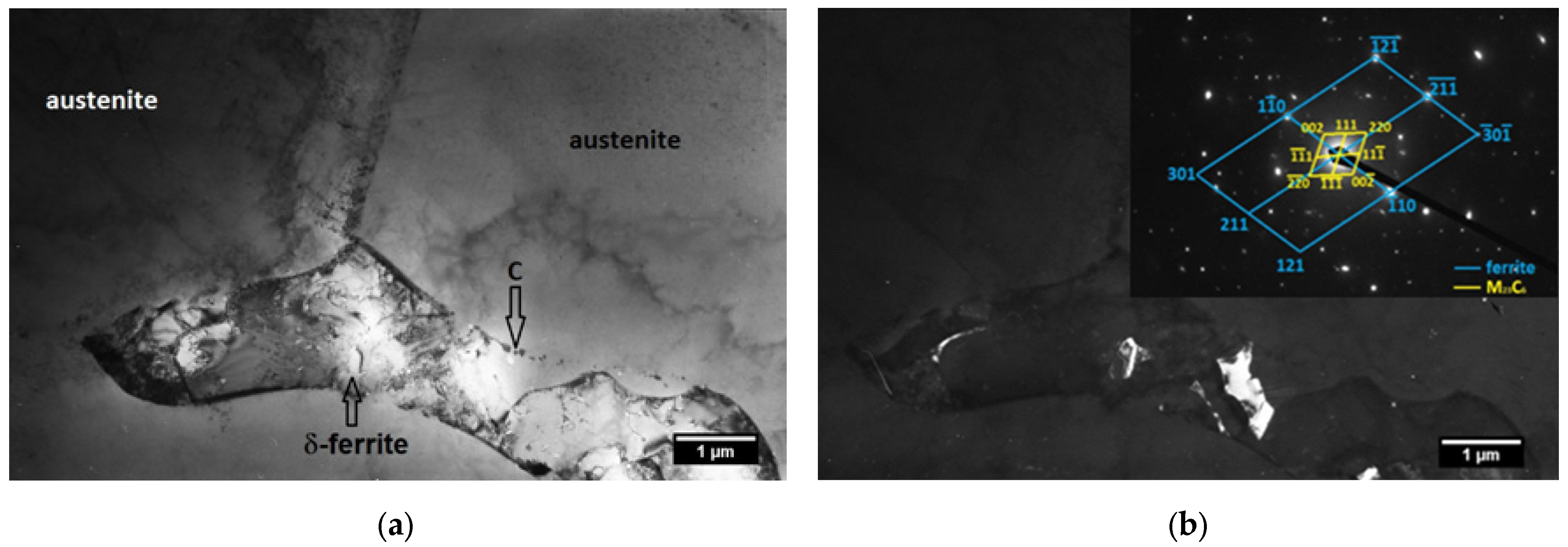
2.2. TEM Analysis of the SS Substrate

For the individual secondary phase identification, transmission electron microscopy (TEM) of the thin SS foils was performed. Small discs of 3 mm in diameter and about 0.1 mm thick were jet-electro-polished in electrolyte HNO3:CH3OH = 3:7, at 0 °C and 15 V, in order to obtain transparent areas near the central hole. The jet-electro-polishing was performed using TENUPOL 5 (Struers A/S, Ballerup, Denmark). TEM observations were performed using JEOL 200 CX (Jeol Ltd., Tokyo, Japan) operated at 200 kV and Philips CM 300 (Philips and Co., Eindhoven, The Netherlands) operating at 300 kV. The analysis was supplemented by selected area electron diffraction for phase identification.

2.3. SEM Analysis of the TiN-Ag Coatings

SEM analysis of the deposited TiN-Ag coatings was performed using JSM-7200F Schottky Field Emission Scanning Electron Microscope (Jeol Ltd., Tokyo, Japan) equipped with EDS. All samples were prepared using a JEOL Cross-Section Polisher (CP) IB-19500CP. The CM milling conditions were 6 kV and 2.5 h.

2.4. Wettability Measurements of the TiN-Ag Coatings

Surface wettability measurements were conducted under room temperature (22.5 ± 0.2 °C) conditions. Samples of TiN-Ag and TiN films (which served as a control) were set onto a measurement bench and a 2 µL drop of deionized water, as a polar component, was placed onto the film’s surface using a micropipette (Finnpipette, Thermo Fisher, Helsinki, Finland), at a distance of 4 mm and angle of 90°. For the non-polar components, diiodomethane and ethylene-glycol, the measurements were conducted in the same manner using a 2 µL drop. Contact angles (θ) were measured 1 s after the liquid achieved contact with the substrate surface by measuring the angle between the plane tangent to the drop and the plane of the underlying surface. The analyzer’s setup consists of a Canon 77D camera equipped with a Canon ultrasonic microlens f100 mm (Tokyo, Japan) and a position stand. Data were analyzed using ImageJ contact angle software, version 1.5t (National Institutes of Health, LOCI, University of Wisconsin, Madison, WI, USA) and the results were subjected to statistical analysis using IBM SPSS Statistics v22 software (SPSS Inc., Chicago, IL, USA). Descriptive measurements and the Mann–Whitney test were used to evaluate differences between TiN and TiN-Ag.
The surface energy of the deposited films could not be evaluated due to a chemical reaction between the silver films and diiodomethane, which could compromise the measurement accuracy [15].

2.5. Cells’ Viability on the Surface of TiN-Ag Coatings

Dental pulp stem cells (DPSCs) were isolated from the pulp tissue of a third molar extracted from a healthy 18-year-old patient, as described previously [16]. All patients were informed about the study and signed written informed consent prior to participation. The study was approved by the Ethical Committee of the School of Dental Medicine (No. 36/8), University of Belgrade, and was conducted according to the Helsinki Declaration.
To determine the viability of the cells, mitochondrial activity assay (MTT) was used. Samples of TiN-Ag- and TiN-coated substrate (with the latter serving as a control) were placed in 24-well plates, sterilized under UV light for 15 min on both sides, and complete medium (Dulbecco’s Modified Eagle Medium (DMEM) supplemented with 10% fetal bovine serum (FBS) and 100 U/mL penicillin–streptomycin, all from Invitrogen, Thermo Fisher Scientific, Waltham, MA, USA) was added. The next day, samples were transferred in new 24-well plates, 1 × 10⁴ cells (cells from the fifth passage were used) were seeded on the samples and cultured under standard conditions in a humidified incubator at 37 °C with 5% CO2. After 24 h and 7 days of culturing, the medium was discarded and a solution containing 3-(4,5-dimethylthiazol-2-yl)-2,5-diphenyltetrazolium bromide (MTT, 0.5 mg/mL; Sigma-Aldrich, St. Louis, MO, USA) was added to each well and incubated. Following a 4 h incubation, the supernatant was removed, and dimethyl sulfoxide (DMSO, Sigma-Aldrich) was added to solubilize the formazan crystals. The plate was then placed on a shaker at 250 rpm for 20 min in the dark at 37 °C. The resulting coloured solutions from the 12-well plates were transferred into a new 96-well plate, and the absorbance was measured at 550 nm using a microplate reader (RT-2100c, Rayto, Shenzhen, China). As a control, cells were cultured on sterile glass discs matching the dimensions of the experimental samples. Mitochondrial activity was expressed as a percentage relative to the control group.

2.6. Osteogenic and Chondrogenic Differentiation of DPSCs in the Presence of TiN-Ag Coatings

The aforementioned DPSCs were used for the osteogenic and chondrogenic differentiation assays. Cells at the fifth passage were used in this study.
To assess differentiation, cells were washed twice with PBS and fixed in 4% paraformaldehyde for 30 min at room temperature. Osteogenic and chondrogenic differentiation were confirmed by staining with 2% Alizarin Red S (Centrohem, Belgrade, Serbia) and 1% Alcian Blue solution in 3% acetic acid (Sigma-Aldrich), respectively. Samples were incubated with the staining solutions for 30 min and subsequently washed with distilled water. To quantify differentiation, the bound dyes were extracted using 250 μL of 1% hydrochloric acid in 70% ethanol for 20 min, and absorbance was measured at 450 nm using an ELISA microplate reader (RT-2100c, Rayto, China).
Additionally, the expression of osteocalcin (OCN) and bone morphogenetic protein 2 (BMP2) was evaluated by immunofluorescence. Cells were fixed with 4% paraformaldehyde for 20 min at room temperature and then incubated for 45 min in a blocking and permeabilization buffer containing 10% bovine serum albumin (BSA) and 0.1% Triton X-100 in PBS. Purified primary antibodies against BMP2 and OCN (Thermo Fisher Scientific; both diluted 1:100 in 5% BSA) were applied, and the samples were incubated at 4 °C for 24 h. This was followed by a 1 h incubation with a secondary Alexa Fluor 488-conjugated antibody (Thermo Fisher Scientific; 1:500 in PBS) in a humidified dark chamber. For nuclear staining, cells were treated with 4′,6-diamidino-2-phenylindole (DAPI; 1:4000, Molecular Probes (now Thermo Fisher), Eugene, OR, USA) for 10 min at room temperature in the dark. Each staining step was followed by three PBS washes, each lasting 5 min. Immunofluorescence images were captured using an epifluorescence microscope (AxioImager A2, Carl Zeiss, Oberkochen, Germany).

3. Results

3.1. TEM Analysis

TEM analysis of the SS substrate was performed in order to determine whether and how its microstructure influences the structure of the deposited coatings. The substructure of the sample, observed by TEM using thin foil specimens, is shown in Figure 1a. The matrix exhibits a dendritic character and consists of austenite, while δ-ferrite was observed in the interdendritic area. Small particles of irregular shape were occasionally observed at the boundaries of austenite and ferrite. Detailed observation revealed the presence of dislocation loops and relatively low density of dislocations within austenitic grains (Figure 1b).
Figure 2 shows the details of δ-ferrite with small particles at the boundaries between austenite and δ-ferrite and some irregularly shaped particles inside ferritic grain (Figure 2a). These particles were identified by electron diffraction as carbide M23C6 (Figure 2b and Table A1 (given in Appendix A)).

3.2. SEM Analysis

The SEM image of TiN-Ag coating (Figure 3) reveals the morphology and thickness of individual TiN and Ag interlayers, formed through simultaneous deposition (Figure 3). A slightly wavy surface morphology is observed, likely resulting from the lattice mismatch between the TiN and Ag layers.
The composition of each layer was determined by EDS mapping, which qualitatively revealed the spatial distribution of Ti, N, and Ag across the multilayer structure (Figure 4). The first TiN layer, which is in direct contact with the SS substrate, is 108 nm thick. In contrast, the upper TiN layers are 16–20 nm thick, while the Ag layers exhibit a thickness of about 20 nm (Figure 3). Figure 4c clearly shows the intercalation of Ag into TiN layers, as well as the intercalation of Ti and N into Ag layers. The topmost layer consists predominantly of Ag (Figure 4c).
It should be noted that a separate analysis of the control TiN film was not conducted, but it is assumed to have the same structure as the first TiN layer in the multilayer TiN-Ag film, given that they were deposited in the same manner.

3.3. Wettability

The contact angle measurements of reference liquids with TiN and TiN-Ag film showed moderately hydrophilic behaviour, without a statistically significant difference between TiN and TiN-Ag for any reference liquids (p > 0.05) (Figure 5).

3.4. Cells Viability

The mitochondrial activity of DPSC was significantly higher on TiN-Ag samples compared to the control TiN samples at both measured time points (Figure 6). The highest level of cellular activity was observed after 24 h of cultivation on the TiN-Ag samples.

3.5. Osteogenic and Chondrogenic Differentiation

DPSCs demonstrated significantly greater differentiation into both osteogenic and chondrogenic lineages when cultured on TiN-Ag samples compared to the control TiN samples (Figure 7). This enhanced differentiation was evident by the abundant red-stained calcium deposits indicating osteogenesis, and the prominent blue-stained proteoglycans associated with chondrogenesis (Figure 7a).

3.6. Immunocytochemistry

After 21 days of cultivation in osteogenic medium, cells seeded on TiN-Ag samples exhibited significantly increased expression of BMP2 and OCN (Figure 8). Additionally, OCN expression was notably elevated in the TiN-Ag group cultured in chondrogenic medium.

4. Discussion

The surface of the sample of AISI 316 SS implant was coated with TiN-Ag multilayer film using a combination of cathodic arc evaporation and DC magnetron sputtering. This approach enabled the formation of stable multilayer coatings with improved biocompatibility.
TEM analysis of SS substrate prior to coating deposition was performed to determine whether its surface structure, particularly at the boundaries of austenite grains, where ferrite and carbide phases are present, influences the structure of the deposited coatings. Further investigations showed that these surface specificities of SS substrate (small islands of ferrite and carbide phases) did not affect the adhesion of the deposited coatings and the orderly growth of TiN-Ag epitaxial layers. Instead, the significant implantation depth of Ti ions into the austenite substrate (approximately 108 nm) and the resulting formation of a graded interface layer play crucial roles in enabling the epitaxial growth of TiN-Ag multilayers. Namely, SEM analysis of the deposited coatings revealed epitaxial growth of the coating layers, showing pronounced waviness on both the film surface and within the interlayers. This wavy morphology is most likely a consequence of the ~3.7% lattice mismatch between the TiN and Ag epitaxial layers, which leads to elastic strain accumulation and subsequent morphological instability [17]. Such behaviour is characteristic of the Stranski–Krastanov growth mode, where initially flat layers evolve into three-dimensional undulations in order to minimize overall strain energy [18]. Although the adhesion of TiN to steel is generally challenging due to the difference in crystal structures, the formation of an interfacial titanium layer at the early stage of deposition process likely caused by intercalation of Ti ions into SS surface (as can be seen in Figure 4, where the green Ti strip is thicker than the N blue strip), may have enhanced the adhesion of TiN to the SS surface [19]. Furthermore, SEM in combination with EDS revealed both the thickness and composition of each coating layer. The initial TiN layer, directly adhering to the SS substrate, was the thickest, while upper TiN layers exhibited similar thickness to Ag layers (Figure 3 and Figure 4). EDS mapping also indicated intercalation of Ag into TiN layers and vice versa, with Ti and N intercalating into Ag layers, likely contributing to the strong interlayer adhesion. Although silver is generally known to reduce the hardness of coatings [7], this significant mixing of TiN and Ag phases throughout the coating volume is expected to help maintain satisfactory mechanical properties. The top layer was predominantly composed of Ag, which is considered to be responsible for providing antibacterial properties of the deposited coatings [7].
The biological behaviour of certain materials/surfaces can be correlated with their wettability properties, and therefore we analyzed the wettability of the deposited coatings. As is known, a surface is classified as hydrophobic when its water contact angle (CA) exceeds 90° and hydrophilic when the CA is below this threshold [13]. Wettability measurements of TiN-Ag films, together with control TiN films, showed that the TiN-Ag films exhibited slightly reduced wettability (~10°) compared to pure TiN films, influenced by the presence of the Ag top layer, which alters the surface energy and possibly the surface roughness as well. Some studies pointed out that surfaces with moderately hydrophilic wetting properties (i.e., a contact angle around 40°), such as our TiN-Ag film, support optimal cell attachment [20] and promote cell differentiation [21]. Moreover, the hydrophilic behaviour of TiN-Ag film is desirable for application as coatings of the orthopedic implants, because good wettability promotes better osteointegration [22].
To evaluate the biological response to TiN-Ag films, and compare it with the TiN film, dental pulp stem cells were used. These cells are widely utilized in implant research due to their demonstrated potential to enhance bone regeneration, promote osseointegration, and support vascularization around dental implants [23,24]. This approach allowed us to assess not only of the cell viability but also the coatings’ potential bioactivity. DPSCs are well established as an excellent source of mesenchymal stem cells, and are particularly suitable for studies focused on mineralization, osteogenic potential, and chondrogenic differentiation [25,26]. The observed increase in mitochondrial activity of DPSCs after 24 h and 7 days of culture on TiN-Ag indicates good biocompatibility and a stimulatory effect of silver on the cells, as previously observed with silver nanoparticles (AgNPs) coated on titanium implant surfaces [27]. In the study by Zhao et al. [10], various configurations of Ag and TiN layers on implant surfaces were investigated. The results revealed that the highest biocompatibility was observed in the group where the TiN-Ag composite layer was positioned at the surface, in direct contact with the cells. Similarly, in our study, the enhanced osteogenic and chondrogenic differentiation observed on TiN-Ag samples indicates the bioactive properties of the investigated coatings. As previously reported by Wan et al. [28], the incorporation of Ag into TiN coatings enhanced osteogenic differentiation, depending on adequate Ag concentration. BMP2, a well-established protein relevant to both osteogenic and chondrogenic differentiation [29], was significantly expressed on the surface of the DPSC cultured on TiN-Ag samples in our study. These findings further confirm that the TiN-Ag coating developed in this study stimulated both osteogenic and chondrogenic differentiation. This suggests that the presence of Ag on the biomaterial surface can modulate cellular proliferation and promote bone tissue differentiation [30].

5. Conclusions

Stainless steel substrate was coated with a multilayer protective coating composed of alternating TiN and Ag layers in order to enhance its biological performances and ensure safer clinical application. The SEM and EDS of the deposited coatings revealed both the thickness and composition of each coating layer—from the initial TiN layer directly adhering to the SS substrate to the top layer predominantly composed of Ag, which imparts antibacterial properties. Additionally, EDS mapping qualitatively illustrated the spatial distribution of Ti, N, and Ag across the layers and clearly indicated interdiffusion at the interfaces which likely contributes to the strong interlayer adhesion. TiN-Ag film exhibited moderately hydrophilic behaviour, which is desirable for application in orthopedic implants. Biological assays demonstrated fair cell viability and good osteogenic and chondrogenic differentiation of dental pulp stem cells on the coating surface. This study, which primarily focused on the structural, morphological, and surface properties of the TiN-Ag multilayer coatings, showed their great potential for surface modification of SS implants. However, further investigations are required to support these findings and fully assess their practical applicability. In particular, future studies should include measurements of coating–substrate adhesion strength, wear resistance, corrosion behaviour and antibacterial properties, to fully evaluate the functional performances of the TiN-Ag implant coating for potential clinical use.

Author Contributions

Conceptualization, B.P. and V.J.; methodology, V.J., A.T.P. and D.M.; investigation, B.P., D.M., M.M.L., M.L., A.T.P. and S.Ž.; resources, I.N.; writing—original draft preparation, B.P., D.M., M.M.L., M.L., A.T.P. and S.Ž.; writing—review and editing, B.P., I.N. and V.J.; supervision, V.J. All authors have read and agreed to the published version of the manuscript.

Funding

This research was funded by Ministry of Science, Technological Development and Innovation of the Republic of Serbia (grant numbers 451-03-136/2025-03/200017 and 451-03-66/2024-03/200129).

Institutional Review Board Statement

The study was approved by the Ethical Committee of the School of Dental Medicine (No. 36/8), University of Belgrade, and was conducted according to Helsinki Declaration.

Informed Consent Statement

Informed consent was obtained from all subjects involved in the study.

Data Availability Statement

The original contributions presented in this study are included in the article. Further inquiries can be directed to the corresponding author.

Conflicts of Interest

Ilija Nasov was employed by the company R&D Center Plasma DOO, Skopje and Vukoman Jokanović was employed by the company ALBOS DOO, Belgrade. The remaining authors declare that the research was conducted in the absence of any commercial or financial relationships that could be construed as a potential conflict of interest.

Abbreviations

The following abbreviations are used in this manuscript:
SSStainless steel.
DCDirect current.
TEMTransmission electron microscope.
SEMScanning electron microscope.
EDSElectron dispersive spectroscopy.
MTT3-(4,5-dimethylthiazol-2-yl)-2,5-diphenyltetrazolium bromide.
DPSCsDental pulp stem cells.
PVDPlasma vapour deposition.
PBSPhosphate-buffered saline.
OCNOsteocalcin.
BMP2Bone morphogenetic protein 2.
BSABovine serum albumin.

Appendix A

The solutions of diffraction patterns of ferrite and precipitates are given in Table A1.
Table A1. Solutions of diffraction patterns of the ferrite and precipitates.
Blue MotiveYellow Motive
Diffraction PatternTable Values of FerriteDiffraction PatternTable Values of Carbide M23C6
No.dhkl (10−10 m)dhkl (10−10 m)(h k l)No.dhkl (10−10 m)dhkl (10−10 m)(h k l)
1.2.042.02 1 1 ¯ 0 1.6.206.15 111
2.1.200.91 211 2.6.206.15 11 1 ¯
3.0.920.77 301 3.3.763.77 220
4.1.200.91 ( 121 ¯ ) 4.5.455.32 ( 00 2 ¯ )
φ1/271°φ1-10/21173.2°φ1/270°φ111/11-170.5°

References

  1. Prasad, K.; Bazaka, O.; Chua, M.; Rochford, M.; Fedrick, L.; Spoor, J.; Symes, R.; Tieppo, M.; Collins, C.; Cao, A.; et al. Metallic Biomaterials: Current Challenges and Opportunities. Materials 2017, 10, 884. [Google Scholar] [CrossRef] [PubMed]
  2. Braic, M.; Zamfir, S.; Balaceanu, M.; Braic, V.; Pavelescu, G.; Zamfir, A.; Vladescu, A. Corrosion resistance of TiN coated 316L stainless steel in artificial physiological solution. J. Optoelectron. Adv. Mater. 2003, 5, 503–510. [Google Scholar]
  3. Bekmurzayeva, A.; Duncanson, W.J.; Azavedo, H.A.; Kanayeva, D. Surface Modification of stainless steel for biomedical applications: Revisiting a century-old material. Mater. Sci. Eng. C 2018, 93, 1073–1089. [Google Scholar] [CrossRef] [PubMed]
  4. Aziz, I.; Mulyani, E.; Yusuf, Y. Morphological, mechanical and antibacterial properties of Ti-Cu-N thin films deposited by sputtering DC. Heliyon 2023, 10, e17170. [Google Scholar] [CrossRef]
  5. Staszuk, M.; Pakuła, D.; Reimann, Ł.; Kloc-Ptaszna, A.; Pawlyta, M.; Kříž, A. Structure and Properties of TiO2/nanoTiO2 Bimodal Coatings Obtained by a Hybrid PVD/ALD Method on 316L Steel Substrate. Materials 2021, 14, 4369. [Google Scholar] [CrossRef]
  6. Wang, Q.; Akhavan, B.; Jing, F.; Cheng, D.; Sun, H.; Xie, D.; Leng, Y.; Bilek, M.M.; Huang, N. Catalytic Formation of Nitric Oxide Mediated by Ti-Cu Coatings Provides Multifunctional Interfaces for Cardiovascular Applications. Adv. Mater. Interfaces 2018, 5, 1701487. [Google Scholar] [CrossRef]
  7. Braceras, I.; Brizuela, M.; Álvarez, N.; Martínez Van Geeteruyen, M.; Azkona, I. TiN-Ag as an antimicrobial and wear resistant coating. Biotribology 2021, 28, 100192. [Google Scholar] [CrossRef]
  8. Ji, X.; Zhao, M.; Dong, L.; Han, X.; Li, D. Influence of Ag/Ca ratio on the osteoblast growth and antibacterial activity of TiN coatings on Ti-6Al-4V by Ag and Ca ion implantation. Surf. Coat. Technol. 2020, 403, 126415. [Google Scholar] [CrossRef]
  9. Ziebert, C.; Ulrich, S. Hard multilayer coatings containing TiN and/or ZrN: A review and recent progress in their nanoscale characterization. J. Vac. Sci. Technol. A 2006, 24, 554–583. [Google Scholar] [CrossRef]
  10. Zhao, M.; Gong, H.; Ma, M.; Dong, L.; Huang, M.; Wan, R.; Gu, H.; Kang, Y.; Li, D. A comparative antibacterial activity and cytocompatibility for different top layers of TiN, Ag or TiN-Ag on nanoscale TiN/Ag multilayers. Appl. Surf. Sci. 2019, 473, 334–342. [Google Scholar] [CrossRef]
  11. Jokanović, V.; Bundaleski, N.; Petrović, B.; Ferarra, M.; Jokanović, B.; Živković, S.; Nasov, I. Detailed phisyco-chemical characterization of the multilayered thin films based on titanium oxynitride and copper doped titanium nitride obtained by different PVD techniques. Vacuum 2022, 195, 110708. [Google Scholar] [CrossRef]
  12. Petrović, B.; Ilić, B.; Biočanin, B.; Potran, M.; Nikitović, A.; Nasov, I.; Jokanović, V. Physico-chemical and biological properties of titanium nitride doped with copper and silver by combined methods of cathodic arc evaporation and magnetron sputtering. Int. J. Surf. Sci. Eng. 2024, 18, 245–259. [Google Scholar] [CrossRef]
  13. Čolović, B.; Kisić, D.; Jokanović, B.; Rakočević, Z.; Nasov, I.; Petkoska, A.T.; Jokanović, V. Wetting Properties of Titanium Oxides, Oxynitrides and Nitrides Obtained by DC and Pulsed Magnetron Sputtering and Cathodic Arc Evaporation. Mater. Sci. Pol. 2019, 37, 173–181. [Google Scholar] [CrossRef]
  14. Basgul, C.; MacDonald, D.W.; Klein, G.R.; Piuzzi, N.S.; Kurtz, S.M. Retrieval Analysis of Titanium Nitride Coatings for Orthopaedic Implants. J. Arthroplast. 2024, 39 (Suppl. 1), S272–S279. [Google Scholar] [CrossRef]
  15. de Arruda, A.C.S.; Mansano, R.D.; Ordonez, N.; Ruas, R.; Durrant, S.F. Ag Behavior on TiN Thin Films for Decorative Coatings. Coatings 2024, 14, 322. [Google Scholar] [CrossRef]
  16. Vukovic, M.; Lazarevic, M.; Mitic, D.; Jaksic Karisik, M.; Ilic, B.; Andric, M.; Jevtic, B.; Roganovic, J.; Milasin, J. Acetylsalicylic-acid (ASA) regulation of osteo/odontogenic differentiation and proliferation of human dental pulp stem cells (DPSCs) in vitro. Arch. Oral Biol. 2022, 144, 105564. [Google Scholar] [CrossRef]
  17. Chawla, J.S.; Gall, D. Epitaxial Ag(001) grown on MgO(001) and TiN(001): Twinning, surface morphology, and electron surface scattering. J. Appl. Phys. 2012, 111, 043708. [Google Scholar] [CrossRef]
  18. Prieto, J.E.; Markov, I. Stranski–Krastanov mechanism of growth and the effect of misfit sign on quantum dots nucleation. Surf. Sci. 2017, 664, 172–184. [Google Scholar] [CrossRef]
  19. Liu, H.; Li, J.; Ma, D.; Jiang, X.; Xie, D.; Leng, Y. Influence of Interlayers on Adhesion Strength of TiN Film on Mg Alloy. Coatings 2024, 14, 121. [Google Scholar] [CrossRef]
  20. Arima, Y.; Iwata, H. Effect of Wettability and Surface Functional Groups on Protein Adsorption and Cell Adhesion Using Well-Defined Mixed Self-Assembled Monolayers. Biomaterials 2007, 28, 3074–3082. [Google Scholar] [CrossRef]
  21. Al-Azzam, N.; Alazzam, A. Micropatterning of Cells via Adjusting Surface Wettability Using Plasma Treatment and Graphene Oxide Deposition. PLoS ONE 2022, 17, e0269914. [Google Scholar] [CrossRef] [PubMed]
  22. Priyanka, C.P.; Keerthi Krishnan, K.; Sudeep, U.; Ramachandran, K.K. Osteogenic and Antibacterial Properties of TiN-Ag Coated Ti-6Al-4V Bioimplants with Polished and Laser Textured Surface Topography. Surf. Coat. Technol. 2023, 474, 130058. [Google Scholar] [CrossRef]
  23. He, P.; Zhang, Q.; Motiwala, F.I.; Shanti, R.M.; Chang, B.M.; Le, A.D. Potential application of dental stem cells in regenerative reconstruction of oral and maxillofacial tissues: A narrative review. Front. Oral Maxillofac. Med. 2022, 4, 14. [Google Scholar] [CrossRef] [PubMed]
  24. Liu, Y.; Xiong, W.; Li, J.; Feng, H.; Jing, S.; Liu, Y.; Zhou, H.; Li, D.; Fu, D.; Xu, C.; et al. Application of dental pulp stem cells for bone regeneration. Front. Med. 2024, 11, 1339573. [Google Scholar] [CrossRef]
  25. Davies, O.G.; Cooper, P.R.; Shelton, R.M.; Smith, A.J.; Scheven, B.A. A comparison of the in vitro mineralisation and dentinogenic potential of mesenchymal stem cells derived from adipose tissue, bone marrow and dental pulp. J. Bone Miner. Metab. 2015, 33, 371–382. [Google Scholar] [CrossRef]
  26. Fernandes, T.L.; Cortez de Santanna, J.P.; Frisene, I.; Gazarini, J.P.; Gomes Pinheiro, C.C.; Gomoll, A.H.; Lattermann, C.; Hernandez, A.J.; Franco Bueno, D. Systematic Review of Human Dental Pulp Stem Cells for Cartilage Regeneration. Tissue Eng. Part. B Rev. 2020, 26, 1–12. [Google Scholar] [CrossRef]
  27. Lampé, I.; Beke, D.; Biri, S.; Csarnovics, I.; Csik, A.; Dombrádi, Z.; Hajdu, P.; Hegedűs, V.; Rácz, R.; Varga, I.; et al. Investigation of silver nanoparticles on titanium surface created by ion implantation technology. Int. J. Nanomed. 2019, 14, 4709–4721. [Google Scholar] [CrossRef]
  28. Wan, R.; Chu, S.; Wang, X.; Lei, L.; Tang, H.; Hu, G.; Dong, L.; Li, D.; Gu, H. Study on the osteogenesis of rat mesenchymal stem cells and the long-term antibacterial activity of Staphylococcus epidermidis on the surface of silver-rich TiN/Ag modified titanium alloy. J. Biomed. Mater. Res. B Appl. Biomater. 2020, 108, 3008–3021. [Google Scholar] [CrossRef]
  29. Zhou, N.; Li, Q.; Lin, X.; Hu, N.; Liao, J.Y.; Lin, L.B.; Zhao, C.; Hu, Z.M.; Liang, X.; Xu, W.; et al. BMP2 induces chondrogenic differentiation, osteogenic differentiation and endochondral ossification in stem cells. Cell Tissue Res. 2016, 366, 101–111. [Google Scholar] [CrossRef]
  30. Damle, A.; Sundaresan, R.; Rajwade, J.M.; Srivastava, P.; Naik, A. A concise review on implications of silver nanoparticles in bone tissue engineering. Biomater. Adv. 2022, 141, 213099. [Google Scholar] [CrossRef]
Figure 1. (a) Overview of the substructure—δ-ferrite can be observed in the interdendritic area, while small particles (C) of irregular shape can be observed at the boundaries between austenite and ferrite. (b) Details of δ-ferrite in the interdendritic area, with large, irregularly shaped particles (C) inside ferritic grain or some small particles at the austenite–ferrite boundaries, and small dislocation loops (DLs) inside austenitic grains.
Coatings 15 00820 g001
Figure 2. (a) Details of δ-ferrite in the interdendritic area, with particles at the austenite–δ-ferrite boundaries (C)—bright field. (b) Highlighted precipitates of irregular shape inside ferritic grain. Reflection (111)M23C6 was used for the dark field image. Inset in upper-right corner shows electron diffraction patterns of particles and ferrite (indexing of diffraction patterns follows from Table A1 (given in Appendix A).
Coatings 15 00820 g002
Figure 3. SEM image obtained by BSE of TiN-Ag sample; magnification 100,000×.
Coatings 15 00820 g003
Figure 4. EDS mapping of TiN-Ag sample: (a) SEM image of analyzed cross-section; (b) distribution of Ti, N, and Ag within the cross-section; (c) overlay of Ti and Ag/N and Ag (accelerating voltage 7 kV, magnification 260,000×).
Coatings 15 00820 g004
Figure 5. The contact angle measurements of TiN and TiN-Ag films with reference liquids.
Coatings 15 00820 g005
Figure 6. MTT (a) 24 h and (b) 7 days after cells were seeded on samples. *** p < 0.001, ** p < 0.01.
Coatings 15 00820 g006
Figure 7. Osteogenic and chondrogenic differentiation of DPSC seeded on samples (a) representative micrographs of TiN and TiN-Ag samples, (b) quantification of osteogenic (osteo) and chondrogenic (chondro) differentiation after 21 days of culturing, ** p < 0.01, *** p < 0.001.
Coatings 15 00820 g007
Figure 8. Expression of BMP2 and OCN from DPSC seeded on TiN and TiN-Ag, and cultured for 21 days in osteogenic and chondrogenic medium.
Coatings 15 00820 g008
Disclaimer/Publisher’s Note: The statements, opinions and data contained in all publications are solely those of the individual author(s) and contributor(s) and not of MDPI and/or the editor(s). MDPI and/or the editor(s) disclaim responsibility for any injury to people or property resulting from any ideas, methods, instructions or products referred to in the content.

