Article

Concentration-Optimized Minocycline-Modified Antimicrobial Coatings on Polyetheretherketone for the Prevention of Implant-Associated Infections

1
Department of Stomatology, North Sichuan Medical College, Nanchong 637000, China
2
School of Pharmacy, Medical Imaging Key Laboratory of Sichuan Province, North Sichuan Medical College, Nanchong 637000, China
3
Department of Clinical Medicine, North Sichuan Medical College, Nanchong 637000, China
*
Author to whom correspondence should be addressed.
These authors contributed equally to this work.
Coatings 2025, 15(6), 622; https://doi.org/10.3390/coatings15060622
Submission received: 21 April 2025 / Revised: 18 May 2025 / Accepted: 21 May 2025 / Published: 22 May 2025

Abstract

:
Implant-associated infections (IAIs) are major complications in dental and orthopedic implants, potentially compromising osseointegration and eventually causing implant loosening or removal. Thus, early prevention of bacterial adhesion and biofilm formation is critical for successful long-term osseointegration. Polyetheretherketone (PEEK) exhibits excellent physicochemical properties and an elastic modulus similar to bone tissue, making it a promising material for dental and orthopedic implants. However, its inherent lack of antibacterial properties limits its ability to prevent IAIs. Herein, an antibacterial coating with controlled drug release and excellent biocompatibility is designed by immobilizing minocycline (Mino)-doped carboxymethyl chitosan (CMCS) onto the PEEK surface via a polydopamine (PDA)-mediated Michael addition and Schiff base reaction. The coating is characterized by SEM, XPS, water contact angle measurements, and in vitro Mino release assays. Antibacterial activity is evaluated using the zone of inhibition (ZOI), turbidity, and colony counting assays, while biocompatibility is assessed through a SEM analysis of cell morphology and CCK-8 assay. The results show that the Mino-modified coating is successfully fabricated on the PEEK surface, achieving sustained Mino release for up to 14 days. Among the three Mino concentrations, the PEEK-0.5Mino group demonstrates the best balance of antibacterial activity and biocompatibility, highlighting its potential for preventing IAIs in orthopedic and dental applications.

1. Introduction

Implant-associated infections (IAIs) are common and severe complications of dental and orthopedic implants, posing significant risks to patient recovery and long-term implant success [1]. They primarily result from bacterial adhesion and biofilm formation, with Staphylococcus aureus (S. aureus) and Escherichia coli (E. coli) being the most frequent pathogens [2]. Once biofilms form, bacteria become highly tolerant to antibiotics and evade immune responses, making conventional treatments ineffective [3]. IAIs induce local inflammation and hinder new bone formation, leading to bone resorption and loss and failure of bone–implant integration [4]. Mild infections may require prolonged antibiotic therapy, while severe cases may necessitate implant removal, increasing healthcare costs and risks [5]. Preventing and controlling IAIs, particularly by inhibiting biofilm formation, remains a major challenge in dental and orthopedic implant research. Current clinical strategies rely on systemic antibiotic administration, either orally or intravenously, but this approach has notable limitations. First, systemic drug distribution often fails to achieve adequate concentrations at the implant site, reducing efficacy [6]. Second, prolonged use of antibiotics promotes bacterial resistance and complicates infection management [3]. Third, systemic antibiotics disrupt the host microbiota, leading to dysbiosis and secondary infections [7]. As a result, systemic antibiotic therapy alone is insufficient for effective IAI management. To address these challenges, surface modifications with sustained antibacterial properties emerge as a promising alternative. These modifications enable localized antimicrobial release and prevent bacterial adhesion and biofilm formation [8]. Recent advances in surface engineering and drug delivery drive the development of antibacterial coatings and drug-loaded implant materials. Compared to systemic antibiotic therapy, antibacterial surface modifications enhance local efficacy while minimizing systemic side effects, offering a promising solution in dental and orthopedic materials research [9].
Polyetheretherketone (PEEK) has been applied in orthopedic, spinal, and dental implants due to its excellent mechanical properties, biocompatibility, and bone-like elastic modulus [10,11,12,13,14]. In addition, it has excellent properties such as corrosion resistance, chemical stability, and radiation transparency of imaging [10,15]. However, as a bioinert material, PEEK’s hydrophobic surface hinders osteoblast adhesion while promoting bacterial attachment and biofilm formation, increasing the risk of IAIs [16]. Biofilm formation enhances antibiotic resistance and immune evasion, complicating infection treatment. Therefore, strategies to endow PEEK with antibacterial functionality while maintaining its biocompatibility become a critical focus in its biomedical research. Several approaches are explored to impart antibacterial properties to implants, including surface modification with antimicrobial metal ions, antibiotic loading, antimicrobial peptide functionalization, and multifunctional composite coatings [17,18,19,20,21]. Metal ions such as silver, copper, and zinc are widely used due to their broad-spectrum antibacterial effects [21,22]. These ions inhibit bacterial growth and biofilm formation by disrupting bacterial membranes, interfering with protein synthesis, and inducing oxidative stress [23,24,25]. However, their antibacterial efficacy is concentration-dependent, with low concentrations being insufficient to eradicate bacteria, while excessive levels may cause cytotoxicity, impair osteoblast adhesion and differentiation, and induce toxicity in surrounding tissues [26]. Additionally, the uncontrolled release of metal ions may lead to diminished long-term antibacterial efficacy. In contrast, antibiotics remain the most effective clinical strategy for preventing and treating IAIs. Functionalizing PEEK surfaces with site-specific antibiotic delivery not only enhances antibacterial efficacy but also reduces the required antibiotic dosage, minimizes systemic side effects, and lowers the risk of antibiotic resistance, thereby providing a safer and more efficient antibacterial strategy [17]. Despite the clinical advantages of antibiotics, many existing antibiotic-loaded coatings suffer from critical limitations. First, most systems rely on physical adsorption or simple encapsulation methods, which often result in burst release and insufficient release duration, failing to provide sustained antibacterial protection throughout the implantation period [27]. Second, some antibiotic systems face a mismatch between minimum inhibitory concentration (MIC) and cytocompatibility, requiring lower doses combined with other antimicrobials or auxiliary methods like photothermal or sonodynamic therapy [28]. However, these combination strategies complicate the fabrication process and pose operational challenges for clinical translation. Therefore, there is a pressing need for the development of antibiotic-based coatings that offer potent antibacterial activity, sustained release behavior, high cytocompatibility, and simplified fabrication procedures.
Minocycline (Mino) is a broad-spectrum tetracycline antibiotic that binds to the A-site of the 30S ribosomal subunit, blocking peptide chain elongation and inhibiting protein synthesis in bacteria and other pathogens [29]. Among tetracycline antibiotics, Mino exhibits the strongest antibacterial activity, with a spectrum similar to tetracycline. It effectively inhibits various gram-positive bacteria and gram-negative bacteria [29]. Additionally, Mino has a low MIC and high lipophilicity, facilitating bacterial membrane penetration and enabling broad-spectrum, low-dose, and highly efficient antibacterial effects [30]. Functionalizing PEEK surfaces with Mino holds promise for enhancing the antibacterial properties of PEEK implants. However, controlled drug release and optimization of the Mino loading concentration remain major challenges in achieving both effective antibacterial performance and good biocompatibility. Carboxymethyl chitosan (CMCS), a water-soluble derivative of chitosan, exhibits excellent biocompatibility, biodegradability, antibacterial activity, and strong adhesion [31]. It is widely used as a drug carrier due to its ability to regulate drug release and improve bioavailability [32]. Polydopamine (PDA) is a biomimetic coating material that forms a stable, adhesive layer on implant surfaces via the self-polymerization of dopamine [33]. PDA provides a biocompatible interface rich in active functional groups, making it an ideal platform for further surface modifications to enhance implant functionality [34]. PDA-based coatings incorporating antibiotics, metal ions, or antimicrobial peptides show great potential in achieving long-term local antimicrobial effects [28,35].
In this study, following previous methodology [36], a PDA coating is first deposited onto the PEEK surface to provide a stable adhesive layer. A mixed solution of Mino and CMCS is then applied to the PDA-modified PEEK surface. CMCS-encapsulated Mino is chemically grafted onto the PDA layer via Michael addition and Schiff base reactions, forming a functional coating with sustained antibacterial activity and favorable biocompatibility. This study aims to determine the optimal Mino concentration for achieving stable antibacterial effects while maintaining biocompatibility. The coating’s biocompatibility is evaluated in vitro using MC3T3-E1 pre-osteoblasts, and its antibacterial efficacy is assessed against S. aureus and E. coli through in vitro antibacterial tests.

2. Materials and Methods

2.1. Materials

PEEK disks (ϕ 15 mm × 2 mm) were acquired from Junhua Special Engineering Plastic Products Co., Ltd. (Suzhou, China) and subsequently surface-polished to achieve optical-grade smoothness prior to experimentation. Dopamine hydrochloride (DA) was obtained commercially from Aladdin Reagent Co., Ltd. (Shanghai, China). Mino and CMCS were purchased from Macklin Biochemical Co., Ltd. (Shanghai, China). Peptone was obtained from Beijing Aoboxing Bio-Tech Co., Ltd. (Beijing, China), yeast extract was purchased from OXOID, and sodium chloride and agarose were supplied by Chengdu Cologne Chemicals Co., Ltd. (Chengdu, China). Cell culture reagents, including α-minimum essential medium (α-MEM), penicillin–streptomycin solution, fetal bovine serum (FBS), and 0.25% trypsin, were provided by Hyclone (Logan, UT, USA). Essential biochemicals such as phosphate-buffered saline (PBS), Tris-HCl buffer, and CCK-8 assay kits originated from Beyotime (Shanghai, China).

2.2. Sample Preparation

PEEK samples were soaked in the 2 mg/mL DA solution for 24 h in the dark to obtain the PDA-modified PEEK samples (PEEK-PDA). Then 100 μL mixed solution of CMCS (5 mg/mL) and Mino (0.2, 0.5, and 1.0 mg/mL) was applied to the PEEK-PDA surface. After drying and washing, the Mino-loaded samples were designated as PEEK-0.2Mino, PEEK-0.5Mino, and PEEK-1.0Mino.

2.3. Surface Characterization

A field-emission scanning electron microscope (FE-SEM, Hitachi S-4200, Tokyo, Japan) was used to characterize surface morphology. X-ray photoelectron spectroscopy (XPS, ESCALAB 250Xi, Thermo Fisher, Leicestershire, UK) was utilized to detect surface elemental composition and chemical states. Hydrophilicity was assessed by measuring the static contact angle of a 4 μL deionized water droplet using a contact angle analyzer (DSA100, Kruss, Hamburg, Germany).

2.4. Release Behavior of Mino

The sustained release behavior of Mino from the three Mino-loaded PEEK samples was monitored over 14 days via UV-Vis spectroscopy (Shimadzu UV-2700, Shimadzu Corporation, Kyoto, Japan). To validate the UV-Vis method and ensure measurement accuracy, the absorbance spectra of Mino and CMCS at different concentrations were first recorded. The maximum absorption wavelength (λmax) of Mino was determined to be approximately 345 nm, and CMCS showed no absorbance at this wavelength, confirming the absence of spectral interference. A standard curve was then established by measuring the absorbance of Mino solutions at known concentrations at 345 nm. During the release experiment, each specimen was immersed in 5 mL of PBS and maintained at 37 °C to mimic physiological conditions. At predetermined time points (6 h, 12 h, and days 1, 3, 5, 7, 10, and 14), the release medium was collected for absorbance measurement, and an equal volume of fresh PBS was replenished to maintain sink conditions. The concentration of released Mino was calculated using the standard curve.

2.5. In Vitro Antibacterial Assays

2.5.1. Bacterial Culture

A liquid culture medium was prepared by dissolving 10 g of peptone, 5 g of sodium chloride, and 5 g of yeast extract in 1 L of deionized water, followed by thorough mixing and autoclaving. For solid medium, 15 g of agar was added to the liquid medium, homogenized, autoclaved, and allowed to cool. S. aureus and E. coli strains were cultured by streaking onto solid media and incubating at 37 °C for 16–18 h. A single colony was then inoculated into 15 mL of liquid medium and incubated at 37 °C with shaking at 200 rpm for 16–18 h. The optical density (OD) of the bacterial suspension was measured, and the concentration was adjusted to 1 × 10⁷ CFU/mL through serial dilution.

2.5.2. Zone of Inhibition (ZOI) Assay

The antibacterial activity of the samples was evaluated using the ZOI assay. A 100 μL bacterial suspension (1 × 10⁷ CFU/mL) was evenly spread onto solid culture media. Sterilized samples were placed in direct contact with the agar surface and incubated at 37 °C for 16–18 h. The inhibition zones surrounding the samples were observed and photographed, and the inhibition zone radius was recorded.

2.5.3. Turbidity Assay

The turbidity assay was used to assess the antibacterial efficacy of the coatings in their surrounding environment. Sterilized samples were placed in 24-well plates with the working surface facing upward. A 100 μL bacterial suspension (1 × 10⁷ CFU/mL) was added onto each sample surface and incubated at 37 °C for 3 h. Subsequently, 1 mL of liquid culture medium was added to each well, followed by further incubation at 37 °C for 24 h. The supernatant from each well was then collected for imaging, and the OD value at 600 nm was measured using a microplate reader (Thermo Fisher Scientific Inc., Sunnyvale, CA, USA).

2.5.4. Colony Counting Assay

The colony counting assay was used to evaluate the bactericidal efficacy of the samples. Sterilized samples were placed in 24-well plates, and 100 μL of a 1 × 10⁷ CFU/mL bacterial suspension was added to each surface, followed by incubation at 37 °C for 3 h. After adding 1 mL of culture medium, samples were incubated for another 24 h. The culture medium was discarded, and the samples were rinsed with PBS to remove adherent bacteria. The bacterial suspension was then serially diluted tenfold, and 100 μL was plated onto agar plates. After 16–18 h of incubation at 37 °C, colonies were counted using NIH ImageJ 1.45 software (America). The antibacterial rate was calculated as follows:
R = (N0 − Nt)/N0 × 100%,
where N0 and Nt represent the average number of viable bacteria on the control and experimental samples, respectively.

2.6. In Vitro Biocompatibility Assays

2.6.1. Cell Culture

MC3T3-E1 pre-osteoblasts were obtained from the Chinese Academy of Sciences Cell Bank (Shanghai, China) and cultured in α-MEM supplemented with 10% FBS and 1% penicillin–streptomycin. The cells were incubated in a humidified atmosphere of 5% CO2 at 37 °C, and the medium was refreshed every 3 days.

2.6.2. Cell Adhesion

Cells were seeded at a density of 2.5 × 10⁴ cells per well and incubated for 1 or 3 days. At each time point, non-adherent cells were removed by rinsing twice with PBS. The adherent cells were fixed in 2.5% glutaraldehyde for 4 h, dehydrated through a graded ethanol series (30%, 50%, 75%, 85%, 95%, and 100%; 15 min each), air-dried, and sputter-coated with gold for SEM observation.

2.6.3. Cell Proliferation

Cell proliferation was evaluated using the CCK-8 assay. Cells were seeded onto each sample at a density of 2 × 10⁴ cells per well and cultured for 1, 3, and 5 days. At each time point, samples were rinsed three times with PBS and incubated with 300 μL of α-MEM containing 30 μL of CCK-8 solution for 1.5 h. The supernatant was then transferred to a 96-well plate, and absorbance at 450 nm was measured using a microplate reader.

2.7. Statistical Analysis

All experiments were performed in triplicate or more. Data are presented as mean ± standard deviation. Statistical analysis was conducted using one-way ANOVA, followed by the least significant difference (LSD) post hoc test. Differences were considered statistically significant at p < 0.05.

3. Results

3.1. Surface Characterization

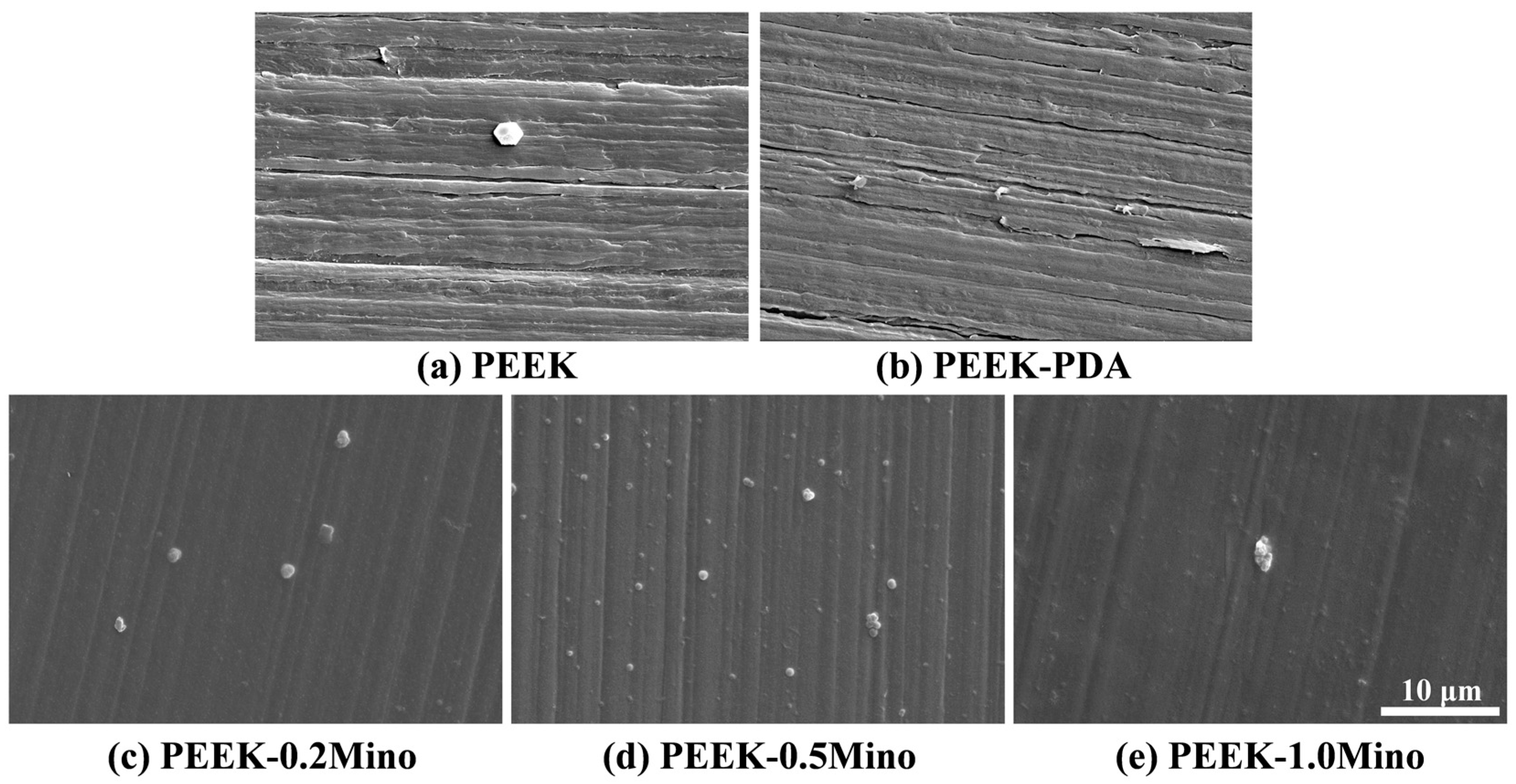
3.1.1. Surface Morphology Characterization by SEM

Figure 1 presents SEM images illustrating the surface morphology of the samples at different modification stages. The pristine PEEK surface exhibits distinct linear polishing marks from mechanical treatment, indicating a relatively smooth but patterned surface. Following PDA coating, the original polishing marks remain visible, suggesting that the PDA layer formed via self-polymerization under weakly alkaline conditions is uniform but relatively thin. After treatment with the CMCS-Mino coating, the surface morphology undergoes a more pronounced transformation. The previously visible polishing marks are no longer observable, suggesting that the CMCS-Mino layer is considerably thicker and effectively masks the underlying topography. Additionally, no apparent differences in surface morphology are observed among PEEK-0.2Mino, PEEK-0.5Mino, and PEEK-1.0Mino.

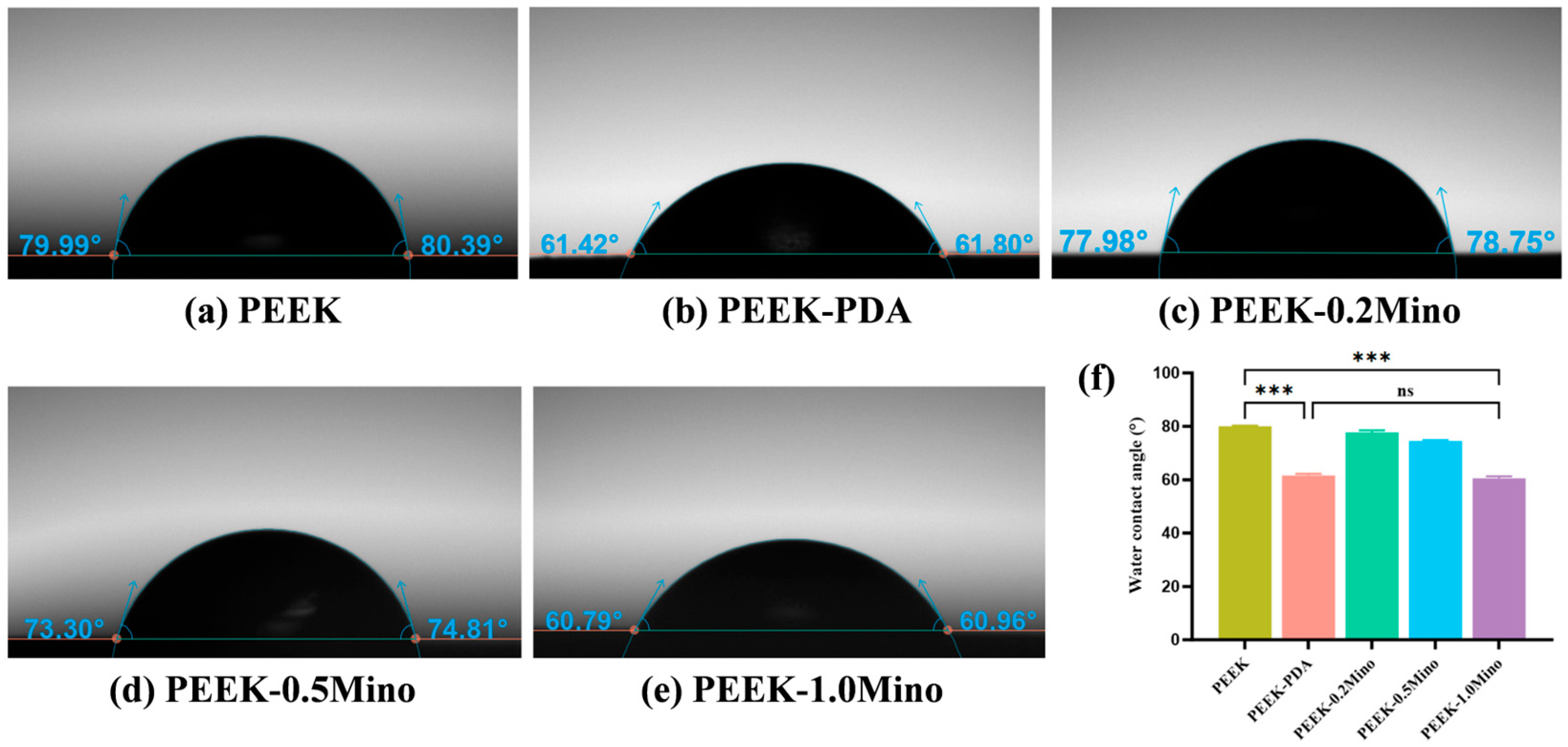
3.1.2. Water Contact Angle and Wettability Analysis

Figure 2 presents the water contact angle images for the five sample groups, with corresponding quantitative measurements summarized. The unmodified PEEK surface exhibits a relatively high contact angle of 80.1°. After PDA coating, the contact angle decreases markedly to 61.6°, indicating enhanced hydrophilicity. Further modification with the CMCS-Mino coating leads to a non-linear trend in surface wettability depending on Mino concentration. PEEK-0.2Mino and PEEK-0.5Mino exhibit contact angles of 77.7° and 74.6°, respectively—lower than that of unmodified PEEK but slightly higher than that of the PDA-coated sample. PEEK-1.0Mino shows a contact angle of 60.5°, which is comparable to that of the PDA-coated sample.

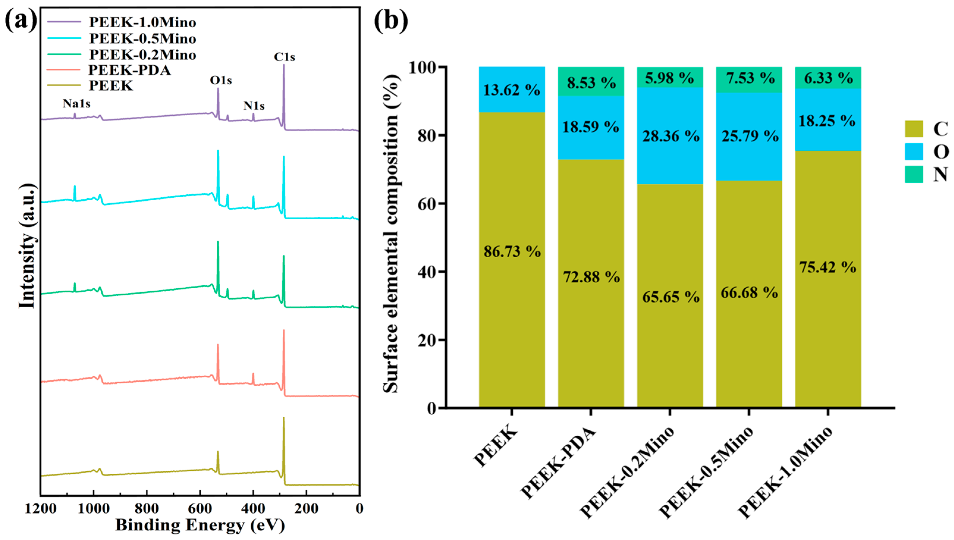
3.1.3. Surface Chemical Composition Analysis by XPS

Figure 3 shows the XPS wide-scan spectra (Figure 3a) and the relative surface elemental composition (Figure 3b) of PEEK, PEEK-PDA, PEEK-0.2Mino, PEEK-0.5Mino, and PEEK-1.0Mino. The pristine PEEK surface displays characteristic C1s and O1s peaks. After PDA coating, a distinct N1s peak appears. Following CMCS and Mino deposition, a Na1s peak is observed. The elemental composition varies with increasing Mino concentration.
Figure 4 presents the high-resolution XPS spectra of C1s and O1s for PEEK, PEEK-PDA, PEEK-0.2Mino, PEEK-0.5Mino, and PEEK-1.0Mino, along with the corresponding fitting peak assignments. The C1s spectrum of PEEK shows peaks at 284.8 eV (C-C/C-H), 286.4 eV (ether C-O), and 287.3 eV (ketonyl C=O). For PEEK-PDA, the C1s spectrum reveals PDA-specific peaks at 284.1 eV (C=C), 284.8 eV (C-C/C-H), 285.7 eV (aromatic and aliphatic C-N), and 287.7 eV (aromatic C=N, catechol C-OH, and quinone C=O) [37]. The O1s spectrum of PEEK-PDA also exhibits PDA-specific peaks, while PEEK-related signals are no longer observed. After deposition of CMCS and Mino, the C1s peaks of PEEK-Mino appear at 284.8 eV (C-C/C-H), 286.2 eV (C-O-C, C-OH, C-N), and 287.8 eV (C=O, COOH, O=C-NH), while the O1s peaks are centered at 531.1 eV (COOH, C=O, O=C-NH) and 532.7 eV (C-O-C, C-OH, bound H2O) [38].
Table 1 and Table 2 list the relative contents of fitted C1s and O1s peaks for PEEK-0.2Mino, PEEK-0.5Mino, and PEEK-1.0Mino. From PEEK-0.2Mino to PEEK-1.0Mino, the area of the C-C/C-H peak at 284.8 eV increases, while that of the C=O, COOH, and O=C-NH peak at 287.8 eV decreases (Table 1). In the O1s spectra, the relative area of the 531.1 eV peak (COOH, C=O, O=C-NH) gradually increases, while the 532.7 eV peak (C-O-C, C-OH, bound H2O) gradually decreases, and both peaks become comparable in PEEK-1.0Mino.

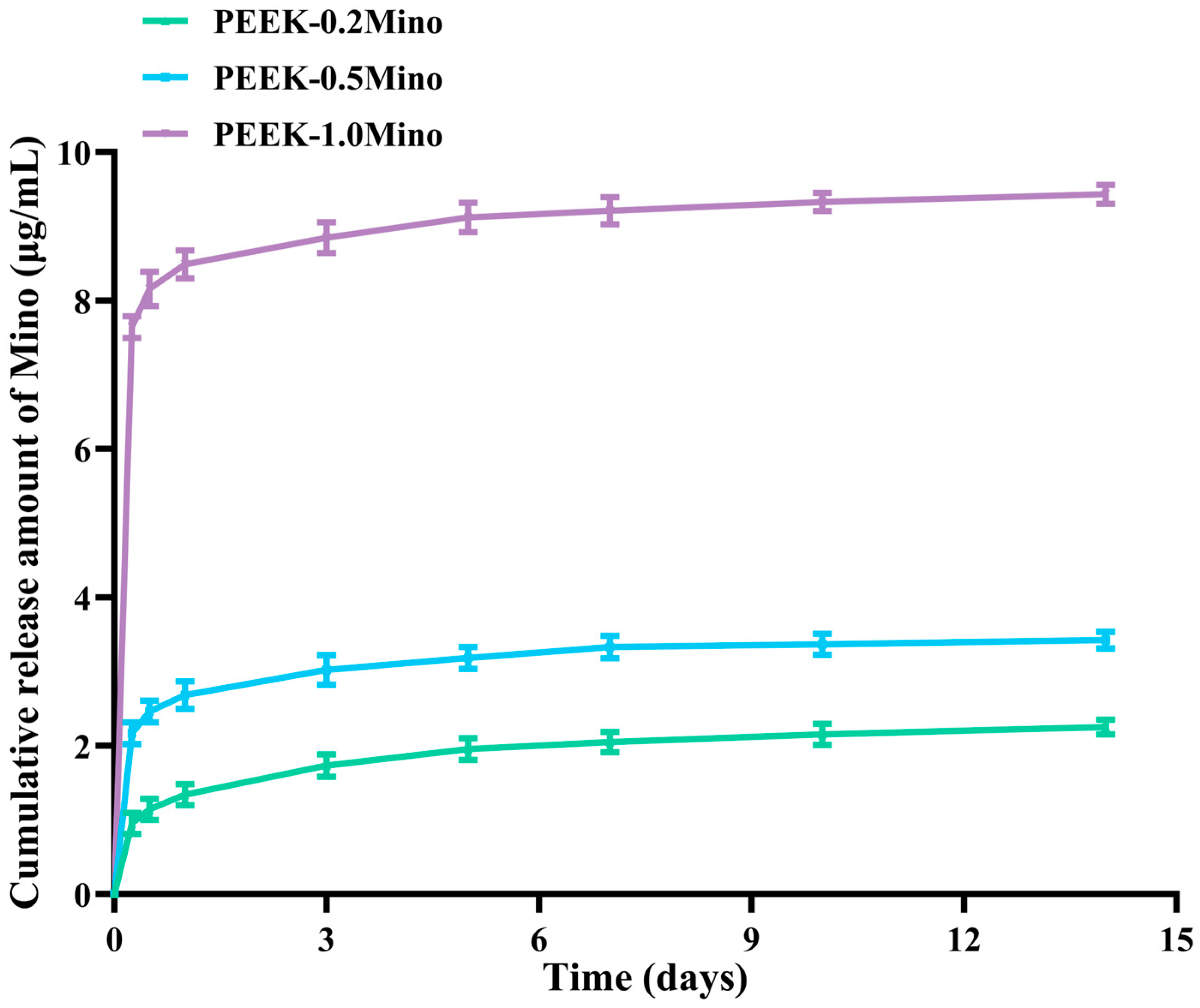
3.2. Release Behavior of Mino

Figure 5 illustrates the cumulative Mino release from PEEK-0.2Mino, PEEK-0.5Mino, and PEEK-1.0Mino samples over a 14-day period in PBS. All three Mino-loaded PEEK samples exhibit sustained drug release behavior. Specifically, during the first 12 h of immersion, the amount of Mino released increases rapidly, likely due to the dissolution of Mino on the coating surface. After this initial phase, the release rate gradually slows down over time.

3.3. Antibacterial Performance Evaluation
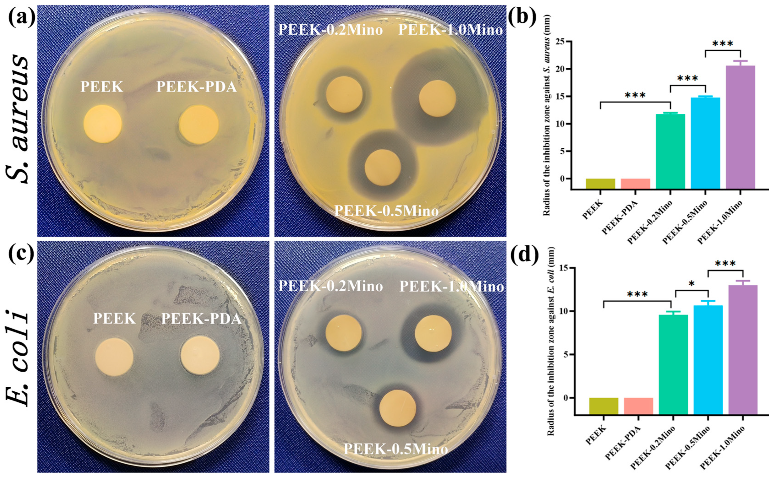
3.3.1. ZOI Assay

The ZOI assay evaluates the antibacterial efficacy of different sample groups against surrounding bacteria. As shown in Figure 6, neither the PEEK nor PEEK-PDA group exhibits an inhibition zone against S. aureus or E. coli, indicating that these materials lack inherent antibacterial activity. In contrast, PEEK-Mino samples show clear inhibition zones. For S. aureus, the inhibition zone diameters are (11.75 ± 0.20) mm for PEEK-0.2Mino, (14.75 ± 0.20) mm for PEEK-0.5Mino, and (20.58 ± 0.72) mm for PEEK-1.0Mino. Similarly, for E. coli, the inhibition zones are (9.58 ± 0.31) mm, (10.67 ± 0.42) mm, and (13.00 ± 0.41) mm, respectively.

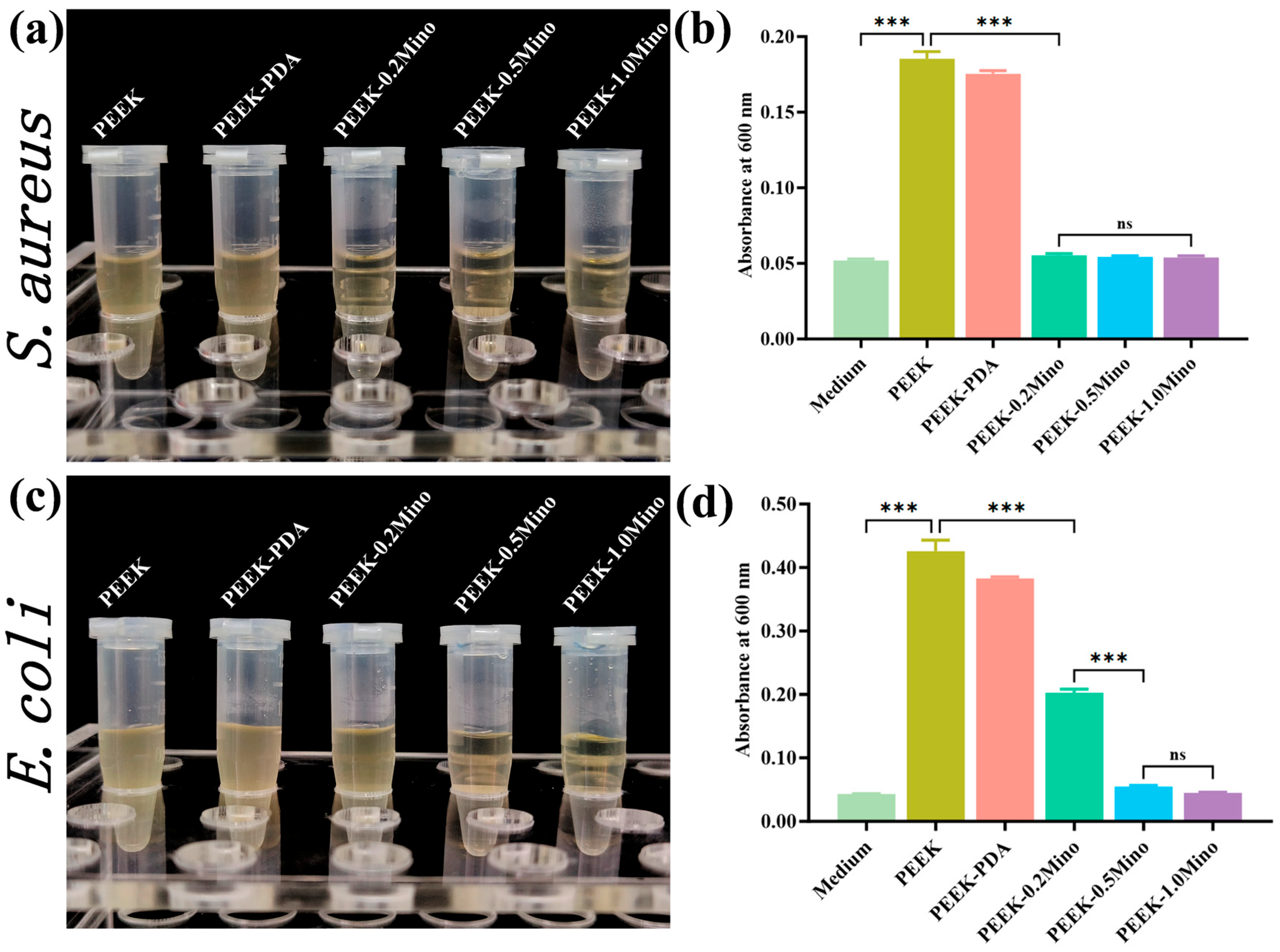
3.3.2. Turbidity Assay

The turbidity assay assesses the antibacterial efficacy of different sample groups by evaluating bacterial growth in the surrounding liquid environment. As shown in Figure 7, the PEEK and PEEK-PDA groups result in visibly turbid culture media after co-incubation with S. aureus and E. coli, and their OD600 values are significantly higher than that of the sterile control. These results indicate extensive bacterial proliferation and confirm the absence of antibacterial activity in these two groups. In contrast, the PEEK-Mino groups exhibit reduced turbidity depending on the bacterial strain and Mino concentration. For S. aureus, the supernatants of PEEK-0.2Mino, PEEK-0.5Mino, and PEEK-1.0Mino remain clear, and their OD600 values are nearly equal to the sterile control, indicating strong antibacterial activity. For E. coli, PEEK-0.5Mino and PEEK-1.0Mino also show clear supernatants and low OD600 values, but the PEEK-0.2Mino group produces visibly turbid media with an intermediate OD600 value, suggesting limited antibacterial efficacy at the lowest Mino concentration.

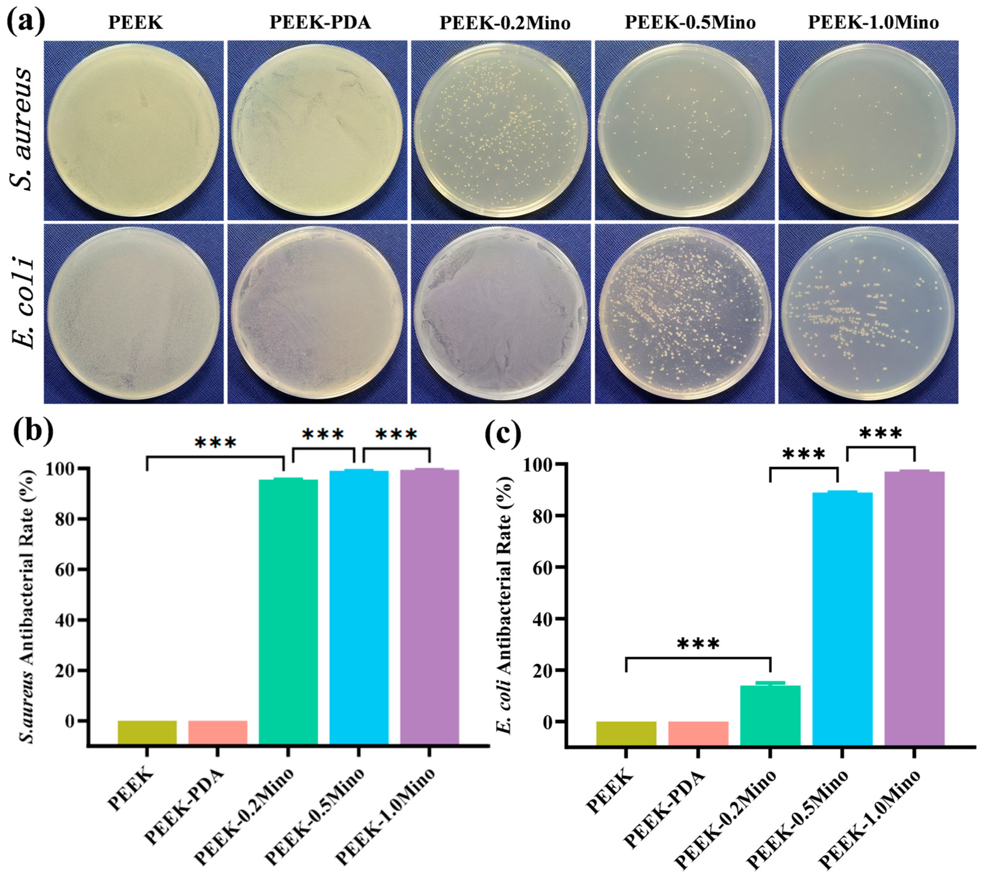
3.3.3. Colony Counting Assay

The colony counting assay is used to quantitatively evaluate the antibacterial efficacy of the modified PEEK surfaces by counting viable bacteria remaining after incubation. As shown in Figure 8, the PEEK and PEEK-PDA groups exhibit dense bacterial colonies after co-incubation with S. aureus, covering the agar surface and indicating a lack of antibacterial activity. In contrast, the PEEK-0.2Mino, PEEK-0.5Mino, and PEEK-1.0Mino groups show a significant reduction in colony numbers, with bacterial survival dropping by more than 95%. A similar trend is observed in the E. coli groups. The PEEK-0.5Mino and PEEK-1.0Mino samples maintain strong antibacterial effects, reducing bacterial counts by over 85%. However, the PEEK-0.2Mino group shows relatively limited efficacy, with a bacterial reduction rate of approximately 10%.

3.4. Biocompatibility Evaluation

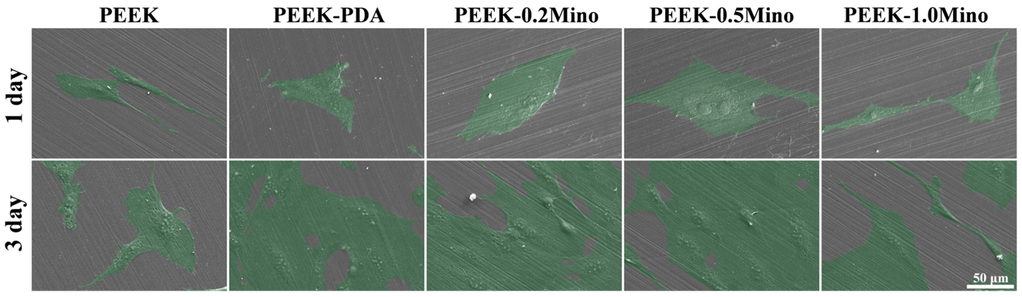
3.4.1. Cell Adhesion

Cell attachment and spreading are critical for initiating osseointegration and are influenced by proteins and growth factors that regulate subsequent cell proliferation and differentiation [39]. Cell attachment and spreading on the material surface are evaluated after 1 and 3 days of incubation. As shown in Figure 9, MC3T3-E1 pre-osteoblasts on PEEK surfaces exhibit a slender, spindle-shaped morphology with limited spreading, indicating suboptimal cell–material interactions on day 1. In contrast, the PEEK-PDA and PEEK-Mino groups promote enhanced cell spreading, with cells on PEEK-0.5Mino showing the largest spread, indicating amendatory cell–material interactions. On day 3, cells on PEEK-PDA, PEEK-0.2Mino, and PEEK-0.5Mino surfaces almost cover the entire field of view, whereas cells on PEEK and PEEK-1.0Mino surfaces spread relatively poorly.

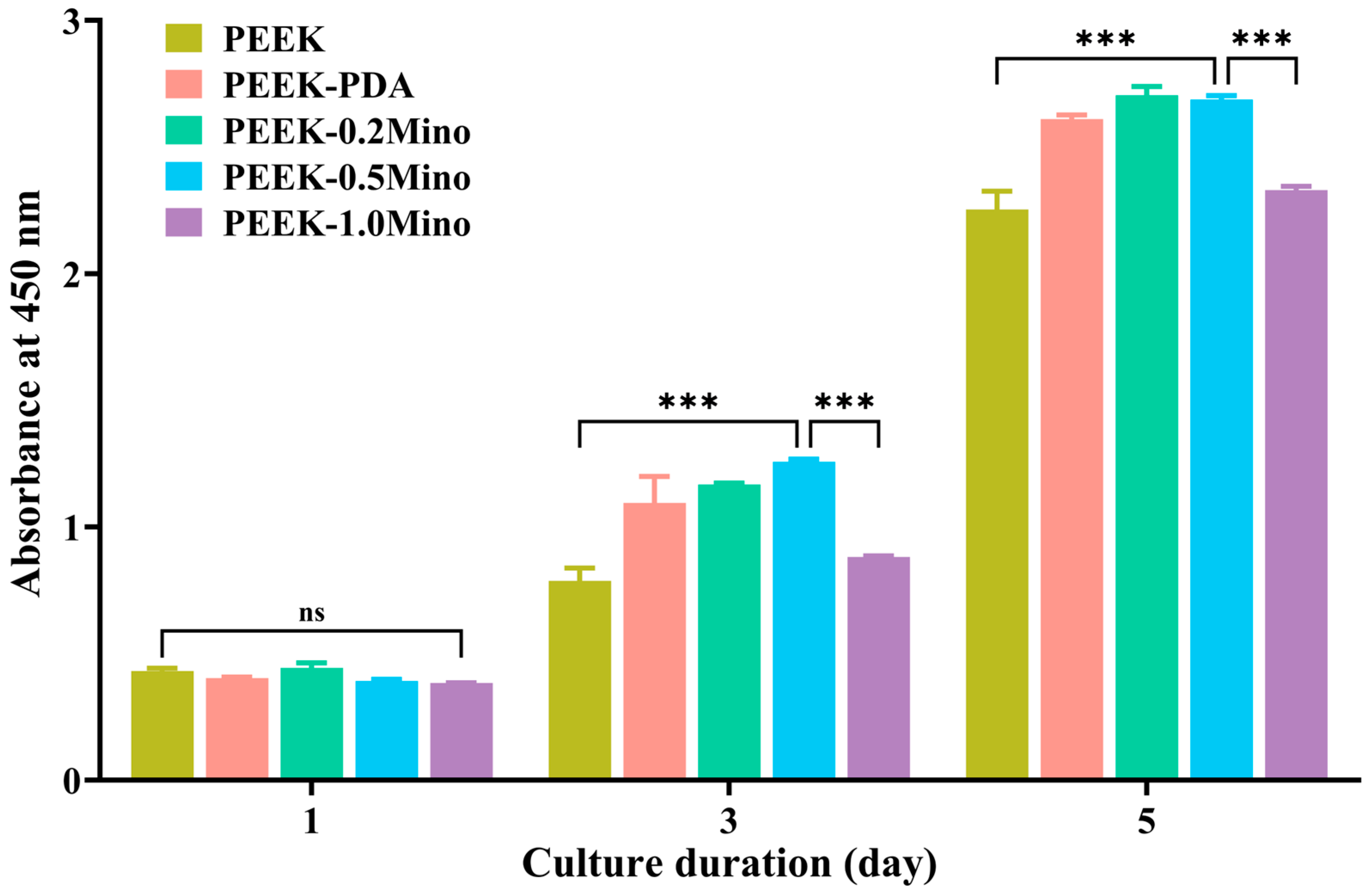
3.4.2. Cell Proliferation

Osteoblast proliferation on the implant surface is essential for bone–implant integration. Cell proliferation is assessed using the CCK-8 assay over 5 days. As shown in Figure 10, all groups exhibit increased proliferation over time. On day 1, no significant differences are observed between the groups. By days 3 and 5, PEEK demonstrates the lowest proliferation, while cell growth increases from PEEK-PDA to PEEK-0.5Mino, peaking at PEEK-0.5Mino. PEEK-1.0Mino exhibits reduced cell proliferation, likely due to the cytotoxic effects of excessive Mino release.

4. Discussion

Effective prevention of IAIs remains a critical challenge in orthopedic and dental applications [1], particularly for bioinert materials such as PEEK, which lacks inherent antibacterial properties and facilitates bacterial adhesion and biofilm formation. In this study, a surface modification strategy combining PDA pre-coating and CMCS-assisted Mino loading is developed to endow PEEK implants with both sustained antibacterial activity and favorable biocompatibility. By systematically optimizing Mino concentration, we aim to balance antibacterial efficacy with cellular compatibility, addressing the dual demands of infection control and osseointegration. The physicochemical characterization, antibacterial assays, and in vitro biocompatibility tests collectively demonstrate the successful fabrication and functional performance of this multilayered coating system.
The SEM results indicate that the PDA layer provides a uniform but thin intermediate coating, consistent with our previous findings [33,36], which confirm the conformal and repeatable nature of the PDA coating process (Figure 1b). The disappearance of polishing marks after CMCS-Mino treatment suggests successful and uniform coverage of the PEEK-PDA surface by the CMCS-Mino composites (Figure 1c–e). The absence of morphology differences among different Mino concentrations implies that the microscopic surface structure is primarily determined by the CMCS matrix rather than the Mino content.
Enhanced wettability is generally considered favorable for subsequent cell adhesion and proliferation, which are critical during the early stages of osseointegration [40]. The decrease in contact angle after PDA coating likely results from the presence of hydrophilic catechol and amine groups introduced by the PDA layer (Figure 2b). The subsequent increase in contact angle for PEEK-0.2Mino and PEEK-0.5Mino may result from a reduction in surface roughness, as the CMCS-Mino layer smooths the sandpaper grinding traces (Figure 2c,d). Additionally, the CMCS matrix may introduce steric effects or molecular domains that are less hydrophilic than pure PDA, thereby influencing water droplet behavior. Interestingly, the contact angle of PEEK-1.0Mino decreases again (Figure 2e), suggesting that a higher density of hydrophilic groups—such as hydroxyl, ketonyl, and amino groups—introduced by increased Mino loading enhances surface wettability. Although SEM reveals no apparent differences in surface morphology among the Mino-modified groups, the increased Mino content may still influence surface chemical composition or hydration behavior at the molecular level. While the precise mechanisms remain to be elucidated, these results suggest that both surface chemistry and microstructure collectively contribute to the wettability of the composite coating.
XPS analysis confirms the stepwise surface modification of PEEK. The appearance of the N1s peak after PDA coating indicates successful formation of the PDA layer on the PEEK surface (Figure 3a). The presence of the Na1s peak confirms successful introduction of CMCS, which typically contains sodium ions (Figure 3a). Although Mino lacks a unique elemental marker, the observed changes in surface elemental composition with increasing Mino content suggest effective Mino loading onto the PEEK surface (Figure 3b).
The exclusive presence of PDA-related bonding signals in both C1s and O1s spectra for PEEK-PDA suggests that the PDA layer fully covers the underlying PEEK surface, indicating a coating thickness above 10 nm (Figure 4). After further modification with CMCS and Mino, the emergence of new characteristic peaks corresponding to the CMCS and Mino functional groups confirms successful deposition of the composite coating. The disappearance of PDA signals in the PEEK-Mino spectra and the presence of CMCS-Mino-specific peaks further support that the final composite layer exceeds 10 nm in thickness, fully masking the underlying PDA layer.
These variations in peak areas are consistent with the increasing Mino content in the composite coating. The molecular structure of Mino contains a higher proportion of C-C/C-H bonds, contributing to the observed increase in the 284.8 eV signal (Table 1). Accordingly, the C1s spectrum of PEEK-0.2Mino mainly exhibits the characteristic bonding of CMCS, whereas PEEK-1.0Mino primarily presents Mino-related bonding features. Similarly, in the O1s spectra of PEEK-1.0Mino, the relative area of fitting peaks at 531.1 eV and 532.7 eV becomes comparable (Table 2), consistent with Mino’s molecular structure. Together, these results confirm the successful PDA pre-treatment and subsequent Mino loading on the PEEK surface.
Drug release experiments reveal a biphasic release profile for Mino (Figure 5). The rapid initial release of Mino is beneficial for addressing bacterial infections during the early stage of implantation, providing a short-term, high-efficiency antibacterial effect. The subsequent slow-release phase is attributed to the stable chemical bonding between Mino and CMCS and PDA formed via Michael addition and Schiff base reactions, and the electrostatic interaction between Mino and CMCS and PDA. This sustained release may help prevent bacterial adhesion and biofilm formation during the healing and stabilization phase of the implant. Moreover, as the host immune system recovers, the continuous low-dose release of Mino provides prolonged antibacterial protection, contributing to the long-term safety of the implant. Importantly, the coating is designed for short-term local release at concentrations above the MIC, which effectively avoids prolonged sub-inhibitory exposure—one of the main drivers of antibiotic resistance [41,42]. In addition, the non-systemic, site-specific delivery further reduces the likelihood of selecting for antibiotic-resistant bacteria by limiting systemic exposure [43,44]. Together, these features suggest that the risk of inducing minocycline resistance with this coating is likely to be low.
The ZOI assay (Figure 6), turbidity assay (Figure 7), and colony counting assay (Figure 8) collectively confirm the significant antibacterial properties of all three PEEK-Mino samples, with effectiveness increasing as Mino concentration increases. PEEK-0.5Mino and PEEK-1.0Mino exhibit stable and potent antibacterial activity across all tests, effectively inhibiting S. aureus and E. coli adhesion and proliferation. However, PEEK-0.2Mino demonstrates relatively weaker antibacterial performance. While it maintains strong inhibition against S. aureus, its efficacy against E. coli is noticeably lower, as indicated by a smaller inhibition zone, higher turbidity in the culture medium, and a significantly reduced bacterial reduction rate in the colony counting assay. The difference in antibacterial efficacy between S. aureus and E. coli may stem from their structural characteristics. S. aureus, a gram-positive bacterium, has a relatively permeable cell wall that facilitates Mino penetration and action [29,45]. In comparison, E. coli is gram-negative and possesses an outer membrane rich in lipopolysaccharides, which acts as a barrier limiting antibiotic permeability [46]. Additionally, the smaller inhibition zones observed for E. coli may stem from its higher resistance to Mino via mechanisms such as efflux pumps, ribosomal protection proteins, and enzymatic modifications [47,48]. These factors collectively reduce the susceptibility of E. coli to Mino, making the antibacterial coating less effective against gram-negative strains. Overall, these findings highlight the importance of adequate Mino loading to ensure consistent and broad-spectrum antibacterial efficacy.
To assess the influence of the antibacterial coating on the biocompatibility of PEEK and determine the optimal Mino concentration that achieves a balance between sustained antibacterial efficacy and favorable cellular responses, the adhesion (Figure 9) and proliferation (Figure 10) of MC3T3-E1 pre-osteoblasts on the modified PEEK surfaces are systematically evaluated. The results show that all Mino-functionalized coatings support osteoblast adhesion and proliferation to varying degrees, indicating overall cytocompatibility. Notably, PEEK-0.5Mino exhibits the most favorable cellular responses, including enhanced cell spreading and the highest proliferation rate, suggesting that this concentration promotes early osteoblast activity without compromising cell viability. In contrast, PEEK-1.0Mino shows reduced cell adhesion and significantly lower proliferation, which can likely be attributed to cytotoxic effects from excessive Mino release. These findings highlight the importance of balancing antibacterial efficacy with cytocompatibility. While higher Mino concentrations enhance antibacterial performance, they impair osteoblast viability and compromise bone–implant integration. The CCK-8 results align with adhesion observations, confirming that PEEK-0.5Mino achieves the optimal balance between antibacterial activity and biocompatibility.
In summary, this study presents a multilayered PEEK coating with concentration-dependent antibacterial activity and favorable cytocompatibility in vitro. Among the tested samples, PEEK-0.5Mino provides the best balance between antibacterial efficacy and osteoblast compatibility under in vitro conditions. However, this study does not evaluate the osteogenic potential of the coating using assays such as ALP activity, mineralization (e.g., Alizarin Red staining), or osteogenic gene expression (e.g., Runx2, OCN, OPN). In addition, no in vivo studies assess the biological performance of the coating under physiological conditions, and the antibacterial activity is evaluated against only two standard strains, S. aureus and E. coli. While these organisms serve as common models, they do not represent the full microbial spectrum relevant to dental infections, particularly anaerobic pathogens such as P. gingivalis, T. denticola, and F. nucleatum. Therefore, the clinical applicability of the coating in dental implants remains uncertain and requires further evaluation. Future work will focus on systematic osteogenic assessments, in vivo studies, and antibacterial testing against oral anaerobic bacteria to verify and expand the application scope of the coating.

5. Conclusions

In conclusion, a sustained-release Mino antibacterial coating is successfully constructed on the PEEK surface, enabling continuous Mino release for up to 14 days. In vitro antibacterial tests show that increasing Mino concentration enhances bacterial inhibition, with PEEK-0.5Mino and PEEK-1.0Mino demonstrating strong antimicrobial effects, while PEEK-0.2Mino proves less effective against E. coli. Biocompatibility assessments reveal that PEEK-0.5Mino achieves the optimal balance, promotes osteoblast adhesion and proliferation, and maintains cell viability, whereas excessive Mino release in PEEK-1.0Mino induces cytotoxicity. These findings suggest that PEEK-0.5Mino offers sustained antibacterial activity and favorable biocompatibility in vitro and shows potential for future development as a surface modification strategy to prevent IAIs and support tissue integration.

Author Contributions

Conceptualization, Y.Z. (Yanyan Zheng); methodology, Y.Z. (Yongheng Zhang) and L.Z.; validation, Y.Z. (Yongheng Zhang), L.Z., P.Y. and Q.H.; investigation, Y.Z. (Yongheng Zhang), L.Z., Y.Z. (Yuehong Zhang), P.Y. and Q.H.; writing—original draft preparation, Y.Z. (Yongheng Zhang) and L.Z.; writing—review and editing, Y.L. and Y.Z. (Yanyan Zheng); supervision, Y.Z. (Yanyan Zheng); funding acquisition, Y.Z. (Yanyan Zheng). All authors have read and agreed to the published version of the manuscript.

Funding

This work was supported by the Specific Cooperative Scientific Research Project of Nanchong (No. 22SXCXTD0001), the Scientific Research Development Plan of Affiliated Hospital of North Sichuan Medical College (No. 2023PTZK011), the Opening Project of Medical Imaging key Laboratory of Sichuan Provine (No. MIKL202410), and the Undergraduate Training Program for Innovation and Entrepreneurship (No. S202510634101, No. S202510634065).

Institutional Review Board Statement

Not applicable.

Informed Consent Statement

Not applicable.

Data Availability Statement

Data will be made available on request.

Conflicts of Interest

The authors declare that they have no known competing financial interests or personal relationships that could appear to have influenced the work reported in this paper.

References

  1. Dong, J.; Wang, W.; Zhou, W.; Zhang, S.; Li, M.; Li, N.; Pan, G.; Zhang, X.; Bai, J.; Zhu, C. Immunomodulatory biomaterials for implant-associated infections: From conventional to advanced therapeutic strategies. Biomater. Res. 2022, 26, 72. [Google Scholar] [CrossRef] [PubMed]
  2. Azad, M.A.; Patel, R. Practical Guidance for Clinical Microbiology Laboratories: Microbiologic diagnosis of implant-associated infections. Clin. Microbiol. Rev. 2024, 37, e0010423. [Google Scholar] [CrossRef]
  3. Li, B.; Webster, T.J. Bacteria antibiotic resistance: New challenges and opportunities for implant-associated orthopedic infections. J. Orthop. Res. 2018, 36, 22–32. [Google Scholar] [CrossRef]
  4. Chu, G.; Guan, M.; Jin, J.; Luo, Y.; Luo, Z.; Shi, T.; Liu, T.; Zhang, C.; Wang, Y. Mechanochemically Reprogrammed Interface Orchestrates Neutrophil Bactericidal Activity and Apoptosis for Preventing Implant-Associated Infection. Adv. Mater. 2024, 36, e2311855. [Google Scholar] [CrossRef] [PubMed]
  5. Zimmerli, W. Clinical presentation and treatment of orthopaedic implant-associated infection. J. Intern. Med. 2014, 276, 111–119. [Google Scholar] [CrossRef] [PubMed]
  6. Wang, M.; Zheng, Y.; Yin, C.; Dai, S.; Fan, X.; Jiang, Y.; Liu, X.; Fang, J.; Yi, B.; Zhou, Q.; et al. Recent Progress in antibacterial hydrogel coatings for targeting biofilm to prevent orthopedic implant-associated infections. Front. Microbiol. 2023, 14, 1343202. [Google Scholar] [CrossRef]
  7. Willing, B.P.; Russell, S.L.; Finlay, B.B. Shifting the balance: Antibiotic effects on host-microbiota mutualism. Nat. Rev. Microbiol. 2011, 9, 233–243. [Google Scholar] [CrossRef]
  8. Ahmadabadi, H.Y.; Yu, K.; Kizhakkedathu, J.N. Surface modification approaches for prevention of implant associated infections. Colloids Surf. B Biointerfaces 2020, 193, 111116. [Google Scholar] [CrossRef]
  9. Li, S.; Yue, Y.; Wang, W.; Han, M.; Wan, X.; Li, Q.; Chen, X.; Cao, J.; Zhang, Y.; Li, J.; et al. Ultrasound-Activated Probiotics Vesicles Coating for Titanium Implant Infections Through Bacterial Cuproptosis-Like Death and Immunoregulation. Adv. Mater. 2024, 36, e2405953. [Google Scholar] [CrossRef]
  10. Zhang, Y.; Zhang, W.; Yang, M.; Li, M.; Zhou, L.; Liu, Y.; Liu, L.; Zheng, Y. Comprehensive review of polyetheretherketone use in dentistry. J. Prosthodont. Res. 2025, 69, 215–232. [Google Scholar] [CrossRef]
  11. Zheng, Y.; Gao, A.; Bai, J.; Liao, Q.; Wu, Y.; Zhang, W.; Guan, M.; Tong, L.; Geng, D.; Zhao, X.; et al. A programmed surface on polyetheretherketone for sequentially dictating osteoimmunomodulation and bone regeneration to achieve ameliorative osseointegration under osteoporotic conditions. Bioact. Mater. 2022, 14, 364–376. [Google Scholar] [CrossRef] [PubMed]
  12. Zheng, Y.; Zhou, H.; Li, M.; Fu, J.; Dong, J.; Liu, Y.; Liu, L. Polyetheretherketone surface engineered with a degradable hybrid coating for accelerating osteogenesis. Mater. Lett. 2023, 331, 133515. [Google Scholar] [CrossRef]
  13. Liu, L.H.; Zhang, W.F.; Yuan, L.; Liu, Y.; Zheng, Y.Y. Ameliorative antibacterial, anti-inflammatory, and osteogenic activity of sulfonate-bearing polyetheretherketone toward orthopedic and dental implants. Mater. Lett. 2021, 305, 130774. [Google Scholar] [CrossRef]
  14. Chen, Y.; Ni, H.; Park, J.-J.; Lv, S. A Review of the Preparation, Modification, and Applications of Polyetheretherketone Coating. Coatings 2024, 14, 1451. [Google Scholar] [CrossRef]
  15. Yee, A.; Xin, X.; Liu, H.; Ma, L.; Cheng, K. Fabrication of PVTF Electroactive Coatings on PEEK Implant to Provide Surface Potential for Enhancing Osteogenesis. Coatings 2025, 15, 261. [Google Scholar] [CrossRef]
  16. Xiao, T.; Fan, L.; Liu, R.; Huang, X.; Wang, S.; Xiao, L.; Pang, Y.; Li, D.; Liu, J.; Min, Y. Fabrication of Dexamethasone-Loaded Dual-Metal-Organic Frameworks on Polyetheretherketone Implants with Bacteriostasis and Angiogenesis Properties for Promoting Bone Regeneration. ACS Appl. Mater. Interfaces 2021, 13, 50836–50850. [Google Scholar] [CrossRef] [PubMed]
  17. Li, D.; Tan, X.; Zheng, L.; Tang, H.; Hu, S.; Zhai, Q.; Jing, X.; Liang, P.; Zhang, Y.; He, Q.; et al. A Dual-Antioxidative Coating on Transmucosal Component of Implant to Repair Connective Tissue Barrier for Treatment of Peri-Implantitis. Adv. Healthc. Mater. 2023, 12, e2301733. [Google Scholar] [CrossRef]
  18. Zhu, J.; Tang, H.; Wang, S.; Zong, Y.; Dai, Q.; Wu, H.; Liang, K.; Yang, J. Mussel-inspired self-assembly platform for staged implant osseointegration: Combining early anti-infection and late osteoinduction. Mater. Design. 2023, 228, 111857. [Google Scholar] [CrossRef]
  19. Garner, S.J.; Dalby, M.J.; Nobbs, A.H.; Barbour, M.E. A novel chlorhexidine-hexametaphosphate coating for titanium with antibiofilm efficacy and stem cell cytocompatibility. J. Mater. Sci. Mater. Med. 2021, 32, 139. [Google Scholar] [CrossRef]
  20. Fang, Y.; Wang, Q.; Yang, Z.; Yang, W.; Wang, L.; Ma, J.; Fu, Y. An efficient approach to endow TiNbTaZr implant with osteogenic differentiation and antibacterial activity in vitro. Mater. Design. 2022, 221, 110987. [Google Scholar] [CrossRef]
  21. Deng, L.; Deng, Y.; Xie, K. AgNPs-decorated 3D printed PEEK implant for infection control and bone repair. Colloids Surf. B Biointerfaces 2017, 160, 483–492. [Google Scholar] [CrossRef] [PubMed]
  22. Yang, X.; Wang, Q.; Zhang, Y.; He, H.; Xiong, S.; Chen, P.; Li, C.; Wang, L.; Lu, G.; Xu, Y. A dual-functional PEEK implant coating for anti-bacterial and accelerated osseointegration. Colloids Surf. B Biointerfaces 2023, 224, 113196. [Google Scholar] [CrossRef] [PubMed]
  23. Bozic Cvijan, B.; Korac Jacic, J.; Bajcetic, M. The Impact of Copper Ions on the Activity of Antibiotic Drugs. Molecules 2023, 28, 5133. [Google Scholar] [CrossRef]
  24. Xue, Y.; Zhang, L.; Zhou, J.; Chen, J.; Ma, Y.; Han, Y. Low-dose Cu ions assisted by mild thermal stimulus inducing bacterial cuproptosis-like death for antibiosis and biointegration. Adv. Funct. Mater. 2024, 34, 2308197. [Google Scholar] [CrossRef]
  25. Lu, J.; Wang, Y.; Jin, M.; Yuan, Z.; Bond, P.; Guo, J. Both silver ions and silver nanoparticles facilitate the horizontal transfer of plasmid-mediated antibiotic resistance genes. Water Res. 2020, 169, 115229. [Google Scholar] [CrossRef]
  26. Nie, P.; Zhao, Y.; Xu, H. Synthesis, applications, toxicity and toxicity mechanisms of silver nanoparticles: A review. Ecotoxicol. Environ. Saf. 2023, 253, 114636. [Google Scholar] [CrossRef]
  27. Jennings, J.A.; Carpenter, D.P.; Troxel, K.S.; Beenken, K.E.; Smeltzer, M.S.; Courtney, H.S.; Haggard, W.O. Novel antibiotic-loaded point-of-care implant coating inhibits biofilm. Clin. Orthop. Relat. Res. 2015, 473, 2270–2282. [Google Scholar] [CrossRef] [PubMed]
  28. Shi, Y.; Lai, Y.; Guo, Y.; Cai, Z.; Mao, C.; Lu, M.; Ren, C.; Ong, J.L.; Chen, W. Aspirin/amoxicillin loaded chitosan microparticles and polydopamine modified titanium implants to combat infections and promote osteogenesis. Sci. Rep. 2024, 14, 7624. [Google Scholar] [CrossRef]
  29. Brogden, R.N.; Speight, T.M.; Avery, G.S. Minocycline: A review of its antibacterial and pharmacokinetic properties and therapeutic use. Drugs 1975, 9, 251–291. [Google Scholar] [CrossRef]
  30. Garrido-Mesa, N.; Zarzuelo, A.; Galvez, J. Minocycline: Far beyond an antibiotic. Br. J. Pharmacol. 2013, 169, 337–352. [Google Scholar] [CrossRef]
  31. Geng, Y.; Xue, H.; Zhang, Z.; Panayi, A.C.; Knoedler, S.; Zhou, W.; Mi, B.; Liu, G. Recent advances in carboxymethyl chitosan-based materials for biomedical applications. Carbohydr. Polym. 2023, 305, 120555. [Google Scholar] [CrossRef] [PubMed]
  32. Riaz Rajoka, M.S.; Zhao, L.; Mehwish, H.M.; Wu, Y.; Mahmood, S. Chitosan and its derivatives: Synthesis, biotechnological applications, and future challenges. Appl. Microbiol. Biotechnol. 2019, 103, 1557–1571. [Google Scholar] [CrossRef] [PubMed]
  33. Zhang, Y.; Liu, L.; Li, M.; Wang, S.; Fu, J.; Yang, M.; Yan, C.; Liu, Y.; Zheng, Y. Dose-dependent enhancement of in vitro osteogenic activity on strontium-decorated polyetheretherketone. Sci. Rep. 2025, 15, 3063. [Google Scholar] [CrossRef] [PubMed]
  34. Jiang, J.; Zhu, L.; Zhu, L.; Zhu, B.; Xu, Y. Surface characteristics of a self-polymerized dopamine coating deposited on hydrophobic polymer films. Langmuir 2011, 27, 14180–14187. [Google Scholar] [CrossRef]
  35. Li, D.; Li, Y.; Shrestha, A.; Wang, S.; Wu, Q.; Li, L.; Guan, C.; Wang, C.; Fu, T.; Liu, W.; et al. Effects of Programmed Local Delivery from a Micro/Nano-Hierarchical Surface on Titanium Implant on Infection Clearance and Osteogenic Induction in an Infected Bone Defect. Adv. Healthc. Mater. 2019, 8, e1900002. [Google Scholar] [CrossRef]
  36. Zhang, W.; Liu, L.; Zhou, H.; He, C.; Yang, X.; Fu, J.; Wang, H.; Liu, Y.; Zheng, Y. Surface bisphosphonation of polyetheretherketone to manipulate immune response for advanced osseointegration. Mater. Design. 2023, 232, 112151. [Google Scholar] [CrossRef]
  37. Li, X.; Yuan, S.; Chen, S.; Luo, R.; Xiong, K.; Yang, Z.; Wang, J.; Huang, N. Proliferation and functionality of human umbilical vein endothelial cells on angiopoietin-1 immobilized 316L stainless steel. J. Mater. Chem. B 2015, 3, 8717–8728. [Google Scholar] [CrossRef]
  38. Yuan, D.; Cadien, K.; Liu, Q.; Zeng, H. Adsorption characteristics and mechanisms of O-Carboxymethyl chitosan on chalcopyrite and molybdenite. J. Colloid Interface Sci. 2019, 552, 659–670. [Google Scholar] [CrossRef]
  39. Nobles, K.; Janorkar, A.V.; Roach, M.D.; Walker, L.; Williamson, R.S. In Vitro Evaluation of Optimized PEEK Surfaces for Enhanced Osseointegration. Coatings 2024, 14, 518. [Google Scholar] [CrossRef]
  40. Liu, L.; Dong, J.; Zhang, W.; He, C.; Liu, Y.; Zheng, Y. The single-step fabrication of a poly (sodium vinylsulfonate)-grafted polyetheretherketone surface to ameliorate its osteogenic activity. Coatings 2022, 12, 868. [Google Scholar] [CrossRef]
  41. Ramsay, K.A.; McTavish, S.M.; Wardell, S.J.; Lamont, I.L. The effects of sub-inhibitory antibiotic concentrations on Pseudomonas aeruginosa: Reduced susceptibility due to mutations. Front Microbiol. 2021, 12, 789550. [Google Scholar] [CrossRef] [PubMed]
  42. Ching, C.; Zaman, M.H. Development and selection of low-level multi-drug resistance over an extended range of sub-inhibitory ciprofloxacin concentrations in Escherichia coli. Sci. Rep. 2020, 10, 8754. [Google Scholar] [CrossRef] [PubMed]
  43. Gao, W.; Chen, Y.; Zhang, Y.; Zhang, Q.; Zhang, L. Nanoparticle-based local antimicrobial drug delivery. Adv. Drug Deliv. Rev. 2018, 127, 46–57. [Google Scholar] [CrossRef]
  44. Qian, H.; Lei, T.; Hua, L.; Zhang, Y.; Wang, D.; Nan, J.; Liu, W.; Sun, Y.; Hu, Y.; Lei, P. Fabrication, bacteriostasis and osteointegration properties researches of the additively-manufactured porous tantalum scaffolds loading vancomycin. Bioact. Mater. 2023, 24, 450–462. [Google Scholar] [CrossRef]
  45. Shin, H.-J.; Yang, S.; Lim, Y. Antibiotic susceptibility of Staphylococcus aureus with different degrees of biofilm formation. J. Anal. Sci. Technol. 2021, 12, 41. [Google Scholar] [CrossRef]
  46. Gauba, A.; Rahman, K.M. Evaluation of Antibiotic Resistance Mechanisms in Gram-Negative Bacteria. Antibiotics 2023, 12, 1590. [Google Scholar] [CrossRef]
  47. Nasrollahian, S.; Graham, J.P.; Halaji, M. A review of the mechanisms that confer antibiotic resistance in pathotypes of E. coli. Front. Cell. Infect. Microbiol. 2024, 14, 1387497. [Google Scholar] [CrossRef]
  48. Fernandez, L.; Hancock, R.E. Adaptive and mutational resistance: Role of porins and efflux pumps in drug resistance. Clin. Microbiol. Rev. 2012, 25, 661–681. [Google Scholar] [CrossRef]
Figure 1. SEM images of various PEEK samples.
Coatings 15 00622 g001
Figure 2. Water contact angles of different PEEK samples: (a) PEEK, (b) PEEK-PDA, (c) PEEK-0.2Mino, (d) PEEK-0.5Mino, (e) PEEK-1.0Mino, and (f) quantified water contact angles (n = 3). Data are mean ± SD. *** p < 0.001; ns = not significant.
Coatings 15 00622 g002
Figure 3. XPS wide-scan spectra (a) and surface elemental composition (b) of different PEEK samples.
Coatings 15 00622 g003
Figure 4. High-resolution C1s spectra (a) and high-resolution O1s spectra (b) of different PEEK samples.
Coatings 15 00622 g004
Figure 5. Cumulative Mino release concentration from PEEK-Mino samples over 14 days in PBS (n = 3).
Coatings 15 00622 g005
Figure 6. Inhibition zone images and radii of various PEEK samples against S. aureus (a,b) and E. coli (c,d) after 16 h (n = 3). Data are mean ± SD. * p < 0.05; *** p < 0.001.
Coatings 15 00622 g006
Figure 7. Turbidity assay images and corresponding OD values of different PEEK samples against S. aureus (a,b) and E. coli (c,d) after 24 h (n = 3). Data are mean ± SD. *** p < 0.001; ns = not significant.
Coatings 15 00622 g007
Figure 8. Colony counting images (a) and quantification (b,c) after 16 h (n = 3). Data are mean ± SD. *** p < 0.001.
Coatings 15 00622 g008
Figure 9. Cell morphology of MC3T3-E1 pre-osteoblasts cultured on different PEEK sample surfaces after 1 and 3 days observed by SEM.
Coatings 15 00622 g009
Figure 10. CCK-8 assay of MC3T3-E1 pre-osteoblast proliferation at 1, 3, and 5 days (n = 3). Data are mean ± SD. *** p < 0.001; ns = not significant.
Coatings 15 00622 g010
Table 1. The content of different C-species for PEEK-0.2Mino, PEEK-0.5Mino, and PEEK-1.0Mino.
B.E. (eV)AttributionRelative Composition of Carbon Species (%)
PEEK-0.2MinoPEEK-0.5MinoPEEK-1.0Mino
284.8C-C/C-H45.0359.7675.84
286.2C-O-C, C-OH, C-N42.7224.4813.85
287.8C=O, COOH, O=C-NH10.1414.2510.31
291.3π→π*2.111.510.00
Table 2. The content of different O-species for PEEK-0.2Mino, PEEK-0.5Mino, and PEEK-1.0Mino.
B.E. (eV)AttributionRelative Composition of Oxygen Species (%)
PEEK-0.2MinoPEEK-0.5MinoPEEK-1.0Mino
531.1COOH, C=O, O=C-NH31.8444.2646.92
532.7C-O-C, C-O-H, bound H2O65.6248.6746.05
535.7gas phase H2O2.547.077.03
Disclaimer/Publisher’s Note: The statements, opinions and data contained in all publications are solely those of the individual author(s) and contributor(s) and not of MDPI and/or the editor(s). MDPI and/or the editor(s) disclaim responsibility for any injury to people or property resulting from any ideas, methods, instructions or products referred to in the content.

