Review

Research Progress of the Coatings Fabricated onto Titanium and/or Titanium Alloy Surfaces in Biomaterials for Medical Applications for Anticorrosive Applications

College & Hospital of Stomatology, Anhui Medical University, Key Lab. of Oral Diseases Research of Anhui Province, Hefei 230032, China
*
Author to whom correspondence should be addressed.
Coatings 2025, 15(5), 599; https://doi.org/10.3390/coatings15050599
Submission received: 25 March 2025 / Revised: 6 May 2025 / Accepted: 12 May 2025 / Published: 17 May 2025
(This article belongs to the Special Issue Innovative Coatings for Corrosion Protection of Alloy Surfaces)

Abstract

:
Titanium (Ti) and its alloys have attracted more interest, as they are widely employed as biomaterials due to their great biocompatibility, excellent strength ratio, and lightweight. However, corrosion occurs slowly due to an electrochemical reaction once the Ti material has been placed in the human body, contributing to infection and failure of implants in medical applications. Thus, the corrosion phenomenon has caused great concern in the biomedical field. It is desirable to make the surface modification to provide better corrosion resistance. The fabrication techniques of the coatings fabricated onto Ti and/or Ti alloy surfaces have been reported, including sol–gel, annealing, plasma spraying, plasma immersion ion implantation, physical vapor deposition, chemical vapor deposition, anodization, and micro-arc oxidation. This review first describes the corrosion types, including localized corrosion (both pitting and crevice corrosion), galvanic corrosion, selective leaching, stress corrosion cracking (SCC), corrosion fatigue (CF), and fretting corrosion. In the second part, the effects of corrosion on the human body were discussed, and the primary cause for clinical failure and allergies has been identified as the excessive release of poisonous and dangerous metal ions (Co, Ni, and Ti) from corroded implants into bodily fluids. The inclusion and exclusion criteria during the selection of literature are described in the third section. In the last section, we emphasized the current research progress of Ti alloy (particularly Ti6Al4V alloy) coatings in biomaterials for medical applications involving dental, orthopedic, and cardiovascular implants for anticorrosive applications. However, there are also several problems to explore and address in future studies, such as the release of excessive metal ions, etc. This review will draw attention to both researchers and clinicians, which could help to increase the coatings fabricated onto Ti and/or Ti alloy surfaces for anticorrosive applications in biomaterials for medical applications.

1. Introduction

During the last few decades, there has emerged a growth in the requirement for the restoration of damaged hard tissues due to numerous illnesses, including dental, orthopedic, and cardiovascular diseases [1]. Biomaterials are frequently utilized for this purpose to repair skeletal tissues in the body. Nowadays, stainless steel [2], magnesium-based alloys, titanium (Ti), and its related alloys are biomaterials employed in dental and orthopedic applications [3]. Among these biomaterials, Ti and its related alloys have been extensively employed in dentistry, orthopedics, and the cardiovascular system [3].
Ti and its alloys, particularly Ti6Al4V alloy, have been extensively used in biomedical materials owing to their outstanding biocompatibility [4,5,6,7,8,9], better mechanical properties like super strength ratio and lightweight, and good corrosion resistance [10,11,12]. However, Ti and its related alloys would interact with extracellular bodily fluids after being implanted in human tissues, and corrosion occurs slowly as a result of complicated electro-biochemical reactions at the implant–tissue interfaces [13], which can cause infection and failure of implants as medical devices. Proteins are involved in a variety of processes that can either inhibit or enhance metal degradation, based on the type of protein, concentration, and the implant material’s properties [14]. The corrosion of metallic implants is prone to be accelerated by the elevated amounts of proteins and chloride ions found in bodily fluids [15]. Proteins are denatured and converted into a coating on the metal surface, which prevents corrosion [14]. Moreover, the released toxic ions (e.g., aluminum and vanadium) from matrix corrosion may infect nearby bone tissue and shorten the longevity of Ti alloy implants [16]. For example, Nitinol Shape Memory Alloy (NiTi SMA), a new promising material, is used more frequently in medical applications [17] such as medical devices due to its unique characteristics, e.g., wear resistance, super-elasticity, and shape memory effect [18]. The development of NiTi SMA as a medical device is significantly constrained by the fact that Ni ions in high concentrations of more than 50% on NiTi alloy could result in harmful effects on the human body, including toxicity, allergy, chronic inflammation, and other problems [19]. As we all know, the surface of a material is important as an interface between the material and the environment. For example, the surface characteristics of NiTi, which serve as an interface between living tissue and the artificial implant, are essential in ensuring the success of implantation, particularly in the early stages [20]. However, modern materials should be modified before application or any additional process, including coating with functional materials, because their surface properties are typically insufficient, such as wettability, adhesion properties, biocompatibility, etc. [21]. The poor antibacterial activity and bioinert surface are other challenges of the common biomaterials, e.g., NiTi, stainless steels, etc., that should be treated. The coatings fabricated onto Ti and/or Ti alloy surfaces, which were composed of metals (Ti, Ni, Al, V) and bioactive materials, such as graphene oxide (GO) [22] and Chitosan–Bioactive glass (CS-BG) [23] have good antibacterial and anticorrosive activity, and bioactive surfaces. Therefore, it is crucial for the surface modification of Ti-based metal materials to provide the implant with better corrosion resistance [24]. Sol–gel [23], annealing [25], plasma spraying [26], plasma immersion ion implantation [27,28], physical vapor deposition [29], chemical vapor deposition [30], anodization [31,32,33] and micro-arc oxidation [34,35] have been reported as the surface modification ways to fabricate Ti alloy coatings.
In this review, we first describe the corrosion types. In the second part, the effects of corrosion on the human body were discussed. In the last section, we emphasized the current research progress of Ti material coating in biomaterials for medical applications involving dental, orthopedic, and cardiovascular implants for anticorrosive applications. A time-line for the development of coatings based on Ti material is shown in Table 1. There are several review articles reporting the same research area [36,37], and the novelty and advancement of the present review is the statement of the different applications of coatings fabricated onto Ti and/or Ti alloy surfaces in biomaterials for medical applications. A summary figure regarding corrosion types and effects, as well as anticorrosive applications of coatings fabricated onto Ti and/or Ti alloy surfaces in biomaterials for medical applications is shown in Figure 1. We anticipate that this review will be beneficial to the prosperous development of coatings fabricated onto Ti and/or Ti alloy surfaces for anticorrosive applications in biomaterials for medical applications.

2. Corrosion of Biomaterials

Corrosion is described as an electrochemical reaction during which a metal interplays with its chemical environment, resulting in substantial material (like an alloy implant) degradation and a structural change [39]. The majority of metallic implants are declared to have strong corrosion resistance, which increases the lifespan of the implant in vivo. The corrosive agents in the body fluid include Cl ions, dissolved oxygen, etc. [50,51,52]. However, research indicates that corrosion occurs gradually as a result of an electrochemical process after a metallic implant is inserted into the human body [53]. Therefore, this section describes the corrosion types and the corresponding effects on the human body.

2.1. Corrosion Types

Implants and various medical devices are frequently subject to different types of corrosion, which include localized corrosion (both pitting and crevice corrosion), galvanic corrosion, selective leaching, stress corrosion cracking (SCC), corrosion fatigue (CF), and fretting corrosion [54,55], as shown in Figure 2. The latter three are caused by the synergy of mechanical and electrochemical factors. That is to say, general (uniform) corrosion happens extremely rarely in vivo.

2.1.1. Pitting Corrosion

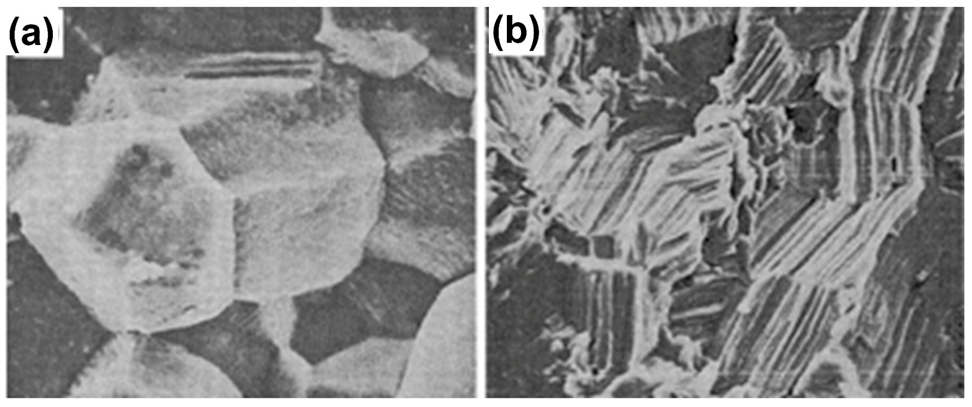
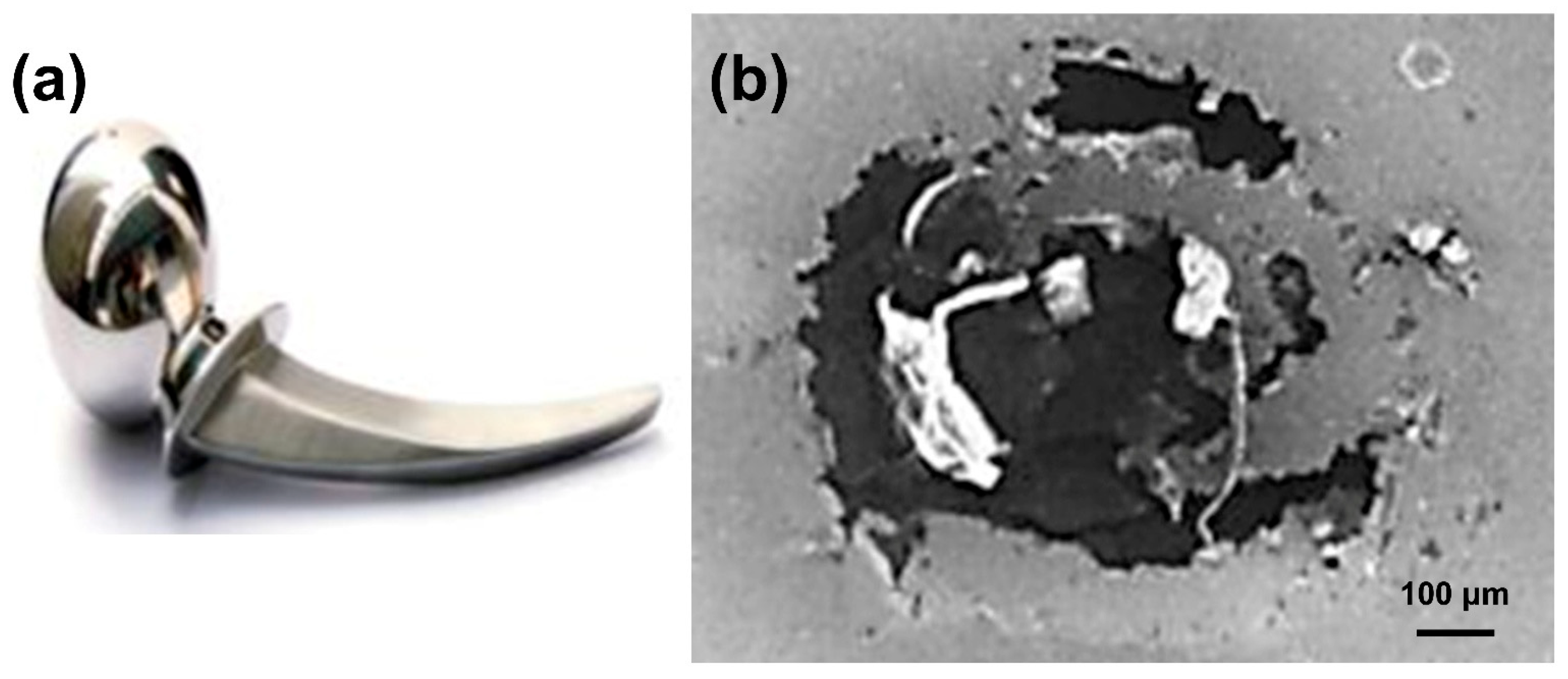
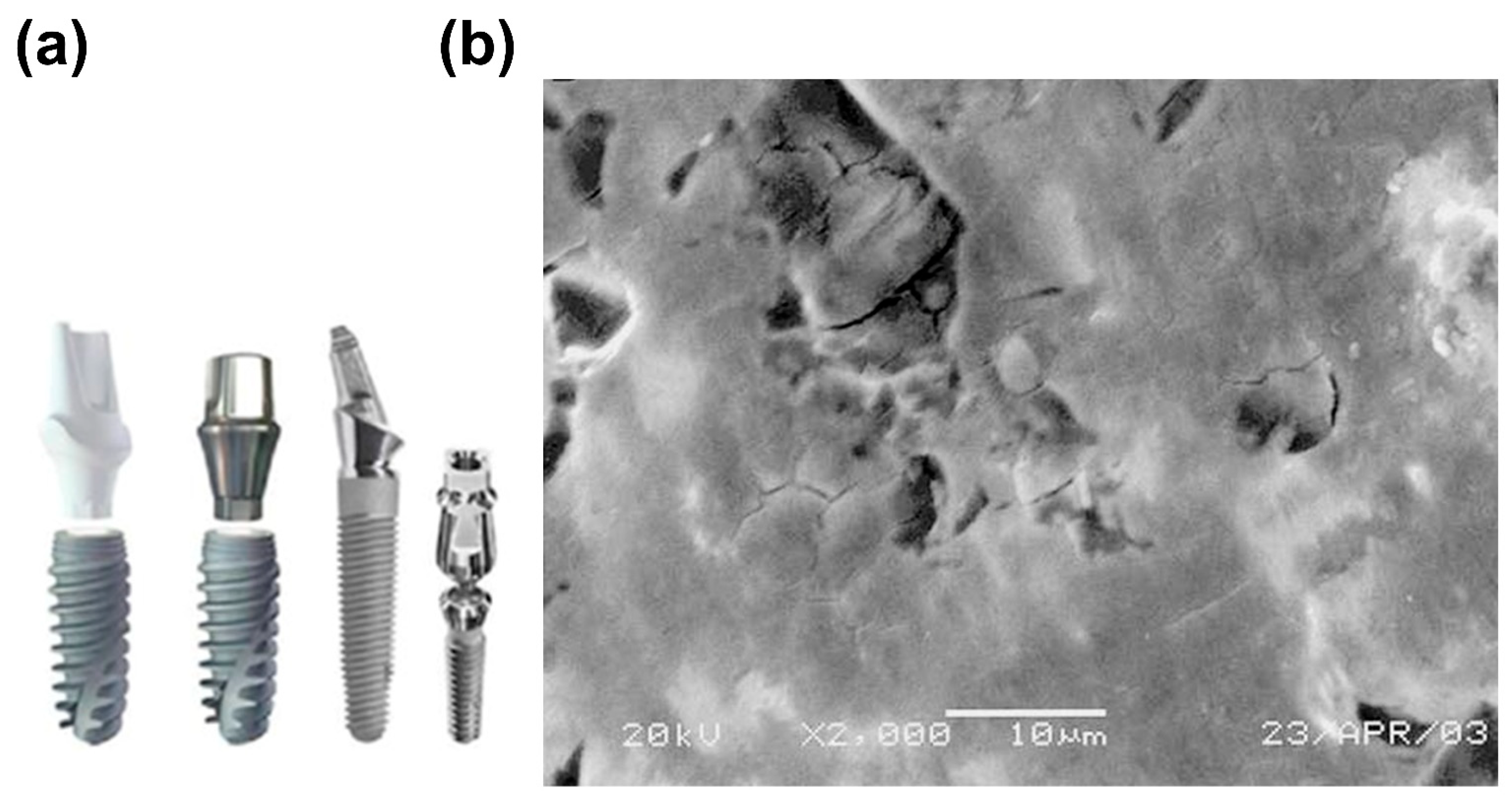
Pitting corrosion is a kind of localized corrosion on the metal surface [36]. Generally, the basic pitting corrosion process could be divided into three steps. Initially, many new stable pit nucleations cause the development of a small bare region on the metal without turning it into a passive film surface [56]. Subsequently, the nucleation of a metastable pit that might have re-passivated causes localized disintegration of the small components of the underlying metal, which is also known as a pit embryo. Finally, the creation of a stable pit caused by the accumulation of damage results in the re-passivation of metastable pits [36,57]. Although Ti has great resistance to pitting corrosion because of the passive oxidized film [58], it is susceptible to pitting corrosion under harsh working environments. Temperature and the concentration of halide ions have an important impact on pitting corrosion, the severity of which often follows the order of F < Cl < I < Br [59,60]. The shape of the implant and the SEM image of pitting corrosion are presented in Figure 2 [55,61].
Figure 2. (a) Shape of the implant and (b) SEM image of pitting corrosion [55,61].
Coatings 15 00599 g002

2.1.2. Crevice Corrosion

Crevice corrosion, like pitting corrosion, is a form of localized corrosion [62]. It develops more frequently in areas where a mass transfer is constrained on the metal surface, like under deposits or in small crevices. The aggressive chloride ion concentration, pH decrease, and oxygen depletion at these occluded regions can quickly trigger surface activation, resulting in crevice corrosion on the metallic surfaces [37,59]. The shape of the implant and the SEM image of crevice corrosion are displayed in Figure 3 [55,59].

2.1.3. Stress Corrosion Cracking (SCC)

SCC is caused by an interaction of static tensile stresses and corrosive conditions. Failures frequently take place in moderate conditions with tensile stress far below the macroscopic yield strength of the metal. Tensile tensions might result from residual stresses, thermal stresses, or external forces. Typically, the time needed to start SCC becomes shorter when these stresses increase [37]. With the exception of several particular conditions [63], such as aqueous halides [64], anhydrous methanol, nitrogen tetroxide (N2O4), and liquid mercury, pure Ti and its alloys are frequently invulnerable to SCC. The SEM images of stress corrosion cracking are shown in Figure 4 [65].

2.1.4. Corrosion Fatigue (CF)

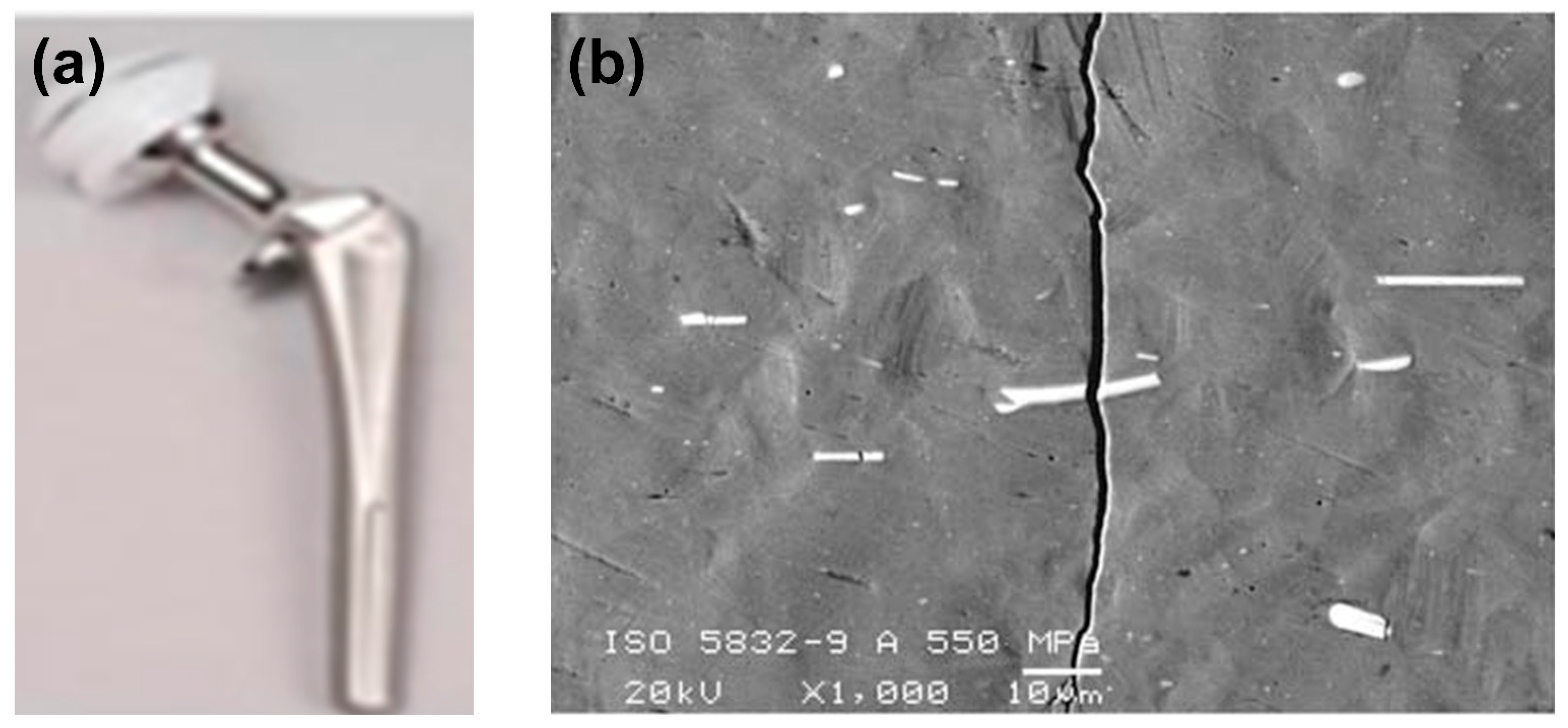
The pits on the metallic surface have been linked to the nucleation of fatigue cracks [66]. The aqueous condition could hasten the emergence of a surface defect and spread to a critical size where it can rupture under fatigue conditions. CF is the term used to describe this phenomenon, which happens when a metal fails as it is simultaneously exposed to cyclic stresses and chemical attacks [67]. It is generally known that CF reduces the fatigue life of metallic implants, such as Ti6Al4V, due to the decrease in fatigue strength, which in turn should arise from the dissolution of Ti2+ ions in the living body, wearing at sliding parts, and fretting [68]. The shape of the implant and the SEM image of CF are represented in Figure 5 [55,68].

2.1.5. Fretting Corrosion

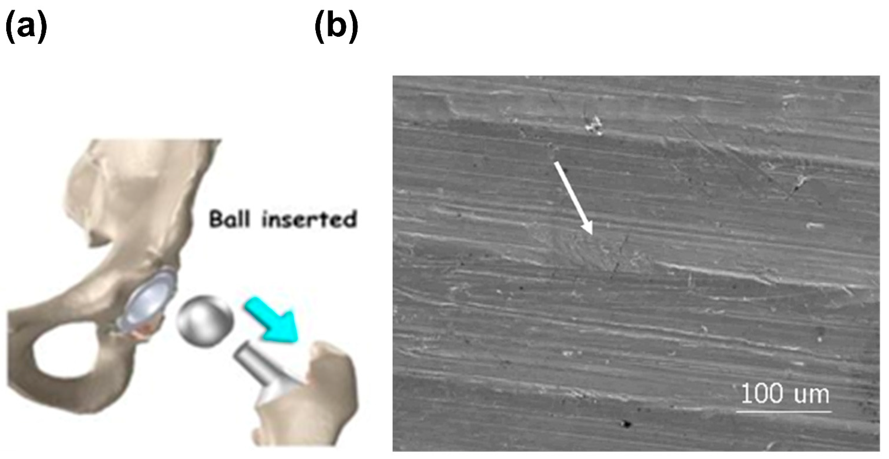
Tribocorrosion is described as a permanent change in the metal resulting from simultaneous mechanical and physicochemical reactions at tribological interactions [69]. Fretting corrosion is a unique form of tribocorrosion that is extremely significant in the field of biomedical implants like knee replacements. It can cause aseptic loosening, the initial decline of mechanical integrity, and ultimately breakdown of the metallic implant, necessitating additional repair surgery and increasing pain to the patients [70]. Moreover, fretting corrosion could significantly alter corrosion behavior through mechanically damaging the passive oxide film. As a result, the capacity to repassivate the metal is essential. Ti and its alloys are prone to fretting corrosion [71], which has become a big problem. The shape of the implant and the SEM image of fretting corrosion are indicated in Figure 6 [55,59].

2.1.6. Galvanic Corrosion

Galvanic corrosion, additionally referred to as bimetallic corrosion, is the rapid corrosion of an anode (a fairly reactive metal) when it comes into electrical contact with a cathode (a more passive metal) in a widespread electrolytic solution. This type of corrosion can occur locally or generally [37]. It may be expected that when cobalt- and Ti-based alloys are used simultaneously in vivo, the more active cobalt alloy will experience faster corrosion and the less active Ti alloy will act as the cathode [72]. Nevertheless, Ti is an inferior cathode due to the slow reduction processes of water and oxygen on Ti surfaces and the fact that its passive current is almost independent of potential and easily polarized, which implies that any metal that is coupled to Ti should only experience the minor acceleration of corrosion. Therefore, Ti-cobalt combinations are steady both in vivo and in vitro, at a minimum when no relative movement takes place [73]. The 316L stainless steel, in contrast, is vulnerable to pitting when it is paired with either cobalt- or Ti-based alloys [74]. The shape of the implant and the SEM image of galvanic corrosion are illustrated in Figure 7 [55,75].

2.2. Effect of Corrosion on the Human Body

The primary cause for clinical failure and allergies has been identified as the excessive release of poisonous and dangerous metal ions (Co, Ni, and Ti) from corroded implants into bodily fluid [76]. When a material begins to corrode, the metal will dissolve and cause erosion, which may ultimately result in brittleness and fracture of the implant, further triggering inflammation in the surrounding tissues [55,77]. Matusiewicz found that greater amounts of metal implants, such as Ni, Co, and Ti, in the blood of patients caused various health problems [78]. The toxicity of Ni and Co-based implants has been identified after implantation for 4 to 5 years, contact-allergy-related dermatitis, red skin, and itchiness were observed for Ni-based implants, while pathology of systemic and neurological problems for Co-based implants, such as dyspnea, severe headaches, and memory difficulties [78,79]. Take Ti alloys as an example. Each constituting element of the Ti alloy could possibly cause health issues. Ti is currently regarded as the element that is most biocompatible when compared to the components of other metallic implants. However, Ti exposure is linked to a “yellow nail syndrome” (YNS), which manifests as a yellowish discoloration of the nails of patients, together with other kinds of symptoms like coughing and sinus infection [80]. Certain YNS symptoms have been directly related to Ti dental implants because they vanished after removing the implant [81].

3. Review of Literature

To be included in this review, studies were required to (1) be published in the last 20 years; (2) have coatings on the Ti and/or Ti alloy surfaces; (3) have better corrosion resistance; (4) be Biomaterials; (5) be applied to medical applications. Any literature that did not meet the inclusion criteria was excluded.

4. Anticorrosive Applications in Biomaterials for Medical Applications

Ti and its related alloys are utilized in a variety of applications, which include fracture fixation implants, dental implants, heart pacemakers, and so on. Ti alloys are widely employed for joint replacement components because of their superior mechanical characteristics as compared to pure Ti [37]. During the last several decades, developments in manufacturing technology have improved the anticorrosive property of medical alloys, but corrosion of biomedical implant alloys can be observed in both orthopedic and cardiovascular implants. Severe corrosion of medical implants might not only have a negative influence on the structure or function of the implant but also trigger a biological response. For orthopedic metallic implants, the corrosion can lead to pseudotumor and unfavorable localized tissue response [82,83,84,85]. The corrosion byproducts in cardiovascular stents could trigger an inflammatory cell activation that accelerates neointimal formation and causes restenosis [86]. Therefore, we summarize the recent progress of coatings fabricated onto Ti and/or Ti alloy surfaces in biomaterials for medical applications, for anticorrosive applications like dental, orthopedic, and cardiovascular applications.

4.1. Dental Applications

Ti and its alloys are widely utilized in dental applications. However, allergies and tissue discoloration in patients caused by Ti have been revealed [87]. In oral environments, commercial mouth rinses, toothpaste, and preventive gels with fluoride are frequently utilized to avoid dental caries and alleviate dental sensitivity [88]. However, the anticorrosive property of Ti can be seriously harmed in the presence of high fluoride ions because of the change in the insoluble Ti and TiO2 oxide layer into soluble Ti-fluorine compounds [89]. Implants and dental crowns typically remain in the oral cavity for years or a lifetime, but the Ti and oxide film can be damaged in saliva for long-term service, which can result in decreased usage in material life [90,91].

4.1.1. Orthodontic Wires and Brackets

As we all know, the anticorrosive property of orthodontic wires is a significant consideration when assessing their biocompatibility, because the process of corrosion can have an adverse influence on their biocompatibility. Additionally, the enamel surface may be stained due to the metal ions that are released by the corrosion of orthodontic wires, which results in an unfavorable esthetic effect [92]. Therefore, substantial research on the corrosion resistance of NiTi orthodontic wires has been published [93].
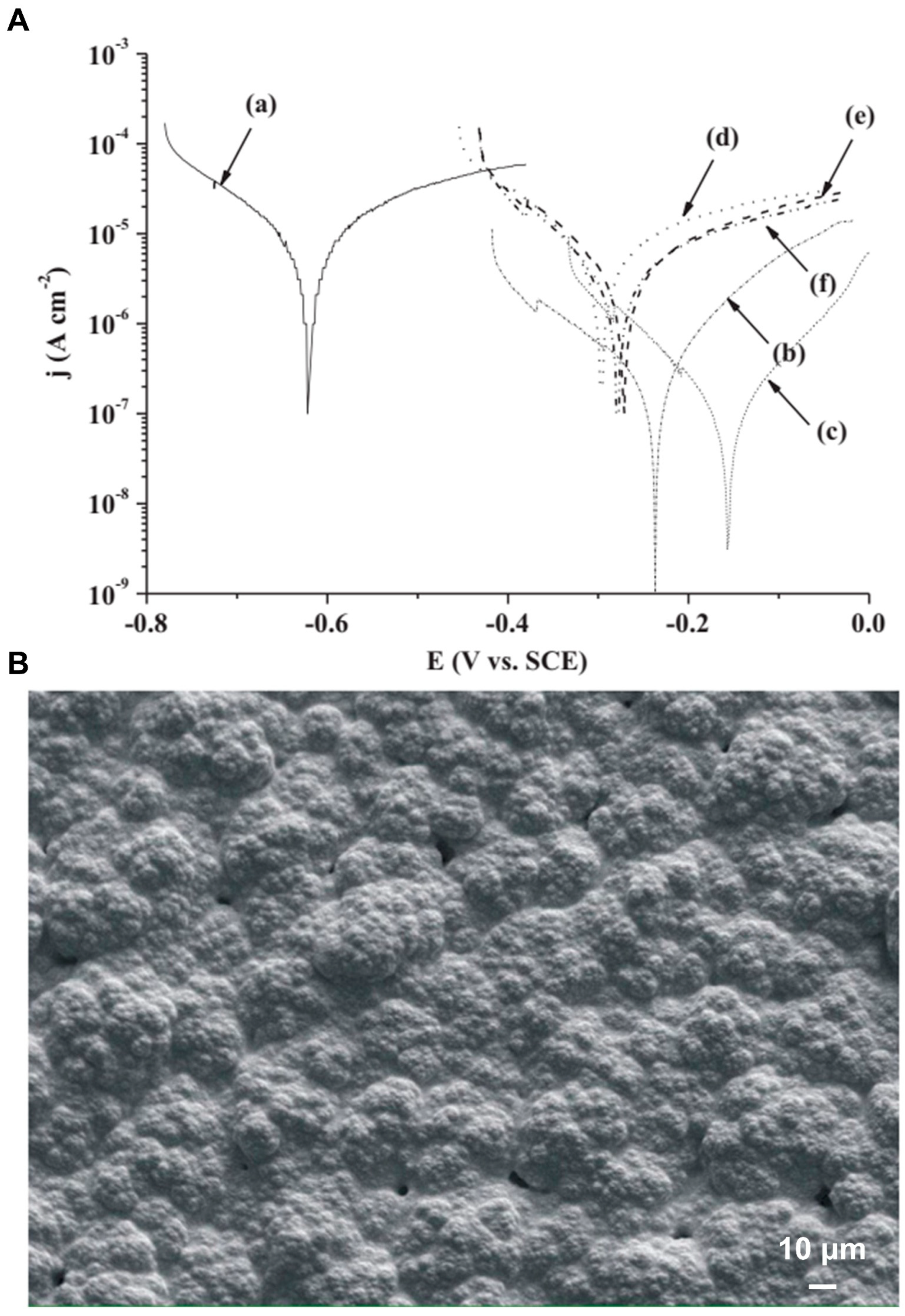
In the previous study, the anticorrosive property of various Ti-containing dental orthodontic wires (Ni-Ti, Ni-Ti-Cu, Ti-Mo-Zr-Sn, and Ti-Nb alloys) was examined in fluoride-containing artificial saliva by cyclic potentiodynamic polarization curve tests [88]. In order to mimic the fluoride level of commercial toothpaste, various NaF concentrations (0%, 0.2%, and 0.5%) were incorporated into the artificial saliva. As shown in Figure 3a, the anticorrosive properties of Ni-Ti, Ni-Ti-Cu, Ti-Mo-Zr-Sn, and Ti-Nb dental orthodontic wires were decreased with the increase of NaF concentration in acidic artificial saliva. The Ni-Ti and Ni-Ti-Cu wires showed a considerable anodic dissolution in the artificial saliva with the 0.5% NaF concentration (Figure 8A(a,b)), although the Ti-Mo-Zr-Sn and Ti-Nb wires still retained a passive zone (Figure 8A(c,d)). Among the evaluated Ti-containing dental orthodontic wires, the Ni-Ti and Ni-Ti-Cu wires with primarily TiO2 on the surface film were more vulnerable to fluoride-enhanced corrosion, whereas the Ti-Mo-Zr-Sn and Ti-Nb wires with MoO3/ZrO2/SnO and Nb2O5, respectively, together with TiO2 on the surface film, were less susceptible to pitting corrosion and fluoride-enhanced corrosion. They also exhibited scanning electron microscopy (SEM) images of the examined Ni-Ti, Ni-Ti-Cu, Ti-Mo-Zr-Sn, and Ti-Nb orthodontic wires both before (as-received situation) and after the corrosion experiments in fluoride-free artificial saliva. As indicated by arrows in Figure 8B(e,f), the pitting corrosion developed on Ni-Ti and Ni-Ti-Cu wires after corrosion experiments in fluoride-free artificial saliva. However, the Ti-Mo-Zr-Sn and Ti–-Nb wires had higher pitting corrosion resistance in the acidic fluoride-free artificial saliva as compared to the Ni-Ti and Ni-Ti-Cu wires according to Figure 8B, which demonstrated that the addition of MoO3/SnO/ZrO2 or Nb2O5 to the TiO2-containing surface film can enhance the pitting corrosion resistance of TiO2-based surface film in acidic artificial saliva. In other words, the passive oxide film properties of the surface of the wire had a major influence on the corrosion resistance of the evaluated Ti-containing orthodontic wires. The aforementioned findings may offer valuable guidance when selecting anticorrosive orthodontic wires in dentistry.

EPD

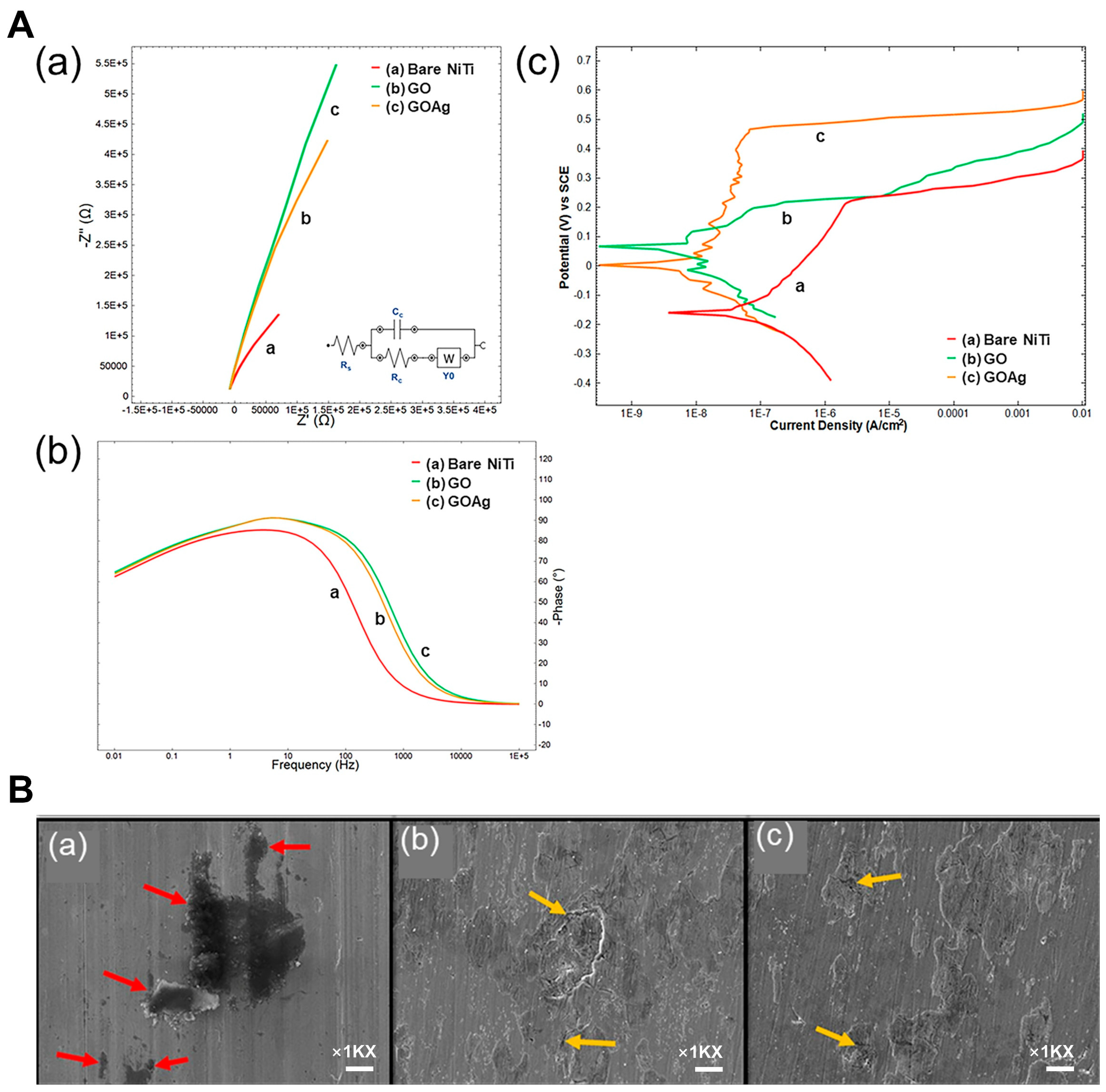
The anticorrosive properties of graphene oxide (GO) and GO/silver (GO/Ag) nanocomposite-treated NiTi alloy via EPD were investigated [22]. Potentiodynamic polarization curves and electrochemical impedance spectroscopy (EIS) tests were used to examine the anticorrosive properties of the bare NiTi and treated NiTi alloys in a 3.5% NaCl solution. As shown in Figure 9A(a,b), the Nyquist plots of the GO/Ag nanocomposite-treated NiTi samples exhibited the biggest diameters in the capacitive loop and a higher and broader phase angle among all samples, indicating the best corrosion resistance and lowest corrosion rates. In contrast to the bare NiTi alloy, the corrosion potential (Ecorr) of GO-coated NiTi and GO/Ag-coated NiTi samples was 0.031 V and 0.008 V, respectively, which both showed a positive shift. The corrosion current density (Icorr) was 0.017 µA and 0.002 µA, respectively, indicating 2 orders of magnitude reduction (Figure 9A(c) and Table 2). Both the GO-treated NiTi and the GO/Ag-treated NiTi alloys exhibited superior anticorrosive properties and greater protective efficiency than the bare one. The SEM images of the GO-coated alloy and GO/Ag-coated substrates showed the smooth and homogeneous morphology. Figure 9B(a–c) displays the SEM micrographs of the uncoated NiTi and coated NiTi alloy surface after the potentiodynamic polarization curve tests. As demonstrated in Figure 9B(a), the bare NiTi showed pitting corrosion due to the attack of chloride ions in 3.5% NaCl solution. However, as shown in Figure 9B(b,c), the GO and GO/Ag-coated NiTi alloys showed corrosion resistance since pitting corrosion did not occur, with only a few small regions of surface deterioration after being attacked by chloride ions. The electrochemical impedance spectroscopy (EIS) parameters of the bare NiTi alloy, GO-coated NiTi, and GO/Ag-coated NiTi are shown in Table 3. The corrosion mechanism of NiTi is attributed to the anodic oxidation, which takes place at the working electrode along with the reduction process in the cathode, which utilizes the electrons released during the oxidation of Ni and Ti. Anodic and cathodic reactions on the electrodes are given below.
At the cathode: reduction of O2 as shown in Equations (1) and (2).
O 2 + 2 H 2 O + 2 e 2 O H + 2 O H
O H + e O H
At the anode: formation of corrosion products as shown in Equations (3)–(6).
3 N i + 6 O H 3 N i O H 2 + 6 e
N i O H 2 N i O · H 2 O
T i + 4 O H T i H 4 O 4 + 4 e
T i H 4 O 4 T i O 2 · 2 H 2 O
The GO-coated NiTi and GO/Ag-coated NiTi restrict either the anodic oxidation or prevent the contact of OH. It is possible to inhibit the corrosion of NiTi.

Ion Plating

Ti nitride (TiN) coating via ion plating is frequently employed in dental applications due to its high hardness and wear resistance. Li et al. [94] prepared a TiN/TiAlN multilayer coating by ion source enhanced hybrid arc ion plating. As compared to the control sample, the TiN/TiAlN multilayer coating has the lowest Icorr, highest Ecorr, and protective efficiency. The corrosion resistance of TiN-coated stainless steel and NiTi orthodontic wires produced through ion plating was reported [95]. In comparison to TiN-coated orthodontic wires, uncoated stainless steel and NiTi wires displayed significant pitting corrosion after anodic polarization measurements in 0.9% NaCl solution. The electrochemical corrosion analysis demonstrated that TiN coating via ion plating enhances the anticorrosive property of orthodontic wires, which are frequently utilized in orthodontic treatment.

4.1.2. Dental Implants

Chemical Vapor Deposition

The dental implants can be in direct contact with the adverse oral environment in the oral cavity. The interactions between the external dental implants and host tissue have a major effect on the stability and longevity of the implants over time. When the implants are exposed to body fluids in vivo, excellent surface properties, especially for corrosion resistance, are desirable [96]. Malhotra et al. [97] produced graphene coating by chemical vapor deposition on Ti6Al4V grade 5 and 23 disks (Gp5 and Gp23, respectively) and exposed them to a corrosive environment with 0.5M NaCl solution with the addition of 2 ppm fluoride (pH 2.0) for up to 30 days. In comparison to the untreated grades 5 and 23 disks (C5 and C23, respectively), they found that there is a positive shift in the open circuit potential (OCP) for the Gp5 and Gp23 at all immersion points. The corresponding Icorr in the Gp5 and Gp23 was decreased, accompanied by a relatively positive shift in Ecorr. The graphene-treated Ti6Al4V was thermodynamically stable, more noble and resistant to corrosion than the control sample, according to the OCP and Ecorr, and the decrease in Icorr at all periods. Furthermore, the corrosion rate of the graphene-treated Ti6Al4V alloys was extremely small and consistent (0.001 mm/y), while those of C5 and C23 reached up to 16 and 5 times after 14 d (~0.091 mm/y), respectively. The research provides a new opportunity for graphene as an anticorrosive coating for dental-implanted devices and metal biomedical alloys.
To enhance the surface characteristics and anticorrosive property of Ti-13Nb-13Zr alloy, it has been reported that Singh et al. [98] fabricated iron oxide (Fe3O4) composite-coated Ti-13Nb-13Zr alloy that included hydroxyapatite (HA) and chitosan (CS), as shown in Figure 10A. The electrophoretic deposition approach (EPD) has been used to create pure HA and composites with various percentages of Fe3O4 (1, 3, and 5 wt%). In comparison to HA-3 wt% Fe3O4 and HA-5 wt% Fe3O4 composite coatings, the HA-1 wt% Fe3O4 coating displayed the superior resistance to corrosion with the smallest Icorr of 5.90 × 10⁻¹¹ µA and the largest Ecorr of −0.309 mV in a Ringer’s solution, as shown in Figure 10B and Table 4. Therefore, it is suggested that the HA-CS composite coating combined with Fe3O4 is an alternative treatment for Ti-based alloys in biological fields like dental and orthopedic applications.

Sol–Gel Method

Chellapa et al. [43] fabricated a composite coating containing SiO2 and ZnO on a Ti-6Al-4V sample by a low thermal volatilization sol–gel method to handle silica and zinc oxide precursors at low temperatures. The corrosion resistance of the coating was assessed through the use of Tafel curves and EIS tests. The electrochemical results indicated that the composite coating has better corrosion resistance than the untreated Ti6Al4V sample. The result suggested that the composite coating could be helpful in biomedical devices, particularly in dental and knee joint implants.

Thermal Oxidation

Zhang et al. [99] thermally oxidized the Zr-20Nb-3Ti alloy with a low elastic modulus at 600 °C for 1.25 h to acquire the compact oxide layer to enhance their resistance to wear and corrosion as screws for a dental implant. The electrochemical corrosion measurement revealed that the Icorr of the biomedical Zr-20Nb-3Ti sample has a smaller order of magnitude and its Ecorr shifts from −0.45992 V to −0.35319 V, as compared to those of the untreated one. The sharp decrease in corrosion rate (from 1.94 μm/year to 0.0597 μm/year) demonstrated the increase in anticorrosive property of the Zr-20Nb-3Ti alloy in artificial saliva via thermal oxidation. In conclusion, oxidized Zr-20Nb-3Ti alloy is considered a suitable candidate for a dental implant.

Acid Etching

Sun et al. [100] produced a bio-functional porous surface coating with structures at the nano-to submicro scale by sandblasting, acid etching, and NaOH leaching. The extracellular matrix (ECM)-like structure coating comprises a porous TiO2 outer layer with a thickness of 150–200 nm and a dense TiO2 inner layer with a thickness of 50–100 nm. The inner layer significantly improved corrosion resistance, as illustrated by an increase in Ecorr, decreases in corrosion rate, and passive current in simulated blood plasma (SBP). The proposed surface treatment method has considerable development potential for the fabrication of dental and orthopedic implants.

4.2. Orthopedic Applications

The aging of the worldwide population is accelerating the increase in bone-associated illnesses. Osteoporosis is a significant health problem affecting over 200 million individuals around the world, requiring the application of excellent orthopedic prostheses for the replacement and regeneration of hard tissue [101,102,103,104]. Ti alloys could replace hard tissues due to their superior chemical stability and adequate strength, which can successfully avoid the stress-shielding effect. As a result, Ti alloy is considered to be the first candidate for orthopedic implants [24,105,106].

4.2.1. Sol–Gel Method and EPD

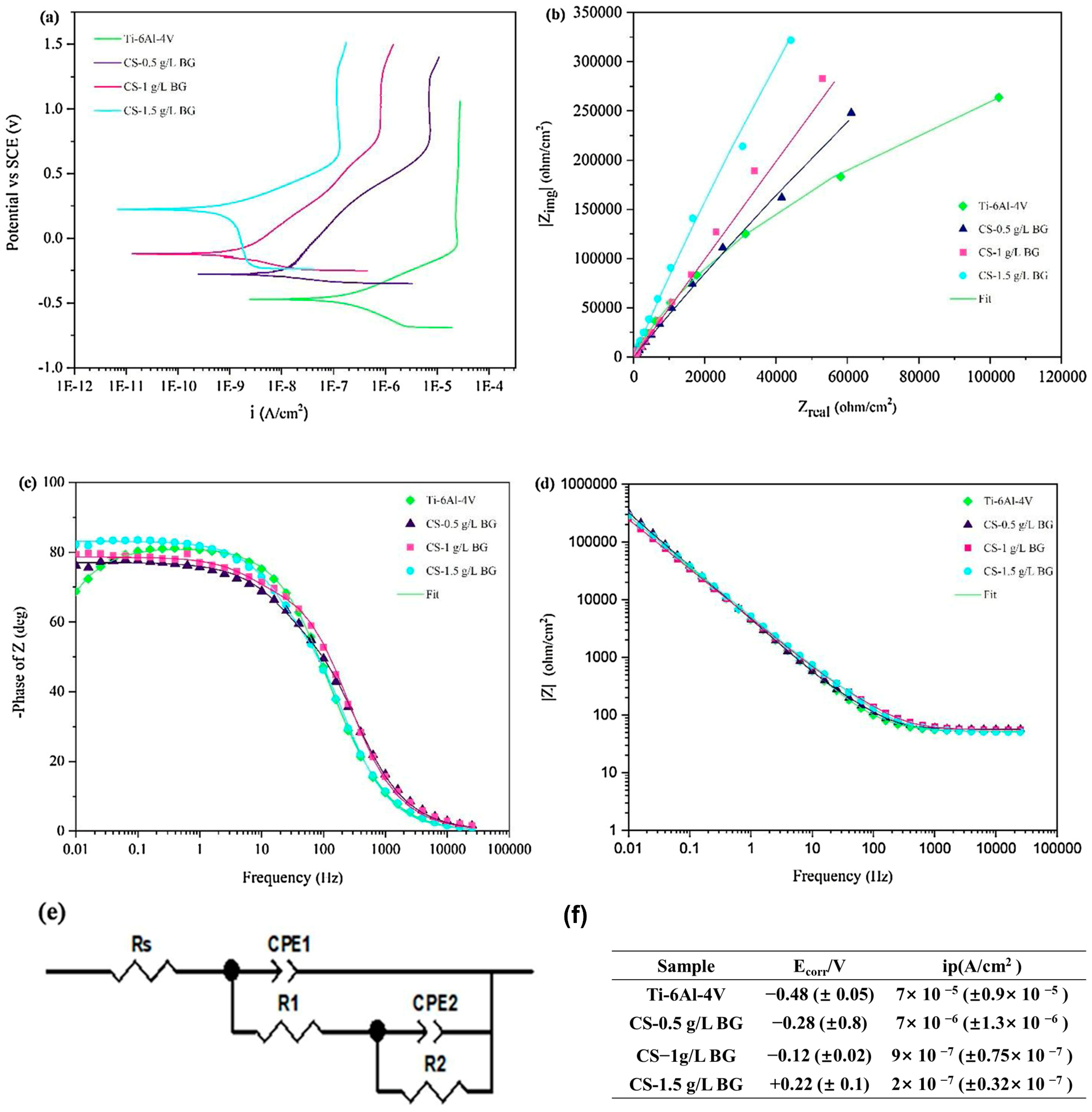
In the previous study, Mahlooji et al. [23] fabricated Chitosan–Bioactive glass (CS-BG) nanocomposite coatings on the Ti-6Al-4V alloy to examine the impact of the BG concentration on the bio-corrosion, biological activity, and wetting behavior, etc., Sol–gel technique was used to synthesize BG nanoparticles first. In addition, three nanocomposite coatings with various BG concentrations (0.5, 1, and 1.5 g/L) were produced by cathodic EPD. As shown in Figure 11a, the CS-BG coating, including 1.5 g/L BG, indicated the highest Ecorr of 0.22 V and the lowest passivation current density (ip) of 2 × 10⁻⁷ µA, which effectively enhanced the corrosion resistance of Ti-6Al-4V alloy. According to the EIS measurements, the impedance of all samples in the simulated body fluid (SBF) solution was enhanced in the following order of Ti-6Al-4V < CS-0.5 g/LBG < CS-1 g/L BG < CS-1.5 g/L BG (Figure 11). The Nyquist plot revealed that CS-1.5 g/L BG coating may be a suitable choice for greater corrosion resistance in orthopedic applications due to its largest diameter in capacity loop, which can be attributed to the more consistent distribution of BG and its greater thickness in comparison to the CS-0.5 g/L BG and CS-1 g/L BG coatings (Figure 11b). According to the EIS results (Table 5), the corrosion resistance of Ti-6Al-4V was significantly improved with the increase in BG content after EPD treatment. Therefore, the CS-1.5 g/L BG nanocomposite coating has considerable potential for utilization in orthopedics. de Lima Almeida et al. [107] synthesized siloxane-poly(hydroxyethyl methacrylate) composite coatings with the incorporation of calcium chloride and calcium sulfate by the sol–gel method, followed by dip-coating them on Ti6Al4V. The corrosion resistance of composite coatings with and without the incorporation of calcium salt was investigated by EIS measurements. The electrochemical examinations in the SBF solution (which were simulated) revealed that coatings with composite systems had improved barrier properties after immersion up to 96 h, as compared to uncoated Ti6Al4V samples. The composite coating has a regular morphology and uniform and consistent coverage that reduces surface irregularities, indicating that the thickness of the coating contributes to the corrosion resistance of the substrate. Therefore, these composite coatings demonstrated promising properties that are suitable for anticorrosive applications in biomaterials. Hameed et al. [108] and El et al. [109] demonstrated that the HA coating on Ti alloy via plasma spraying and sol–gel process has a protective impact, which results in superior anticorrosive properties. Nevertheless, the HA-coated Ti alloy has considerable disadvantages, including poor mechanical characteristics [110,111], which causes the coating to crack and tear off on the Ti6Al4V substrate.

4.2.2. Magnetron Sputtering and MAO

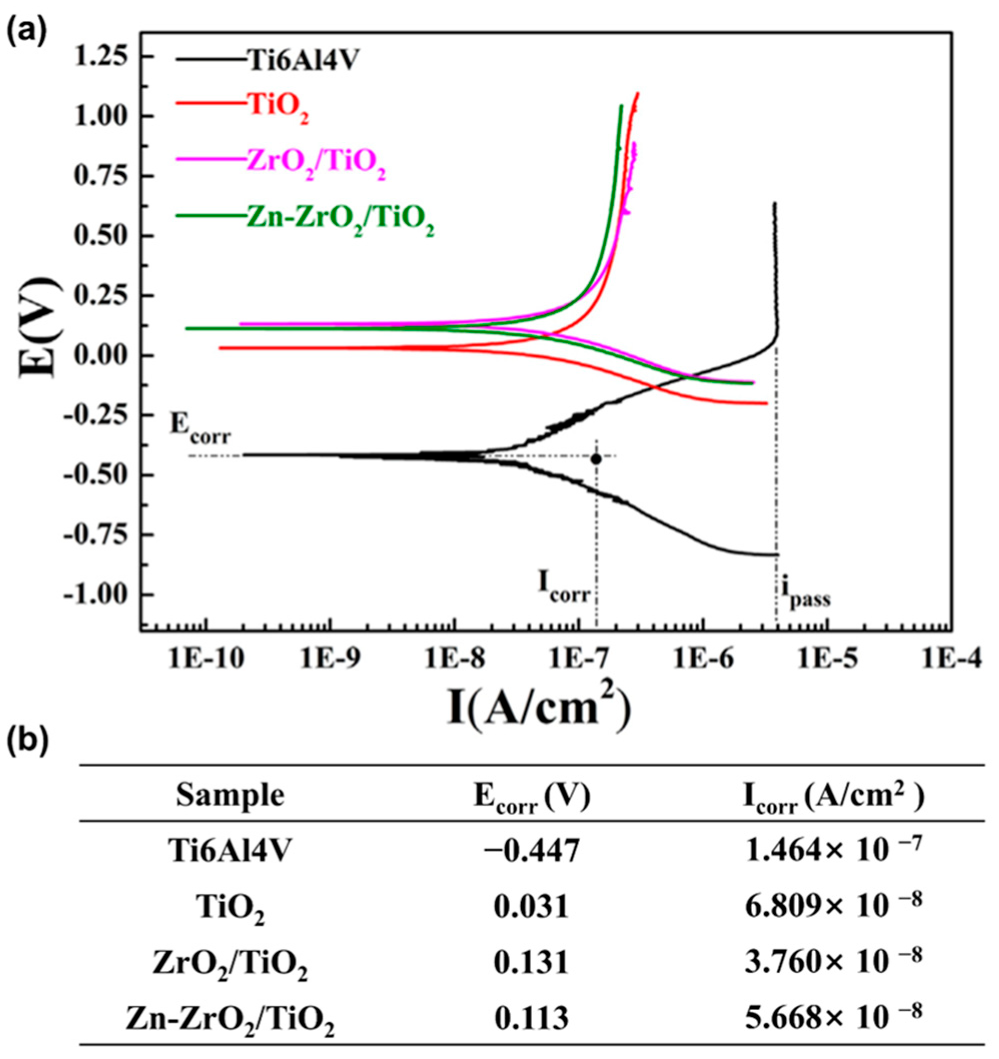
Wang et al. [44] fabricated Zn-doped ZrO2/TiO2 coatings (Zn-ZrO2/TiO2) on Ti6Al4V alloy through magnetron sputtering in combination with MAO. As shown in Figure 12a,b, the MAO coatings have a larger Ecorr in comparison to the bare Ti6Al4V alloy. There is a decreasing trend for Icorr of Ti6Al4V, TiO2, Zn-ZrO2/TiO2, and ZrO2/TiO2 in the SBF. The above results demonstrated that the MAO coatings can significantly reduce the corrosion rate of the Ti6Al4V substrate. Thus, it is anticipated that Ti alloy implants can be coated with Zn-ZrO2/TiO2 because of their better corrosion resistance, superior antibacterial properties, and great biocompatibility in the fields of orthopedics and dentistry. Zaveri et al. [39] evaluated the corrosion behaviors of the TiO2 nanoparticles coated Ti6Al4V implant subjected to three simulated biofluids, i.e., NaCl solution, Hanks’ solution, and Cigada solution, by electrochemical measurements and SEM. The Tafel curves showed that the bare Ti6Al4V has more negative Ecorr and higher Icorr than TiO2 nanoparticle-coated Ti6Al4V in the three different simulated biofluids. In other words, the TiO2 nanoparticle-coated Ti6Al4V has better corrosion resistance as compared to the bare Ti6Al4V. Furthermore, the TiO2 nanoparticle coatings thickened the previously existing oxide film on the Ti6Al4V, improving the corrosion resistance of the bioimplant.
A Zinc (Zn)-doped calcium phosphate (CaP) coating was prepared on the Ti6Al4V substrate via the MAO technique [112]. Electrochemical tests in SBF were performed to evaluate corrosion resistance. As compared to uncoated Ti6Al4V alloy, the Zn-doped CaP coating revealed lower Icorr and higher polarization resistance (Rp), indicating superior corrosion resistance. The Zn-doped CaP coating on the Ti6Al4V implants significantly improved corrosion resistance, reducing the implant failure risks. Prosolov et al. [113] proposed 3D porous calcium phosphate CaP coatings on Ti substrates modified with poly(lactic-co-glycolic acid) (PLGA). The CaP coating on Ti substrate was created via the ultrasonic-assisted micro-arc oxidation (UMAO) method, and then modified with PLGA by a dip coating process at concentrations of 5%, 8%, and 10%, respectively. Corrosion resistance was evaluated through gravimetric and electrochemical methods in 0.9% NaCl and PBS solutions, revealing that PLGA dramatically reduced corrosion rates, with the corrosion current decreasing by two orders of magnitude even for the 5% PLGA/CaP/Ti coating. Higher PLGA concentrations offered even improved anticorrosive properties. This study highlights the potential of employing CaP- and PLGA-modified coating to extend the life and performance of orthopedic implants, addressing a major challenge in biomaterials for medical applications.

4.2.3. Hydrothermal Process

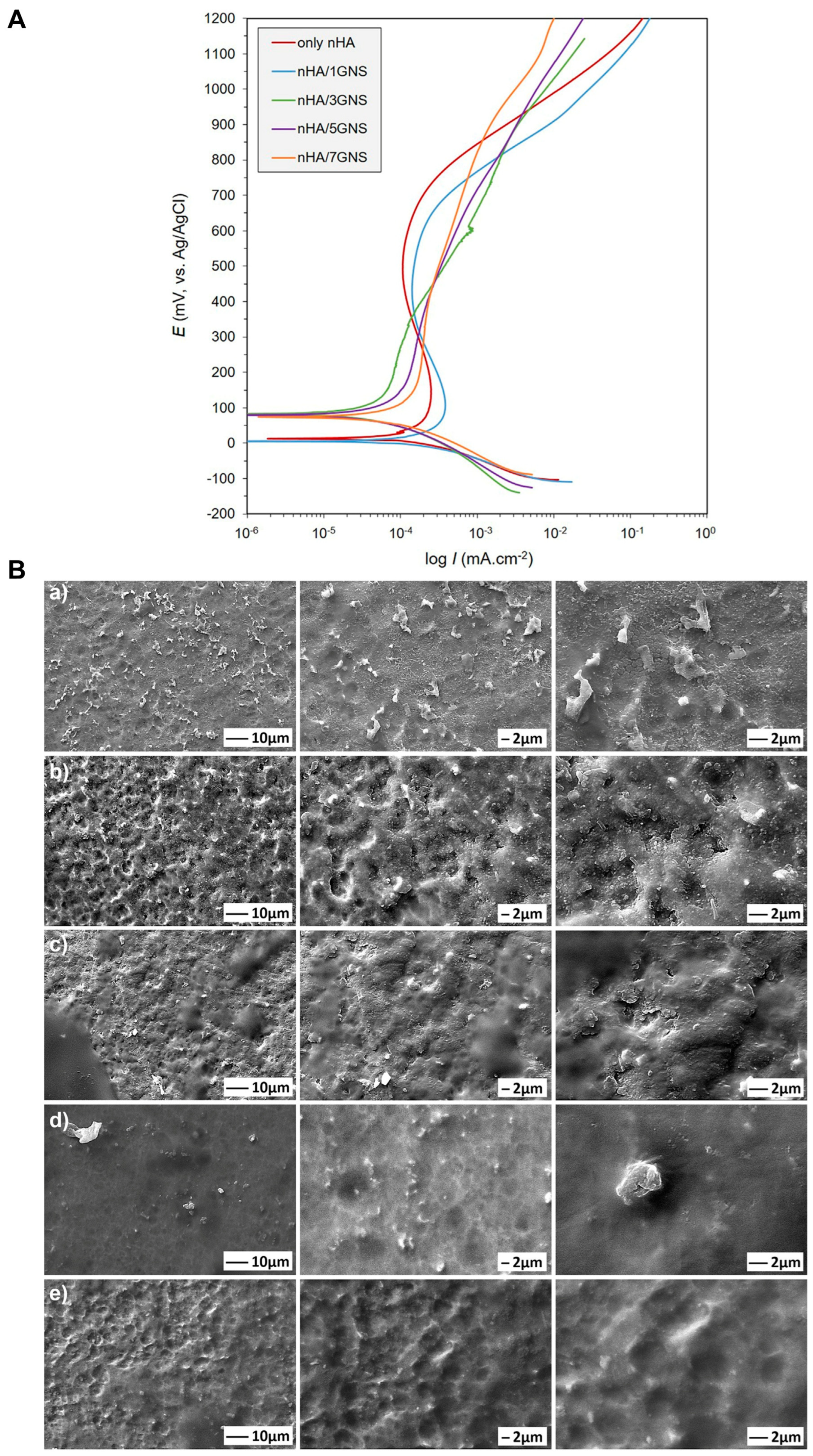
It is stated that Yigit et al. [46] synthesized nanoHA/graphene nanosheet (nHA/GNS) composite coating on Ti6Al4V surface via hydrothermal process. The potentiodynamic polarization curves of the only nHA and nHA/1GNS coatings are extremely similar (Figure 13A). However, the Icorr of the nHA/1GNS coating is larger than that of only the nHA coating because of the composite structure of the oxidized layer on the nHA/1GNS coating. Furthermore, the Ecorr of the nHA/GNS hybrid coatings with the larger addition of GNSs (>3 wt%) is higher than that of both only nHA and nHA/1GNS coatings, as shown in Figure 13A and Table 6. That is to say, nHA/3GNS and nHA/5GNS coatings were more anticorrosive as compared to other coatings in the SBF solution. The addition of 3 wt% GNS results in an even distribution of HA particles on the surface of the GNS layer, resulting in a crack-free nHA/GNS coating that covers the entire substrate surface. As shown in Figure 13B, only nHA and nHA/1GNS coated surfaces developed severe corrosion pits, particularly, while nHA/3GNS, nHA/5GNS, and nHA/7GNS composite coating surfaces obviously appeared to have a protective oxide layer. The nHA/GNS composite coating has enhanced the anticorrosive property of Ti6Al4V alloys in the SBF solution, which has potential applications in orthopedics.

4.2.4. Laser Treatment

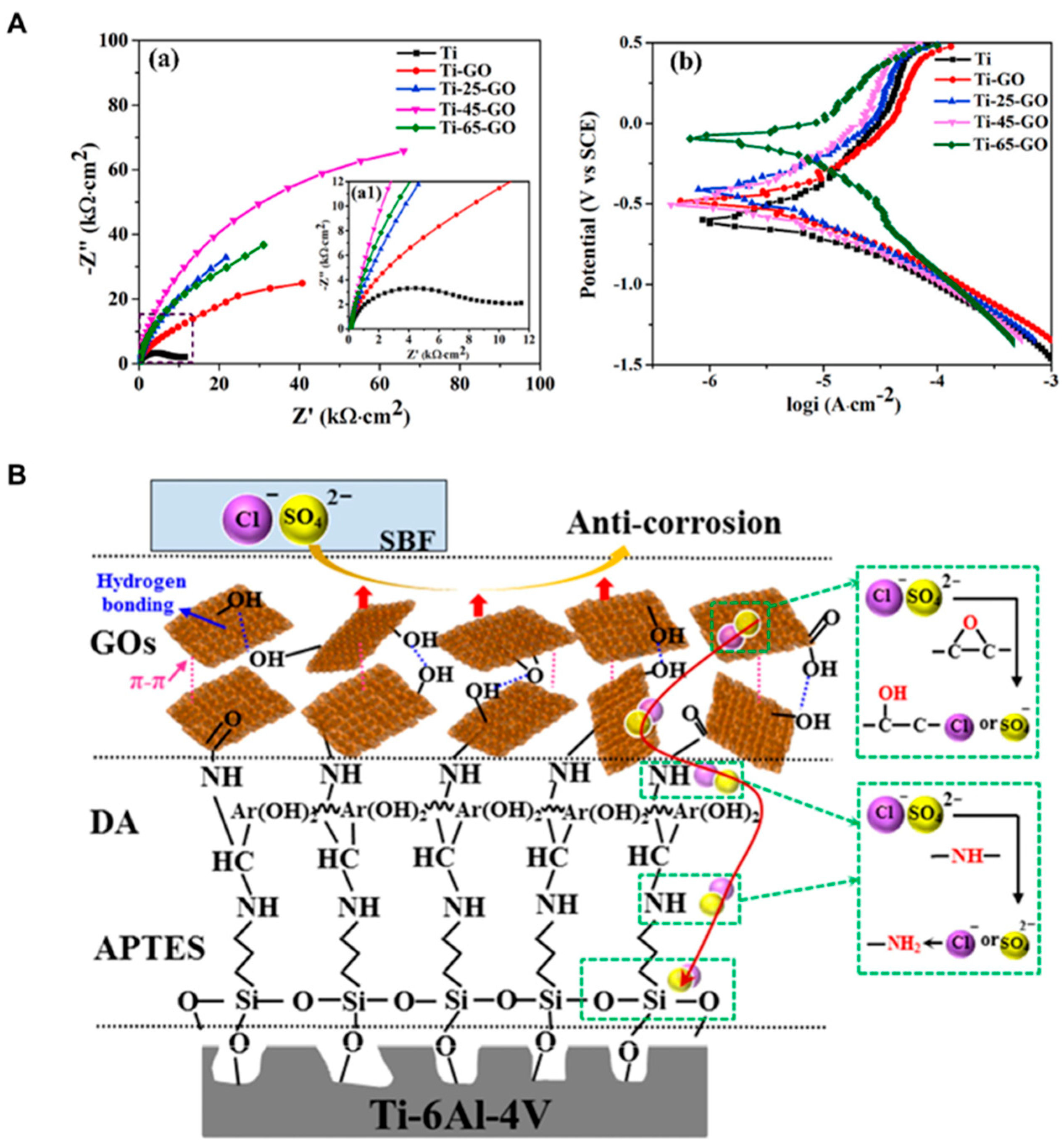
Wang et al. [47] fabricated microgrooves with different groove widths and graphene oxide (GO) coating on the Ti6Al4V alloy via laser treatment and chemical assembly, respectively. Based on the width (0, 25, 45, and 65 μm, respectively) of the microgrooves on the Ti6Al4V surfaces, these GO-coated coatings can be referred to as Ti-GO, Ti-25-GO, Ti-45-GO, and Ti-65-GO, respectively. The influence of microgroove width on the corrosion resistance and biotribological characteristics of Ti6Al4V alloys coated with GOs was thoroughly examined in SBF solution. The greater the curvature radius (R) of Nyquist plots, the better the corrosion resistance. The R of Ti-GO is noticeably higher than that of the Ti6Al4V alloy, as shown in Figure 14A(a1). After the Ti6Al4V alloys are processed via laser texturing, the R of GOs-treated Ti6Al4V with microgrooves indicates the following order of Ti-GO < Ti-25-GO < Ti-65-GO < Ti-45-GO, revealing that the Ti-45-GO sample has the greatest corrosion resistance, and the laser processing could enhance the anticorrosive property of the Ti6Al4V surface. Moreover, the Tafel curves show similar trends (Figure 14A(b) and Table 7), and the Ti-45-GO sample exhibits the lowest Icorr (1.425 × 10⁻⁷ µA), indicating its great corrosion resistance. The protective mechanisms of GO on the Cl and SO42− in SBF solution could be described in Figure 14B after applying onto the Ti-6Al-4V surface [114]. The GO sheets are randomly deposited over the Ti alloy surface and then linked to a certain thickness by hydrogen, π, and van der Waals bonds. It can be seen that these GOs could reduce direct contact with corrosive ions and delay the surface corrosion of Ti alloy. The above results indicated that combining a suitable microgroove of 45 μm width with GO coatings could dramatically improve the corrosion resistance of Ti6Al4V alloys because of the homogenization of the surface microstructure from the Ti-45 sample, and the protective impact of GO coatings has significant potential for biomedical applications.
Yang et al. [48] presented a novel approach to construct a superhydrophobic surface with a water contact angle (WCA) of 155.4° ± 0.9° and a water sliding angle (WSA) of 4.4° ± 1.1° on NiTi SMA by the combination of laser treatment and polydimethylsiloxane (PDMS) modification. The corrosion resistance of the superhydrophobic surface with regular grid patterns or grooves was evaluated in the SBF solution by using potentiodynamic polarization curves and EIS measurements. The Icorr is utilized to measure the corrosion rate and the efficiency of corrosion inhibition. In other words, a lower Icorr is considered to have better corrosion resistance. The potentiodynamic polarization curves showed that the superhydrophobic surface has lower Icorr values than the untreated NiTi SMA and purely ablated NiTi SMA by ns-laser. Therefore, the superhydrophobic surface samples exhibited superior corrosion resistance among the three untreated and treated NiTi SMA samples, indicating that creating superhydrophobic surfaces on the NiTi SMA by combining laser irradiation with PDMS modification can obviously improve corrosion resistance. In addition, the superhydrophobic surface samples will be developed on a scalpel surface for a medical device in this study.
Zhang et al. [115] modified the Ti6Al4V substrate using plasma nitriding and plasma-enhanced chemical vapor deposition (PN + PECVD), and laser remelting (LR) processes, respectively, to increase its wear resistance and anticorrosive property in vivo. The corrosion current of the bare Ti6Al4V alloy, LR, and PN + PECVD samples followed the order of PN + PECVD < LR < Ti6Al4V. That is to say, the corrosion resistance of Ti6Al4V, LR, and PN + PECVD samples is also in the order of Ti6Al4V < LR < PN + PECVD. As a result, it is believed that the PN + PECVD and LR processing could significantly improve the corrosion resistance of Ti6Al4V alloy in vivo. Hussein et al. [116] created a novel biomedical Ti-20Nb-13Zr at.% alloy (TNZ), which was treated with an Nd: YAG laser in a nitrogen environment to improve its anticorrosive property and surface characteristics. The corresponding corrosion resistance was examined via potentiodynamic polarization curves and EIS measurements in the SBF solution. The SEM observation showed that a thick and compact layer of TiN micro-/nanosized tiny particles was formed underneath the surface with a size of 9.1 μm. The electrochemical tests showed that the existence of tiny grains in the TiN film as a consequence of laser nitriding may significantly improve the protective ability of the laser-nitrided TNZ surfaces in the SBF solution. Moreover, both uncoated and coated Ti-20Nb-13Zr alloy demonstrated better anticorrosive properties than the commercially pure Ti and Ti6Al4V alloy. Laser nitriding has been shown to enhance the surface hardness and anticorrosive properties of different Ti alloys in a simulated body, which can provide a potential way to enhance the characteristics of biomaterials applied in orthopedics, implants, and dentistry.

4.2.5. Chemical Self-Assembly

Tian et al. [49] created HA coating and Fe3O4-coated HA (Fe3O4/HA) coating on the Ti6Al4V via chemical self-assembly, respectively. The corrosion performance was thoroughly assessed through an electrochemical analyzer in the SBF solution. As demonstrated in Figure 15a, Ti-HA and Ti-Fe3O4/HA have a higher R than untreated Ti6Al4V, indicating the smaller electrochemical reaction rate and improved corrosion resistance on the Ti-HA and Ti-Fe3O4/HA [117]. Furthermore, Ti-Fe3O4/HA displayed the highest R, demonstrating that the Fe3O4/HA coating has a beneficial shielding impact on the Ti6Al4V alloy. The impedance modulus Z (f = 0.01 Hz) showed the order of Fe3O4/HA > Ti-HA > Ti6Al4V (Figure 15b), whose changing trend is similar to that in Figure 15a,c. As shown in Figure 15c and Table 8, the Ti-Fe3O4/HA has the highest Ecorr of −324.60 mV and the lowest Icorr of 153.55 nA, which effectively improved the corrosion resistance of Ti6Al4V alloy. These findings exhibited that the Fe3O4/HA coating on the Ti6Al4V has superior corrosion resistance due to its capacity to shield the active ions in the SBF solution, which may explore application in the biomedical field. Ti-Cu alloy has drawn considerable interest in orthopedic and dental applications.

4.2.6. Laser Shock Peening (LSP)

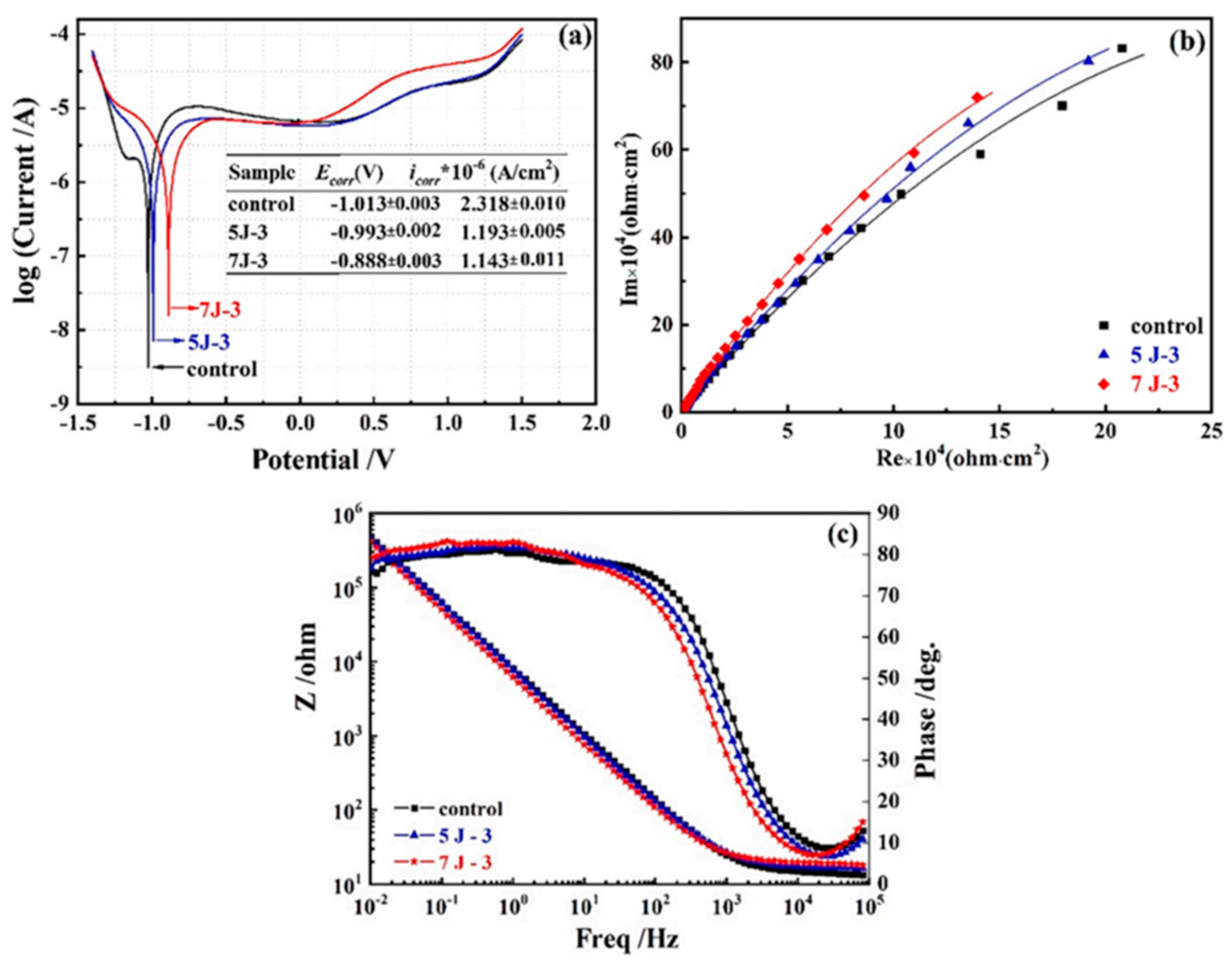
The present type of Cu has an essential effect on the corrosive property. The application of LSP is increased to enhance the characteristics, particularly the anticorrosive properties of various metallic components. Yang et al. [118] investigated the impacts of the LSP process on the phase composition, the surface grain size of Ti-3Cu alloy, and surface-associated characteristics like wettability and corrosion resistance to the SBF solution. The EIS fitting results are presented in Table 9. As shown in Figure 16a, the Ecorr for 5 J-3 (Ti-3Cu alloy after being impacted with the laser pulse energy of 5 J for 3 times) and 7 J-3 (Ti-3Cu alloy after being impacted with the laser pulse energy of 7 J for 3 times) turned toward a positive trend, and the Icorr was decreased dramatically as compared to the control sample (Ti-3Cu alloy), implying that the usage of LSP process can enhance the corrosion resistance of Ti-3Cu alloy. The 7 J-3 sample had a higher Ecorr and a lower Icorr as compared to the 5 J-3 sample, demonstrating that greater laser energy can lead to improved corrosion resistance. The 5 J-3 and 7 J-3 samples had evidently higher R than the bare Ti-3Cu alloy, revealing that the existence of Ti2Cu exposed to the LSP process enhances the anticorrosive property of Ti-3Cu alloy (Figure 16b). As shown in Figure 16c, the highest phase angle is approximately 80°, and the impedance modulus rises with a linear slope of around −1 when the frequency is changing from high frequency to low frequency, exhibiting the typical properties of capacitive behavior. The broad phase peak in the low frequency region between 10−1 and 102 Hz was apparent in the Bode phase plots, which is a typical property of the single-layer film. The above results exhibited that the Ti-3Cu alloy after the LSP process has better anticorrosion properties than the Ti-3Cu alloy. In conclusion, the LSP process with a larger laser energy density on Ti-3Cu alloy was recommended as a feasible method for enhancing the general characteristics of Ti-3Cu alloy, and thus preventing infections and failure in implants as a medical device.

4.2.7. EPD and Magnetron Sputtering

According to the previous work, Mthisi et al. [45] fabricated laser cladding Ti-Al2O3 coatings with different Al2O3 percentages (5, 8, and 10 wt%) to enhance the anticorrosive property and hardness of Ti6Al4V by using the electrophoretic deposition technique and magnetron sputtering. A potentiostat was used to evaluate the corrosion of the acquired coatings in Hanks’ solution. After applying various Ti-Al2O3 coatings on the Ti6Al4V via laser deposition, the anticorrosive property of Ti6Al4V was significantly increased. Owing to the limited porosity within the coating layer, Ti-5 wt% Al2O3 coating resulted in an ideal decrease of 81% in corrosion rate and a rise of 709% in polarization resistance over the substrate. It is recognized that producing Ti-Al2O3 on Ti6Al4V using a laser can increase the anticorrosive property and hardness of the Ti6Al4V substrate. Liu et al. [40] fabricated a new coating (F-DLC/Ti) by depositing diamond-like carbon (DLC) on Ti6Al4V substrates and then grafting 1H,1H,2H,2H-perfluorodecyl acrylate on DLC/Ti that formed a thick monolayer, as shown in Figure 17A. Figure 17B and Table 10 showed that F-DLC/Ti has a larger Ecorr of 0.0399 V and a smaller Icorr of 82.025 nA, exhibiting excellent corrosion resistance. The steady DLC coating and hydrophobic grafted monolayer function by synergistic effect to significantly prevent corrosion on the Ti6Al4V alloy in the SBF solution. It is expected to offer a promising and efficient method for enhancing current DLC-treated and other bioimplant materials because DLC could be deposited on the majority of substrate surfaces and photochemical grafting is likely to have an extensive variety of applications.

4.2.8. Pre-Anodization (PA) and MAO Techniques

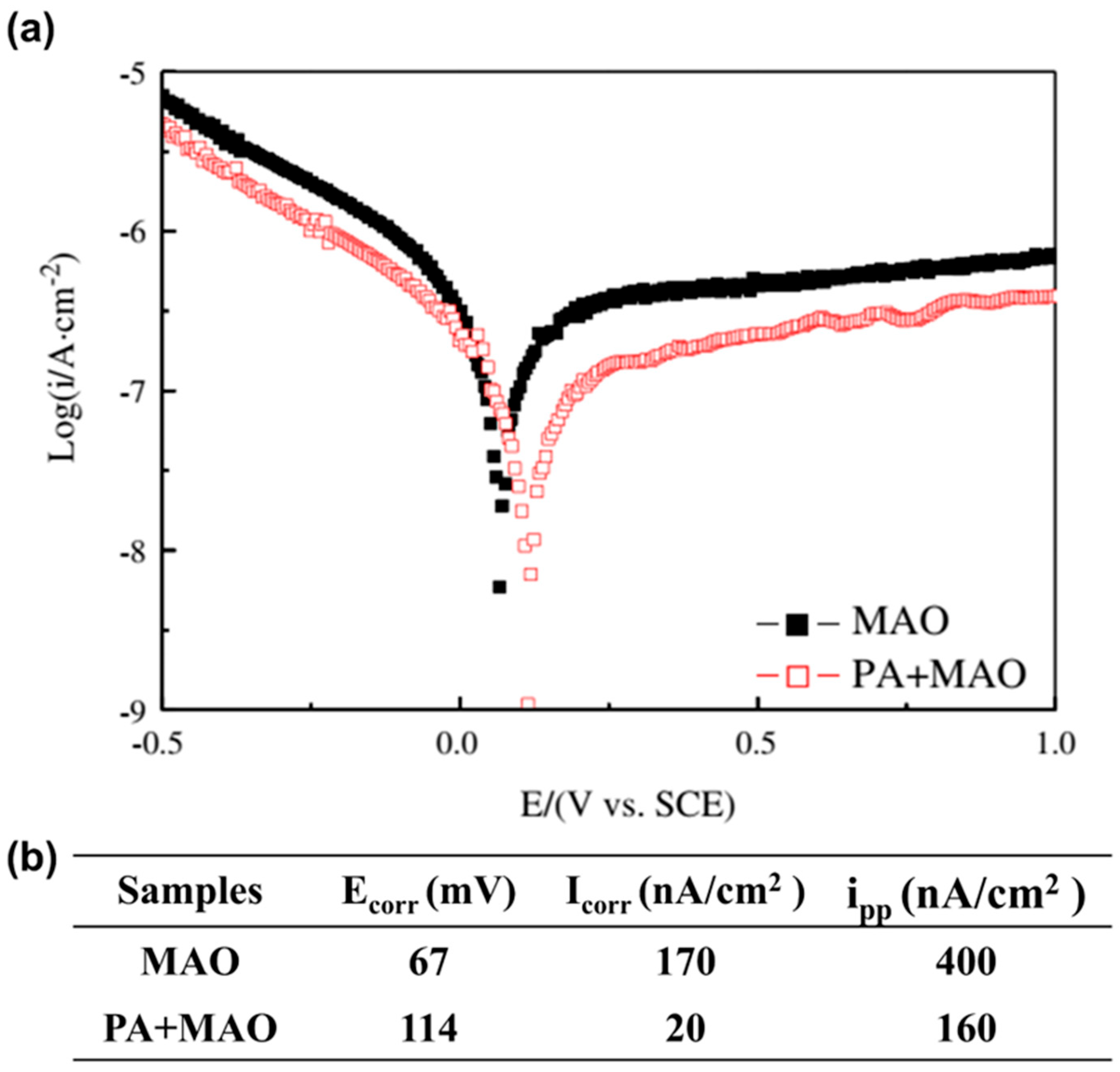
It is reported that a porous TiO2 coating was fabricated on biomedical β Ti alloy via PA and MAO techniques [41]. In comparison to the non-pre-anodized Ti alloy, the pre-anodized Ti alloy exhibited a greater Ecorr and a lower Icorr (Figure 18), which was ascribed to the thick and adhesive inner layer produced via pre-anodization to support the passive treatment of the Ti surface. It is important for orthopedic implants used in physiological environments containing fluoride or chloride. In addition to enhancing the biological activity of the Ti alloy implants, the pre-anodized MAO coating also makes them more resistant to corrosion during the crucial synostosis period. However, further research on improving pre-anodization parameters is desired to enhance the continuous anticorrosion property of MAO coating.

4.2.9. Plasma Immersion Ion Implantation and Deposition (PIII&D)

Moreover, NiTi shape memory alloy (SMA) is possibly appropriate for orthopedic implants because of its super-elasticity and the shape memory effect. Poon et al. [119] improved the anticorrosion and other characteristics of NiTi alloy by PIII&D. The corrosion in SBF experiments showed that either a direct carbon PIII or an amorphous PIII&D coating could significantly increase corrosion resistance and prevent Ni from diffusing out of the materials. Hendry et al. [120] deposited TiN, zirconium nitride (ZrN), or amorphous carbon (AC) coatings with a thickness of 3–4 μm onto the Ti6Al4V orthopedic implant surfaces by physical vapor deposition (PVD) cathodic arc evaporation technologies. They employed a kind of original fretting equipment to assess the fretting corrosion resistance of treated and untreated Ti6Al4V samples in Hanks’ solution. The results indicated that the TiN-treated alloy has enhanced anticorrosive properties in contrast to the TiN-free coated alloy, which displays significant corrosion damage.

4.2.10. Powder Immersion Reaction Assisted Coating (PIRAC) Technique

Starosvetsky et al. [121] prepared NiTi SMA by the PIRAC technique to change its surface characteristics. After PIRAC anneals at 900 and 1000 °C, two-layer TiN/Ti2Ni coatings with toughness were fabricated on the NiTi SMA surface. It was discovered that the PIRAC process significantly increased the anticorrosive property of NiTi SMA in the Ringer’s solution. The NiTi SMA samples that PIRAC nitrided at 1000 °C, 1 h up to 1.1 V showed no pitting corrosion in comparison to the uncoated NiTi SMA samples. The coated samples also had extremely low metal ion release rates and rather low anodic currents in the passive zone. The results of this research show that the PIRAC nitriding process can enhance the in vivo behavior of NiTi SMA exposed to human tissue. Pohrelyuk et al. [122] created nitride coatings on the Ti6Al4V surface of different constituents, thickness, and surface properties via varying nitrogen partial pressure from 1 to 105 Pa and nitriding temperature from 850 to 900 °C, and examined their corrosion behavior in Ringer’s solution at 36 and 40 °C. The studies revealed that the content of TiN in the nitride region of coating I (nitrogen partial pressure of 1 Pa at nitriding temperature of 850 °C), coating II (nitrogen partial pressure of 105 Pa at nitriding temperature of 850 °C) and coating III (nitrogen partial pressure of 105 Pa at nitriding temperature of 900 °C) is only 4%, 67% and 58%. TiN is dominant in the nitride region of coatings II and III, while Ti2N is common in the nitride region of coating I. The potentiodynamic polarization results demonstrated that nitride coatings enhanced the corrosion resistance of the alloy at two solution temperatures as compared to the untreated Ti6Al4V alloy (Figure 19A,B, and Table 11). With an increase in TiN phase composition in the nitride layer, the anticorrosive property of the Ti6Al4V alloy is enhanced. The ability of the nitride coating has a considerable impact on the corrosion resistance of the Ti6Al4V alloy with the nitriding temperature rise from 36 to 40 °C.

4.2.11. AIP

Tian et al. [38] deposited TiN and TiN/Ti films on Ti6Al4V substrate through AIP. The corrosion behavior of TiN and TiN/Ti coatings was evaluated by open circuit potential, potentiodynamic polarization curves, and EIS tests in Hanks’ balanced salt solution (HBSS). After the potentiodynamic polarization measurements, the surface characteristics of the corroded samples were observed by SEM. The electrochemical measurements exhibited that the TiN and the TiN/Ti coatings could effectively protect the Ti6Al4V substrate, and the anticorrosive property of the TiN/Ti composite coating was higher than that of the TiN coating. As compared to the untreated Ti6Al4V alloy, no pitting corrosion occurred on the TiN film deposited on the Ti6Al4V substrate after potentiodynamic polarization measurement, which illustrated that the TiN film coated by AIP offers adequate protection for the orthopedic Ti6Al4V substrate. In the previous study, Manhabosco et al. [123] evaluated the corrosion resistance of plasma nitrided Ti6Al4V samples in phosphate-buffered saline (PBS) solution through polarization plots and EIS tests. The Ecorr of the Ti6Al4V increased after nitriding treatment, and Icorr was lower than the bare Ti6Al4V, revealing an improvement of corrosion resistance, which was likely ascribed to the development of the composite layer made up of TiN and Ti2N, and/or to the enhancement of the metallic matrix in nitrogen. All of the characteristics indicated that the nitrided Ti6Al4V samples have greater corrosion resistance in contrast to the bare Ti6Al4V surface.

4.3. Cardiovascular Applications

In 2017, it was reported that there were approximately 1.8 cases of congenital cardiac illness, such as atrial septal defect, ventricular septal defect, and patent foramen ovale per 100 live births, a 4.2% growth since 1990 [124]. External and internal biomedical devices, such as cardiovascular stents, are frequently employed by usage of NiTi alloy because of its special shape memory effect and super-elasticity [125].
Kong et al. [126] investigated the permanent anticorrosive performance of the NiTi alloy in laboratory and biological conditions. In total, 64 Amplatzer® Septal Occluders of different sizes, randomly chosen from 240 devices that had been subjected to the NaCl solution for 14 months, were examined according to SEM. The tested devices looked like new, without corrosion or wire fractures, in vitro, after cleaning the NaCl solution. SEM images showed that both surfaces of the tested and control wires are identical, illustrating the complete Ti oxidized layer. Additionally, they used SEM to examine three Amplatzer® devices explanted in dogs after implantation for 18 to 36 months, revealing a similar sign that a complete Ti oxidized layer without corrosion in vivo. Therefore, Nitinol wires applied in Amplatzer® septal occlusion devices exhibited anticorrosive properties both in vitro and in vivo when subjected to physiological saline solution, demonstrating that NiTi is an inert, anticorrosive alloy. Stacey et al. [127] fabricated 4 groups of Nitinol stents by utilizing various treatment ways (including salt pot (SP), mechanical polish (MP), air furnace (AF), and oxidized tube (OT), respectively) to develop different surface characteristics. After 6 months, the stents were removed from the iliac arteries of minipigs for corrosion examination. It was found that Nitinol stent samples with breakdown potentials (Eb) smaller than 200 mV showed corrosion in vivo after 6 months of implantation. On the other hand, Nitinol stents with pits beginning at potentials greater than roughly 600 mV exhibited no evidence of corrosion in vivo after 6 months of implant surgery. These results show that functional corrosion measurements, in combination with a thorough comprehension of the surface properties of Nitinol biomedical devices, could offer insight into corrosion resistance in vivo.
In 2014, Flamini et al. [42] modified the Nitinol surface composed of a self-assembled film of alkylsilane compounds (propyltrichlorosilane (C3H7SiCl3, C3) or octadecyltrichlorosilane (C18H37SiCl3, C18) and polypyrrole (PPy) with the addition of sodium bis(2-ethylhexyl) sulfosuccinate (AOT). In this study, the corrosion behavior of different systems, including uncoated NiTi alloy (NiTi), NiTi alloy treated with PPy film (NiTi/PPy), NiTi alloy coated by both self-assembled films of alkylsilane (NiTi/C3 or NiTi/C18) and PPy film (NiTi/C3/PPy or NiTi/C18/PPy), was examined. The oxide film on NiTi alloy, which serves as a physical shield between the NiTi alloy and the aqueous media, causes the samples modified with the self-assembled film of alkylsilane to exhibit 2 orders of magnitude in Icorr and show higher Ecorr than the unmodified NiTi alloy (Figure 20A(a–c)). Despite a change in a slight magnitude, the silane/PPy film also results in a positive shift in the Ecorr (Figure 20A(e,f)). Meanwhile, the Icorr is 1 order of magnitude lower than that of the untreated NiTi alloy, while 1 order of magnitude higher than that of NiTi/C3 or NiTi/C18 due to the galvanic reaction between the polymer and the substrate that produced oxidation of the substrate and the reduction in the polymer. Additionally, the NiTi/C18/PPy film also showed no evidence of pitting corrosion after 15 h of polarization at 0.65 V in 0.15 M NaCl solution (Figure 20B). The resistance to pitting corrosion of the substrate in NaCl solution is enhanced by the conjunction of alkylsilanes and the existence of AOT trapped within the PPy films. The improved corrosion resistance of NiTi alloy makes it an attractive alternative as an anticorrosive treatment in a variety of biomedical applications, e.g., cardiovascular, orthodontics, orthopedics, and urology.

5. Conclusions and Future Prospects

In this review, we first describe the corrosion types. In the second part, the effects of corrosion on the human body were discussed. The inclusion and exclusion criteria during the selection of literature are described in Section 3. In the last section, we summarized and discussed the current research progress of coatings fabricated onto Ti and/or Ti alloy surfaces in biomaterials for medical applications involving dental, orthopedic, and cardiovascular implants for anticorrosive applications.
More importantly, the review described coatings fabricated onto Ti and/or Ti alloy surfaces based on different deposition methods, offering a comprehensive solution to the challenges associated with the corrosion susceptibility of titanium implants. By enhancing the corrosion resistance of Ti and its alloys, these coatings hold significant promise for improving success rates and longevity of dental, orthopedic, and cardiovascular implants. This review is expected to be a valuable reference for anticorrosive applications in the field of biomaterials. Future research should concentrate on in vivo evaluations and long-term clinical trials to further validate these results and explore the possibility for broad clinical applications.

Author Contributions

Conceptualization, S.Z.; methodology, Q.R.; software, Q.R.; validation, S.Z.; formal analysis, Q.R., J.Z., Y.C. and Y.Y.; investigation, Q.R., X.C., D.L., R.Z., A.L. and Y.L.; resources, S.Z.; data curation, S.Z. and Q.R.; writing—original draft preparation, S.Z. and Q.R.; writing—review and editing, S.Z. and Q.R.; visualization, S.Z. and Q.R.; supervision, S.Z.; project administration, S.Z.; funding acquisition, S.Z. All authors have read and agreed to the published version of the manuscript.

Funding

This work was supported by National Natural Science Foundation of China (grant no. 82101070); the Key Research and Development Program of Anhui Province (grant no. 2022e07020051); College Young and Middle-Aged Teachers Training Action Project of Anhui Education Department (grant no. YQZD2023024); Research Fund of Anhui Institute of Translational Medicine (grant no. 2021zhyxC51); Natural Science Foundation of Anhui Province (grant no. 2008085QH374); Anhui Medical University Basic and Clinical Collaborative Research Enhancement Program (grant no. 2019xkjT019); Grants for Scientific Research of BSKY (grant no. XJ201918) from Anhui Medical University; 2023 Disciplinary Construction Project in the School of Dentistry, Anhui Medical University (grant no. 2023xkfyts02); 2022 Disciplinary Construction Project in the School of Dentistry, Anhui Medical University (grant no. 2022xkfyts09); 2021 Disciplinary Construction Project in the School of Dentistry, Anhui Medical University (grant no. 2021kqxkFY17) and Research Improvement Program in Stomatologic Hospital and College of Anhui Medical University (grant no. 2020kqkyT02).

Institutional Review Board Statement

Not applicable.

Informed Consent Statement

Not applicable.

Data Availability Statement

Not applicable.

Conflicts of Interest

The authors declare no conflict of interest.

References

  1. Tang, S.; Dong, Z.; Ke, X.; Luo, J.; Li, J. Advances in biomineralization-inspired materials for hard tissue repair. Int. J. Oral Sci. 2021, 13, 42. [Google Scholar] [CrossRef]
  2. Liu, B.; Li, J.; Guo, W.; Xu, P.; Zhang, S.; Zhang, Y. Progress in corrosion-resistant coatings on surface of low alloy steel. J. Iron Steel Res. Int. 2023, 30, 193–215. [Google Scholar] [CrossRef]
  3. Ananth, K.P.; Suganya, S.; Mangalaraj, D.; Ferreira, J.M.; Balamurugan, A. Electrophoretic bilayer deposition of zirconia and reinforced bioglass system on Ti6Al4V for implant applications: An in vitro investigation. Mater. Sci. Eng. C 2013, 33, 4160–4166. [Google Scholar] [CrossRef] [PubMed]
  4. Choy, M.T.; Tang, C.Y.; Chen, L.; Wong, C.T.; Tsui, C.P. In vitro and in vivo performance of bioactive Ti6Al4V/TiC/HA implants fabricated by a rapid microwave sintering technique. Mater. Sci. Eng. C 2014, 42, 746–756. [Google Scholar] [CrossRef] [PubMed]
  5. Lecocq, M.; Felix, M.S.; Bernard, C.; Linares, J.M.; Chaves-Jacob, J.; Decherchi, P.; Dousset, E. Biocompatibility of four common orthopedic biomaterials following neuroelectromyostimulation: An in-vivo study. J. Biomed. Mater. Res. Part B 2018, 106, 1156–1164. [Google Scholar] [CrossRef] [PubMed]
  6. Li, J.; Li, Z.; Tu, J.; Jin, G.; Li, L.; Wang, K.; Wang, H. In vitro and in vivo investigations of a-C/a-C:Ti nanomultilayer coated Ti6Al4V alloy as artificial femoral head. Mater. Sci. Eng. C 2019, 99, 816–826. [Google Scholar] [CrossRef]
  7. Moon, B.S.; Kim, S.; Kim, H.E.; Jang, T.S. Hierarchical micro-nano structured Ti6Al4V surface topography via two-step etching process for enhanced hydrophilicity and osteoblastic responses. Mater. Sci. Eng. C 2017, 73, 90–98. [Google Scholar] [CrossRef]
  8. Salaie, R.N.; Besinis, A.; Le, H.; Tredwin, C.; Handy, R.D. The biocompatibility of silver and nanohydroxyapatite coatings on titanium dental implants with human primary osteoblast cells. Mater. Sci. Eng. C 2020, 107, 110210. [Google Scholar] [CrossRef]
  9. Xiong, Y.; Wang, W.; Gao, R.; Zhang, H.; Dong, L.; Qin, J.; Wang, B.; Jia, W.; Li, X. Fatigue behavior and osseointegration of porous Ti-6Al-4V scaffolds with dense core for dental application. Mater. Des. 2020, 195, 108994. [Google Scholar] [CrossRef]
  10. Rafiee, K.; Naffakh-Moosavy, H.; Tamjid, E. The effect of laser frequency on roughness, microstructure, cell viability and attachment of Ti6Al4V alloy. Mater. Sci. Eng. C 2020, 109, 110637. [Google Scholar] [CrossRef]
  11. Sidambe, A.T.; Todd, I.; Hatton, P.V. Effects of build orientation induced surface modifications on the in vitro biocompatibility of electron beam melted Ti6Al4V. Powder Metall. 2016, 59, 57–65. [Google Scholar] [CrossRef]
  12. Amirnejad, M.; Rajabi, M.; Jamaati, R. The effect of crystallographic texture as a distinct effective parameter on the biocorrosion performance of Ti6Al4V alloy in PBS solution. Corros. Sci. 2021, 179, 109100. [Google Scholar] [CrossRef]
  13. Rao, Q.; Weng, L.; Zhang, J.; Liu, D.; Zhang, W.; Chen, S.; Chen, J.; Li, X.; Qiu, H.; Cao, Y.; et al. Research progress in superhydrophobic titanium-based implants for antibacterial applications. Coatings 2023, 13, 419. [Google Scholar] [CrossRef]
  14. Talha, M.; Ma, Y.; Kumar, P.; Lin, Y.; Singh, A. Role of protein adsorption in the bio corrosion of metallic implants—A review. Colloids Surf. B 2019, 176, 494–506. [Google Scholar] [CrossRef]
  15. Rafieerad, A.R.; Bushroa, A.R.; Zalnezhad, E.; Sarraf, M.; Basirun, W.J.; Baradaran, S.; Nasiri-Tabrizi, B. Microstructural development and corrosion behavior of self-organized TiO2 nanotubes coated on Ti–6Al–7Nb. Ceram. Int. 2015, 41, 10844–10855. [Google Scholar] [CrossRef]
  16. Wang, C.; Zhang, G.; Li, Z.; Zeng, X.; Xu, Y.; Zhao, S.; Hu, H.; Zhang, Y.; Ren, T. Tribological behavior of Ti-6Al-4V against cortical bone in different biolubricants. J. Mech. Behav. Biomed. Mater. 2019, 90, 460–471. [Google Scholar] [CrossRef]
  17. Li, J.; Chen, D.; Zhang, Y.; Yao, Y.; Mo, Z.; Wang, L.; Fan, Y. Diagonal-symmetrical and midline-symmetrical unit cells with same porosity for bone implant: Mechanical properties evaluation. J. Bionic Eng. 2019, 16, 468–479. [Google Scholar] [CrossRef]
  18. Khalili, V.; Naji, H. Developing a mechanochemical surface pretreatment to increase the adhesion strength of hydroxyapatite electrophoretic coating on the NiTi alloy as a bone implant. Surf. Coat. Technol. 2020, 397, 125985. [Google Scholar] [CrossRef]
  19. Genchi, G.; Carocci, A.; Lauria, G.; Sinicropi, M.S.; Catalano, A. Nickel: Human health and environmental toxicology. Int. J. Environ. Res. Public Health 2020, 17, 679. [Google Scholar] [CrossRef]
  20. Safavi, M.S.; Khalil-Allafi, J.; Visai, L. Improved osteogenic activity of NiTi orthopedic implant by HAp-Nb2O5 composite coatings: Materials and biological points of view. Biomater. Adv. 2023, 150, 213435. [Google Scholar] [CrossRef]
  21. Mozetič, M. Surface Modification to Improve Properties of Materials. Materials 2019, 12, 441. [Google Scholar] [CrossRef] [PubMed]
  22. Srimaneepong, V.; Rokaya, D.; Thunyakitpisal, P.; Qin, J.; Saengkiettiyut, K. Corrosion Resistance of Graphene oxide/Silver Coatings on Ni-Ti alloy and Expression of IL-6 and IL-8 in Human Oral Fibroblasts. Sci. Rep. 2020, 10, 3247. [Google Scholar] [CrossRef] [PubMed]
  23. Mahlooji, E.; Atapour, M.; Labbaf, S. Electrophoretic deposition of bioactive glass—Chitosan nanocomposite coatings on Ti-6Al-4V for orthopedic applications. Carbohydr. Polym. 2019, 226, 115299. [Google Scholar] [CrossRef]
  24. He, X.; Zhang, G.; Wang, X.; Hang, R.; Huang, X.; Qin, L.; Tang, B.; Zhang, X. Biocompatibility, corrosion resistance and antibacterial activity of TiO2/CuO coating on titanium. Ceram. Int. 2017, 43, 16185–16195. [Google Scholar] [CrossRef]
  25. Abd El-Rahman, A.M. Synthesis and annealing effects on the properties of nanostructured Ti–Al–V–N coatings deposited by plasma enhanced magnetron sputtering. Mater. Chem. Phys. 2015, 149–150, 179–187. [Google Scholar] [CrossRef]
  26. Bhattacharjee, A.; Bandyopadhyay, A.; Bose, S. Plasma sprayed fluoride and zinc doped hydroxyapatite coated titanium for load-bearing implants. Surf. Coat. Technol. 2022, 440, 128464. [Google Scholar] [CrossRef]
  27. Cisternas, M.; Bhuyan, H.; Retamal, M.J.; Casanova-Morales, N.; Favre, M.; Volkmann, U.G.; Saikia, P.; Diaz-Droguett, D.E.; Mändl, S.; Manova, D.; et al. Study of nitrogen implantation in Ti surface using plasma immersion ion implantation & deposition technique as biocompatible substrate for artificial membranes. Mater. Sci. Eng. C 2020, 113, 111002. [Google Scholar]
  28. Tian, X.; Zhang, P.; Xu, J. Incorporating zinc ion into titanium surface promotes osteogenesis and osteointegration in implantation early phase. J. Mater. Sci. Mater. Med. 2023, 34, 55. [Google Scholar] [CrossRef] [PubMed]
  29. Kylychbekov, S.; Allamyradov, Y.; Khuzhakulov, Z.; Majidov, I.; Banga, S.; ben Yosef, J.; Duta, L.; Er, A.O. Bioactivity and mechanical properties of hydroxyapatite on Ti6Al4V and Si(100) surfaces by pulsed laser deposition. Coatings 2023, 13, 1681. [Google Scholar] [CrossRef]
  30. Xiao, G.; Zeng, H.; Xu, S.; Chen, C.; Zhao, Q.; Liu, X. Preparation of Ti species coating hydrotalcite by chemical vapor deposition for photodegradation of azo dye. J. Environ. Sci. 2017, 60, 14–23. [Google Scholar] [CrossRef]
  31. Zong, M.; Bai, L.; Liu, Y.; Wang, X.; Zhang, X.; Huang, X.; Hang, R.; Tang, B. Antibacterial ability and angiogenic activity of Cu-Ti-O nanotube arrays. Mater. Sci. Eng. C 2017, 71, 93–99. [Google Scholar] [CrossRef] [PubMed]
  32. Zheng, S.; Li, C.; Fu, Q.; Hu, W.; Xiang, T.; Wang, Q.; Du, M.; Liu, X.; Chen, Z. Development of stable superhydrophobic coatings on aluminum surface for corrosion-resistant, self-cleaning, and anti-icing applications. Mater. Des. 2016, 93, 261–270. [Google Scholar] [CrossRef]
  33. Zheng, S.; Li, C.; Fu, Q.; Xiang, T.; Hu, W.; Wang, J.; Ding, S.; Liu, P.; Chen, Z. Fabrication of a micro-nanostructured superhydrophobic aluminum surface with excellent corrosion resistance and anti-icing performance. Rsc Adv. 2016, 6, 79389–79400. [Google Scholar] [CrossRef]
  34. Zhang, X.; Wu, H.; Geng, Z.; Huang, X.; Hang, R.; Ma, Y.; Yao, X.; Tang, B. Microstructure and cytotoxicity evaluation of duplex-treated silver-containing antibacterial TiO2 coatings. Mater. Sci. Eng. C 2014, 45, 402–410. [Google Scholar] [CrossRef]
  35. Yan, Q.; Xue, T.; Liu, S.; Wang, W.; Wang, Y.; Song, X.; Yang, X.; Shang, W. A comparative study of surface characterization and corrosion behavior of micro-arc oxidation treated Ti-6Al-4V alloy prepared by SEBM and SLM. J. Iron Steel Res. Int. 2023, 30, 165–175. [Google Scholar] [CrossRef]
  36. Asri, R.I.M.; Harun, W.S.W.; Samykano, M.; Lah, N.A.C.; Ghani, S.A.C.; Tarlochan, F.; Raza, M.R. Corrosion and surface modification on biocompatible metals: A review. Mater. Sci. Eng. C 2017, 77, 1261–1274. [Google Scholar] [CrossRef] [PubMed]
  37. Eliaz, N. Corrosion of metallic biomaterials: A review. Materials 2019, 12, 407. [Google Scholar] [CrossRef]
  38. Tian, B.; Xie, D.B.; Wang, F.H. Corrosion behavior of TiN and TiN/Ti composite films on Ti6Al4V alloy in Hank’s solution. J. Appl. Electrochem. 2008, 39, 447–453. [Google Scholar] [CrossRef]
  39. Zaveri, N.; McEwen, G.D.; Karpagavalli, R.; Zhou, A. Biocorrosion studies of TiO2 nanoparticle-coated Ti-6Al-4V implant in simulated biofluids. J. Nanopart. Res. 2009, 12, 1609–1623. [Google Scholar] [CrossRef]
  40. Liu, Y.; Fu, X.; Bu, Y.; Zhang, J.; Zhang, J.; Wu, L. Photochemical grafting of fluorinate alkenes on DLC coated Ti6Al4V to improve in vitro cytocompatibility, friction and corrosion resistance. Surf. Coat. Technol. 2012, 208, 51–56. [Google Scholar] [CrossRef]
  41. Cui, W.F.; Jin, L.; Zhou, L. Surface characteristics and electrochemical corrosion behavior of a pre-anodized microarc oxidation coating on titanium alloy. Mater. Sci. Eng. C 2013, 33, 3775–3779. [Google Scholar] [CrossRef] [PubMed]
  42. Flamini, D.O.; Saidman, S.B. Corrosion behaviour of Nitinol alloy coated with alkylsilanes and polypyrrole. Mater. Sci. Eng. C 2014, 44, 317–325. [Google Scholar] [CrossRef]
  43. Chellappa, M.; Vijayalakshmi, U. Electrophoretic deposition of silica and its composite coatings on Ti-6Al-4V, and its in vitro corrosion behaviour for biomedical applications. Mater. Sci. Eng. C 2017, 71, 879–890. [Google Scholar] [CrossRef] [PubMed]
  44. Wang, R.; He, X.; Gao, Y.; Zhang, X.; Yao, X.; Tang, B. Antimicrobial property, cytocompatibility and corrosion resistance of Zn-doped ZrO2/TiO2 coatings on Ti6Al4V implants. Mater. Sci. Eng. C 2017, 75, 7–15. [Google Scholar] [CrossRef]
  45. Mthisi, A.; Popoola, A.P.I. Influence of Al2O3 addition on the hardness and in vitro corrosion behavior of laser synthesized Ti-Al2O3 coatings on Ti-6Al-4V. Int. J. Adv. Manuf. Technol. 2018, 100, 917–927. [Google Scholar] [CrossRef]
  46. Yigit, O.; Dikici, B.; Senocak, T.C.; Ozdemir, N. One-step synthesis of nano-hydroxyapatite/graphene nanosheet hybrid coatings on Ti6Al4V alloys by hydrothermal method and their in-vitro corrosion responses. Surf. Coat. Technol. 2020, 394, 125858. [Google Scholar] [CrossRef]
  47. Wang, C.; Li, Z.; Zhao, H.; Zhang, G.; Ren, T.; Zhang, Y. Enhanced anticorrosion and antiwear properties of Ti-6Al-4V alloys with laser texture and graphene oxide coatings. Tribol. Int. 2020, 152, 106475. [Google Scholar] [CrossRef]
  48. Yang, C.; Cao, W.; Yang, Z.; Wang, M.; Jing, X.; Tian, Y. The study on the anti-corrosion performance of NiTi alloy in human body solution with the fabricating processes of laser irradiation and PDMS modification. J. Bionic Eng. 2021, 18, 77–91. [Google Scholar] [CrossRef]
  49. Tian, P.; Zhao, X.; Sun, B.; Cao, H.; Zhao, Y.; Yan, J.; Xue, Y.; Lin, H.; Han, S.; Ren, T.; et al. Enhanced anticorrosion and tribological properties of Ti6Al4V alloys with Fe3O4/HA coatings. Surf. Coat. Technol. 2022, 433, 128118. [Google Scholar] [CrossRef]
  50. Safavi, M.S.; Walsh, F.C.; Surmeneva, M.A.; Surmenev, R.A.; Khalil-Allafi, J. Electrodeposited hydroxyapatite-based biocoatings: Recent progress and future challenges. Coatings 2021, 11, 110. [Google Scholar] [CrossRef]
  51. Safavi, M.S.; Surmeneva, M.A.; Surmenev, R.A.; Khalil-Allafi, J. RF-magnetron sputter deposited hydroxyapatite-based composite & multilayer coatings: A systematic review from mechanical, corrosion, and biological points of view. Ceram. Int. 2021, 47, 3031–3053. [Google Scholar]
  52. Safavi, M.S.; Walsh, F.C.; Visai, L.; Khalil-Allafi, J. Progress in niobium oxide-containing coatings for biomedical applications: A critical review. ACS Omega 2022, 7, 9088–9107. [Google Scholar] [CrossRef] [PubMed]
  53. Hanawa, T. In vivo metallic biomaterials and surface modification. Mater. Sci. Eng. A 1999, 267, 260–266. [Google Scholar] [CrossRef]
  54. Gilbert, J.L. Corrosion in the human body: Metallic implants in the complex body environment. Corrosion 2017, 73, 1478–1495. [Google Scholar] [CrossRef]
  55. Manivasagam, G.; Dhinasekaran, D.; Rajamanickam, A. Biomedical implants corrosion and its prevention—A review. Recent Pat. Corros. Sci. 2010, 2, 40–54. [Google Scholar] [CrossRef]
  56. Liens, A.; Ter-Ovanessian, B.; Courtois, N.; Fabregue, D.; Wada, T.; Kato, H.; Chevalier, J. Effect of alloying elements on the microstructure and corrosion behavior of TiZr-based bulk metallic glasses. Corros. Sci. 2020, 177, 108854. [Google Scholar] [CrossRef]
  57. Shtefan, V.; Navas, N.F.; Kaban, I.; Hantusch, M.; Gebert, A. Pitting corrosion mechanisms of Ti-Cu-(Pd-) based metallic glasses in simulated physiological solution. Corros. Sci. 2025, 251, 112913. [Google Scholar] [CrossRef]
  58. Qiao, Y.; Xu, D.; Wang, S.; Ma, Y.; Chen, J.; Wang, Y.; Zhou, H. Effect of hydrogen charging on microstructural evolution and corrosion behavior of Ti-4Al-2V-1Mo-1Fe alloy. J. Mater. Sci. Technol. 2021, 60, 168–176. [Google Scholar] [CrossRef]
  59. Virtanen, S.; Milosev, I.; Gomez-Barrena, E.; Trebse, R.; Salo, J.; Konttinen, Y.T. Special modes of corrosion under physiological and simulated physiological conditions. Acta Biomater. 2008, 4, 468–476. [Google Scholar] [CrossRef]
  60. Liu, J.; Alfantazi, A.; Asselin, E. Effects of temperature and sulfate on the pitting corrosion of titanium in high-temperature chloride solutions. J. Electrochem. Soc. 2015, 162, C189–C196. [Google Scholar] [CrossRef]
  61. Jirarungsatian, C.; Prateepasen, A. Pitting and uniform corrosion source recognition using acoustic emission parameters. Corros. Sci. 2010, 52, 187–197. [Google Scholar] [CrossRef]
  62. McCafferty, E. Introduction to Corrosion Science, 3rd ed.; Springer: New York, NY, USA, 2010; pp. 263–313. [Google Scholar]
  63. Prando, D.; Brenna, A.; Diamanti, M.V.; Beretta, S.; Bolzoni, F.; Ormellese, M.; Pedeferri, M. Corrosion of titanium: Part 1: Aggressive environments and main forms of degradation. J. Appl. Biomater. Funct. Mater. 2017, 15, e291–e302. [Google Scholar] [CrossRef] [PubMed]
  64. Hollis, A.C.; Scully, J.C. The stress corrosion cracking and hydrogen embrittlement of titanium in methanolhydrochloric acid solutions. Corros. Sci. 1993, 34, 821–835. [Google Scholar] [CrossRef]
  65. Jafari, S.; Harandi, S.E.; Singh Raman, R.K. A review of stress-corrosion cracking and corrosion fatigue of magnesium alloys for biodegradable implant applications. Jom 2015, 67, 1143–1153. [Google Scholar] [CrossRef]
  66. Sudarshan, T.S.; Srivatsan, T.S.; Harvey, D.P., II. Fatigue processes in metals—Role of aqueous environments. Eng. Fract. Mech. 1990, 36, 827–852. [Google Scholar] [CrossRef]
  67. Larrosa, N.O.; Akid, R.; Ainsworth, R.A. Corrosion-fatigue: A review of damage tolerance models. Int. Mater. Rev. 2017, 63, 283–308. [Google Scholar] [CrossRef]
  68. Giordani, E.J.; Guimarães, V.A.; Pinto, T.B.; Ferreira, I. Effect of precipitates on the corrosion-fatigue crack initiation of ISO 5832-9 stainless steel biomaterial. Int. J. Fatigue 2004, 26, 1129–1136. [Google Scholar] [CrossRef]
  69. Landolt, D.; Mischler, S.; Stemp, M.; Barril, S. Third body effects and material fluxes in tribocorrosion systems involving a sliding contact. Wear 2004, 256, 517–524. [Google Scholar] [CrossRef]
  70. Li, W.; Li, N.; Zheng, Y.; Yuan, G. Fretting properties of biodegradable Mg-Nd-Zn-Zr alloy in air and in Hank’s solution. Sci. Rep. 2016, 6, 35803. [Google Scholar] [CrossRef]
  71. Ali, M.M.; Raman, S.G.S.; Pathak, S.D.; Gnanamoorthy, R. Influence of plasma nitriding on fretting wear behaviour of Ti-6Al-4V. Tribol. Int. 2010, 43, 152–160. [Google Scholar]
  72. Warlimont, H. Springer Handbook of Materials Data, 3rd ed.; Springer: Cham, Switzerland, 2018; pp. 271–278. [Google Scholar]
  73. Lucas, L.C.; Buchanan, R.A.; Lemons, J.E. Investigations on the galvanic corrosion of multialloy total hip prostheses. J. Biomed. Mater. Res. 1981, 15, 731–747. [Google Scholar] [CrossRef]
  74. Rostoker, W.; Pretzel, C.W.; Galante, J.O. Couple corrosion among alloys for skeletal prostheses. J. Biomed. Mater. Res. 1974, 8, 407–419. [Google Scholar] [CrossRef] [PubMed]
  75. Serhan, H. Is galvanic corrosion between titanium alloy and stainless steel spinal implants a clinical concern? Spine J. 2004, 4, 379–387. [Google Scholar] [CrossRef] [PubMed]
  76. Morais, L.S.; Serra, G.G.; Muller, C.A.; Andrade, L.R.; Palermo, E.F.; Elias, C.N.; Meyers, M. Titanium alloy mini-implants for orthodontic anchorage: Immediate loading and metal ion release. Acta Biomater. 2007, 3, 331–339. [Google Scholar] [CrossRef]
  77. Latysh, V.; Krallics, G.; Alexandrov, I.; Fodor, A. Application of bulk nanostructured materials in medicine. Curr. Appl. Phys. 2006, 6, 262–266. [Google Scholar] [CrossRef]
  78. Matusiewicz, H. Potential release of in vivo trace metals from metallic medical implants in the human body: From ions to nanoparticles-A systematic analytical review. Acta Biomater. 2014, 10, 2379–2403. [Google Scholar] [CrossRef]
  79. Hussain, H.D.; Ajith, S.D.; Goel, P. Nickel release from stainless steel and nickel titanium archwires—An in vitro study. J. Oral Biol. Craniofac. Res. 2016, 6, 213–218. [Google Scholar] [CrossRef]
  80. Berglund, F.; Carlmark, B. Titanium, sinusitis, and the yellow nail syndrome. Biol. Trace Elem. Res. 2011, 143, 1–7. [Google Scholar] [CrossRef]
  81. Ataya, A.; Kline, K.P.; Cope, J.; Alnuaimat, H. Titanium exposure and yellow nail syndrome. Respir. Med. Case Rep. 2015, 16, 146–147. [Google Scholar] [CrossRef]
  82. Kwon, Y.M.; Ostlere, S.J.; McLardy-Smith, P.; Athanasou, N.A.; Gill, H.S.; Murray, D.W. “Asymptomatic” pseudotumors after metal-on-metal hip resurfacing arthroplasty: Prevalence and metal ion study. J. Arthroplast. 2011, 26, 511–518. [Google Scholar] [CrossRef]
  83. Campbell, P.; Ebramzadeh, E.; Nelson, S.; Takamura, K.; De Smet, K.; Amstutz, H.C. Histological features of pseudotumor-like tissues from metal-on-metal hips. Clin. Orthop. Relat. Res. 2010, 468, 2321–2327. [Google Scholar] [CrossRef] [PubMed]
  84. Cooper, H.J.; Della Valle, C.J.; Berger, R.A.; Tetreault, M.; Paprosky, W.G.; Sporer, S.M.; Jacobs, J.J. Corrosion at the head-neck taper as a cause for adverse local tissue reactions after total hip arthroplasty. J. Bone Jt. Surg. 2012, 94, 1655–1661. [Google Scholar] [CrossRef]
  85. Cooper, H.J.; Urban, R.M.; Wixson, R.L.; Meneghini, R.M.; Jacobs, J.J. Adverse local tissue reaction arising from corrosion at the femoral neck-body junction in a dual-taper stem with a cobalt-chromium modular neck. J. Bone Jt. Surg. 2013, 95, 865–872. [Google Scholar] [CrossRef] [PubMed]
  86. Wataha, J.C.; O’Dell, N.L.; Singh, B.B.; Ghazi, M.; Whitford, G.M.; Lockwood, P.E. Relating nickel-induced tissue inflammation to nickel release in vivo. J. Biomed. Mater. Res. 2001, 58, 537–544. [Google Scholar] [CrossRef]
  87. Fage, S.W.; Muris, J.; Jakobsen, S.S.; Thyssen, J.P. Titanium: A review on exposure, release, penetration, allergy, epidemiology, and clinical reactivity. Contact Dermat. 2016, 74, 323–345. [Google Scholar] [CrossRef]
  88. Lee, T.H.; Wang, C.C.; Huang, T.K.; Chen, L.K.; Chou, M.Y.; Huang, H.H. Corrosion resistance of titanium-containing dental orthodontic wires in fluoride-containing artificial saliva. J. Alloys Compd. 2009, 488, 482–489. [Google Scholar] [CrossRef]
  89. Stancheva, M.; Bojinov, M. Influence of fluoride content on the barrier layer formation and titanium dissolution in ethylene glycol–water electrolytes. Electrochim. Acta 2012, 78, 65–74. [Google Scholar] [CrossRef]
  90. Barao, V.A.R.; Mathew, M.T.; Assuncao, W.G.; Yuan, J.C.; Wimmer, M.A.; Sukotjo, C. Stability of cp-Ti and Ti-6Al-4V alloy for dental implants as a function of saliva pH—An electrochemical study. Clin. Oral Implant. Res. 2012, 23, 1055–1062. [Google Scholar] [CrossRef]
  91. Vieira, A.C.; Ribeiro, A.R.; Rocha, L.A.; Celis, J.P. Influence of pH and corrosion inhibitors on the tribocorrosion of titanium in artificial saliva. Wear 2006, 261, 994–1001. [Google Scholar] [CrossRef]
  92. Topolska, J.M.; Jagielska, A.; Motyl, S.; Kozub-Budzyń, G.A.; Kępa, L.; Wagner, B.; Wątor, K. Metal leakage from orthodontic appliances chemically alters enamel surface during experimental in vitro simulated treatment. Sci. Rep. 2024, 14, 5412. [Google Scholar] [CrossRef]
  93. Arora, V.; Pawar, V.R.; Sukumaran, K.; Varthini, N.P.; Kumar, A.V.; Seema, S.; Krishnan, M. Corrosion resistance of surface modified nickel titanium archwires. Angle Orthod. 2014, 84, 358–367. [Google Scholar]
  94. Li, G.; Zhang, L.; Cai, F.; Yang, Y.; Wang, Q.; Zhang, S. Characterization and corrosion behaviors of TiN/TiAlN multilayer coatings by ion source enhanced hybrid arc ion plating. Surf. Coat. Technol. 2019, 366, 355–365. [Google Scholar] [CrossRef]
  95. Sugisawa, H.; Kitaura, H.; Ueda, K.; Kimura, K.; Ishida, M.; Ochi, Y.; Kishikawa, A.; Ogawa, S.; Takano-Yamamoto, T. Corrosion resistance and mechanical properties of titanium nitride plating on orthodontic wires. Dent. Mater. J. 2018, 37, 286–292. [Google Scholar] [CrossRef] [PubMed]
  96. Liu, C.; Chu, P.K.; Lin, G.; Yang, D. Effects of Ti/TiN multilayer on corrosion resistance of nickel–titanium orthodontic brackets in artificial saliva. Corros. Sci. 2007, 49, 3783–3796. [Google Scholar] [CrossRef]
  97. Malhotra, R.; Han, Y.M.; Morin, J.L.P.; Luong-Van, E.K.; Chew, R.J.J.; Castro Neto, A.H.; Nijhuis, C.A.; Rosa, V. Inhibiting corrosion of biomedical-grade Ti-6Al-4V alloys with graphene nanocoating. J. Dent. Res. 2020, 99, 285–292. [Google Scholar] [CrossRef]
  98. Singh, S.; Singh, G.; Bala, N. Electrophoretic deposition of hydroxyapatite-iron oxide-chitosan composite coatings on Ti–13Nb–13Zr alloy for biomedical applications. Thin Solid Film. 2020, 697, 137801. [Google Scholar] [CrossRef]
  99. Zhang, J.; Gan, X.; Tang, H.; Zhan, Y. Enhancement of wear and corrosion resistance of low modulus beta-type Zr-20Nb-xTi (x = 0, 3) dental alloys through thermal oxidation treatment. Mater. Sci. Eng. C 2017, 76, 260–268. [Google Scholar] [CrossRef]
  100. Sun, Y.-S.; Huang, H.-H.; Tsai, Y.-H.; Kuo, Y.-L.; Lee, J.-W.; Lee, Y.-J.; Linn, T.Y.; Chen, P. Creating an extracellular matrix-like three-dimension structure to enhance the corrosion resistance and biological responses of titanium implants. J. Res. Sci. 2024, 19, S70–S80. [Google Scholar] [CrossRef]
  101. de Lima, G.G.; de Souza, G.B.; Lepienski, C.M.; Kuromoto, N.K. Mechanical properties of anodic titanium films containing ions of Ca and P submitted to heat and hydrothermal treatment. J. Mech. Behav. Biomed. Mater. 2016, 64, 18–30. [Google Scholar] [CrossRef]
  102. Prodana, M.; Duta, M.; Ionita, D.; Bojin, D.; Stan, M.S.; Dinischiotu, A.; Demetrescu, I. A new complex ceramic coating with carbon nanotubes, hydroxyapatite and TiO2 nanotubes on Ti surface for biomedical applications. Ceram. Int. 2015, 41, 6318–6325. [Google Scholar] [CrossRef]
  103. Lei, Z.; Zhang, H.; Zhang, E.; You, J.; Ma, X.; Bai, X. Antibacterial activities and biocompatibilities of Ti-Ag alloys prepared by spark plasma sintering and acid etching. Mater. Sci. Eng. C 2018, 92, 121–131. [Google Scholar] [CrossRef] [PubMed]
  104. Chen, M.; Yang, L.; Zhang, L.; Han, Y.; Lu, Z.; Qin, G.; Zhang, E. Effect of nano/micro-Ag compound particles on the bio-corrosion, antibacterial properties and cell biocompatibility of Ti-Ag alloys. Mater. Sci. Eng. C 2017, 75, 906–917. [Google Scholar] [CrossRef]
  105. Zhang, R.; Liu, X.; Xiong, Z.; Huang, Q.; Yang, X.; Yan, H.; Ma, J.; Feng, Q.; Shen, Z. Novel micro/nanostructured TiO2/ZnO coating with antibacterial capacity and cytocompatibility. Ceram. Int. 2018, 44, 9711–9719. [Google Scholar] [CrossRef]
  106. Zhang, X.; Wang, H.; Li, J.; He, X.; Hang, R.; Huang, X.; Tian, L.; Tang, B. Corrosion behavior of Zn-incorporated antibacterial TiO2 porous coating on titanium. Ceram. Int. 2016, 42, 17095–17100. [Google Scholar] [CrossRef]
  107. de Lima Almeida, L.; da Silva Ferreira, D.W.F.; de Andrade Santana, J.; Huck-Iriart, C.; Kunst, S.R.; Ferreira, J.Z.; Oliveira, C.T.; Sarmento, V.H.V. Effect of the addition of calcium salts on the structure and anticorrosion properties of siloxane-poly(hydroxyethyl methacrylate) hybrid coating applied on Ti-6Al-4V alloy. J. Sol-Gel Sci. Technol. 2020, 96, 690–701. [Google Scholar] [CrossRef]
  108. Hameed, P.; Gopal, V.; Bjorklund, S.; Ganvir, A.; Sen, D.; Markocsan, N.; Manivasagam, G. Axial suspension plasma spraying: An ultimate technique to tailor Ti6Al4V surface with HAp for orthopaedic applications. Colloids Surf. B 2019, 173, 806–815. [Google Scholar] [CrossRef]
  109. El Hadad, A.A.; Peon, E.; Garcia-Galvan, F.R.; Barranco, V.; Parra, J.; Jimenez-Morales, A.; Galvan, J.C. Biocompatibility and corrosion protection behaviour of hydroxyapatite sol-gel-derived coatings on Ti6Al4V alloy. Materials 2017, 10, 94. [Google Scholar] [CrossRef]
  110. Ren, J.; Zhao, D.; Qi, F.; Liu, W.; Chen, Y. Heat and hydrothermal treatments on the microstructure evolution and mechanical properties of plasma sprayed hydroxyapatite coatings reinforced with graphene nanoplatelets. J. Mech. Behav. Biomed. Mater. 2020, 101, 103418. [Google Scholar] [CrossRef]
  111. Kumar, A.; Biswas, K.; Basu, B. On the toughness enhancement in hydroxyapatite-based composites. Acta Biomater. 2013, 61, 5198–5215. [Google Scholar] [CrossRef]
  112. Parau, A.C.; Büyüksungur, S.; Li, G.; Liu, Q.; Badillo, E.; Blum, L.; Schmidt, J.; Pana, I.; Vitelaru, C.; Marinescu, I.M.; et al. Zn-doped CaP coating equips Ti implants with corrosion resistance, biomineralization, antibacterial and immunotolerant activities. J. Adv. Res. 2025; in press. [Google Scholar] [CrossRef]
  113. Prosolov, K.A.; Komarova, E.G.; Kazantseva, E.A.; Luginin, N.A.; Kashin, A.D.; Uvarkin, P.V.; Sharkeev, Y.P. Enhanced Corrosion Resistance and Mechanical Durability of the Composite PLGA/CaP/Ti Scaffolds for Orthopedic Implants. Polymers 2024, 16, 826. [Google Scholar] [CrossRef] [PubMed]
  114. Wang, C.; Zhang, G.; Li, Z.; Xu, Y.; Zeng, X.; Zhao, S.; Deng, J.; Hu, H.; Zhang, Y.; Ren, T. Microtribological properties of Ti 6Al 4V alloy treated with self-assembled dopamine and graphene oxide coatings. Tribol. Int. 2019, 137, 46–58. [Google Scholar] [CrossRef]
  115. Zhang, J.; Li, K.; Hu, J. Performances investigation of Ti6Al4V alloy modified by plasma nitriding + plasma enhanced chemical vapor deposition and laser remelting process in simulated body fluid. Met. Mater. Int. 2020, 27, 4757–4767. [Google Scholar] [CrossRef]
  116. Hussein, M.A.; Kumar, A.M.; Yilbas, B.S.; Al-Aqeeli, N. Laser nitriding of the newly developed Ti-20Nb-13Zr at.% biomaterial alloy to enhance its mechanical and corrosion properties in simulated body fluid. J. Mater. Eng. Perform. 2017, 26, 5553–5562. [Google Scholar] [CrossRef]
  117. Zhang, Y.; Addison, O.; Yu, F.; Troconis, B.C.R.; Scully, J.R.; Davenport, A.J. Time-dependent Enhanced Corrosion of Ti6Al4V in the Presence of H2O2 and Albumin. Sci. Rep. 2018, 8, 3185. [Google Scholar] [CrossRef] [PubMed]
  118. Yang, Y.; Lu, Y.; Shi, W.; Hou, B.; Qiao, H.; Qi, J.; Zhang, E.; Qin, G. Improvement of biocorrosion resistance and antibacterial performance of Ti-3Cu alloy subjected to laser shock peening. Opt. Laser Technol. 2023, 158, 108794. [Google Scholar] [CrossRef]
  119. Poon, R.W.Y.; Yeung, K.W.K.; Liu, X.Y.; Chu, P.K.; Chung, C.Y.; Lu, W.W.; Cheung, K.M.C.; Chan, D. Carbon plasma immersion ion implantation of nickel-titanium shape memory alloys. Biomaterials 2005, 26, 2265–2272. [Google Scholar] [CrossRef]
  120. Hendry, J.A.; Pilliar, R.M. The fretting corrosion resistance of PVD surface-modified orthopedic implant alloys. J. Biomed. Mater. Res. 2001, 58, 156–166. [Google Scholar] [CrossRef]
  121. Starosvetsky, D.; Gotman, I. Corrosion behavior of titanium nitride coated Ni-Ti shape memory surgical alloy. Biomaterials 2001, 22, 1853–1859. [Google Scholar] [CrossRef]
  122. Pohrelyuk, I.M.; Fedirko, V.M.; Tkachuk, O.V.; Proskurnyak, R.V. Corrosion resistance of Ti-6Al-4V alloy with nitride coatings in Ringer’s solution. Corros. Sci. 2013, 66, 392–398. [Google Scholar] [CrossRef]
  123. Manhabosco, T.M.; Tamborim, S.M.; dos Santos, C.B.; Müller, I.L. Tribological, electrochemical and tribo-electrochemical characterization of bare and nitrided Ti6Al4V in simulated body fluid solution. Corros. Sci. 2011, 53, 1786–1793. [Google Scholar] [CrossRef]
  124. Zimmerman, M.S.; Smith, A.G.C.; Sable, C.A.; Echko, M.M.; Wilner, L.B.; Olsen, H.E.; Atalay, H.T.; Awasthi, A.; Bhutta, Z.A.; Boucher, J.L. Global, regional, and national burden of congenital heart disease, 1990–2017: A systematic analysis for the Global Burden of Disease Study 2017. Lancet Child Adolesc. Health 2020, 4, 185–200. [Google Scholar] [CrossRef] [PubMed]
  125. Yachia, D.; Beyar, M. New treatment modality for penile urethral strictures using a self-expanding and self-retaining coil stent: Urocoil®. Eur. Urol. 1993, 24, 500–504. [Google Scholar] [CrossRef]
  126. Kong, H.F.; Wilkinson, J.L.; Coe, J.Y.; Gu, X.P.; Urness, M.; Kim, T.H.; Bass, J.L. Corrosive behaviour of Amplatzer® devices in experimental and biological environments. Cardiol. Young 2002, 12, 260–265. [Google Scholar] [CrossRef] [PubMed]
  127. Sullivan, S.J.L.; Madamba, D.; Sivan, S.; Miyashiro, K.; Dreher, M.L.; Trepanier, C.; Nagaraja, S. The effects of surface processing on in-vivo corrosion of Nitinol stents in a porcine model. Acta Biomater. 2017, 62, 385–396. [Google Scholar] [CrossRef]
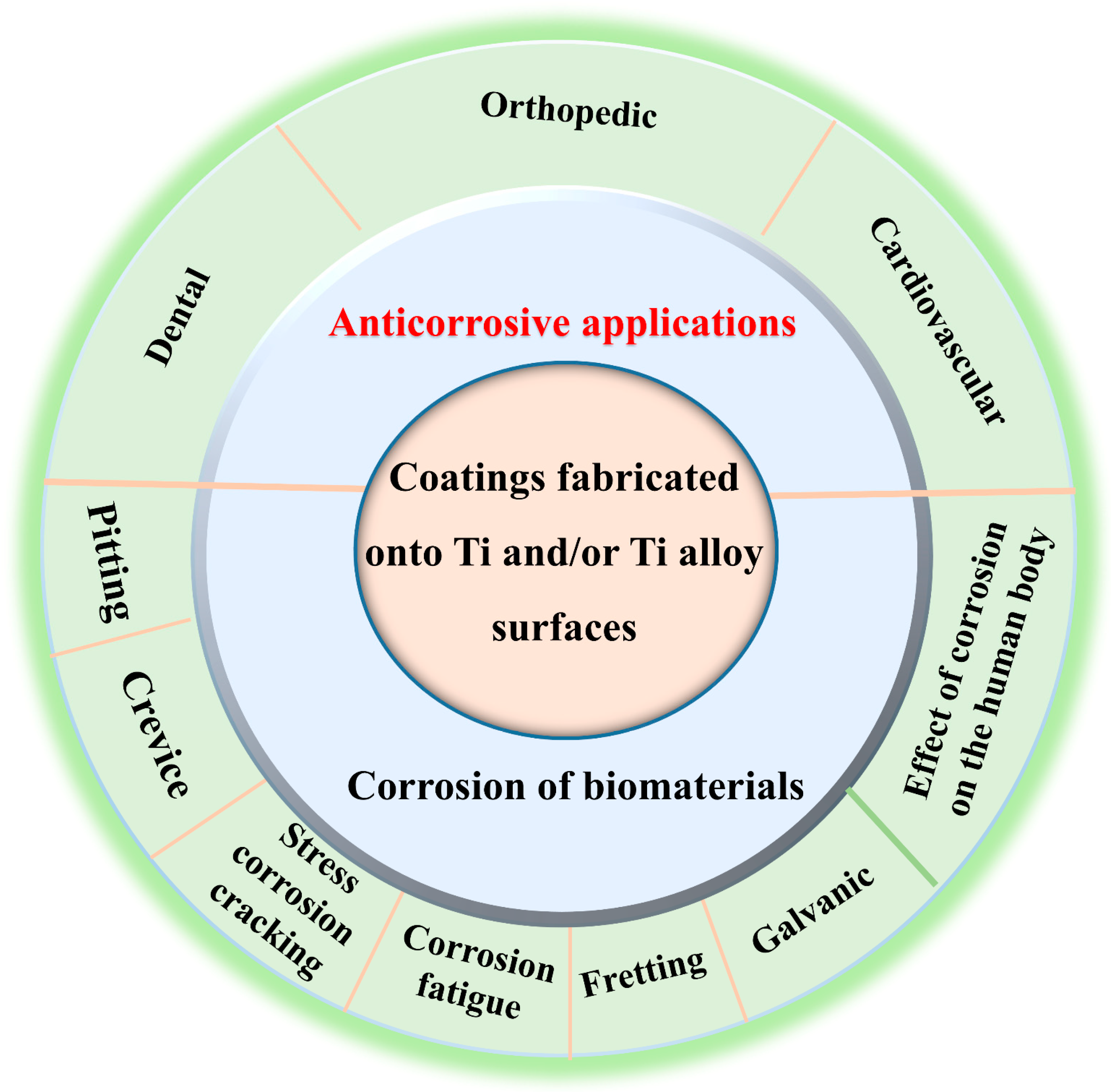
Figure 1. Corrosion types and effects, as well as anticorrosive applications of coatings fabricated onto Ti and/or Ti alloy surfaces in biomaterials for medical applications.
Coatings 15 00599 g001
Figure 3. (a) Shape of the implant and (b) SEM image of crevice corrosion (magnification 70×) [55,59].
Coatings 15 00599 g003
Figure 4. SEM images of (a) intergranular stress corrosion cracks (SCC) and (b) transgranular SCC [65].
Coatings 15 00599 g004
Figure 5. (a) shape of the implant and (b) SEM image of corrosion fatigue (CF) [55,68].
Coatings 15 00599 g005
Figure 6. (a) Shape of the implant and (b) SEM image of fretting corrosion [55,59].
Coatings 15 00599 g006
Figure 7. (a) Shape of the implant and (b) SEM image of galvanic corrosion [55,75].
Coatings 15 00599 g007
Figure 8. (A) Cyclic potentiodynamic polarization curves of Ti-containing (Ni-Ti, Ni-Ti-Cu, Ti-Mo-Zr-Sn, and Ti-Nb) dental orthodontic wires in acidic artificial saliva with various NaF concentrations (black arrows indicated hysteresis loop). (B) SEM images of the examined Ni-Ti, Ni-Ti-Cu, Ti-Mo-Zr-Sn, and Ti-Nb wires both before (as-received situation) and after corrosion examination in fluoride-free artificial saliva [88].
Coatings 15 00599 g008
Figure 9. (A) (a) Nyquist plots with equivalent circuit model, (b) Bode plots, and (c) potentiodynamic polarization plots of the untreated NiTi, GO-treated NiTi (GO), and GO/Ag-treated NiTi (GOAg) alloy after immersion in a 3.5% NaCl solution. Rs, Rct, Cdl, W, and SCE represent electrolyte solution resistance, charge transfer resistance of the corrosion reaction, capacitance, Warburg impedance related to the diffusion of O2 of the NiTi alloy, and saturated calomel electrode, respectively. (B) SEM micrographs of (a) bare NiTi, (b) GO, and (c) GOAg-treated NiTi alloys after the potentiodynamic polarization curve tests. The pitting corrosion is represented by the arrows [22].
Coatings 15 00599 g009
Figure 10. (A) Diagrammatic representation of the synthesis of composite coatings on a Ti-13Nb-13Zr alloy, and (B) potentiodynamic polarization curves of different samples. HA, Fe3O4, HA@1Fe3O4, HA@3Fe3O4, and HA@5Fe3O4 represent pure hydroxyapatite (HA), pure Fe3O4, pure HA and Fe3O4 with various concentrations (1, 3, and 5 wt%) composite coatings, respectively [98].
Coatings 15 00599 g010
Figure 11. (a) Tafel curves, (b) Nyquist plots, and (c,d) Bode plots of Ti-6Al-4V and different concentrations of BG in simulated body fluid (SBF) solution (pH 7.40), (e) the corresponding equivalent electrical circuit model: CS-0.5 g/L BG, CS-1 g/L BG and CS-1.5 g/L BG represent Chitosan–Bioactive glass (CS-BG) hybrid coatings with different BG levels of 0.5, 1 and 1.5 g/L on the Ti-6Al-4V surface, respectively. (f) Electrochemical corrosion parameters for Ti-6Al-4 V and the coated samples in SBF. Ecorr and ip represent corrosion potential and passivation current density, respectively [23].
Coatings 15 00599 g011
Figure 12. (a) Tafel curves of different samples in the simulated body fluid (SBF) solution. TiO2, ZrO2/TiO2, and Zn-ZrO2/TiO2 represent TiO2 coating, ZrO2/TiO2 coating, and Zn-doped ZrO2/TiO2 porous coatings on Ti6Al4V alloy, respectively; (b) corrosion parameters of corrosion potential (Ecorr) and corrosion current density (Icorr) [44].
Coatings 15 00599 g012
Figure 13. (A) Potentiodynamic polarization curves of synthesized composite coatings on Ti6Al4V. (B) SEM images of synthetic composite coatings on Ti6Al4V sample surfaces after corrosion experiments in vitro in the simulated body fluid (SBF). (a) Only nHA, (b) nHA/1GNS, (c) nHA/3GNS, (d) nHA/5GNS, and (e) nHA/7GNS represent nano-hydroxyapatite (nHA) composite coatings added with various graphene nanosheets (GNSs) of 0, 1, 3, 5, and 7 wt% on the Ti6Al4V alloy, respectively [46].
Coatings 15 00599 g013
Figure 14. (A) (a) Nyquist plots and (b) Tafel plots of different samples. The enlarged picture (a1) of the blue dash-line area is displayed in the inset in (a), and (B) A schematic illustration for anticorrosion mechanisms of graphene oxide (GO) coatings. Ti-GO, Ti-25-GO, Ti-45-GO and Ti-65-GO represent GO coating with 0, 25, 45 and 65 μm width of the microgrooves on the Ti-6Al-4V alloy, respectively [47,114].
Coatings 15 00599 g014
Figure 15. (a) Nyquist plots, (b) Bode plots, and (c) Tafel plots of different samples. Ti-HA and Ti-Fe3O4/HA represent hydroxyapatite (HA) coating and Fe3O4 coated with HA coating on the Ti6Al4V, respectively [49].
Coatings 15 00599 g015
Figure 16. (a) Tafel plots, (b) Nyquist plots, and (c) Bode plots of Ti-3Cu alloys before and after laser shock peening (LSP) treatment. Control, 5 J-3 and 7 J-3 represent Ti-3Cu alloy, sample obtained after being impacted 3 times with the laser pulse energy of 5 J and 7 J on the Ti-3Cu alloy, respectively [118].
Coatings 15 00599 g016
Figure 17. (A) Schematic representation of the modification process of composite coatings on the Ti6Al4V substrate and (B) Tafel plots of Ti6Al4V (blue), DLC/Ti (red): diamond-like carbon (DLC) coating on the Ti6Al4V, and F-DLC/Ti (black): 1H,1H,2H,2H-perfluorodecyl acrylate coated on DLC coating on the Ti6Al4V [40].
Coatings 15 00599 g017
Figure 18. (a) The potentiodynamic polarization curves of a porous TiO2 coating on biomedical β Ti alloy via MAO (micro-arc oxidation) and PA+MAO (pre-anodization followed by MAO technique) in the 0.9% NaCl solution. (b) Corrosion parameters of corrosion potential (Ecorr), corrosion current density (Icorr), and passivation current density (ipp) [41].
Coatings 15 00599 g018
Figure 19. Tafel plots of (A) Ti6Al4V alloy at 36 °C (1) and 40 °C (2), and (B) Ti6Al4V alloy with nitride coatings I, II, and III in the Ringer’s solution at 36 °C (a) and 40 °C (b). The nitriding parameters of nitride coatings I, II, and III are nitrogen partial pressure of 1 Pa at nitriding temperature of 850 °C, nitrogen partial pressure of 105 Pa at nitriding temperature of 850 °C, and nitrogen partial pressure of 105 Pa at nitriding temperature of 900 °C, respectively [122].
Coatings 15 00599 g019
Figure 20. (A) Tafel plots recorded after open circuit potential measurements in 0.15 M NaCl solution for (a) untreated NiTi alloy (NiTi), (b) NiTi alloy treated with C3 (NiTi/C3), (c) NiTi alloy coated with C18 (NiTi/C18), (d) NiTi alloy treated with PPy film (NiTi/PPy), (e) NiTi alloy coated by both self-assembled films of C3 and PPy film (NiTi/C3/PPy), and (f) NiTi alloy coated by both self-assembled films of C18 and PPy film (NiTi/C18/PPy). (B) SEM images of the NiTi/C18/PPy after 15 h of polarization at 0.65 V in 0.15 M NaCl solution. C3, C18, and PPy represent propyltrichlorosilane (C3H7SiCl3), octadecyltrichlorosilane (C18H37SiCl3), and polypyrrole, respectively [42].
Coatings 15 00599 g020
Table 1. A time-line for the development of coatings fabricated onto Ti and/or Ti alloy surfaces.
Fabrication
Technique
CoatingSubstrateImproved PropertiesApplicationYear of PublicationReference
Arc ion plating (AIP)TiN and TiN/Ti
coating
Ti-6Al-4V alloyCorrosion resistanceOrthopedics2008[38]
Spin techniqueTiO2 Nano-particles coatingTi-6Al-4V alloyCorrosion resistanceOrthopedics2009[39]
Magnetron sputtering and photochemical functionalizationF-DLC/
Ti coating
Ti-6Al-4V alloyAnti-corrosion and friction, biocompatibility, and functional propertiesVarious applications2012[40]
Pre-anodization (PA) and micro-arc oxidation (MAO) techniquesPorous TiO2 coatingbiomedical β Ti alloyCorrosion resistanceVarious applications2013[41]
Polydimethylsiloxane (PDMS) modificationSelf-assembled filmNiTi alloy (NiTi)Corrosion resistanceBiomedical applications2014[42]
Low thermal volatilization sol–gel methodSiO2 and ZnO composite coatingTi-6Al-4V alloyCorrosion resistance, mechanical propertiesDental and knee joint implants2017[43]
Magnetron sputtering in combination with MAOZn-doped ZrO2/TiO2 coatingTi-6Al-4V alloyAnti-corrosive, antibacterial properties, and biocompatibilityorthopedics and dentistry2017[44]
Electrophoretic deposition technique and magnetron sputteringTi-Al2O3 coatingTi-6Al-4V alloyAnti-corrosive property and hardnessOrthopedics2018[45]
Sol–gel technique and
cathodic electrophoretic deposition (EPD)
Chitosan–Bioactive glass (CS-1.5 g/L BG) nanocomposite coatingTi-6Al-4V alloyBio-corrosion, biological activity, and wetting behaviorOrthopedics2019[23]
EPDGraphene oxide (GO) and GO/silver (GO/Ag) nanocomposite coatingsNiTi alloyCorrosion resistance, biocompatibilityOrthodontic wires and brackets2020[22]
Hydrothermal processNanoHA/graphene nanosheet (nHA/GNS) composite coatingTi-6Al-4V alloyCorrosion resistanceOrthopedics2020[46]
Laser treatment and chemical assemblyGraphene oxide (GO) coatingTi-6Al-4V alloyCorrosion resistanceBiomedical application2020[47]
Laser treatment and polydimethylsiloxane (PDMS) modificationSuperhydrophobic surfaceNiTi alloyCorrosion resistanceMedical device2021[48]
Chemical self-assemblyFe3O4-coated HA coatingTi-6Al-4V alloyCorrosion resistanceBiomedical application2022[49]
Table 2. Corrosion parameters of the bare NiTi alloy, GO-coated NiTi (GO), and GO/Ag-coated NiTi (GOAg) substrates. Ecorr, Icorr, and η represent corrosion potential, corrosion current density, and protection efficiency, respectively [22].
SpecimensEcorr
(V vs. SCE)
Icorr (µA/cm2)η (%)
Bare NiTi−0.1700.158
GO0.0310.01789.24
GOAg0.0080.00298.73
Table 3. Electrochemical impedance spectroscopy (EIS) parameters of the bare NiTi alloy, GO-coated NiTi (GO), and GO/Ag-coated NiTi (GOAg) substrate. Rs = Electrolyte solution resistance, Rct = Charge transfer resistance of the corrosion reaction, Cdl = Capacitance, and Yo = Warburg impedance related to the diffusion of O2 of the NiTi alloy [22].
SpecimenRs (Ω)Rct (kΩ)Cdl (µF)Yo (µMho)
Bare NiTi16.2016.8036.908.29
GO16.50−6.1226.6015.60
GOAg16.50−5.0447.2023.20
Table 4. Corrosion parameters of corrosion potential (Ecorr) and corrosion current density (Icorr) [98].
SampleEcorr (V)Icorr (μA/cm2)
Uncoated−0.6021.1.5 × 10−8
HA−0.4821.15 × 10−10
Fe3O4−0.5162.68 × 10−10
HA@1Fe3O4−0.3095.90 × 10−11
HA@3Fe3O4−0.3648.20 × 10−11
HA@5Fe3O4−0.3931.02 × 10−10
Table 5. EIS fitting results for uncoated and coated samples; Rs = Electrolyte solution resistance, R1 = charge transfer resistance of the coating layer; R2 = charge transfer resistance of the double layer related to the barrier/inner layer; CPE1 and CPE2 = constant phase elements (CPE) of the coating layer/solution interface; and CPE2 = CPE of the barrier/inner layer [23].
SampleRs (Ω × cm2)R1 (Ω × cm2)CPE1-T (×10−5, Fsn−1cm−2)CPE1-PR2 (Ω × cm2)CPE2-T (×10−5, Fsn−1cm−2)CPE2-PChi-Squared (X2)
Ti-6Al-4V52.3179.13.230.911.25 × 1066.910.943 × 10−4
CS-0.5 g/L BG55779.73.210.891071.110.937 × 10−4
CS-1 g/L BG56.95595.42.880.931.83 × 1071.080.921 × 10−3
CS-1.5 g/L BG51.3835831.450.871.25 × 10101.030.891 × 10−3
Table 6. Corrosion parameters of the synthesized hybrid coatings on Ti6Al4V, Ecorr, Icorr, and Rp represent corrosion potential, corrosion current density, and the polarization resistance of the layer [46].
CoatingEcorr (mV)Icorr (×10−9, A·cm−2)Corr. Rate (mpy)Rp (Ω·cm2)
Only nHA12.871150.05275,303
nHA/1GNS5.871251090120,429
nHA/3GNS83.11500.037597,065
nHA/5GNS79.89550.065776,167
nHA/7GNS74.90850.077385,321
Table 7. Electrochemical corrosion parameters of Ti alloy plates in SBF solution [47].
SamplesCorrosion Potential (V)Corrosion Current Density (A/cm2)
Ti−0.5721.894 × 10−6
Ti-GO−0.4671.112 × 10−6
Ti-25-GO−0.4254.031 × 10−7
Ti-45-GO−0.4421.425 × 10−7
Ti-65-GO−0.1023.894 × 10−7
Table 8. Electrochemical parameters of Ti6Al4V, Ti-HA, and Ti-Fe3O4/HA in SBF solution [49].
SamplesCorrosion Potential (mV)Corrosion Current
Density (nA/cm2)
Inhibition Efficiency (η)
Ti6Al4V−489.531123.03-
Ti-HA−532.59157.9985.93%
Ti-Fe3O4/HA−324.60153.3386.33%
Table 9. The EIS fitting parameters of different samples. Control, 5 J-3 and 7 J-3 represent Ti-3Cu alloy, sample obtained after being impacted 3 times with the laser pulse energy of 5 J and 7 J on the Ti-3Cu alloy, respectively. Rs = solution resistance; CPE = constant phase element; and Rp = the polarization resistance of the layer [118].
SamplesRs (Ω⋅cm2)CPE (μF/cm2)nRp (107 Ω⋅cm2)
control35.8 ± 1.109.55 ± 0.840.886 ± 0.202.36 ± 0.54
5 J-343.7 ± 1.0510.01 ± 0.840.891 ± 0.213.58 ± 0.85
7 J-344.1 ± 1.2611.92 ± 1.030.893 ± 0.265.05 ± 1.74
Table 10. Corrosion potentials (Ecorr) and corrosion current densities (Icorr) for the Ti6Al4V before and after modification [40].
SamplesEcorr (V)Icorr (nA·cm−2)
Ti6Al4V−0.2380837.96
DLC/Ti−0.1010164.42
F-DLC/Ti0.039982.025
Table 11. Corrosion parameters of Ti-6Al-4V alloy in Ringer’s solution without surface treatment and after nitriding [122].
T, °CCorrosion ParametersSurface Condition
UntreatedCoating ICoating IICoating III
36Ecor, V vs. Ag/AgCl−0.425−0.225−0.24−0.11
icor × 10−4, A/cm20.0410.00550.00270.0037
40Ecor, V vs. Ag/AgCl−0.430−0.375−0.34−0.125
icor × 10−4, A/cm20.0190.00500.00700.02
Disclaimer/Publisher’s Note: The statements, opinions and data contained in all publications are solely those of the individual author(s) and contributor(s) and not of MDPI and/or the editor(s). MDPI and/or the editor(s) disclaim responsibility for any injury to people or property resulting from any ideas, methods, instructions or products referred to in the content.