Article

Physical Vapor Deposited TiN and TiAlN on Biomedical β-Type Ti-29Nb-13Ta-4.6Zr: Microstructural Characteristics, Surface Hardness Enhancement, and Antibacterial Activity

Department of Metallurgical and Materials Engineering, Yıldız Technical University, 34220 Istanbul, Turkey
Coatings 2025, 15(10), 1126; https://doi.org/10.3390/coatings15101126
Submission received: 11 July 2025 / Revised: 18 September 2025 / Accepted: 18 September 2025 / Published: 29 September 2025
(This article belongs to the Special Issue Films and Coatings with Biomedical Applications)

Abstract

Beta (β)-type Ti-29Nb-13Ta-4.6Zr (TNTZ) alloys combine low modulus with biocompatibility but require improved surface properties for long-term implantation. This study aimed to enhance the surface mechanical strength and antibacterial performance of TNTZ by applying TiN and TiAlN coatings via PVD. Notably, TiAlN was deposited on TNTZ for the first time, enabling a direct side-by-side comparison with TiN under identical deposition conditions. Dense TiN (~1.06 μm) and TiAlN (~1.73 μm) coatings were deposited onto solution-treated TNTZ and characterized by X-ray diffraction, scanning probe microscopy, Vickers microhardness, Rockwell indentation test (VDI 3198), static water contact angle measurements, and a Kirby–Bauer disk-diffusion antibacterial assay against Escherichia coli (E. coli). Both coatings formed face-centered cubic (FCC) structures with smooth interfaces (Ra ≤ 5.3 nm) while preserving the single-phase β matrix of the substrate. The hardness increased from 192 HV (uncoated) to 1059 HV (TiN) and 1468 HV (TiAlN), and the adhesion quality was rated as HF2 and HF1, respectively. The surface wettability changed from hydrophilic (48°) to moderately hydrophobic (82°) with TiN and highly hydrophobic (103°) with TiAlN. Similarly, the diameter of the no-growth zones increased to 18.02 mm (TiN) and 19.09 mm (TiAlN) compared to 17.65 mm for uncoated TNTZ. The findings indicate that TiAlN, in particular, provided improved hardness, adhesion, and hydrophobicity. Preliminary bacteriostatic screening under diffusion conditions suggested a modest relative antibacterial response, though the effect was not statistically significant between coated and uncoated TNTZ. Statistical analysis confirmed no significant difference between the groups (p > 0.05), indicating that only a preliminary bacteriostatic trend— rather than a definitive antibacterial effect—was observed. Both nitride coatings strengthened TNTZ without compromising its structural integrity, making TiAlN-coated TNTZ a promising candidate for next-generation orthopedic implants.

1. Introduction

Titanium and its alloys have become useful materials in biomedical applications, especially in orthopedic and dental implants, due to their excellent biocompatibility, mechanical strength, corrosion resistance, and favorable elastic modulus [1,2,3]. Among various titanium alloys, β-type titanium alloys have gained prominence because their modulus closely matches that (~10–30 GPa) of human bone, reducing stress shielding effects commonly observed in implants [4,5,6]. One of the most promising β-type titanium alloys is Ti-29Nb-13Ta-4.6Zr (TNTZ); an abbreviation derived from Ti, Nb, Ta, and Zr, developed by Niinomi and his colleagues, specifically tailored for biomedical purposes [7,8]. This alloy offers excellent mechanical properties, such as high fatigue strength, good ductility, and a low elastic modulus (~60 GPa), making it ideal for load-bearing orthopedic implants [9,10]. Clinical applications of TNTZ alloys are hindered by inherent limitations, particularly with respect to wear resistance and bacterial colonization. Wear-induced particle debris generated from the implant surface can lead to inflammation, osteolysis, and eventually, implant loosening [11,12]. Furthermore, bacterial adhesion and subsequent biofilm formation on implant surfaces cause serious problems, often leading to severe infections and implant failure [13,14]. Therefore, improving the surface properties of TNTZ alloys to increase wear resistance and provide antibacterial properties has become a major research goal.
Surface modification techniques such as physical vapor deposition (PVD), chemical vapor deposition (CVD), anodizing, and thermal oxidation have been widely used to improve the surface properties of titanium alloys [15,16,17]. Among these methods, PVD has attracted much attention due to its ability to produce thick, uniform, and continuous coatings with superior mechanical, chemical, and biological properties [16,18]. Within PVD coatings, titanium nitride (TiN) and titanium aluminum nitride (TiAlN) coatings are widely used in the biomedical field due to their high hardness, good wear resistance, chemical inertness, and promising antibacterial potential [19,20,21]. For example, TiN coatings have been used on load-bearing orthopedic prostheses (such as hip and knee joint replacements) to create a hard, wear-resistant, biocompatible surface [22]. This protective TiN layer has been shown to reduce wear debris and metal ion release from implants, thereby improving longevity and biocompatibility. Likewise, TiAlN and related hard nitride coatings are being explored for orthopedic implants (e.g., fracture fixation screws and joint prostheses) due to their excellent wear and corrosion resistance [23].
TiN coatings, which are widely studied for their biocompatibility and durability, significantly improve the wear and corrosion resistance of underlying surfaces, prolonging the lifetime of implants in physiological environments [24,25]. Mani et al. showed that TiN coatings significantly increase corrosion resistance and electrical conductivity on stainless steel surfaces, highlighting their beneficial effects in aggressive biological environments [24]. Similarly, TiAlN coatings showed better mechanical properties than traditional TiN coatings; these properties are due primarily to their higher hardness, thermal stability, and higher chemical inertia, which are particularly favorable in biomedical applications where corrosive and abrasive conditions coexist [26,27].
Many studies have highlighted the positive effects of TiN and TiAlN coatings in improving the surface properties of biomedical titanium alloys. For example, Lisoń-Kubica et al. evaluated TiN coatings on Ti-13Nb-13Zr alloy and observed significant improvements in mechanical hardness and corrosion resistance, further demonstrating its biocompatibility and suitability for implant placement [28]. Similarly, TiAlN-coated implants showed reduced friction coefficients, increased surface hardness, and improved corrosion resistance, which significantly prolonged their service life [29,30]. In addition to mechanical and corrosion properties, the biocompatibility of coatings should be thoroughly evaluated, especially regarding bacterial interactions. Bacterial accumulation and subsequent biofilm formation pose serious problems for orthopedic implants and often require implant removal and reoperation, affecting the patient’s quality of life and healthcare costs [31]. Therefore, providing antibacterial function through surface treatment methods has become essential for the development of orthopedic implants. The antibacterial function of coatings such as TiN and TiAlN primarily depends on their surface chemistry and topography, which affect the kinetics of bacterial adhesion and biofilm formation [32]. Oh et al. pointed out that surface chemistry strongly affects the kinetics and thermodynamics of bacterial adhesion; therefore, surface modification is an essential strategy to reduce bacterial accumulation [32].
While TiN and TiAlN coatings are well established on conventional titanium substrates, their performance on the β-type Ti-29Nb-13Ta-4.6Zr (TNTZ) alloy—a low-modulus, high-biocompatibility material designed for biomedical use—remains largely unexplored. This work provides the first systematic, side-by-side comparison of TiN and TiAlN coatings on TNTZ, deposited via industrial cathodic arc PVD under identical pretreatment and bias conditions to ensure comparability. Within a single, consistent framework, we assess microstructure (XRD, SEM, AFM), adhesion (VDI 3198), microhardness, wettability, and antibacterial activity against Escherichia coli (E. coli). Notably, TiAlN is introduced here for the first time as a functional coating for TNTZ, demonstrating a unique combination of high hardness, strong adhesion, and slightly enhanced bacteriostatic effect, despite exhibiting greater hydrophobicity.

2. Materials and Methods

2.1. Material

In this study, hot-forged Ti-29Nb-13Ta-4.6Zr (TNTZ) alloy bars with a diameter of 25 mm and a length of 50 mm were obtained from Dr. Mitsuo Niinomi, Tohoku University, Sendai, Japan. As a starting condition, the alloys were subjected to solution heat treatment at 1063 K under vacuum conditions, followed by quenching to maintain a single BCC (β) phase structure. The chemical composition of the alloy is presented in Table 1. Rectangular specimens with dimensions of 10 mm × 15 mm × 3 mm were sectioned from the forged rods using electrical discharge wire cutting. The specimen surfaces were then ground using waterproof abrasive papers up to #2500 and subsequently polished with a 0.3 μm colloidal SiO2 suspension to achieve a fine finish suitable for surface modification processes. This heat-treatment regimen was adopted to obtain a homogeneous single-phase β microstructure and to relieve residual stresses prior to coating.

2.2. Physical Vapor Deposition (PVD) on TNTZ Samples

TiN and TiAlN thin films were deposited on TNTZ samples in the Ionbond IHI Group (İstanbul, Turkey) company using an industrial cathodic arc PVD system (Hauzer Flexicoat 850). Deposition was carried out in a cathodic arc PVD system with separate cathodes for Ti and TiAl. For the TiN coating, a pure Ti target (99.98% Ti) was used in a nitrogen atmosphere; for the TiAlN coating, a composite Ti–Al (50:50 wt%) target was utilized under the same conditions to supply both Ti and Al for the nitride coating. The PVD system was initially evacuated and heated to 480 °C for 2 h. Before starting the coating process, a high vacuum level of approximately 10−4 mbar was created. For surface pretreatment, argon gas was introduced into the chamber at a flow rate of 44 sccm and converted to plasma using a gas filament source. To perform plasma-assisted surface cleaning, a pulsed negative bias voltage of 100 V was applied to the samples for 40 min. After this step, nitrogen gas was introduced at a flow rate of 1000 sccm to bring the chamber pressure to approximately 10−2 mbar. During the coating process, an arc discharge was created by applying a current of 125 A to the cathode. A negative bias voltage of −50 V and −100 V was applied to the substrate to facilitate TiN and TiAlN film deposition. During deposition, the gas flow was maintained at 800–900 sccm. The coating process duration varies between 45 min and 1 h 45 min, depending on the coated material type.

2.3. Microstructural Characterization

X-ray diffraction (XRD) was performed using X’Pert PRO (Malvern PANalytic) with Cu Kα radiation (λ = 0.15418 nm) at 40 kV and 40 mA. X-ray measurements were performed from a thickness of 1 µm. The diffraction patterns were collected over a 2θ range from 30° to 90° with a step size of 0.04°. The crystallite size (D) was estimated from the XRD peak broadening by applying the following Scherrer formula [33,34]:
D = K λ β cos θ
where K is the shape factor (0.9), β is the FWHM of the peak in radians, and θ is the Bragg angle (half of the 2θ peak position, in degrees). The β determined from measured Full Width at Half Maximum (FWHM) (degrees) values of the peaks having the highest intensity, which is called Bobs in the following equation.
β = B o b s × π 180
To accurately find the FWHM of each peak, we performed a Gaussian fit to the intensity vs. 2θ data in the vicinity of the peak. The Gaussian fitting yields the peak width at half maximum intensity.
Cross-sectional microstructures and elemental composition of the coatings were observed and analyzed using scanning electron microscopy (Zeiss-Sigma Evo ls10) equipped with an energy-dispersive X-ray spectroscopy (EDS) detector. Prior to SEM/EDS analysis, the specimens were sputter-coated with a thin gold (Au) conductive layer to avoid charging effects during imaging.

2.4. Atomic Force Microscopy

Surface topography and roughness were characterized by atomic force microscopy (AFM) (Shimadzu SPM-9600 scanning probe microscope) in tapping mode over 1 μm × 1 μm scan areas. The arithmetic roughness (Ra), maximum height (Rz), Ten-point mean roughness (Rzjis), root-mean-squared roughness (Rq), maximum profile peak height (Rp), and maximum profile valley depth (Rv) were measured for each sample.

2.5. Hardness Measurement

Vickers microhardness measurements of the thin film-coated specimens were performed at room temperature using a Wilson Hardness TUKON 1102 microhardness tester. A load of 25 g was applied with a dwell time of 15 s for each indentation. To ensure accuracy and reproducibility, measurements were conducted at multiple locations across the specimen surface, with a minimum of five indentations per sample.

2.6. Adhesion Evaluation

The adhesion quality of the coatings was qualitatively assessed using the Rockwell indentation test in accordance with the VDI 3198 standard [35]. Indentations were performed using a Rockwell C hardness tester with an applied load of 150 kgf. The resulting indentation traces were examined under an optical microscope to evaluate coating adhesion. The classification principle of the VDI 3198 test is illustrated in Figure 1. Based on the crack patterns and delamination around the indentation, adhesion was categorized into six classes (HF1 to HF6) [35]. Coating adhesion levels classified as HF1, HF2, HF3, and HF4 are considered acceptable, whereas HF5 and HF6 indicate poor adhesion, with unacceptable bonding between the coating and substrate.

2.7. Contact Angle Measurement

Static contact angles for both coated and uncoated samples were determined using distilled water at 25 °C by a sessile-drop goniometer (KSV Cam200). A high-resolution digital camera captured the droplet profile, and the contact angle was extracted from the cross-sectional image 10 s after deposition. The goniometer has a ±2° standard deviation. To ensure accuracy and reproducibility, measurements were conducted multiple times on five different specimens.

2.8. Bacterial Sensitivity Tests

Antibacterial activity was quantified by the Kirby–Bauer disk-diffusion assay [36]. Overnight cultures of Escherichia coli (ATCC 25922) were diluted to a 0.5 McFarland turbidity (≈1.5 × 10⁸ CFU/mL) and uniformly spread onto Mueller–Hinton agar using 0.1 mL of the suspension. Alloy disks were ultrasonically cleaned in deionized water and ethanol (3 min each) and UV-sterilized at 254 nm for 30 min before testing. Sterile disks were gently pressed onto the inoculated agar and incubated at 37 °C for 24 h. Resulting inhibition zones were recorded with a measurement microscope (Mitotuyo, Japan) having a Cross-travel Stage (X-Y directions) for a linear measurement with a resolution of 0.01 mm. All conditions were evaluated in triplicate, each on independently prepared plates, to identify the most antibacterial coating.
Statistical analysis: Inhibition-zone dimensions are presented as mean ± standard deviation. Group differences were evaluated with a one-way analysis of variance (ANOVA); when the ANOVA indicated significance, pairwise comparisons were performed using independent-sample t-tests. All statistical tests were two-tailed, and differences were considered significant at p < 0.05.

3. Results and Discussion

3.1. Microstructural Analysis

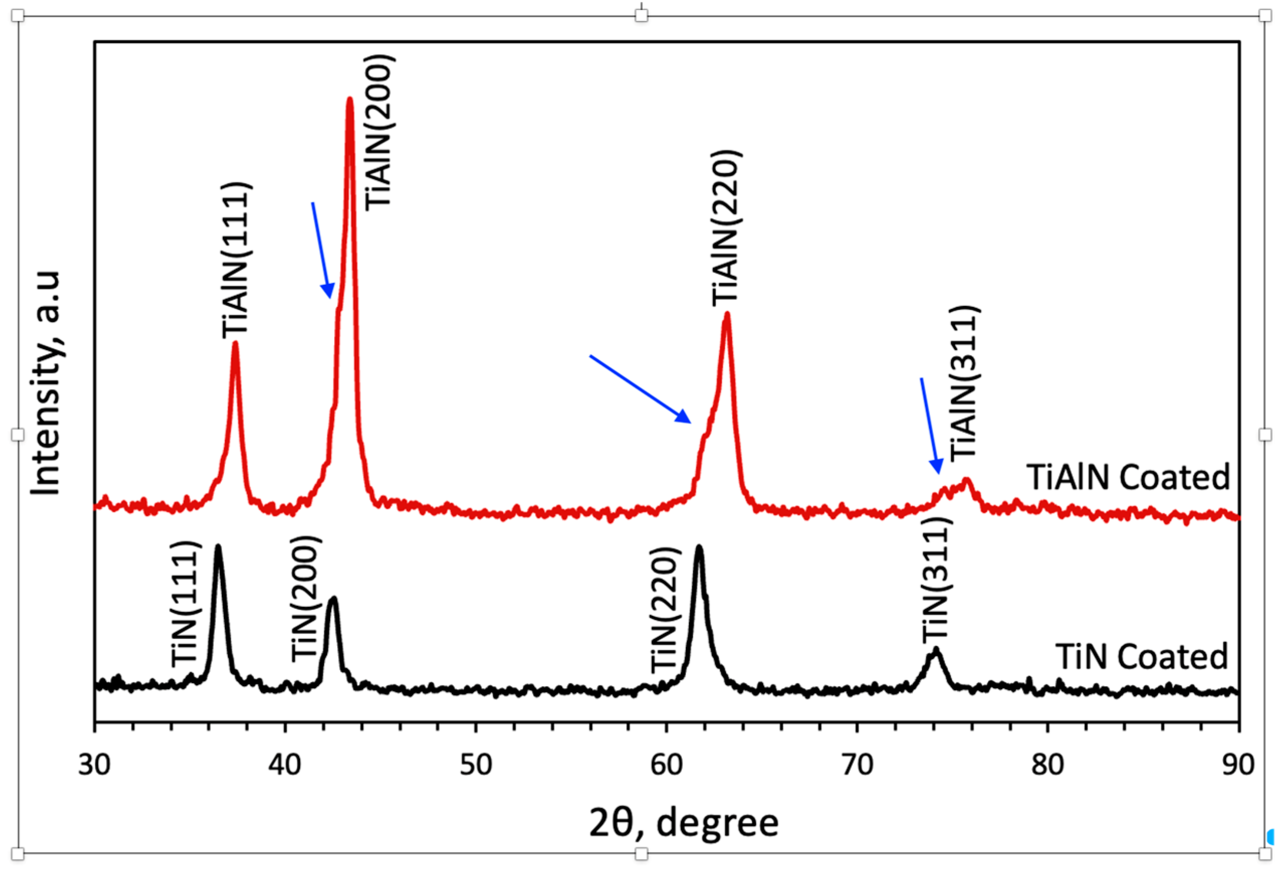
Ti-29Nb-13Ta-4.6Zr (TNTZ) alloy is confirmed to be a single-phase β (BCC) structure in its solution-treated form, as expected for this quaternary, Nb/Ta-stabilized Ti alloy [1,4,7]. When the peaks of the XRD profiles obtained from the thin coating on the specimen are examined in Figure 2, it is evident that the TiN-coated specimen has a face-centered cubic (FCC) crystal structure characteristic of TiN. When these results are compared with those in previous studies [37,38], they appear to be consistent. When the TiAlN peaks were examined, it was determined that they exhibited similar orientations of (111), (200), and (311) with slightly different angles. TiAlN and TiN are isostructural (both face-centered cubic), but Al substitution in TiN reduces the lattice parameter (Al atoms are smaller). Consequently, a homogeneous TiAlN (with ~50% Al) exhibits peaks at a higher 2θ (smaller d-spacing) compared to pure TiN [39]. In the TiAlN coating’s XRD profile, the primary peaks are indeed shifted to higher angles relative to TiN, confirming incorporation of Al into the NaCl-type lattice [39]. According to the TiN and TiAlN coating study of Qiu [40], Al atoms were substitutionally located at the same lattice points as Ti atoms in this lattice, which can change the lattice parameters and, consequently, the interspace distance of planes.
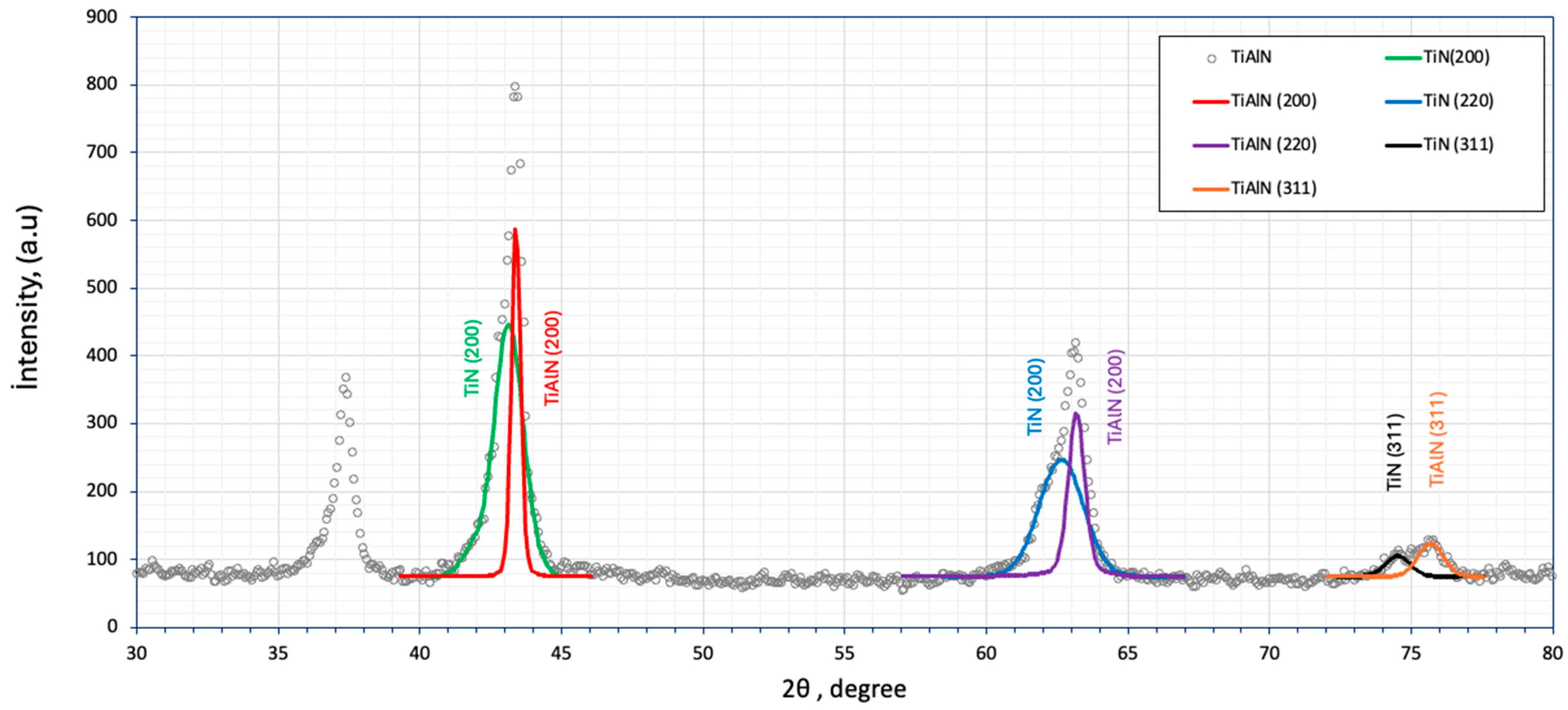
The TiAlN coating exhibited FCC reflections with slight shoulders (marked by blue arrows) near the (200), (220), and (311) peaks, indicating the presence of minor TiN-like domains. No evidence of wurtzite-AlN or other secondary phases was observed. In essence, the TiAlN (200), (220), (311) reflections each seem to be a convolution of two contributions: one from the intended TiAlN phase, and a weaker one from a TiN-like phase. The peaks were deconvoluted from the main TiAlN peaks by Origin Lab Pro 8.0 software using Gaussian fitting in Figure 3. The fact that shoulders appear toward the lower-angle side of these peaks—closely at the angles where TiN would diffract—implies there are domains with a larger lattice parameter. This produces an asymmetric broadening rather than a single symmetric peak. Such peak asymmetry and splitting are signs of either a second phase or a compositional gradient in the lattice parameter. Notably, there is no sign of the emergence of any new independent peaks for wurtzite-AlN, which would appear at distinct positions of 2θ: ~33°–36° 2θ for (100)/(002), suggesting that any Al-rich domains. The shoulders here correspond specifically to FCC TiN type reflections, reinforcing that the secondary phase is TiN or a Ti-rich cubic TiAlN [41]. However, because these features appear only as subtle shoulders (not distinct new peaks) in the conventional XRD patterns, they cannot by themselves definitively identify a separate TiN phase. Our interpretation of these shoulders as TiN-like domains is therefore guided by analogous observations in the literature (Bartosik et al. [41]) and should be considered tentative. Bartosik et al. also reported that the XRD peaks of TiAlN can shift or broaden asymmetrically, signaling the formation of Ti-rich and Al-rich domains [41]. TiAlN coatings are known to form a metastable solid solution cubic structure when deposited by PVD, up to a relatively high Al content (up to about 70 at% Al under typical conditions) [42]. However, this Ti–Al–N system has a wide miscibility gap, meaning that the TiN and AlN phases are not thermodynamically prone to mixing at lower temperatures [42]. Thus, TiAlN is thermodynamically metastable; given sufficient thermal energy or high Al content, it tends to phase-separate into Ti-rich and Al-rich nitride domains (essentially TiN and AlN phases) [41]. In the present case of a TiAlN coating (50:50 wt% Ti:Al target) deposited at 480 °C, the appearance of shoulders corresponding to TiN diffraction suggests that the alloy may be approaching a solubility or metastability limit, leading to incipient phase separation. Essentially, a portion of the coating behaves like TiN (with little Al in the lattice), even as the main phase remains TiAlN.
One possibility of the appearing of TiN-like phase is incomplete alloying during growth—the Al and Ti may not distribute uniformly at the atomic scale under the given deposition conditions. CA-PVD involves energetic ion bombardment (here with a 100 V pulsed bias), which can induce re-sputtering of lighter elements; indeed, studies have shown that increasing substrate bias can selectively deplete Al from a growing TiAlN film, yielding a lower Al/Ti ratio and shifting XRD peaks toward the TiN positions [43]. From a thermodynamic standpoint, the high Al content (≈50 at% Al) in TiAlN puts it inside the spinodal decomposition regime at typical growth/annealing temperatures [42]. Although 480 °C is moderate, it may still allow limited atomic mobility over the deposition time. This can result in nano-scale compositional modulations: any spontaneous fluctuation toward a Ti-rich and an Al-rich composition will lower the free energy (since the single-phase solid solution is unstable inside the miscibility gap).
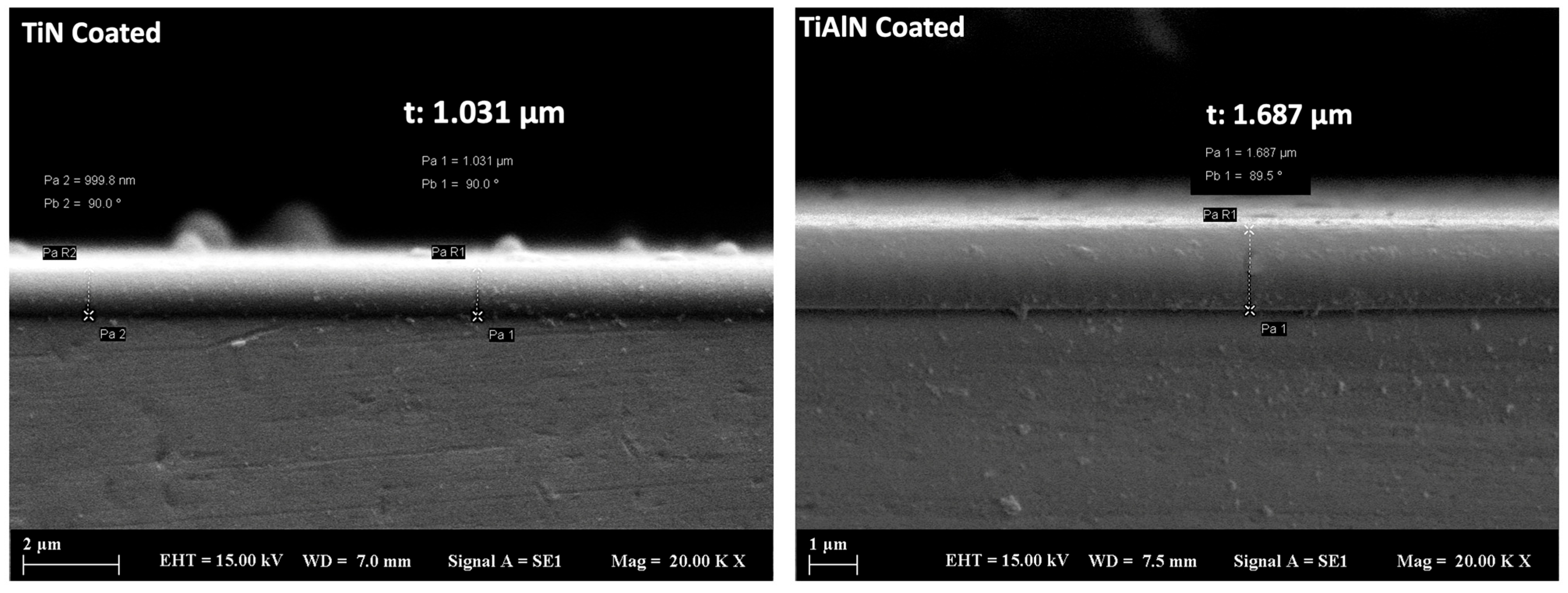
For both TiN- and TiAlN-coated TNTZ substrates, the coating thicknesses were measured from SEM cross-sectional images shown in Figure 4. This approach permitted for examination of the coating structure and its uniformity across the sample. Figure 4 provides a cross-sectional perspective for the TiN-coated and TiAlN-coated samples, which allowed the accurate determination of the average thickness values. The average thickness of the coatings was determined from these measurements. The TiN coating was found to have a thickness of 1.03 ± 0.04 µm, whereas the TiAlN coating was thicker at 1.687 ± 0.05 µm. Considering the usage of the same substrate materials and close surface quality, the thickness variation is primarily related to differences in process parameters, particularly the applied bias voltage and deposition duration required for TiN versus TiAlN. This thickness variation may influence the direct comparison of properties such as hardness and roughness.
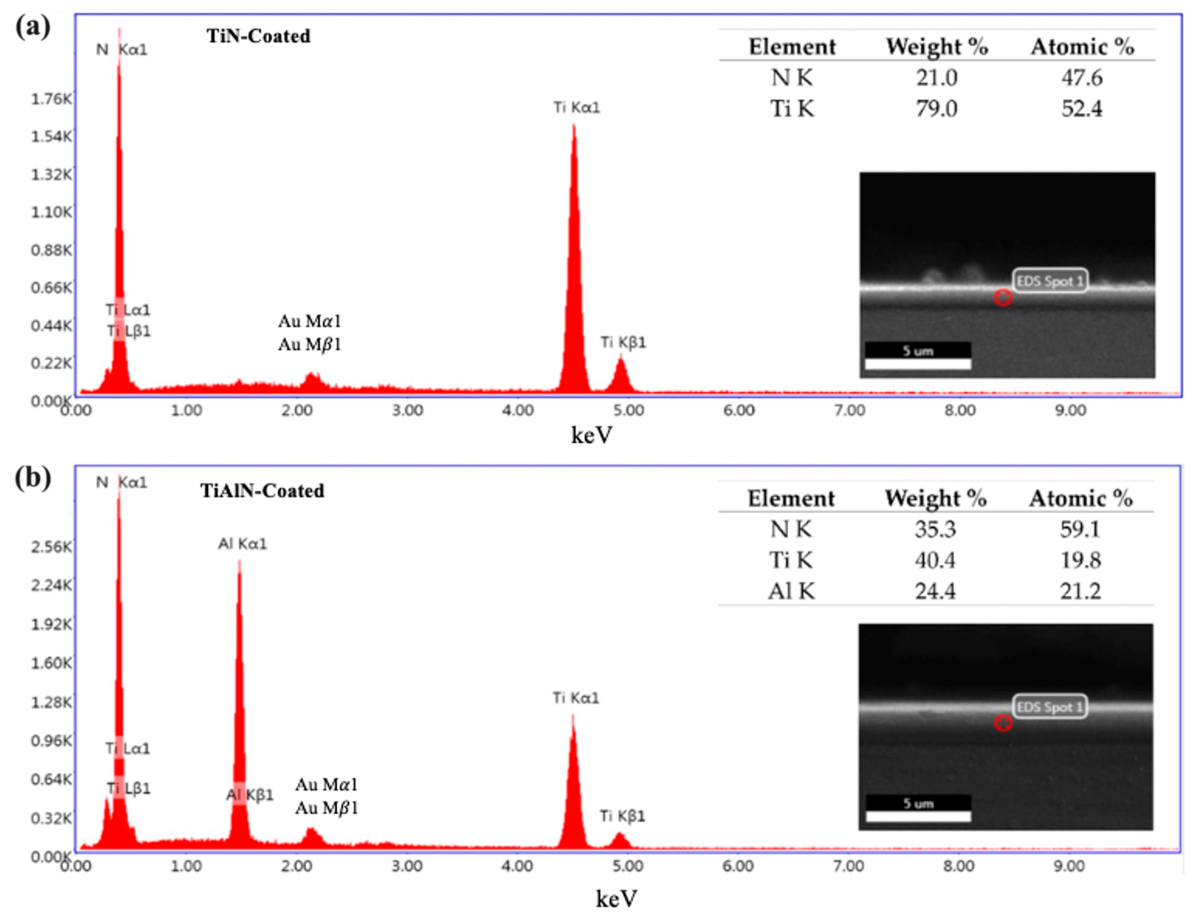
EDS analysis results for both TiN-coated and TiAlN-coated specimens are shown in Figure 5. The data provide elemental distribution patterns that confirm the successful deposition of the intended coatings. For the TiN coating, Figure 5a shows a nearly equal atomic ratio of titanium and nitrogen, indicating a proper stoichiometric ratio for TiN. Similarly, the elemental distribution in Figure 5b confirms that the TiAlN coating was successfully deposited on the surface. A thorough analysis of SEM images from the cross-sectional areas of thin-film-coated specimens created via the PVD method revealed the absence of defects at the interface.
It should be noted that SEM-EDS provides only semi-quantitative elemental analysis and thus indicates the presence of Ti, Al, N, but does not yield precise film stoichiometry. For example, the TiAlN coating was deposited using a 50:50 wt% Ti–Al target (intended to produce roughly equiatomic Ti and Al), but the actual Ti:Al ratio in the film may deviate from this ideal. Therefore, the measured EDS ratios should be viewed as approximate rather than exact.

3.2. Coating Formation Mechanisms: Crystallite Size Estimation and AFM Topography

The diffraction peaks are noticeably broad, reflecting the nanocrystalline nature of the PVD coatings. Peak broadening in XRD is principally caused by the finite size of crystallites (coherent domains) and, to a lesser extent, microstrain. In the present arc-PVD films, lattice microstrain is minimal (unlike heavily strained bulk alloys), so the peak width can be attributed mainly to small subgrain (crystalite domain) size. For TiN, the (111) peak’s full-width at half maximum (FWHM) is ~0.86°, corresponding to an average crystallite size of D ≈ 9.7 nm (Table 2). The as-measured TiAlN (200) reflection was broader (FWHM ≈ 1.33°), suggesting ~6 nm crystallites. However, this peak was a convolution of TiN-like structures as a doublet component. After deconvoluting the (200) peak to isolate the physical broadening, we obtained a narrower effective FWHM (~0.82°), which yields an updated TiAlN crystallite size of approximately D ≈ 10 nm. Both coatings consist of very fine grains on the order of 10 nm. The TiAlN coating’s grains are comparable in size to or slightly larger than those of TiN—a somewhat unexpected result, since aluminum additions in TiN are often reported to refine grain size. In our case, the XRD peak for TiAlN is marginally narrower than for TiN, indicating that Al did not significantly reduce the crystallite size.
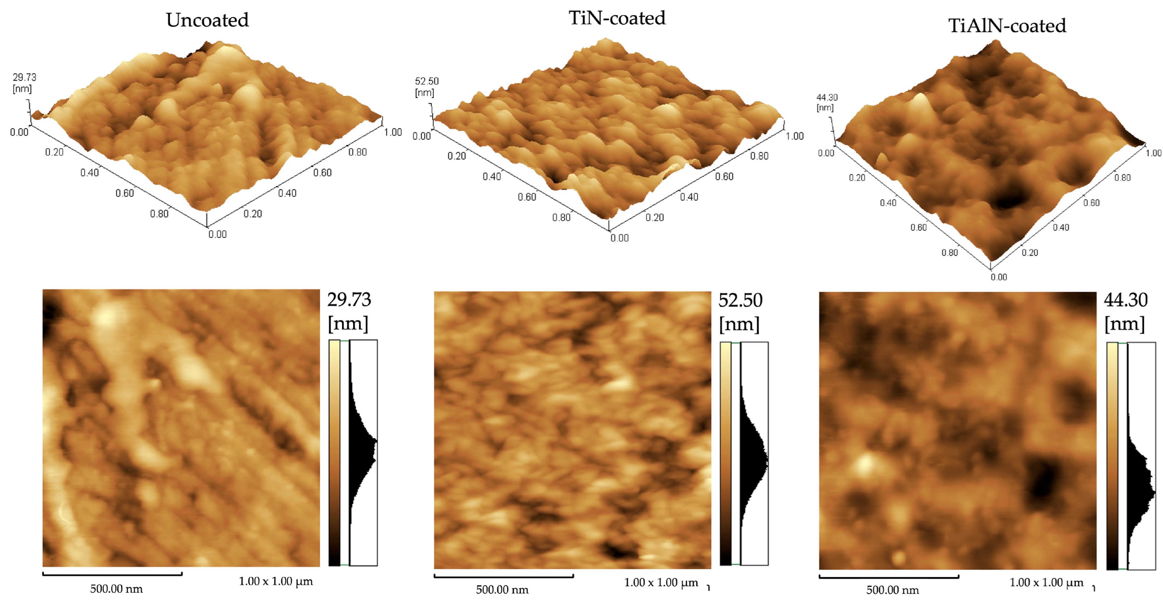
Atomic force microscopy (AFM) reveals how these nanoscale grains/subgrains manifest in surface topography. The AFM scans (Figure 6) show that the uncoated polished TNTZ substrate is extremely smooth (Ra ≈ 3.1 nm, Rq ≈ 4.0 nm). Coating deposition increases the roughness modestly: TiN-coated surfaces have Ra ≈ 5.2 nm (Rq ≈ 6.6 nm), while TiAlN-coated surfaces have Ra ≈ 4.2 nm (Rq ≈ 5.3 nm). The peak-to-valley height (Rz) is more strongly affected, roughly doubling from ~30 nm (uncoated) to ~52.5 nm for TiN and ~44.2 nm for TiAlN. The fact that both coatings are so smooth (Ra in the 4–5 nm range) underscores the highly controlled nature of the PVD growth process—even with some droplets present, the atomic-scale uniformity of the coating growth leads to an almost atomically flat surface.
The TiAlN coating has slightly fluctuated between bigger and smaller grains (subgrains (crytallite domain) and lower roughness. This trend is consistent with earlier observations that TiAlN coatings (Ra ≈ 209 nm) tend to be smoother than TiN (246 nm) under similar conditions [44]. Indeed, hard PVD coatings with smaller crystallite size are generally found to be smoother [44,45]. The particularly smooth TiAlN surface (despite its small grain size) likely reflects its more uniform, dense microstructure: PVD-induced growth defects like cathodic-arc droplets and nodular particles—which dominate roughness—may be fewer or smaller in the TiAlN layer [46]. Both coatings retain nanometric Ra and Rz values, but the TiAlN film (with ~10.39 nm grains) presents a more uniform, fine-grained morphology and lower roughness than the TiN film (with ~9.7 nm grains), reflecting subtle differences in droplet morphology and film densification during deposition.
The difference becomes clear upon examining the AFM images: both coatings exhibit micrometer-scale “droplet” features characteristic of cathodic arc deposition. These appear as bright, hemispherical bumps a few hundred nanometers in lateral size. In the TiN coating, the droplets are relatively larger and taller—individual protrusions are ~25–30 nm high (comparable to the Rp values in Table 3) and up to several hundred nm across. The TiAlN coating shows the same phenomenon but with smaller, more rounded droplets, resulting in a lower Rz. Such macroparticles are a known byproduct of the cathodic arc PVD process: molten metal droplets are ejected from the cathode spots and deposit on the growing film, creating raised nodules that increase surface roughness [44,47]. Unfiltered arc systems (like that used here) invariably produce these defects [48,49]. Indeed, prior studies confirm that the presence of undissolved droplets can increase PVD coating roughness significantly [50,51]. In our results, TiAlN’s smoother finish can be attributed to its smaller/less pronounced droplets. Apart from the droplets, the underlying intrinsic roughness of the coatings is very low—on the order of only a few nanometers Ra—reflecting the nano-scale grain structure. The fine grains themselves do not produce significant surface texture at this scale; instead, roughness is dominated by the arc-induced particles. This is why TiAlN, despite having similar (or slightly larger) crystallites than TiN, achieves a smoother surface: the topography is governed more by droplet defect morphology than by grain size. We note that even the “rougher” TiN-coated surface (Ra ~5 nm) is still biologically smooth (far below the micron-scale roughness levels that affect cell responses), so the nano-topographical differences here are mainly of coating quality and mechanical interest rather than a concern for biocompatibility [46].
The mechanisms of coating formation in the cathodic arc PVD process explain the observed microstructural and surface differences. Deposition occurs via energetic condensation: a high-current arc (125 A) on the target continuously evaporates and ionizes metal, which condenses as a film on the heated, biased substrate [52]. Nucleation of TiN or TiAlN on the substrate leads to a dense array of grains that grow competitive columns through the coating thickness. Cross-sectional microscopy (Figure 4) indeed shows a uniform coating with a columnar microstructure and a well-bonded interface, characteristic of arc-PVD nitride films. The grain size and surface morphology are sensitive to key deposition parameters such as arc current, substrate temperature, and bias voltage [53]. In general, higher arc currents increase the cathode spot activity and deposition rate, but also produce more macroparticles (droplets) ejected from the cathode [52]. In our process, the arc current was fixed, so both coatings experienced a similar droplet flux; however, the cathode material differed (pure Ti vs. Ti–Al alloy), which can influence droplet size distribution. The substrate temperature was ~480 °C (a typical value for arc deposition of nitrides), which is high enough to afford appreciable adatom mobility. Higher substrate temperatures promote grain coarsening and better crystallinity in PVD films [42]. With the TiAlN run, a longer deposition time (yielding a thicker ~1.7 μm film vs. ~1.06 μm for TiN) combined with this elevated temperature likely allowed grains to grow slightly larger on average, consistent with the XRD results. By contrast, a shorter deposition or lower temperature tends to freeze in smaller grains. Substrate bias voltage also plays a crucial role. The TiN coating was deposited at –50 V bias, whereas TiAlN was used at –100 V bias. A higher bias accelerates more ions towards the substrate, increasing ion bombardment energy during film growth. This energetic bombardment has two main effects: it can re-sputter adatoms from protruding features (helping to smooth the film), and it can peen the growing film, creating defects that act as additional nucleation sites—both of which tend to refine the microstructure. In various studies, increasing the bias voltage has been shown to produce smaller grain sizes in arc-PVD nitride coatings [54] along with higher compressive stress. In our case, the –100 V bias for TiAlN likely contributed to a denser, more homogenously nucleated film compared to TiN’s lower bias. This may be one reason the TiAlN coating developed a uniformly fine-grained morphology and did not exhibit any exceptionally large crystallites despite the longer deposition. The higher bias also could aid in “flattening” the film by preferentially sputtering high points (such as the tops of droplets) during growth, which would mitigate roughness. In effect, the TiAlN process parameters (greater bias and thickness) yielded a coating that, although nanocrystalline like TiN, is slightly more compact and topographically smooth.
The addition of aluminum (Al) itself influences the growth mechanism. Alloying TiN with Al alters the crystal chemistry and can modify growth kinetics [39]. Al has a high affinity for N and tends to stabilize the TiAlN phase with a slightly smaller lattice parameter, which may change the preferred growth orientation (indeed, TiAlN showed a prominent (200) reflection, whereas TiN’s strongest peak was (111)). Many reports suggest that Al incorporation in nitrides results in grain refinement, attributing this to reduced surface diffusivity or growth disruption by differing atomic flux [52,55,56]. In the present study, we did not observe a dramatic grain size reduction with Al—in fact, the TiAlN grains are of similar size to that of TiN. This indicates that the deposition conditions (especially the higher bias and temperature for TiAlN) counteracted any inherent grain-refining effect of Al. Instead, the influence of Al is seen in the morphology of the coating. TiAlN formed a very uniform, dense structure with fewer defects.
The smoother surface of TiAlN can thus be understood as a result of both material and process factors: the material (TiAlN) may nucleate in a manner that yields a high nuclei density, and it hardens the film (which could limit grain boundary mobility), while the process (higher bias, longer growth) leads to effective densification and smaller droplet imprint. The formation of the TiN and TiAlN coatings via cathodic arc PVD produces a nanocrystalline columnar microstructure in both cases, but the specific parameters and alloying differentiate their grain size and surface features. TiN grew with ~10 nm crystallites and relatively large arc droplets, giving a slightly rougher nanostructured surface. TiAlN grew under more energetic conditions, achieving an equally fine grain size (~10 nm) and incorporating Al atoms into the lattice, which together yielded a more compact grain arrangement and smaller surface droplets. These microstructural distinctions are directly reflected in the AFM-measured roughness (TiAlN being ~20% smoother than TiN).
All samples have extremely low nanoscopic roughness (Ra on the order of 1–5 nm), far below the micron-scale roughness known to enhance bone growth [57]. Implant studies show moderately rough surfaces (Ra ~1–2 µm) maximize osteoblast response [52], while very smooth (Ra < 0.5 µm) or extremely rough surfaces are less ideal. Here, even the “roughest” coated surface (Ra ≈ 5 nm = 0.005 µm) is essentially smooth biologically. Therefore, changes in Ra at this scale will likely have minimal direct effect on cell adhesion or osseointegration. Crucially, TiN and TiAlN themselves are highly biocompatible. TiN-coated implants have a long clinical history. Studies report that nitride coatings can actually improve cell viability [57,58]. For example, human fibroblasts showed higher proliferation on TiN- or TiAlN-coated alloy surfaces than on uncoated metal [52]. Another study found that mammalian cells grew and spread better on TiN films than on bare titanium [58]. The chemistry (inert nitrides) and enhanced corrosion resistance (less ion release) generally boost biocompatibility [28]. The slightly increased nanotopography of the coatings should not harm biocompatibility; on the contrary, the hard, stable nitride surface tends to reduce metal ion release and support cell attachment.

3.3. Surface Hardness

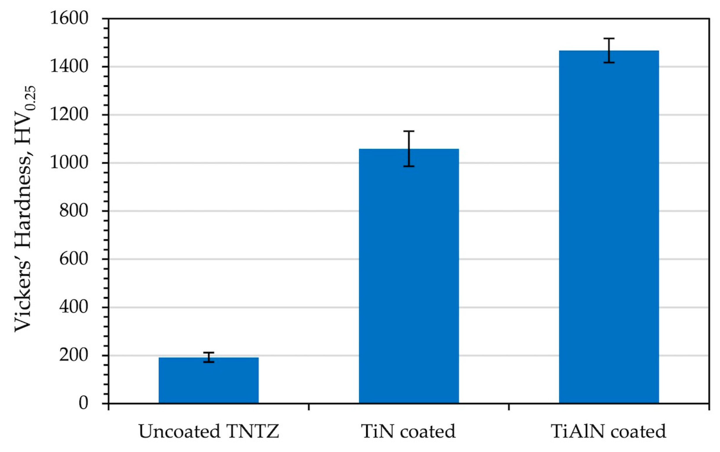
Hardness measurements were taken on uncoated TNTZ and coated specimens. The averages of the measurements taken from different points of the surfaces of the specimens are shown in Figure 7. The hardness value of the TNTZ alloy was measured as 192.2 HV on average. In a previous study, Akahori et al. raised the hardness (175 HV) of TNTZ to over 600 HV with a surface of TiN and Ti2N formed during gas nitriding [59]. The hardness of the coatings was measured as 1059.4 HV for TiN and 1467.6 HV for TiAlN. There was a significant difference between the surface hardness of the uncoated and coated specimens. The measurements showed that the TiAlN coating exhibited considerably higher hardness than the TiN coating. This difference may be attributed to the presence of substitutional Al atoms in the FCC crystal structure, in addition to interstitial N atoms [60]. Al atoms occupy the same lattice positions as Ti atoms in the structure, causing the structure to change [40]. Another contributing factor is the coating thickness. The TiAlN coating (~1.73 μm) is thicker than the TiN coating (~1.06 μm), which likely reduces substrate influence during indentation and thus leads to a higher measured hardness. In general, a thicker hard coating will yield a higher apparent microhardness because the indenter engages more of the hard coating and less of the softer substrate beneath it.

3.4. Coating Adhesion Performance

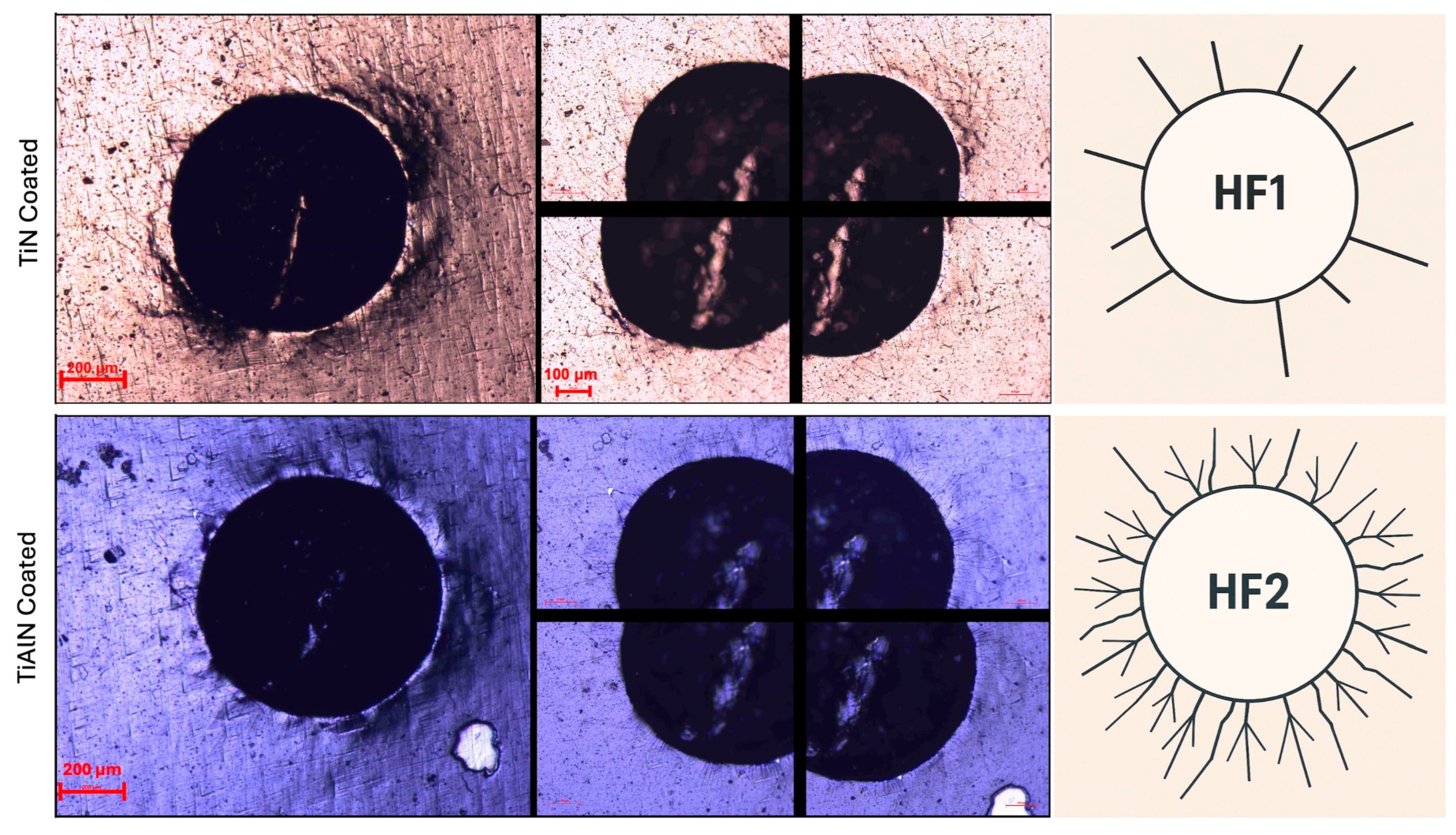
The Rockwell C indentation test was performed on the PVD TiN and TiAlN coatings according to VDI 3198 to evaluate their adhesion to the TNTZ substrate. The optical micrographs of the indented areas (Figure 8) show extensive radial cracking for the TiN film but no coating delamination or spalling. The TiAlN coating likewise showed no film peeling and even fewer and finer cracks around the indent. Under the VDI 3198 classification (from HF1 = pristine adhesion to HF6 = severe failure), these observations correspond to HF1–HF2 quality, that is, minor crack formation without any flaking. In other words, both coatings maintained their integrity under the severe Hertzian stress (no portion of the film detached), which VDI 3198 interprets as good adhesion [35,61]. This is consistent with other studies; for example, Grenadyorov et al. found that TiN/TiAlN PVD coatings typically score HF1–HF2 under the VDI 3198 test [61]. Similarly, Chang et al. reported that various Ti-based nitride films on hard substrates achieved an HF1–HF2 rating [62,63]. It was also observed that the TiN and TiAlN PVD coatings did not delaminate, and the cracking of the coating was very low [37,64,65].
According to VDI 3198, an HF1 rating signifies essentially no observable flaking (only very fine, hardly visible cracks), while an HF2 rating allows some circumferential microcracking but still no large delamination [35,66]. The TiAlN-coated sample behaved like an HF1 case (virtually no film detachment), and the TiN-coated sample behaved like an HF2 case (fine radial cracks without chipping) [35,67]. By convention, HF1–HF4 are considered acceptable adhesion modes—only HF5 and HF6 are deemed failures [35]. Thus, both coatings easily meet the VDI 3198 acceptance criteria. The fact that the TiAlN coating showed fewer cracks than the TiN coating suggests slightly better toughness or stress accommodation, but in either case, the adhesion is strong. These results imply that the TiN and TiAlN films are securely bonded to the TNTZ alloy and should perform reliably in service. The lack of any delamination or spallation under the Rockwell load indicates that substrate–film interfacial strength is high. In practical terms, HF1–HF2 coatings are routinely considered suitable for applications requiring wear resistance and biocompatibility. To further improve the coating structure, one could adopt graded or multilayer designs: for example, Elmkhah et al. [68] found that a TiN/TiAlN/TiN multilayer (introducing a hardness gradient) yielded better adhesion than a single-layer TiAlN film.

3.5. Surface Wettability

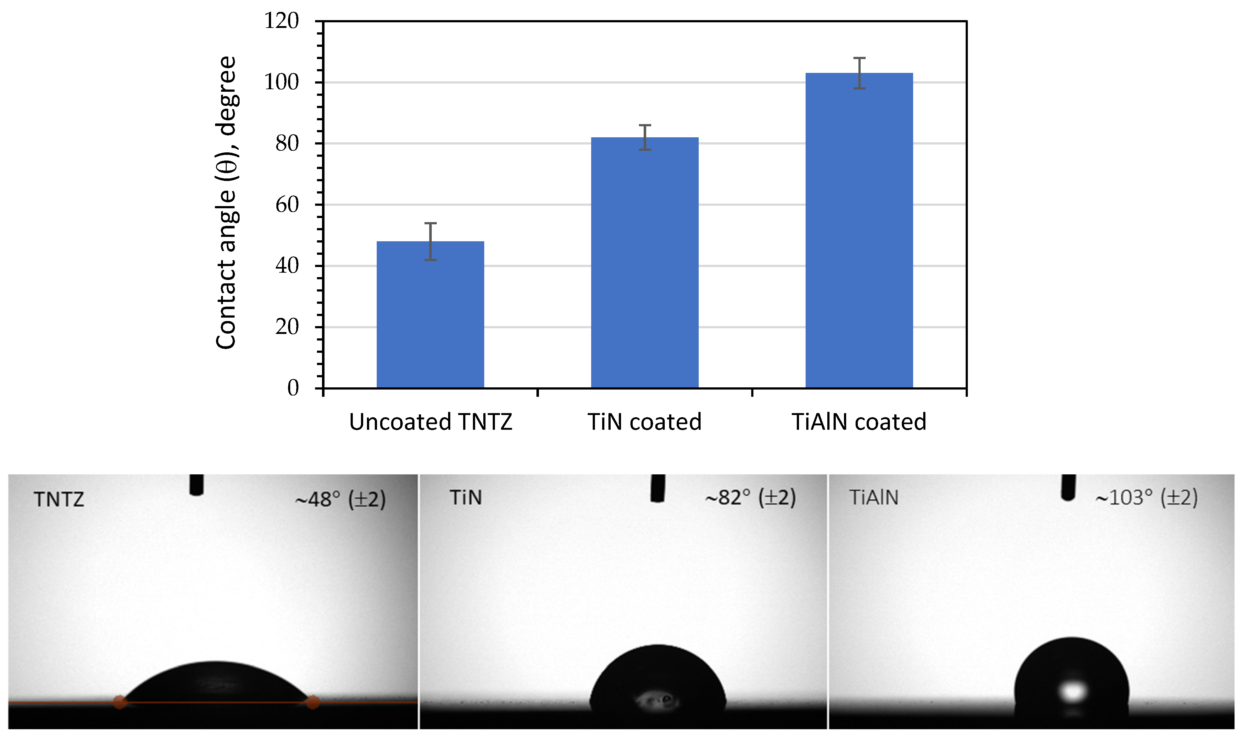
The water contact-angle measurements showed a clear trend in Figure 9. The uncoated TNTZ surface had a low contact angle (~48°), indicating strong hydrophilicity, whereas the TiN coating raised the angle to ~82°, and the TiAlN coating raised it to ~103°. This indicates that these coatings alter the surface chemistry and surface energy of TNTZ. By convention, θ ≈ 90° separates hydrophilic and hydrophobic behavior [28], so the uncoated and TiN-coated samples (θ < 90°) remain hydrophilic, but the TiAlN-coated surface becomes distinctly hydrophobic. Comparably, Ti-13Nb-13Zr alloys had a contact angle of ~43°, which increased to ~68° after TiN PVD coating [28]. Mani et al. also reported a similar tendency: 316L stainless steel had a contact angle of ~65° on its uncoated surface, which rose to ~87° with a TiN coating and ~93° with a TiAlN coating [24]. In all cases, the nitride coatings greatly increase the contact angle. This systematic increase in contact angle reflects changes in surface energy and chemistry. TiN and especially TiAlN form ceramic nitride coatings that are less polar than the native titanium oxide on uncoated Ti. These coatings exhibit “high contact angles” and “low surface energy” compared to their metallic substrates [24]. A higher angle implies a more water-repellent, hydrophobic surface, which in their study was attributed to coating roughness and non-polar character [24].
In our case, the TiAlN coating (with its aluminum component) likely forms a surface that is effectively more non-polar (e.g., due to surface Al–O or Al–N chemistry) than either uncoated TNTZ or TiN-coated TNTZ. Thus, TiAlN pushes the contact angle above 90°, whereas TiN (containing only Ti–N only) yields an intermediate value. These wettability changes have practical implications for biocompatibility. Surfaces with θ < 90° are generally considered hydrophilic and tend to promote protein adsorption and cell attachment, whereas θ > 90° indicates hydrophobicity that can hinder initial wetting [28].
In orthopedic implants, moderate hydrophilicity often aids osseointegration. For example, Piotrowska and Madej emphasized that even with a TiN coating (θ ≈ 68°), the surface remained hydrophilic, which they linked to favorable cell proliferation [28]. By contrast, the TiAlN-coated TNTZ (θ ≈ 103°) may repel water and cells more strongly. Thus, while TiN and TiAlN coatings can greatly enhance hardness and corrosion resistance, they must be balanced against potential loss of wettability. In our TNTZ system, the TiN-coated surface (θ ≈ 82°) sits just below the hydrophilic–hydrophobic threshold, retaining good wettability, whereas the TiAlN-coated surface is clearly hydrophobic. These findings are consistent with the notion that nitrided Ti surfaces tend to become more hydrophobic than the bare alloy [24,28].

3.6. Bacterial Sensitivity

The inhibition zones observed were narrow, respectively (≈17.7 mm for uncoated, 18.0 mm for TiN-coated, and 19.1 mm for TiAlN-coated TNTZ) (Figure 10b). ANOVA analysis did not reveal any statistically significant difference in inhibition zone diameter between the samples (p > 0.05). Given the small absolute differences (≤1.4 mm) and the inherent variability in measuring inhibition zones, these results should be interpreted with caution as only a preliminary indication of bacteriostatic effect. The findings are, therefore, discussed as a relative bacteriostatic tendency under disk diffusion conditions, rather than evidence of strong antibacterial activity.
It should be noted that the disk diffusion method has limitations when applied to inert nitride coatings, which do not release active agents into the medium. The modest differences observed are consistent with previous reports on TiN and TiAlN coatings, where antibacterial effects were subtle or non-significant under diffusion conditions [69]. A more conclusive assessment would require direct-contact methods such as bacterial colony counting or biofilm assays. Accordingly, the present results are considered preliminary indications rather than definitive proof of antibacterial performance.”
Many studies have noted that hydrophobic surfaces tend to promote bacterial adhesion and biofilm formation [30,70]. In our case, however, the opposite trend emerged: the highly hydrophobic TiAlN surface best resisted bacterial growth. This suggests that factors beyond simple wettability—such as surface chemistry or topography—are important. For example, TiN coatings are known to reduce bacterial colonization on titanium implants [70,71], and indeed our TiN-coated TNTZ showed slightly better inhibition than the bare TNTZ substrate. It is plausible that the TiAlN coating, with its different chemical composition (a mixed Ti–Al nitride), further impedes bacterial adhesion or viability. In particular, an aluminum-containing nitride surface can develop a modified oxide layer (e.g., involving Al2O3) that is more chemically inert and less hospitable to bacteria compared to the titanium-oxide surface on TiN. These findings are consistent with the prevailing view that nitride or ceramic-coated titanium enhances resistance to bacterial adhesion, thereby potentially reducing the risk of infection in biomedical applications [72,73]. The TiAlN-coated TNTZ exhibited the greatest bacteriostatic effect (largest inhibition zone), highlighting the coating’s potential to enhance the antibacterial performance of this β-type alloy.

3.7. Comparative Analysis with Literature

Table 4 compares the key properties of the TiN and TiAlN coatings from this study with representative literature results for similar coatings produced by various methods. The comparison includes deposition method, coating thickness, Vickers hardness, average contact angle, and reported biological properties. As shown, the hardness values for our TiN (1059 HV) and TiAlN (1468 HV) coatings are quite higher than gas nitriding results on TNTZ, but are lower than some magnetron-sputtered counterparts and are in line with other arc-PVD results, reflecting differences in deposition parameters and substrate type. The contact angles (82° for TiN and 102° for TiAlN) also fall within the ranges reported for nitride coatings, with TiAlN exhibiting the expected higher hydrophobicity. Importantly, our coatings demonstrated measurable antibacterial activity against E. coli, with inhibition zones (18.02 mm for TiN and 19.09 mm for TiAlN) comparable to or exceeding values reported for similar coatings in the literature. This consolidated comparison highlights that, while TiN and TiAlN are well-studied coating materials, their performance on β-type TNTZ alloy—particularly in terms of combined mechanical, surface, and antibacterial properties—shows competitive or superior results compared to established analogs.

4. Conclusions

This study evaluated the effects of PVD-applied TiN and TiAlN coatings on a biomedical β-type Ti-29Nb-13Ta-4.6Zr (TNTZ), focusing on microstructure, morphology, mechanical performance, wettability, and antibacterial response. The results demonstrated that both TiN and TiAlN coatings formed dense FCC lattice structures on TNTZ without altering the single-phase β matrix of the substrate. The average coating thicknesses were ~1.06 ± 0.04 µm for TiN and ~1.73 ± 0.05 µm for TiAlN, and cross-sections showed uniform, defect-free interfaces. AFM measurements indicated that the coatings were extremely smooth (Ra ≈ 4–5 nm), only slightly rougher than the polished substrate (~3 nm).
The hard nitride coatings substantially enhanced the surface mechanical properties of TNTZ. Vickers microhardness increased from about 192 HV (uncoated) to 1059 HV with TiN coating and 1468 HV with TiAlN coating. Rockwell C adhesion tests (VDI 3198) classified the TiN coating as HF2 and the TiAlN coating as HF1, indicating strong adhesion in both cases with only minor cracking and no delamination. These results confirm that both TiN and TiAlN coatings are firmly bonded to the TNTZ substrate and can withstand significant mechanical stress without failure.
The surface wettability was markedly changed by the coatings: the uncoated TNTZ was hydrophilic (water contact angle ~48°), whereas TiN-coated TNTZ was moderately hydrophobic (~82°) and TiAlN-coated TNTZ was strongly hydrophobic (~103°). Despite the higher hydrophobicity of TiAlN (which, in general, could encourage biofilm formation), the TiAlN-coated samples showed a slightly larger inhibition zone (~19.1 mm) compared with TiN-coated (~18.0 mm) and uncoated (~17.7 mm) TNTZ under disk diffusion conditions. Given the small differences observed, these results should be regarded as preliminary and not conclusive evidence of antibacterial action. Statistical analysis confirmed that the observed differences were not significant (p > 0.05), reinforcing that only a tentative bacteriostatic trend has been observed. They suggest a possible bacteriostatic tendency of TiAlN surfaces, which warrants further investigation using direct-contact assays and biofilm quantification.
Furthermore, the arc-PVD process used for coating is industrially scalable and capable of uniformly treating complex implant geometries. This implies that the improvements achieved in TNTZ through TiN and TiAlN coatings can be translated to actual orthopedic implant components. In summary, TNTZ alloy coated with TiAlN (in particular) emerges as a promising candidate for next-generation biomedical implants, offering a superior combination of mechanical durability and slightly resistance to bacterial colonization.

5. Future Directions and Biomedical Validation

Although preliminary bacteriostatic tests indicated reduced bacterial adhesion on TiN and TiAlN coatings, more rigorous assays are needed to validate their anti-biofilm efficacy. Future work will include quantitative biofilm formation assays (e.g., crystal violet staining or CFU counts) and viability imaging. A confocal laser scanning microscopy (CLSM)/Fluorescence microscopy with LIVE/DEAD fluorescent probes (SYTO®9/propidium iodide) to quantify live and dead bacteria on the coated surfaces [76,77]. The BacLight viability kit is a well-established method for assessing bactericidal efficiency on biomaterial surfaces [78]. In parallel, in vitro cytocompatibility will be evaluated by culturing osteoblast-like cells on the coated TNTZ alloy. Cell adhesion, proliferation, and morphology will be assessed by metabolic assays (MTT/WST-1) and fluorescence imaging of the actin cytoskeleton (e.g., phalloidin/DAPI staining) [79,80]. Live/dead staining of mammalian cells will further verify cell viability on the coatings [79].
We will also perform comprehensive electrochemical and surface analyses in simulated physiological media. Potentiodynamic polarization testing will be carried out in Hank’s Balanced Salt Solution (HBSS) at 37 °C (pH ~6.8) to determine corrosion potentials and current densities of TiN- and TiAlN-coated TNTZ [76]. Such polarization curves (optionally supplemented by impedance spectroscopy) are standard for evaluating implant corrosion resistance in vitro [81]. Finally, wettability and surface energy will be characterized using dynamic contact angle measurements. Specifically, advancing and receding water contact angles will be measured (e.g., with a tilting-stage method) to determine contact angle hysteresis, which quantifies liquid adhesion to the surface. Dynamic wettability is especially relevant for biomaterial surfaces, as hysteresis and surface energy influence protein adsorption and cell behavior on implants.

Funding

This research received no external funding.

Institutional Review Board Statement

Not applicable.

Informed Consent Statement

Not applicable.

Data Availability Statement

The original contributions are presented in this study’s article; further inquiries can be directed to the corresponding author.

Acknowledgments

The author gratefully acknowledges Mitsuo Niinomi for generously providing the β-type Ti-29Nb-13Ta-4.6Zr (TNTZ) alloy samples used in this study. The PVD coatings were deposited with the assistance of Ionbond IHI Group (Istanbul, Turkey), which the author sincerely appreciates.

Conflicts of Interest

The author declares no conflicts of interest.

References

  1. Niinomi, M. Mechanical biocompatibilities of titanium alloys for biomedical applications. J. Mech. Behav. Biomed. Mater. 2008, 1, 30–42. [Google Scholar] [CrossRef]
  2. Geetha, M.; Singh, A.K.; Asokamani, R.; Gogia, A.K. Ti based biomaterials, the ultimate choice for orthopaedic implants—A review. Prog. Mater. Sci. 2009, 54, 397–425. [Google Scholar] [CrossRef]
  3. Rack, H.J.; Qazi, J.I. Titanium alloys for biomedical applications. Mater. Sci. Eng. C 2006, 26, 1269–1277. [Google Scholar] [CrossRef]
  4. Niinomi, M.; Nakai, M. Titanium-based biomaterials for preventing stress shielding between implant devices and bone. Int. J. Biomater. 2011, 2011, 836587. [Google Scholar] [CrossRef] [PubMed]
  5. Biesiekierski, A.; Wang, J.; Abdel-Hady Gepreel, M.; Wen, C. A new look at biomedical Ti-based shape memory alloys. Acta Biomater. 2012, 8, 1661–1669. [Google Scholar] [CrossRef]
  6. Banerjee, R.; Nag, S.; Fraser, H.L. A novel combinatorial approach to the development of beta titanium alloys for orthopedic implants. Mater. Sci. Eng. C 2005, 25, 282–289. [Google Scholar] [CrossRef]
  7. Niinomi, M. Recent metallic materials for biomedical applications. Metall. Mater. Trans. A 2002, 33, 477–486. [Google Scholar] [CrossRef]
  8. Niinomi, M. Development of new metallic alloys for biomedical applications. Acta Biomater. 2010, 6, 3888–3903. [Google Scholar] [CrossRef]
  9. Sakaguchi, N.; Niinomi, M.; Akahori, T.; Takeda, J.; Toda, H. Relationships between tensile deformation behavior and microstructure in Ti–Nb–Ta–Zr system alloys. Mater. Sci. Eng. C 2005, 25, 363–369. [Google Scholar] [CrossRef]
  10. Niinomi, M.; Hattori, T.; Morikawa, K.; Kasuga, T.; Suzuki, A.; Fukui, H.; Niwa, S. Development of Low Rigidity β-type Titanium Alloy for Biomedical Applications. Mater. Trans. 2002, 43, 2970–2977. [Google Scholar] [CrossRef]
  11. Long, M.; Rack, H.J. Titanium alloys in total joint replacement—A materials science perspective. Biomaterials 1998, 19, 1621–1639. [Google Scholar] [CrossRef]
  12. Li, J.; Hastings, G.W. Chapter 5 Oxide Bioceramics: Inert Ceramic Materials in Medicine and Dentistry. In Handbook of Biomaterial Properties; Murphy, W., Black, J., Hastings, G., Eds.; Springer: New York, NY, USA, 2016. [Google Scholar] [CrossRef]
  13. Campoccia, D.; Montanaro, L.; Arciola, C.R. The significance of infection related to orthopedic devices and issues of antibiotic resistance. Biomaterials 2006, 27, 2331–2339. [Google Scholar] [CrossRef]
  14. Arciola, C.R.; Campoccia, D.; Montanaro, L. Implant infections: Adhesion, biofilm formation and immune evasion. Nat. Rev. Microbiol. 2018, 16, 397–409. [Google Scholar] [CrossRef]
  15. Liu, X.; Chu, P.K.; Ding, C. Surface modification of titanium, titanium alloys, and related materials for biomedical applications. Mater. Sci. Eng. R Rep. 2004, 47, 49–121. [Google Scholar] [CrossRef]
  16. Colombo, D.A.; Mandri, A.D.; Echeverría, M.D.; Massone, J.M.; Dommarco, R.C. Mechanical and tribological behavior of Ti/TiN and TiAl/TiAlN coated austempered ductile iron. Thin Solid Film. 2018, 647, 19–25. [Google Scholar] [CrossRef]
  17. Mohseni, E.; Zalnezhad, E.; Bushroa, A.R. Comparative investigation on the adhesion of hydroxyapatite coating on Ti–6Al–4V implant: A review paper. Int. J. Adhes. Adhes. 2014, 48, 238–257. [Google Scholar] [CrossRef]
  18. Musil, J. Hard and superhard nanocomposite coatings. Surf. Coat. Technol. 2000, 125, 322–330. [Google Scholar] [CrossRef]
  19. Nose, M.; Zhou, M.; Honbo, E.; Yokota, M.; Saji, S. Colorimetric properties of ZrN and TiN coatings prepared by DC reactive sputtering. Surf. Coat. Technol. 2001, 142–144, 211–217. [Google Scholar] [CrossRef]
  20. Shin, C.-S.; Gall, D.; Hellgren, N.; Patscheider, J.; Petrov, I.; Greene, J.E. Vacancy hardening in single-crystal TiNx(001) layers. J. Appl. Phys. 2003, 93, 6025–6028. [Google Scholar] [CrossRef]
  21. Jeong, Y.H.; Choe, H.C.; Brantley, W.A. Nanostructured thin film formation on femtosecond laser-textured Ti–35Nb–xZr alloy for biomedical applications. Thin Solid Film. 2011, 519, 4668–4675. [Google Scholar] [CrossRef]
  22. Safin Kaosar Saad, K.; Saba, T.; Bin Rashid, A. Application of PVD coatings in medical implantology for enhanced performance, biocompatibility, and quality of life. Heliyon 2024, 10, e35541. [Google Scholar] [CrossRef]
  23. Amirtharaj Mosas, K.K.; Chandrasekar, A.R.; Dasan, A.; Pakseresht, A.; Galusek, D. Recent Advancements in Materials and Coatings for Biomedical Implants. Gels 2022, 8, 323. [Google Scholar] [CrossRef]
  24. Mani, S.P.; Kalaiarasan, M.; Ravichandran, K.; Rajendran, N. Corrosion resistant and conductive TiN/TiAlN multilayer coating on 316L SS: A promising metallic bipolar plate for proton exchange membrane fuel cell. J. Mater. Sci. 2021, 56, 10575–10596. [Google Scholar] [CrossRef]
  25. Kang, B.S.; Sul, Y.T.; Johansson, C.B.; Oh, S.J.; Lee, H.J.; Albrektsson, T. The effect of calcium ion concentration on the bone response to oxidized titanium implants. Clin. Oral Implant. Res. 2012, 23, 690–697. [Google Scholar] [CrossRef]
  26. PalDey, S.; Deevi, S.C. Properties of single layer and gradient (Ti,Al)N coatings. Mater. Sci. Eng. A 2003, 361, 1–8. [Google Scholar] [CrossRef]
  27. Shugurov, A.R.; Kuzminov, E.D.; Garanin, Y.A. Structure and Mechanical Properties of Ti-Al-Ta-N Coatings Deposited by Direct Current and Middle-Frequency Magnetron Sputtering. Metals 2023, 13, 512. [Google Scholar] [CrossRef]
  28. Lisoń-Kubica, J.; Taratuta, A.; Wilk, K.; Kolasa, J.; Antonowicz, M.; Paszenda, Z.; Walke, W.; Gümüş, S.; Basiaga, M. The effect of TiN coating on the physicochemical properties of Ti-13Nb-13Zr alloy for biomedical applications. Langmuir 2025, 41, 13264–13271. [Google Scholar] [CrossRef] [PubMed]
  29. Kelly, P.J.; Arnell, R.D. Magnetron sputtering: A review of recent developments and applications. Vacuum 2000, 56, 159–172. [Google Scholar] [CrossRef]
  30. Grips, V.W.; Ezhil Selvi, V.; Barshilia, H.C.; Rajam, K.S. Electrochemical behavior of single layer CrN, TiN, TiAlN coatings and nanolayered TiAlN/CrN multilayer coatings prepared by reactive direct current magnetron sputtering. Thin Solid Film. 2006, 514, 204–211. [Google Scholar] [CrossRef]
  31. Hall-Stoodley, L.; Costerton, J.W.; Stoodley, P. Bacterial biofilms: From the natural environment to infectious diseases. Nat. Rev. Microbiol. 2004, 2, 95–108. [Google Scholar] [CrossRef]
  32. Oh, J.K.; Yegin, Y.; Yang, F.; Zhang, M.; Li, J.; Huang, S.; Verkhoturov, S.V.; Schweikert, E.A.; Perez-Lewis, K.; Scholar, E.A.; et al. The influence of surface chemistry on the kinetics and thermodynamics of bacterial adhesion. Sci. Rep. 2018, 8, 17247. [Google Scholar] [CrossRef]
  33. Holzwarth, U.; Gibson, N. The Scherrer equation versus the ‘Debye-Scherrer equation’. Nat. Nanotech 2011, 6, 534. [Google Scholar] [CrossRef]
  34. Aissani, L.; Belgroune, A.; Saoudi, A.; Hmima, A.; Fellah, M.; Obrosov, A.; Alhussein, A. Tribo-mechanical performance and antibacterial activity in (Cu, Zr)-alloyed Ti(Al)N coatings synthesized by reactive magnetron sputtering. J. Mater. Sci. 2022, 57, 19612–19630. [Google Scholar] [CrossRef]
  35. Vidakis, N.; Antoniadis, A.; Bilalis, N. The VDI 3198 indentation test evaluation of a reliable qualitative control for layered compounds. J. Mater. Process. Technol. 2003, 143–144, 481–485. [Google Scholar] [CrossRef]
  36. Bauer, A.W.; Kirby, W.M.M.; Sherris, J.C.; Turck, M. Antibiotic susceptibility testing by a standardized single disk method. Am. J. Clin. Pathol. 1966, 45, 493–496. [Google Scholar] [CrossRef]
  37. Xia, Y.; Cui, C.; Han, B.; Geng, H.; Liu, L. Preparation of in situ TiC@TiN core–shell and Ti2N–Al4C3 nanoparticles and their effects on Al–Zn–Mg–Cu alloy. J. Mater. Sci. 2021, 56, 17011–17027. [Google Scholar] [CrossRef]
  38. Ahlgren, M.; Blomqvist, H. Influence of bias variation on residual stress and texture in TiAlN PVD coatings. Surf. Coat. Technol. 2005, 200, 157–160. [Google Scholar] [CrossRef]
  39. Hörling, A.; Hultman, L.; Odén, M.; Sjölén, J.; Karlsson, L. Thermal stability of arc evaporated high aluminum-content thin films. J. Vac. Sci. Technol. A 2002, 20, 1815–1823. [Google Scholar] [CrossRef]
  40. Qiu, R. Electron Microscopy Investigation of Detailed Microstructures of CVD TiAlN and TiN Coatings: Effects of Gas Flow and Substrate on Coating Microstructure. Ph.D. Thesis, Chalmers University of Technology, Göteborg, Sweden, 2020. Available online: https://research.chalmers.se/publication/516330/file/516330_Fulltext.pdf (accessed on 17 September 2025).
  41. Bartosik, M.; Rumeau, C.; Hahn, R.; Zhang, Z.L.; Mayrhofer, P.H. Fracture toughness and structural evolution in the TiAlN system upon annealing. Sci. Rep. 2017, 7, 16476. [Google Scholar] [CrossRef]
  42. Abrikosov, I.A.; Knutsson, A.; Alling, B.; Tasnádi, F.; Lind, H.; Hultman, L.; Odén, M. Phase Stability and Elasticity of TiAlN. Materials 2011, 4, 1599–1618. [Google Scholar] [CrossRef] [PubMed]
  43. Elmkhah, H.; Zhang, T.F.; Abdollah-zadeh, A.; Kim, K.H.; Mahboubi, F. Surface characteristics for the TiAlN coatings deposited by high power impulse magnetron sputtering technique at the different bias voltages. J. Alloys Comp. Part A 2016, 688, 820–827. [Google Scholar] [CrossRef]
  44. Matei, A.A.; Turcu, R.N.; Pencea, I.; Herghelegiu, E.; Petrescu, M.I.; Niculescu, F. Comparative Characterization of the TiN and TiAlN Coatings Deposited on a New WC-Co Tool Using a CAE-PVD Technique. Crystals 2023, 13, 112. [Google Scholar] [CrossRef]
  45. Long, T.; Zeng, J.; Yu, D.; Wu, S. Microstructure of TiAlN and CrAlN coatings and cutting performance of coated silicon nitride inserts in cast iron turning. Ceram. Intern. 2014, 40, 9889–9894. [Google Scholar] [CrossRef]
  46. Panjan, P.; Drnovšek, A.; Mahne, N.; Čekada, M.; Panjan, M. Surface Topography of PVD Hard Coatings. Coatings 2021, 11, 1387. [Google Scholar] [CrossRef]
  47. Zhang, Z.; Zhang, L.; Yuan, H.; Qiu, M.; Zhang, X.; Liao, B.; Zhang, F.; Ouyang, X. Tribological Behaviors of Super-Hard TiAlN Coatings Deposited by Filtered Cathode Vacuum Arc Deposition. Materials 2022, 15, 2236. [Google Scholar] [CrossRef]
  48. Anders, A. Unfiltered and filtered cathodic arc deposition. In Handbook of Deposition Technologies for Films and Coatings, 3rd ed.; Martin, P.M., Ed.; William Andrew Publishing: Norwich, NY, USA, 2010; pp. 466–531. [Google Scholar]
  49. Vlasveld, A.C.; Harris, S.G.; Doyle, E.D.; Lewis, D.B.; Münz, W.D. Characterisation and performance of partially filtered arc TiAlN coatings. Surf. Coat. Technol. 2002, 149, 217–224. [Google Scholar] [CrossRef]
  50. Martin, P.J.; Bendavid, A.; Netterfield, R.P.; Kinder, T.J.; Jahan, F.; Smith, G.B. Plasma deposition of tribological and optical thin film materials with a filtered cathodic arc source. Surf. Coat. Technol. 1999, 112, 257–260. [Google Scholar] [CrossRef]
  51. Adesina, A.Y.; Gasem, Z.M.; Mohammed, A.S. Comparative investigation and characterization of the scratch and wear resistance behavior of TiN, CrN, AlTiN and AlCrN cathodic arc PVD coatings. Arab. J. Sci. Eng. 2019, 44, 10355–10371. [Google Scholar] [CrossRef]
  52. Muhammed, M.; Javidani, M.; Ebrahimi Sadrabadi, T.; Heidari, M.; Levasseur, T.; Jahazi, M. A Comprehensive Review of Cathodic Arc Evaporation Physical Vapour Deposition (CAE-PVD) Coatings for Enhanced Tribological Performance. Coatings 2024, 14, 246. [Google Scholar] [CrossRef]
  53. Warcholinski, B.; Gilewicz, A.; Ratajski, J.; Kuklinski, Z.; Rochowicz, J. An analysis of macroparticle-related defects on CrCN and CrN coatings in dependence of the substrate bias voltage. Vacuum 2012, 86, 1235–1239. [Google Scholar] [CrossRef]
  54. Guruvenket, S.; Rao, G.M. Effect of ion bombardment and substrate orientation on structure and properties of titanium nitride films deposited by unbalanced magnetron sputtering. J. Vac. Sci. Technol. A 2002, 20, 678–682. [Google Scholar] [CrossRef]
  55. Chen, L.; Du, Y.; Wang, A.J.; Wang, S.Q.; Zhou, S.Z. Effect of Al content on microstructure and mechanical properties of Ti–Al–Si–N nanocomposite coatings. Int. J. Ref. Met. Hard Mater. 2009, 27, 718–721. [Google Scholar] [CrossRef]
  56. Cao, H.; Ouyang, X.; Wu, X.; Chen, L.; Wu, J.; Wu, J.; Wang, J.; Liao, B. Mechanical and Electrochemical Properties of Titanium Aluminum Nitride Coatings with Different Nitrogen Flow Rates on CrMnSi Steel by Filter Cathode Vacuum Arc Technology. Coatings 2025, 15, 379. [Google Scholar] [CrossRef]
  57. Boyan, B.D.; Hummert, T.W.; Dean, D.D.; Schwartz, Z. Role of material surfaces in regulating bone and cartilage cell response. Biomaterials 1996, 17, 137–146. [Google Scholar] [CrossRef] [PubMed]
  58. Chien, C.-C.; Liu, K.-T.; Duh, J.-G.; Chang, K.-W.; Chung, K.-H. Effect of nitride film coatings on cell compatibility. Dent. Mater. Off. Publ. Acad. Dent. Mater. 2008, 24, 986–993. [Google Scholar] [CrossRef]
  59. Akahori, T.; Niinomi, M.; Nakai, M.; Nishimura, H.; Takei, Y.; Fukui, H.; Ogawa, M. Wear and Mechanical Properties, and Cell Viability of Gas-Nitrided Beta-Type Ti-Nb-Ta-Zr System Alloy for Biomedical Applications. Mater. Trans. 2008, 49, 166–174. [Google Scholar] [CrossRef]
  60. Van Trinh, T.; Le, H.T.; Le Thi, B. Fabrication of Titanium Nitride Thin Film on Titanium Using Cathodic Arc Plasma Evaporation for Biomedical Application. Eur. J. Dent. 2025. [Google Scholar] [CrossRef] [PubMed]
  61. Shum, P.W.; Tam, W.C.; Li, K.Y.; Zhou, Z.F.; Shen, Y.G. Mechanical and tribological properties of titanium–aluminium–nitride films deposited by reactive close-field unbalanced magnetron sputtering. Wear 2004, 257, 1030–1040. [Google Scholar] [CrossRef]
  62. Grenadyorov, A.; Oskirko, V.; Zakharov, A.; Oskomov, K.; Rabotkin, S.; Semenov, V.; Solovyev, A.; Shmakov, A. Properties of TiAlN coatings obtained by Dual-HiPIMS with short pulses. Materials 2023, 16, 1348. [Google Scholar] [CrossRef]
  63. Prajapati, J.; Bhatt, P.M.; Patel, N.S. Force and wear analysis of PVD coated cutting tool—A review. Int. J. Adv. Res. Innov. Ideas Educ. 2015, 1, 9–16. [Google Scholar]
  64. Chang, Y.Y.; Lai, H.M. Wear behavior and cutting performance of CrAlSiN and TiAlSiN hard coatings on cemented carbide cutting tools for Ti alloys. Surf. Coat. Technol. 2014, 259, 152–158. [Google Scholar] [CrossRef]
  65. Hagarová, M.; Jakubéczyová, D.; Baranová, G.; Eliáš, M. Adhesion determination of thin wear resistant coatings. Mater. Sci. Forum 2019, 952, 107–113. [Google Scholar] [CrossRef]
  66. Estrada-Martínez, F.F.; Gómez-Vargas, O.A.; Vega-Moron, R.C.; Melo-Maximo, D.V. Adhesion and wear study on 316L steel coated with titanium nitride by PVD. J. Technol. Innov. 2022, 9, 9–25. [Google Scholar] [CrossRef]
  67. Fernandes, L.; Silva, F.J.G.; Alexandre, R. Study of TiAlN PVD coating on stamping dies used in tinplate food package production. Micromachines 2019, 10, 182. [Google Scholar] [CrossRef]
  68. Elmkhah, H.; Mahboubi, F.; Abdollah-Zadeh, A.; Rouhaghdam, A.R.S. A new approach to improve the surface properties of H13 steel for metal forming applications by applying the TiAlN multi-layer coating. J. Manuf. Process. 2018, 32, 873–877. [Google Scholar] [CrossRef]
  69. Parreira, P.; Magalhães, A.; Gonçalves, I.C.; Gomes, J.; Vidal, R.; Reis, C.A.; Leckband, D.E.; Martins, M.C.L. Effect of surface chemistry on bacterial adhesion, viability, and morphology. J. Biomed. Mater. Res. A 2011, 99, 344–353. [Google Scholar] [CrossRef] [PubMed]
  70. Oliva, S.; Trubiani, O.; Piattelli, M.; Capogreco, M.; Murmura, G. Antibacterial activity of titanium nitride coating: A mini review. Ital. J. Anat. Embryol. 2023, 127, 73–76. [Google Scholar] [CrossRef]
  71. Cunliffe, D.; Smart, C.A.; Alexander, C.; Vulfson, E.N. Bacterial adhesion at synthetic surfaces. Appl. Environ. Microbiol. 1999, 65, 4995–5002. [Google Scholar] [CrossRef]
  72. Ji, M.-K.; Park, S.-W.; Lee, K.; Kang, I.-C.; Yun, K.-D.; Kim, H.-S.; Lim, H.-P. Evaluation of antibacterial activity and osteoblast-like cell viability of TiN, ZrN and (Ti1−xZrx)N coating on titanium. J. Adv. Prosthodont. 2015, 7, 166–171. [Google Scholar] [CrossRef]
  73. Größner-Schreiber, B.; Griepentrog, M.; Haustein, I.; Müller, W.; Briedigkeit, H.; Göbel, U.B.; Lange, K. Plaque formation on surface modified dental implants: An in vitro study. Clin. Oral Implant. Res. 2001, 12, 543–551. [Google Scholar] [CrossRef] [PubMed]
  74. Shah, A.; Izman, S.; Ismail, S.N.F.; Ayu, H.M.; Kob, C.G.C.; Daud, R.; Kadir, M.R.A. The Influence of Ultrasonic Vibration Frequency on the Properties of TiN Coated Biomedical Ti–13Zr–13Nb. Metals 2018, 8, 317. [Google Scholar] [CrossRef]
  75. Subramanian, B.; Muraleedharan, C.V.; Ananthakumar, R.; Jayachandran, M. A comparative study of titanium nitride (TiN), titanium oxy nitride (TiON) and titanium aluminum nitride (TiAlN), as surface coatings for bio implants. Surf. Coat. Technol. 2011, 205, 5014–5020. [Google Scholar] [CrossRef]
  76. Cao, Y.; Su, B.; Chinnaraj, S.; Jana, S.; Bowen, L.; Charlton, S.; Duan, P.; Jakubovics, N.S.; Chen, J. Nanostructured titanium surfaces exhibit recalcitrance towards Staphylococcus epidermidis biofilm formation. Sci. Rep. 2018, 8, 1071. [Google Scholar] [CrossRef] [PubMed]
  77. Lorenzetti, M.; Dogša, I.; Stošicki, T.; Stopar, D.; Kalin, M.; Kobe, S.; Novak, S. The Influence of Surface Modification on Bacterial Adhesion to Titanium-Based Substrates. ACS Appl. Mater. Interfaces 2015, 7, 1644–1651. [Google Scholar] [CrossRef]
  78. Berney, M.; Hammes, F.; Bosshard, F.; Weilenmann, H.U.; Egli, T. Assessment and Interpretation of Bacterial Viability by Using the LIVE/DEAD BacLight Kit in Combination with Flow Cytometry. Appl. Environ. Microbiol. 2007, 73, 3283–3290. [Google Scholar] [CrossRef]
  79. Yin, C.; Zhang, T.; Wei, Q.; Cai, H.; Cheng, Y.; Tian, Y.; Leng, H.; Wang, C.; Feng, S.; Liu, Z. Surface treatment of 3D printed porous Ti6Al4V implants by ultraviolet photofunctionalization for improved osseointegration. Bioact. Mater. 2021, 7, 26–38. [Google Scholar] [CrossRef] [PubMed]
  80. Levin, M.; Spiro, R.C.; Jain, H.; Falk, M.M. Effects of Titanium Implant Surface Topology on Bone Cell Attachment and Proliferation in vitro. Med. Devices 2022, 15, 103–119. [Google Scholar] [CrossRef]
  81. Anderson, S.; Affi, J.; Yuli, Y.; Niinomi, M.; Akahori, T.; Gunawarman. Improving the Corrosion Resistance of TNTZ in Hanks’ Solution after Thermomechanical Treatment. J. Hunan Univ. Nat. Sci. 2022, 49, 96–109. [Google Scholar] [CrossRef]
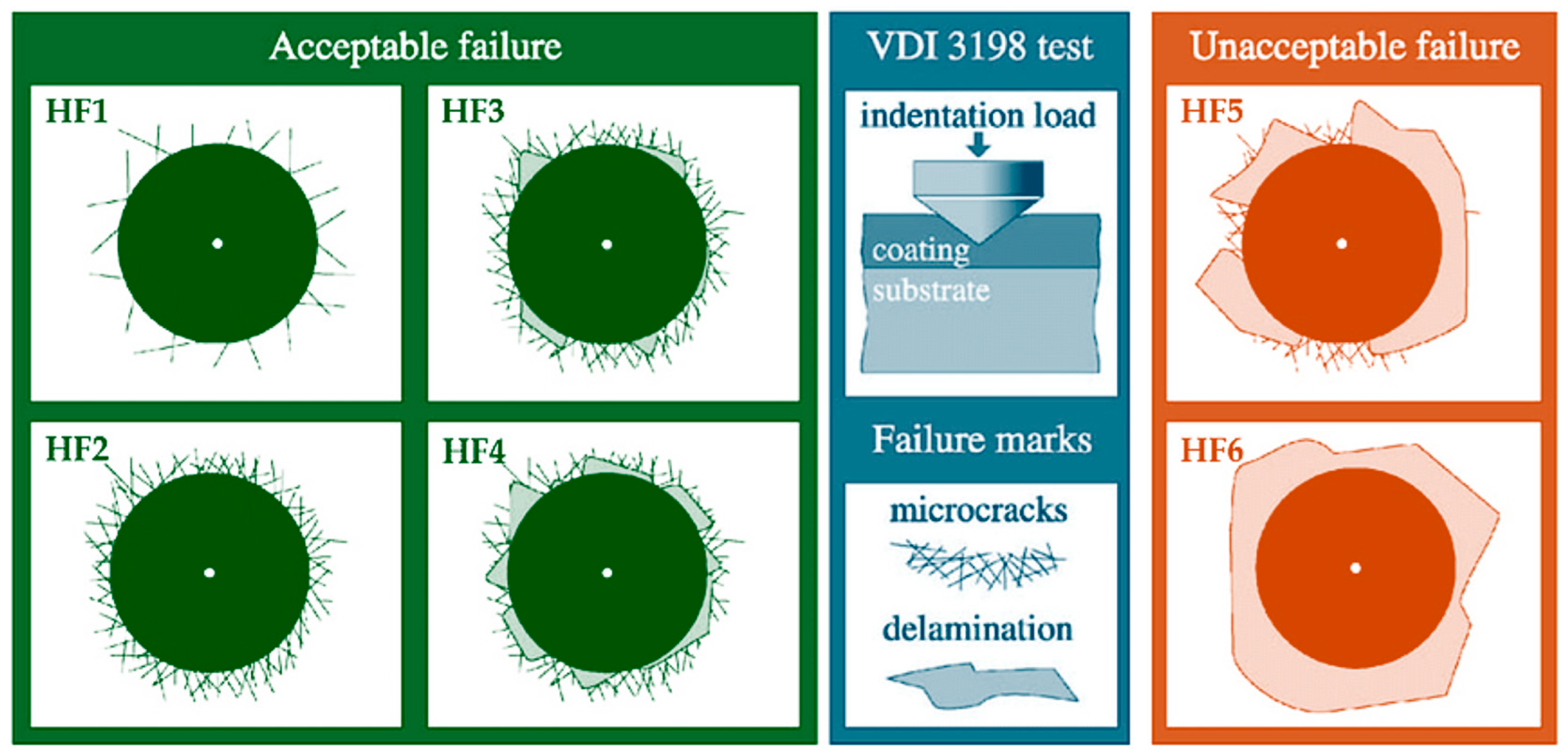
Figure 1. Illustration of the principle of the VDI 3198 qualitative coating adhesion evaluation.
Coatings 15 01126 g001
Figure 2. XRD profiles of TiN-coated and TiAlN-coated TNTZ specimens. The blue arrows show the shoulders near the (200), (220), and (311) peaks.
Coatings 15 01126 g002
Figure 3. The deconvoluted XRD peak profiles of TiAlN-coated TNTZ specimen using Origin Lab program.
Coatings 15 01126 g003
Figure 4. SEM images taken from the cross-section of TiN-coated and TiAlN-coated TNTZ. Upper black part is polymeric mounting material, and bottom part is TNTZ substrate. These micrographs show the coating thickness and a uniform, well-bonded interface with the substrate. (A higher-magnification cross-sectional image will be provided in the final publication to improve clarity.).
Coatings 15 01126 g004
Figure 5. SEM-EDS results obtained from the cross-section of (a) TiN-coated and (b) TiAlN-coated TNTZ specimens. Measurement points are marked in red on the SEM images. The corresponding EDS spectra indicated strong Ti and N signals for the TiN coating, and Ti, Al, and N for the TiAlN coating, confirming the expected elemental makeup of each coating. The small Au peak originates from the conductive coating layer applied for SEM imaging.
Coatings 15 01126 g005
Figure 6. Representative surface-topography maps—displayed in both three-dimensional (3D) (upper images) and two-dimensional (2D) (lower images)—were recorded for uncoated, TiN-coated, and TiAlN-coated. Each panel corresponds to a 1 µm × 1 µm scan acquired with a scanning-probe microscope (AFM). In the 2D maps, color indicates surface height (bright = low, dark = high). The coatings exhibit extremely low roughness on the nanoscale. No large droplet particles are evident on the coated surfaces.
Coatings 15 01126 g006
Figure 7. Average micro Vickers’ hardness values of uncoated, TiN-coated, and TiAlN-coated TNTZ.
Coatings 15 01126 g007
Figure 8. Optical microscope images after the Rockwell adhesion test on TiN-coated and TiAlN-coated TNTZ, with the standard comparison key figures of the HF1 and HF2 for the Rockwell adhesion tests. In the VDI 3198 ranking [35], HF1 signifies excellent adhesion (virtually no coating damage), whereas HF2 indicates good adhesion (minor cracking without delamination).
Coatings 15 01126 g008
Figure 9. Average contact angles and contact angle pictures of the distilled water on the substrates of TNTZ and TiN and TiAlN-coated TNTZ at 25 °C.
Coatings 15 01126 g009
Figure 10. (a) Pictures obtained after the antibacterial sensitivity tests after 24 h against E. coli. incubation of uncoated, TiN-coated, and TiAlN-coated TNTZ. (b) Histogram of the average inhibition zone lengths obtained after antibiotic susceptibility test against E. coli incubation. The length of the sample is 15 mm.
Coatings 15 01126 g010
Table 1. Elemental composition (mass %) of the hot-forged Ti-29Nb-13Ta-4.6Zr.
NbTaZrFeCNOHTi
28.612.34.750.220.020.010.090.04Balance
Table 2. Estimated XRD data and calculated crystalite size using Scherrer formula.
Samples2θFHWM (Bobs)FHWM (β)Crystalite Size (D)
TiN (111)36.46≈0.86°≈0.015 rad9.74 nm
TiAlN (200)
(Figure 2)
43.36°≈1.33°≈0.023 rad.6.39 nm
TiAlN (200)
(Figure 3)
43.36°≈0.82°≈0.014 rad.10.39 nm
Table 3. Topographical parameters obtained from AFM analysis of uncoated, TiN-coated, and TiAlN-coated TNTZ samples.
AFM Topographical Parameters (nm)Uncoated TNTZTiN CoatedTiAlN Coated
Arithmetic roughness (Ra)3.055.234.02
Maximum height (Rz)29.7352.5044.16
Ten-point mean roughness (Rzjis)14.8225.2821.88
root-mean-squared roughness (Rq)3.966.585.29
Maximum profile peak height (Rp)14.3727.3227.83
Maximum profile valley depth (Rv)15.3725.1816.33
Table 4. Comparison of TiN and TiAlN coating properties in this study vs. literature reports.
Coating
(Substrate)
Deposition MethodCoating
Thickness (µm)
Hardness (HV)Contact Angle (°)Biological Properties Refs.
TiN (TNTZ) Arc-PVD1.06 ± 0.041059 ± 1582 ± 2Inhibition zone: 18.02 mm (E. coli)This study
TiAlN (TNTZ)Arc-PVD1.73 ± 0.051468 ± 20102 ± 2Inhibition zone: 19.09 mm (E. coli)This study
TiN (Ti-13Nb-13Zr)Arc-PVD~2~1800~68Increased osteoblast proliferation; reduced bacterial adhesion (S. aureus)[28]
TiN (Ti-13Nb-13Zr)ultrasonic vibrations assisted Arc-PVD.~1.58 to 1.86.n.r.~75Improved corrosion properties[74]
TiN, TiON and TiAlN (pure titanium)DC magnetron sputter depositionn.r.n.r.n.r.TiAlN exhibited superior corrosion resistance, acceptable cytotoxicity[75]
TiAlN (316L SS)Arc-PVD~22500~93Reduced bacterial adhesion; improved wear resistance[24]
TiAlN (M2 steel) (AISI 321 steel)Magnetron sputtering2–32300–2500~90Maintained cell viability; reduced biofilm[26]
TiN and Ti2N, (TNTZ)Gas nitridingA fewOver 600n.r.Better cell viability[59]
Notes: n.r. = not reported.
Disclaimer/Publisher’s Note: The statements, opinions and data contained in all publications are solely those of the individual author(s) and contributor(s) and not of MDPI and/or the editor(s). MDPI and/or the editor(s) disclaim responsibility for any injury to people or property resulting from any ideas, methods, instructions or products referred to in the content.