
Influence of Co-Content on the Optical and Structural Properties of TiOx Thin Films Prepared by Gas Impulse Magnetron Sputtering


 ,
,
Abstract
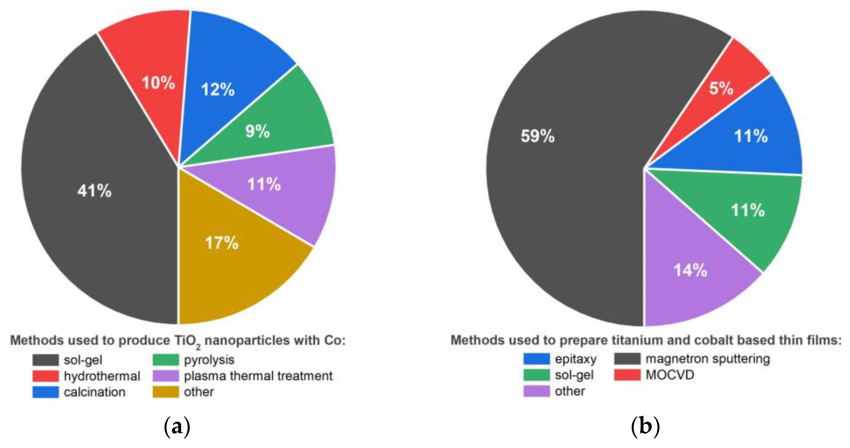
1. Introduction
2. Materials and Methods
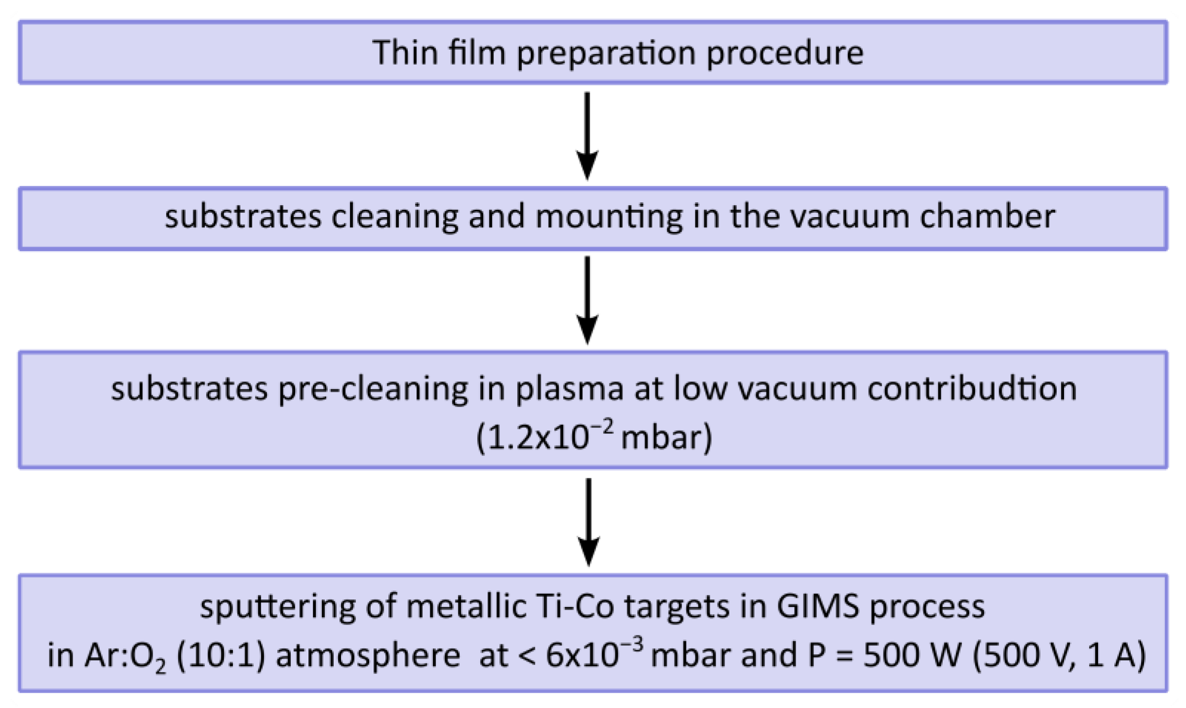
2.1. Preparation of Thin Films
2.2. Methods of Thin Film Characterisation
3. Results and Discussion
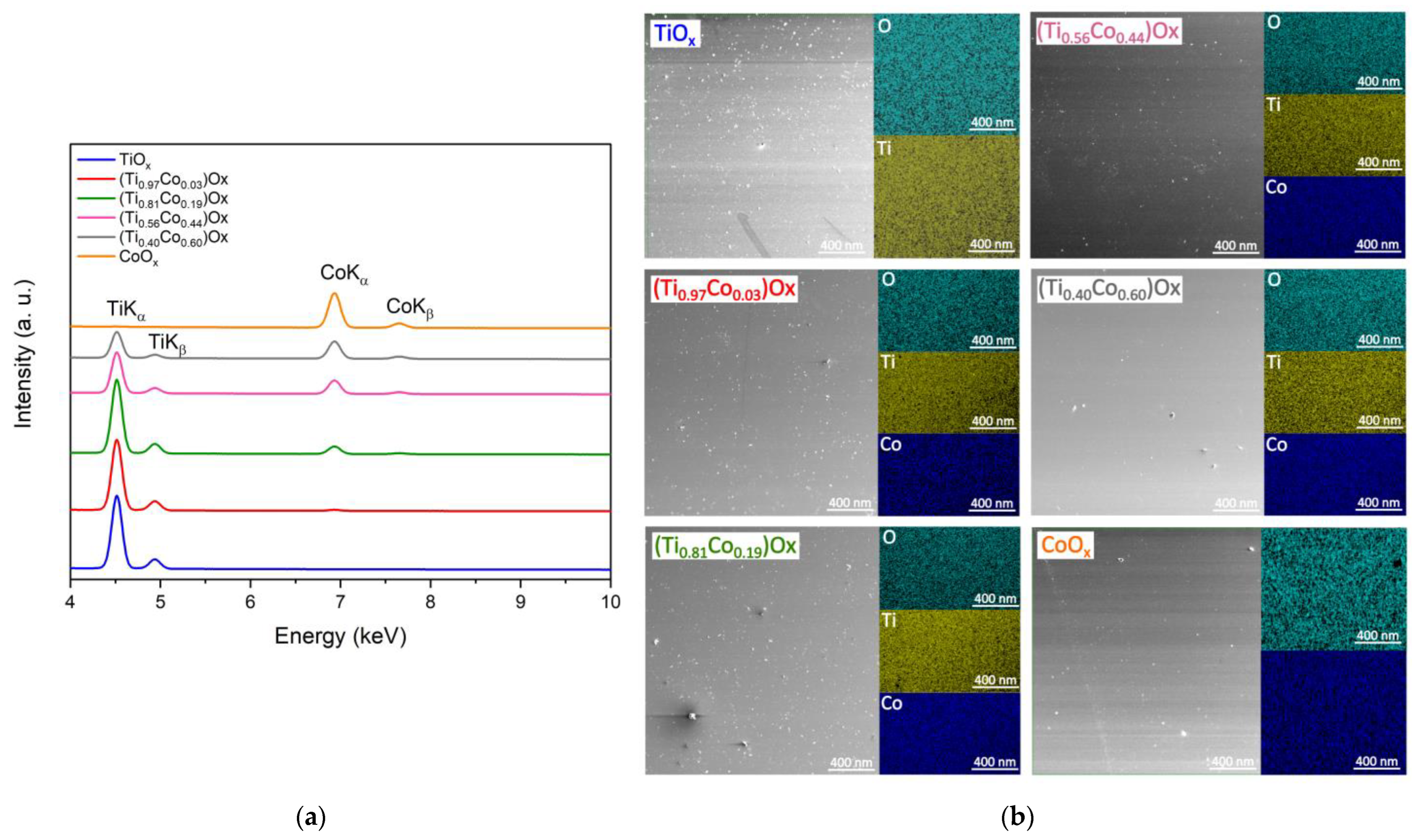
3.1. Material Composition of (Ti,Co)Ox Thin Films
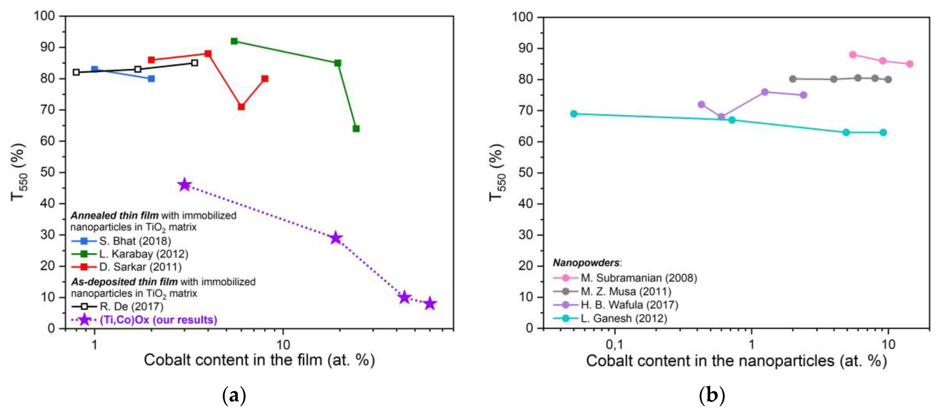
3.2. Optical Characterisation of (Ti,Co)Ox Thin Films
3.3. Structural Characterisation of Thin Films of (Ti,Co)Ox
4. Conclusions
Author Contributions
Funding
Institutional Review Board Statement
Informed Consent Statement
Data Availability Statement
Conflicts of Interest
Abbreviations
| a | lattice parameters |
| d | interplane distance |
| dPDF | standard interplane distance (according to PDF card) |
| D | average crystallite size |
| E | energy |
| Eg | width of the energy gap |
| Egopt. | width of the optical energy gap |
| Hsubstart | hardness of the substrate |
| Hthin film | hardness of the thin film |
| h | Planck’s constant |
| Tλ | light transmission coefficient |
| T550 | average value of the light transmission coefficient for the wavelength λ = 550 nm |
| Tλa | average transmission in the range of 300 to 900 nm |
| t | thickness of the films |
| α | absorption coefficient |
| λ | wavelength of electromagnetic radiation |
| λcut-off | position of the edge of optical absorption |
| EDS | energy-dispersive spectroscopy |
| (Ti0.97Co0.03)Ox | abbreviated notation of the thin film as a mixture of oxides in which the atomic content is: 97% at. Ti to 3% at. Co |
| powder diffraction files | |
| SEM | transmission electron microscopy |
| XPS | X-ray photoelectron spectroscopy |
| XRD | grazing incidence X-ray diffraction |
References
- Venturini, J.; Bonatto, F.; Guaglianoni, W.C.; Lemes, T.; Arcaro, S.; Alves, A.K.; Bergmann, C.P. Cobalt-doped titanium oxide nanotubes grown via one-step anodization for water splitting applications. Appl. Surf. Sci. 2019, 464, 351–359. [Google Scholar] [CrossRef]
- Liu, C.; Wang, F.; Zhu, S.; Xu, Y.; Liang, Q.; Chen, Z. Controlled charge-dynamics in cobalt-doped TiO2 nanowire photoanodes for enhanced photoelectrochemical water splitting. J. Colloid Interface Sci. 2018, 530, 403–411. [Google Scholar] [CrossRef] [PubMed]
- Yang, C.; Makabu, C.M.; Du, X.; Li, J.; Sun, D.; Liu, G. Cobalt nanorods decorated titanium oxide arrays as efficient and stable electrocatalyst for oxygen evolution reaction. Electrochim. Acta 2021, 396, 139213. [Google Scholar] [CrossRef]
- Khan, W.; Ahmad, S.; Hassan, M.M.; Naqvi, A.H. Structural phase analysis, band gap tuning and fluorescence properties of Co doped TiO2 nanoparticles. Opt. Mater. 2014, 38, 278–285. [Google Scholar] [CrossRef]
- Yamada, Y.; Toyosaki, H.; Tsukazaki, A.; Fukumura, T. Epitaxial growth and physical properties of a room temper ferromagnetic semiconductor: Anatase phase Ti1-xCoxO2. J. Appl. Phys. 2004, 96, 5097. [Google Scholar] [CrossRef]
- Husain, S.; Alkhtaby, L.A.; Giorgetti, E.; Zoppi, A.; Miranda, M.M. Influence of cobalt doping on the structural, optical and luminescence properties of sol-gel derived TiO2 nanoparticles. Philos. Mag. 2017, 97, 17–27. [Google Scholar] [CrossRef]
- Bhat, S.; Sandeep, K.M.; Kumar, P.; Dharmaprakash, S.M.; Byrappa, K. Characterization of transparent semiconducting cobalt doped titanium dioxide thin films prepared by sol–gel process. J. Mater. Sci. Mater. Electron. 2018, 29, 1098–1110. [Google Scholar] [CrossRef]
- Jiang, P.; Xiang, W.; Kuang, J.; Liu, W.; Cao, W. Effect of cobalt doping on the electronic, optical and photocatalytic properties of TiO2. Solid State Sci. 2015, 46, 27–32. [Google Scholar] [CrossRef]
- Musa, M.Z.; Ameran, Z.F.; Mamat, M.H.; Malek, M.F.; Rasheid, B.A.; Noor, U.M.; Rusop, M. Effects of cobalt doping concentration on the structural, electrical, and optical properties of titanium dioxide thin films. In Proceedings of the 2011 International Conference on Electronic Devices, Systems and Applications (ICEDSA), Kuala Lumpur, Malaysia, 25–27 April 2011; pp. 339–342. [Google Scholar] [CrossRef]
- Wojcieszak, D.; Mazur, M.; Pokora, P.; Wrona, A.; Bilewska, K.; Kijaszek, W.; Kotwica, T.; Posadowski, W.; Domaradzki, J. Properties of metallic and oxide thin films based on Ti and Co prepared by magnetron sputtering from sintered targets with different Co-content. Materials 2021, 14, 3797. [Google Scholar] [CrossRef]
- Wojcieszak, D.; Domaradzki, J.; Pokora, P.; Sikora, M.; Mazur, M.; Chodasewicz, P.; Morgiel, J.; Gibson, D. Optical and structural properties of gradient (Ti,Co)Ox thin-film coatings with a resistive switching effect. Appl. Opt. 2022, 34, 10283–10289. [Google Scholar] [CrossRef]
- Mei, J.; Liao, T.; Ayoko, G.A.; Bell, J.; Sun, Z. Cobalt oxide-based nanoarchitectures for electrochemical energy applications. Prog. Mater. Sci. 2019, 103, 596–677. [Google Scholar] [CrossRef]
- Verma, M.; Mitan, M.; Kim, H.; Vaya, D. Efficient photocatalytic degradation of malachite green dye using facilely synthesized cobalt oxide nanomaterials using citric acid and oleic acid. J. Phys. Chem. Solids 2021, 155, 110125. [Google Scholar] [CrossRef]
- Gajbhiye, N.S.; Sharma, S.; Nigam, A.K.; Ningthoujam, R.S. Tuning of single to multi-domain behavior for monodispersed ferromagnetic cobalt nanoparticles. Chem. Phys. Lett. 2008, 466, 181–185. [Google Scholar] [CrossRef]
- Kaloyeros, A.E.; Pan, Y.; Goff, J.; Arles, B. Cobalt thin films: Trends in processing technologies and emerging applications. ECS J. Solid State Sci. Technol. 2019, 8, 119–152. [Google Scholar] [CrossRef]
- Hirohata, A.; Yamada, K.; Nakatani, Y.; Prejbeanu, I.L.; Dieny, B.; Pirro, P.; Hillebrands, B. Review on spintronics: Principles and device applications. J. Magn. Magn. Mater. 2020, 509, 166711. [Google Scholar] [CrossRef]
- Bewan, S.; Ndolomingo, M.J.; Meijboom, R.; Bingwa, N. Cobalt oxide promoted tin oxide catalysts for highly selective glycerol acetalization reaction. Inorg. Chem. Commun. 2021, 128, 108578. [Google Scholar] [CrossRef]
- Svegl, F.; Orel, B.; Hutchins, M.G.; Kalcher, K. Structural and Spectroelectrochemical Investigations of sol-gel derived electrochromic spinel Co3O4 films. J. Electrochem. Soc. 1996, 143, 1532. [Google Scholar] [CrossRef]
- Bahlawane, N.; Rivera, E.F.; Hoinghaus, K.K.; Brechling, A.; Kleineberg, U. Characterization and tests of planar Co3O4 model catalysts prepared by chemical vapor deposition. Appl. Catal. B Environ. 2004, 53, 245–255. [Google Scholar] [CrossRef]
- Yao, W.; Fang, H.; Ou, E.; Wang, J.; Yan, Z. Highly efficient catalytic oxidation of cyclohexane over cobalt-doped mesoporous titania with anatase crystalline structure. Catal. Commun. 2006, 7, 387–390. [Google Scholar] [CrossRef]
- Ganesh, I.; Gupta, A.K.; Kumar, P.P.; Sekhar, P.S.C.; Radha, K.; Padmanabham, G.; Sundararajan, G. Preparation and characterization of Co-doped TiO2 materials for solar light induced current and photocatalytic applications. Mater. Chem. Phys. 2012, 135, 220–234. [Google Scholar] [CrossRef]
- Wang, H.; Wang, J.; Wu, Z.; Liu, Y. NO catalytic oxidation behaviors over CoOx/TiO2 catalysts synthesized by sol–gel method. Catal. Lett. 2010, 134, 295–302. [Google Scholar] [CrossRef]
- Yang, W.H.; Kim, M.H.; Ham, S.W. Effect of calcination temperature on the low-temperature oxidation of CO over CoOx/TiO2 catalysts. Catal. Today 2007, 123, 94–103. [Google Scholar] [CrossRef]
- Sadanandam, G.; Lalitha, K.; Kumari, V.D.; Shankar, M.V.; Subrahmanyam, M. Cobalt doped TiO2: A stable and efficient photocatalyst for continuous hydrogen production from glycerol: Water mixtures under solar light irradiation. Int. J. Hydrogen Energy 2013, 38, 9655–9664. [Google Scholar] [CrossRef]
- Wafula, H.B.; Musembi, R.J.; Juma, A.; Tonui, P.; Simiyu, J.; Sakwa, T.; Prakash, D.; Verma, K.D. Compositional analysis and optical properties of Co doped TiO2 thin films fabricated by spray pyrolysis method for dielectric and photocatalytic applications. Optik 2017, 128, 212–217. [Google Scholar] [CrossRef]
- Li, J.G.; Buchel, R.; Isobe, M.; Mori, T.; Ishigaki, T. Cobalt-doped TiO2 nanocrystallites: Radio-frequency thermal plasma processing, phase structure, and magnetic properties. J. Phys. Chem. C 2009, 113, 8009–8015. [Google Scholar] [CrossRef]
- Subramanian, M.; Vijayalakshmi, S.; Venkataraj, S.; Jayavel, R. Effect of cobalt doping on the structural and optical properties of TiO2 films prepared by sol-gel process. Thin Solid Films 2008, 516, 3776–3782. [Google Scholar] [CrossRef]
- Seong, N.J.; Yoon, S.G. Effects of Co-doping level on the microstructural and ferromagnetic properties of liquid-delivery metalorganic-chemical-vapor-deposited Ti1-xCoxO2 thin films. Appl. Phys. Lett. 2002, 81, 4209. [Google Scholar] [CrossRef]
- Song, H.Q.; Mei, L.M.; Zhang, Y.P.; Yan, S.S.; Ma, X.; Wang, Y.; Zhang, Z.; Chen, L.Y. Magneto-optical Kerr rotation in amorphous TiO2/Co magnetic semiconductor thin films. Phys. B Condens. Matter 2007, 388, 130–133. [Google Scholar] [CrossRef]
- Logacheva, V.A.; Lukin, A.N.; Afonin, N.N.; Serbin, O.V. Synthesis and optical properties of cobalt-modified titanium oxide films. Opt. Spectrosc. 2019, 126, 674–680. [Google Scholar] [CrossRef]
- Ko, Y.; Park, D.S.; Seo, B.S.; Yang, H.J.; Shin, H.J.; Kim, J.Y.; Lee, J.H.; Lee, W.H.; Reucroft, P.J.; Lee, J.G. Studies of cobalt thin films deposited by sputtering and MOCVD. Mater. Chem. Phys. 2003, 80, 560–564. [Google Scholar] [CrossRef]
- Quiroz, H.P.; Calderon, J.A.; Dussan, A. Magnetic switching control in Co/TiO2 bilayer and TiO2:Co thin films for magnetic-resistive random access memories (M-RRAM). J. Alloys Compd. 2020, 840, 155674. [Google Scholar] [CrossRef]
- Quiroz, H.; Galindez, E.F.; Dussan, A. Ferromagnetic-like behavior of Co doped TiO2 flexible thin films fabricated via co-sputtering for spintronic applications. Heliyon 2020, 6, e03338. [Google Scholar] [CrossRef]
- Afonin, N.N.; Logacheva, V.A. Cobalt modification of thin rutile films magnetron-sputtered in vacuum. Tech. Phys. 2018, 63, 605–611. [Google Scholar] [CrossRef]
- Griffin, K.A.; Pakhomov, A.B. Cobalt-doped anatase TiO2: A room temperature dilute magnetic dielectric material. J. Appl. Phys. 2005, 97, 10D320. [Google Scholar] [CrossRef]
- Ahmad, M.K.; Rasheid, N.A.; Ahmed, A.Z.; Abdullah, S.; Rusop, M. Study of cobalt doping on the electrical and optical properties of titanium dioxide thin film prepared by sol-gel method. In Proceedings of the 2008 IEEE International Conference on Semiconductor Electronics, Johor Bahru, Malaysia, 25–27 November 2008; pp. 561–565. [Google Scholar] [CrossRef]
- Park, W.; Ortega-Hertogs, R.J.; Moodera, J.S. Semiconducting and ferromagnetic behavior of sputtered Co-doped TiO2 thin films above room temperature. J. Appl. Phys. 2002, 91, 8093. [Google Scholar] [CrossRef]
- Tian, J.; Deng, H.; Sun, L.; Kong, H.; Yang, P.; Chu, J. Effects of Co doping on structure and optical properties of TiO2 thin films prepared by sol–gel method. Thin Solid Films 2012, 520, 5179–5183. [Google Scholar] [CrossRef]
- Quiroz, H.P.; Dussan, A. Synthesis temperature dependence on magnetic properties of cobalt doped TiO2 thin films for spintronic applications. Appl. Surf. Sci. 2019, 484, 688–691. [Google Scholar] [CrossRef]
- Park, Y.R.; Kim, K.J. Structural and optical properties of rutile and anatase TiO2 thin films: Effects of Co doping. Thin Solid Films 2005, 484, 34–38. [Google Scholar] [CrossRef]
- Chodun, R.; Dypa, M.; Wicher, B.; Langier, K.N.; Okrasa, S.; Minikayev, R.; Zdunek, K. The sputtering of titanium magnetron target with increased temperature in reactive atmosphere by gas injection magnetron sputtering technique. Appl. Surf. Sci. 2022, 574, 151597. [Google Scholar] [CrossRef]
- Zdunek, K.; Nowakowska-Langier, K.; Dora, J.; Chodun, R. Gas injection as a tool for plasma process control during coating deposition. Surf. Coat. Technol. 2013, 228, S367–S373. [Google Scholar] [CrossRef]
- Skowronski, L.; Zdunek, K.; Nowakowska-Langier, K.; Chodun, R.; Trzcinski, M.; Kobierski, M.; Kustra, M.; Wachowiak, A.; Wachowiak, W.; Hiller, T.; et al. Characterization of microstructural, mechanical and optical properties of TiO2 layers deposited by GIMS and PMS methods. Surf. Coat. Technol. 2015, 282, 16–23. [Google Scholar] [CrossRef]
- Wiatrowski, A.; Mazur, M.; Obstarczyk, A.; Wojcieszak, D.; Kaczmarek, D.; Morgiel, J.; Gibson, D. Comparison of the physicochemical properties of TiO2 thin films obtained by magnetron sputtering with continuous and pulsed gas flow. Coatings 2018, 8, 412. [Google Scholar] [CrossRef]
- Mazur, M. Analysis of the properties of functional titanium dioxide thin films deposited by pulsed DC magnetron sputtering with various O2: Ar ratios. Opt. Mater. 2017, 69, 96–104. [Google Scholar] [CrossRef]
- Mazur, M.; Wojcieszak, D.; Wiatrowski, A.; Kaczmarek, D.; Lubanska, A.; Domaradzki, J.; Mazur, P.; Kalisz, M. Analysis of amorphous tungsten oxide thin films deposited by magnetron sputtering for application in transparent electronics. Appl. Surf. Sci. 2021, 570, 151151. [Google Scholar] [CrossRef]
- Lei, C.; Du, Y.; Zhu, M.; Huo, W.; Wu, H.; Zhang, Y. Microstructure and mechanical properties of in situ TiC/Ti composites with a laminated structure synthesized by spark plasma sintering. Mater. Sci. Eng. A 2021, 812, 141136. [Google Scholar] [CrossRef]
- Wimler, D.; Lindemann, J.; Gammer, C.; Spoerk-Erdely, P.; Stark, A.; Clemens, H.; Mayer, S. Novel intermetallic-reinforced near-α Ti alloys manufactured by spark plasma sintering. Mater. Sci. Eng. A 2020, 792, 139798. [Google Scholar] [CrossRef]
- Lis, M.; Wrona, A.; Mazur, J.; Dupont, C.; Kamińska, M.; Kopyto, D.; Kwarciński, M. Fabrication And Properties Of Silver Based Multiwall Carbon Nanotube Composite Prepared By Spark Plasma Sintering Method. Arch. Metall. Mater. 2015, 60, 1351–1355. [Google Scholar] [CrossRef]
- Oliver, W.; Pharr, G. An improved technique for determining hardness and elastic modulus using load and displacement sensing indentation experiments. J. Mater. Res. 1992, 7, 1564–1583. [Google Scholar] [CrossRef]
- Jung, Y.-G.; Lawn, B.; Martyniuk, M.; Huang, H.; Hu, X. Evaluation of elastic modulus and hardness of thin films by nanoindentation. J. Mater. Res. 2004, 19, 3076–3080. [Google Scholar] [CrossRef]
- Karabay, I.; Yuksel, S.A.; Ongul, F.; Ozturk, S.; Asli, M. Structural and optical characterization of TiO2 thin films prepared by sol-gel process. Acta Phys. Pol. A 2012, 121, 265–267. [Google Scholar] [CrossRef]
- Sarkar, D.; Ghosh, C.K.; Maiti, U.N.; Chattopadhyay, K.K. Effect of spin polarization on the optical properties of Co-doped TiO2 thin films. Phys. B 2011, 406, 1429–1435. [Google Scholar] [CrossRef]
- De, R.; Tripathi, S.; Naidu, S.C.; Prathap, C.; Tripathu, J.; Singh, J.; Haque, S.M.; Rao, K.D.; Sahoo, N.K. Investigation on optical properties of spin coated TiO2/Co composite thin films. AIP Conf. Proc. 2017, 1832, 080038. [Google Scholar] [CrossRef]
- Adewinbi, S.A.; Buremoh, W.; Owoeye, V.A.; Ajayeoba, Y.A.; Salau, A.O.; Busari, H.K.; Tijani, M.A.; Taleatu, B.A. Preparation and characterization of TiO2 thin film electrode for optoelectronic and energy storage potentials: Effects of Co incorporation. Chem. Phys. Lett. 2021, 779, 138854. [Google Scholar] [CrossRef]
- Islam, M.N.; Podder, J. The role of Al and Co co-doping on the band gap tuning of TiO2 thin films for applications in photovoltaic and optoelectronic devices. Mater. Sci. Semicond. Process. 2021, 121, 105419. [Google Scholar] [CrossRef]
- Boutlala, A.; Bourfaa, F.; Mahtili, M.; Bouaballou, A. Deposition of Co-doped TiO2 thin films by sol-gel method. IOP Conf. Ser. Mater. Sci. Eng. 2016, 108, 012048. [Google Scholar] [CrossRef]
- Waseda, Y.; Matsubara, E.; Shinoda, K. X-ray Diffraction Crystallography: Introduction, Examples and Solved Problems; Springer: Berlin/Heidelberg, Germany, 2011; ISBN 978-3-642-16634-1. [Google Scholar] [CrossRef]
- Jauch, W.; Reehuls, M.; Bleif, H.J.; Kubanek, F.; Pattison, P. Crystallographic symmetry and magnetic structure of CoO. Phys. Rev. B 2001, 64, 052102. [Google Scholar] [CrossRef]
- Wdowik, U.D.; Parlinski, K. Lattice dynamics of CoO from first principles. Phys. Rev. B 2007, 75, 104306. [Google Scholar] [CrossRef]
- Moulder, J.F.; Stickle, W.F.; Sobol, P.E.; Bomben, K.D. Handbook of X-ray Photoelectron Spectroscopy; Physical Electronics, Inc.: Eden Prairie, MN, USA, 1995. [Google Scholar]
- Crist, B.V. Handbooks of Monochromatic XPS Spectra: Volume 1—The Elements and Native Oxides; XPS International, Inc.: Ames, IA, USA, 1999. [Google Scholar]
- Cole, K.M.; Kirk, D.; Thorpe, S. Co3O4 nanoparticles characterized by XPS and UPS. Surf. Sci. Spectrs. 2021, 28, 014001. [Google Scholar] [CrossRef]
- Velhal, N.B.; Yun, T.H.; Ahn, J.; Kim, T.; Kim, J.; Yim, C. Tailoring cobalt oxide nanostructures for stable and high-performance energy storage applications. Ceram. Int. 2023, 49, 4889–4897. [Google Scholar] [CrossRef]
- Gong, B.; Luo, X.; Bao, N.; Ding, J.; Li, S.; Yi, J. XPS study of cobalt TiO2 films prepared by pulsed laser deposition. Surf. Interface Anal. 2014, 46, 1043–1046. [Google Scholar] [CrossRef]
- Naseem, S.; Pinchuk, I.V.; Luo, Y.K.; Kawakami, R.K.; Khan, S.; Husain, S.; Khan, W. Epitaxial growth of cobalt doped TiO2 thin films on LaAlO3(100) substrate by molecular beam epitaxy and their opto-magnetic based applications. Appl. Surf. Sci. 2019, 493, 691–702. [Google Scholar] [CrossRef]
- Kim, J.; Livonen, T.; Hamalainen, J.; Kemell, M.; Meinander, K.; Mizohata, K.; Wang, L.; Raisanen, J.; Beranek, R.; Leskela, M.; et al. Low-temperature atomic layer deposition of cobalt oxide as effective catalyst for photoelectrochemical water-splitting devices. Chem. Mater. 2017, 29, 5796–5805. [Google Scholar] [CrossRef]
- Biesinger, M.C.; Payne, B.P.; Grosvenor, A.P.; Lau, L.W.M.; Gerson, A.R.; Smart, R.S.C. Resolving surface chemical states in XPS analysis of first row transition metals, oxides and hydroxides: Cr, Mn, Fe, Co and Ni. Appl. Surf. Sci. 2011, 257, 2717–2730. [Google Scholar] [CrossRef]
- Liu, T.; Guo, Y.F.; Yan, Y.M.; Wang, F.; Deng, C.; Rooney, D.; Sun, K.N. CoO nanoparticles embedded in the tree-dimensional nitrogen/sulfur co-doped carbon nanofiber networks as a bifunctional catalyst for oxygen reduction/evolution reactions. Carbon 2016, 106, 84–92. [Google Scholar] [CrossRef]
- Li, X.C.; She, F.S.; Shen, D.; Liu, C.P.; Chen, L.H.; Li, Y.; Deng, Z.; Chen, Z.H.; Wang, H.E. Coherent nanoscale cobalt/cobalt oxide heterostructures embedded in porous carbon for the oxygen reduction reaction. R. Soc. Chem. 2018, 8, 28625. [Google Scholar] [CrossRef] [PubMed]
- Chuang, T.J.; Brundle, C.R.; Rice, D.W. Interpretation of the X-ray photoemission spectra of cobalt oxides and cobalt oxide surfaces. Surf. Sci. 1976, 59, 413–429. [Google Scholar] [CrossRef]


| Target | Co-Content in the Film (at.%) |
|---|---|
| Ti | - |
| Ti0.98Co0.02 | 3 |
| Ti0.88Co0.12 | 19 |
| Ti0.88Co0.12 + Ti0.50Co0.50 | 44 |
| Ti0.50Co0.50 | 60 |
| Co | 100 |
| Thin Film | Phase (-) | (hkl) (-) | D (nm) | d (nm) | dPDF (nm) | a 1 (Å) |
|---|---|---|---|---|---|---|
| CoOx | CoO | (111) | 4.1 | 0.24597 | 0.24807 | 4.26033 |
| CoO | (200) | 4.0 | 0.21302 | 0.21687 | 4.33740 | |
| CoO | (220) | 4.1 | 0.15062 | 0.15125 | 4.26018 | |
| (Ti0.40Co0.60)Ox | amorphous | |||||
| (Ti0.56Co0.44)Ox | ||||||
| (Ti0.81Co0.19)Ox | ||||||
| (Ti0.97Co0.03)Ox | ||||||
| TiOx | ||||||
Disclaimer/Publisher’s Note: The statements, opinions and data contained in all publications are solely those of the individual author(s) and contributor(s) and not of MDPI and/or the editor(s). MDPI and/or the editor(s) disclaim responsibility for any injury to people or property resulting from any ideas, methods, instructions or products referred to in the content. |
© 2023 by the authors. Licensee MDPI, Basel, Switzerland. This article is an open access article distributed under the terms and conditions of the Creative Commons Attribution (CC BY) license (https://creativecommons.org/licenses/by/4.0/).
Share and Cite
Pokora, P.; Wojcieszak, D.; Mazur, P.; Kalisz, M.; Sikora, M. Influence of Co-Content on the Optical and Structural Properties of TiOx Thin Films Prepared by Gas Impulse Magnetron Sputtering. Coatings 2023, 13, 955. https://doi.org/10.3390/coatings13050955
Pokora P, Wojcieszak D, Mazur P, Kalisz M, Sikora M. Influence of Co-Content on the Optical and Structural Properties of TiOx Thin Films Prepared by Gas Impulse Magnetron Sputtering. Coatings. 2023; 13(5):955. https://doi.org/10.3390/coatings13050955
Chicago/Turabian StylePokora, Patrycja, Damian Wojcieszak, Piotr Mazur, Małgorzata Kalisz, and Malwina Sikora. 2023. "Influence of Co-Content on the Optical and Structural Properties of TiOx Thin Films Prepared by Gas Impulse Magnetron Sputtering" Coatings 13, no. 5: 955. https://doi.org/10.3390/coatings13050955
APA StylePokora, P., Wojcieszak, D., Mazur, P., Kalisz, M., & Sikora, M. (2023). Influence of Co-Content on the Optical and Structural Properties of TiOx Thin Films Prepared by Gas Impulse Magnetron Sputtering. Coatings, 13(5), 955. https://doi.org/10.3390/coatings13050955