Ciprofloxacin-Loaded Composite Granules Enriched in Silver and Gallium Ions—Physicochemical Properties and Antimicrobial Activity
Abstract
1. Introduction
2. Materials and Methods
2.1. Synthesis of Silver- and Gallium-Containing Calcium Phosphate Powders
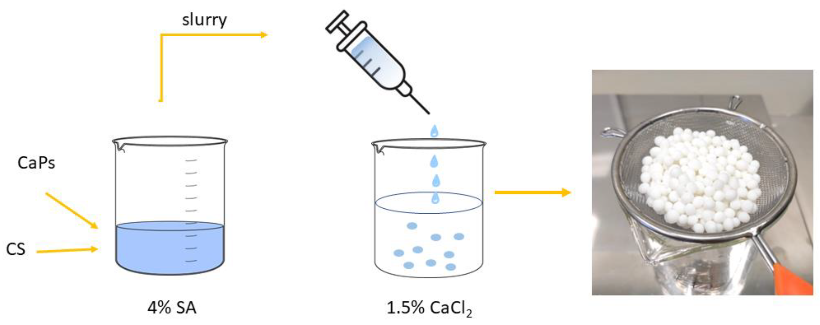
2.2. Preparation of Biphasic Granules
2.3. Physicochemical Analysis of CaP Powders
2.4. Physicochemical Analysis of Granules
2.5. In Vitro Cytotoxicity Study
2.6. Microbiological Activity Studies
3. Results
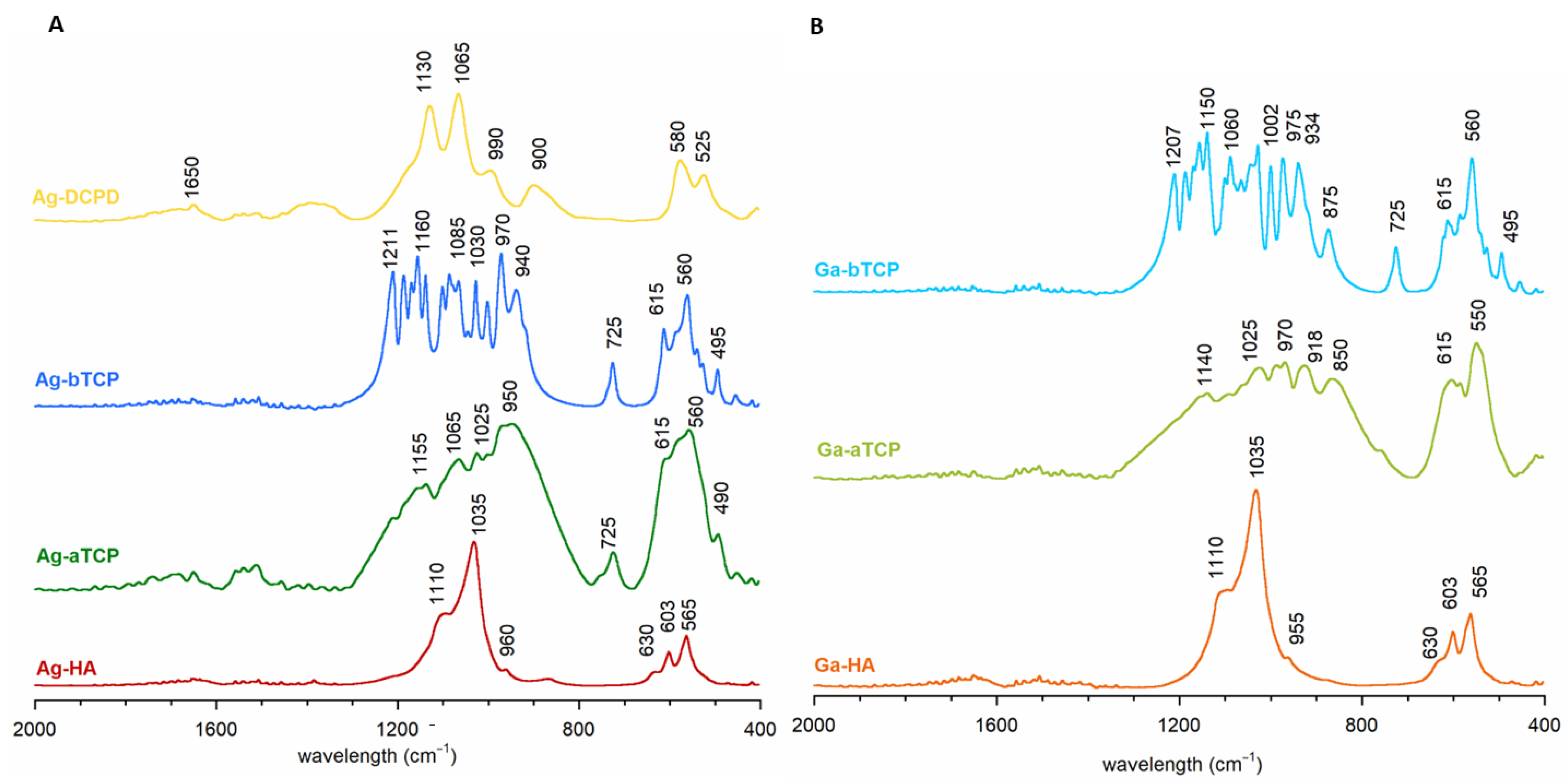
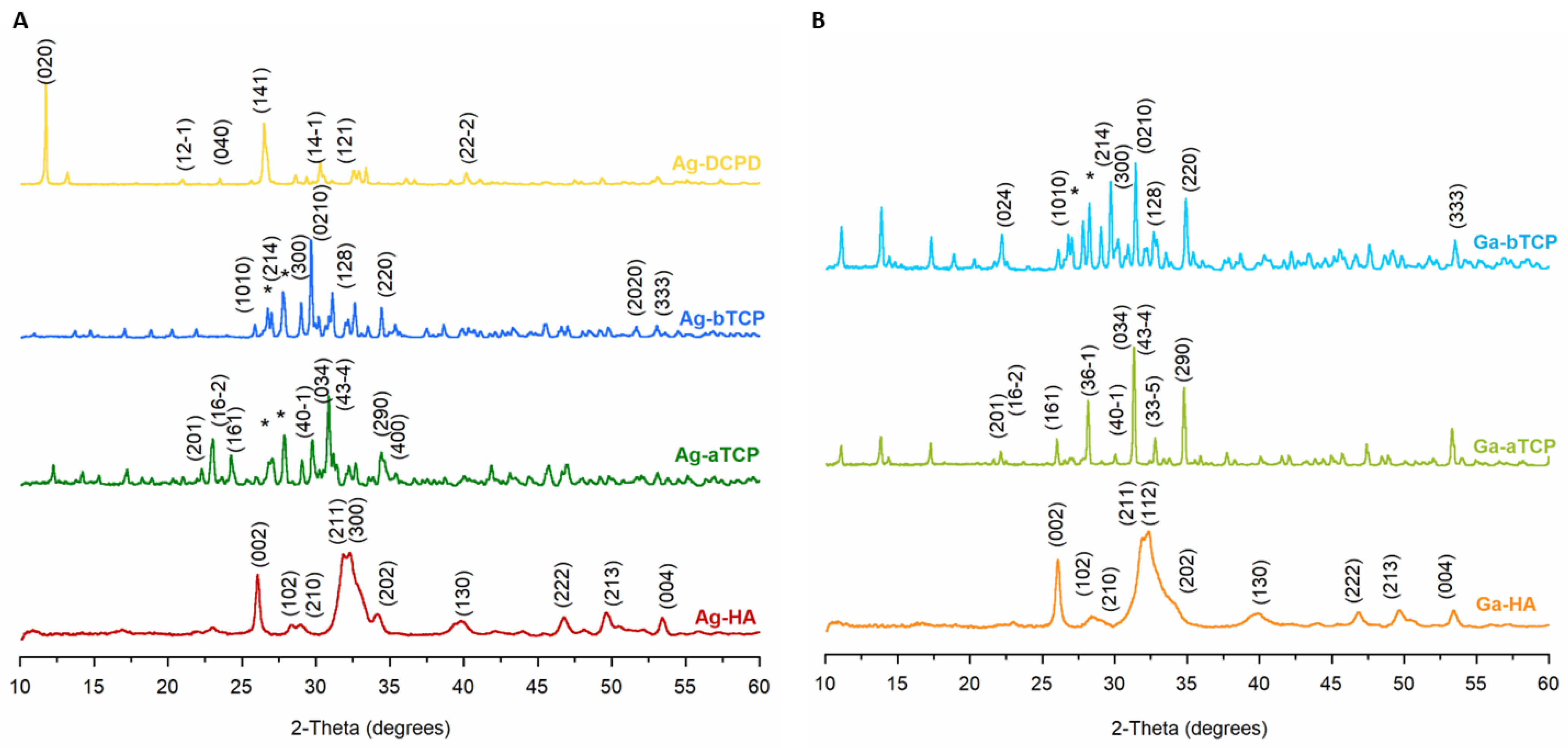
3.1. Characterization of Synthesized Powders
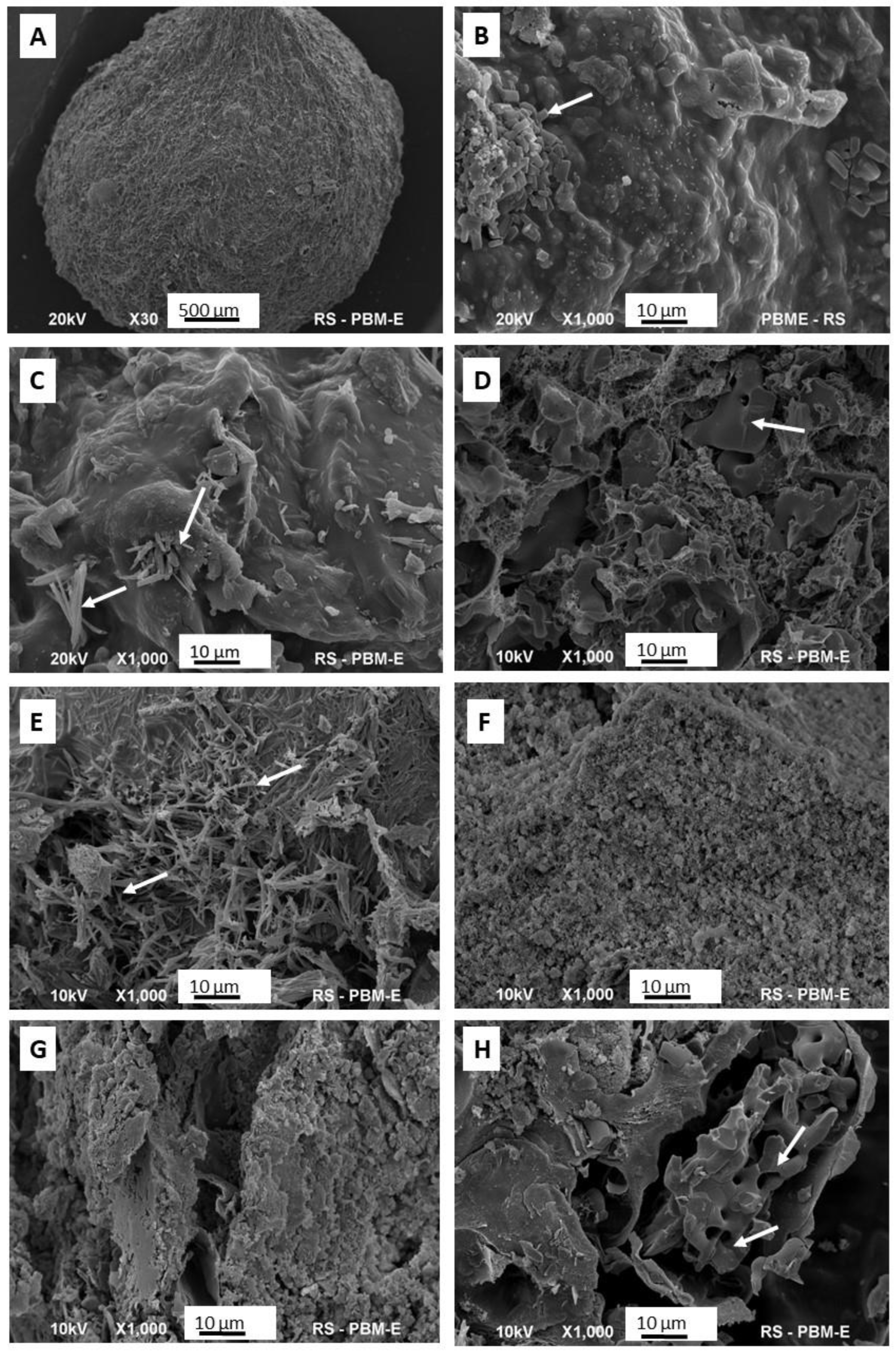
3.2. Morphology of the Synthetic Granules
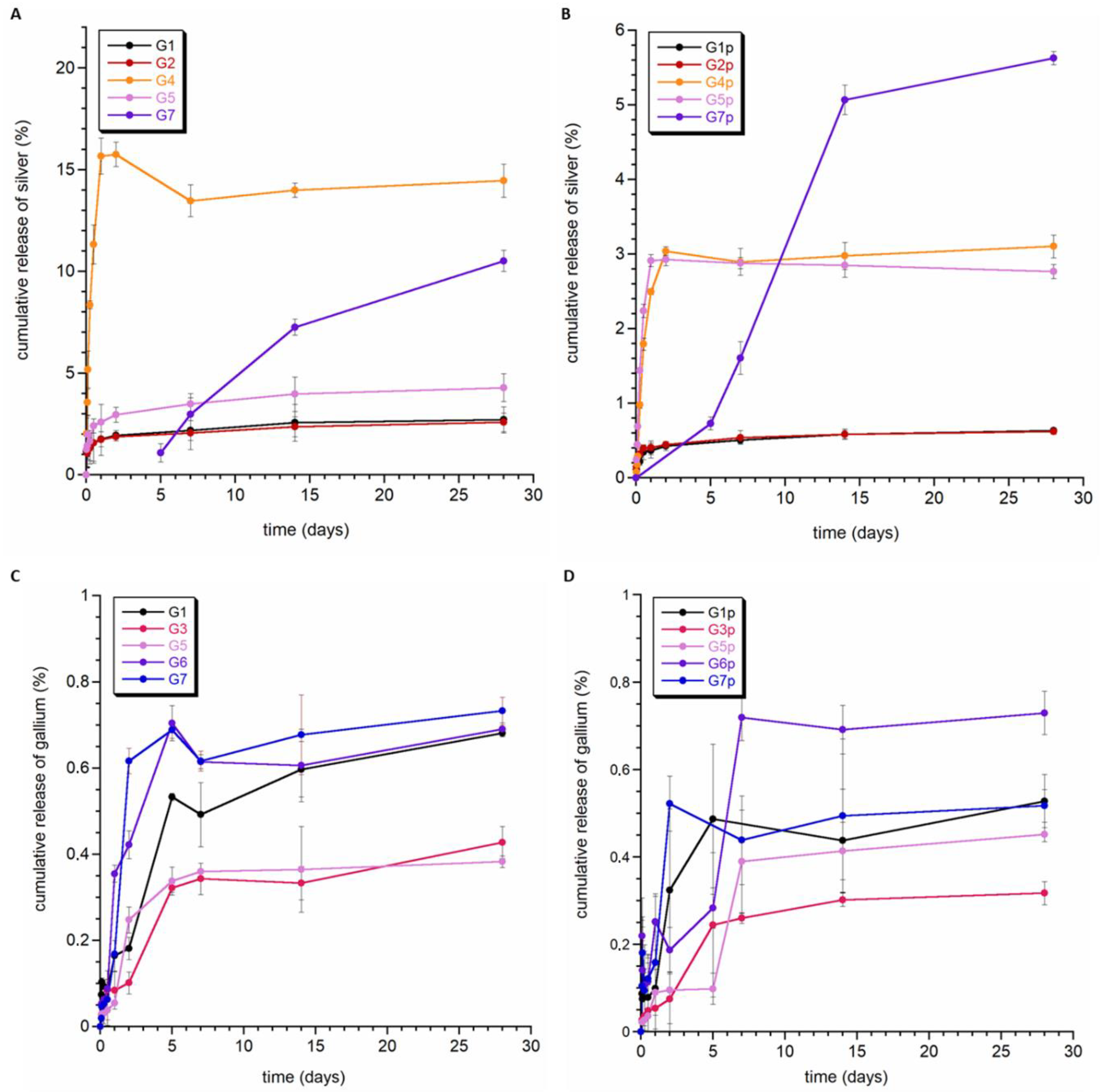
3.3. Ion Release Studies
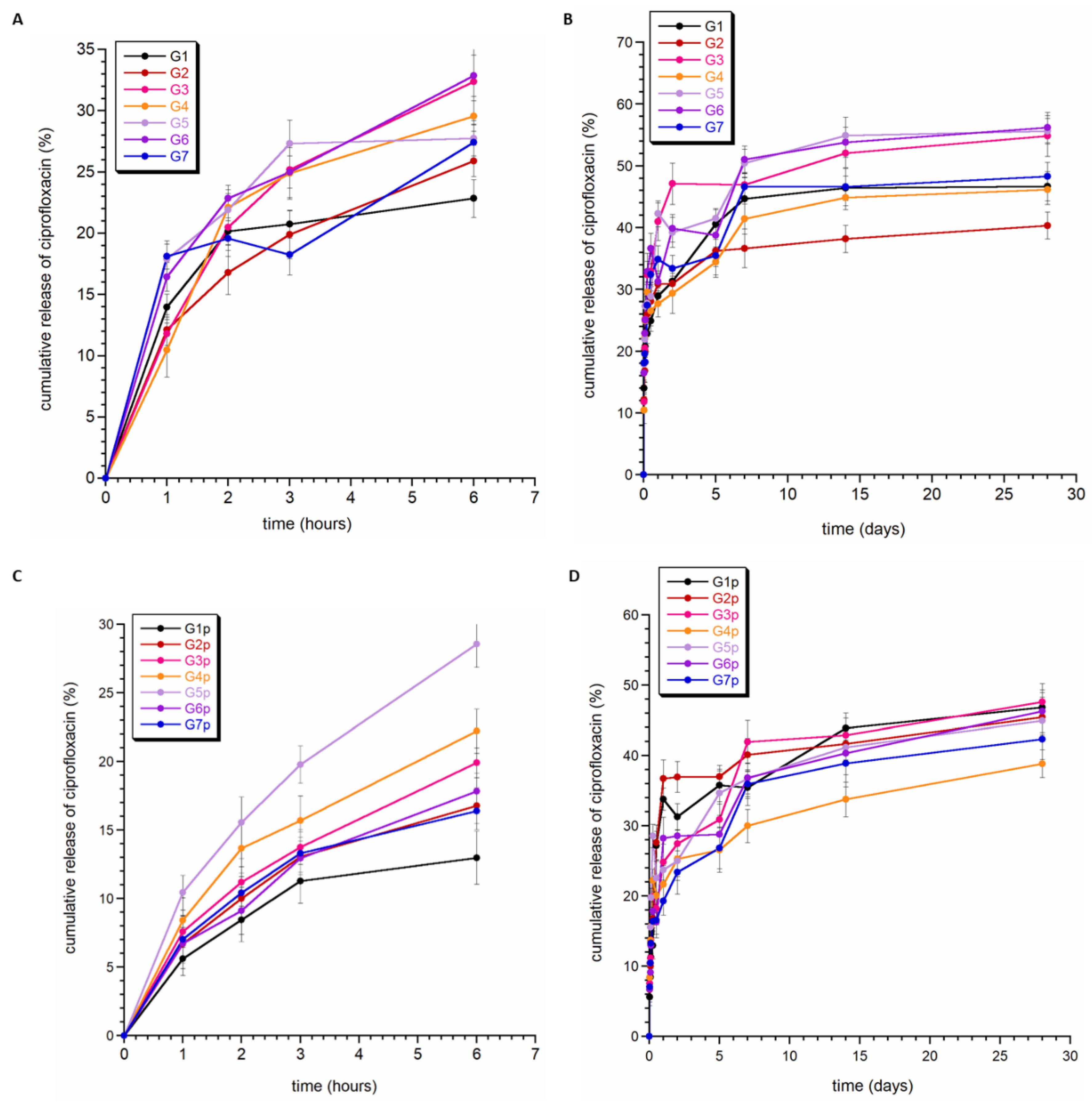
3.4. Ciprofloxacin Release
3.5. Cytotoxicity Studies
3.6. Antibacterial Activity
4. Conclusions
Supplementary Materials
Author Contributions
Funding
Institutional Review Board Statement
Informed Consent Statement
Data Availability Statement
Conflicts of Interest
References
- Chen, A.F.; Wessel, C.B.; Rao, N. Staphylococcus aureus screening and decolonization in orthopedic surgery and reduction of surgical site infections. Clin. Orthop. Relat. Res. 2013, 471, 2383–2399. [Google Scholar] [CrossRef] [PubMed]
- Cheng, L.; Li, R.; Liu, G.; Zhang, Y.; Tang, X.; Wang, J.; Liu, H.; Qin, Y. Potential antibacterial mechanism of silver nanoparticles and the optimization of orthopedic implants by advanced modification technologies. Int. J. Nanomed. 2018, 13, 3311. [Google Scholar]
- Chae, K.; Jang, W.Y.; Park, K.; Lee, J.; Kim, H.; Lee, K.; Lee, C.K.; Lee, Y.; Lee, S.H.; Seo, J. Antibacterial infection and immune-evasive coating for orthopedic implants. Sci. Adv. 2020, 6, eabb0025. [Google Scholar] [CrossRef]
- Jenks, P.; Laurent, M.; McQuarry, S.; Watkins, R. Clinical and economic burden of surgical site infection (SSI) and predicted financial consequences of elimination of SSI from an English hospital. J. Hosp. Infect. 2014, 86, 24–33. [Google Scholar] [CrossRef]
- Greene, L.R. Guide to the elimination of orthopedic surgery surgical site infections: An executive summary of the Association for Professionals in Infection Control and Epidemiology elimination guide. Am. J. Infect. Control. 2012, 40, 384–386. [Google Scholar] [CrossRef] [PubMed]
- Khalid, H.; Nafees, F.; Khaliq, M.A. Infective Organisms and their Changing Antibiotic Sensitivity Trends in Surgical Site Infection after Orthopedic Implant Surgeries. Pak. J. Med. Health Sci. 2018, 12, 1256–1258. [Google Scholar]
- Li, B.; Webster, T.J. Bacteria antibiotic resistance: New challenges and opportunities for implant-associated orthopedic infections. J. Orthop. Res. 2018, 36, 22–32. [Google Scholar] [CrossRef] [PubMed]
- Blondeau, J.M. Fluoroquinolones: Mechanism of action, classification, and development of resistance. Surv. Ophthalmol. 2004, 49, S73–S78. [Google Scholar] [CrossRef] [PubMed]
- Manchon, A.; Prados-Frutos, J.C.; Rueda-Rodriguez, C.; Salinas-Goodier, C.; Alkhraisat, M.H.; Rojo, R.; Rodriguez-Gonzalez, A.; Berlanga, A.; Lopez-Cabarcos, E. Antibiotic release from calcium phosphate materials in oral and maxillofacial surgery. molecular, cellular, and pharmaceutical aspects. Curr. Pharm. Biotechnol. 2017, 18, 52–63. [Google Scholar] [CrossRef]
- Rehman, A.; Patrick, W.M.; Lamont, I.L. Mechanisms of ciprofloxacin resistance in Pseudomonas aeruginosa: New approaches to an old problem. J. Med. Microbiol. 2019, 68, 1–10. [Google Scholar] [CrossRef]
- Lu, M.; Liao, J.; Dong, J.; Wu, J.; Qiu, H.; Zhou, X.; Li, J.; Jiang, D.; He, T.-C.; Quan, Z. An effective treatment of experimental osteomyelitis using the antibacterial titanium/silver-containing nHP66 (nano-hydroxyapatite/polyamide-66) nanoscaffold biomaterials. Sci. Rep. 2016, 6, 39174. [Google Scholar] [CrossRef]
- Sorinolu, A.J.; Godakhindi, V.; Siano, P.; Vivero-Escoto, J.L.; Munir, M. Influence of silver ion release on the inactivation of antibiotic-resistant bacteria using light-activated silver nanoparticles. Mater. Adv. 2022, 3, 9090–9102. [Google Scholar] [CrossRef]
- McRee, A.E. Therapeutic review: Silver. J. Exot. Pet Med. 2015, 2, 240–244. [Google Scholar] [CrossRef]
- Barras, F.; Aussel, L.; Ezraty, B. Silver and antibiotic, new facts to an old story. Antibiotics 2018, 7, 79. [Google Scholar] [CrossRef] [PubMed]
- Kolmas, J.; Groszyk, E.; Kwiatkowska-Różycka, D. Substituted hydroxyapatites with antibacterial properties. BioMed. Res. Int. 2014, 2014, 178123. [Google Scholar] [CrossRef] [PubMed]
- Best, M.G.; Cunha-Reis, C.; Ganin, A.Y.; Sousa, A.; Johnston, J.; Oliveira, A.L.; Smith, D.G.; Yiu, H.H.; Cooper, I.R. Antimicrobial properties of gallium (III)-and iron (III)-Loaded polysaccharides affecting the growth of Escherichia coli, Staphylococcus aureus, and Pseudomonas aeruginosa, in vitro. ACS Appl. Biol. Mater. 2020, 3, 7589–7597. [Google Scholar] [CrossRef]
- Łapa, A.; Cresswell, M.; Campbell, I.; Jackson, P.; Goldmann, W.H.; Detsch, R.; Boccaccini, A.R. Gallium-and cerium-doped phosphate glasses with antibacterial properties for medical applications. Adv. Eng. Mater. 2020, 22, 1901577. [Google Scholar] [CrossRef]
- Li, H.-K.; Rombach, I.; Zambellas, R.; Walker, A.S.; McNally, M.A.; Atkins, B.L.; Lipsky, B.A.; Hughes, H.C.; Bose, D.; Kümin, M. Oral versus intravenous antibiotics for bone and joint infection. N. Engl. J. Med. 2019, 380, 425–436. [Google Scholar] [CrossRef] [PubMed]
- Romanò, C.L.; Scarponi, S.; Gallazzi, E.; Romanò, D.; Drago, L. Antibacterial coating of implants in orthopedics and trauma: A classification proposal in an evolving panorama. J. Orthop. Surg. Res. 2015, 10, 1–11. [Google Scholar] [CrossRef] [PubMed]
- Dorozhkin, S.V. Bioceramics of calcium orthophosphates. Biomaterials 2010, 31, 1465–1485. [Google Scholar] [CrossRef]
- Dorozhkin, S.V. Nanosized and nanocrystalline calcium orthophosphates. Acta Biomater. 2010, 6, 715–734. [Google Scholar] [CrossRef] [PubMed]
- Bal, Z.; Kaito, T.; Korkusuz, F.; Yoshikawa, H. Bone regeneration with hydroxyapatite-based biomaterials. Emergent Mater. 2020, 3, 521–544. [Google Scholar] [CrossRef]
- Bouler, J.-M.; Pilet, P.; Gauthier, O.; Verron, E. Biphasic calcium phosphate ceramics for bone reconstruction: A review of biological response. Acta Biomater. 2017, 53, 1–12. [Google Scholar] [CrossRef] [PubMed]
- Owen, G.R.; Dard, M.; Larjava, H. Hydroxyapatite/beta-tricalcium phosphate biphasic ceramics as a regenerative material for the repair of complex bone defects. J. Biomed. Mater. Res. Part B Appl. Biomater. 2018, 106, 2493–2512. [Google Scholar] [CrossRef]
- Schmitz, J.P.; Hollinger, J.O.; Milam, S.B. Reconstruction of bone using calcium phosphate bone cements: A critical review. J. Oral Maxillofac. Surg. 1999, 57, 1122–1126. [Google Scholar] [CrossRef]
- Mofakhami, S.; Salahinejad, E. Biphasic calcium phosphate microspheres in biomedical applications. J. Control. Release 2021, 338, 527–536. [Google Scholar] [CrossRef]
- Shao, R.; Quan, R.; Zhang, L.; Wei, X.; Yang, D.; Xie, S. Porous hydroxyapatite bioceramics in bone tissue engineering: Current uses and perspectives. J. Ceram. Soc. Japan 2015, 123, 17–20. [Google Scholar] [CrossRef]
- Predoi, D.; Iconaru, S.L.; Predoi, M.H.; Buton, N. Development of novel tetracycline and ciprofloxacin loaded silver doped hydroxyapatite suspensions for biomedical applications. Antibiotics 2022, 12, 74. [Google Scholar] [CrossRef]
- Jackson, J.; Lo, J.; Hsu, E.; Burt, H.M.; Shademani, A.; Lange, D. The combined use of gentamycin and silver nitrate in bone cement for a synergistic and extended antibiotic action against Gram-positive and Gram-negative bacteria. Materials 2021, 14, 3413. [Google Scholar] [CrossRef]
- Wan, G.; Ruan, L.; Yin, Y.; Yang, T.; Ge, M.; Cheng, X. Effects of silver nanoparticles in combination with antibiotics on the resistant bacteria Acinetobacter baumannii. Int. J. Nanomed. 2016, 11, 3789–3800. [Google Scholar] [CrossRef]
- Pajor, K.; Pajchel, Ł.; Zgadzaj, A.; Piotrowska, U.; Kolmas, J. Modifications of hydroxyapatite by gallium and silver ions—Physicochemical characterization, cytotoxicity, and antibacterial evaluation. Int. J. Mol. Sci. 2020, 21, 5006. [Google Scholar] [CrossRef] [PubMed]
- EN ISO 10993-5; 2009 Biological Evaluation of Medical Devices—Part 5: Tests for in Vitro Cytotoxicity (ISO 10993-5:2009), Annex A Neutral Red Uptake (NRU) Cytotoxicity Test. International Organization for Standardization: Geneva, Switzerland, 2009.
- EN ISO 10993-12; 2012 Biological Evaluation of Medical Devices—Part 12: Sample Preparation and Reference Materials (ISO 10993-12:2012). International Organization for Standardization: Geneva, Switzerland, 2012.
- Sayahi, M.; Santos, J.; El-Feki, H.; Charvillat, C.; Bosc, F.; Karacan, I.; Milthorpe, B.; Drouet, C. Brushite (Ca,M)HPO4, 2H2O doping with bioactive ions (M = Mg2+, Sr2+, Zn2+, Cu2+, and Ag+): A new path to functional biomaterials? Mater. Today Chem. 2020, 16, 10030. [Google Scholar] [CrossRef]
- Fadeeva, I.V.; Gafurov, M.R.; Kiiaeva, I.A.; Orlinskii, S.B.; Kuznetsova, L.M.; Filippov, Y.Y.; Fomin, A.S.; Davydova, G.A.; Selezneva, I.I.; Barinov, S.M. Tricalcium phosphate ceramics doped with silver, copper, zinc and iron (III) ions in concentration of less than 0.5% for bone tissue regeneration. BioNanoScience 2017, 7, 434–438. [Google Scholar] [CrossRef]
- Kolmas, J.; Groszyk, E.; Piotrowska, U. Nanocrystalline hydroxyapatite enriched in selenite and manganese ions: Physicochemical and antibacterial properties. Nanoscale Res. Lett. 2015, 10, 989. [Google Scholar] [CrossRef]
- Gopi, D.; Shinyjoy, E.; Kavitha, L. Synthesis and spectral characterization of silver/magnesium co-substituted hydroxyapatite for biomedical applications. Spectrochim. Acta Part A Mol. Biomol. Spectrosc. 2014, 127, 286–291. [Google Scholar] [CrossRef]
- Okada, M.; Furuzono, T. Hydroxylapatite nanoparticles: Fabrication methods and medical applications. Sci. Technol. Adv. Mater. 2012, 13, 064103. [Google Scholar] [CrossRef]
- Jillavenkatesa, A.; Condrate Sr, R. The infrared and Raman spectra of β-and α-tricalcium phosphate (Ca3(PO4)2). Spectrosc. Lett. 1998, 31, 1619–1634. [Google Scholar] [CrossRef]
- Martínez, T.; Espanol, M.; Charvillat, C.; Marsan, O.; Ginebra, M.; Rey, C.; Sarda, S. α-tricalcium phosphate synthesis from amorphous calcium phosphate: Structural characterization and hydraulic reactivity. J. Mater. Sci. 2021, 56, 13509–13523. [Google Scholar] [CrossRef]
- Kolmas, J.; Kaflak, A.; Zima, A.; Ślósarczyk, A. Alpha-tricalcium phosphate synthesized by two different routes: Structural and spectroscopic characterization. Ceram. Int. 2015, 41, 5727–5733. [Google Scholar] [CrossRef]
- Boonchom, B.; Baitahe, R. Synthesis and characterization of nanocrystalline manganese pyrophosphate Mn2P2O7. Mater. Lett. 2009, 63, 2218–2220. [Google Scholar] [CrossRef]
- El Kady, A.M.; Mohamed, K.R.; El-Bassyouni, G.T. Fabrication, characterization and bioactivity evaluation of calcium pyrophosphate/polymeric biocomposites. Ceram. Int. 2009, 35, 2933–2942. [Google Scholar] [CrossRef]
- dos Santos Tavares, D.; de Oliveira Castro, L.; de Almeida Soares, G.D.; Alves, G.G.; Granjeiro, J.M. Synthesis and cytotoxicity evaluation of granular magnesium substituted β-tricalcium phosphate. J. Appl. Oral Sci. 2013, 21, 37–42. [Google Scholar] [CrossRef] [PubMed]
- Walczyk, D.; Malina, D.; Krol, M.; Pluta, K.; Sobczak-Kupiec, A. Physicochemical characterization of zinc-substituted calcium phosphates. Bull. Mater. Sci. 2016, 39, 525–535. [Google Scholar] [CrossRef]
- Lee, D.; Kumta, P.N. Chemical synthesis and stabilization of magnesium substituted brushite. Mater. Sci. Eng. C 2010, 30, 934–943. [Google Scholar] [CrossRef]
- Arbez, B.; Kun-Darbois, J.D.; Convert, T.; Guillaume, B.; Mercier, P.; Huber, L.; Chappard, D. Biomaterial granules used for filling bone defects constitute 3D scaffolds: Porosity, microarchitecture, and molecular composition analyzed by microCT and Raman microspectroscopy. J. Biomed. Mater. Res. B 2019, 107, 415–423. [Google Scholar] [CrossRef]
- Szurkowska, K.; Zgadzaj, A.; Kuras, M.; Kolmas, J. Novel hybrid material based on Mg2+ and SiO44− co-substituted nanohydroxyapatite, alginate and chondroitin sulfate for potential use in biomaterials engineering. Ceram. Int. 2018, 44, 18551–18559. [Google Scholar] [CrossRef]
- Pajor, K.; Michalicha, A.; Belcarz, A.; Pajchel, L.; Zgadzaj, A.; Wojas, F.; Kolmas, J. Antibacterial and cytotoxicity evaluation of new hydroxyapatite-based granules containing silver or gallium ions with potential use as bone substitutes. Int. J. Mol. Sci. 2022, 23, 7102. [Google Scholar] [CrossRef]
- Kurtjak, M.; Vucomanovic, M.; Krajnc, A.; Kramer, L.; Turk, B.; Suvorow, D. Designing Ga(III)-containing hydroxyapatite with antibacterial activity. RSC Adv. 2016, 6, 112839–112852. [Google Scholar] [CrossRef]
- Cavallaro, G.; Lazzara, G.; Milioto, S.; Parisi, F.; Evtugyn, V.; Roshina, E.; Fakhrullin, R. Nanohydrogel formation within the halloysite lument for triggered and sustained release. ACS Appl. Mater. Interface 2018, 10, 8265–8273. [Google Scholar] [CrossRef]
- Fosca, M.; Rau, J.V.; Uskokovic, V. Factors influencing the drug release from calcium phosphate cements. Bioactive Mater. 2022, 7, 341–363. [Google Scholar] [CrossRef]
- Lisuzzo, L.; Cavallaro, G.; Milioto, S.; Lazzara, G. Halloysite nanotubes coated by chitosan for controlled release of khellin. Polymers 2020, 12, 1766. [Google Scholar] [CrossRef] [PubMed]
| Material | Comment | Nominal wt% of Doped Ion |
|---|---|---|
| Ag-HA | Silver-containing hydroxyapatite | 0.22 |
| Ag-aTCP | Silver-containing tricalcium phosphate α type | 0.23 |
| Ag-bTCP | Silver-containing tricalcium phosphate β type | 0.23 |
| Ag-DCPD | Silver-containing brushite | 1.26 |
| Ga-HA | Gallium-containing hydroxyapatite | 3.35 |
| Ga-aTCP | Gallium-containing tricalcium phosphate α type | 3.61 |
| Ga-bTCP | Gallium-containing tricalcium phosphate β type | 3.61 |
| Standard Granules | Granules Layered with PCL | Silver Component | Gallium Component |
|---|---|---|---|
| G1 | G1p | Ag-DCPD | Ga-HA |
| G2 | G2p | Ag-DCPD | Ga-aTCP |
| G3 | G3p | Ag-aTCP | Ga-bTCP |
| G4 | G4p | Ag-HA | Ga-aTCP |
| G5 | G5p | Ag-HA | Ga-bTCP |
| G6 | G6p | Ag-bTCP | Ga-HA |
| G7 | G7p | Ag-aTCP | Ga-HA |
| Sample | k/min−n | n | R2 |
|---|---|---|---|
| G1 | 17.0 ± 1.1 | 0.17 ± 0.01 | 0.9791 |
| G2 | 11.5 ± 2.1 | 0.23 ± 0.03 | 0.9351 |
| G3 | 17.9 ± 1.4 | 0.13 ± 0.02 | 0.9979 |
| G4 | 21.8 ± 2.4 | 0.15 ± 0.02 | 0.9238 |
| G5 | 11.1 ± 1.2 | 0.16 ± 0.01 | 0.9331 |
| G6 | 11.0 ± 1.1 | 0.23 ± 0.02 | 0.9791 |
| G7 | 17.7 ± 1.8 | 0.15 ± 0.02 | 0.9324 |
| G1p | 12.7 ± 0.9 | 0.17 ± 0.02 | 0.9683 |
| G2p | 21.0 ± 1.5 | 0.16 ± 0.02 | 0.9655 |
| G3p | 14.9 ± 1.5 | 0.15 ± 0.02 | 0.9494 |
| G4p | 21.4 ± 1.7 | 0.15 ± 0.02 | 0.9555 |
| G5p | 10.6 ± 1.2 | 0.23 ± 0.02 | 0.9711 |
| G6p | 18.9 ± 1.8 | 0.15 ± 0.02 | 0.9384 |
| G7p | 9.4 ± 0.2 | 0.24 ± 0.02 | 0.9855 |
| Sample | Cell Viability ± SD [%] | IC50 [mg/mL] | Classification |
|---|---|---|---|
| Pure HA | 101 ± 2 | N | Non-cytotoxic |
| Ag-HA | 102 ± 4 | N | Non-cytotoxic |
| Ga-HA | 94 ± 4 | N | Non-cytotoxic |
| Pure β-TCP | 105 ± 2 | N | Non-cytotoxic |
| Ag-bTCP | 98 ± 3 | N | Non-cytotoxic |
| Ga-bTCP | 114 ± 1 | N | Non-cytotoxic |
| Pure α-TCP | 101 ± 3 | N | Non-cytotoxic |
| Ag-aTCP | 106 ± 3 | N | Non-cytotoxic |
| Ga-bTCP | 99 ± 1 | N | Non-cytotoxic |
| Pure DCPD | 111 ± 6 | N | Non-cytotoxic |
| Ag-DCPD | 21 ± 10 | 83 | Cytotoxic |
| G1 | 10 ± 7 | 76 | Cytotoxic |
| G2 | 44 ± 4 | 94 | Cytotoxic |
| G3 | 99 ± 3 | N | Non-cytotoxic |
| G4 | 87 ± 4 | N | Non-cytotoxic |
| G5 | 100 ± 4 | N | Non-cytotoxic |
| G6 | 85 ± 4 | N | Non-cytotoxic |
| G7 | 99 ± 3 | N | Non-cytotoxic |
| LT | 0 ± 0 | <10 | Cytotoxic |
| PE | 102 ± 7 | N | Non-cytotoxic |
| Type of Granules | Bacterial Growth [CFU/mL] | |
|---|---|---|
| Staphylococcus aureus | Escherichia coli | |
| G1 | 1.25 × 10³ | 0 |
| G2 | 1 × 10² | 0 |
| G3 | 5 × 101 | 0 |
| G4 | 5 × 101 | 0 |
| G5 | 5 × 101 | 0 |
| G6 | 5 × 101 | 0 |
| G7 | 0 | 0 |
| G1p | 1.5 × 10³ | 0 |
| G2p | 2.75 × 10³ | 0 |
| G3p | 0 | 0 |
| G4p | 0 | 0 |
| G5p | 0 | 0 |
| G6p | 0 | 0 |
| G7p | 0 | 0 |
| Type of Granules | Bacterial Growth—Staphylococcus aureus [CFU/mL] | ||
|---|---|---|---|
| 1st Week | 2nd Week | 3rd Week | |
| G1 | 1.25 × 10³ | 2.61 × 10⁴ | 1.55 × 10⁵ |
| G1p | 1.5 × 10³ | 1.65 × 10³ | 4.96 × 10⁵ |
| G2 | 1 × 10² | 1.45 × 10³ | 2.51 × 10⁵ |
| G2p | 2.75 × 10³ | 4.68 × 10⁴ | 1.6 × 10⁵ |
Disclaimer/Publisher’s Note: The statements, opinions and data contained in all publications are solely those of the individual author(s) and contributor(s) and not of MDPI and/or the editor(s). MDPI and/or the editor(s) disclaim responsibility for any injury to people or property resulting from any ideas, methods, instructions or products referred to in the content. |
© 2023 by the authors. Licensee MDPI, Basel, Switzerland. This article is an open access article distributed under the terms and conditions of the Creative Commons Attribution (CC BY) license (https://creativecommons.org/licenses/by/4.0/).
Share and Cite
Pajor, K.; Pajchel, Ł.; Zgadzaj, A.; Kowalska, P.; Kowalczuk, A.; Kolmas, J. Ciprofloxacin-Loaded Composite Granules Enriched in Silver and Gallium Ions—Physicochemical Properties and Antimicrobial Activity. Coatings 2023, 13, 494. https://doi.org/10.3390/coatings13030494
Pajor K, Pajchel Ł, Zgadzaj A, Kowalska P, Kowalczuk A, Kolmas J. Ciprofloxacin-Loaded Composite Granules Enriched in Silver and Gallium Ions—Physicochemical Properties and Antimicrobial Activity. Coatings. 2023; 13(3):494. https://doi.org/10.3390/coatings13030494
Chicago/Turabian StylePajor, Kamil, Łukasz Pajchel, Anna Zgadzaj, Paulina Kowalska, Anna Kowalczuk, and Joanna Kolmas. 2023. "Ciprofloxacin-Loaded Composite Granules Enriched in Silver and Gallium Ions—Physicochemical Properties and Antimicrobial Activity" Coatings 13, no. 3: 494. https://doi.org/10.3390/coatings13030494
APA StylePajor, K., Pajchel, Ł., Zgadzaj, A., Kowalska, P., Kowalczuk, A., & Kolmas, J. (2023). Ciprofloxacin-Loaded Composite Granules Enriched in Silver and Gallium Ions—Physicochemical Properties and Antimicrobial Activity. Coatings, 13(3), 494. https://doi.org/10.3390/coatings13030494


