Article

Nanohydroxyapatite Loaded with 5-Fluorouracil and Calendula officinalis L. Plant Extract Rich in Myo-Inositols for Treatment of Ovarian Cancer Cells

by
Magdalena Osial
1,*,
Sławomir Wilczewski
2,
Joanna Szulc
2,
Hai Dang Nguyen
3,
Thi Kieu Oanh Nguyen
3,
Katarzyna Skórczewska
2,
Agnieszka Majkowska-Pilip
4,
Kinga Żelechowska-Matysiak
4,
Dorota Nieciecka
5,
Agnieszka Pregowska
1,
Thu Phuong Nguyen
6,
Alicja Tymoszuk
7,
Dariusz Kulus
7 and
Michael Giersig
1,*
1
Institute of Fundamental Technological Research, Polish Academy of Sciences, Pawinskiego 5B Street, 02-106 Warsaw, Poland
2
Faculty of Chemical Technology and Engineering, Bydgoszcz University of Science and Technology, Seminaryjna 3 Street, 85-326 Bydgoszcz, Poland
3
University of Science and Technology of Hanoi, Vietnam Academy of Science and Technology, 18 Hoang Quoc Viet, Cau Giay, Hanoi, Vietnam
4
Centre of Radiochemistry and Nuclear Chemistry, Institute of Nuclear Chemistry and Technology, Dorodna 16 Street, 03-195 Warsaw, Poland
5
Faculty of Chemistry, University of Warsaw, Pasteura 1 Street, 02-093 Warsaw, Poland
6
Institute for Tropical Technology, Vietnam Academy of Science and Technology, 18 Hoang Quoc Viet, Cau Giay, Hanoi, Vietnam
7
Laboratory of Ornamental Plants and Vegetable Crops, Faculty of Agriculture and Biotechnology, Bydgoszcz University of Science and Technology, Bernardyńska 6 Street, 85-029 Bydgoszcz, Poland
*
Authors to whom correspondence should be addressed.
Coatings 2023, 13(11), 1944; https://doi.org/10.3390/coatings13111944
Submission received: 20 September 2023 / Revised: 12 November 2023 / Accepted: 12 November 2023 / Published: 14 November 2023

Abstract

:
In this study, the hydroxyapatite (HAp) in the form of nanoparticles was synthesized through the wet co-precipitation method and loaded with plant extract from Calendula officinalis L. rich in biologically active myo-inositol, and the 5-fluorouracil anticancer drug. The obtained nanomaterials have rod-like structures approx. 30 nm in diameter and 100 nm in length. FT-IR analysis results proved the immobilization of biologically active compounds. The loading of plant extract into the carrier improved the stability of colloidal suspension, which was confirmed with turbidimetry. The composite modified with calendula extract and drug (HAp@Cal@5-flu) effectively scavenges the DPPH radicals, with a radical scavenging activity (RSA) of about 20.0 ± 1.3%. The effect is supported by the DFT calculations of the HOMO-LUMO, presenting the chemical reactivity of the molecules loaded into the HAp. The in vitro cytotoxicity results on SKOV-3 ovarian cancer cells show the pronounced cytotoxic effect of the HAp@Cal@5-flu. The calendula extract loading into the carrier provided better interactions with the tumorous biomimetic membranes studied with a Langmuir trough, making it a promising material in nano-biomedicine, including drug delivery and targeted cancer treatment.

1. Introduction

Medicinal plants are an important source of pharmacological molecules. They have been used for centuries as part of a history of traditional medicine [1]. The role of herbs in healthcare has held a strong position over time. Currently, phytotherapy and the acquisition of natural metabolites from plants are attracting increasing interest. Hence, studies in this field are needed and justified. One plant that contains bioactive molecules with a potential anticancer effect is Calendula officinalis L. (commonly known as pot marigold) [2]. Even though the species is valued for its decorative properties, it is mainly cultivated as a medicinal plant for the pharmaceutical and cosmetic industries [3]. C. officinalis L. contains various secondary metabolites—i.e., flavonoids, carotenoids, steroids, and terpenoids—that are a potential source of antioxidants as well as antibacterial, antiviral, and anti-inflammatory phytoconstituents [4]. Extracts of marigolds are used in treating skin disorders, such as wounds, dermatological lesions, tumors, swellings, ulcers, and exfoliative cheilitis, but also for gastrointestinal inflammations and dysmenorrhea [2,5]. Moreover, pot marigold has been used as an oilseed crop due to the presence of unsaturated fatty acids and α- and γ-tocopherols, which are used in paint and food industries [6]. C. officinalis L. is particularly rich in nitrogenous hetero-cycles with indole cores, which are considered to be responsible for some of its health-stimulating activities.
Inositols are compounds that can be found in nature. They are derived from sugars, possessing a molecular structure very similar to simple sugars, and thus, they are placed at the boundary between primary and secondary [7]. Myo-inositol is the most represented of the nine stereoisomers of C6 sugar alcohol (cyclitol), which belongs to the vitamin B complex group [8]. Myo-inositol is emerging as a central feature in plant biochemistry and physiology [9]. It is involved in cell morpho-genesis, cytogenesis, and lipid synthesis. The role of myo-inositol as a co-factor of enzymes and messenger molecules in signal transduction has also been proven [8]. Increasing evidence supports its physiological and therapeutic effects on human health. It plays a significant role in human reproduction and oogenesis [10]. Moreover, myo-inositol is one of the most abundant metabolites in the human brain and is located mainly in glial cells. The concentration of that compound is altered in several brain disorders, such as Alzheimer’s disease and brain tumors [11]. Inositol has been used as a supplement in treating several pathologies, including polycystic ovary syndrome, metabolic syndrome, and gestational diabetes. Refs. [12,13] reported the presence of D-pinitol, D-chiro-inositol, bornesitol, scylloinositol, and myo-inositol in calendula anthodium. The protective and anticancer effects of inositols, particularly myo-inositol, have been studied for decades. The anticancer activity of inositols has been described as a “broad spectrum” due to their ability to modulate the entire cell cycle (progression, apoptosis, and differentiation). In addition, inositols seem to be crucial for increasing the likelihood of curing cancer [7,14,15]. Such compounds can be loaded into the nanocarriers for local therapy or diagnostics [16]. Besides many nanocarriers, one of the promising candidates is nanostructural hydroxyapatite (HAp), which is widely studied for several biological uses, including antibacterial, drug delivery, dental, and orthopedic applications [17]. Depending on its application, much attention has been paid to incorporating different metals into HAp to change its properties, e.g., HAp doped with Fe and Co displayed increased antibacterial activity against Shigella dysenteriae, while Mn doping improved activity against Staphylococcus aureus [18]. Other sources also show the antimicrobial activity of HAp [19,20]. Due to its high surface-area-to-volume ratio and chemical composition, it favors chemical and physical modifications. Nanohydroxyapatite is an ideal platform for anticancer drug delivery [21,22], including, e.g., methotrexate [23] paclitaxel [24], toceranib [25], doxorubicin [26,27,28], and 5-fluorouracil [29]. HAp has also recently been used in randomized clinical trials in bone regeneration for its biocompatibility [30]. For these reasons, we used it as a drug carrier. According to the chemotherapy, it still needs improvement. Saif et al. present the role of the 5-fluorouracil adjustment in anticancer treatment, mentioning the scale of overdosing and underdosing of patients with direct chemotherapy [31]. Other sources also present the importance of individual dose adjustment for patients to maintain the optimal time of treatment for effective treatment [32,33,34,35]. Therefore, drug delivery by the nanosized composite can reduce the toxic effect on healthy tissues.
In this work, we present a nanostructural composite based on hydroxyapatite, 5-fluorouracil, and calendula extract for cancer treatment. The plant extract obtained from Calendula officinalis L. was proposed as a source of the biologically active components, mainly myo-inositol (56.9%), and confirmed via gas chromatography, offering antioxidative properties and improving drug uptake by cancer cells in vitro. We demonstrated the facile modification of HAp with calendula extract and the potential of the proposed nanostructural platform as a therapeutic agent for effective cancer treatment, presenting its cytotoxicity against SKOV-3 cells. The potential of the use against cancer cells was confirmed with an in vitro test and Langmuir trough technique using analogs of biological membranes—in particular, healthy DOPC and tumorous DOPS lipid-based layers.

2. Materials and Methods

2.1. Chemicals

The hydroxyapatite was obtained from the salts Ca(NO3)2 (calcium nitrate) and (NH4)2HPO4 (diammonium hydrogen phosphate), where the precipitating agent was 25% NH3 ammonia aqueous solution of analytical grade purchased from POCH, Gliwice, Poland. Solutions were prepared using deionized water conductivity 100 μS cm−1 at 25 °C from HYDROLAB, Gliwice, Poland. The Calendula officinalis L. petals were purchased from Oldfarm, Łódź, Poland. Methanol used for extraction of >96% grade was supplied by Chempur, Poland, and NaCl of analytical grade was purchased from Warchem, Poland. Chloroform was supplied by POCH, Gliwice, Poland. The anticancer drug, 5-fluorouracil of ≥99% grade, was supplied by Sigma-Aldrich, Germany. Lipids: DOPC (1,2-dioleoyl-sn-glycero-3-phosphocholine, purity >99%), DOPS 1,2-dioleoyl-sn-glycero-3-phospho-L-serine (sodium salt) were purchased from Avanti Polar Lipids, Birmingham, AL, USA.

2.2. Determining the Morphology, Hydrodynamic Diameter, and Zeta Potential

The morphology and nanocomposites of Hap were investigated using scanning electron microscopy (SEM)—Merlin, ZEISS, Stuttgart, Germany—and transmission electron microscopy (TEM)—Zeiss Libra 120 Plus, Stuttgart, Germany, operating at 120 kV.
The hydrodynamic diameter and the zeta potential of the nanostructures were determined with a Malvern Zetasizer instrument (Malvern, UK) fitted with a He-Ne laser with λ = 632.8 nm. The scattering angle was 173°. The zeta potential studies were performed at 0.01 M NaCl solution.

2.3. Investigating Chemical Composition and Crystallinity

The chemical composition was studied via Fourier-transform infrared spectroscopy (FT-IR) using the ATR technique Alpha apparatus supplied by Bruker (Ettlingen, Germany). The spectra were recorded in the range of 4000–400 cm−1, where 32 scans at a resolution of 4 cm−1 were performed.
The thermal stability of the nanocomposites was assessed by the thermogravimetric method (TGA) using a TG 209 F3 Tarsus apparatus supplied by Netzsch (Selb, Germany). The heating rate was about 10 °C min−1 in an open ceramic crucible under a nitrogen atmosphere in the temperature range of 30 to 725 °C.
Crystallinity studies were performed using the X-ray diffraction (XRD) DSH method on a Malvern Panalytical X’Pert PRO MPD powder diffractometer, (Malvern, UK). The XRD patterns were recorded in the (2θ) range from 10° to 70°, with a scan rate of 1° per min and a total record time of 67 min. Analysis was performed using Co Kα at 40 kV and 40 mA. Phase identification was performed via the use of X’Pert Plus HighScore software v3.0. with access to the COD (Crystallography Open Database) database, and the size of the crystallites was calculated using a Scherrer formula.

2.4. Stability Studies

The stability of the colloidal suspension was carried out using the multiple light scattering method by the Turbiscan Lab apparatus (Formulation SA, Toulouse, France). Measurements were performed in a cylindrical glass vial (54 mm in height). Then, the samples were placed in a Turbiscan Lab apparatus from Formulation SA, France, and were measured using light (880 nm wavelength). Measurements were carried out every 5 min for 50 min and the Turbiscan Stability Index (TSI) was determined.

2.5. Determining the Composition of Calendula officinalis L. Plant Extract

Profiling analysis was performed using a Thermo Scientific™ TRACE™ 1310 GC system equipped with a Thermo Scientific™ 1310 autosampler connected to a ISQ 7000 mass spectrometer (Waltham, MA, USA). A Thermo Scientific™ Trace™ GC column 15 m × 0.25 mm × 0.25 μm was equipped with purified helium as the carrier gas at a constant flow rate of 1.2 mL min−1. The oven temperature was held at 50 °C for 5 min, increased to 220 °C at a rate of 3 °C min−1, held at 220 °C for 5 min, increased to 240 °C at a rate of 10 °C min−1, and finally kept at 240 °C for 5 min. The injector and ion source temperature were set at 200 °C and 250 °C respectively. The split mode was applied with a ratio of 1:40. The mass spectrometer was operating in full scan mode at 45 eV over the range of 50–800 a.m.u. The identification of the selected peaks was made by searching in the NIST2017 data library. Selected peaks were analyzed based on the relative ratio by taking the peak area of this peak divided by the sum of peak areas of all detected peaks in the tea leaf sample. The GC measurement was performed as follows: approximately 10 mg of crude extracts was accurately weighed into a 2 mL lock-cap centrifuge tube. Then, 1 mL of HPLC-grade methanol (CH3OH) was added and shaken. 200 µL of ex-tracts were taken and evaporated under purified nitrogen gas. The sample was then methoxylated by resuspending the pellet in 50 μL of methoxyamine hydrochloride solution (20 mg mL−1 in pyridine) and incubating with constant agitation for 2 h at 37 °C to protect carbonyl moieties. Acidic protons were then trimethylsilylated by adding 70 μL of N-methyl-N-(trimethylsilyl)-trifluoroacetamide (MSTFA) and incubating at 37 °C with constant agitation for 30 min.

2.6. Determining the Radical Scavenging Activity

The antioxidative studies of the nanocarrier were determined according to the method of [36]. The scavenging capacity of free radical DPPH was tested with pure hydroxyapatite colloidal suspension, 5-fluorouracil solution, calendula extract (0.1 mg g−1), where the 2 mL of the sample was extracted with 8 mL water. Samples were placed in 50 mL tubes, sealed, shaken, and stored in the dark at room temperature for 48 h. The extracted solvent was centrifuged for 10 min at 3500 rpm (Hettich UNIVERSAL 320R, Germany). Obtained extracts (2 mL) were mixed with 2 mL of methanol solution containing DPPH radicals (0.1 mM L−1). The reaction mixture was vortexed thoroughly and left in the dark for 15 min. The absorbance of the mixture was measured with UV-vis spectrometry at line λ = 517 nm (Agilent 8453 UV-visible Spectroscopy System, Santa Clara, California, USA). The radical scavenging activity (RSA) was calculated as a percentage of DPPH discoloration using the equation:
R S A = A D P P H A S A D P P H 100 % ,
where AS stands for the absorbance of the solution when the sample extract is mixed with DPPH methanolic solution. ADPPH is the absorbance of the DPPH solution.

2.7. Stastistical Analysis

A one-way variance analysis ANOVA was used to evaluate significant statistical differences between the studied parameters and storage periods. The comparison of means was performed using Tukey’s method. Statistical data were performed in Microsoft Excel 2016. Significance was defined as p < 0.05.

2.8. In Vitro Studies

The in vitro studies were performed using the following materials: McCoy’s medium, Eagle’s minimal essential medium (EMEM), trypsin-EDTA, fetal bovine serum, L-glutamine, phosphate-buffered saline (PBS), and penicillin/streptomycin solutions. These compounds were supplied from Biological Industries (Biological Industries, Beth Haemek, Israel). The dimethyl sulfoxide (DMSO) was purchased from Sigma-Aldrich, St. Louis, MO, USA. The Cell Titer 96® Aqueous One Solution Reagent (a tetrazolium compound (3-(4,5-dimethylthiazol-2-yl)-5-(3-carboxymethoxyphenyl)-2-(4-sulfophenyl)-2H-tetrazolium, inner salt) (MTS)) was ordered from Promega, Madison, WI, USA.
Cell viability was studied with a human-derived SKOV-3 cancer cell line obtained from the American Type Culture Collection from ATCC, Rockville, MD, USA. The cell line was cultured in McCoy’s medium that was supplemented with 10% fetal bovine serum and 1% penicillin/streptomycin at 37 °C in a humidified atmosphere containing 5% CO2, where 2.5 × 10³ SKOV-3 cells/well were placed in 96-well plates and incubated. Then, the medium was removed and prepared nanocomposites as well as HAp, calendula extract, and the drug were added and incubated for 24 h and 48 h, respectively. After a suitable time, the medium with compounds was removed, the wells with cells were washed with PBS, and fresh medium and MTS reagent were added. After two hours of incubation, the absorbance was measured at 490 nm and the cell viability was evaluated.

2.9. Studying Interactions between Nanoparticles and Biomimetic Membranes
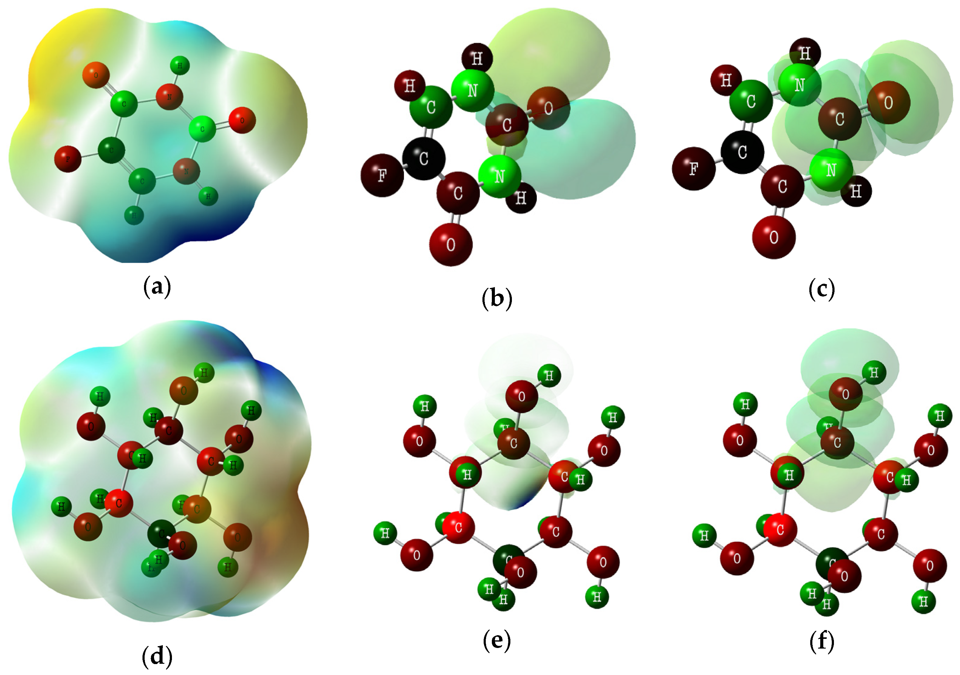
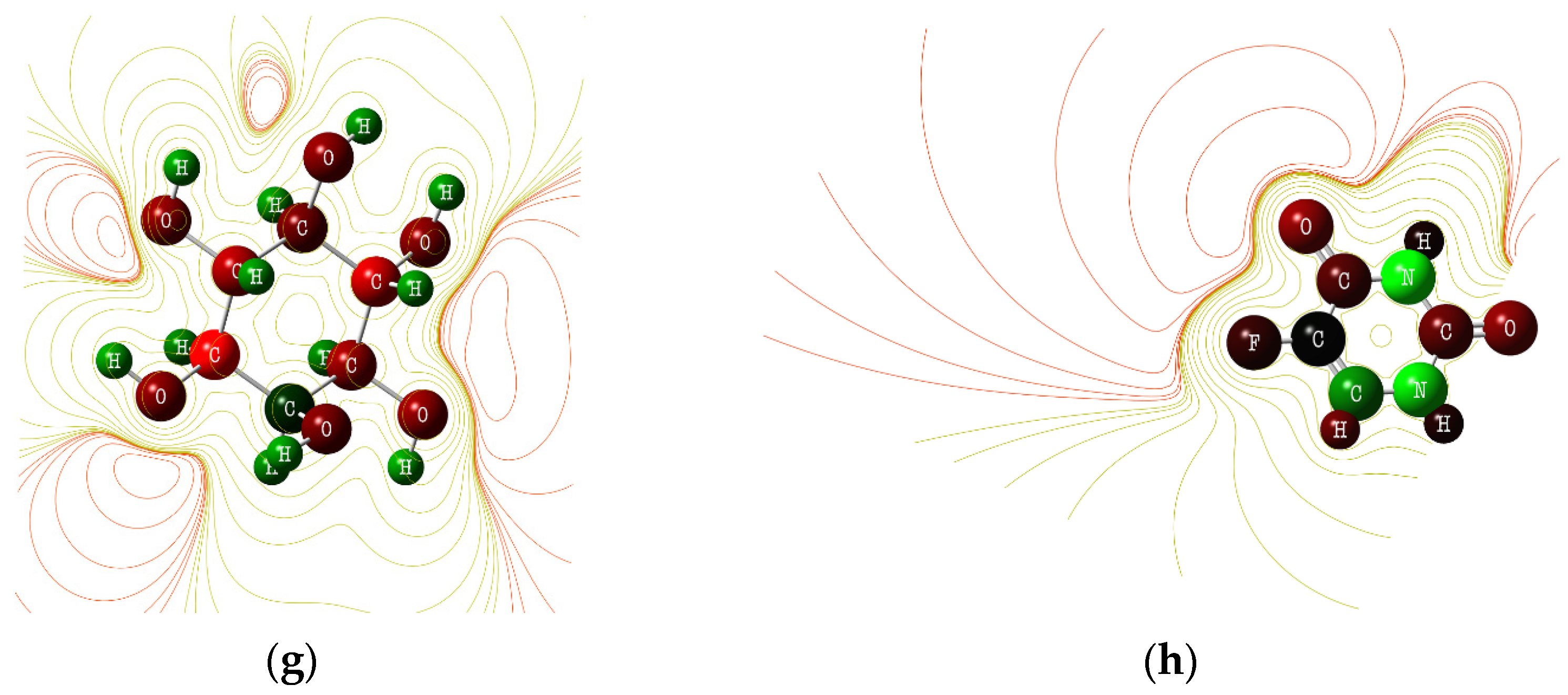
The Langmuir trough technique was performed to investigate the interactions between the nanocarrier and biomimetic membranes. Prior to the measurements, the trough and barriers were washed from potential impurities with chloroform and methanol, while Wilhelmy’s platinum plate was flamed. The Langmuir trough was filled with the subphase solution (distilled water or solution of drug/nanocarriers), and then 40 µL of a chloroform solution of the selected lipid—in particular DOPC, DOPS with concentrations of 2 mg mL−1—was dropped onto the water surface. Then, chloroform was evaporated, and lipids were compressed with a barrier rate of 5 cm2 min−1 until they reached the surface pressure of about 30 mN m−1.
By recording the surface pressure–time dependence, a lipid layer was formed on the pure water (in the way described above), and then an appropriate solution of hydroxyapatite or drug was injected under the surface of the subphase.

2.10. DTF Studies

The spectral and electronic properties of 5-fluorouracil and myo-inositol were analyzed via the application of the density functional theory (DFT). The calculations were made using Gaussian ver. 09 software packages [37]. The geometry optimization, molecular electrostatic potential (MEP), and frontier molecular orbitals (FMOs) analyses were carried out using the B3LYP method with a 6-23G**(d, p) basis set with the default convergence criteria [38,39,40]. GaussView 5.0 was employed to plot the highest occupied molecular (HOMO) and lowest unoccupied molecular orbital (LUMO) as well as molecular electrostatic potential [37].

2.11. Procedures

Synthesis of hydroxyapatite
Nanostructural hydroxyapatite (HAp) was synthesized using the wet co-precipitation method in a glass beaker, wherein 590 mg of Ca(NO3)2 (calcium nitrate) was dissolved in 25 mL of deionized water. The beaker was placed on the magnetic stirrer, and the solution was stirred at 600 rpm. Then, 330 mg of (NH4)2HPO4 (diammonium hydrogen phosphate) was dissolved in 25 mL of distilled water and placed in the burette above the beaker. The second burette was filled with a 25% ammonia solution. Then, the (NH4)2HPO4 was added dropwise to the Ca(NO3)2 with continuous stirring, while the ammonia solution was added to adjust the pH to 10.5 during synthesis. The obtained suspension was stirred for 2 h. Next, the suspension was washed several times with distilled water until a neutral pH was achieved. The product had about 440 mg of HAp, which was suspended in 40 mL of distilled water and placed in a 50 mL Falcon tube.
Preparation of Calendula officinalis L. extract
Calendula officinalis L. extract was obtained with the B-811 Büchi Soxhlet extractor (Switzerland), where the 50 g of dry petals was placed in a glass vial, and the Soxhlet standard procedure was used. As an extracting solvent absolute methyl alcohol was used. The extraction of calendula flowers lasted for 3h. Collected calendula extract was evaporated in a Rotavapor R-100 (Büchi, Switzerland) rotary evaporator and dried for 24 h at 50 °C in a vacuum to remove the remaining solvent.
Modification of hydroxyapatite with Calendula officinalis L.
The 40 mg of Calendula officinalis L. extract was added to the HAp aqueous suspension, and 10 mL of methanol was added. Initially, the suspension was sonicated for 15 min. Then, it was shaken mechanically for 4 h on the orbital spinner. Then, it was sonicated for 15 min again and left for mechanical shaking overnight. Next, the suspension was cooled down (to about 10 °C) for 1 h, centrifuged for 5 min at 9000 rpm, and washed with water. The HAp coated with active ingredients from the Calendula officinalis L. extract (HAp@Cal) was subsequently characterized and loaded with 5-fluorouracil.
Modification of HAp@Cal with 5-fluorouracil
The next step was the immobilization of the 5-fluorouracil and calendula extract onto nano-hydroxyapatite, 15 mg of the drug was placed in the colloidal suspension of HAp@Cal and stirred overnight with 300 rpm at room temperature. Subsequently, the suspension was cooled down for 2 h to ~5 °C, and then the suspension was centrifuged at about 1000 rpm for 5 min to remove the solution. Next, the suspension was washed with distilled water. After the subsequent centrifuging, it was suspended in 50 mL of water, and finally, it was shaken on a vortex for 3 min, obtaining HAp@Cal@5-flu.

3. Results

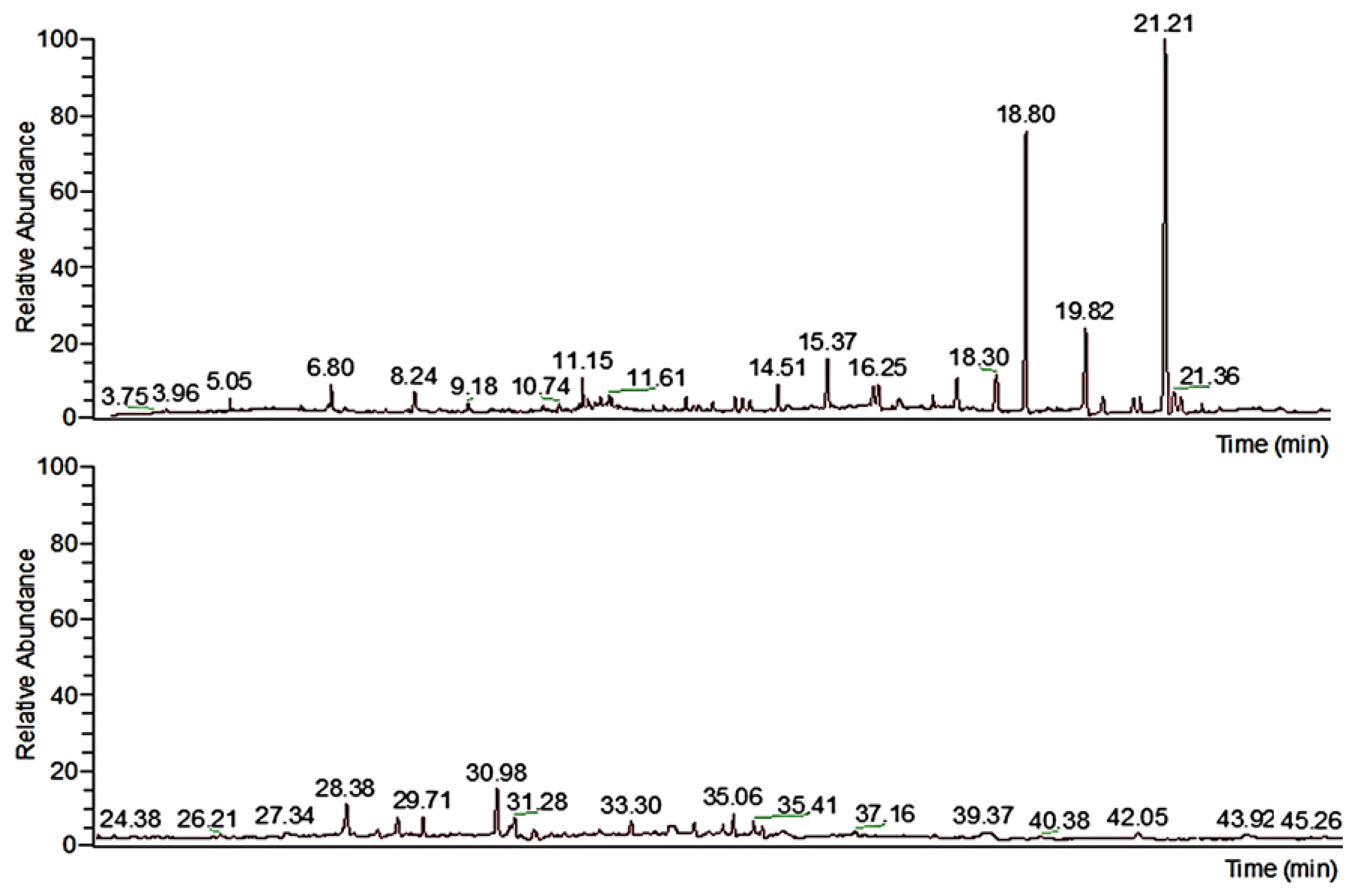
3.1. Chemical Composition Studies of Calendula officinalis L. Extract

The chemical composition of the extract was determined via gas chromatography. Figure 1 presents the chromatogram obtained for the calendula extract (the upper image shows a scale of up to 23 min, and the lower image shows a timescale of up to 46 min). As can be seen, the extract contains 35 compounds that have different retention times. All molecules that were detected are presented in Table 1, relating to the retention time. The highest content (as a percentage) among many other biologically active ingredients is myo-inositol; there is about 56.9% of this compound in the extract. Myo-inositol plays an important role in various cellular processes [41]. Its biological activity is essential for a wide range of cell functions, like the development and function of peripheral nerves [42], osteogenesis [43], and reproduction [30]. It naturally occurs in the natural biological membranes of the cells and is an essential nutrient required by human cells for growth and survival [44,45].
As the extract is rich in this compound, it was chosen to be loaded into the HAp to check the anticancer and antioxidant properties of the nanocomposite. Such an effect is proven in the literature, indicating high biological activity including anticancer [34,46] and antioxidant [47] effects and regulatory activity on the immune system [48]. Another active ingredient, with 7.69% content, is palmitic acid. Its presence in the extract can improve the interactions of the nanocomposite with the biological membranes during in vitro studies.

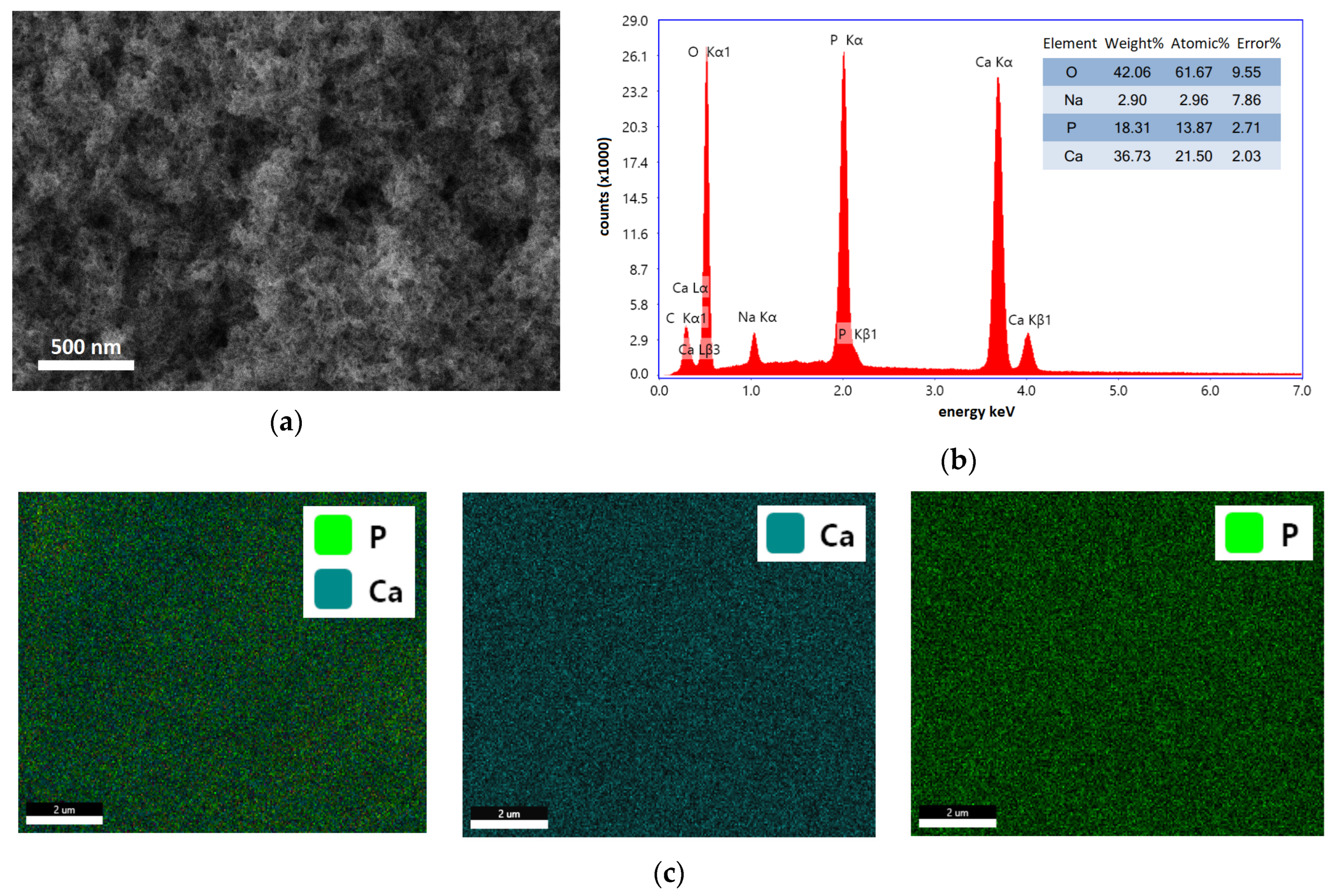
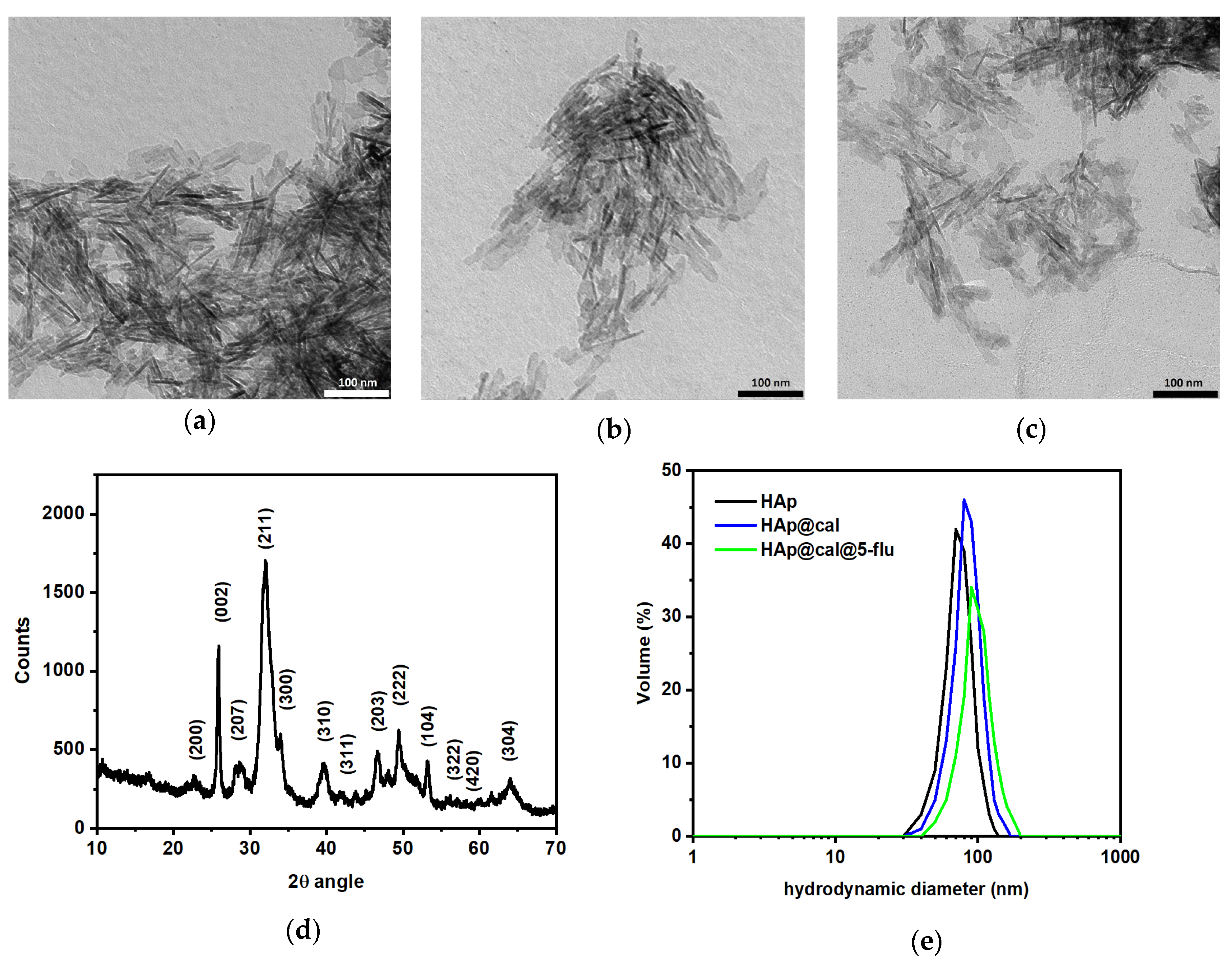
3.2. Morphology, Composition, and Crystallinity Studies

The morphology of HAp was investigated with scanning electron microscopy (SEM). As can be seen in Figure 2a, the sample contains granular structures that are uniformly distributed in the whole bulk, similar to the HAp-based structures demonstrated in the literature [49,50]. To determine the elemental composition of the obtained HAp, X-ray energy dispersive spectroscopy (EDS) was used, see Figure 2b. The recorded spectrum demonstrates the peaks characteristic to Ca, P, O, and traces of Na. The atomic content for Ca is 21.50 ± 2.03%, and that for P is about 13.87 ± 2.71%; the Ca:P ratio is about ~1.6, close to the values presented in the literature for stoichiometric HAp [51,52]. The spectrum also presents the C and Na in the sample, which come from the conducting tape that was used to glue the HAp to the aluminum-based sample holder. The EDS mapping presented in Figure 2c revealed the uniform distribution of Ca and P in the sample.
Complementary to the SEM analysis, the morphologies of HAp and its composites were also investigated with transmission electron microscopy (TEM), see Figure 3a. The suspension taken for the analysis was about 10 µL diluted in 1 mL of distilled water. Before TEM analyses, the suspension was shaken with a vortex for 1 min and placed on a copper mesh (300 grids) covered with a formvar layer. The recorded image for the bare HAp reveals rod-shaped structures similar to the structures described in the literature [53,54]. HAp structures overlap for fast drying from the ethanol suspension; however, the structures with lengths up to 100 ± 15 nm and diameters of about 25 ± 15 nm can be distinguished. The dark spots come from the overlapping of the particular structures during the drying of the samples. Figure 3b and Figure 3c present the hydroxyapatite loaded with the calendula extract (HAp@Cal) and both extract and 5-fluorouracil (HAp@Cal@5-flu), respectively. The organic shell is not clearly seen for HAp@Cal, while the shape and size of the structures are similar to all samples. In the case of HAp@Cal@5-flu, the additional shades from the organic layer can be distinguished on the TEM image.
Subsequently, the crystallinity of the as-synthesized HAp was studied with X-ray diffraction (XRD). Figure 3d shows the presence of reflections peaks at certain 2θ angles, where they can be ascribed as follows: 22.59° (200), 25.90° (002), 28.56° (207), 32.00° (211), 33.85° (300), 39.47° (310), 42.03° (311), 46.44° (203), 49.49° (222), 52.27° (104), 56.32° (322), 60.01° (420), and 64.02° (304). Recorded peaks can be ascribed to the hydroxyapatite, which is in good agreement with the literature [55,56,57,58]. The reflection peaks were also identified based on the Crystallography Open Database (COD)—in particular, the 96-901-1092 card. The crystallite size D was estimated using the Scherrer formula, Equation (2) [59]:
D = λ K β cos θ ,
where β is the full width at half the maximum length of the reflection, θ stands for the Bragg angle, λ is the X-ray wavelength, and K is a dimensionless shape factor (0.94).
The size of crystallites estimated based on the formula is about 28.6 ± 2.7 nm which refers to the diameter of the obtained structures. Such a value is in good agreement with the TEM studies.
Next, the hydrodynamic sizes of the HAp and its nanocomposites after the organics modification were investigated with dynamic light scattering (DLS) technique. Figure 3e shows a hydrodynamic size of the bare HAp of about 64.0 ± 3.0 nm. The value is larger than that for the dry HAp, for the hydration of nanostructures. The hydrodynamic diameter for the HAp@Cal is about 84.0 ± 5.0 nm, and that of HAp@Cal@5-flu is about 89.0 ± 4.0 nm. The surface potential was estimated at about −10.2 mV for HAp, −22.7 mV for HAp@Cal, and −26.6 mV for HAp@Cal@5-flu. The change in surface potential is caused by the presence of the organic shell on the surface of HAp. Subsequent loading with the 5-flu also changes the surface potential for the presence of negatively charged functional groups.
After the loading of the HAp with calendula extract and the drug, the thermal stability of the nanocomposite was studied. Based on thermogravimetry analysis (TGA) the content of loaded agents was estimated. Figure 3f shows the TGA of HAp and nanocomposite materials, and the inset shows the derivative that precisely presents the change of the recorded mass loss in percent per min (DTG). The mass loss of the unmodified hydroxyapatite sample was 3.5%, occurring at temperatures between 50 and ~100–130 °C. This was related to the evaporation of water and dehydration of the nanomaterial. At higher temperatures, HAp exhibited relative thermal stability within the range of the study, i.e., up to 700 °C. The results obtained are in agreement with the literature data on the thermal stability of hydroxyapatite [60,61,62]. The thermal decomposition of HAp@Cal and HAp@Cal@5-flu was in three stages. The first, related to dehydration, was similar to hydroxyapatite between 50 and ~100–130 °C. The next two steps occurred between up to 350 °C and between 350 and 550 °C, respectively, and were related to the decomposition of organic compounds in the calendula extract and the thermal degradation of the drug [63,64,65]. Based on the differences in mass loss at 550 °C between HAp, HAp@Cal, and HAp@Cal@5-flu, the percentage of calendula extract and drug in the composites was estimated. The analysis showed that the composites contained 6.4% extract and 2.2% 5-fluorouracil.
The FT-IR analysis presented in Figure 3g shows the spectra of HAp, HAp@Cal, and HAP@Cal@5-flu. On the hydroxyapatite spectrum, distinct bands were observed at 470, 559, 599, and 1025 cm−1 which were attributed to vibrations of the PO 4 3 group [66,67,68,69]. Whereas, asymmetric stretching (1454 cm−1) and out-of-plane bending mode (874 cm−1) vibrations, were assigned to the CO 3 2 group. The band at 3565 cm−1 is typical to ion-stretching OH group vibrations in HAp. The wide band at around 3250 cm−1 and the band at 1640 cm−1 are due to the presence of H2O in the hydroxyapatite structure [68,70]. The spectra of HAp@Cal and HAp@Cal@5-flu are similar to unmodified HAp; however, the inset reveals an increase in the bands, mainly for the HAp@Cal@5-flu. The bands in the range of 1500 to 1680 cm−1 can be ascribed to the C=C, C=N, and C=O vibrations for 5-fluorouracil, while the double band at 1160 and 1255 cm−1 relates to the C-O and C-N vibrations also from the drug [71]. Additionally, some changes can be observed; the bands at 2926 cm−1 and 2856 cm−1 are due to the presence of O-H carboxylic acids and alkyl C-H groups in calendula extract [72,73]. Another source shows that bands were observed at 1688, 1255, and 821 cm−1, which were respectively attributed to C=C and ring-stretching vibrations; C-F and ring-stretching vibrations; and ring deformations related to in-plane vibrations [74,75,76]. The FT-IR analysis confirms the adsorption of calendula extract and 5-fluorouracil on hydroxyapatite.

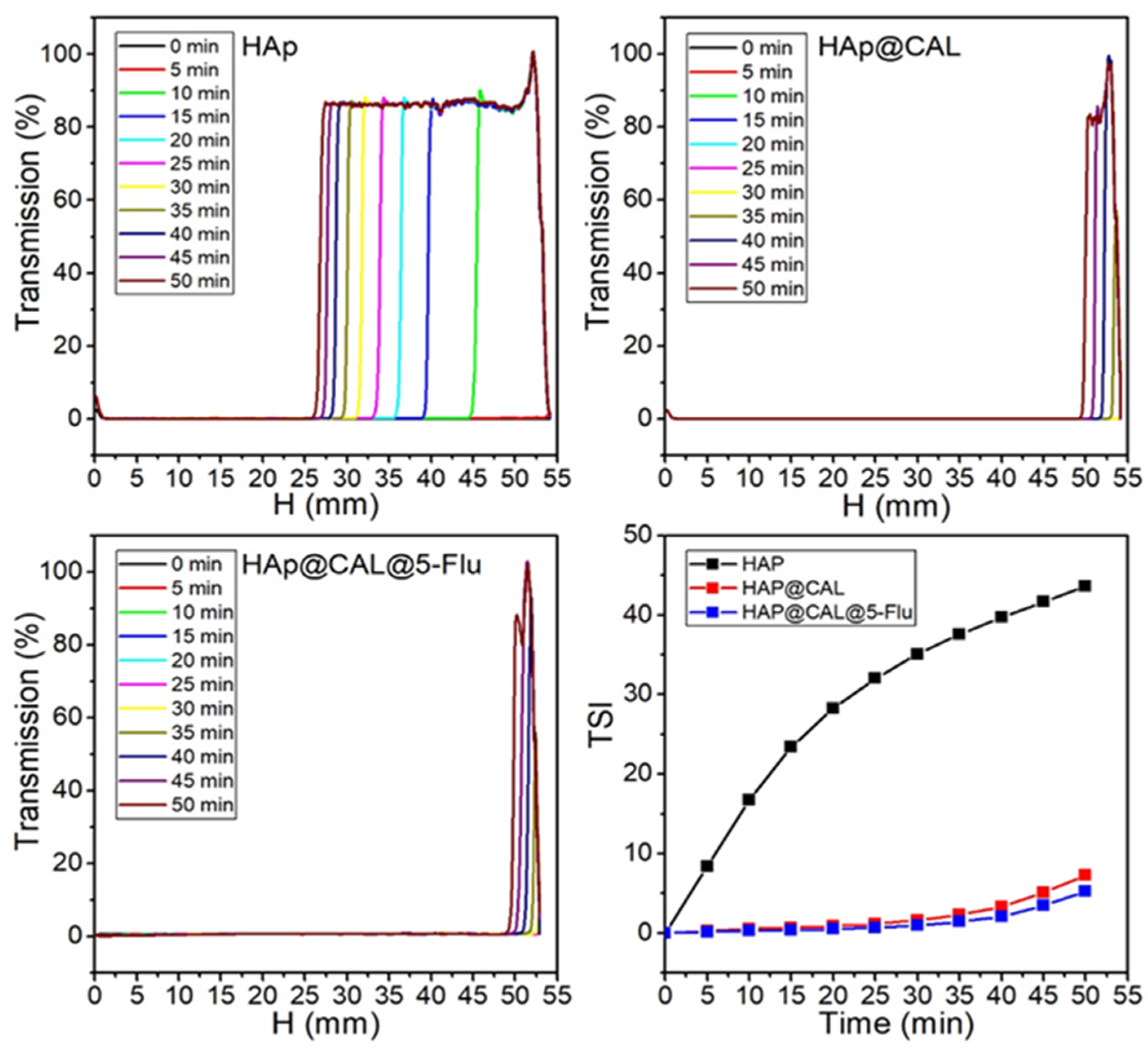
3.3. Turbiscan Analysis

Research into the use of drug delivery systems for anticancer therapy requires the stability of dispersions of the above-mentioned carriers in water. The Turbiscan Lab apparatus (Formulation SA, France) was used to analyze the water dispersions of the obtained materials. The stability of the aqueous suspensions was assessed using the percentage of transmitted light as a function of sample height. The results seen in Figure 4 showed that destabilization of HAp, HAp@Cal, and HAp@Cal@5-flu dispersion occurs due to sedimentation. This is evidenced by an increase in the clarity of the suspension in the upper layer with increasing time tests [77,78,79]. To analyze the destabilization kinetics of the obtained dispersions, the Turbiscan Stability Index (TSI) (Turbiscan-specific parameter) was used, which allows us to compare several systems to each other. In general, the higher the TSI value, the less stable the system [80,81]. Other sources suggest that the dispersion shows a diffuse state when the TSI is less than 5, while when this value is between 5 and 20, the dispersion shows a swollen state. The dispersion is sedimentary when the TSI is greater than 20 [82]. The TSI analysis reveals that unmodified HAp was stable for 5 min, while after 15 min, the dispersion was sedimentary. Adsorption of calendula extract on the surface significantly improved the stability of the dispersion, as evidenced by the low TSI values of both HAp@Cal and HAP@Cal@5-flu. The obtained results indicate that the presented systems are suitable for further research, e.g., using in vitro techniques.

3.4. Antioxidant Properties
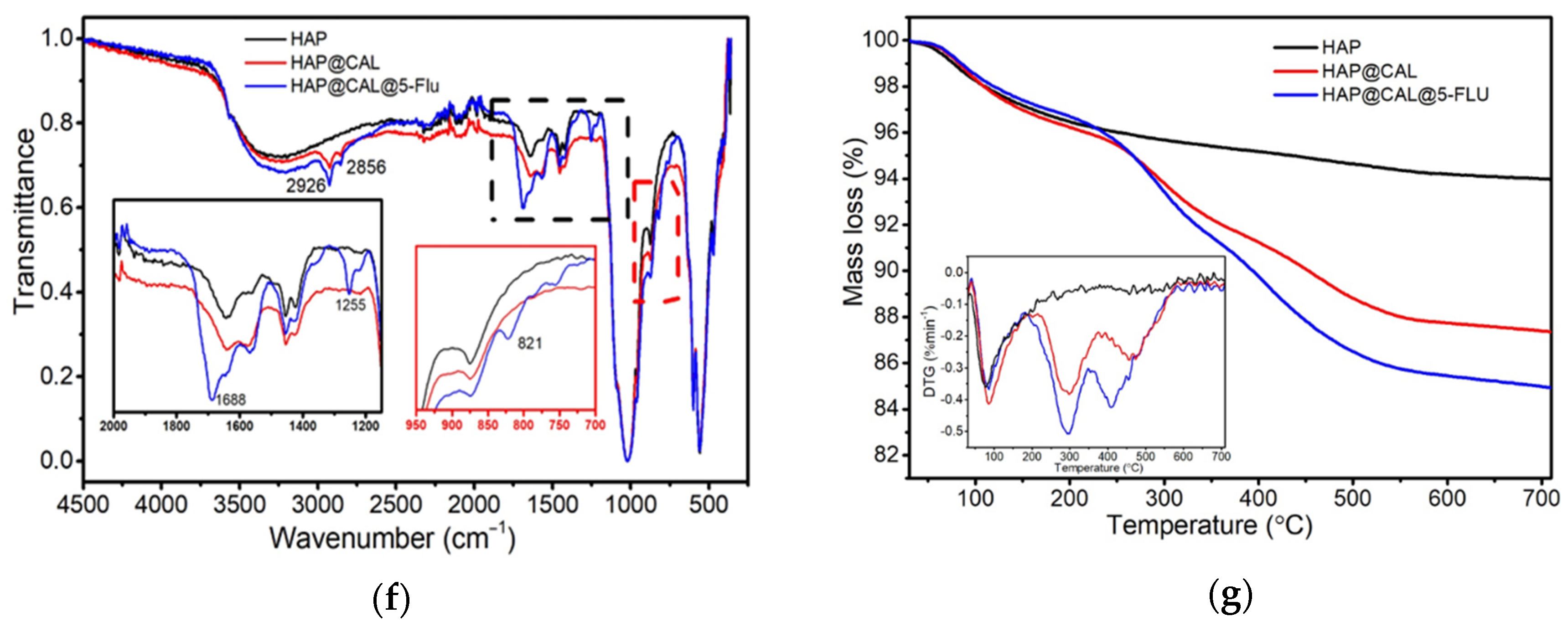
Calendula extract was found to scavenge the free radical DPPH˙, while both HAp and the drug have very low effects against DPPH˙, see Table 2. As the Calendula extract is an effective source of antioxidant agents, its loading into the HAp matrix improved activity, especially for the HAp@Cal@5-flu. Since calendula extract contains phenolic compounds like caffeic acid and chlorogenic acid, which have strong antioxidant properties, it is expected that the extract will present high radical scavenging activity [83,84,85]. Some studies show that calendula extracts have been reported to possess cytotoxic and antitumor activities [83]. On the other hand, there are theories that antioxidant administration during chemotherapy reduces or prevents certain types of ROS-induced toxicities of chemotherapeutics [86]. Adding antioxidant agents—calendula extract in the current research—to obtain stable colloidal suspension not only improves physical properties of dispersion but also benefits in potentially acting against ROS during the therapy.

3.5. DTF Analysis

Additionally, to investigate the antioxidative potential of HAp modified with myo-inositol and 5-fluorouracil, the density functional theory (DFT) was implemented in the studies to determine the geometry optimization. Therefore, the molecular electrostatic potential (MEP) was used as a general measure for the substituent effects in chemical reactions and interactions between molecules, and the frontier molecular orbitals (FMOs) were used to understand the reactivity of both compounds.
The MEP surface of 5-flu is shown in Figure 5a, where the red color denotes the electrophilic attack reactivity, the blue color is responsible for nucleophilic attack reactivity, and the green color represents the neutral region. The dipole moment is 5.39, while the total energy is −510.522 au. The highest occupied molecular orbital (HOMO) and lowest unoccupied molecular orbital (LUMO) are shown in Figure 5b and Figure 5c, respectively. The energy gap is obtained from the difference between the orbital energy value, LUMO, and HOMO. The value of the energy gap (i.e., HOMO–LUMO gap) affects the reactivity of the molecule and its kinetic stability. The greater the value of the HOMO–LUMO gap, the lower the reactivity of the molecule and the greater its kinetic stability. In the case of 5-fluorouracil, the energy gap is equal to 0.382 eV. Figure 5d presents the MEP surface of myo-inositol, Figure 5e shows the HOMO, and Figure 5f reveals the LUMO. The HOMO–LUMO gap is equal to 0.578 eV, showing higher kinetic stability and lower reactivity of myo-inositol than 5-fluorouracil.
The electrostatic potential contour maps for positive and negative potentials for myo-inositol and 5-fluorouracil, respectively, are shown in Figure 5g,h. Based on the map, the partial distribution of change along the molecule’s surface is presented, making it useful tool for the determination of the molecular polarity as well as the dipole arrow placement. As can be seen in the HOMO–LUMO and electrostatic potential contour map, both compounds have different reactivities, and myo-inositol can easily undergo the oxidation pathway [87]. The drug has two potential sites of protonation and deprotonation entering the cell using the same mechanism of transport as uracil [88,89]. This shows that the combination of both compounds in the therapy can provide additive effects, making it even more effective in biological applications.

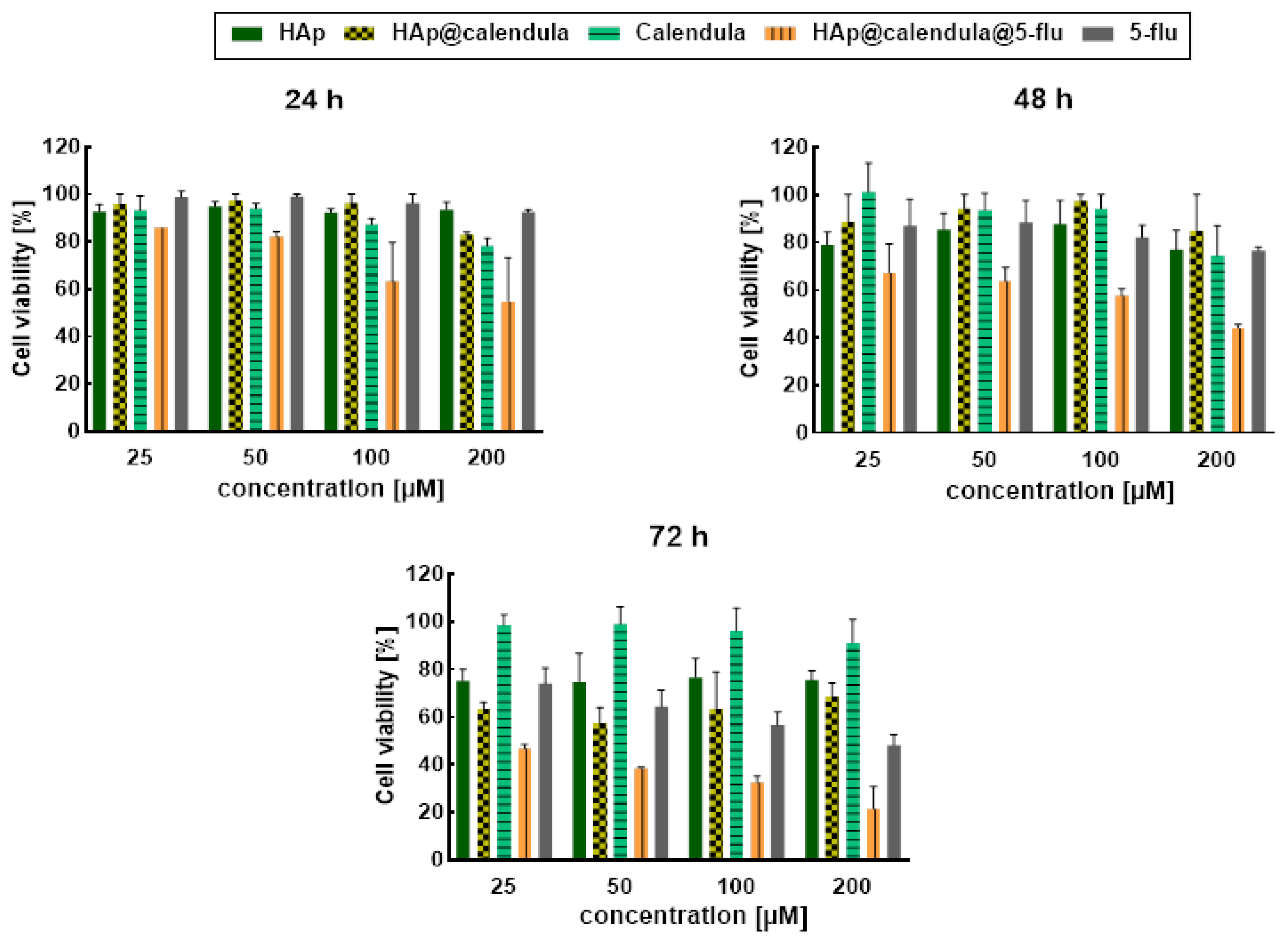
3.6. In Vitro Cytotoxicity Results

To evaluate the cytotoxicity of nanocomposite loaded with calendula extract and the anticancer drug, in vitro cell experiments with the use of an MTS reagent were performed. Figure 6 shows the results of the cytotoxicity studies after 24, 48, and 72 h where the drug concentrations were about 25, 50, 100, and 200 µM. The concentrations of HAp, HAp@Cal, and calendula extract separately were the same like the concentration of these ingredients in the composite HAp@Cal@5-flu. The cytotoxic effect of HAp@Cal@5-flu nanocomposite is even observed for the lowest drug concentration and the shortest exposure time. This effect becomes stronger with the increase in drug concentration and incubation time, reaching 60% cell viability for 100 µM after 24 h. Interestingly, the bare calendula extract exhibits no cytotoxicity. However, according to the bare HAp, Figure 6 shows the decrease in cell viability to ~80%, which can be explained by the lack of stabilization of HAp and subsequent agglomeration onto cells [90]. Additionally, it can also be caused by the specific shape and size of the obtained nanostructures. The literature also refers to similar effects [91,92,93]. A significant reduction in cell viability (~70%) was achieved for HAp@Cal@5-flu at 200 µM drug concentration and 72 h of incubation time. This effect can occur due to the presence of myo-inositol and palmitic acid in the calendula extract. These molecules are noncytotoxic alone. While they interact with biological membranes, the drug can be more easily released and accumulated into the cells. The proposed nanocomposite demonstrated high effectiveness on SKOV-3 cancer cells. Furthermore, a greater effect after treatment with both calendula extract and 5-fluorouracil bound to HAp was achieved in comparison to these compounds used separately. Since 5-fluorouracil has adverse side effects, the use of HAp@Cal@5-flu will allow the concentration of the drug to be reduced while keeping the drug highly effective. This can be caused by the biological activity of myo-inositol. The level of this compound in the body is decreased during carcinogenesis [87,94]. Several studies show supplementation of myo-inositol effect during anticancer therapy on the inhibition of cancer growth [95,96].

3.7. Interaction of Conjugate with Model Membrane System

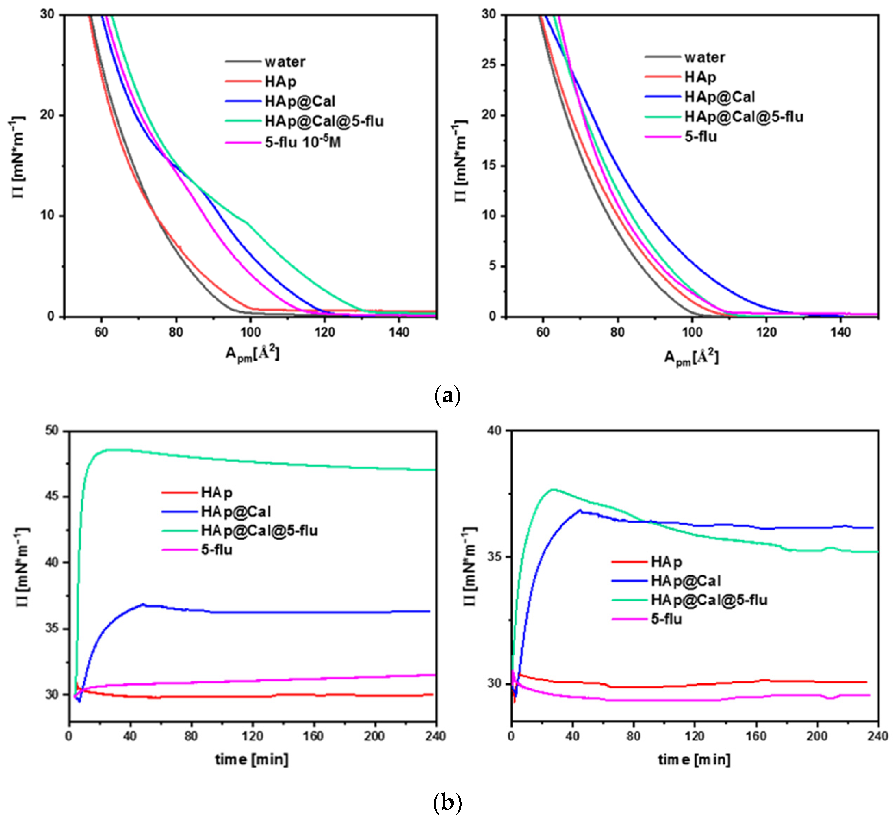
3.7.1. Langmuir Studies

The Langmuir method is the technique that is most commonly used to obtain and study artificial lipid monolayers, which—in their compositions and properties—correspond to biological membranes found in living organisms. Langmuir monolayers are simplified membrane models as they mimic half of the natural cell membranes [97]. For that reason, it was decided that research would be conducted to obtain information on how the conjugate and its components affect the structure of the lipid membranes formed using this method. The biological membranes of normal and cancerous cells differ in terms of composition and structure. In the following studies, two types of lipids, DOPC (1,2-dioleoyl-sn-glycero-3-phosphocholine) and DOPS (1,2-dioleoyl-sn-glycero-3-phospho-L-serine), were used to mimic the structure of the membrane of a normal cell and a cancer cell, respectively [98]. The films were formed on a subphase consisting of water or appropriate solutions of hydroxyapatite and/or drug.
Based on the measurements, it can be seen that the addition of pure hydroxyapatite to the subphase does not change the structure of the membrane or the shape of the isotherm, see Figure 7a. This does not depend on the type of used lipid or the concentration of HAp (even more than 30 mg L−1). The observed effect is very promising and consistent with the in vitro studies because the carrier should be inert to lipid structures and living cells [99]. Hydroxyapatite modified with calendula extract has a strong affinity for both lipids. Even at low surface pressures, the surface area per lipid molecule increases significantly. In the case of DOPC film a slight transition on the isotherm at 22 mN m−1 is visible, see Figure 7b, which indicates a change in the state of organization of molecules in the membrane under the influence of the presence of natural substances contained in calendula. That effect is mitigated by the addition of a drug adsorbed on the HAp surface. 5-fluorouracil is physically adsorbed in the outer shell of the carrier, which limits its direct contact with lipids and may reduce the interaction of calendula substances with the membrane. The interaction of nanostructures with the DOPS film is much stronger than in the case of the phosphatidylcholine membrane. The adsorption effect of the conjugate on the DOPS film is several times higher than on the DOPC layer. The modification of the nanocarrier with fluorouracil does not reduce its affinity for the DOPS membrane. On the contrary, this phenomenon is enhanced. Lipid DOPS is used in research as a model of the cancer cell membrane. Hence, the strong penetration effect of such a membrane by the conjugate is a very interesting and promising phenomenon. The carrier modified with calendula extract and the drug 5-fluorouracil has a chance to act selectively in relation to cancer cells while omitting normal cells. The presented studies confirm that cytotoxicity to selected cancer cells is much stronger for conjugate than its individual components. It makes the opportunity to develop new carriers as combinations of drugs and substances of natural origin with wide potential applications in medicine.

3.7.2. Surface Pressure–Time Dependence

The following studies were intended to investigate the effects of HAp, HAp@Cal, and HAp@Cal@5-flu on an organized biomimetic membrane. For this purpose, lipid layers were compressed at a surface pressure of 30 mN m−1. Then, an appropriate solution was injected into the subphase under the formed film. In therapy, the conjugates or drugs are injected into the bloodstream and then must overcome the biological membranes. Thus, the active compound is in contact with the already organized membrane. The proposed system was to imitate the model of interaction of hydroxyapatite and drugs with cell membranes in the body. The selection of the pressure value of about 30 mN m−1 allows experiments for a membrane with a similar degree of organization that occurs in natural cell membranes to be conducted [100,101]. The injection of a pure hydroxyapatite solution under the organized layer does not cause any changes in the surface pressure value, so the carrier does not interact with the biomimetic membrane. This result confirms the presented results of the Langmuir isotherm and cytotoxicity tests—hydroxyapatite is inert to the lipid structures and cells of selected lines.
The addition of a pure drug with a concentration of 10−5 M does not significantly affect the organization of lipids in any of the tested membranes [98,102]. In the presence of the DOPS layer for the drug, only slight pressure changes of about 1.5 mN m−1 are visible, which suggests easier adsorption of fluorouracil on the tumor cell model. Application of hydroxyapatite modified with calendula extract shows that the nanocarrier penetrates the biomimetic membrane within about 50 min, increasing the ∏ value to 37 mN m−1. The type of lipid does not play a role in the adsorption kinetics of the nanosystem. Calendula extract cannot selectively interact with one of the cell membrane models (DOPC or DOPS). The situation changes when the carrier has been modified with a plant extract and a drug at the same time. In the case of the DOPS membrane, the pressure value and adsorption time increase significantly. The higher effects of both compounds (calendula and 5-fluorouracil) affect the kinetics of the conjugate penetration into the membrane. In less than 20 min, the DOPS layer is completely saturated with the conjugate, while the pressure increases by about 60% relative to HAp@Cal. In the right panel of Figure 7 (for the DOPC lipid), the lack of synergism associated with the coexistence of both substances can be observed. After modification of the conjugate with drugs, the value of surface pressure decreases compared to the HAp@Cal nanocarrier.
The results obtained from the Langmuir technique confirm the higher adsorption efficiency of the conjugate in the case of the tumor cell membrane model, at the same time indicating the possible synergism of calendula extract and 5-fluorouracil. The possibility of greater affinity of the conjugate for tumor cells may have a promising effect in future therapies using the proposed material.

4. Conclusions

This work presents nanostructural hydroxyapatite as a platform for the immobilization of biologically active ingredients from the plant extract and anticancer drug for nano-biomedicinal use. A rod-shaped HAp ~30 nm in diameter and ~100 nm in length was prepared using the wet co-precipitation method. The EDS and XRD studies confirmed the formation of the HAp with a Ca:P ratio of ~1.6, which is similar to the natural hydroxyapatites. Nanostructures were loaded with the calendula extract from the petals of C. officinalis L.—rich with biologically active myo-inositol—and then the cytostatic drug 5-fluorouracil. Calendula extract was loaded into the HAp carrier to interact with biological membranes and work as a stabilizing agent to obtain stable colloidal suspension. FT-IR, DLS, and turbidimetry studies show the changes in the surface properties of the conjugate after loading of the extract. The nanocomposite HAp@Cal@5-flu reveals a cytotoxic effect on SKOV-3 cancer cells, where the effect is much stronger than for bare 5-fluorouracil. Based on the Langmuir trough results, it can be seen that the composite exhibits strong adsorption to the analogue of tumorous cell membrane. Moreover, calendula extract is a source of antioxidant agents, leading to the effective scavenging of DPPH radicals. The conducted research confirms the potential of HAp@Cal@5-flu to be applied in anticancer therapy, making it a promising nanocarrier to be used in the biomedical field.

Author Contributions

Conceptualization: M.O., S.W., J.S. and K.S.; data curation: M.O., S.W., J.S., H.D.N., T.K.O.N., K.S., A.M.-P., K.Ż.-M., D.N., A.P., T.P.N., A.T. and D.K.; formal analysis: M.O., S.W., J.S., H.D.N., T.K.O.N., A.M.-P. and K.Ż.-M.; funding acquisition: M.O., M.G. and T.P.N.; investigation: M.O., S.W., J.S., H.D.N., T.K.O.N., K.S., A.M.-P., K.Ż.-M., A.P., D.N. and T.P.N.; methodology: M.O., S.W., J.S., K.S., H.D.N., T.K.O.N., T.P.N., A.M.-P., K.Ż.-M. and A.P.; project administration: M.O.; resources: M.O. and M.G.; validation: M.O.; supervision: M.O. and M.G.; visualization: M.O.; writing—original draft: M.O., S.W., J.S., H.D.N., A.M.-P., K.Ż.-M., D.N., A.P., A.T., D.K. and M.G.; writing—review and editing: M.O. and M.G. All authors have read and agreed to the published version of the manuscript.

Funding

This work was partially supported by the Vietnam Academy of Science and Technology under grant No. CT0000.09/21-23.

Institutional Review Board Statement

Not applicable.

Informed Consent Statement

Not applicable.

Data Availability Statement

Data will be available upon request.

Acknowledgments

M.O. and P.T.N. would like to thank Thi Mai Thanh Dinh from the University of Science and Technology of Hanoi, Vietnam Academy of Science and Technology for the consultations.

Conflicts of Interest

The authors declare no conflict of interest.

References

  1. Ramawat, K.; Dass, S.; Mathur, M. The chemical diversity of bioactive molecules and therapeutic potential of medicinal plants. In Herbal Drugs: Ethnomedicine to Modern Medicine; Ramawat, K., Ed.; Springer: Berlin/Heidelberg, Germany, 2009; pp. 7–30. [Google Scholar]
  2. Abdelwahab, S.I.; Taha, M.M.E.; Taha, S.M.E.; Alsayegh, A.A. Fifty-year of Global Research in Calendula Officinalis L. (1971−2021): A Bibliometric Study. Clin. Complement. Med. Pharmacol. 2022, 2, 100059. [Google Scholar] [CrossRef]
  3. Rogowska, A.; Stpiczyńska, M.; Pączkowski, C.; Szakiel, A. The Influence of Exogenous Jasmonic Acid on the Biosynthesis of Steroids and Triterpenoids in Calendula officinalis Plants and Hairy Root Culture. Int. J. Mol. Sci. 2022, 23, 12173. [Google Scholar] [CrossRef]
  4. Akhtar, G.; Faried, H.N.; Razzaq, K.; Ullah, S.; Wattoo, F.M.; Shehzad, M.A.; Sajjad, Y.; Ahsan, M.; Javed, T.; Dessoky, E.S.; et al. Chitosan-Induced Physiological and Biochemical Regulations Confer Drought Tolerance in Pot Marigold (Calendula officinalis L.). Agronomy 2022, 12, 474. [Google Scholar] [CrossRef]
  5. Raal, A.; Kirsipuu, K.; Must, R.; Tenno, S. Content of Total Carotenoids in Calendula officinalis L. from Different Countries Cultivated in Estonia. Nat. Prod. Commun. 2009, 4, 35–38. [Google Scholar] [CrossRef]
  6. Joly, R.; Forcella, F.; Peterson, D.; Eklund, J. Planting depth for oilseed calendula. Ind. Crop. Prod. 2013, 42, 133–136. [Google Scholar] [CrossRef]
  7. Siracusa, L.; Napoli, E.; Ruberto, G. Review Novel Chemical and Biological Insights of Inositol Derivatives in Mediterranean Plants. Molecules 2022, 27, 1525. [Google Scholar] [CrossRef] [PubMed]
  8. Chhetri, D.R. Myo-Inositol and Its Derivatives: Their Emerging Role in the Treatment of Human Diseases. Front. Pharmacol. 2019, 10, 1172. [Google Scholar] [CrossRef] [PubMed]
  9. Loewus, F.A.; Murthy, P.P.N. myo-Inositol metabolism in plants. Plant Sci. 2000, 150, 1–19. [Google Scholar] [CrossRef]
  10. Papaleo, E.; Unfer, V.; Baillargeon, J.P.; Chiu, T. Contribution of myo-inositol to reproduction. Eur. J. Obstet. Gynecol. Reprod. Biol. 2009, 147, 120–123. [Google Scholar] [CrossRef]
  11. Haris, M.; Cai, K.; Singh, A.; Hariharan, H.; Reddy, R. In vivo mapping of brain myo-inositol. NeuroImage 2011, 54, 2079–2085. [Google Scholar] [CrossRef]
  12. Ratiu, I.A.; Al-Suod, H.; Ligor, M.; Ligor, T.; Krakowska, A.; Górecki, R.; Buszewski, B. Simultaneous Determination of Cyclitols and Sugars Following a Comprehensive Investigation of 40 Plants. Food Anal. Methods 2019, 12, 1466–1478. [Google Scholar] [CrossRef]
  13. Pintaudi, B.; Di Vieste, G.; Bonomo, M. The Effectiveness of Myo-Inositol and D-Chiro Inositol Treatment in Type 2 Diabetes. Int. J. Endocrinol. 2016, 2016, 9132052. [Google Scholar] [CrossRef]
  14. Amabile, M.I.; De Luca, A.; Tripodi, D.; D’alberti, E.; Melcarne, R.; Imbimbo, G.; Picconi, O.; D’andrea, V.; Vergine, M.; Sorrenti, S.; et al. Personalized Medicine Effects of Inositol Hexaphosphate and Myo-Inositol Administration in Breast Cancer Patients during Adjuvant Chemotherapy. J. Pers. Med. 2021, 11, 756. [Google Scholar] [CrossRef]
  15. El Hafny-Rahbi, B.; Brodaczewska, K.; Collet, G.; Majewska, A.; Klimkiewicz, K.; Delalande, A.; Grillon, C.; Kieda, C. Tumour angiogenesis normalized by myo-inositol trispyrophosphate alleviates hypoxia in the microenvironment and promotes antitumor immune response. J. Cell. Mol. Med. 2021, 25, 3284–3299. [Google Scholar] [CrossRef]
  16. Naahidi, S.; Jafari, M.; Edalat, F.; Raymond, K.; Khademhosseini, A.; Chen, P. Biocompatibility of engineered nanoparticles for drug delivery. J. Control. Release 2013, 166, 182–194. [Google Scholar] [CrossRef]
  17. Sebastiammal, S.; Fathima, A.S.L.; Henry, J.; Wadaan, M.A.; Mahboob, S.; Wadaan, A.M.; Manzoor, I.; Gopinath, K.; Rajeswary, M.; Govindarajan, M. Synthesis, Characterization, Antibacterial, Antifungal, Antioxidant, and Anticancer Activities of Nickel-Doped Hydroxyapatite Nanoparticles. Fermentation 2022, 8, 677. [Google Scholar] [CrossRef]
  18. Panneerselvam, R.; Anandhan, N.; Gopu, G.; Ganesan, K.P.; Marimuthu, T. Impact of different transition metal ions in the structural, mechanical, optical, chemico-physical and biological properties of nanohydroxyapatite. Appl. Surf. Sci. 2020, 506, 144802. [Google Scholar] [CrossRef]
  19. Sobierajska, P.; Dorotkiewicz-Jach, A.; Zawisza, K.; Okal, J.; Olszak, T.; Drulis-Kawa, Z.; Wiglusz, R.J. Preparation and antimicrobial activity of the porous hydroxyapatite nanoceramics. J. Alloys Compd. 2018, 748, 179–187. [Google Scholar] [CrossRef]
  20. Zawisza, K.; Sobierajska, P.; Nowak, N.; Kedziora, A.; Korzekwa, K.; Pozniak, B.; Tikhomirov, M.; Miller, J.; Mrowczynska, L.; Wiglusz, R.J. Preparation and preliminary evaluation of bio-nanocomposites based on hydroxyapatites with antibacterial properties against anaerobic bacteria. Mater. Sci. Eng. C 2020, 106, 110295. [Google Scholar] [CrossRef]
  21. Mondal, S.; Dorozhkin, S.V.; Pal, U. Recent progress on fabrication and drug delivery applications of nanostructured hydroxyapatite. WIREs Nanomed. Nanobiotechnology 2018, 10, 1504. [Google Scholar] [CrossRef] [PubMed]
  22. Song, J.; Cui, N.; Mao, X.; Huang, Q.; Lee, E.-S.; Jiang, H. Sorption Studies of Tetracycline Antibiotics on Hydroxyapatite (001) Surface—A First-Principles Insight. Materials 2022, 15, 797. [Google Scholar] [CrossRef] [PubMed]
  23. Vechasilp, J.; Tangtrakulwanich, B.; Oungbho, K.; Yuenyongsawad, S. The efficacy of methotrexate-impregnated hydroxyapatite composites on human mammary carcinoma cells. J. Orthop. Surg. 2007, 15, 56–61. [Google Scholar] [CrossRef] [PubMed]
  24. Watanabe, K.; Nishio, Y.; Makiura, R.; Nakahira, A.; Kojima, C. Paclitaxel-loaded hydroxyapatite/collagen hybrid gels as drug delivery systems for metastatic cancer cells. Int. J. Pharm. 2013, 446, 81–86. [Google Scholar] [CrossRef] [PubMed]
  25. Sobierajska, P.; Serwotka-Suszczak, A.; Targonska, S.; Szymanski, D.; Marycz, K.; Wiglusz, R.J. Synergistic Effect of Toceranib and Nanohydroxyapatite as a Drug Delivery Platform—Physicochemical Properties and In Vitro Studies on Mastocytoma Cells. Int. J. Mol. Sci. 2022, 23, 1944. [Google Scholar] [CrossRef] [PubMed]
  26. Jiang, X.; Zhao, Y.; Wang, C.; Sun, R.; Tang, Y. Effects of physico-chemical properties of ions-doped hydroxyapatite on adsorption and release performance of doxorubicin as a model anticancer drug. Mater. Chem. Phys. 2022, 276, 125440. [Google Scholar] [CrossRef]
  27. Zhao, Q.; Zhang, D.; Sun, R.; Shang, S.; Wang, H.; Yang, Y.; Wang, L.; Liu, X.; Sun, T.; Chen, K. Adsorption behavior of drugs on hydroxyapatite with different morphologies: A combined experimental and molecular dynamics simulation study. Ceram. Int. 2019, 45, 19522–19527. [Google Scholar] [CrossRef]
  28. Kundu, B.; Ghosh, D.; Sinha, M.K.; Sen, P.S.; Balla, V.K.; Das, N.; Basu, D. Doxorubicin-intercalated nano-hydroxyapatite drug-delivery system for liver cancer: An animal model. Ceram. Int. 2013, 39, 9557–9566. [Google Scholar] [CrossRef]
  29. Luo, H.; Ji, D.; Li, C.; Zhu, Y.; Xiong, G.; Wan, Y. Layered nanohydroxyapatite as a novel nanocarrier for controlled delivery of 5-fluorouracil. Int. J. Pharm. 2016, 513, 17–25. [Google Scholar] [CrossRef]
  30. Grocholewicz, K.; Matkowska-Cichocka, G.; Makowiecki, P.; Droździk, A.; Ey-Chmielewska, H.; Dziewulska, A.; Tomasik, M.; Trybek, G.; Janiszewska-Olszowska, J. Effect of nano-hydroxyapatite and ozone on approximal initial caries: A randomized clinical trial. Sci. Rep. 2020, 10, 11192. [Google Scholar] [CrossRef]
  31. Saif, M.W.; Choma, A.; Salamone, S.J.; Chu, E. Pharmacokinetically guided dose adjustment of 5-fluorouracil: A rational approach to improving therapeutic outcomes. J. Natl. Cancer Inst. 2009, 101, 1543–1552. [Google Scholar] [CrossRef]
  32. Bocci, G.; Barbara, C.; Vannozzi, F.; Dipaolo, A.; Melosi, A.; Barsanti, G.; Allegrini, G.; Falcone, A.; Deltacca, M.; Danesi, R. A pharmacokinetic-based test to prevent severe 5-fluorouracil toxicity. Clin. Pharmacol. Ther. 2006, 80, 384–395. [Google Scholar] [CrossRef]
  33. Gamelin, E.; Delva, R.; Jacob, J.; Merrouche, Y.; Raoul, J.L.; Pezet, D.; Dorval, E.; Piot, G.; Morel, A.; Boisdron-Celle, M. Individual fluorouracil dose adjustment based on pharmacokinetic follow-up compared with conventional dosage: Results of a multicenter randomized trial of patients with metastatic colorectal cancer. J. Clin. Oncol. 2008, 26, 2099–2105. [Google Scholar] [CrossRef] [PubMed]
  34. Fang, L.; Jiang, Y.; Yang, Y.; Zheng, Y.; Zheng, J.; Jiang, H.; Zhang, S.; Lin, L.; Zheng, J.; Zhang, S.; et al. Determining the optimal 5-FU therapeutic dosage in the treatment of colorectal cancer patients. Oncotarget 2016, 7, 81880–81887. [Google Scholar] [CrossRef] [PubMed]
  35. Chavani, O.P. 5-Fluorouracil Response Prediction and Blood Level–Guided Therapy in Oncology: Existing Evidence Fundamentally Supports Instigation. Ther. Drug Monit. 2020, 42, 660–664. [Google Scholar] [CrossRef] [PubMed]
  36. Brand-Williams, W.; Cuvelier, M.E.; Berset, C. Use of a free radical method to evaluate antioxidant activity. LWT Food Sci. Technol. 1995, 28, 25–30. [Google Scholar] [CrossRef]
  37. Frisch, M.J.; Trucks, G.W.; Schlegel, H.B.; Scuseria, G.E.; Robb, M.A.; Cheeseman, J.R. Gaussian 09, Revision C.02.; Gaussian Inc.: Wallingford, CT, USA, 2010. [Google Scholar]
  38. Alghamdi, S.; Abbas, F.; Hussein, R.; Alhamzani, A.; El-Shamy, N. Spectroscopic characterization (IR, UV-Vis), and HOMO-LUMO, MEP, NLO, NBO Analysis and the Antifungal Activity for 4-Bromo-N-(2-nitrophenyl) benzamide; Using DFT Modeling and In silico Molecular Docking. J. Mol. Struct. 2023, 1271, 134001. [Google Scholar] [CrossRef]
  39. Raghavachari, K. Perspective on density functional thermochemistry. III. The role of exact exchange. Theor. Chem. Acc. 2000, 103, 361–363. [Google Scholar] [CrossRef]
  40. Elzupir, A.O.; Hussein, R.K.; Ibnaouf, K.H. Intermolecular CH-π Electrons Interaction in Poly (9,9-dioctylfluorenyl-2,7-diyl) (PFO): An Experimental and Theoretical Study. Molecules 2022, 27, 1488. [Google Scholar] [CrossRef]
  41. Croze, M.L.; Soulage, C.O. Potential role and therapeutic interests of myo-inositol in metabolic diseases. Biochimie 2013, 95, 1811–1827. [Google Scholar] [CrossRef]
  42. Chau, J.F.L.; Lee, M.K.; Law, J.W.S.; Chung, S.K.; Chung, S.S.M. Sodium/myo-inositol cotransporter-1 is essential for the development and function of the peripheral nerves. FASEB J. 2005, 19, 1887–1889. [Google Scholar] [CrossRef]
  43. Dai, Z.; Chung, S.K.; Miao, D.; Lau, K.S.; Chan, A.W.; Kung, A.W. Sodium/myo-Inositol Cotransporter 1 and myo-Inositol Are Essential for Osteogenesis and Bone Formation. J. Bone Miner. Res. 2011, 26, 582–590. [Google Scholar] [CrossRef] [PubMed]
  44. Lam, S.; McWilliams, A.; Leriche, J.; MacAulay, C.; Wattenberg, L.; Szabo, E. A phase I study of myo-inositol for lung cancer chemoprevention. Cancer Epidemiol. Biomark. Prev. 2006, 15, 1526–1531. [Google Scholar] [CrossRef] [PubMed]
  45. Eagle, H.; Oyama, V.I.; Levy, M.; Freeman, A.E. myo-Inositol as an essential growth factor for normal and malignant human cells in tissue culture. J. Biol. Chem. 1957, 226, 191–205. [Google Scholar] [CrossRef] [PubMed]
  46. Bizzarri, M.; Dinicola, S.; Bevilacqua, A.; Cucina, A. Broad Spectrum Anticancer Activity of Myo-Inositol and Inositol Hexakisphosphate. Int. J. Endocrinol. 2016, 2016, 5616807. [Google Scholar] [CrossRef]
  47. Graf, E.; Eaton, J.W. Antioxidant functions of phytic acid. Free Radic. Biol. Med. 1990, 8, 61–69. [Google Scholar] [CrossRef]
  48. Zhang, Z.; Song, Y.; Wang, X.-L. Inositol hexaphosphate-induced enhancement of natural killer cell activity correlates with suppression of colon carcinogenesis in rats. World J. Gastroenterol. 2005, 11, 5044–5046. [Google Scholar] [CrossRef]
  49. Chang, L.; Huang, S.; Zhao, X.; Hu, Y.; Ren, X.; Mei, X.; Chen, Z. Preparation of ROS active and photothermal responsive hydroxyapatite nanoplatforms for anticancer therapy. Mater. Sci. Eng. C 2021, 125, 112098. [Google Scholar] [CrossRef]
  50. Barbosa, A.A.; Júnior, S.A.; Mendes, R.L.; de Lima, R.S.; Ferraz, A.D.V. Multifunctional hydroxyapatite with potential for application in theranostic nanomedicine. Mater. Sci. Eng. C 2020, 116, 111227. [Google Scholar] [CrossRef]
  51. Thi Thom, N.; Thi Mai Thanh, D.; Thi Nam, P.; Phuong, N.T.; Buess-Herman, C. Adsorption behavior of Cd2+ ions using hydroxyapatite (HAp) powder. Green Process. Synth. 2018, 7, 409–416. [Google Scholar] [CrossRef]
  52. Thanh, D.T.M.; Trang, P.T.T.; Huong, H.T.; Nam, P.T.; Phuong, N.T.; Trang, N.T.T.; Hoang, T.; Lam, T.D.; Park, J.S. Fabrication of poly (lactic acid)/hydroxyapatite (PLA/HAp) porous nanocomposite for bone regeneration. Int. J. Nanotechnol. 2015, 12, 391–404. [Google Scholar] [CrossRef]
  53. Wu, Y.; Zhou, Y.; Guo, J.; Yuan, J. Features of impinging streams intensifying processes and their applications. Int. J. Chem. Eng. 2010, 2010, 681501. [Google Scholar] [CrossRef]
  54. Zebarjad, S.M.; Sajjadi, S.A.; Sdrabadi, T.E.; Yaghmaei, A.; Naderi, B. A Study on Mechanical Properties of PMMA/Hydroxyapatite Nanocomposite. Engineering 2011, 3, 795–801. [Google Scholar] [CrossRef]
  55. Rout, A.; Agrawal, S. Structural, morphological and electrical properties of new type Dy doped Ca6−xNa2Y2(SiO4)6(OH)2 hydroxyapatite compound synthesized by co-precipitation method. J. Electroceramics 2022, 48, 74–94. [Google Scholar] [CrossRef]
  56. Serhiienko, A.; Dontsova, T.; Yanushevska, O.; Lapinskyi, A.; Krymets, G. Synthesis and characterization of hydroxyapatite and composite based on it with collagen/alginate. Chem. Pap. 2022, 76, 385–392. [Google Scholar] [CrossRef]
  57. Ragunath, C.; Kousalya, L.; Venkatachalam, R.; Anitha, S. Green Synthesis of Hydroxyapatite Nanoparticles from Wrightia tinctoria and Its Antibacterial Activity. BioNanoScience 2022, 12, 723–730. [Google Scholar] [CrossRef]
  58. Scharnweber, T.; Santos, C.; Franke, R.-P.; Almeida, M.M.; Costa, M.E.V. Influence of Spray-dried Hydroxyapatite-5-Fluorouracil Granules on Cell Lines Derived from Tissues of Mesenchymal Origin. Molecules 2008, 13, 2729–2739. [Google Scholar] [CrossRef] [PubMed]
  59. Holzwarth, U.; Gibson, N. The Scherrer equation versus the ‘Debye-Scherrer equation’. Nat. Nanotechnol. 2011, 6, 534. [Google Scholar] [CrossRef] [PubMed]
  60. Balu, S.; Sundaradoss, M.V.; Andra, S.; Jeevanandam, J. Facile biogenic fabrication of hydroxyapatite nanorods using cuttlefish bone and their bactericidal and biocompatibility study. Beilstein J. Nanotechnol. 2020, 11, 285–295. [Google Scholar] [CrossRef]
  61. Liu, Y.; Liu, M.; Ji, S.; Zhang, L.; Cao, W.; Wang, H.; Wang, S. Preparation and application of hydroxyapatite extracted from fish scale waste using deep eutectic solvents. Ceram. Int. 2021, 47, 9366–9372. [Google Scholar] [CrossRef]
  62. Wilson, O.C.; Hull, J.R. Surface modification of nanophase hydroxyapatite with chitosan. Mater. Sci. Eng. C 2008, 28, 434–437. [Google Scholar] [CrossRef]
  63. Rajapakse, H.D.; Adikary, S.U. Synthesis and characterization of chitosan/hydroxyapatite nanocomposite for bone tissue engineering applications. In Proceedings of the 2021 Moratuwa Engineering Research Conference (MERCon), Moratuwa, Sri Lanka, 27–29 July 2021; IEEE: New York, NY, USA, 2021. [Google Scholar]
  64. Slavov, A.; Panchev, I.; Kovacheva, D.; Vasileva, I. Physico-chemical characterization of water-soluble pectic extracts from Rosa damascena, Calendula officinalis and Matricaria chamomilla wastes. Food Hydrocoll. 2016, 61, 469–476. [Google Scholar] [CrossRef]
  65. Gupta, A.; Tiwari, G.; Tiwari, R.; Srivastava, R. Factorial designed 5-fluorouracil-loaded microsponges and calcium pectinate beads plugged in hydroxypropyl methylcellulose capsules for colorectal cancer. Int. J. Pharm. Investig. 2015, 5, 234. [Google Scholar] [CrossRef]
  66. Phuong, N.T.; Nam, N.H.; Hong, C.T.; Thi Hai, D.; Osial, M.; Giersig, M. Apatite ore-based nanostructures: Novel and eco- friendly sorbent for e cient removal of wastewater containing Pb2+ and Fe3+. Res. Sq. 2022, 8, 1–16. [Google Scholar] [CrossRef]
  67. Manoj, M.; Mangalaraj, D.; Ponpandian, N.; Viswanathan, C. Core–shell hydroxyapatite/Mg nanostructures: Surfactant free facile synthesis, characterization and their in vitro cell viability studies against leukaemia cancer cells (K562). RSC Adv. 2015, 5, 48705–48711. [Google Scholar] [CrossRef]
  68. Mir, M.; Leite, F.L.; Herrmann, P.S.D.P.; Pissetti, F.L.; Rossi, A.M.; Moreira, E.L.; Mascarenhas, Y.P. XRD, AFM, IR and TGA study of nanostructured hydroxyapatite. Mater. Res. 2012, 15, 622–627. [Google Scholar] [CrossRef]
  69. Sobierajska, P.; Wiglusz, R.J. Influence of Li+ ions on the physicochemical properties of nanocrystalline calcium–strontium hydroxyapatite doped with Eu3+ ions. New J. Chem. 2019, 43, 14908–14916. [Google Scholar] [CrossRef]
  70. Chandrasekar, A.; Sagadevan, S.; Dakshnamoorthy, A. Synthesis and characterization of nano-hydroxyapatite (n-HAP) using the wet chemical technique. Int. J. Phys. Sci. 2013, 8, 1639–1645. [Google Scholar] [CrossRef]
  71. Santos, C.; Rovath, C.F.; Franke, R.P.; Almeida, M.; Costa, M. Spray-dried hydroxyapatite-5-Fluorouracil granules as a chemotherapeutic delivery system. Ceram. Int. 2009, 35, 509–513. [Google Scholar] [CrossRef]
  72. Fierascu, I.; Bunghez, I.-R.; Fierascu, R.-C.; Ion, R.M.; Dinu Pîrvu, C.E.; Nuta, D. Characterization and Antioxidant Activity of Phytosynthesised Silver Na-noparticles Using Calendula. Farmacja 2014, 62, 129–136. [Google Scholar]
  73. Al-Mussawi, Z.K.; Al-Hussani, I.M. Phytochemical study of Calendula officinalis plant by used GC-MS and FTIR techniques. Plant Arch. 2019, 19, 845–851. [Google Scholar]
  74. Almeida, M.O.; Barros, D.A.S.; Araujo, S.C.; Faria, S.H.; Maltarollo, V.G.; Honorio, K.M. Study on molecular structure, spectroscopic properties (FTIR and UV–Vis), NBO, QTAIM, HOMO-LUMO energies and docking studies of 5-fluorouracil, a substance used to treat cancer. Spectrochim. Acta Part A Mol. Biomol. Spectrosc. 2017, 184, 169–176. [Google Scholar] [CrossRef]
  75. Jubeen, F.; Liaqat, A.; Amjad, F.; Sultan, M.; Iqbal, S.Z.; Sajid, I.; Niazi, M.B.K.; Sher, F. Synthesis of 5-Fluorouracil Cocrystals with Novel Organic Acids as Coformers and Anticancer Evaluation against HCT-116 Colorectal Cell Lines. Cryst. Growth Des. 2020, 20, 2406–2414. [Google Scholar] [CrossRef]
  76. Lin, F.H.; Lee, Y.H.; Jian, C.; Wong, J.-M.; Shieh, M.-J.; Wang, C.-Y. A study of purified montmorillonite intercalated with 5-fluorouracil as drug carrier. Biomaterials 2002, 23, 1981–1987. [Google Scholar] [CrossRef] [PubMed]
  77. Delforce, L.; Hofmann, E.; Nardello-Rataj, V.; Aubry, J.M. TiO2 nanoparticle dispersions in water and nonaqueous solvents studied by gravitational sedimentation analysis: Complementarity of Hansen Parameters and DLVO interpretations. Colloids Surfaces A Physicochem. Eng. Asp. 2021, 628, 127333. [Google Scholar] [CrossRef]
  78. Yue, M.; Huang, M.; Zhu, Z.; Huang, T.; Huang, M. Effect of ultrasound assisted emulsification in the production of Pickering emulsion formulated with chitosan self-assembled particles: Stability, macro, and micro rheological properties. LWT 2022, 154, 112595. [Google Scholar] [CrossRef]
  79. Santos, J.; Trujillo-Cayado, L.A.; Carrillo, F.; López-Castejón, M.L.; Alfaro-Rodríguez, M.C. Relation between Droplet Size Distributions and Physical Stability for Zein Microfluidized Emulsions. Polymers 2022, 14, 2195. [Google Scholar] [CrossRef]
  80. Pascaud, K.; Mercé, M.; Roucher, A.; Destribats, M.; Backov, R.; Schmitt, V.; Sescousse, R.; Brouillet, F.; Sarda, S.; Ré, M. Pickering emulsion as template for porous bioceramics in the perspective of bone regeneration. Colloids Surfaces A Physicochem. Eng. Asp. 2022, 643, 128748. [Google Scholar] [CrossRef]
  81. Navarrete-Segado, P.; Tourbin, M.; Frances, C.; Grossin, D. Masked stereolithography of hydroxyapatite bioceramic scaffolds: From powder tailoring to evaluation of 3D printed parts properties. Open Ceram. 2022, 9, 100235. [Google Scholar] [CrossRef]
  82. Dai, J.; Wang, G.; Ma, L.; Wu, C. Study on the surface energies and dispersibility of graphene oxide and its derivatives. J. Mater. Sci. 2015, 50, 3895–3907. [Google Scholar] [CrossRef]
  83. Preethi, K.C.; Kuttan, G.; Kuttan, R. Antioxidant Potential of an Extract of Calendula officinalis. Flowers in Vitro. and in Vivo. Pharm. Biol. 2008, 44, 691–697. [Google Scholar] [CrossRef]
  84. Braga, P.C.; Dal Sasso, M.; Culici, M.; Spallino, A.; Falchi, M.; Bertelli, A.; Morelli, R.; Scalzo, R.L. Antioxidant activity of calendula officinalis extract: Inhibitory effects on chemiluminescence of human neutrophil bursts and electron paramagnetic resonance spectroscopy. Pharmacology 2009, 83, 348–355. [Google Scholar] [CrossRef]
  85. Martin, D.; Navarro Del Hierro, J.; Villanueva Bermejo, D.; Fernández-Ruiz, R.; Fornari, T.; Reglero, G. Bioaccessibility and Antioxidant Activity of Calendula officinalis Supercritical Extract as Affected by in Vitro Codigestion with Olive Oil. J. Agric. Food Chem. 2016, 64, 8828–8837. [Google Scholar] [CrossRef] [PubMed]
  86. Kasapović, J.; Pejić, S.; Stojiljković, V.; Todorović, A.; Radošević-Jelić, L.; Saičić, Z.S.; Pajović, S.B. Antioxidant status and lipid peroxidation in the blood of breast cancer patients of different ages after chemotherapy with 5-fluorouracil, doxorubicin and cyclophosphamide. Clin. Biochem. 2010, 43, 1287–1293. [Google Scholar] [CrossRef] [PubMed]
  87. Alok, A.; Singh, S.; Kumar, P.; Bhati, K.K. Potential of engineering the myo-inositol oxidation pathway to increase stress resilience in plants. Mol. Biol. Rep. 2022, 49, 8025–8035. [Google Scholar] [CrossRef] [PubMed]
  88. Longley, D.B.; Harkin, D.P.; Johnston, P.G. 5-Fluorouracil: Mechanisms of action and clinical strategies. Nat. Rev. Cancer 2003, 3, 330–338. [Google Scholar] [CrossRef]
  89. Wielińska, J.; Nowacki, A.; Liberek, B. 5-Fluorouracil—Complete Insight into Its Neutral and Ionised Forms. Molecules 2019, 24, 3683. [Google Scholar] [CrossRef] [PubMed]
  90. Motskin, M.; Wright, D.M.; Muller, K.; Kyle, N.; Gard, T.; Porter, A.; Skepper, J. Hydroxyapatite nano and microparticles: Correlation of particle properties with cytotoxicity and biostability. Biomaterials 2009, 30, 3307–3317. [Google Scholar] [CrossRef] [PubMed]
  91. Rao, C.Y.; Sun, X.Y.; Ouyang, J.M. Effects of physical properties of nano-sized hydroxyapatite crystals on cellular toxicity in renal epithelial cells. Mater. Sci. Eng. C 2019, 103, 109807. [Google Scholar] [CrossRef]
  92. Montazeri, N.; Jahandideh, R.; Biazar, E. Synthesis of fluorapatite–hydroxyapatite nanoparticles and toxicity investigations. Int. J. Nanomed. 2011, 6, 197–201. [Google Scholar] [CrossRef]
  93. Shi, Z.; Huang, X.; Cai, Y.; Tang, R.; Yang, D. Size effect of hydroxyapatite nanoparticles on proliferation and apoptosis of osteoblast-like cells. Acta Biomater. 2009, 5, 338–345. [Google Scholar] [CrossRef]
  94. Weinberg, S.E.; Sun, L.Y.; Yang, A.L.; Liao, J.; Yang, G.Y. Overview of Inositol and Inositol Phosphates on Chemoprevention of Colitis-Induced Carcinogenesis. Molecules 2021, 26, 31. [Google Scholar] [CrossRef] [PubMed]
  95. Kassie, F.; Kalscheuer, S.; Matise, I.; Ma, L.; Melkamu, T.; Upadhyaya, P.; Hecht, S.S. Inhibition of vinyl carbamate-induced pulmonary adenocarcinoma by indole-3-carbinol and myo-inositol in A/J mice. Carcinogenesis 2010, 31, 239–245. [Google Scholar] [CrossRef] [PubMed]
  96. Wattenberg, L.W. Chemoprevention of pulmonary carcinogenesis by myo-inositol. Anticancer Res. 1999, 19, 3659–3661. [Google Scholar]
  97. Elderdfi, M.; Sikorski, A.F. Langmuir-monolayer methodologies for characterizing protein-lipid interactions. Chem. Phys. Lipids 2018, 212, 61–72. [Google Scholar] [CrossRef] [PubMed]
  98. Olusegun, S.; Osial, M.; Majkowska-Pilip, A.; Żelechowska-Matysiak, K.; Nieciecka, D.; Krajewski, M.; Pękała, M.; Krysiński, P. Synthesis and characterization of Sr2+ and Gd3+ doped magnetite nanoparticles for magnetic hyperthermia and drug delivery application. Ceram. Int. 2023, 49, 19851–19860. [Google Scholar] [CrossRef]
  99. Nieciecka, D.; Rękorajska, A.; Cichy, D.; Konska, P.; Żuk, M.; Krysiński, P. Synthesis and characterization of magnetic drug carriers modified with Tb3+ ions. Nanomaterials 2022, 12, 795. [Google Scholar] [CrossRef]
  100. Janmey, P.A.; Kinnunen, P.K.J. Biophysical properties of lipids and dynamic membranes. Trends Cell Biol. 2006, 16, 538–546. [Google Scholar] [CrossRef]
  101. Jabłonowska, E.; Matyszewska, D.; Nazaruk, E.; Godlewska, M.; Gaweł, D.; Bilewicz, R. Lipid membranes exposed to dispersions of phytantriol and monoolein cubosomes: Langmuir monolayer and HeLa cell membrane studies. Biochim. Biophys. Acta (BBA) Gen. Subj. 2021, 1865, 129738. [Google Scholar] [CrossRef]
  102. Nguyen, P.T.; Wilczewski, S.; Lewandowski, J.; Majkowska-Pilip, A.; Żelechowska-Matysiak, K.; Nieciecka, D.; Studziński, W.; Olusegun, S.J.; Syczewski, M.; Giersig, M.; et al. 5-fluorouracil and curcuminoids extract from Curcuma longa L. loaded into nanohydroxyapatite as a drug delivery carrier for SKOV-3 and HEpG2 cancer cells treatment. Ceram. Int. 2023, 49, 25775–25787. [Google Scholar] [CrossRef]
Figure 1. GC-MS chromatograms of derivatized methanolic extracts of Calendula officinalis L.
Coatings 13 01944 g001
Figure 2. (a) SEM image, (b) EDS spectrum for the HAp, and (c) elemental maps presenting the Ca and P distribution in HAp.
Coatings 13 01944 g002
Figure 3. TEM images of (a) HAp, (b) HAp@Cal, (c) HAp@Cal@5-flu, and of the XRD patterns (d) HAp and (e) hydrodynamic diameter, (f) TGA curves, and (g) FT-IR spectra for HAp, HAp@Cal, and HAp@Cal@5-flu.
Coatings 13 01944 g003aCoatings 13 01944 g003b
Figure 4. Turbiscan analysis of HAp, HAp@Cal, and HAp@Cal@5-flu.
Coatings 13 01944 g004
Figure 5. (a) The total electron density isosurface is mapped with the molecular electrostatic potential of 5-fluorouracil, (b) HOMO, and (c) LUMO, for 5-fluorouracil in solvent water. (d) The total electron density isosurface is mapped with the molecular electrostatic potential of 5-fluorouracil, (e) HOMO, and (f) LUMO for myo-inositol in solvent water. The electrostatic potential contour of (g) myo-inositol and (h) 5-fluorouracil.
Coatings 13 01944 g005aCoatings 13 01944 g005b
Figure 6. Cytotoxicity studies performed on SKOV-3 cells treated with various concentrations (25, 50, 100, and 200 µM) of HAp, calendula, HAp@Cal, 5-flu, and HAp@Cal@5-flu after 24 h, 48 h, and 72 h of incubation.
Coatings 13 01944 g006
Figure 7. (a) Langmuir isotherms and (b) surface pressure–time dependence for DOPS (left panel) and DOPC (right panel) lipids.
Coatings 13 01944 g007
Table 1. Chemical composition of the Calendula officinalis L., with the content in percent.
Retention Time, minName of MoleculeContent,
%
5.06lactic acid0.6625
6.80Acetin0.9923
9.18salicylic acid0.3617
11.15Ledene1.5297
11.242,4-ditert butylphenol0.3777
11.612(4H) benzofuranone 5,6,7,7a tetrahydro-4,4,7a-trimethyl-R0.3285
11.67dodecanoic acid0.4141
12.942,4-ditert butylphenol0.5055
13.39tau-murrolol0.5326
13.78((1R,4S,4aS,8aS)-4-isopropyl-1,6-dimethyl-1,2,3,4,4a,7,8,8a-octahydronaphthalen-1-yl)oxy0.8539
13.91tau-cadinol0.7169
14.04myristic acid0.6400
14.51((1R,4S,4aS,8aS)-4-isopropyl-1,6-dimethyl-1,2,3,4,4a,7,8,8a-octahydronaphthalen-1-yl)oxy1.7623
15.373-(2-Hydroxyethyl)-2,2,4-trimethyl-3-cyclohexene-1-carbaldehyde3.4663
16.177-(3-Hydroxy-propyl)-bicyclo[2.2.1]heptan-2-one1.3798
16.25myristic acid TMS1.8612
16.60D-fructose0.5646
17.61hexadecanoic acid TMS1.8856
18.30n-hexadecanoic acid3.4895
18.79myo-inositol23.6283
19.82palmitic acid7.6933
20.13scyllo-inositol1.1784
20.669,12-octadecadienoic acid methyl ester1.0332
20.789,12,15-octadecatrienoic acid methyl ester ZZZ1.1167
21.21myo-inositol33.2834
21.369,12-octadecadienoic acid ZZ1.5959
21.499,12,15-octadecatrienoic acid1.2011
21.85octadecanoic acid0.3762
28.38hexadecanoic acid1.6693
29.266βBicyclo[4.3.0]nonane, 5β-iodomethyl-1β-isopropenyl-4α,5α-dimethyl-,1.1223
31.61methyl galactoside0.8303
34.382-methyl-4-octenal0.6879
35.06cedranoxide 8,142.2590
Table 2. Antioxidant effect of nanostructural platform for drug delivery and its compounds.
MaterialRSA, % (SD)
Hydroxyapatite1.1 d (0.8)
Calendula extract20.7 a (0.5)
5-fluorouracil4.0 c (1.2)
HAp@Cal9.9 b (0.7)
HAp@Cal@5-flu20.0 a (1.3)
a,b,c,d Average values in columns denoted with the same letters do not differ statistically significantly at p < 0.05, SD—standard deviation.
Disclaimer/Publisher’s Note: The statements, opinions and data contained in all publications are solely those of the individual author(s) and contributor(s) and not of MDPI and/or the editor(s). MDPI and/or the editor(s) disclaim responsibility for any injury to people or property resulting from any ideas, methods, instructions or products referred to in the content.

