Article

Biopaper Based on Ultralong Hydroxyapatite Nanowires and Cellulose Fibers Promotes Skin Wound Healing by Inducing Angiogenesis

1
Department of Gerontology, Xinhua Hospital Affiliated to Shanghai Jiaotong University School of Medicine, Kongjiang Road 1665, Shanghai 200092, China
2
The Second School of Medicine, Wenzhou Medical University, Wenzhou 325027, China
3
Advanced Institute for Medical Sciences, Dalian Medical University, Dalian 116044, China
4
State Key Laboratory of High Performance Ceramics and Superfine Microstructure, Shanghai Institute of Ceramics, Chinese Academy of Sciences, Shanghai 200050, China
*
Authors to whom correspondence should be addressed.
These authors contributed equally to this work.
Coatings 2022, 12(4), 479; https://doi.org/10.3390/coatings12040479
Submission received: 24 February 2022 / Revised: 23 March 2022 / Accepted: 25 March 2022 / Published: 1 April 2022
(This article belongs to the Special Issue Natural Fiber Based Composites II)

Abstract

:
Skin injury that is difficult to heal caused by various factors remains a major clinical challenge. Hydroxyapatite (HAP) has high potential for wound healing owing to its high biocompatibility and adequate angiogenic ability, while traditional HAP materials are not suitable for wound dressing due to their high brittleness and poor mechanical properties. To address this challenge, we developed a novel wound dressing made of flexible ultralong HAP nanowire-based biopaper. This biopaper is flexible and superhydrophilic, with suitable tensile strength (2.57 MPa), high porosity (77%), and adequate specific surface area (36.84 m2·g−1) and can continuously release Ca2+ ions to promote the healing of skin wounds. Experiments in vitro and in vivo show that the ultralong HAP nanowire-based biopaper can effectively induce human umbilical vein endothelial cells (HUVECs) treated with hypoxia and rat skin tissue to produce more angiogenic factors. The as-prepared biopaper can also enhance the proliferation, migration, and in vitro angiogenesis of HUVECs. In addition, the biopaper can promote the rat skin to achieve thicker skin re-epithelialization and the formation of new blood vessels, and thus promote the healing of the wound. Therefore, the ultralong HAP nanowire-based biopaper has the potential to be a safe and effective wound dressing and has significant clinical application prospects.

1. Introduction

Trauma refers to human tissue damage and dysfunction caused by various injury factors. Skin wound injury is the most common trauma, and its treatment has become an important research area. Some pathological conditions caused by acute and chronic diseases, such as burns, diabetes, senile diseases, etc., can cause skin damage that is difficult to heal [1]. As the first barrier of the human body, the integrity of the skin is essential for various life activities. Moreover, skin is the major temperature perception and regulation organ of the human body. The occurrence of skin trauma seriously disrupts the stable state and functions of the body. Therefore, promoting the rapid healing of skin wounds and ensuring the integrity of the skin are key in maintaining a stable internal environment for all life activities. Delayed skin wound healing is at risk of infection, chronic wounds, and various complications, and even worse, serious skin wound repair is usually costly, with long-term care and expensive treatment products [2]. It is bound to bring a heavy burden to the medical system, the patient’s family, and society [3,4]. Therefore, exploring the way to promote rapid skin wound healing has become an important research topic in the biomedical field.
Wound healing, as one of the most complex biological processes in the body, is affected by many factors. The most important factor in wound healing is the condition of blood vessels at the site of injury [5]. Vascularization and ischemia–reperfusion are signs of early tissue repair [6] and cornerstones of wound healing [7]. Mature endothelial cells are in a static and stable state under physiological conditions with a balance of pro- and anti-angiogenic factors. Only when the body has a pathological condition such as trauma are endothelial cells activated, migrated, and differentiated and form new blood vessels [8]. Angiogenesis is highly dependent on the interactions between cells, and it is mainly achieved by the expressions of cytokines such as endothelial nitric oxide synthase (eNOS) and the activation of various signal pathways [9]. The newly formed blood vessels are essential for transmitting oxygen, nutrients, and small molecules to the wound site as well as providing circulating stem cells and active factors critical to wound repair, thus facilitating wound healing [10]. Moreover, angiogenesis provides a suitable local microenvironment for wound healing and promotes the growth and reconstruction of tissues around the wound [11]. Therefore, the induction of angiogenesis at the wound site is of great significance for wound healing.
Clinically, most wounds are disinfected with 75% alcohol or iodine, and then covered with gauze [12,13]. This has been successful in preventing deterioration of the chronic wound; however, some disadvantages should not be ignored, including the lack of ability to induce angiogenesis, simple function, low bioactivity, and unsatisfactory biocompatibility. Therefore, researchers have been dedicated to developing safer and more effective wound dressings. At present, hydrogel has become popular for medical wound dressings owing to its unique structure and physicochemical properties [14,15,16], but the high cost seriously hinders its widespread application. In addition, dressings should be easy to remove and change frequently to prevent wound infection for chronic wounds, but most hydrogel dressings are difficult to remove because of their excellent bonding effect with wound tissues [17]. To this end, it is urgent to develop an inexpensive, replaceable, bioactive, and biocompatible wound dressing with the ability to induce angiogenesis for wound closure and rapid healing.
HAP, the main inorganic component of hard tissues, has high biocompatibility, bioactivity, and biodegradability [18,19,20], making it a popular biomaterial in biomedical applications [21,22]. As a common bone defect repair material, HAP-based biomaterials not only promote new bone formation, but also exhibit excellent angiogenic potential [23,24,25,26,27]. Our previous studies have found that highly porous and elastic aerogel consisting of pure ultralong HAP nanowires could simultaneously induce angiogenesis and osteogenesis around the bone defect area in vivo. This indicates that HAP is able to promote angiogenesis and is expected to be a safe and effective dressing material for wound healing [27,28]. In addition, the calcium ions released from HAP degradation may regulate the proliferation and differentiation of keratinocytes, and play an important role in the formation of fibroblasts and keratinocytes [29,30].
Unfortunately, traditional HAP materials show poor mechanical properties, and are usually composed of HAP particles or rods, and are difficult to form into a stable porous structure, let alone wound dressings. Based on the idea of exploring the potential use of ultralong HAP nanowires (HAPNWs) in the context of promoting chronic wound healing, in this work, we developed a novel wound dressing made of flexible ultralong HAP nanowire-based biopaper with a small quantity of cellulose fibers (CFs) as the reinforcement, and revealed its mechanism for promoting angiogenesis and skin wound healing. The as-prepared HAPNW/CF biopaper is highly biocompatible, hydrophilic, and shows suitable mechanical properties, beneficial to promoting wound healing. Moreover, the HAPNW/CF biopaper possesses a porous networked structure, which provides a favorable environment for blood vessel formation and ingrowth.
Specifically, the HAPNW/CF biopaper can continuously release calcium ions during the degradation process to promote wound healing. Therefore, the HAPNW/CF biopaper as the wound dressing can up-regulate the expression of angiogenic factors at the wound site, and promote the formation of more blood vessels, nutrition transport, and a stable internal environment, thereby accelerating wound healing. Studies in vitro reveal that the HAPNW/CF biopaper dressing can up-regulate the expression of angiogenesis-related proteins such as eNOS, AKT, and vascular endothelial growth factor A (VEGFA). That effect can be partially offset by specific eNOS inhibitors, indicating that angiogenesis is achieved through the activation of eNOS-related signaling pathways. Animal models also suggest the greater re-epithelialization ability of the HAPNW/CF biopaper than the blank control group. In brief, we developed a wound dressing made of HAPNW/CF biopaper with the advantages of angiogenesis promotion, high bioactivity, high biocompatibility, simple preparation, stable covering, and easy replacement. Our work not only suggests excellent prospects of the HAPNW/CF biopaper dressing in fast wound healing, but also proves the high potential and mechanism of ultralong HAP nanowires in promoting angiogenesis and chronic wound healing.

2. Materials and Methods

2.1. Materials and Chemicals

Calcium chloride (CaCl2), sodium dihydrogen phosphate dihydrate (NaH2PO4·2H2O), and sodium hydroxide (NaOH) were purchased from Sinopharm Chemical Reagent Co., Ltd. (Shanghai, China). Methanol was obtained from Shanghai Lingfeng Chemical Reagent Co., Ltd. (Shanghai, China). Oleic acid was purchased from Aladdin Industrial Corporation (Shanghai, China). All chemicals were of analytical grade and used as received without further purification.

2.2. Preparation of Ultralong HAP Nanowires (HAPNWs)

HAPNWs were synthesized by the calcium oleate precursor solvothermal method previously reported [31,32,33]. Firstly, 135 mL of deionized water, 105 mL of oleic acid (0.33 mol), and 60 mL of methanol were mixed together under mechanical stirring. Then, NaOH (10.500 g) aqueous solution (150 mL), CaCl2 (3.330 g, 0.03 mol) aqueous solution (120 mL), and NaH2PO4·2H2O (9.360 g, 0.06 mol) aqueous solution (180 mL) were slowly added to the above mixture. After that, the resulting reaction system was transferred into a 1 L Teflon-lined stainless-steel autoclave, sealed, and thermally treated at 185 °C for 25 h. The solvothermal product was cooled down to room temperature, collected, and washed with ethanol twice and deionized water twice, and the obtained HAPNWs were dispersed in deionized water for further use.

2.3. Fabrication of the HAPNW/CF Biopaper Consisting of HAPNWs and CFs

CFs were added to the above aqueous suspension containing HAPNWs under stirring. The weight ratio of HAPNWs to CFs was 80:20. The HAPNW/CF biopaper was fabricated through a simple vacuum-assisted filtration method. Specifically, the aqueous suspension containing HAPNWs and CFs was poured onto a filter paper under the suction generated by a vacuum pump, and then the wet HAPNW/CF paper was formed on the filter paper. Finally, the HAPNW/CF biopaper was dried at 95 °C for 10 min. The HAPNW paper consisting of only HAPNWs or the CF paper consisting of only CFs was also prepared by the vacuum-assisted filtration method using the aqueous suspension containing HAPNWs or CFs.

2.4. Materials Characterization

2.4.1. Structural Characterization

The microstructures of the samples were observed by scanning electron microscopy (SEM, S-4800 and TM-3000, Hitachi, Tokyo, Japan) and transmission electron microscope (TEM, JEM-2100, JEOL, Tokyo, Japan. The HAPNW/CF biopaper was identified by X-ray diffraction (XRD) with a Rigaku D/max 2550 V instrument with Cu Kα radiation (λ = 1.54178 Å). Fourier transform infrared spectroscopy (FTIR) spectra were recorded on an FTIR spectrometer (FTIR-7600, Lambda Scientific, Edwardstown, Australia) in the wavenumber range of 400 to 4000 cm−1 at a resolution of 4 cm−1. Thermogravimetric (TG) analysis was performed on a thermal analyzer (STA 409/PC, Netzsch, Selb, Germany) with a heating rate of 10 °C·min−1 in a flowing air atmosphere.

2.4.2. Degradation and Ca2+ Ions Release Test

The HAPNW/CF biopaper (80 mg) was soaked in 40 mL of normal saline at 36.5 °C under constant shaking (160 rpm) in a desk-type constant temperature oscillator (HZQ-X 160, Peiying, Suzhou, China). The supernatant solution (1 mL) was withdrawn for inductively coupled plasma (ICP) analysis (JY 2000-2, Horiba, Paris, France) to measure the concentrations of Ca2+ ions at different intervals and then replaced with the same volume of fresh normal saline.

2.4.3. Specific Surface Area Measurement

The Brunauer–Emmett–Teller (BET) specific surface area of the sample was obtained by analyzing N2 adsorption–desorption through a surface area and pore size analyzer (TriStar II 3020, Micromeritics, Norcross, GA, USA).

2.4.4. Density and Porosity Test

The density of the sample was calculated by dividing the weight by the apparent volume. The porosity was determined by immersing the sample into ethanol for 30 min and calculated according to the following equation:
Porosity (%) = (∆m/ρ)/V0 × 100%
where ∆m is the weight difference of the sample after and before the absorption with ethanol, ρ is the density of ethanol (0.789 g·cm−3), and V0 is the apparent volume of the sample.

2.4.5. Water Absorption and Water Contact Angle Measurements

The water absorption capacity of the sample was calculated as the following formula: water absorption (%) = (mm0)/m0 × 100%, where m0 and m are the weight of the sample before and after immersion in deionized water, respectively.
The water contact angle was measured using an optical contact angle system (model SL200A/B/D, Solon, Shanghai, China).

2.4.6. Mechanical Tests

The tensile behaviors of the samples were measured on a universal testing machine (DRK 101A, Drick, Jinan, China) at a displacement rate of 5 mm·min−1. The elastic modulus was calculated as the slope of the linear fraction of the tensile stress–strain curve. The fracture behavior of the HAPNW/CF biopaper was observed by the SEM (TM-3000, Hitachi, Tokyo, Japan) at an acceleration voltage of 15 kV.

2.5. In Vitro Cellular Studies

2.5.1. Cell Culture

HUVECs were purchased from American Type Culture Collection (Manassas, VA, USA). Cells were cultured under standard culture conditions (37 °C, 5% CO2) and in Dulbecco’s Modified Eagle’s Medium (DMEM) supplemented with 10% fetal bovine serum (Gibco, Grand Island, NE, USA) and 1% penicillin/streptomycin. The cells were cultured in a humidified air incubator (HERAcell 150i, Thermo Fisher Scientific Inc., Waltham, MA, USA). Each experiment was repeated 3 times.

2.5.2. Establishment of a Model of Cellular Ischemia and Hypoxia

According to previous studies, HUVECs were transferred to a three-gas incubator, where the original medium was replaced with a serum-free DMEM medium and incubated at 37 °C for 24 h under the conditions of 1% oxygen, 5% carbon dioxide, and 94% nitrogen [34].

2.5.3. Cell Viability

HUVECs after hypoxia treatment were seeded onto 96-well plates at a density of 2 × 10⁵ cells·mL−1. Cells were intervened with the HAPNW/CF biopaper (weight ratio was 80:20) at different doses of 0, 62.5, 125, 250, 500, and 1000 μg·mL−1 for 24 h. The CCK-8 kit (Beyotime Biotechnology, Shanghai, China) was used to determine cell viability. Following the manufacturer’s instructions, the cells were incubated with 10 μL of CCK-8 solution at 37 °C for 2 h. The OD value was measured at 450 nm using a microplate reader (Epoch, BioTek, Winooski, VT, USA).

2.5.4. Western Blot Analysis

Total cell protein was extracted using 1 × LDS sample buffer (Thermo Fisher Scientific, Waltham, MA, USA). Proteins from skin tissues were extracted in radio-immunoprecipitation assay (RIPA) buffer (Beyotime Biotechnology, Shanghai, China) containing phosphatase inhibitors and protease inhibitors (Thermo Fisher Scientific, Waltham, MA, USA). An equal amount of protein sample was loaded on a sodium dodecyl sulfate polyacrylamide gel electrophoresis (SDS-PAGE) gel, and then transferred to a polyvinylidene fluoride (PVDF) membrane for Western blot analysis. The main antibodies used were as follows: anti-eNOS (Cell Signaling Technology, Danvers, MA, USA), anti-AKT (Cell Signaling Technology, Danvers, MA, USA), and anti-VEGFA (Abcam, Waltham, MA, USA) diluted at 1:1000. Anti-β-actin (Beyotime Biotechnology, Shanghai, China) was diluted with 1:5000 as an internal reference. The ECL Western Blot Detection System (AI600, GE Healthcare, Buckinghamshire, UK) was used for detection. The results were normalized to the control group ratio.

2.5.5. Cell Migration and Tube Formation

The migration ability of HUVECs was evaluated using 8.0 μm Transwell plates (Corning, Corning, NY, USA). HUVECs with a density of 5 × 10⁵ cell·mL−1 were inoculated in the upper lumen of the Transwell plate using the serum-free medium. The sample and the specific eNOS inhibitor N-nitro-L-arginine methyl ester (L-NAME) were placed in the lower lumen, and cultured in a cell incubator for 24 h. The upper compartment of the Transwell plate was removed and the cells in the upper compartment were wiped, fixed in 4% paraformaldehyde for 15 min, and stained with crystal violet (Beyotime Biotechnology, Shanghai, China) for 20 min. The number of migrating HUVECs was counted by a light microscope. The tubeforming ability of HUVECs was evaluated by the tubeforming experiments. Matrigel (BD Biosciences, San Jose, CA, USA) was pre-coated on a 96-well plate, and HUVECs with a density of 5 × 10⁵ cell·mL−1 were inoculated on it. The cells were intervened with or without the sample and L-NAME, respectively. After culturing in a cell culture incubator for 6–8 h, the formation of small tubes was observed with an optical microscope. The results were processed using Image J software.

2.5.6. EdU Cell Proliferation Assay

To observe the effect of the HAPNW/CF biopaper (developed by Shanghai Institute of Ceramics, Chinese Academy of Sciences) and L-NAME intervention on cell proliferation, the EdU kit (Beyotime Biotechnology, Shanghai, China) was used. According to the manufacturer’s instructions, HUVECs were intervened with or without the sample and L-NAME, incubated in the cell incubator with EdU working solution for 2 h, fixed at room temperature with immunostaining fixative for 15 min, incubated at room temperature with the permeating solution for 15 min, incubated at room temperature with click reaction solution for 30 min, and finally incubated at room temperature with Hoechst 33342 for 10 min. The sample was imaged using a fluorescence microscope.

2.6. In Vivo Experiments

2.6.1. Establishment of the Animal Model of Skin Wound

Eight-week-old male Sprague–Dawley (SD) rats were purchased from Shanghai Jihui Laboratory Animal Breeding Co., Ltd. (Shanghai, China) and placed in a temperature and humidity control room. The light:dark cycle was 12:12 h, and they could drink and eat freely. All experimental protocols were approved by the Ethics Committee of Xinhua Hospital Affiliated to Shanghai Jiao Tong University School of medicine under approval number XHEC-F-2021-079, and the experiments were in accordance with relevant institutional guidelines. After weighing, rats were anesthetized by intraperitoneal injection of 1% pentobarbital sodium at a dose of 40 mg·kg−1. Two round full-thickness skin wounds with the same diameter of 1 cm were made on the back of all 5 rats after anesthesia. One side was used as the control group, and the other side was implanted with the HAPNW/CF biopaper with an equal size. Full-thickness skin samples were taken after 7 days.

2.6.2. Histological Analysis

The full-thickness skin samples of rats were taken 7 days after surgery, fixed in 10% formalin for 24 h, paraffin-embedded, sliced into 5 μm thickness according to the standard procedure, and stained with hematoxylin and eosin (H&E). Images were obtained using a microscope to measure the thickness of the re-epithelialized skin. Immunohistochemical staining was performed on paraffin sections using CD34 antibodies (Abclonal, Wuhan, China). The images were obtained by a microscope, and the positive rate of CD34 staining was calculated to evaluate the formation of blood vessels.

2.7. Statistical Analysis

All data were presented as mean ± standard deviation (SD). The differences between groups were compared multiple times by one-way analysis of variance (ANOVA) and post hoc tests using the least significant difference, and p-values less than 0.05 were considered statistically significant.

3. Results

3.1. Characterization of the HAPNW/CF Biopaper and Control Samples

In this work, HAPNWs were synthesized by our previously reported calcium oleate precursor solvothermal method with minor modifications using CaCl2, NaH2PO4·2H2O, and NaOH in the water/methanol/oleic acid reaction system with a Ca/P molar ratio of 0.5 and a Ca/oleic acid molar ratio of 0.09 at 185 °C for 25 h. Then, the HAPNWs and CFs were hybridized to form a porous HAPNW/CF biopaper by a simple vacuum-assisted filtration process, and the HAPNW/CF biopaper was investigated for promoting skin wound healing, as shown in Figure 1.
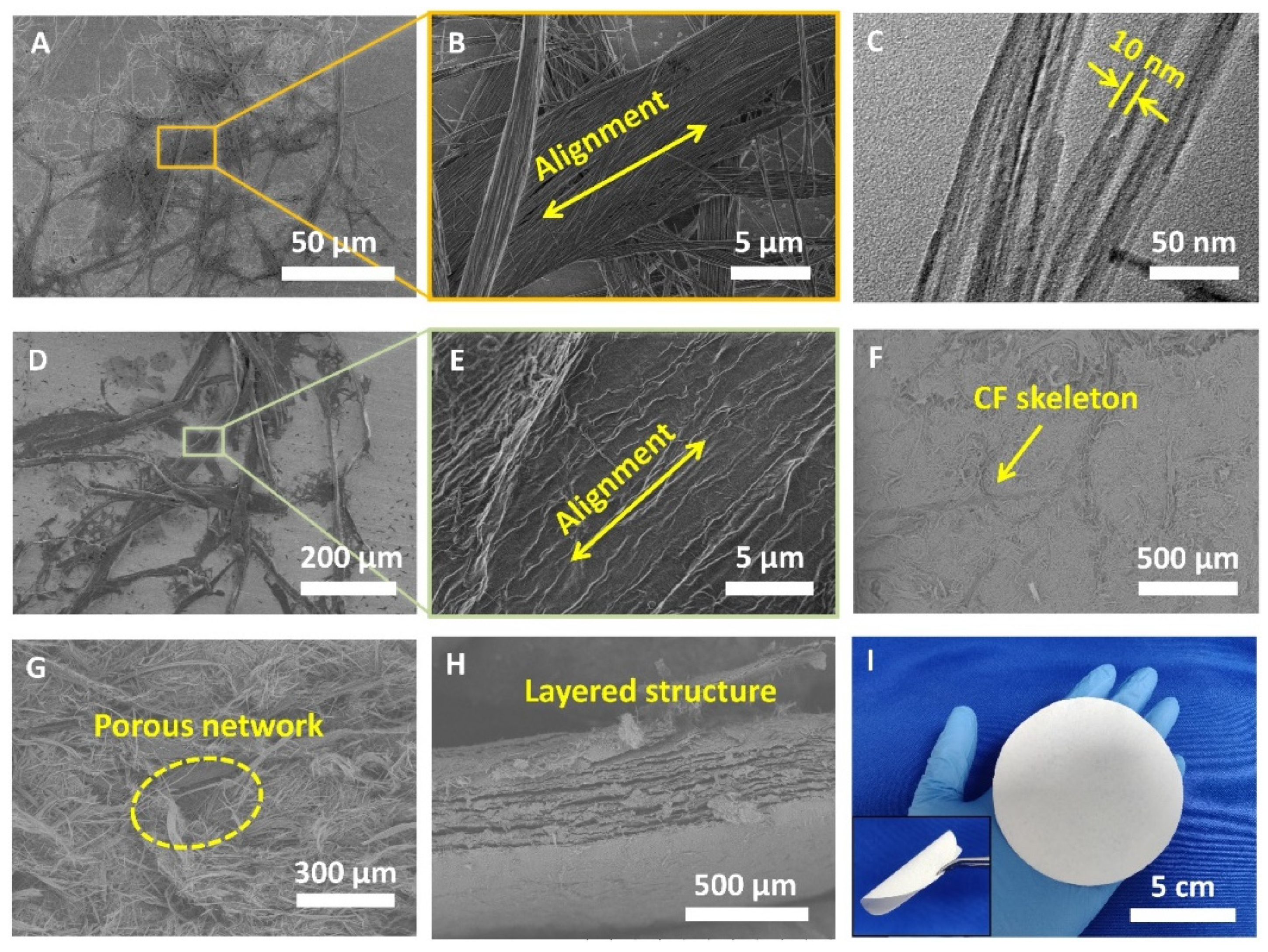
The morphologies of CFs, HAPNWs, and the HAPNW/CF biopaper are shown in Figure 2. In many cases, HAPNWs are self-assembled along their longitudinal direction into highly aligned nanowire bundles with larger diameters, and the diameter of a single nanowire is only about 10 nm (Figure 2A–C). The self-assembled alignment structure of HAPNWs (nanowire bundles) can enhance their mechanical strength and flexibility. It is observed that ultralong HAP nanowire bundles can naturally bend without fracture. The diameters of CFs are tens of micrometers, and a higher-resolution SEM image reveals that the CFs also consists of many approximately parallel fine fibers (Figure 2D,E).
Through a vacuum-assisted filtration process, HAPNWs and CFs were synergistically interwoven with each other to form the HAPNW/CF biopaper. The SEM image of the HAPNW/CF biopaper shows that CFs were embedded in the ultralong HAP nanowire network, which plays an important role as the reinforcement agent, similar to the steel bars in reinforced cement (Figure 2F). In a higher-magnification SEM image of the HAPNW/CF biopaper, a porous network structure constructed with HAPNWs and CFs is clearly observed (Figure 2G). Moreover, the cross-section SEM observation of the HAPNW/CF biopaper exhibits a layered structure, and the thickness of one single layer is about 10 μm (Figure 2H). Thus, the CF-reinforced ultralong HAP nanowire network as well as the layered structure in the HAPNW/CF biopaper cooperate to facilitate the high mechanical performance. Compared with the conventional brittle HAP ceramics, the as-prepared HAPNW/CF biopaper is highly flexible, and can be bent at a large angle (nearly 180°) without obvious cracking (the inset of Figure 2I). In addition, a large-sized HAPNW/CF biopaper with a diameter of 9 cm (Figure 2I) is fabricated rapidly and conveniently through the above-mentioned method for large-area skin wound dressings.
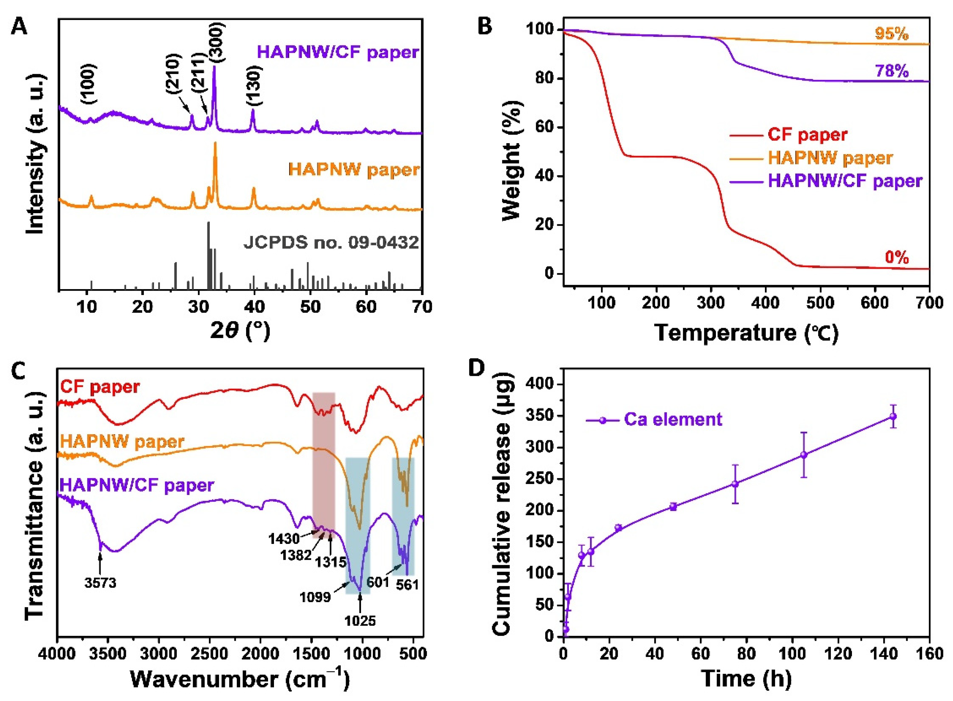
XRD patterns of the as-prepared HAPNW paper and HAPNW/CF biopaper are shown in Figure 3A, which reveals that both the HAPNW paper and the HAPNW/CF biopaper are composed of the crystal phase of hexagonal HAP (JCPDS No. 09-0432), and the peak intensity corresponding to the (0 0 2) crystal plane at 2θ = 25.8° is obviously diminished because of the preferential growth of ultralong HAP nanowires along its c-axis direction. The CFs do not exhibit strong diffraction peaks except for a broad peak locating around 2θ = 10–20° in the XRD pattern of HAPNW/CF biopaper. The TG curves of the samples are shown in Figure 3B. As expected, the weight ratio of the CFs in the HAPNW/CF biopaper is about 22 wt.% calculated by the TG analysis (Figure 3B). CFs are not thermally stable and are completely decomposed and oxidized at temperatures below 450 °C. In contrast, HAPNWs are highly stable at high temperatures, and only a small amount of weight loss (mainly due to the loss of adsorbed water) is observed.
FTIR spectra of the HAPNW paper made of only HAPNWs, the CF paper made of only CFs, and the HAPNW/CF biopaper are shown in Figure 3C. The absorption peak at 3573 cm−1 is the characteristic peak of the OH group from HAP. The absorption peaks at 1099 and 1025 cm−1 correspond to the PO43− group with the stretching mode, and the absorption peaks at 601 and 561 cm−1 originate from the bending mode of the O–P–O bond of HAP. In addition, the absorption peaks of CFs at 1430 cm−1 (C=O), 1382 cm−1 (O–CH3), and 1315 cm−1 (C–O–C) can be observed in the FTIR spectrum of the HAPNW/CF biopaper, confirming the presence of CFs in the biopaper. Specifically, both HAPNWs and CFs show a broad absorption peak from 3500 to 3300 cm−1 ascribed to the hydroxyl groups on their surface, while this peak for HAPNWs is slightly shifted from 3446 to 3436 cm−1 after hybridization with CFs, indicating the interactions between HAPNWs and CFs including hydrogen bonding and van der Waals forces.
In addition, the Ca2+ ion release and degradation behavior of the HAPNW/CF biopaper in vitro are investigated, and the experimental results are shown in Figure 3D, indicating that the cumulative released amount of Ca2+ ions from the HAPNW/CF biopaper in normal saline continuously increases with the increase in the release time. The continuous release of Ca2+ ions from the HAPNW/CF biopaper is beneficial to promoting the skin wound healing. In the degradation and Ca2+ ion release test, the HAPNW/CF biopaper (80 mg) was soaked in 40 mL of normal saline. The cumulative release of Ca2+ ions from the HAPNW/CF biopaper is about 0.35 mg at a release time of about 150 h, indicating that the percentage of the released Ca2+ ions from the HAPNW/CF biopaper is about 0.44 wt.%.
The Brunauer–Emmett–Teller (BET) specific surface areas are measured by the N2 adsorption/desorption isotherms. As shown in Figure 4A,B, the BET specific surface area of the HAPNW paper is measured to be 47.29 m2·g−1, and that of the CF paper is very small (0.84 m2·g−1). Although the presence of CFs will decrease the specific surface area of the HAPNW/CF biopaper, the value is maintained at a relatively high level of 36.84 m2·g−1. The high specific surface area of the HAPNW/CF biopaper is beneficial for providing more sites for cell and protein adhesion and enhancing the angiogenesis in the skin wound region. The differences in the densities of the HAPNW paper (0.416 g·cm−3), CF paper (0.425 g·cm−3), and HAPNW/CF biopaper (0.414 g·cm−3) are not significant (Figure 4C). Their low densities are ascribed to the porous structure and high porosities. The porosities of the HAPNW paper, the HAPNW/CF biopaper, and the CF paper are 80%, 77%, and 70%, respectively, as shown in Figure 4C.
The as-prepared HAPNW paper, the CF paper, and the HAPNW/CF biopaper are highly hydrophilic. As shown in Figure 4D, the water uptake percentage of the HAPNW paper is about 291%, and that of the CF paper is higher (427%). The abundant hydroxyl groups on the surface of CFs are ascribed to its high water absorption capacity. Therefore, the introduction of CFs in the HAPNW/CF biopaper can improve its water absorption capacity, and the water uptake percentage of the as-prepared HAPNW/CF biopaper is about 320%. Moreover, the thickness of the HAPNW/CF biopaper has no significant effect on its water absorption capacity (Figure 4E). The water contact angle of the HAPNW/CF biopaper is 0°, and the water permeates into the biopaper within 5 s, which is consistent with the superhydrophilicity of the as-prepared HAPNW/CF biopaper (Figure 4F).
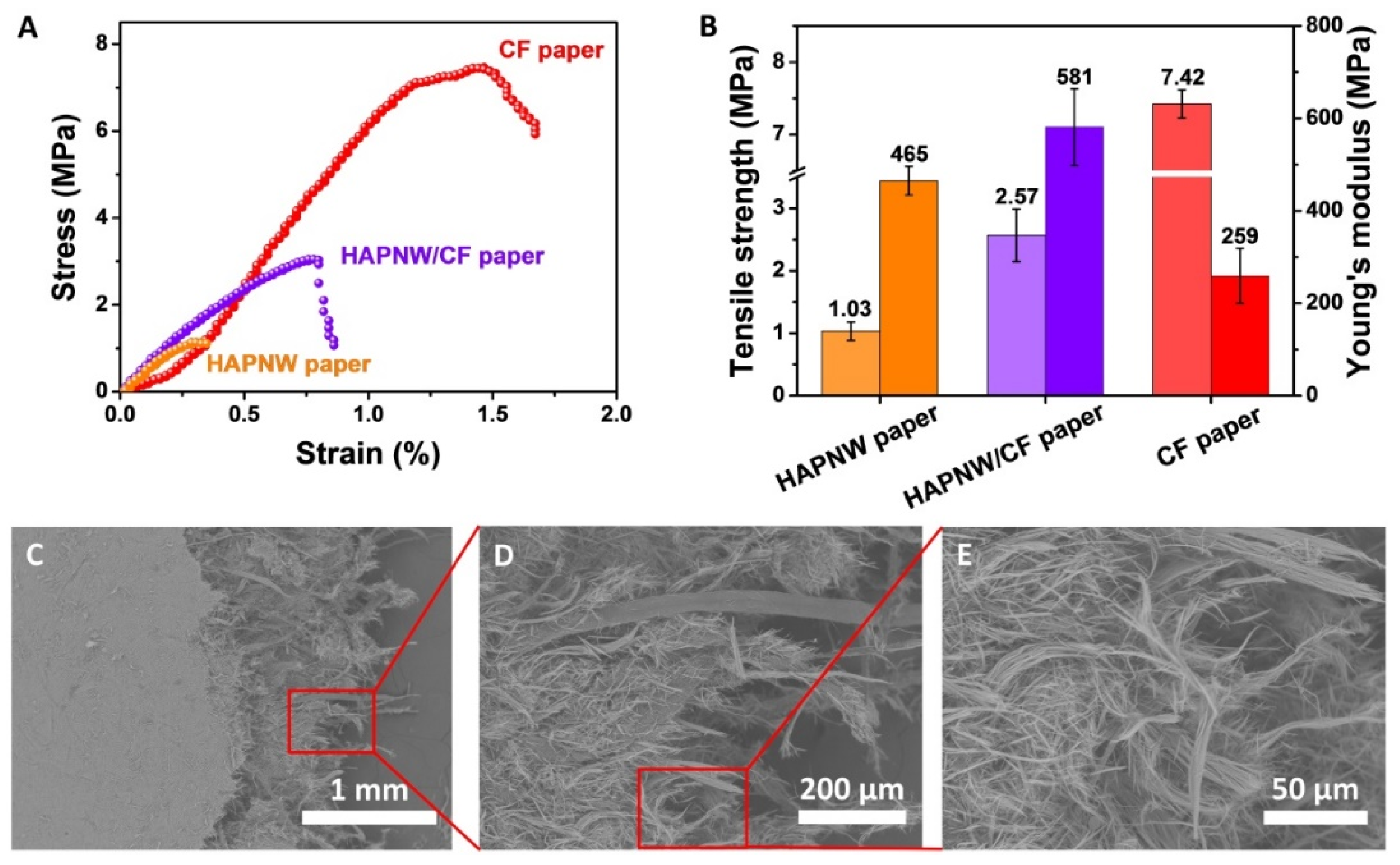
The mechanical properties of the HAPNW paper can be improved by the addition of CFs. As shown in Figure 5A,B, the tensile strength and Young’s modulus of the HAPNW paper are 1.03 MPa and 465 MPa, respectively, and those of the CF paper are 7.42 MPa and 259 MPa, respectively. The tensile strength of the HAPNW paper is relatively low, and this will limit its applications, whereas the CF paper has a relatively high tensile strength but low Young’s modulus, ascribed to the intrinsic soft structure of CFs. Thus, the addition of CFs in the HAPNW paper can improve its mechanical strength and toughness. Compared to the HAPNW paper, the tensile strength of the HAPNW/CF biopaper reaches 2.57 MPa, and the Young’s modulus increases to 581 MPa. These experimental results are also consistent with the high flexibility of the HAPNW/CF biopaper (Figure 2I).
To further demonstrate the mechanical enhancement mechanism, the fracture behaviors of the HAPNW/CF biopaper have been investigated by the SEM observation. As shown in Figure 5C–E, the breakage and pull out of HAPNWs are observed in the broken region of the HAPNW/CF biopaper. Moreover, the twisting and deformation of HAPNWs as well as the fractured CFs are clearly revealed in the fracture area. The obvious tearing surfaces, accompanied by the twisting, deformation, breakage, and pull-out of HAPNWs and CFs, indicate the stretching of HAPNWs and CFs under the external force.

3.2. Safety Evaluation of the HAPNW/CF Biopaper

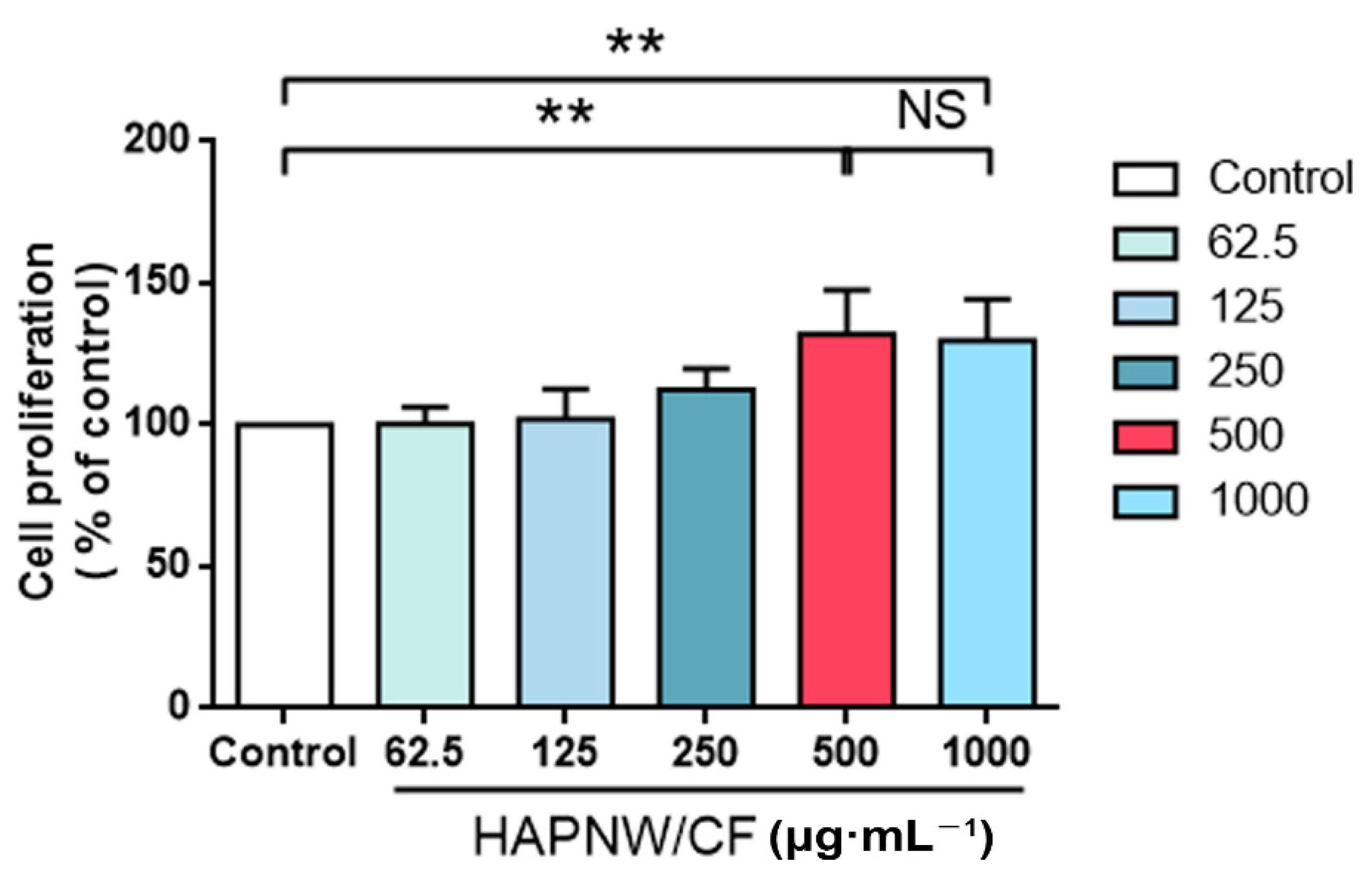
The CCK8 kit was used to evaluate the cell viability of the HAPNW/CF biopaper (weight ratio 80:20) with different doses (62.5, 125, 250, 500, 1000 μg·mL−1) using the CCK8 kit and HUVECs cultured for 24 h, as shown in Figure 6. We found that the cell viabilities of the 500 and 1000 μg·mL−1 HAPNW/CF treatment groups are significantly higher than that of the control group, and the difference is statistically significant (p < 0.01). There is no statistical difference between the 500 and 1000 μg·mL−1 HAPNW/CF treatment groups. Compared with the control group, the remaining treatment groups have no statistical difference. These experimental results indicate the high biocompatibility and safety of the HAPNW/CF biopaper.

3.3. Evaluation of the Effect of the HAPNW/CF Biopaper on HUVECs

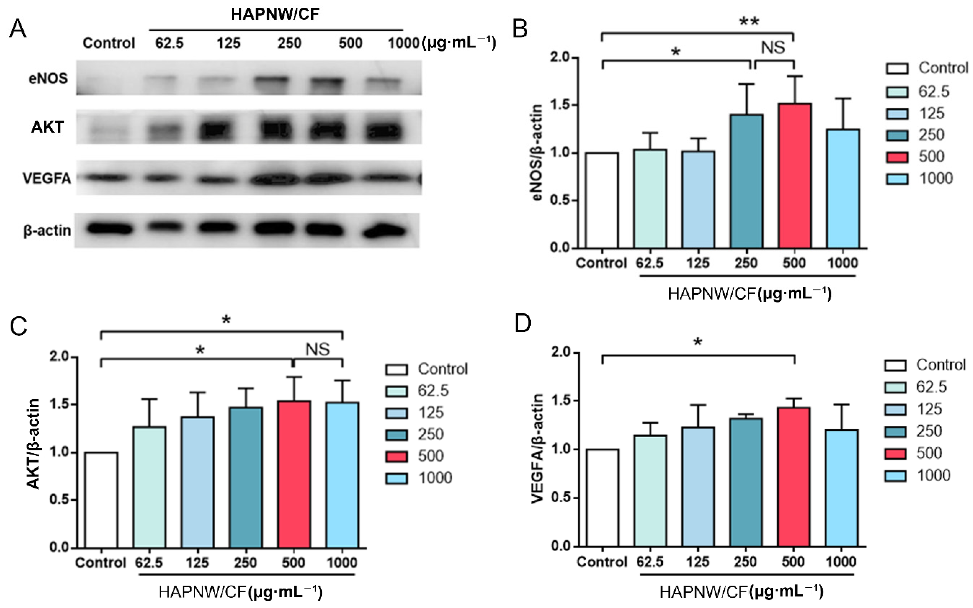
Western blot analysis was used to analyze the expressions of angiogenesis-related proteins eNOS, AKT, and VEGFA after 24 h intervention with different doses (62.5, 125, 250, 500, 1000 μg·mL−1) of HAPNW/CF (weight ratio 80:20) (Figure 7). Compared with the control group, the expressions of eNOS in the 250 and 500 μg·mL−1 HAPNW/CF treatment groups significantly increased, which are statistically significant (p < 0.05 and p < 0.01). There is no statistical difference between 250 and 500 μg·mL−1 HAPNW/CF treatment groups (Figure 7B). Compared with the control group, the expressions of AKT in the 500 and 1000 μg·mL−1 HAPNW/CF treatment groups are significantly higher, which are statistically significant (p < 0.05). There is no statistical difference between the 500 and 1000 μg·mL−1 HAPNW/CF treatment groups (Figure 7C). Compared with the control group, the expression of VEGFA in the 500 μg·mL−1 HAPNW/CF treatment group is significantly increased, which is statistically significant (p < 0.05). There is no statistical difference between the remaining treatment groups and the control group (Figure 7D). These experimental results indicate that the HAPNW/CF biopaper can significantly up-regulate the expressions of angiogenesis-related proteins eNOS, AKT, and VEGFA, and this effect is related to the concentration of the material.

3.4. Effect of L-NAME on the Ability of the HAPNW/CF Biopaper to Promote Angiogenesis

Angiogenesis is realized through multiple signaling pathways. We used specific eNOS inhibitor L-NAME to observe its effect on the ability of the HAPNW/CF biopaper to promote angiogenesis. Western blot analysis (Figure 8A,B) shows that there are significant differences between the control group and the HAPNW/CF treatment group, the HAPNW/CF treatment group and the HAPNW/CF + L-NAME treatment group, and the control group and the HAPNW/CF + L-NAME treatment group (p < 0.001, p < 0.01, p < 0.05). These experimental results indicate that HAPNW/CF can promote the expression of the pro-angiogenic protein eNOS, and L-NAME can partially counteract this effect. Transwell migration experiments (Figure 8C,D) show that compared with the control group and the HAPNW/CF + L-NAME treatment group, the migration ability of cells in the HAPNW/CF treatment group is stronger, and the experimental results have statistical differences (p < 0.01), but there was no statistical difference between the control group and the HAPNW/CF + L-NAME treatment group. These experimental results indicate that HAPNW/CF can enhance the migration ability of HUVECs, and L-NAME can counteract this effect.
Tubeforming experiments (Figure 8E,F) indicate that compared with the control group and the HAPNW/CF + L-NAME treatment group, the HAPNW/CF treatment group has a stronger cell tubeforming ability, and the experimental results are statistically different (p < 0.01). There is also a statistical difference between the control group and the HAPNW/CF + L-NAME treatment group (p < 0.05). This means that HAPNW/CF can enhance the tubeforming ability of HUVECs, while L-NAME partially offsets this effect. The EdU proliferation experiments (Figure 8G,H) show that compared with the control group and the HAPNW/CF + L-NAME treatment group, the HAPNW/CF treatment group has a higher cell proliferation, and the experimental results are statistically different (p < 0.01). However, there is no statistical difference between the control group and the HAPNW/CF + L-NAME treatment group. The experimental results indicate that HAPNW/CF can enhance the proliferation ability of HUVECs, and L-NAME can counteract this effect.

3.5. Effect of the HAPNW/CF Biopaper on the Expression of Pro-Angiogenic Protein in a Full-Thickness Skin Trauma Model

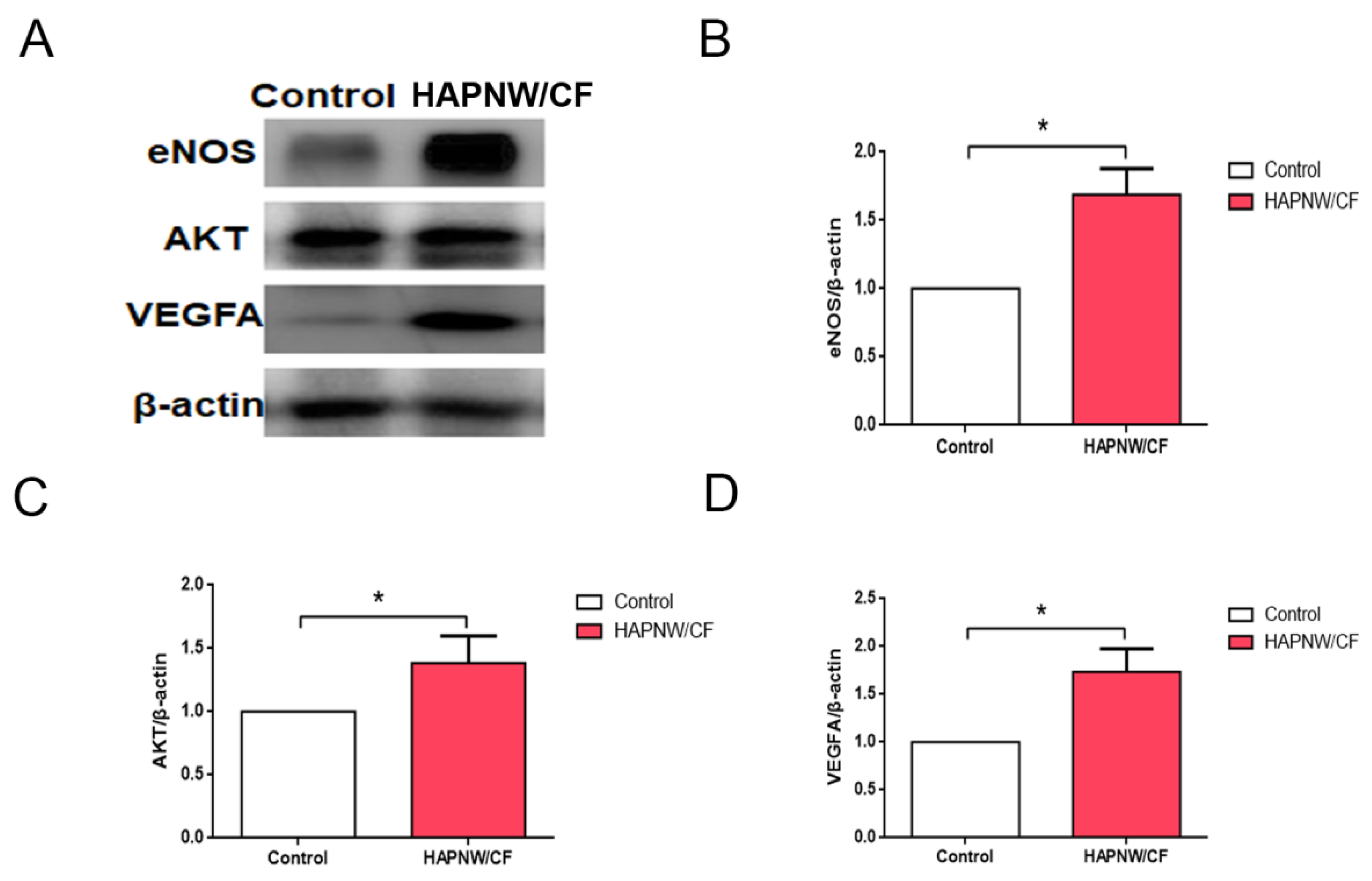
Western blot analysis was used to analyze the expression of angiogenesis-related proteins eNOS, AKT, and VEGFA on the 7th day after the HAPNW/CF biopaper intervention in the full-thickness skin trauma model (Figure 9). Compared with the control group, the expressions of eNOS, AKT, and VEGFA in the HAPNW/CF biopaper treatment group are significantly higher with statistical significance (p < 0.05). The experimental results indicate that the HAPNW/CF biopaper can significantly up-regulate the expression of angiogenesis-related proteins eNOS, AKT, and VEGFA in full-thickness skin trauma model of SD rats.

3.6. Histological Analysis of Angiogenesis in Full-Thickness Skin Trauma Model

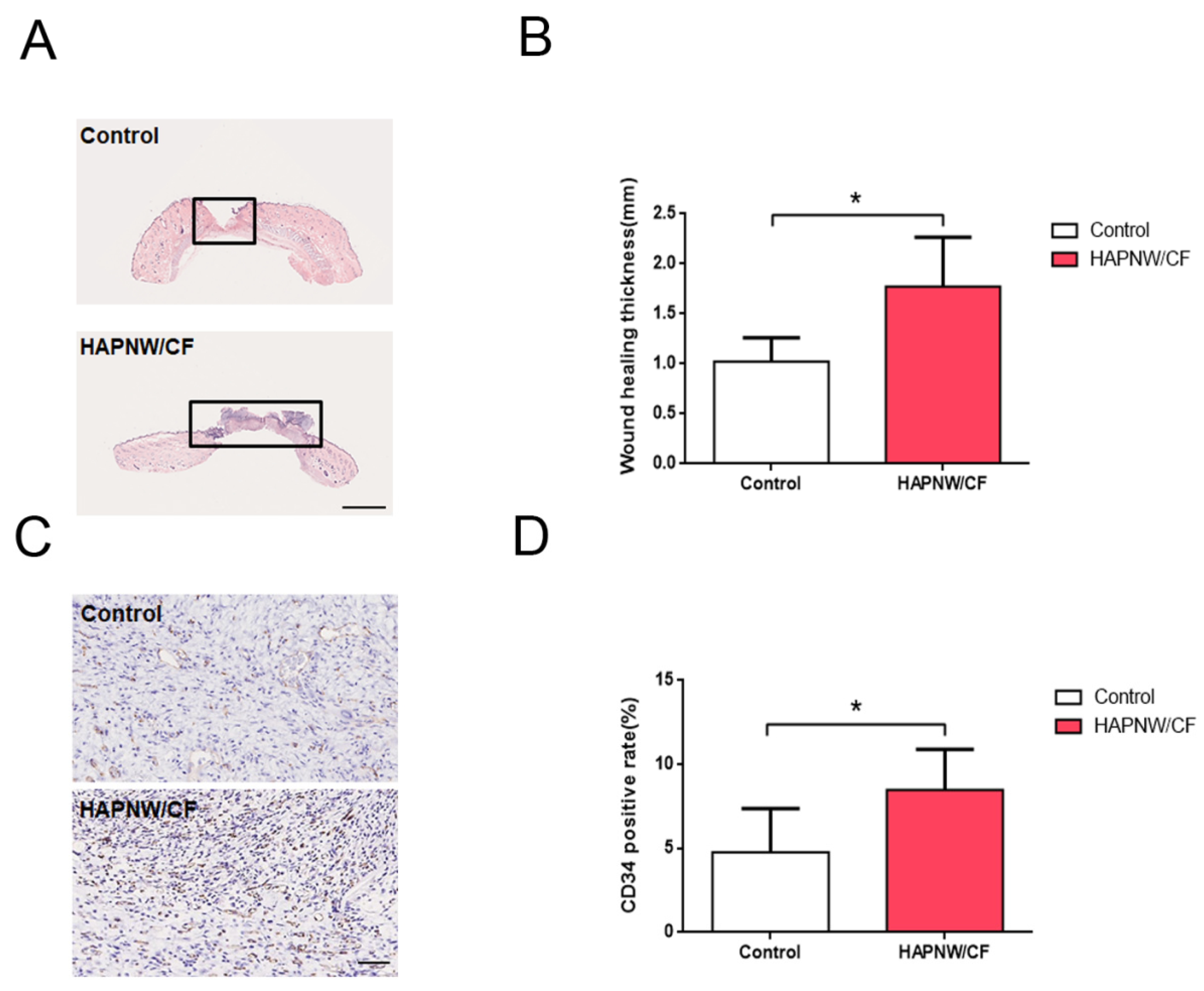
The H&E staining results after 7 days of the HAPNW/CF biopaper intervention in the full-thickness skin trauma model (Figure 10A,B) show that compared with the control group, the thickness of wound healing in the HAPNW/CF biopaper treatment group significantly increases, and the difference is statistically significant (* p < 0.05), indicating that the HAPNW/CF biopaper can promote skin wound healing, and the wound healing depends more on promoting the re-epithelialization of the skin wound rather than shrinking the skin around the wound. CD34 immunohistochemical staining (Figure 10C,D) shows that compared with the control group, the percentage of CD34-positive cells in the HAPNW/CF biopaper treatment group is higher, and the difference is statistically significant (* p < 0.05). The experimental results indicate that the HAPNW/CF biopaper can promote angiogenesis at the wound area, and thus promote skin wound healing.

4. Discussion

The effective healing of skin wounds is essential to ensure the important barrier function of the skin. Skin wound healing is a very complex process that includes hemostasis, inflammation, angiogenesis, migration of various types of cells to the wound, and formation of extracellular matrix [35]. HAP, as a popular kind of bone defect repair biomaterial, can simultaneously induce angiogenesis and osteogenesis around the bone defect area, and may be a potential wound dressing. To this end, we fabricated a biopaper comprising ultralong HAPNWs and CFs by a simple vacuum-assisted filtration method. The as-prepared HAPNW/CF biopaper possesses a layered and porous structure for the ingrowth of new vessels. Both ultralong HAPNWs and CFs cooperate to improve the mechanical performance of the biopaper, which can also be bent and recover to its original state without obvious cracks. A large-sized HAPNW/CF biopaper (with a diameter of 9 cm) was also fabricated rapidly and conveniently for large-area skin wound dressing. The continuous release of Ca2+ ions from the degradation of the HAPNW/CF biopaper is beneficial to promoting skin wound healing. Moreover, the HAPNW/CF biopaper is highly hydrophilic and has high specific surface area, favorable for cell adhesion.
Our in vitro study revealed that the HAPNW/CF intervention in HUVECs treated with hypoxia can induce more expression of eNOS, AKT, and VEGFA compared with the control group, and this effect is related to the concentration of HAPNW/CF. The CCK-8 experiments also show that HAPNW/CF has high biocompatibility with HUVECs. HAPNW/CF at appropriate doses has the highest effectiveness and safety. The expressions of these angiogenesis-related proteins are conducive to angiogenesis and are very important for the progress of skin wound healing [36]. The new blood vessels are beneficial to the improvement of the ischemic and hypoxic conditions of the skin wound, and ensure the nutrient supply of the wound. They can also promote the formation of granulation tissue and re-epithelialization of the skin wound.
Pro-angiogenic factors can promote the formation of blood vessels. After the activation of endothelial cells, the cells form new blood vessels through adhesion, migration, and angiogenesis. The basement membrane of endothelial cells is degraded, the budding appears on the edge of the skin wound, and a new vascular system is formed [37]. Our further research has found that the pro-angiogenic effect of HAPNW/CF at the cellular levels can be partially offset by L-NAME as the specific eNOS inhibitor. HAPNW/CF can induce cells to express more eNOS. However, when HAPNW/CF and L-NAME are applied at the same time, the expression of eNOS is partially suppressed. Additionally, HAPNW/CF can also enhance the proliferation, migration, and tube formation capabilities of HUVECs, and this effect can be completely or partially offset when HAPNW/CF and L-NAME are used at the same time. These results suggest that HAPNW/CF promotes angiogenesis partly through the eNOS signaling pathway. The eNOS is the major source of NO in the cardiovascular system [38]. Under ischemic and hypoxic conditions, NO can play a role through the phosphatidylinositol-3-kinase (PI3K) signaling pathway, affecting the activation, recruitment, migration, and differentiation in endothelial cells [39]. Moreover, HAPNW/CF can continuously release Ca2+ ions, which may be a key early trigger for skin wound healing [40]. The effects of Ca2+ ions on cell proliferation, differentiation, signal transduction, and gene expression were also reported [41]. This may further promote HAPNW/CF-mediated wound healing.
The effect of the HAPNW/CF biopaper on promoting wound healing has also been proven in animal models. The latest research shows that this kind of material has obvious advantages in wound healing. Compared with traditional materials, it has better hemostasis, excellent cell compatibility, and stronger skin wound healing function [42]. In the rat model of hypoxia and ischemia, we found higher levels of eNOS, AKT, and VEGFA proteins expressed in the HAPNW/CF biopaper intervention group than controls. It has been found that the HAPNW/CF biopaper could also induce more expressions of angiogenesis-related factors in the animal model and promote skin wound healing. The histological analysis further confirmed this point. The H&E staining showed that the HAPNW/CF biopaper group had a thicker re-epithelialization level than the control group. The healing of skin wounds was mainly achieved by restoring the thickness of the skin re-epithelialization, rather than the shrinkage of the wound. The CD34 staining experiments showed more angiogenesis in the HAPNW/CF biopaper group than in the control group, indicating that the HAPNW/CF biopaper could promote skin wound healing by inducing more angiogenesis. Moreover, it benefits from the material itself as an excellent wound dressing for the physical protection of wounds [43]. Some studies reported that dressings containing calcium could better promote wound healing [44]. Due to the characteristics of the HAPNW/CF biopaper, the material has the characteristics of high porosity and high specific surface area, which are conducive to the migration of cells and diffusion of nutrients. Studies show that the surface topography of nanomaterials plays an important role in guiding the orientation of microvessels and the morphogenesis of blood vessels [45]. The existence of pores in nanostructured materials provides a basis for cell infiltration and blood vessel formation. The existence of fibers in the HAPNW/CF biopaper is favorable for guiding the directional migration and proliferation of cells during angiogenesis [46]. This angiogenic effect of nanomaterials is significantly dose- and size-dependent [47]. Wound dressings with excellent biocompatibility, biodegradability, non-toxicity, and functionality have great clinical application prospects [48,49]. We hope our work can improve wound healing treatment for more patients and solve more clinical problems.

5. Conclusions

In conclusion, we developed a HAPNW/CF biopaper consisting of ultralong HAP nanowires and CFs with adequate flexibility, mechanical properties, and superhydrophilicity for promoting skin wound healing. The as-prepared HAPNW/CF biopaper has a porous structure with high porosity, which is favorable for blood vessel formation and ingrowth. In addition, the HAPNW/CF biopaper can continuously release Ca2+ ions to promote skin wound healing. We have proven that the HAPNW/CF biopaper has high biocompatibility and the ability to induce angiogenesis for promoting the skin wound healing. Furthermore, we have revealed that the angiogenesis promotion of the HAPNW/CF biopaper is achieved through the activation of eNOS-related signaling pathways. The animal experiments show that the HAPNW/CF biopaper can promote thicker skin re-epithelialization and the formation of new blood vessels, thus promoting the healing of the wound. It is expected that the HAPNW/CF biopaper is a promising and effective dressing for accelerating wound healing with strong potential in clinical applications.

Author Contributions

Conceptualization, J.G. and L.-S.H.; methodology, J.G., L.-S.H. and B.-B.N.; data curation, Y.-K.Z., J.-B.G. and H.-W.R.; writing—original draft preparation, J.G. and L.-S.H.; writing—review and editing, J.G., L.-S.H., B.-B.N., Y.-K.Z., J.-B.G., H.-W.R., H.-P.Y., Y.-J.Z. and J.-L.D.; project administration, H.-P.Y., Y.-J.Z. and J.-L.D.; funding acquisition, J.-L.D. All authors have read and agreed to the published version of the manuscript.

Funding

This research was funded by the National Natural Science Foundation of China (12074253, 12174289), and the Medical and Industrial Cross Research Fund of “Star of Jiaotong University” Program of Shanghai Jiao Tong University (YG2019ZDA21).

Institutional Review Board Statement

This study’s experimental protocol was approved by the Ethics Committee of Xinhua Hospital Affiliated to the Shanghai Jiao Tong University School of Medicine under approval number XHEC-F-2021-079, and the experiments were in accordance with relevant institutional guidelines.

Informed Consent Statement

Not applicable.

Data Availability Statement

Not applicable.

Conflicts of Interest

The authors declare no conflict of interest.

References

  1. Subramaniam, T.; Fauzi, M.B.; Lokanathan, Y.; Law, J.X. The role of calcium in wound healing. Int. J. Mol. Sci. 2021, 22, 6486. [Google Scholar] [CrossRef] [PubMed]
  2. Järbrink, K.; Ni, G.; Sönnergren, H.; Schmidtchen, A.; Pang, C.; Bajpai, R.; Car, J. The humanistic and economic burden of chronic wounds: A protocol for a systematic review. Syst. Rev. 2017, 6, 15. [Google Scholar] [CrossRef] [Green Version]
  3. Keyes, B.E.; Liu, S.; Asare, A.; Naik, S.; Levorse, J.; Polak, L.; Lu, C.P.; Nikolova, M.; Pasolli, H.A.; Fuchs, E. Impaired epidermal to dendritic T cell signaling slows wound repair in aged skin. Cell 2016, 167, 1323–1338. [Google Scholar] [CrossRef] [Green Version]
  4. Kim, H.S.; Sun, X.; Lee, J.H.; Kim, H.W.; Fu, X.; Leong, K.W. Advanced drug delivery systems and artificial skin grafts for skin wound healing. Adv. Drug Deliv. Rev. 2019, 146, 209–239. [Google Scholar] [CrossRef] [PubMed]
  5. Carmeliet, P. Angiogenesis in health and disease. Nat. Med. 2003, 9, 653–660. [Google Scholar] [CrossRef]
  6. Liu, W.; Zhang, G.; Wu, J.; Zhang, Y.; Liu, J.; Luo, H.; Shao, L. Insights into the angiogenic effects of nanomaterials: Mechanisms involved and potential applications. J. Nanobiotechnol. 2020, 18, 9. [Google Scholar] [CrossRef]
  7. Hendrickx, B.; Verdonck, K.; Van den Berge, S.; Dickens, S.; Eriksson, E.; Vranckx, J.J.; Luttun, A. Integration of blood outgrowth endothelial cells in dermal fibroblast sheets promotes full thickness wound healing. Stem Cells 2010, 28, 1165–1177. [Google Scholar] [CrossRef] [PubMed]
  8. Risau, W. Mechanisms of angiogenesis. Nature 1997, 386, 671–674. [Google Scholar] [CrossRef]
  9. Senger, D.R.; Davis, G.E. Angiogenesis. Cold Spring Harb. Perspect. Biol. 2011, 3, a005090. [Google Scholar] [CrossRef] [Green Version]
  10. Greco Song, H.H.; Rumma, R.T.; Ozaki, C.K.; Edelman, E.R.; Chen, C.S. Vascular tissue engineering: Progress, challenges, and clinical promise. Cell Stem Cell 2018, 22, 340–354. [Google Scholar] [CrossRef] [Green Version]
  11. Bae, H.; Puranik, A.S.; Gauvin, R.; Edalat, F.; Carrillo-Conde, B.; Peppas, N.A.; Khademhosseini, A. Building vascular networks. Sci. Transl. Med. 2012, 4, 160ps23. [Google Scholar] [CrossRef] [PubMed] [Green Version]
  12. Yang, Z.; Huang, R.; Zheng, B.; Guo, W.; Li, C.; He, W.; Wei, Y.; Du, Y.; Wang, H.; Wu, D.; et al. Highly stretchable, adhesive, biocompatible, and antibacterial hydrogel dressings for wound healing. Adv. Sci. 2021, 8, 2003627. [Google Scholar] [CrossRef]
  13. Guo, M.; Wang, Y.; Gao, B.; He, B. Shark tooth-inspired microneedle dressing for intelligent wound management. ACS Nano 2021, 15, 15316–15327. [Google Scholar] [CrossRef]
  14. Kim, M.S.; Oh, G.W.; Jang, Y.M.; Ko, S.C.; Park, W.S.; Choi, I.W.; Kim, Y.M.; Jung, W.K. Antimicrobial hydrogels based on PVA and Diphlorethohydroxycarmalol (DPHC) derived from brown alga Ishige okamurae: An in vitro and in vivo study for wound dressing application. Mater. Sci. Eng. C 2020, 107, 110352. [Google Scholar] [CrossRef] [PubMed]
  15. Qiu, X.; Zhang, J.; Cao, L.; Jiao, Q.; Zhou, J.; Yang, L.; Zhang, H.; Wei, Y. Antifouling antioxidant zwitterionic dextran hydrogels as wound dressing materials with excellent healing activities. ACS Appl. Mater. Interfaces 2021, 13, 7060–7069. [Google Scholar] [CrossRef] [PubMed]
  16. Liu, H.; Li, Z.; Zhao, Y.; Feng, Y.; Zvyagin, A.V.; Wang, J.; Yang, X.; Yang, B.; Lin, Q. Novel diabetic foot wound dressing based on multifunctional hydrogels with extensive temperature-tolerant, durable, adhesive, and intrinsic antibacterial properties. ACS Appl. Mater. Interfaces 2021, 13, 26770–26781. [Google Scholar] [CrossRef] [PubMed]
  17. Zhao, X.; Liang, Y.P.; Huang, Y.; He, J.H.; Han, Y.; Guo, B.L. Physical double-network hydrogel adhesives with rapid shape adaptability, fast self-healing, antioxidant and NIR/pH stimulus-responsiveness for multidrug-resistant bacterial infection and removable wound dressing. Adv. Funct. Mater. 2020, 30, 1910748. [Google Scholar] [CrossRef]
  18. Deng, C.; Xu, C.; Zhou, Q.; Cheng, Y. Advances of nanotechnology in osteochondral regeneration. Wiley Interdiscip. Rev. Nanomed. Nanobiotechnol. 2019, 11, e1576. [Google Scholar] [CrossRef]
  19. Mofazzal Jahromi, M.A.; Sahandi Zangabad, P.; Moosavi Basri, S.M.; Sahandi Zangabad, K.; Ghamarypour, A.; Aref, A.R.; Karimi, M.; Hamblin, M.R. Nanomedicine and advanced technologies for burns: Preventing infection and facilitating wound healing. Adv. Drug Deliv. Rev. 2018, 123, 33–64. [Google Scholar] [CrossRef]
  20. Augustine, R.; Prasad, P.; Khalaf, I.M.N. Therapeutic angiogenesis: From conventional approaches to recent nanotechnology-based interventions. Mater. Sci. Eng. C 2019, 97, 994–1008. [Google Scholar] [CrossRef]
  21. Lu, B.Q.; Zhu, Y.J. One-dimensional hydroxyapatite materials: Preparation and applications. Can. J. Chem. 2017, 95, 1091–1102. [Google Scholar] [CrossRef]
  22. Zhu, Y.J.; Lu, B.Q. Deformable biomaterials based on ultralong hydroxyapatite nanowires. ACS Biomater. Sci. Eng. 2019, 5, 4951–4961. [Google Scholar] [CrossRef] [PubMed]
  23. Son, J.; Kim, J.; Lee, K.; Hwang, J.; Choi, Y.; Seo, Y.; Jeon, H.; Kang, H.C.; Woo, H.M.; Kang, B.J.; et al. DNA aptamer immobilized hydroxyapatite for enhancing angiogenesis and bone regeneration. Acta Biomater. 2019, 99, 469–478. [Google Scholar] [CrossRef]
  24. Song, Y.; Wu, H.; Gao, Y.; Li, J.; Lin, K.; Liu, B.; Lei, X.; Cheng, P.; Zhang, S.; Wang, Y.; et al. Zinc silicate/nano-hydroxyapatite/collagen scaffolds promote angiogenesis and bone regeneration via the p38 MAPK pathway in activated monocytes. ACS Appl. Mater. Interfaces 2020, 12, 16058–16075. [Google Scholar] [CrossRef] [PubMed]
  25. Ji, X.; Yuan, X.; Ma, L.; Bi, B.; Zhu, H.; Lei, Z.; Liu, W.; Pu, H.; Jiang, J.; Jiang, X.; et al. Mesenchymal stem cell-loaded thermosensitive hydroxypropyl chitin hydrogel combined with a three-dimensional-printed poly(ε-caprolactone)/nano-hydroxyapatite scaffold to repair bone defects via osteogenesis, angiogenesis and immunomodulation. Theranostics 2020, 10, 725–740. [Google Scholar] [CrossRef]
  26. Dong, T.; Duan, C.; Wang, S.; Gao, X.; Yang, Q.; Yang, W.; Deng, Y. Multifunctional surface with enhanced angiogenesis for improving long-term osteogenic fixation of poly (ether ether ketone) implants. ACS Appl. Mater. Interfaces 2020, 12, 14971–14982. [Google Scholar] [CrossRef]
  27. Huang, G.J.; Yu, H.P.; Wang, X.L.; Ning, B.B.; Gao, J.; Shi, Y.Q.; Zhu, Y.J.; Duan, J.L. Highly porous and elastic aerogel based on ultralong hydroxyapatite nanowires for high-performance bone regeneration and neovascularization. J. Mater. Chem. B 2021, 9, 1277–1287. [Google Scholar] [CrossRef]
  28. Zhu, Y.J. Multifunctional fire-resistant paper based on ultralong hydroxyapatite nanowires. Chin. J. Chem. 2021, 39, 2296–2314. [Google Scholar] [CrossRef]
  29. Salama, A. Recent progress in preparation and applications of chitosan/calcium phosphate composite materials. Int. J. Biol. Macromol. 2021, 178, 240–252. [Google Scholar] [CrossRef]
  30. Kawai, K.; Larson, B.J.; Ishise, H.; Carre, A.L.; Nishimoto, S.; Longaker, M.; Lorenz, H.P. Calcium-based nanoparticles accelerate skin wound healing. PLoS ONE 2011, 6, e27106. [Google Scholar] [CrossRef] [Green Version]
  31. Lu, B.Q.; Zhu, Y.J.; Chen, F. Highly flexible and nonflammable inorganic hydroxyapatite paper. Chem. Eur. J. 2014, 20, 1242–1246. [Google Scholar] [CrossRef] [PubMed]
  32. Zhang, Y.G.; Zhu, Y.J.; Chen, F.; Wu, J. Ultralong hydroxyapatite nanowires synthesized by solvothermal treatment using a series of phosphate sodium salts. Mater. Lett. 2015, 144, 135–137. [Google Scholar] [CrossRef]
  33. Jiang, Y.Y.; Zhu, Y.J.; Chen, F.; Wu, J. Solvothermal synthesis of submillimeter ultralong hydroxyapatite nanowires using a calcium oleate precursor in a series of monohydroxy alcohols. Ceram. Int. 2015, 41, 6098–6102. [Google Scholar] [CrossRef]
  34. Huang, J.J.; Shi, Y.Q.; Li, R.L.; Hu, A.; Lu, Z.Y.; Weng, L.; Han, Y.P.; Wang, S.Q.; Zhang, L.; Hao, C.N.; et al. Therapeutic ultrasound protects huvecs from Ischemia/Hypoxia-induced apoptosis via the PI3K-akt pathway. Am. J. Transl. Res. 2017, 9, 1990–1999. [Google Scholar]
  35. Reinke, J.M.; Sorg, H. Wound repair and regeneration. Eur. Surg. Res. 2012, 49, 35–43. [Google Scholar] [CrossRef] [PubMed]
  36. Wan, X.; Liu, S.; Xin, X.; Li, P.; Dou, J.; Han, X.; Kang, I.-K.; Yuan, J.; Chi, B.; Shen, J. S-nitrosated keratin composite mats with no release capacity for wound healing. Chem. Eng. J. 2020, 400, 125964. [Google Scholar] [CrossRef]
  37. Tonnesen, M.G.; Feng, X.; Clark, R.A. Angiogenesis in wound healing. J. Investig. Dermatol. Symp. Proc. 2000, 5, 40–46. [Google Scholar] [CrossRef] [Green Version]
  38. Dudzinski, D.M.; Igarashi, J.; Greif, D.; Michel, T. The regulation and pharmacology of endothelial nitric oxide synthase. Annu. Rev. Pharmacol. Toxicol. 2006, 46, 235–276. [Google Scholar] [CrossRef]
  39. Balligand, J.L.; Feron, O.; Dessy, C. eNOS Activation by physical forces: From short-term regulation of contraction to chronic remodeling of cardiovascular tissues. Physiol. Rev. 2009, 89, 481–534. [Google Scholar] [CrossRef]
  40. Wood, W. Wound healing: Calcium flashes illuminate early events. Curr. Biol. 2012, 22, R14–R16. [Google Scholar] [CrossRef] [Green Version]
  41. Pastar, I.; Stojadinovic, O.; Yin, N.C.; Ramirez, H.; Nusbaum, A.G.; Sawaya, A.; Patel, S.B.; Khalid, L.; Isseroff, R.R.; Tomic-Canic, M. Epithelialization in wound healing: A comprehensive review. Adv. Wound Care 2014, 3, 445–464. [Google Scholar] [CrossRef] [PubMed] [Green Version]
  42. Zheng, Y.; Ma, W.; Yang, Z.; Zhang, H.; Ma, J.; Li, T.; Niu, H.; Zhou, Y.; Yao, Q.; Chang, J.; et al. An ultralong hydroxyapatite nanowire aerogel for rapid hemostasis and wound healing. Chem. Eng. J. 2022, 430, 132912. [Google Scholar] [CrossRef]
  43. Dhand, C.; Venkatesh, M.; Barathi, V.A.; Harini, S.; Bairagi, S.; Goh Tze Leng, E.; Muruganandham, N.; Low, K.Z.W.; Fazil, M.; Loh, X.J.; et al. Bio-inspired crosslinking and matrix-drug interactions for advanced wound dressings with long-term antimicrobial activity. Biomaterials 2017, 138, 153–168. [Google Scholar] [CrossRef]
  44. Zhao, W.Y.; Fang, Q.Q.; Wang, X.F.; Wang, X.W.; Zhang, T.; Shi, B.H.; Zheng, B.; Zhang, D.D.; Hu, Y.Y.; Ma, L.; et al. Chitosan-calcium alginate dressing promotes wound healing: A preliminary study. Wound Repair Regen. 2020, 28, 326–337. [Google Scholar] [CrossRef]
  45. Sharma, D.; Ross, D.; Wang, G.; Jia, W.; Kirkpatrick, S.J.; Zhao, F. Upgrading prevascularization in tissue engineering: A review of strategies for promoting highly organized microvascular network formation. Acta Biomater. 2019, 95, 112–130. [Google Scholar] [CrossRef] [PubMed]
  46. Formentín, P.; Catalán, Ú.; Fernández-Castillejo, S.; Alba, M.; Baranowska, M.; Solà, R.; Pallarès, J.; Marsal, L.F. Human aortic endothelial cell morphology influenced by topography of porous silicon substrates. J. Biomater. Appl. 2015, 30, 398–408. [Google Scholar] [CrossRef] [PubMed]
  47. Yu, H.; Chen, X.; Cai, J.; Ye, D.; Wu, Y.; Fan, L.; Liu, P. Novel porous three-dimensional nanofibrous scaffolds for accelerating wound healing. Chem. Eng. J. 2019, 369, 253–262. [Google Scholar] [CrossRef]
  48. Bibire, T.; Yilmaz, O.; Ghiciuc, C.M.; Bibire, N.; Dănilă, R. Biopolymers for surgical applications. Coatings 2022, 12, 211. [Google Scholar] [CrossRef]
  49. Hamza, R.Z.; Al-Motaani, S.E.; Al-Talhi, T. Therapeutic and ameliorative effects of active compounds of combretum molle in the treatment and relief from wounds in a diabetes mellitus experimental model. Coatings 2021, 11, 324. [Google Scholar] [CrossRef]
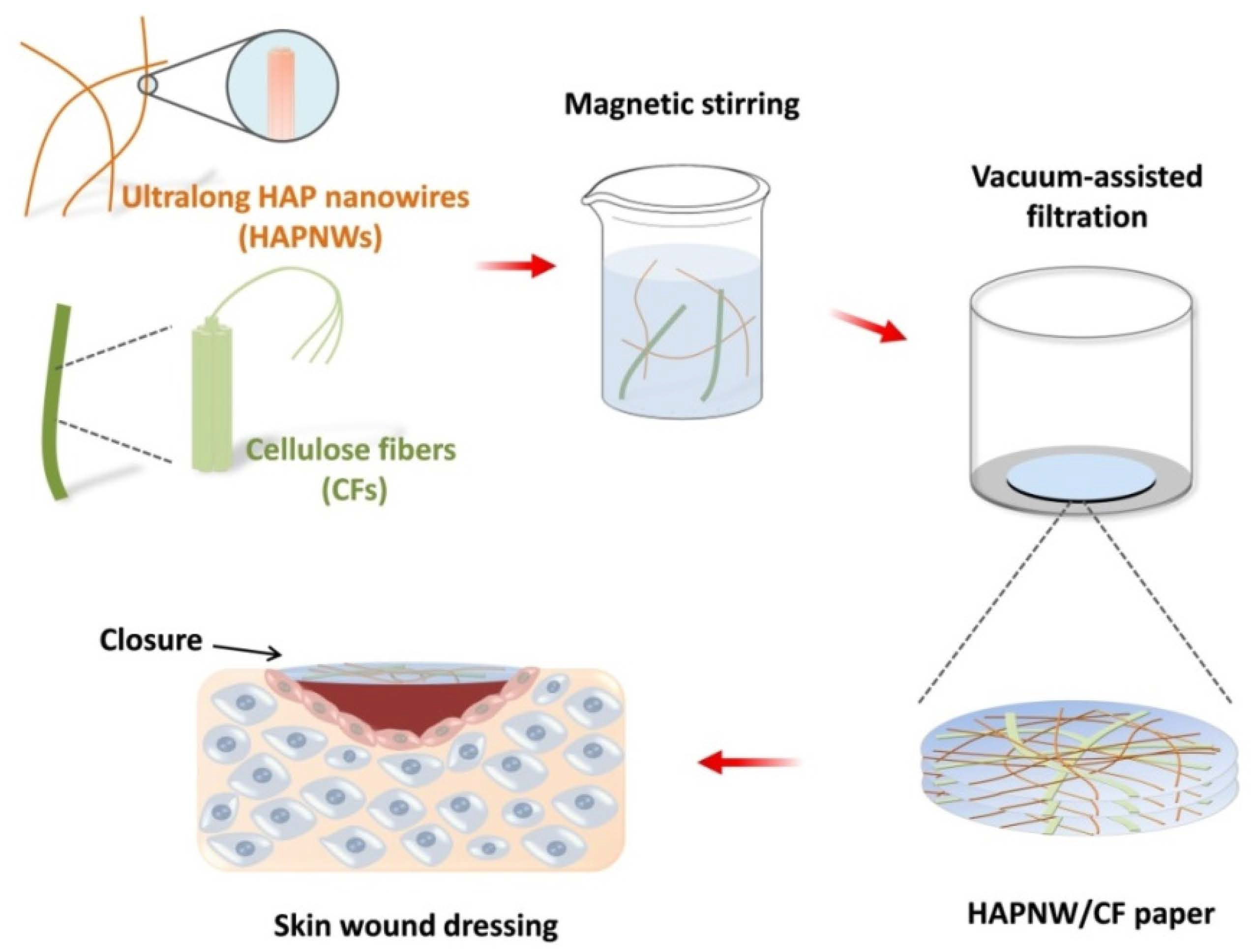
Figure 1. Schematic illustration for the structures of HAPNWs and CFs, and the preparation of the HAPNW/CF biopaper for the skin wound dressing.
Coatings 12 00479 g001
Figure 2. (A,B) Scanning electron microscopy (SEM) images of the HAPNWs with the self-assembled alignment structure. (C) Transmission electron microscopy (TEM) image of the HAPNWs. (D,E) SEM images of the CFs. (FH) The surface morphology (F,G) and the cross-section (H) of the as-prepared HAPNW/CF biopaper. (I) A digital image of a large-sized HAPNW/CF biopaper with a diameter of 9 cm, and the insert is a digital image of the HAPNW/CF biopaper showing high flexibility.
Coatings 12 00479 g002
Figure 3. (A) X-ray diffraction (XRD) patterns of the HAPNW paper and HAPNW/CF biopaper. (B) Thermogravimetric (TG) curves and (C) Fourier transform infrared (FTIR) spectra of the HAPNW paper, the CF paper, and the HAPNW/CF biopaper. (D) The cumulative release curve of Ca2+ ions from the HAPNW/CF biopaper immersed in the normal saline for different times.
Coatings 12 00479 g003
Figure 4. (AD) Nitrogen adsorption–desorption isotherms (A), specific surface areas (B), densities and porosities (C), and water absorption percentages (D) of the HAPNW paper, the CF paper, and the HAPNW/CF biopaper. (E) Water absorption percentages of the HAPNW/CF biopaper with different thicknesses. (F) Water contact angle of the HAPNW/CF biopaper.
Coatings 12 00479 g004
Figure 5. (A) Tensile stress–strain curves of the HAPNW paper, the CF paper, and the HAPNW/CF biopaper. (B) Tensile strengths and Young’s moduli of the HAPNW paper, the CF paper, and the HAPNW/CF biopaper. (CE) SEM images of the fractured HAPNW/CF biopaper.
Coatings 12 00479 g005
Figure 6. Safety analysis of HAPNW/CF (weight ratio 80:20) with different doses (62.5, 125, 250, 500, 1000 μg·mL−1) using the CCK8 kit and HUVECs cultured for 24 h (** p < 0.01).
Coatings 12 00479 g006
Figure 7. Effect of different doses (62.5, 125, 250, 500, 1000 μg·mL−1) of HAPNW/CF (weight ratio 80:20) in HUVECs intervention. (AD) Western blot analysis of protein expressions of eNOS, AKT, and VEGFA after 24 h intervention with different doses of HAPNW/CF (weight ratio 80:20; 62.5, 125, 250, 500, 1000 μg·mL−1) (* p < 0.05; ** p < 0.01).
Coatings 12 00479 g007
Figure 8. The effect of L-NAME on the ability of the HAPNW/CF biopaper to promote angiogenesis. (A,B) Western blot analysis of eNOS protein expressions in the control group, the HAPNW/CF (500 μg·mL−1) group, and the HAPNW/CF (500 μg·mL−1) + L-NAME (100 μM) group. (C,D) Transwell migration experiments, scale bar = 100 μm. (E,F) Tube-forming experiments, scale bar = 100 μm. (G,H) EdU proliferation tests, scale bar = 50 μm (* p < 0.05; ** p < 0.01; *** p < 0.001).
Coatings 12 00479 g008
Figure 9. The effect of the HAPNW/CF biopaper on the expression of pro-angiogenic protein in a full-thickness skin trauma model. (AD) Western blot analysis of eNOS, AKT, and VEGFA protein expressions in the control group and the HAPNW/CF biopaper group after intervention in the SD rat back skin full-thickness trauma model for 7 days (* p < 0.05).
Coatings 12 00479 g009
Figure 10. Histological analysis of angiogenesis in the presence of the HAPNW/CF biopaper in a full-thickness skin trauma model. (A,B) H&E staining of trauma 7 days after operation, scale bar = 2.5 mm. (C,D) CD34 immunohistochemical staining of trauma 7 days after operation, scale bar = 50 μm (* p < 0.05).
Coatings 12 00479 g010
Publisher’s Note: MDPI stays neutral with regard to jurisdictional claims in published maps and institutional affiliations.