Abstract
The placement of bone–level dental implants can lead to the detachment of particles in the surrounding tissues due to friction with the cortical bone. In this study, 60 bone–level dental implants were placed with the same design: 30 made of commercially pure grade 4 titanium and 30 made of Ti6Al4V alloy. These implants were placed in cow ribs following the company’s placement protocols. Particles detached from the dental implants were isolated and their size and specific surface area were characterized. The irregular morphology was observed by scanning electron microscopy. Ion release to the medium was determined at different immersion times in physiological medium. Cytocompatibility studies were performed with fibroblastic and osteoblastic cells. Gene expression and cytokine release were analysed to determine the action of inflammatory cells. Particle sizes of around 15 μM were obtained in both cases. The Ti6Al4V alloy particles showed significant levels of vanadium ion release and the cytocompatibility of these particles is lower than that of commercially pure titanium. Ti6Al4V alloy presents higher levels of inflammation markers (TNFα and Il–1β) compared to that of only titanium. Therefore, there is a trend that with the alloy there is a greater toxicity and a greater pro-inflammatory response.
1. Introduction
Long–term success of titanium dental implants is related to the osseointegration—quality and quantity of the new bone in contact with the dental implant. The cortical bone gives the mechanical stability due to the bone cavity presents a smaller diameter than the dental implant inserted. Bone level implants can produce stresses on the microroughness surfaces and can produce release of debris [1,2,3,4].
In general, all dental implants are sand–blasted with abrasive materials such as aluminium oxide, silicon oxide or carbides that produce roughness for better osseointegration. This surface generally presents important residual stress values due to the impact of the abrasive particles that have caused plastic deformation on the surface. The stresses suffered in these areas due to friction between the cortex and the implant cause the release of particles that fracture the surface of the dental implant [5,6].
Another source of titanium particle generation is implantoplasty techniques, which mechanism parts of the implant and/or prosthesis where bacterial plaque may have colonized. There is much controversy as to whether or not this machining should be carried out, but it is being applied more and more frequently. Even if the particles are aspirated, a large part of them, especially the smaller ones, remain in the patient and their presence can be dangerous [7,8,9]. Another problem that can arise from these high stresses leading to fracture of the titanium is the nucleation of possible cracks that can affect the fatigue behaviour of the dental implant. The cracks generated on the surface with cyclic chewing loads lead to crack growth that can lead to catastrophic failure of the dental implant [10,11].
In addition to the mechanical problems that can arise, it is very important that the detached particles have very high accumulated energy and generally exhibit behaviour close to cytotoxicity, with an increase in cell death being observed. Recent investigations have demonstrated that those particles and ions are not bioinert metals. It is very important to understand the biological mechanisms and implications of these particles and their relationship to the periimplantitis. Several research works have suggested the contribution of Ti particles to the development and progression of periimplantitis [12,13,14,15]. These particles present different types of oxides due to the high quantity of surface energy and play an important role with the activation of the inflammatory response and release of pro-inflammatory cytokines, such as TNF-α, IL-1β, and RANKL [12,13,14,15]. In addition, titanium particles showed a reduced viability of bone marrow stem cells [16], and disruption of epithelial homeostasis, increasing DNA damage response, and potentially compromising the oral epithelial barrier [17,18,19,20,21,22].
The aim of this study is to characterize the particles released during the insertion of bone level dental implants and to determine their biological behaviour. Furthermore, the study also aims to determine the influence of the chemical composition of dental implants (cp Ti and Ti6Al4V).
2. Materials and Methods
2.1. Sample Preparation and Collection
Sixty c.p.–Ti bone level dental implants made of grade 4 (Figure 1), and sixty made of Ti6Al4V alloy with the same design (Vega, Klockner, Escaldes Engordany, Andorra), were implanted in fresh cow ribs by the same investigator (JCV) following the drilling protocol described by the company [23]. In Figure 2, implantation in the fresh cow bones can be observed using a GENTLEsilence LUX 8000B turbine (KaVo Dental GmbH, Biberach an der Riβ, Germany) under constant irrigation. The surface was sequentially modified with a fine–grained tungsten carbide bur (reference H379.314. 014 KOMET; GmbH & Co. KG, Lemgo, Germany), a course–grained silicon carbide polisher (order no. 9608.314.030 KOMET; GmbH & Co. KG) and a fine–grained silicon carbide polisher (order no. 9618.314.030 KOMET; GmbH & Co. KG).
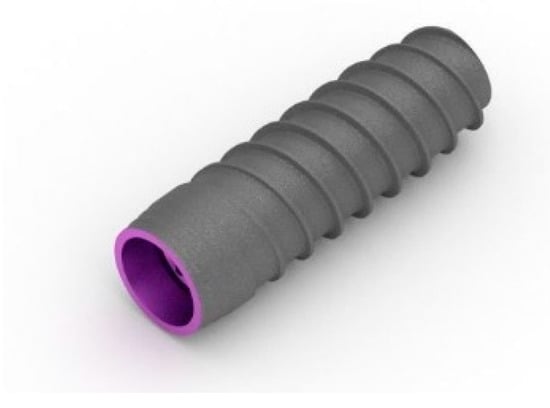
Figure 1.
Bone level dental implant studied. This design was manufactured from grade 4 titanium and Ti6Al4V.

Figure 2.
Bone level dental implant inserted in the fresh cow ribs.
Once the dental implants were placed at bone level, the samples are taken to a high–resolution micro–CT scanner (Skyscan 1272CMOS, Bruker, Billerica, MA, USA) (Figure 3), which makes it possible to observe the titanium particles that have detached from the rough part of the dental implant in the cortical bone. The scans were very slow to improve the resolution of detection of the particles present. Once these particles were observed, the ribs with the implants were placed in an oven at a temperature of 900 °C, causing the bone to toast for 5 h. The remains of the roasting were the mineral content of the bone (apatite) and the detached titanium particles.
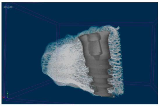
Figure 3.
Dental implant implanted in the fresh cow rib. Image of micro CT.
2.2. Specific Surface Area
The specific surface area is defined as the area in contact with the biological environment. This determination was realized under vacuum conditions at 10 µmHg for metal debris degassed at 100 °C using an ASAP 2020 equipment (Micromeritics, Norcross, GA, USA). Nitrogen was used as an adsorbate. The specific surface area was analysed by applying mathematical calculations described by the BET (Brunauer–Emmett–Teller) theory [24].
2.3. Granulometry
The particle size of the metal debris was measured using the Mastersizer 3000 (Malvern Panalytical, Malvern, UK). This unit uses the laser diffraction technique to measure particle size by measuring the intensity of scattered light as a laser beam passes through the sample of particles. This test was carried out in a wet medium, using ethanol as the liquid scattering medium, and with the necessary amount of metal debris to bring the scattering obscuration and the sample within the optimum range of 5% to 10%, which was finally adjusted to 7%. The particle size range that can be analysed with this equipment is between 10 nm and 3.5 mm. Mechanical and ultrasonic agitation methods (2500 rpm and 50% sonication, respectively) were used to avoid possible agglomeration of the metal debris during the particle size test.
2.4. Scanning Electron Microscopy
The morphology of the obtained particles was evaluated by scanning electron microscopy (SEM) using the Jeol 6400 Sacnnin Electron Microscopy (JEOL, Tokyo, Japan) with an accelerating voltage of 20 keV. The particles were placed on a conductive adhesive carbon tape in order to improve the images. This microscope is equipped with an energy dispersive X-ray microanalysis system EDS Oxford, Oxford, UK).
2.5. Ion Release
The release of metal ions from the sample into the medium was evaluated according to ISO 10993–12–2009. In accordance with standards, a medium/powder ratio of 1 mL per 0.2 g of sample was used. Three samples (n = 3) were prepared for analysis (10 mL of medium and 2 g of metal debris per sample). The liquid medium used for ion release was Hank’s saline solution (Sigma–Aldrich, Co., Life Science, St. Louis, MO, USA), which, being certified and commercially available, ensures homogeneity. The liquid in contact with the metal debris was recovered and filtered through a filter with a pore size of 0.22 µm. For analysis, it was acidified with 2% nitric acid (HNO3 69.99%, Suprapur, Merck, Darmstadt, Germany) to avoid precipitation of the metal ions prior to measurement of their concentration by inductively coupled plasma emission mass spectrometry (ICP–MS).
Samples were extracted at 5 timepoints: 1, 3, 7, 14 days and 21 days, as in similar studies [16]. Samples were stored at 37°C in an incubator oven and were shaken at 250 rpm, with an inclination angle of 30°, to avoid settling of the metal debris during the assay and to ensure continuous exposure of all particles of the powder samples to the medium.
The samples were analysed by ICP–MS (Perkin Elmer Elan 6000, Perkin Elmer Inc., Waltham, MA, USA). This technique allows quantitative multi–elemental analysis with an accuracy of 1 ppt for 90% of the elements of the periodic table.
2.6. Sterilization of Samples for Cell Assays
Prior to cell culture assays, both samples (c.p.–Ti and Ti6Al4V alloy) were separately sterilized with 96% ethanol. Briefly, samples were covered with ethanol for 30 min and ethanol was then eliminated by 3 cycles of centrifugation (7200 rpm, 5 min). Then, samples were washed three times with Dulbecco’s Phosphate Buffered Saline (DPBS) (Sigma–Aldrich®, Sant Louis, MO, USA). After the last centrifugation, the required volume of cell culture medium was added in order to obtain a final concentration of 0.2 g of sample per mL of medium, in accordance to ISO 10993–5.
2.7. Cytotoxicity Assay
The cytotoxicity of the sample was evaluated by indirect exposure determination according to ISO 10993.
The cytotoxicity assays were performed in triplicate (n = 3). The samples studied were test sample (Ti and Ti6Al4V metal debris), positive control (cells seeded directly onto the plate), and negative control: medium without cells.
Samples were handled aseptically throughout the assay. The cytotoxicity test consists of evaluating the percentage cell survival of a known cell line when exposed to the medium that has been in contact with a given material. In this case, an indirect contact cytotoxicity test was performed according to the guidelines specified in ISO 10993–5 “Biological evaluation of medical devices”, part 5 “In vitro cytotoxicity tests”. To quantify cytotoxicity, the cell survival rate, which indicates cytotoxicity if <70%, was calculated.
Since these metal particles are in contact with both bone and soft tissue, two human cell lines were used: SAOS–2 osteoblastic (ATCC® HTB–85, New York, NY, USA) and HFF–1 fibroblastic cells (ATCC® SCRC–1041, New York, NY, USA). Moreover, in order to evaluate the inflammatory response, THP–1 macrophage line (DSMZ, ACC 16) was also used. The cells were stored with dimethyl sulfoxide as a cryopreservative at −180 °C and were assayed bimonthly for the absence of mycoplasma.
Cells were cultured in a humidity–controlled incubator with 5% CO2 supply. As recommended by the manufacturer, McCoy’s Medium (Thermo Fisher Scientific, Waltham, MA, USA) was used for SAOS–2 culture; Dulbecco’s Modified Eagle Medium (DMEM; Thermo Fisher Scientific, Waltham, MA, USA) supplemented with 10% Foetal Bovine Serum (FBS; Thermo Fisher Scientific, Waltham, MA, USA), 1% L–glutamine (Thermo Fisher Scientific, Waltham, MA, USA) and 1% penicillin/streptomycin (Thermo Fisher Scientific, Waltham, MA, USA) was used for HFF–1 culture, and Roswell Park Memorial Institute (RPMI) 1640 medium (Sigma–Aldrich®, Sant Louis, MO, USA) supplemented with 10% foetal bovine serum (FBS) (Sigma–Aldrich®, Sant Louis, MO, USA) and 1% penicillin–streptomycin (Fisher Scientific, Hampton, USA) was used for THP–1 culture. The medium was stored at 4 °C, and the supplements at −20 °C.
The extracts were assayed according to Section 8.2 of ISO 10993–5. To do so, the material was incubated in supplemented medium at a ratio of 1 mL per 0.2 g of sample, for 72 h at 37 °C. Cells were seeded at a density of 2–104 cells/mL for 24 h before contact with the sample extracts.
Cells were incubated for 24 h with undiluted and 1/2, 1/10, 1/100 and 1/1000 diluted extract, using complete medium for dilutions. Cells were inspected for adhesion and morphology before and after contact with the extracts. Once the assay was completed, cells were lysed with Mammalian Protein Extraction Reagent (mPER), and cell viability was assessed as lactate dehydrogenase enzyme activity (LDH; Roche Applied Science, Penzberg, Germany). Viability was calculated according to the manufacturer’s recommendations, measuring absorbance at 492 nm.
2.8. Gene Expression Analysis
Gene expression was analysed by quantitative real time polymerase chain reaction (qPCR). Shortly, total RNA was isolated using NucleoSpin RNA kit (Macherey–Nagel, Düren, Germany), which included DNAse treatment, following the manufacturer’s instructions. One µg of RNA with a ratio of intensities at the wavelengths of 260/280 nm between 1.8–2 was then reversed transcribed into cDNA using Transcriptor First Strand cDNA Synthesis Kit (Roche, Basel, Switzerland) according to the manufacturer’s recommendations. Specific primers for inflammatory response and FastStart Universal SYBR Green Master (Roche, Basel, Switzerland) were used to amplify the desired cDNA. As shown in Table 1, these primers were pro–inflammatory markers (CCR7, TNF–α and IL–1β genes) and anti–inflammatory markers (CD206, TGF–β and IL–10 genes). Gene expression was normalized to the constitutive β–actin gene. and a housekeeping gene (β–actin). Finally, the amplifications were performed in a CFX96 Real–Time PCR Detection System (Bio–Rad, Hercules, California, USA).

Table 1.
Sequences of primers used for quantitative real time polymerase chain reaction.
2.9. Cytokine Release Analysis
Cell culture supernatants were collected at 24 and 48 h in order to quantify the release of cytokines by THP–1 cells. Two pro–inflammatory (TNF–α and IL–1β) and one anti–inflammatory (IL–10) cytokines were analysed. Quantification was performed using commercially available ELISA kits (Thermofisher scientific, Waltham, MA, USA) following the manufacturer’s recommendations.
2.10. Statistical Analysis
Data were recorded using a Microsoft Excel spreadsheet (Microsoft®, Redmond, Washington DC, WA, USA) and subsequently processed with the Stata 14 package (StataCorp®, College Station, San Antonio, TX, USA). Means and standard deviations were calculated, except for the granulometry test, where the mode and percentiles were used.
3. Results and Discussion
The particles released for each dental implant observed by micro–CT range from 3 to 7 in all cases and the average equivalent dimeter were 11.5 μM for cp Ti and 12.2 μM for Ti6Al4V alloy, as can be observed in Figure 4. These results are obtained by following the dental implant manufacturer’s drilling protocols. The specific surfaces obtained are shown in Table 2. From the results obtained we determine that there are no statistically significant differences between Ti and T6Al4V alloy and they have a good reproducibility given the correlation coefficient close to 1.
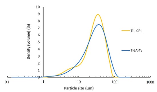
Figure 4.
Granulometry of the particles released.

Table 2.
Specific surface of the Ti and Ti6Al4V particles.
Electron microscopy has shown, at different magnifications, an irregular morphology of the particles in both samples evaluated (Figure 5). At the morphological level, SEM analysis has not shown major differences between the Ti and Ti6Al4V samples, although a higher amount of fine particles seems to be observed in the Ti6Al4V µm samples. These results would confirm the data provided by the laser grain size measurements. The SEM analysis has shown a heterogeneous geometry, with a rough and irregular finish in all the powders evaluated, which would be the product of some kind of synthesis process by friction [25,26,27].
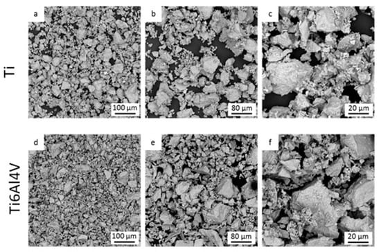
Figure 5.
Particles released observed by SEM. (a–c) Titanium particles observed at different magnifications. (d–f) Ti-6Al-4V alloy particles observed at different magnifications.
X-ray energy dispersive microanalysis does not detect the presence of contaminants in its composition, nor does it detect the presence of iron that could come from the steels of the drills used in the surgery.
Figure 6 shows the release curves of titanium, aluminium and vanadium ions into the liquid medium (Hanks’ solution) as a function of incubation time for both powders evaluated. A comparative analysis of the titanium ion release level has shown similar behaviour of both powders analysed, with an initial stage of high titanium ion release in the first 3 days of study, followed by a second stage of progressive stabilisation of the titanium ion release level between 3 and 21 days of incubation.
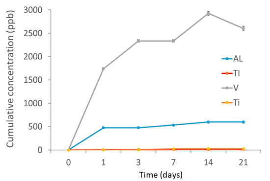
Figure 6.
Cumulative concentration of release of titanium, aluminium and vanadium ions. The release of titanium ions from commercially pure Ti grade 4 and Ti–6Al–4V alloy implants overlap. The values are very similar and very low (<30 ppb in both material types).
The quantitative comparative analysis of titanium ion release between the two powders tested showed differences between the samples in terms of the amount of ions as a function of incubation time. Despite the low values of ion concentration released by both powders, the Ti6Al4V powder released approximately twice the amount of Ti ions, which could be explained by a galvanic couple effect of the alloy with respect to Al and V. The comparative analysis of ion release between the two powders showed a preferential release of V and Al ions with respect to Ti from the Ti6Al4V powder. The possible influence of the galvanic effect on the Ti6Al4V alloy would have caused the preferential release of V and Al between one and two orders of magnitude higher than Ti, respectively [28,29]. The significant release of vanadium ions could be due to the fact that this element favours the beta phase of Ti6Al4V. In this alloy, it has approximately 4% beta phase and is more unstable than the majority alpha phase and with a higher stability [9].
3.1. Cytotoxicity Assays
The results obtained from the cytotoxicity test are shown below in the form of percentage cell survival (Figure 7). Seventy per cent is set as the lower limit of cell survival for a material not to be considered cytotoxic. The cells were adhered to the substrate plate and presented the expected morphology, both before and after incubation with the extracts. The samples evaluated in the cytotoxicity assay presented values higher than 70% cell survival in all cases. The samples analysed under the conditions tested were found not to be cytotoxic to any of the cell types studied.
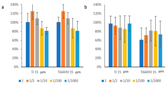
Figure 7.
Cellular viability in the cytotoxicity tests: (a) SAOS–1 and (b) HFF–1.
Cytotoxicity tests have shown values slightly above 80% cell survival in both cell lines tested for the Ti–15 µm powder. However, the Ti6Al4V powder showed mean cell survival values below 80% for the HFF–1 cell line. In view of the results, a certain tendency towards an increased cell cytotoxic effect on human fibroblasts (gingival tissue) of Ti6Al4V powder particles compared to titanium particles is confirmed. A cytotoxicity assay was also performed for THP–1 cells at 24 and 48 h to assess cell survival. For this, these cells were cultured in medium containing the extracts at different concentrations: [ISO] (0.2 g/mL), and their dilutions 1:2, 1:10, 1:100 and 1:1000. According to regulations, a cell survival rate of less than 70% was considered cytotoxic. The cells were adherent to the substrate plate and presented the expected morphology, both before and after incubation with the extracts [30,31,32]. As can be seen in Figure 8, cells with concentrations of [ISO], and their 1:2 dilutions were considered cytotoxic at 24 and 48 h, for the titanium aluminium vanadium alloy Ti6Al4V (TiAL15), but not for the titanium microparticles (Ti15) where only the [ISO] concentration is considered cytotoxic. In addition, extracts of the alloy microparticles (TiAL15) were also cytotoxic at a 1:10 dilution. For this reason, the 1:100 dilution was used for further experiments [18,33].
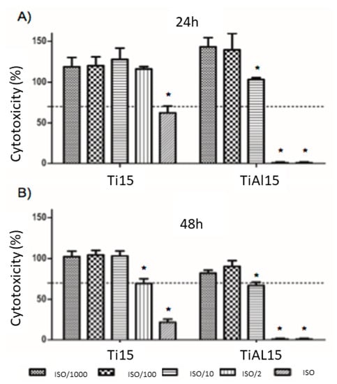
Figure 8.
Cytotoxicity levels of THP–1 cells in extracts of microparticles of Titanium. (Ti15) and the alloy of titanium, aluminium and vanadium Ti6Al4V (TiAL15), carried out at 24 h (A) and 48 h (B). * p < 0.05 vs. its [ISO]/1000.
3.2. Gene Expression Analysis
Once the cytotoxicity of the extracts in culture with THP–1 cells had been assessed, the inflammatory response was tested at the concentration considered to be non–cytotoxic. As can be seen in Figure 9, in the case of gene expression, the pro–inflammatory markers (CCR7, TNFα and IL–1β) showed similar levels to the control (TCP), and these were significantly lower when cultured with the pro–inflammatory LPS medium, the positive control for inflammation (Figure 9A). Specifically, the values for the titanium aluminium vanadium Ti6Al4V alloy (TiAL15) extracts were significantly lower at 24 h In contrast, in the case of IL–1β interleukin, the values for titanium (Ti) extracts were significantly lower at 48 h.
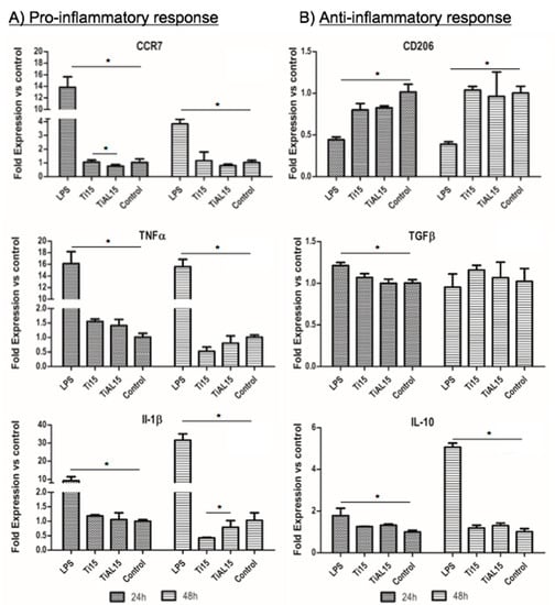
Figure 9.
RNA analysis of the inflammatory response in THP–1 cells for pro–inflammatory (A) and anti–inflammatory (B) markers. Cells cultured with extracts of microparticles of Titanium (Ti15) and the alloy of titanium, aluminium and vanadium Ti6Al4V (TiAL15). * p < 0.05.
On the other hand, the anti–inflammatory markers (CD206, TGF–β and IL–10) followed the same trend as the control sample (TCP) as they presented similar values (Figure 9B). These results indicate that the cells do not show an inflammatory response when cultured in medium conditioned with the samples [18,33].
3.3. Cytokine Release
Once the response was evaluated at the gene level, protein analysis was performed. Figure 10 shows that the pro–inflammatory markers (TNFα and IL–1β) showed similar levels to those of the control (TCP), and these were significantly lower than the culture with the pro–inflammatory LPS medium, the positive control of inflammation (Figure 10A). On the other hand, the anti–inflammatory marker tested (IL–10) showed similar results to the control sample (TCP). These results indicate that the cells do not show an inflammatory response when cultured in the conditioned medium of the samples. Furthermore, these results are in agreement with the cytotoxicity results, indicating that the general trend is that the alloy sample (TiAL15) shows a higher cell toxicity and a higher pro–inflammatory response [34,35,36,37].
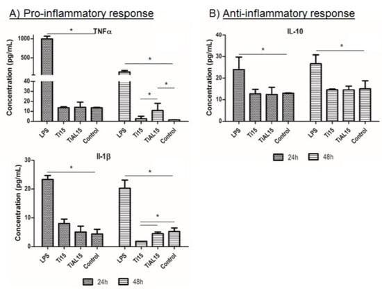
Figure 10.
Protein analysis of the inflammatory response in THP–1 cells for pro–inflammatory (A) and anti–inflammatory (B) markers. Cells cultured with extracts of microparticles of Titanium (Ti15) and the alloy of titanium, aluminium and vanadium Ti6Al4V (TiAL15). * p < 0.05.
Debris of titanium, Ti6Al4V and other metals have been proposed as a risk factor of bone loss and inflammation of the peri–implant mucosa [12,14,16,38]. Periimplantitis has been associated with a greater accumulation of Ti in peri–implant tissues, in comparison with healthy implants [18,33]. Moreover, it has been reported that Ti debris can promote DNA damage in oral epithelial cells by activating the molecular markers CHK2 and BRCA1 [35]. Metal particles can be released into the peri–implant tissues through different mechanisms, including implant insertion, fatigue, corrosion, wear, or surface decontamination methods of the dental implant, such as implantoplasty. Although Ti particles seem to induce the expression of pro–inflammatory cytokines and decrease the viability of osteogenic cells [16,39,40] the immunological properties (inflammatory and osteogenic response) of metal particles released during insertion dental implant is still unknown.
4. Conclusions
According to the tests carried out to evaluate the inflammatory response of THP–1 cells in contact with extracts of 15 µm titanium microparticles of different composition: titanium (Ti15) and an alloy of titanium, aluminium and vanadium Ti6Al4V (TiAL15), we can conclude that cell survival in cultures with extracts of the Ti6AL4V alloy is lower compared to that of only titanium (Ti15), which obtained a significantly higher percentage of cytotoxicity. At the gene level, cells do not show an inflammatory response grown in conditioned medium from the samples compared to the control. Even so, at 48 h, the Ti6AL4V alloy presents higher levels of the inflammation marker Il–1β compared to that of only titanium (Ti15). From studies concerning cytokine release, the cells do not show an inflammatory response grown in conditioned medium from the samples compared to the control. Even so, at 48 h, the Ti6AL4V alloy presents higher levels of inflammation markers (TNFα and Il–1β) compared to that of only titanium (Ti15). Therefore, there is a trend that with the alloy there is a greater toxicity and a greater pro–inflammatory response.
Author Contributions
Conceptualization, J.G., J.C.V., A.B., J.D., A.E.-M. and B.B.; methodology, J.C.V., E.P.-P. and A.B.; validation, J.G., J.C.V., E.P.-P. and A.B.; formal analysis, E.P.-P., J.C.V., J.G. and A.B.; investigation, R.P., J.C.V., B.B., J.G. and A.B.; resources, J.C.V. and J.G.; data curation, J.G.; writing—original draft preparation, J.G., A.B. and J.C.V.; writing—review and editing, J.D., A.E.-M. and E.P.-P.; visualization and J.D.; supervision, J.G.; project administration, J.G. and J.C.V.; funding acquisition, J.C.V. All authors have read and agreed to the published version of the manuscript.
Funding
The work was supported by the Spanish government and the Ministry of Science and Innovation of Spain by the research project numbers RTI2018–098075–B–C21 and RTI2018–098075–BC22, cofounded by the EU through the European Regional Development Funds (MINECO–FEDER, EU). Authors also acknowledge Generalitat de Catalunya for funding through the 2017SGR–1165 project and the 2017SGR708 project.
Institutional Review Board Statement
Not applicable.
Informed Consent Statement
Not applicable.
Data Availability Statement
Not applicable.
Conflicts of Interest
The authors declare no conflict of interest.
References
- Velasco-Ortega, E.; Alfonso-Rodríguez, C.; Monsalve-Guil, L.; España-López, A.; Jiménez-Guerra, A.; Garzón, I.; Alaminos, M.; Gil, F. Relevant aspects in the surface properties in titanium dental implants for the cellular viability. Mater. Sci. Eng. C 2016, 64, 1–10. [Google Scholar] [CrossRef]
- Aparicio, C.; Rodriguez, D.; Gil, F.J. Variation of roughness and adhesion strength of deposited apatite layers on titanium dental implants. Mater. Sci. Eng. C 2011, 31, 320–324. [Google Scholar] [CrossRef]
- Albrektsson, T.; Wennerberg, A. Oral implant surfaces: Part 1-review focusing on topographic and chemical properties of different surfaces and in vivo responses to them. Int. J. Prosthodont. 2004, 17, 536–543. [Google Scholar]
- Herrero-Climent, M.; Lázaro, P.; Rios, J.V.; Lluch, S.; Marqués, M.; Guillem-Martí, J.; Gil, F.J. Influence of acid-etching after grit-blasted on osseointegration of titanium dental implants: In vitro and in vivo studies. J. Mater. Sci. Mater. Med. 2013, 24, 2047–2055. [Google Scholar] [CrossRef]
- Ríos-Carrasco, B.; Ferreira-Lemos, B.; Herrero-Climent, M.; Gil., F.J.; Ríos-Santos, J.V. Effect of the acid–etching on grit–blasted dental implants to improve osseointegration: Histomorphometric analysis of the bone–implant contact in the Rabbit Tibia Model. Coatings 2021, 11, 1426. [Google Scholar] [CrossRef]
- Ordon, M.; Nawrotek, P.; Stachurska, X.; Mizielińska, M. Polyethylene Films Coated with Antibacterial and Antiviral Layers Based on CO2 Extracts of Raspberry Seeds, of Pomegranate Seeds and of Rosemary. Coatings 2021, 11, 1179. [Google Scholar] [CrossRef]
- Gil, J.; Pérez, R.; Herrero-Climent, M.; Rizo-Gorrita, M.; Torres-Lagares, D.; Gutierrez, J.L. Benefits of residual aluminum oxide for sand blasting titanium dental implants: Osseointegration and bactericidal effects. Materials. 2021, 15, 178. [Google Scholar] [CrossRef] [PubMed]
- Costa-Berenguer, X.; García-García, M.; Sánchez-Torres, A.; Sanz-Alonso, M.; Figueiredo, R.; Valmaseda-Castellon, E. Effect of implantoplasty on fracture resistance and surface roughness of standard diameter dental implants. Clin. Oral Implant. Res. 2017, 29, 46–54. [Google Scholar] [CrossRef] [PubMed]
- Toledano-Serrabona, J.; GilF, J.; Camps-Font, O.; Valmaseda-Castellón, E.; Gay-Escoda, C.; Sánchez-Garcés, M. Physicochemical and biological characterization of Ti6Al4V particles obtained by implantoplasty: An in vitro study. Part I. Materials 2021, 14, 6507. [Google Scholar] [CrossRef] [PubMed]
- Toledano-Serrabona, J.; Sánchez-Garcés, M.A.; Gay-Escoda, C.; Valmaseda-Castellon, E.; Camps-Font, O.; Verdeguer, P.; Molmeneu, M.; Gil, F.J. Mechanical properties and corrosión behavior of Ti6Al4V particles obtained by implatoplasty. An in vivo study. Part II. Materials 2021, 14, 6519. [Google Scholar] [CrossRef]
- Velasco-Ortega, E.; Flichy-Fernández, A.; Punset, M.; Jiménez-Guerra, A.; Manero, J.M.; Gil, J. Fracture and fatigue of titanium narrow dental implants: New trends in order to improve the mechanical response. Materials 2019, 12, 3728. [Google Scholar] [CrossRef]
- Pérez, R.A.; Gargallo, J.; Altuna, P.; Herrero-Climent, M.; Gil, F. Fatigue of narrow dental implants: Influence of the hardening method. Materials 2020, 13, 1429. [Google Scholar] [CrossRef] [PubMed]
- Bannon, B.; Mild, E. Titanium alloys for biomaterial application: An overview. In Titanium Alloys in Surgical Implants; Luckey, H.A., Kubli, F., Eds.; ASTM International: West Conshohocken, PA, USA, 2009; p. 7. [Google Scholar]
- Cadosch, D.; Sutanto, M.; Chan, E.; Mhawi, A.; Gautschi, O.P.; von Katterfeld, B.; Simmen, H.-P.; Filgueira, L. Titanium uptake, induction of RANK-L expression, and enhanced proliferation of human T-lymphocytes. J. Orthop. Res. 2010, 28, 341–347. [Google Scholar] [CrossRef]
- Liu, R.; Lei, T.; Dusevich, V.; Yao, X.; Liu, Y.; Walker, M.P.; Wang, Y.; Ye, L. Surface characteristics and cell adhesion: A comparative study of four commercial dental implants. J. Prosthodont. 2013, 22, 641–651. [Google Scholar] [CrossRef]
- Pettersson, M.; Kelk, P.; Belibasakis, G.; Bylund, D.; Thorén, M.M.; Johansson, A. Titanium ions form particles that activate and execute interleukin-1β release from lipopolysaccharide-primed macrophages. J. Periodontal Res. 2017, 52, 21–32. [Google Scholar] [CrossRef]
- Meng, B.; Chen, J.; Guo, D.; Ye, Q.; Liang, X. The effect of titanium particles on rat bone marrow stem cells in vitro. Toxicol. Mech. Methods 2009, 19, 552–558. [Google Scholar] [CrossRef]
- Fretwurst, T.; Buzanich, G.; Nahles, S.; Woelber, J.P.; Riesemeier, H.; Nelson, K. Metal elements in tissue with dental peri-implantitis: A pilot study. Clin. Oral Implant. Res. 2015, 27, 1178–1186. [Google Scholar] [CrossRef] [PubMed]
- Magone, K.; Luckenbi, L.L.D.; Goswami, T. Metal ions as inflammatory initiators of osteolysis. Arch. Orthop. Trauma. Surg. 2015, 135, 683–695. [Google Scholar] [CrossRef] [PubMed]
- Shanbhag, A.S.; Jacobs, J.J.; Glant, T.T.; Gilbert, J.L.; Black, J.; Galante, J.O. Composition and morphology of wear debris in failed uncemented total hip replacement. J Bone Joint Surg Br. 1994, 76, 60–67. [Google Scholar] [CrossRef]
- Archibeck, M.J.; Jacobs, J.; A Roebuck, K.; Glant, T.T. The basic science of periprosthetic osteolysis. Instr Course Lect 2001, 50, 185–195. [Google Scholar] [CrossRef] [PubMed]
- Olmedo, D.G.; Paparella, M.L.; Spielberg, M.; Brandizzi, D.; Guglielmotti, M.B.; Cabrini, R.L. Oral mucosa tissue response to titanium cover screws. J. Periodontol. 2012, 83, 973–980. [Google Scholar] [CrossRef]
- Purdue, P.E.; Koulouvaris, P.; Nestor, B.J.; Sculco, T.P. The central role of wear debris in periprosthetic osteolysis. HSS J.® Musculoskelet. J. Hosp. Spéc. Surg. 2006, 2, 102–113. [Google Scholar] [CrossRef]
- Wimhurst, J.A.; Brooks, R.A.; Rushton, N. Inflammatory responses of human primary macrophages to particulate bone cements in vitro. J Bone Joint Surg Br. 2001, 83, 278–282. [Google Scholar] [CrossRef] [PubMed][Green Version]
- Dental Implant system. 2021. Available online: https://www.klockner.es/producto/Implantes–de–colocaci%C3%B3n-crestal/VEGA%C2%AE (accessed on 18 December 2021).
- Brunauer, S.; Emmett, P.H.; Teller, E. Adsorption of gases in multimolecular layers. J. Am. Chem. Soc. 1938, 60, 309–319. [Google Scholar] [CrossRef]
- Case, C.P.; Langkamer, V.G.; James, C.; Palmer, M.R.; Kemp, A.J.; Heap, P.F.; Solomon, L. Widespread dissemination of metal debris from implants. J. Bone Jt. Surgery. Br. Vol. 1994, 76-B, 701–712. [Google Scholar] [CrossRef]
- Nicholson, J.W. Titanium alloys for dental implants: A review. Prosthesis 2020, 2, 100–116. [Google Scholar] [CrossRef]
- Senna, P.; Antoninha Del Bel Cury, A.; Kates, S.; Meirelles, L. Surface damage on dental implants with release of loose particles after insertion into bone. Clin. Implant. Dent. Relat. Res. 2015, 17, 681–692. [Google Scholar] [CrossRef]
- Aparicio, C.; Gil, F.J.; Fonseca, C.; Barbosa, M.; Planell, J.A. Corrosion behaviour of commercially pure tianium shot blasted with different materials and sizes of shot particles for dental implant applications. Biomaterial 2003, 24, 263–273. [Google Scholar] [CrossRef]
- Rodrigues, D.C.; Valderrama, P.; Wilson, J.T.G.; Palmer, K.; Thomas, A.; Sridhar, S.; Adapalli, A.; Burbano, M.; Wadhwani, C. Titanium corrosion mechanisms in the oral environment: A retrieval study. Materials 2013, 6, 5258–5274. [Google Scholar] [CrossRef] [PubMed]
- Dalago, H.R.; Filho, G.S.; Rodrigues, M.A.P.; Renvert, S.; Bianchini, M.A. Risk indicators for peri-implantitis. A cross-sectional study with 916 implants. Clin. Oral Implant. Res. 2016, 28, 144–150. [Google Scholar] [CrossRef]
- Halperin-Sternfeld, M.; Sabo, E.; Akrish, S. The pathogenesis of implant-related reactive lesions: A clinical, histologic and polarized light microscopy study. J. Periodontol. 2016, 87, 502–510. [Google Scholar] [CrossRef] [PubMed]
- He, X.; Reichl, F.-X.; Wang, Y.; Michalke, B.; Milz, S.; Yang, Y.; Stolper, P.; Lindemaier, G.; Graw, M.; Hickel, R. Analysis of titanium and other metals in human jawbones with dental implants: A case series study. Dent. Mater. 2016, 32, 1042–1051. [Google Scholar] [CrossRef] [PubMed]
- Olmedo, D.G.; Nalli, G.; Verdú, S.; Paparella, M.L.; Cabrini, R.L. Exfoliative cytology and titanium dental implants: A pilot study. J. Periodontol. 2013, 84, 78–83. [Google Scholar] [CrossRef] [PubMed]
- Olmedo, D.; Paparella, M.; Brandizzi, D.; Cabrini, R. Reactive lesions of peri-implant mucosa associated with titanium dental implants: A report of 2 cases. Int. J. Oral Maxillofac. Surg. 2010, 39, 503–507. [Google Scholar] [CrossRef] [PubMed]
- Wilson, T.G., Jr.; Valderrama, P.; Burbano, M.; Blansett, J.; Levine, R.; Kessler, H.; Rodrigues, D.C. Foreign bodies associated with peri-implantitis human biopsies. J. Periodontol. 2015, 86, 9–15. [Google Scholar] [CrossRef] [PubMed]
- Del Amof, S.-L.; Rudek, I.; Wagner, V.; Martins, M.; O’valle, F.; Galindo-Moreno, P.; Giannobile, W.; Wang, H.-L.; Castilho, R. Titanium activates the DNA damage response pathway in oral epithelial cells: A pilot study. Int. J. Oral Maxillofac. Implant. 2017, 32, 1413–1420. [Google Scholar] [CrossRef]
- Mardashev, S.R.; Nikolaev Ya., A.; Sokolov, N.N. Isolation and properties of a homogenous L asparaginase preparation from Pseudomonas fluorescens AG (Russian). Biokhimiya 1975, 40, 984–989. [Google Scholar]
- Del Amof, S.-L.; Garaicoa-Pazmino, C.; Fretwurst, T.; Castilho, R.M.; Squarize, C.H. Dental implants-associated release of titanium particles: A systematic review. Clin. Oral Implant. Res. 2018, 29, 1085–1100. [Google Scholar] [CrossRef]
Publisher’s Note: MDPI stays neutral with regard to jurisdictional claims in published maps and institutional affiliations. |
© 2022 by the authors. Licensee MDPI, Basel, Switzerland. This article is an open access article distributed under the terms and conditions of the Creative Commons Attribution (CC BY) license (https://creativecommons.org/licenses/by/4.0/).