Article

Synthesis, Characterization, and Antimicrobial of MnO and CdO Nanoparticles by Using a Calcination Method

1
Department of Chemistry, College of Science, Qassim University, Buraidah 51452, Saudi Arabia
2
Taiz University, Taiz 3086, Yemen
3
Laboratory of Physico-Chemistry of Materials, University of Monastir, Avenue of the Environment, Monastir 5019, Tunisia
*
Author to whom correspondence should be addressed.
Coatings 2022, 12(2), 215; https://doi.org/10.3390/coatings12020215
Submission received: 2 December 2021 / Revised: 19 January 2022 / Accepted: 20 January 2022 / Published: 7 February 2022
(This article belongs to the Special Issue Nanoparticles for Energy, Sensing and Biomedical Applications)

Abstract

:
Nano-sized manganese oxide and cadmium oxide were formed quantitatively via chemical routes, using calcination from an aqueous solution containing metal chloride as a precursor, to create polyvinyl alcohol and para-aminobenzoic acid complexes with the following formulae: [Mn (PVA)(P-ABA) (H2O)3] H2O and [Cd (PVA)(P-ABA) (H2O)3]. The synthesized complexes and metal oxide nanoparticles were characterized using elemental analysis, thermal analyses (TGA and DTA), FT-IR spectroscopy, XRD analysis, UV-vis spectra, and SEM and TEM electron microscopes. The kinetic and thermodynamic parameters (∆H*, ∆G* and ∆S*) for the Mn(II) and Cd(II) coordination compounds were calculated. The antimicrobial properties of the samples were assessed using five bacterial strains and three fungal strains. Three strains of (G+) bacteria, two strains of (G−) bacteria, one stain of yeast-like fungi, and two molds were used in this study.

1. Introduction

Nanotechnology is a new and rapidly developing field that involves the industrial processing and application of structures, devices, and systems by controlling shape and size in the nanometer range, having a length less than 100 nm [1,2]. Many great researchers have given attention to the synthesis of metal oxide nanoparticles, due to their essential importance and the extensive range of potential technological applications, as well as their unique properties, such as large surface area, high crystallinity, chemical purity, and phase selectivity, which are different from their bulk counterparts [3,4]. Many methods have been used to synthesis metal oxides, such as the hydrothermal method [5,6,7,8], sol-gel synthesis, co-precipitation method, solvothermal [9,10,11,12], thermal decomposition technique, micro-emulation, mechanochemical method sputtering, chemical etching, atomic condensation, and laser pyrolysis [13,14,15,16,17,18,19,20]. Manganese oxide and cadmium oxide nanoparticles are commonly used in diverse fields, such as in electrodes, catalysis [21,22], sensors, and optoelectronics [23,24]. MnO is a transition metal oxide, having interesting physical and chemical properties. It has been used in optoelectronic applications and is also used as an electrode material, and in rechargeable batteries, catalysts, electrochemical capacitors, sensors, and magneto-electronic devices [25,26].
Nanoparticles of manganese oxide are attracting much attention, such as for positive electrode materials for lithium and alkaline batteries [27,28], magneto-electric materials [29], electrochemical energy storage technologies [30], water purification, and biomedical drug delivery applications; while its supplementation has a blood glucose lowering effect [31,32,33].
Cadmium oxide nanoparticles have also attracted tremendous attention due to their interesting properties, including a direct band gap of 2.3 electron volts (eV) [34]. They have also been used in solar cells, IR reflectors, transparent electrodes, phototransistors [35,36,37,38], and gas sensors [39,40]. The photocatalytic activity of CdO nanomaterials using methyl red dye as an organic contaminate has been studied, and they are often exposed to water sources in the textile industry. The results showed that nanoparticles of CdO are very efficient as a photocatalyst for the degradation of Congo red dye solution [41]. Cadmium oxide NPs were chosen due to their chemical stability, low cost, and widespread availability [42]. Polyvinyl alcohols are artificial, water soluble, nontoxic, and biocompatible polymers that have been used since the early 1930s in a wide range of commercial and industrial medical applications, including in resins, lacquers, surgical threads, and drug delivery systems [43,44,45]. Para-aminobenzoic acid is a well-known amino acid compound in medicinal chemistry and biochemistry, and has many industrial applications; while, P-ABA is a necessary substrate for the fabricate of diverse biological scaffolds. 4-aminobenzoic acid (P–ABA) is an essential substrate of the folic acid biosynthetic pathway in bacteria. Although it is not a vitamin, it is still referred to as a member of the B vitamins [46,47]. As mentioned above, there are many methods used to obtain metal oxide nanoparticles. In this work, calcination was used to synthesize them from their complexes, because this is an easy and inexpensive approach.

2. Experimental

2.1. Materials

The chemicals employed in this work were reagent grade and were not purified. Polyvinyl alcohol (PVA), P-aminobenzoic acid, ethanol, manganese (II) chloride tetrahydrates, and cadmium (II) chloride hydrate (≥99%) were purchased from Sigma- Aldrich.

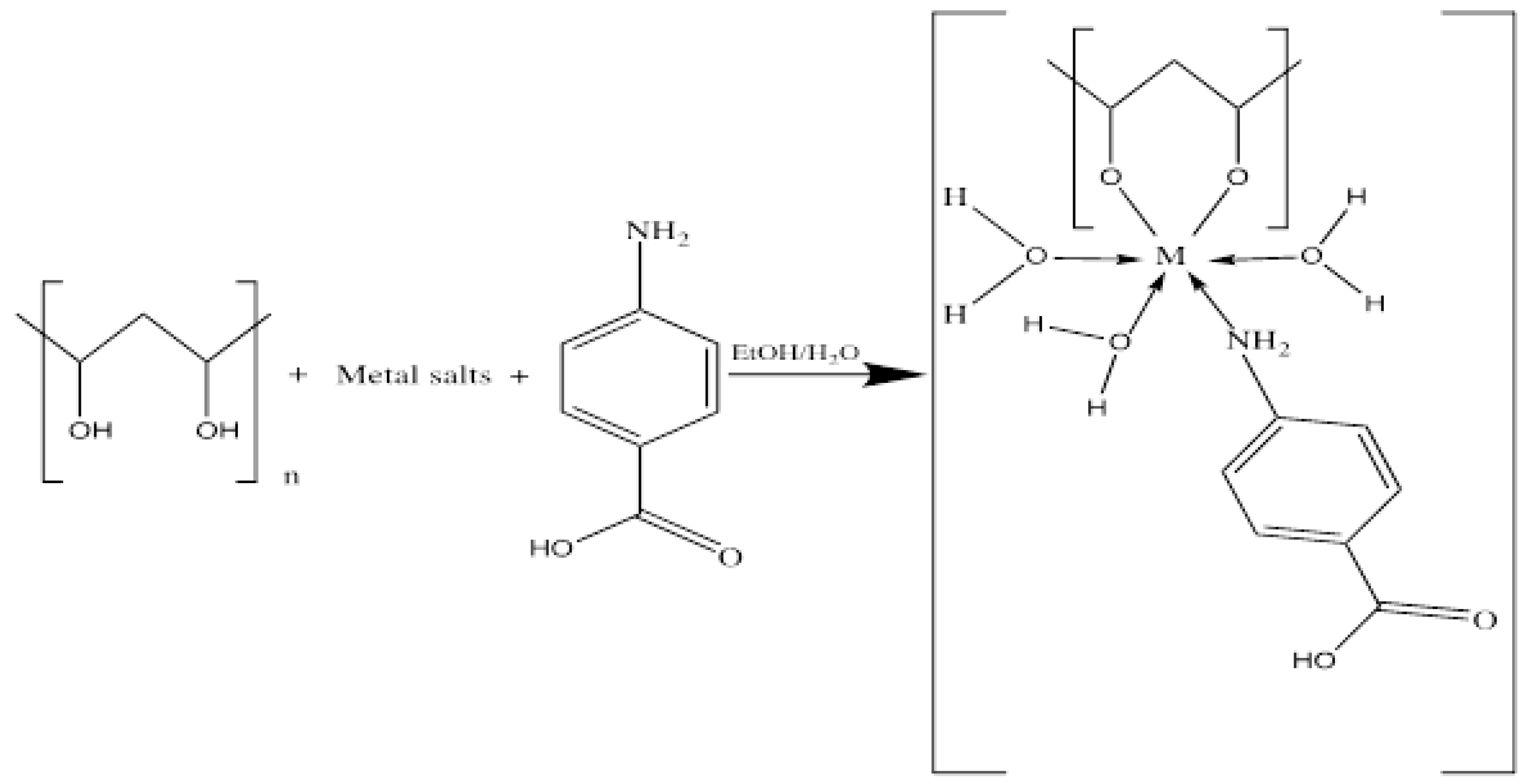
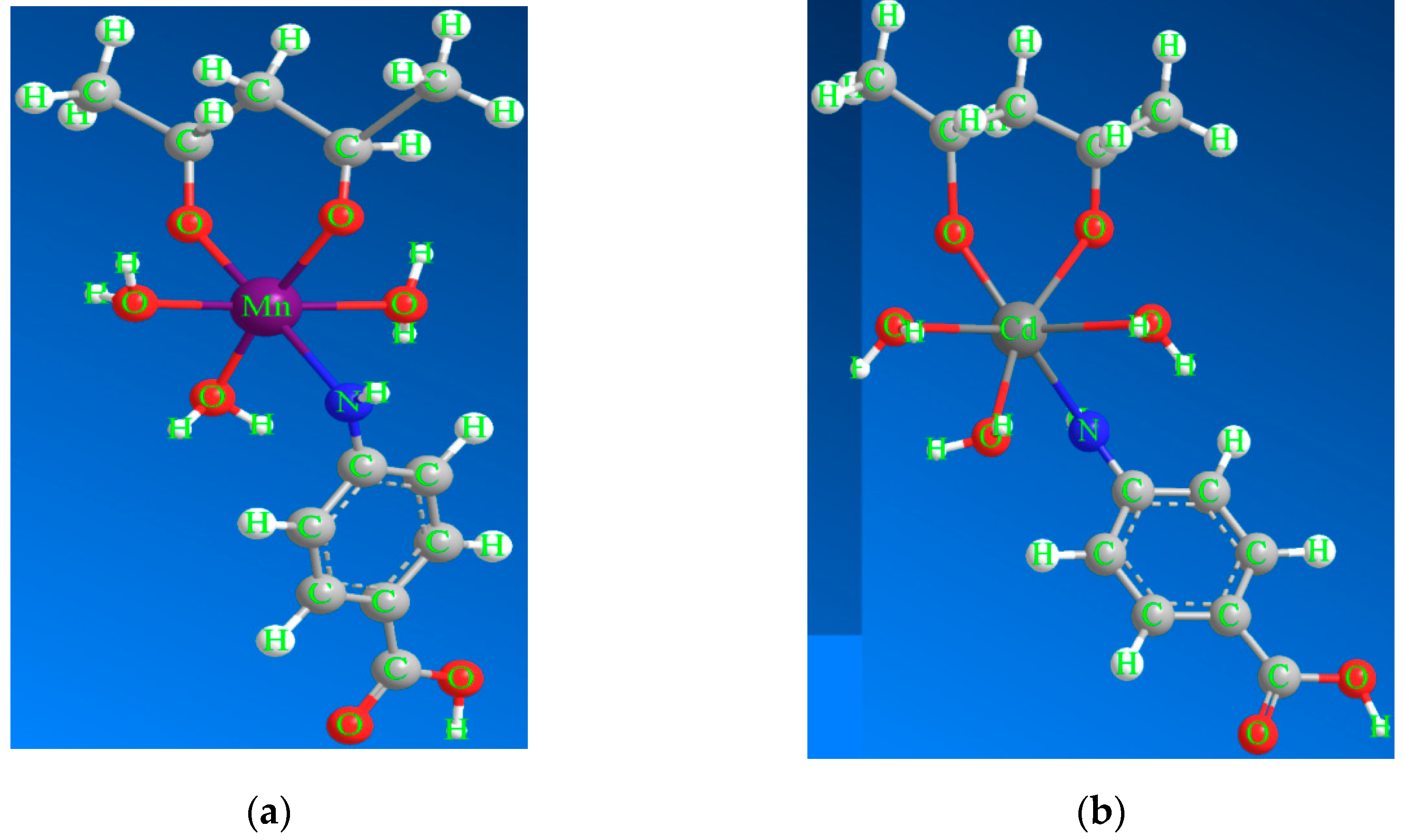
2.2. Synthesis of the [Mn(PVA)(P-ABA)(H2O)3]·H2O and [Cd(PVA)(P-ABA)(H2O)3] Complexes

Polyvinyl alcohol ligand (2 g) was dissolved in 100 mL distilled water by stirring at 50 °C for 60 min. A solution of 0.001 M NaOH in 1.5 mL distilled water was added to the PVA solution. After cooling, MnCl2·4H2O solution (5.35 g in 35 mL distilled water) was added dropwise to the PVA solution under rapid stirring, then P-ABA solution (3.70 g in 30 mL ethanol) was added to the mixture. Finally, the mixture was refluxed for 3 h and then cooled. A light brown substance was produced, the latter was collected by filtration. For Cd(II) complex, we measured 5.44 g of CdCl2·H2O and followed the previous procedure.

2.3. Synthesis of Manganese Oxide and Cadmium Oxide Nanoparticles

A precipitate of Mn(II) and Cd(II) complexes was washed with ethanol and then dried at 40 °C. Then calcined at 550 ◦C and 450 ◦C for 4 h, respectively.

2.4. Material Characterization

The elemental analyzers were determined using a GmbhVario El analyzer, structural information was obtained from FT-IR spectra on a Thermo Nicolet (6700) FT-IR spectrophotometer, with a wavenumber range of 400–4000 cm−1, and UV-vis absorption were recorded on a UV–2102 PC Shimadzu using a 1 cm matched quartz cuvette in the wavelength range of 200 to 900 nanometers. At room temperature, magnetic susceptibility was used to measure the Mn(II) and Cd(II) complexes using a magnetic susceptibility balance from MSB–Auto. Thermogravimetry analysis (TGA) and differential thermal analysis (DTA) were conducted in air atmosphere on a Shimadzu DTG 60 thermal analyzer, at a heating rate 10 C min−1. Measurements of the X-ray diffraction (XRD) were carried out using an XRD diffractometer Model (PW 1710) (Cu-Kα radiation). The morphology and structure of the synthesized materials were characterized using a scanning electron microscope and transmission electron microscope, JEOL/JEM 1010 High-Resolution TEM.

2.5. Antimicrobial Activity of Manganese Oxide and Cadmium Oxide Nanoparticles

The antimicrobial activities of MnO and CdO nanoparticles were extracted and evaluated in this study using five bacterial strains and three fungal strains: Enterococcus faecalis, Staphylococcus epidermidis, and Staphylococcus aureus as Gram-positive cocci; E. coli and P. aeruginosa as Gram-negative bacilli, one strain of yeast-like fungi (Candida albicans), and two molds (Aspergillus fumigatus and Aspergillus flavus). These were prepared from pure cultures in Muller–Hinton broth and each was compared with a standard of 0.5 McFarland. Mold samples were inoculated directly into Sabouraud dextrose agar, then each extract (100 µL) was added to a certain Petri dish. These dishes were incubated for 18–24 h at 37 °C. After this, we observed the zone of inhibition around the dishes and measured the zone diameter in millimeters (mm) using a ruler.

3. Result and Discussion

Complex formation of PVA and P–ABA was carried out with the following (Scheme 1), and the suggestion of a coordinated structure around the manganese (II) or cadmium (II) in their complexes is illustrated in Figure 1.

3.1. Characterization

3.1.1. Elemental Analyses of Complexes

The elemental analysis (%) of [Mn(PVA)(P–ABA)(H2O)3]·H2O was C 35.84, H 6.05, and N 8.09, which is in good agreement with the calculated elemental analysis (%): C 34.30, H 5.46, and N 7.99. Other complexes are shown in Table 1.

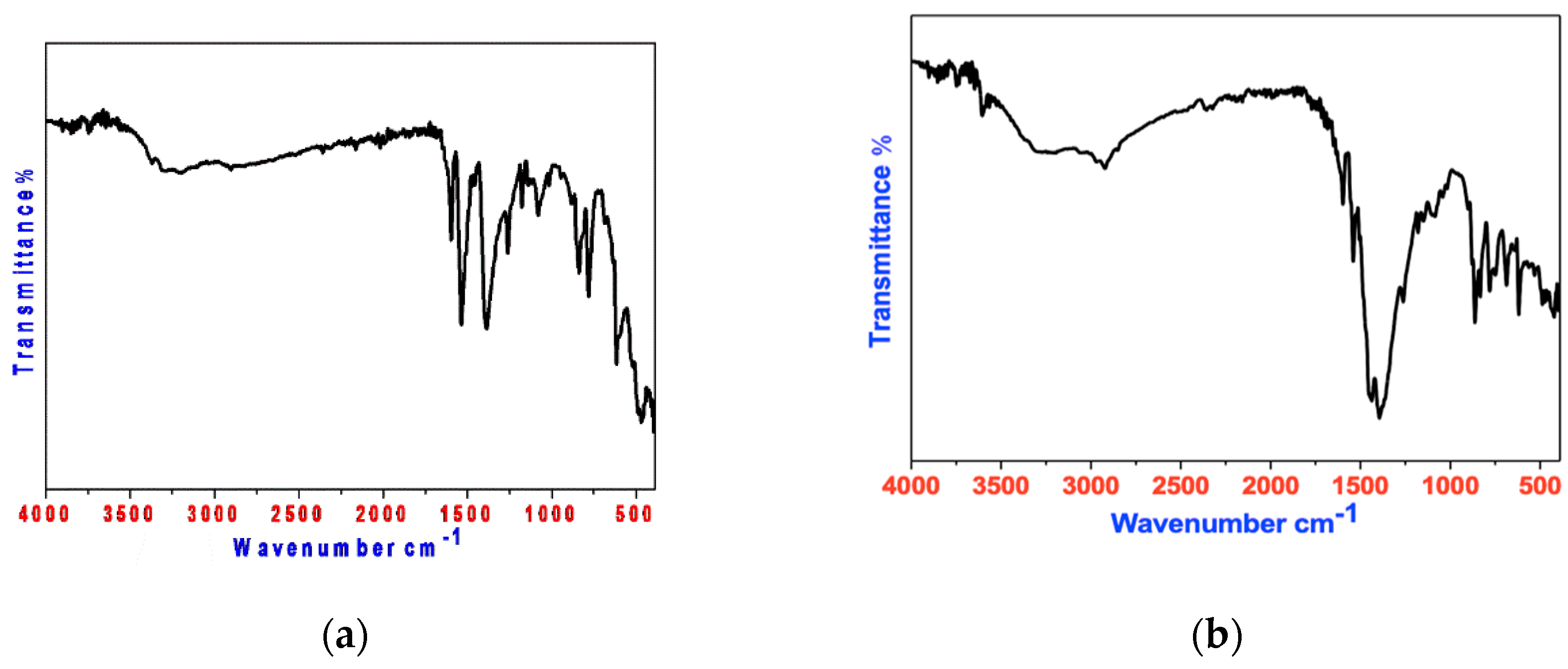
3.1.2. FT-IR Spectroscopy

Infrared spectra were obtained in KBr disks using a Thermo-Nicolet-6700 spectrometer, between 400 and 4000 cm−1. Table 2 shows the patterns of FT-IR of pure PVA, P-ABA, and their complexes. The identities of the Mn(II) complex and Cd(II) complex were confirmed by FT-IR. They clearly show a different fingerprint from the parent PVA and P–ABA [43,48]. Figure 2 shows a closer view of the fingerprint area. We found that the ν(O–H) band, which appears at 3400 cm−1 in the spectrum of PVA, was shifted to a lower wave number (3280–3250 cm−1), indicating a sharing of this group in the bonding with the metal ions. The stretching frequency of ν(CH2) was at 2890–2930 cm−1 (a slight shift compared to the PVA spectrum at 2905 cm−1). The peak of the ν(C–OC) stretch vibration was recorded at 1150 cm−1 in the ligand spectrum and 1180–1178 cm−1 for the complexes [43]. The stretching frequency of ν(NH2) was observed at 3234 cm−1 for P-ABA as a free ligand, while this value was shifted to 3243–3220 cm−1 after complexation [48]. Moreover, the ν(C=O) stretch vibration was recorded as a medium intensity strip at 1680 cm−1 of P-ABA in all complexes, and the spectrum underwent almost no shifting. The tape characteristic of the ν(C=N) stretch vibration was recorded at 1640 cm−1 for P–ABA (a slight shift compared to the ligand spectrum) at 1650–1620 cm−1 and the para substitution was present at 750 cm−1 [48]. Briefly, PVA coordinated to the metal ions in bidentate mode through oxygen atoms, while P–ABA coordinated in the monodentate mode through the nitrogen atom. A broad band with medium intensity located in the range 3388–3337 cm−1 was assigned to ν(OH) of (H2O) for all complexes. In addition, the appearance of the absorption bands, which are represented by characteristic absorption peaks at 522–515 cm−1 and 430–422 cm−1, corresponded to ν(metal–O) and ν(metal–N), respectively [49].

3.1.3. Optical Characterization and Magnetic Moment

The UV-visible absorption spectrum of PVA, P–AMB (DMSO, 5 × 10⁻⁵ M), and their Mn(II) and Cd(II) complexes are shown in Table 3, absorption peaks were found at 45,248–35,087 cm−1 [50,51], which can be attributed to the π–π*, n–π* transition of the PVA and P-ABA. The electronic spectrum of manganese (II) complex, displaying the electronic spectral band at 19,685 assigned to d-d transition ‘6A1g → 4Eg’ (Figure 3), was drawn to show UV-visible of the Mn(II) oxide nanoparticles. The magnetic moment was 5.53 B.M., which is representative of the octahedral geometry of Mn+2, while Cd+2 is the nonmagnetic ion; it only has bands for π–π*, n–π*, which are assigned to the intra-ligand charge–transfer transitions [52,53].

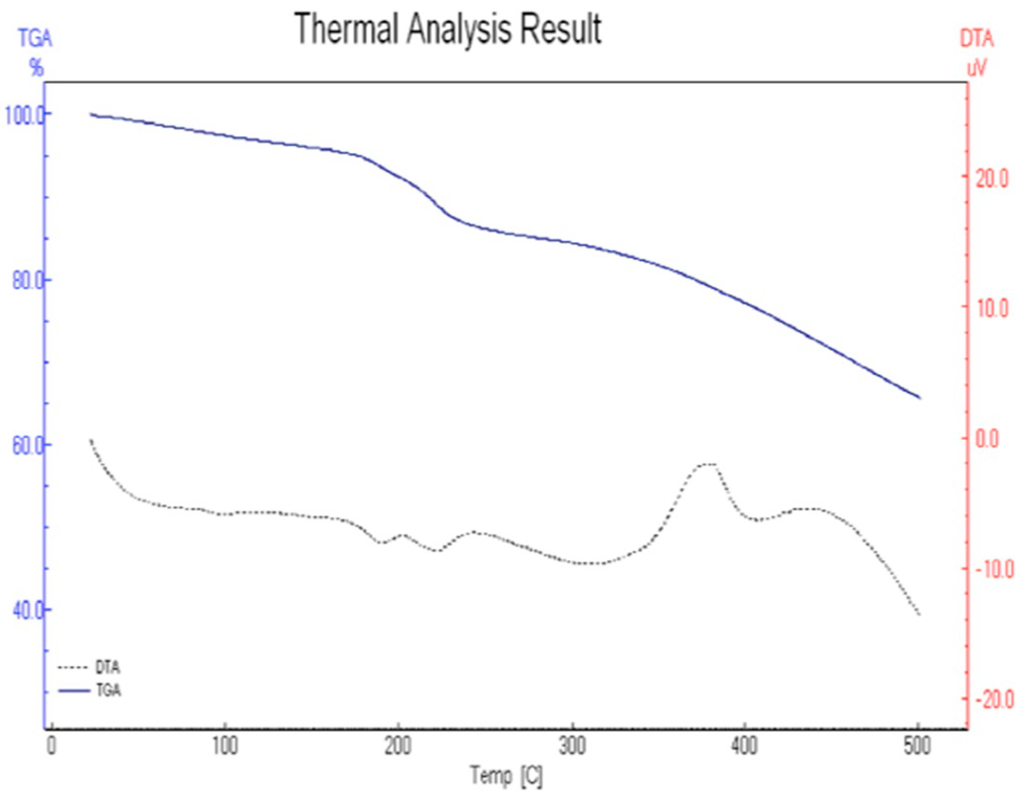
3.1.4. Thermal Analysis

Thermal Analysis of [Mn(PVA)(P–ABA)(H2O)3]·H2O

Figure 4 depicts the TG, DTG, and DTA. The complex’s thermal decomposition process (1) includes five stages. These steps occur in the temperature ranges 70–140, 142–244, 246–378, 380–403, and 405–600 °C. The first and second mass loss correlates well with the release of one crystalline water and three coordinated water molecules (calc. 20.59%, found 18.02%) (DTG peak at 88 °C). This corresponds to a broad endothermic peak in the DTA curve at 90 °C. The third step represents a loss of weight, indicating expulsion of a para-aminobenzoate ligand (calc. 39.16%, found 37.78%). This step composed a DTG peak at 302 °C, associated with an endothermic peak at 304 °C in the DTA trace. The fourth and fifth steps were decomposition products of the remainder of the polyvinyl alcohol ligand (calc. 21.14%, found 20.06%). These steps are manifested in the DTG curves as peaks at 392 and 435 °C, and the DTA trace shows an exothermic effect at 394 and 437 °C. The ultimate product at 600 °C is consistent with the formation of MnO as a residual part (calc. 20.25%, found 19.45%).

Thermal Analysis of [Cd(PVA)(P–ABA)(H2O)3]

The thermal decomposition processes of the Cd(II) complex involve three stages, which occur in the temperature ranges 60–235, 237–368, and 370–600 °C. The first stage mass loss corresponds to the liberation of the three coordinated water molecules, with a corresponding DTG midpoint at 198 °C (calc. 13.88%, found 12.78%), with an endothermic peak at 200 °C in the DTA trace. The second mass loss corresponds to the liberation of para-aminobenzoic acid (calc. 35.20%, found 34.18%) (DTG peak at 340 °C), and the DTA trace furnishes an endothermic effect at 342 °C. The third mass loss represents the expulsion of the rest of the organic ligand. The decomposition of the complex proceeds in the next step, leaving a stable residue of CdO (calc.32.95%, found 31.80%).

3.1.5. Kinetic Analysis

The Coats–Redfern [54] method was used to perform non-isothermal kinetic studies for the thermal decomposition of the complexes (Figure 5). Table 4 lists the kinetic parameters that were determined.

3.1.6. Thermodynamic Parameters

The activation variables ∆H*, ∆S*, and ∆G* for the degradation stages of the mentioned complexes are recorded in Table 5. The negative ∆S* values of stages for the Mn(II) complex and Cd(II) complex decomposition suggest that the stimulated compound was forced to be high by the reactants; and the reactions were lower than normal. The positive ∆G* values indicate the nonspontaneous decomposition of the reaction.

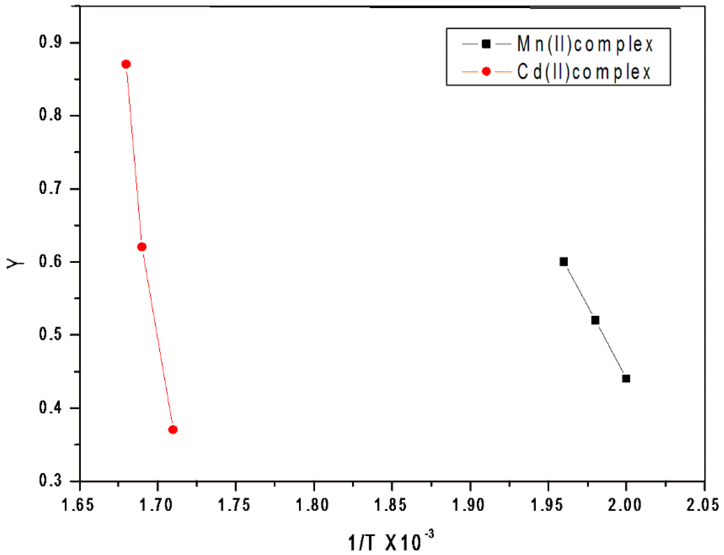
Kinetic Compensation Impact

The kinetic compensation impact (KCI) states that the rate drop expected as the activation energy increases does not materialize in a specific set of processes. The KCI, which was observed in a number of nonthermal processes, appears to be valid here. It was noted that the value of energy (E) for the specific processes conformed to a straight line, as determined by the linear square technique for the two complexes (Figure 6), and is drawn as follows:

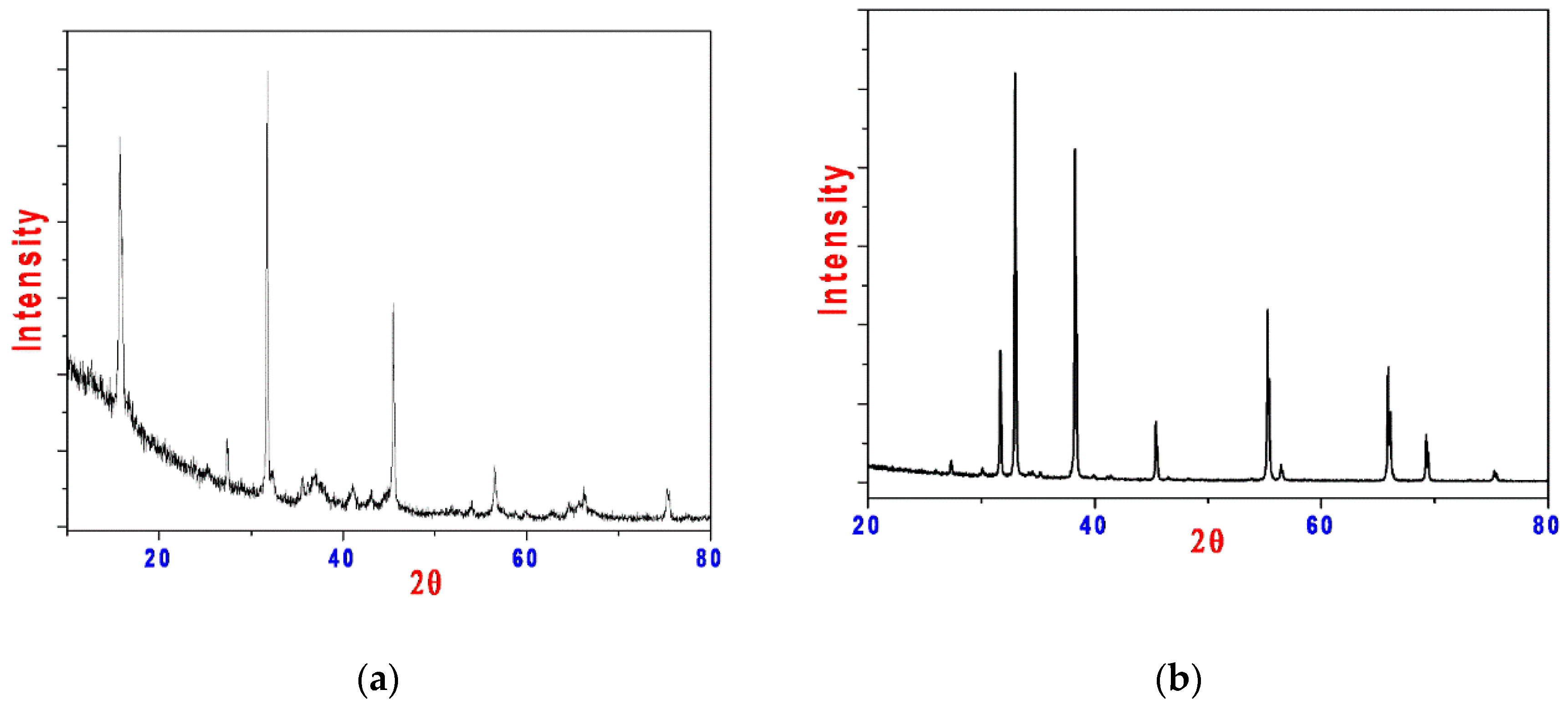
3.1.7. X-ray Powder Diffraction (XRD)

For the produced determination, the XRD patterns of the manganese oxide NP and cadmium oxide NP samples were obtained (Figure 7). The outstanding peaks were used to calculate the grain size using the Scherrer equation. The spectra of crude MnO and CdO nanoparticles showing the diffraction peaks at 2θ values are tabulated in Table 6; Joint Committee on Powder Diffraction Standards (JCPDS) of MnO and CdO card no. 057-1088 and 065-2908, respectively.

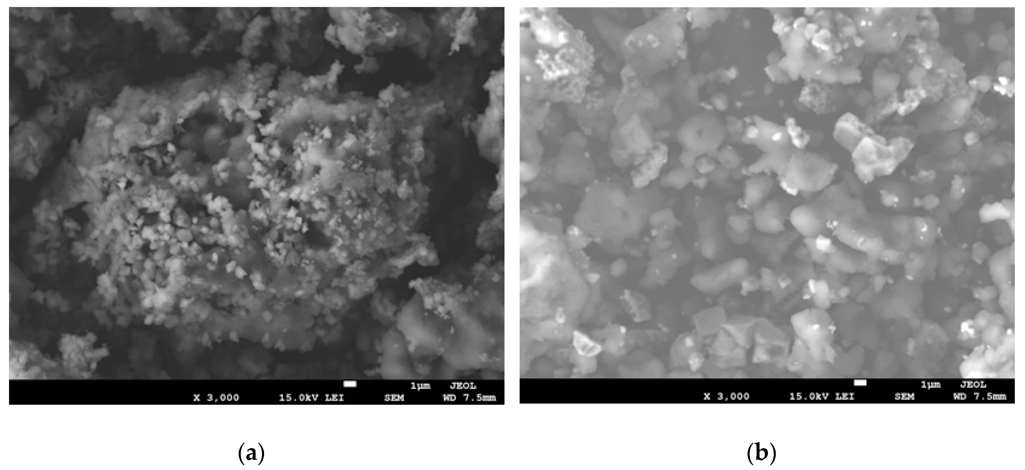
3.1.8. Morphological Studies

Morphology and shape structure were examined using scanning electron microscope (SEM) and transmission electron microscope (TEM). Figure 8 shows the SEM and TEM images of the as-synthesized MnO and CdO as nanomaterials. The nanoparticles exhibited various morphologies. The SEM picture shows that the CdO NPs had a fractured rock-like structure, while the MnO NPs had a coral-like structure. The microstructure of these materials can be observed in the SEM images as many aggregates of smaller nanoparticles. The structure appears as spherical in nature in the TEM pictures of CdO and MnO. Apart from some cubic nanoparticles, the majority of nanoparticles are sphere-shaped.

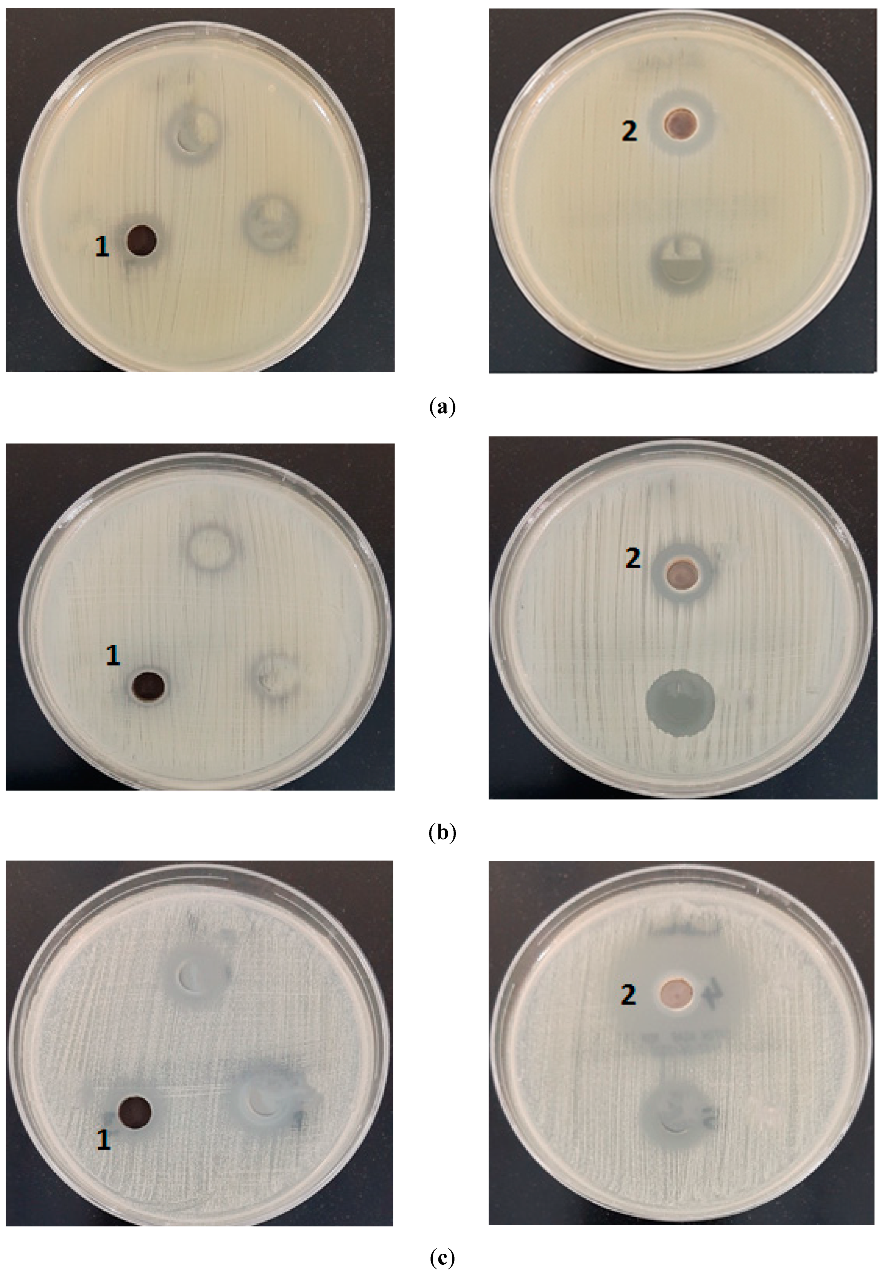
4. Biological Activity

Antimicrobial activities were evaluated on different bacterial and fungal strains, as mentioned in Section 2.5. The data are as presented in Table 7. From the data obtained, it was found that both of the metal oxide NPs displayed very good activity against the bacterial strains and fungal strains; we can clearly notice that manganese oxide NPs had better antimicrobial activity against Gram-negative than Gram-positive bacteria, but it only had an effect on one strain of Gram-positive bacteria (staphylococcus aureus) (Figure 9). Candida albicans, as seen in Figure 9, proved more sensitive to CdO than the extractor, with a significantly higher zone of inhibition than the other bacterial and fungal strains. When our findings were compared with previously published data, it was found that the novel metal oxides had an excellent activity.

5. Conclusions

In summary, manganese oxide nanoparticles and cadmium oxide nanoparticles with orthorhombic and cubic structures were synthesized by calcination at 550 °C and 450 °C for 4 h. From the XRD, scanning electron microscope (SEM), and transmission electron microscope (TEM) data, the nanoparticle sizes were ~42 and 58 nm for MnO and CdO NPs, respectively. The advantages of the calcination method include that it is convenient for the synthesis of CdO nanoparticles in normal laboratory conditions, low cost, and high purity nanosized products. The synthesized MnO and CdO NPs demonstrated excellent antibacterial and antifungal activities.

Author Contributions

M.S.A.-F. (Supervision, writing—original draft preparation, review and editing), N.A. (Review and editing), R.O.A. (Writing—original draft preparation) and G.A.A. (Writing—review and editing). All authors have read and agreed to the published version of the manuscript.

Funding

This research received no external funding.

Institutional Review Board Statement

Not applicable.

Informed Consent Statement

Not applicable.

Data Availability Statement

Not applicable.

Acknowledgments

The researchers would like to thank the Deanship of Scientific Research, Qassim University for funding the publication of this project.

Conflicts of Interest

The authors declare no conflict of interest.

References

  1. López–Serrano, A.; Muñoz–Olivas, R.; Sanz–Landaluze, J.; Olasagasti, M.; Rainieri, S.; Cámara, C. Comparison of bioconcen-tration of ionic silver and silver nanoparticles in zebrafish eleutheroembryos. Environ. Pollut. 2014, 191, 207–214. [Google Scholar] [CrossRef] [PubMed]
  2. Burda, C.; Chen, X.; Narayanan, R.; El-Sayed, M.A. Chemistry and properties of nanocrystals of different shapes. Chem. Rev. 2005, 105, 1025–1102. [Google Scholar] [CrossRef] [PubMed]
  3. Athar, T.; Topnani, N.; Hakeem, A.; Ahmed, W. Synthesis and characterization of MnO2 and CdO nanoparticles. Adv. Sci. Lett. 2012, 5, 39–42. [Google Scholar] [CrossRef]
  4. Marouzi, S.; Sabouri, Z.; Darroudi, M. Greener Synthesis and medical applications of metal oxide nanoparticles. J. ceramint. 2021, 47, 19632–19650. [Google Scholar] [CrossRef]
  5. Xiao, W.; Wang, D.; Lou, X.W. Shape–controlled synthesis of MnO2 nanostructures with enhanced electrocatalytic activity for oxygen reduction. J. Phys. Chem. 2010, 114, 1694–1700. [Google Scholar] [CrossRef]
  6. Xu, G.; Xu, Y.; Sun, H.; Fu, F.; Zheng, X.; Taoli, J.; Yang, S.; Sun, S. Facile synthesis of porous MnO/C nanotubes as a high capacity anode material for lithium ion batteries. Chem. Comm. 2012, 48, 8502–8504. [Google Scholar] [CrossRef]
  7. Yan, D.; Yan, P.; Yue, G.; Chang, J.; Yang, Q.; Qu, D.; Geng, Z.; Chen, J.; Zang, G.; Zhuo, R. Self-assembled flower- like hier-archical spheres and nanobelts of manganese oxide by hydrothermal method and morphology control of them. Chem.Phys. Lett. 2007, 440, 134–138. [Google Scholar] [CrossRef]
  8. Wang, X.; Li, Y. Synthesis formation mechanism of manganese dioxide nanowires/nanorods. Chem. 2003, 9, 300–306. [Google Scholar] [CrossRef]
  9. Ching, S.; Welch, E.; Hughes, S.; Bahadoor, A.; Suib, S. Nonaqueous sol-gel syntheses of microporous manganese oxides. Chem. Mater. 2002, 14, 1292–1299. [Google Scholar] [CrossRef]
  10. D’Souza, L.; Richards, R. Synthesis of metal-oxide nanoparticles: Liquid–solid transformations. In Synthesis, Properties, and Applications of Oxide Nanomaterials; Rodriguez, J.A., Fernández-García, M., Eds.; International University Bremen: Bremen, Germany, 2007; pp. 81–117. [Google Scholar]
  11. Suslick, K.S.; Choe, S.B.; Cichowlas, A.A.; Grinstaff, M.W. Sonochemical synthesis of amorphous iron. Nature 1991, 353, 414–416. [Google Scholar] [CrossRef]
  12. Askarinejad, A.; Morsali, A. Syntheses and Charactrization of CdCO3 and CdO Nanoparticles by Using Sonochemical Method. Mater. Lett. 2008, 62, 478–482. [Google Scholar] [CrossRef]
  13. Gulino, A.; Castelli, F.; Dapporto, P.; Rossi, P.; Fragalà, I. Synthesis and Characterization of Thin Films of Cadmium Oxide. Chem. Mater. 2002, 14, 704–709. [Google Scholar] [CrossRef]
  14. Mohamed, M.; Halawy, S. Non-isothermal decomposition of cadmium itaconate monohydrate in different atmospheres. J. Anal. Appl. Pyrolysis 2002, 65, 287–300. [Google Scholar] [CrossRef]
  15. Ni, X.; Li, M.J.; Jiao, D.; Huang, J.; Zhang, X. Microwave characteristics of Co/TiO2 nanocomposites prepared by mechanochemical synthesis. J. Alloys Compd. 2009, 468, 386–391. [Google Scholar] [CrossRef]
  16. Xu, X.; Tang, J.; Nishimura, T.; Hao, L. Synthesis of Ca–a–SiAlON phosphors by a mechanochemical activation route. Acta Mater. 2011, 59, 1570–1576. [Google Scholar] [CrossRef]
  17. Anner, G.E. Physical Vapor Deposition; Sputtering. In Planar Processing Primer; Springer: Dordrecht, The Netherlands, 1990. [Google Scholar] [CrossRef]
  18. Swain, A.K. Review on green synthesis of silver nanoparticles by physical, chemical and biological methods. Int. J. Sci. Eng. Res 2016, 7, 551–554. [Google Scholar] [CrossRef]
  19. Uskoković, V.; Drofenik, M. Synthesis of materials within reverse micelles. Surf. Rev. Lett. 2005, 12, 239–277. [Google Scholar] [CrossRef]
  20. Kumar, D.S.; Kumar, B.J.; Mahesh, H.M. Quantum nanostructures (QDs): An overview. Syn. Inorg. Nanomater. 2018, 59–88.–88. [Google Scholar] [CrossRef]
  21. Thiagarajan, S.; Tsai, T.H.; Chen, S.M. 099. Electrochemical Fabrication of Nano Manganese Oxide Modified Electrode for Detection of H2O2. Int. J. Electrochem. Sci. 2011, 6, 2235–2245. [Google Scholar]
  22. Li, J.; Wang, Y.J.; Zou, B.S.; Wu, X.C.; Lin, J.G.; Guo, L.; Li, Q.S. Magnetic properties of nanostructured Mn oxide particles. Appl. Phys. Lett. 1997, 70, 3047–3049. [Google Scholar] [CrossRef]
  23. Shchukin, D.G.; Sviridov, D.V.; Kulak, A.I. Integrating photoelectrochemical semiconductor sensor for sulphide ion deter-mination. Sens. Actuators B Chem. 2001, 76, 556–559. [Google Scholar] [CrossRef]
  24. Dong, W.; Zhu, C. Optical properties of surface-modified CdO nanoparticles. Opt. Mater. 2003, 22, 227–233. [Google Scholar] [CrossRef]
  25. Pandey, B.K.; Shahi, A.K.; Gopal, R. Optical and electrical transport properties of MnO nanoparticles. Mater. Focus. 2013, 2, 221–226. [Google Scholar] [CrossRef]
  26. Ould-Ely, T.; Prieto-Centurion, D.; Kumar, A.; Guo, W.; Knowles, W.V.; Asokan, S.; Wong, M.S.; Rusakova, I.; Luüttge, A.; Whitmire, K.H. Manganese (II) oxide nanohexapods: Insight into controlling the form of nanocrystals. Chem. Mater. 2006, 18, 1821–1829. [Google Scholar] [CrossRef]
  27. Thackeray, M.; Kock, A.; de Piocciotto, L.; Pistoia, G. Synthesis and Characterization of γ-MnO2 from LiMn2O4. J. Power Sources 1989, 26, 355–363. [Google Scholar] [CrossRef]
  28. Kaushik, A.; Jayant, R.; Sagar, V.; Nair, M. The Potential of Magneto-Electronic Nanocarriers Drug Delivery. Expert Opin. Drug Deliv. 2014, 11, 1635–1646. [Google Scholar] [CrossRef]
  29. Chen, T.; Valentin, O. Controllable Catalysis with Nanoparticles: Bimetallic Alloy Systems and Surface Adsorbates. ACS Catal. 2016, 6, 4025–4033. [Google Scholar] [CrossRef] [Green Version]
  30. Lee, W.J.; Jeong, S.; Lee, H.; Kim, B.J.; An, K.H.; Park, Y.K.; Jung, S.C. Facile synthesis of iron-ruthenium bimetallic oxide nanoparticles on carbon nanotube composites by liquid phase plasma method for supercapacitor. Korean J Chem Eng. 2017, 34, 2993–2998. [Google Scholar] [CrossRef]
  31. Saxena, S.; Saxena, U. Development of Bimetal Oxide Doped Multifunctifunctional Polymer nanocomposite for water treat-ment. Int. Nano Lett. 2016, 6, 223–234. [Google Scholar] [CrossRef] [Green Version]
  32. Kumar, R.; Gokulakrishnan, N.; Kumar, R.; Krishna, V.M.; Saravanan, A.; Supriya, S.; Somanathan, T. Can Be a Bimetal Oxide ZnO-MgO Nanoparticles Anticancer Drug Carrier and Deliver Doxorubicin Adsorption/Release Study. J. Nanosci. Nanotechnol. 2015, 15, 1543–1553. [Google Scholar] [CrossRef]
  33. Shaukat, A.; Anwar, H.; Mahmood, A.; Hussain, G.; Rasul, A.; Ijaz, M.; Faisal, M.; Ibrahim, M.; Ali, A. Synthesis cum char-acterization of MgO and MnO nanoparticles and their assessment as antidiabetic and antioxidative agents in diabetic rat model. Physica B Condens. Matter 2021, 602, 412–570. [Google Scholar] [CrossRef]
  34. Somasundaram, G.; Rajan, J.; Sangaiya, P. Dilip, Hydrothermal synthesis of CdO nanoparticles for photocatalytic and anti-microbial activities. Results Materials 2019, 4, 100044. [Google Scholar] [CrossRef]
  35. Su, L.M.; Grote, N.; Schmitt, F. Diffuse Planar InP Biopolar Junction Transistor with a Cadmium Oxide Film Emitter. Electron. Lett. 1985, 20, 716–717. [Google Scholar] [CrossRef]
  36. Su, L.M.; Grote, N.; Schmitt, F. Diffused planar InP bipolar transistor with a cadmium oxide film emitter. Electron. Lett. 1984, 20, 716–717. [Google Scholar] [CrossRef]
  37. Ocampo, I.; Ferandez, A.; Sabastian, P. Low Resistivity Transparent Conducting Thin Film Deposited by DC Reactive Magnetron Sputtering at Room Temperature. Semicond. Sci. Technol. 1993, 8, 750. [Google Scholar] [CrossRef]
  38. Benko, F.; Koffyberg, F. Quantum efficiency and Optical Transition of CdO Photoanodes. Solid State Commun. 1986, 57, 901–990. [Google Scholar] [CrossRef]
  39. Gurumugan, K.; Mangalarag, D.; Narayandass, A.; Sekar, K.; Vallabham, C. Characterization of Transparent conducting cdO films Deposited by Spray Pyrolysis. Semicond. Sci. Technol. 1994, 9, 1827. [Google Scholar] [CrossRef]
  40. Xiangfeng, C.; Xingqin, L.; Guangyao, M. Effects of CdO dopant on The Gas Sensitivity Properties of ZnFe2O4 Semi-conductors. Sens. Actuators B Chem. 1995, 65, 64–67. [Google Scholar] [CrossRef]
  41. Rane, Y.; Raghuwanshi, M.; Koli, R.; Gosavi, S.; Deshpande, N. Visible-light assisted CdO nanowires photocatalyst for toxic dye degradation studies. Optic. 2019, 133, 535–544. [Google Scholar] [CrossRef]
  42. Sivakumar, S.; Venkatesan, A.; Soundhirarajan, P.; Khatiwada, C. Synthesis, characterizations and anti-bacterial activities of pure and Ag Doped CdO Nanoparticles by Chemical Method. Spectrochim. Acta A Mol. Biomol. Spectrosc. 2014, 136, 1751–1759. [Google Scholar] [CrossRef]
  43. Hendrawan, H.; Khoerunnisa, F.; Sonjaya, Y.; Putri, A. Poly (vinyl alcohol)/glutaraldehyde/Premna oblongifolia merr extract hydrogel for controlled-release and water absorption application. IOP Conf. Ser.: Mater. Sci. Eng. 2019, 509, 012048. [Google Scholar] [CrossRef]
  44. Nagarkar, R.; Patel, J. Polyvinyl Alcohol: A Comprehensive Study. ASPS 2019, 4, 34–44. [Google Scholar]
  45. Hernandez, G.; Ricardo, M.; Morales, P.; Sanchez, M. Polyvinyl alcohol based-drug delivery system for cancer treatment. Int. J. Pharm. 2021, 600, 120478. [Google Scholar] [CrossRef] [PubMed]
  46. Al-Fakeh, M.; Alsaedi, R. Synthesis, Characterization, and Antimicrobial Activity of CoO Nanoparticles from a Co(II) Complex Derived from Polyvinyl Alcohol and Aminobenzoic Acid Derivative. Sci. World J. 2021, 2021, 1–11. [Google Scholar] [CrossRef] [PubMed]
  47. Un Nisa, Z.; Tashfeen, A. Para-Aminobenzoic Acid-A Substrate of Immense Significance. Mini-Rev. Org. Chem. 2020, 6, 686–700. [Google Scholar] [CrossRef]
  48. Roik, N.; Belyakova, L. IR Spectroscopy, X-ray Diffraction and Thermal Analysis Studies of Solid “β-Cyclodextrin—Pa-ra-Aminobenzoic Acid” Inclusion Complex. Phys. Chem. Solid State. 2011, 12, 168–173. [Google Scholar]
  49. Aly, A.A.; Ghandour, M.; Alfakeh, M.S. Synthesis and characterization of transition metal coordination polymers derived from 1, 4-benzenedicarboxylate and certain azoles. Turk. J. Chem. 2012, 36, 69–79. [Google Scholar]
  50. Siddaiah, T.; Ojha, P.; Kumar, G.; Ramu, C. Structural, Optical and Thermal Characterizations of PVA/MAA: EA Polyblend Films. Mater. Res. 2018, 21, e20170987. [Google Scholar] [CrossRef] [Green Version]
  51. Gainar, A.; Stevens, J.; Suljoti, E.; Xiao, J.; Gonlak, R.; Aziz, E.; Schrorder, S. The Structure of p-Aminobenzoic Acid in Water: Studies Combining UV-Vis, NEXAFS and RIXS Spectroscopies. J. Phys. Conf. Ser. 2016, 712, 012034. [Google Scholar] [CrossRef] [Green Version]
  52. Al-Fakeh, M.; Allazzam, G.; Yarkandi, N. Ni(II), Cu(II), Mn(II), and Fe(II) Metal Complexes Containing 1,3-Bis(diphenylphosphino)propane and Pyridine Derivative: Synthesis, Characterization, and Antimicrobial Activity. Int. J. Biomater. 2021, 2021, 1–12. [Google Scholar] [CrossRef]
  53. OBA, A.; Osowole, A.A.; Malumi, E.O.; Wakil, S.M. Synthesis and characterization of transition metal coordination polymers derived from 1, 4-benzenedicarboxylate and certain azoles. Rev. J. Chem. 2015, 36, 69–79. [Google Scholar]
  54. Coats, A.; Redfern, J. Kinetic Parameters from Thermogravimetric Data. Nature 1964, 201, 68–69. [Google Scholar] [CrossRef]
Scheme 1. Synthesis of metal complexes. M = Mn(II) and Cd(II).
Coatings 12 00215 sch001
Figure 1. Coordination geometry around (a) Mn(II) complex and (b) Cd(II) complex.
Coatings 12 00215 g001
Figure 2. FT–IR of (a) Mn(II) complex and (b) Cd(II) complex.
Coatings 12 00215 g002
Figure 3. UV–visible of Mn(II) oxide nanoparticles.
Coatings 12 00215 g003
Figure 4. Thermograph of Mn(II) Complex.
Coatings 12 00215 g004
Figure 5. Coats–Redfern of Mn(II) and Cd(II) complexes.
Coatings 12 00215 g005
Figure 6. Impact of kinetic compensation of Mn(II) complex.
Coatings 12 00215 g006
Figure 7. XRD of (a) MnO nanoparticles and (b) CdO nanoparticles.
Coatings 12 00215 g007
Figure 8. (a) SEM image of MnO nanoparticles; (b) SEM image of CdO nanoparticles; (c) TEM image of MnO nanoparticles; (d) TEM image of CdO nanoparticles.
Coatings 12 00215 g008aCoatings 12 00215 g008b
Figure 9. Microbiological effect of MnO NPs and CdO NPs on (a) E. coli, (b) staphylococcus aureus, and (c) Candida albicans.
Coatings 12 00215 g009
Table 1. Color, elemental analysis, and decomposition point of the mixed ligand complexes.
CompoundsM.F
(M.Wt)
ColorFound (Calcd. %)m.p. °C
(Decom.)
Λm
Scm2 mol−1
CHN
[Mn(PVA)(P-ABA)(H2O)3]·H2O
1
C10H19N2MnO8 (350.13)Light brown35.84
(34.30)
6.05
(5.46)
8.09
(7.99)
239–24158
[Cd(PVA)(P-ABA)(H2O)3]
2
C10H17N2CdO7 (389.58)White31.07
(30.83)
4.78
(4.39)
7.63
(7.18)
290–29236
Table 2. FTIR patterns of pure PVA, P–ABA, and their complexes.
AssignmentPVAP-ABAMn(II) ComplexCd(II) Complex
VO-H3400-32503280
VO-H water--33373339
VN-H2-323432433220
VC=N-164016201650
VC-OC1150-11781180
M-O--522515
M-N--430422
Table 3. Electronic spectra of PVA, P-AMB, and their complexes.
CompoundWave Number cm−1Type of Transition
Mn(II) complex19,685d-d transition
35,971n→π*
41,841π→π*
Cd(II) complex36,363n→π*
42,918π→π*
Table 4. Kinetic data of the thermal degradation for Mn(II) complex and Cd(II) complex.
ComplexesStepCoats–Redfern
nrEZ × 102
[Mn(PVA)(P-ABA)(H2O)3]·H2O2nd0.000.993055.343.73
0.330.998864.364.34
0.500.999369.524.69
0.660.999573.604.97
1.000.999985.035.76
2.001.000124.028.49
[Cd(PVA)(P-ABA)(H2O)3]2nd0.000.980821.901.47
0.330.985726.411.78
0.500.988129.111.96
0.660.990831.742.14
1.000.993538.182.50
2.000.997662.454.25
Table 5. Thermodynamic parameters for the compounds.
CompoundStep∆S*∆H*∆G*
Mn(II) complex2nd−249.6058.7184.22
−250.3162.25188.13
−250.4971.67197.64
−250.5676.91202.95
−250.6282.14208.24
−250.75138.24264.25
Cd(II) complex2nd−248.2842.91160.65
−248.48129.61246.80
−248.91161.36278.26
−249.71165.17281.69
−250.33182.58298.90
−251.50192.01308.24
∆H*, ∆G* in Kj mol−1 and ∆S* in Kj mol−1 K−1
Table 6. X-ray diffraction data of MnO and CdO NPs.
ParametersManganese Oxide (NPS)Cadmium Oxide (NPS)
Empirical formulaMnOCdO
Formula weight70.92128.40
Crystal systemOrthorhombic Cubic
a (Å)5.6304.694
b (Å)5.6444.694
c (Å)7.9724.694
α (°)90.0090.00
β (°)90.0090.00
γ (°)90.0090.00
Volume of unit cell (Å3)253.35103.46
Particle size (nm)4258
Table 7. The antimicrobial properties of the samples.
Tested Organisms1
MnO
2
CdO
Zone of Inhibition in mm
Staphylococcus aureus1216
Staphylococcus epidermidis-20
Enterococcus faecalis-14
Escherichia coli1216
Pseudomonas aeruginosa1114
Candida albicans1530
Aspergillus fumigatus1223
Aspergillus flavus1215
Publisher’s Note: MDPI stays neutral with regard to jurisdictional claims in published maps and institutional affiliations.

