Article

Physical Vapor Deposition Technology in Personal Protective Equipment Production: Improved Antibacterial and Hydrophobic Character of Textiles

1
Department of Mechanical Engineering, CEMMPRE, Centre for Mechanical Engineering, Materials and Processes, University of Coimbra, Rua Luis Reis Santos, 3030-788 Coimbra, Portugal
2
CEB, Centre of Biological Engineering, LIBRO—Laboratório de Investigação em Biofilmes Rosário Oliveira, University of Minho, Campus of Gualtar, 4710-057 Braga, Portugal
3
LABBELS–Associate Laboratory, Braga/Guimarães, Portugal
4
Laboratory for Wear, Testing & Materials, Instituto Pedro Nunes, Rua Pedro Nunes, 3030-199 Coimbra, Portugal
*
Author to whom correspondence should be addressed.
Coatings 2022, 12(10), 1399; https://doi.org/10.3390/coatings12101399
Submission received: 30 August 2022 / Revised: 20 September 2022 / Accepted: 21 September 2022 / Published: 25 September 2022
(This article belongs to the Special Issue Trends in Coatings and Surface Technology)

Abstract

:
Personal protective equipment (PPE) has been adapted as biological threats have emerged, such as increasingly drug-resistant bacteria and the emergence of new viruses such as COVID-19. PPE must be increasingly resilient to prevent the proliferation of pathogens, but using sustainable raw materials and environmentally friendly technologies. The aim of this study is to show a new way of modifying the surface of various types of fabrics to enable their efficient use as PPE. The Ag/DLC coating was successfully deposited by sputtering onto several types of textiles using different chemical compositions of Ag/DLC (0, 8, 10, and 12Ag). As a crucial parameter, wettability was evaluated, showing that silver addition increases the hydrophobicity character of the coated fabrics, namely in cotton, changing from hydrophilic to hydrophobic. Antibacterial activity and cytotoxicity were evaluated on all coatings, revealing that they are efficient in eliminating the spread of bacteria (Staphylococcus aureus and Klebsiella pneumoniae) and pose no risk to the human body. The results presented here are promising in protecting healthcare workers, with the next steps being to study the efficiency of these coatings against viruses. In addition, this study reveals an opportunity to use sustainable fabrics, such as cotton, with high efficiency in protection against pathogens, instead of synthetic fiber textiles.

1. Introduction

Healthcare-associated infections (HAIs) have long been seen as a public health threat. It is estimated that 3.8 million people acquire HAIs/year in EU hospitals [1], leading to about 30,000 deaths/year [2]. Such infections cost millions of euros to health care systems. The number of deaths increased in the last years with COVID-19, which reveals the importance of the protection against any pathogen (viruses, fungus or bacteria) of the patients and professionals in healthcare systems.
Alongside surface disinfection, the use of technical textiles in a healthcare environment is a necessary tool to avoid the dissemination of the more resistant pathogens, since the capacity of protection the healthcare workers (HCWs) against the pathogens is attributed to this type of fabric. The necessity for medical textiles became more noticeable during the COVID-19 pandemic, where the personal protective equipment (PPEs) available, such as masks, gowns or coveralls [3,4], was critical to protect HCWs.
A virus can spread via aerosols generated by coughing and sneezing in the air, by vectors such as insects, or by the transmission of body fluids such as saliva, blood, or semen [4]. In particular, for respiratory pathogens, they can be transmitted via droplets coming from an infected person when talking, sneezing, coughing or exhaling [4]. These droplets can spread the viruses through airborne, contact and fomites [5]. For this reason, microbial contamination is of great concern for the production of textiles used in hospitals.
Due to their large surface area and ability to retain moisture, textiles promote the growth of microorganisms such as bacteria and fungi, which can be found almost everywhere and are able to quickly multiply under certain circumstances. The growth of microorganisms on textiles has several undesirable effects: the generation of unpleasant odours, the diminishing of mechanical strength, stains, discoloration, and the increased chance of user contamination [6].
Another problem commonly associated with the use of textiles is the transmission of liquid through them by penetration and permeation, which involves the flow of gas, vapor, or liquid through a porous material, and the diffusion of gas or vapor through a porous material, respectively. Pathogens are larger in size than gas and vapor molecules and are believed to penetrate and not permeate materials. The coronavirus, which causes COVID-19, has been found to be transmitted via aerosols, being able to penetrate some textiles [4,7].
To provide textiles with antimicrobial ability, there are different approaches being studied and applied, such as the inclusion of antimicrobial compounds in the polymeric fibres that can leach from the polymeric matrix, grafting of certain moieties onto the polymer surface or the physical modification of fibres surface [6,8]. Some studies have reported that just by modifying the surface properties of a material, such as free energy, polarity or topography, it is possible to decrease the bacterial adhesion to the surface during the initial stage of the biofilm formation process, without using chemical antimicrobial agents. Those modifications may create new functional groups and/or change the surface roughness [6]. Also, fluid repellent finishes can be used to create a barrier which prevents adsorbed fluids from penetrating fabrics [4].
Any antimicrobial treatment performed on a textile needs to satisfy certain requirements besides being efficient against microorganisms. The main challenge is for it not to be cytotoxic or cause allergies, irritations and sensitization [6,8]. Moreover, antimicrobial treatments performed on textiles also need to be suitable for textile processing, present durability to laundering, dry cleaning and hot pressing, be environmentally friendly and should not damage the textile quality or appearance [6,9].
The antibacterial property of surfaces is acquired by using a bioactive agent, namely, metallic nanoparticles (NPs), such as Ag, nano-ZnO, nano-TiO2, Cu, SiO2, or Au [4,10,11,12,13]. These NPs can be applied using various techniques such as spray, sonochemical, sol-gel, foam coating, hot-melt extrusion, or spray-dry techniques [11,13].
Another method used to modify surfaces by forming a thin film is a physical vapor deposition (PVD) process, of which there are different types, such as cathode arc deposition, electron beam physical vapor deposition, evaporative deposition, sputtering or ion plating [14,15,16]. Among the basic sputtering deposition techniques, magnetron sputtering (MS) is the most advantageous. MS uses powerful magnets confining the plasma to the region closest to the substrate, which greatly improves deposition because it maintains a higher ion density making the electron/gas molecule collision process much more efficient [17]. Also, MS allows to deposit metallic or non-metallic films on various types of substrate surfaces such as textiles (cotton, wool, silk…), metal or ceramics by controlling the parameters of the sputtering process and the target materials [18]. Due to the combination of metallic nanoparticles and a matrix film, the coatings created through this process are characterized by a large surface area with better functionality and durability. In addition, this technique allows the combination of oxide, metallic and composite films to obtain different characteristics, such as antibacterial and hydrophobic properties, which has attracted great interest for medical applications [19]. Textiles coated with copper (Cu) and titanium dioxide (TiO2) films have demonstrated UV resistance and antibacterial characteristics. Also, deposition of silver (Ag) films on textiles has led to an improvement in antibacterial activity [20].
In addition to the variety of types of coatings, PVD technology has important advantages over those mentioned above to incorporate NPs modifying the surface of the substrates. In the case of polymer fibers, the deposition may take place at low temperatures and the coatings have strong adhesion to the fibrous substrates [21]. Moreover, this method is considered more environmentally friendly than other techniques since it does not use chemical agents during the process.
To ensure the controlled release of NPs, thereby increasing the lifetime of the antibacterial capacity of the substrate, they are usually deposited on a matrix. As well as being biocompatible, diamond-like carbon (DLC), an amorphous carbon-based thin film grown by PVD, exhibits high properties such as high hardness, hydrophobicity, wear resistance, chemical stability, gas barrier properties, anti-burn characteristics, infrared permeability, biocompatibility, and a low coefficient of friction, which makes it preferable as a matrix for incorporating metallic NPs as biocidal agents for use in PPEs that come into contact with skin [22,23,24,25,26,27,28,29].
Depending on their functionality and purpose, a huge variety of textiles can be used in such medical protective equipment. In the case of disposable medical textiles like surgical masks or surgical gowns, they are usually made of synthetic fibres because of their superior liquid barrier properties. The non-woven fabric typically made by polypropylene (PP), known as TNT, is very popular for medical clothing, in particular for disposable medical clothing, because of its comparatively low cost and speed of production, as well as its excellent levels of sterility and infection control, which are critical in such applications [3,4,30].
In the case of scrubs, masks or bedding textiles and drapes present in healthcare facilities, woven fabrics typically made from cotton or polyester/cotton blends are commonly used [4,30]. When compared with TNTs, these types of textiles have weaker barrier properties against liquids and bacteria, although they normally provide more wearer comfort to the user and can endure several washing/cleaning processes, giving them a reusability property that TNTs usually do not have.
As mentioned above, the COVID-19 pandemic highlighted the limitations of PPE. The manufacture of sterile nonwovens is not sufficient to cover demands after this period. In addition, nonwoven fabrics are typically made of propylene with a bond-melt-blown-spun-bond, which negatively impacts the environment. Fabrics, such as cotton or polyester/cotton blends, require more treatment to gain the proper properties to protect healthcare workers [4]. Given the environmental issues and sustainability of raw materials, it is crucial to continue researching technologies and new products for PPEs to be more efficient and more eco-friendly alternatives such as natural fibres. Since the recycling process of medical textiles has not yet been implemented, the search for more efficient and reused fabrics to control the spread of pathogens is still ongoing, as more resistant bacteria and viruses are emerging.
Several studies have been carried out which apply PVD to textiles to improve their qualities according to commercial demands [17,21]. However, as far we know, the application of DLC with silver to promote antibacterial properties in textiles has not yet been tested. Moreover, it is known that the type of substrate is an important parameter that needs to be taken into account to optimise the deposition parameters. Our group has considerable experience in using this kind of coating on hard surfaces, but this is the first time we transfer their knowledge to textiles [28,29,31,32,33,34,35,36,37]. Using different fabrics as substrates it is possible to compare how the finishing works with each type of fabric.
In the present study, fabrics commonly used in PPEs (cotton, jersey (cotton + elastane) and TNT) were chosen and coated using MS with diamond-like carbon doped with silver (Ag/DLC). The coatings deposited on the different fabrics, which had varying Ag contents, were characterized with respect to their chemical composition, morphology and wettability. The toxicity of the coatings as well as their antimicrobial activity were also evaluated. The combination of results allows for a comparison of the efficiency of the MS process in the deposition of different types of fabrics and its use as PPE.

2. Materials and Methods

2.1. Coatings Preparation

The Ag/DLC coatings were deposited by dual target MS onto four substrates: silicon (15 × 15 mm2), cotton fabric (100 × 20 mm2), jersey format fabric (100 × 20 mm2) and Polypropylene TNT (100 × 20 mm2). Two targets were used, one of graphite and one of silver, with the dimensions of 150 × 150 × 10 mm3 and with a purity of 99.95%.
Before the deposition process, the Si, cotton, jersey format and TNT substrates were cleaned in an ultrasonic bath of ethanol (96%), for 10 min. After the ultrasonic bath, the samples were dried using a dryer and were then glued to the substrate holder using glue tape, except for the Si substrates, for which silver glue was used.
After introducing the substrate holder into the vacuum chamber, an air pumping was performed until a pressure of 10−5 Pa was reached. Then, before starting the deposition, the targets were submitted to a cleaning process by applying a direct current (DC) to the targets (one at a time) for 5 min, so that any contamination present on the surface of the targets could be eliminated. During the etching cleaning process, the substrate holder was protected by placing a stainless-steel plate between it and the targets, in order to prevent any kind of cross-contamination.
The depositions were performed in an argon (Ar) atmosphere (21.9 sccm), with a working pressure of 10−1 Pa. A constant rotation of 23.5 rpm was applied to the substrate holder to maintain the homogeneity of the chemical composition and thickness of the coatings. The depositions were performed with an applied power of 1500 W to the C target and, in the case of the Ag target, the power applied was between 70 W and 155 W, according to the required Ag at. %. The deposition working pressure was also varied in the depositions performed between 1.0 Pa and 0.8 Pa. The deposition time was maintained for all the samples in order to have a similar thickness in the coatings, estimated to be ~50 nm. Some more experimental details are depicted in Table 1, such as the deposition time, deposition rate or power density (JC) from each target. The samples were labelled according to the amount of silver, e.g., Ag/DLC with 0% of Ag content was named 0Ag.

2.2. Characterization Analysis

The surface morphology was examined by Scanning electron microscope (SEM), using a field emission scanning electron microscope (FESEM) ZEISS MERLIN Compact/VP Compact. This equipment was an energy dispersive X-ray spectrometer (EDS) Oxford, X-MAXN to evaluate the chemical composition of the films. The measurements were performed on the samples surface at a magnification of 50 k and with an acceleration voltage of 10 kV. The hydrophobic characteristics analysis was obtained by measuring the contact angles between the substrates and a water droplet placed on the surface the uncoated and coated substrates, using an automated contact angle measurement apparatus (OCA 15 Plus; Dataphysics, Filderstadt, Germany). If the contact angle θ ≥ 90°, the sample is considered to have hydrophobic properties.
The cytotoxicity tests were performed using a colorimetric method to verify if the fabrics coated with Ag/DLC cause toxicity in the animal cells in order to prevent any toxic effect, as the main application of these textile will be for the use of PPEs (e.g., masks). The cytotoxicity was tested using fibroblasts 3T3 (CCL-163) obtained from the American Type Cell Collection (ATCC) by an indirect test performed with the saline AS used in the analysis of the ICP-OES that was in contact with fabrics substrates uncoated (virgin) and coated with Ag/DLC for 168 h, in accordance with ISO 10993-5:2009. The cytotoxicity procedure was according to [38]. The assays were performed at least three times and in triplicate.
In order to evaluate the antibacterial activity of the coated fabrics, zone of inhibition tests, adapted from a Kirby-Bauer test, were carried out to determine the diffusion of silver from the surface of the coatings. The evaluation of the antibacterial activity was performed against two bacteria, one gram-positive, Staphylococcus aureus (ATCC 6538 obtained from American Type Cell Collection) and one gram-negative Klebsiella pneumoniae (ATCC 11296). The choice of these microorganisms relates to the applicability, as S. aureus is often found on the skin and K. pneumoniae causes different types of healthcare-associated infections, including respiratory infections such as pneumonia. Initially, the inoculation of a single colony was carried out in 30 mL Tryptic soy broth (TSB, Merck, Kenilworth, NJ, USA) culture and incubated at 37 °C overnight at 120 rpm. The cell suspension obtained was adjusted to an optical density (OD) of 0.8 at 620 nm and properly diluted in culture media to 1 × 10⁸ CFU/mL. An aliquot of cellular suspension (100 μL) was spread in Tryptic Soy Agar (TSA, Merck) petri dishes. After medium solidification, the samples (previously sterilized by exposure to ±1 h to UV light) were placed separately on the top of the agar plate, placing the side with treatment in contact with the agar, and incubated for 24 h at 37 °C. After the incubation period, the inhibition zone (zone of transparent medium, which means that there is no bacteria growth) formed around the sample was photographed to record the results (images captured with Image Lab™ software). After the inhibition zone test, the samples were removed and carefully washed three times with distilled water. Samples were dehydrated by immersion in increasing ethanol concentration solutions: 70, 95 and 100% (v/v) for 10, 10 and 20 min, respectively, and placed in a sealed desiccator. Afterwards, the samples were mounted on aluminum bases with carbon tape, sputter-coated with gold and observed on a Phenom Charge Reduction Sample Holder (CRH) at 10 kV and a spot size of 3.3, with a desktop scanning electron microscope (SEM) coupled with energy-dispersive X-ray spectroscopy (EDS) analysis (Phenom ProX with EDS detector (Phenom-World BV, Eindhoven, The Netherlands)). The measurements were performed on the surface of the samples at a magnification of 5 k.

3. Results and Discussion

3.1. Chemical and Morphological Analysis

The silver percentage range was chosen keeping in mind the toxicity of this chemical element to human when in contact with skin (see Section 3.3). EDS analysis of the Si coated samples showed Ag percentages of 8 at. %, 10 at. %, and 12 at. %, in the DLC-doped thin films.
The surface of the Ag/DLC coatings deposited on the cotton, jersey and TNT fabrics are shown in Figure 1, as well as the surface of the three fabrics without the coating (virgin fabrics). The presence of AgNP on the surface was observed for all coated fabrics, with its agglomeration more easily identifiable as the amount of Ag increases. The images also show the presence of cracks, in the 10Ag-cotton and 12Ag-jersey samples. It is known that the adhesion between magnetron sputtering coatings and fabrics is better than other methods [21]. However, this adhesion depends on the sputtering process and the type of substrates, because they have different chemical and morphological properties on the surface and the porosity of the fibers is also a critical parameter [18]. In this case, all other samples have no adhesion deficiency and therefore do not depend on the deposition method or the type of substrate. Therefore, the cracks may result from the foldability and elasticity of the fabrics.

3.2. Wettability

The fluid repellent finish is a barrier which prevents the absorption of fluids into textile. Pathogens are most easily transmitted through liquids such as respiratory droplets or body fluids [4]. Therefore, the hydrophobicity of the PPE surface is of utmost importance, to prevent contamination of people wearing this protection. The wettability of the samples was evaluated and the results of the contact angle analysis of the coated fabrics are shown in Table 2. All coated samples have hydrophobic properties (contact angle θ ≥ 90°), regardless of the Ag concentration in the coating. Since the composition of the jersey shape and TNT fabrics has a specific amount of polymer structure in its uncoated state (textile), both already have a hydrophobic character, whereas the cotton is hydrophilic. Applying the coating of DLC without silver, cotton becomes hydrophobic, the jersey format maintains the contact angle value and the TNT slightly decreases the hydrophobic character. All coated textiles become less hydrophobic when some silver content is introduced into the carbon matrix. In the cotton, as the silver content increases, the coated textiles are more efficient at repelling water. For sample 12Ag, it is possible to say that all the textiles promote similar liquid barrier properties.

3.3. Cytotoxicity Evaluation

When using silver nanoparticles in biomedical applications it is essential to ascertain the toxicity that this nanomaterial can impart to the human body. The values of the nanomaterials amount, which can be toxic, are still under study because they depend on several parameters (amount, shape, capping binder, or surface charge) [39,40,41]. Since these values are not fully defined, the coatings produced in this study were evaluated regarding the cell viability using 3T3 cells.
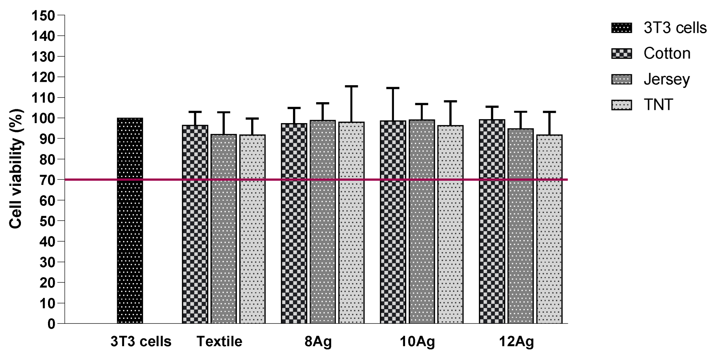
Figure 2 shows the cytotoxicity results obtained for all uncoated and Ag/DLC-coated fabrics. The percentage of viable cells above 70% confirms the non-cytotoxic activity of Ag/DLC coatings according to ISO 10993-5:2009. The viability of the cells confirms that the coated textiles are non-toxic to the cells used, even at the values of Ag released.

3.4. Antibacterial Activity

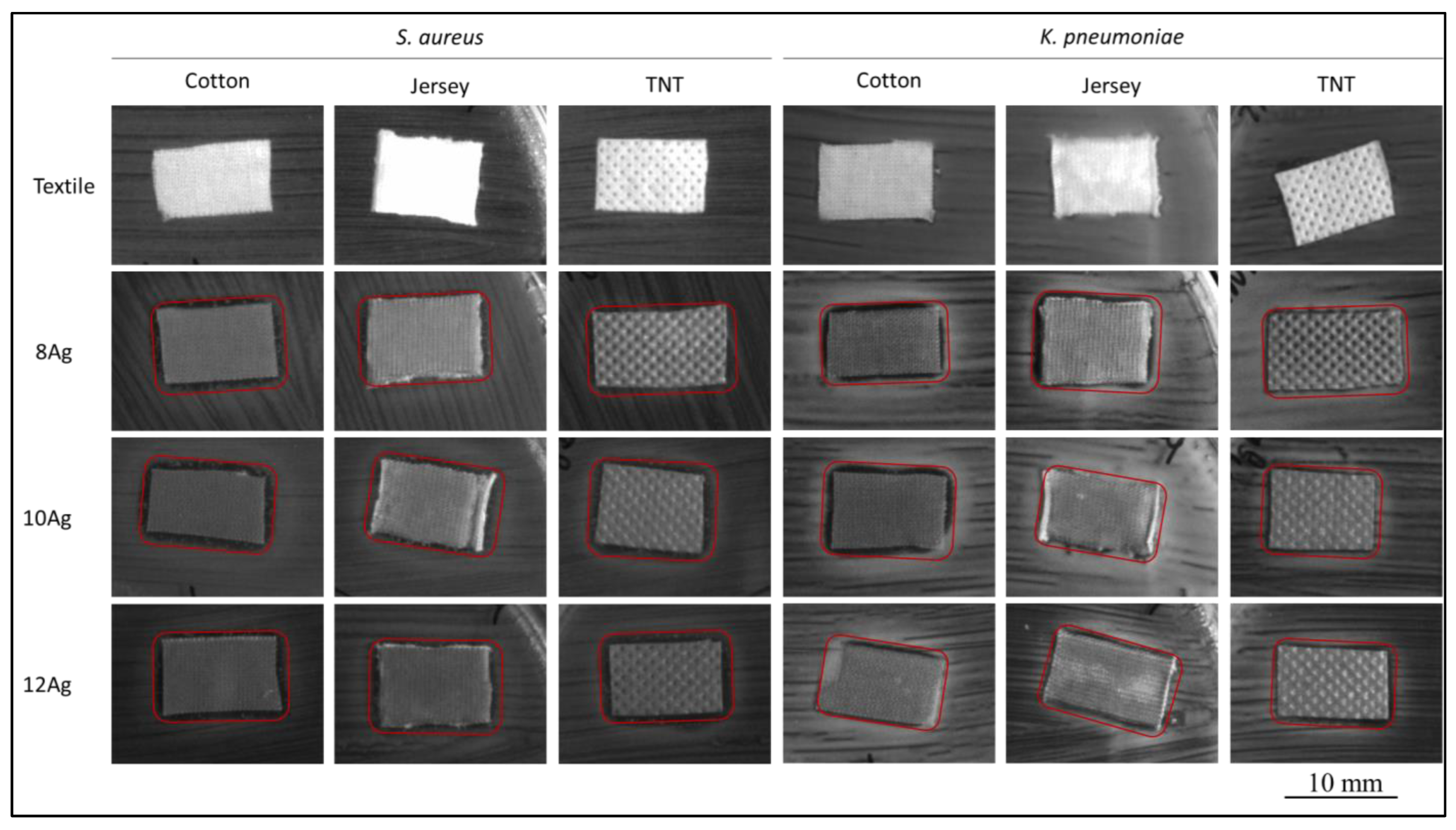
The antibacterial activity of the coatings deposited on the different types of fabric must be evaluated before these devices can be used as PPE. As mentioned in Section 2.2, the coatings were tested against two types of bacteria. The zone of inhibition (ZoI) tests showed promising results for both bacteria, S. aureus and K. pneumoniae, as shown in Figure 3. The coated fabrics achieved a ZoI around them, while the virgin fabrics showed no antibacterial activity, as expected. Silver-free DLC coatings are also known to promote bacterial growth, since it is not a bioactive agent, as shown by Carvalho et al. [36].
For the fabrics coated with the film with less Ag content (8Ag), the inhibition zones are more difficult to identify, especially for the TNT fabric for both types of bacteria (Figure 3). With an increasing amount of Ag, the inhibition zones of the coated fabrics become more easily identifiable, especially for the cases of the S. aureus bacteria. For the K. pneumoniae cases, the zones of inhibition are more irregular, however, the textiles still show antibacterial activity. The coated cotton fabric seems to acquire antibacterial activity for all the silver content thin films, whereas the other types of textiles do not show clear activity against the bacteria for all the coatings.
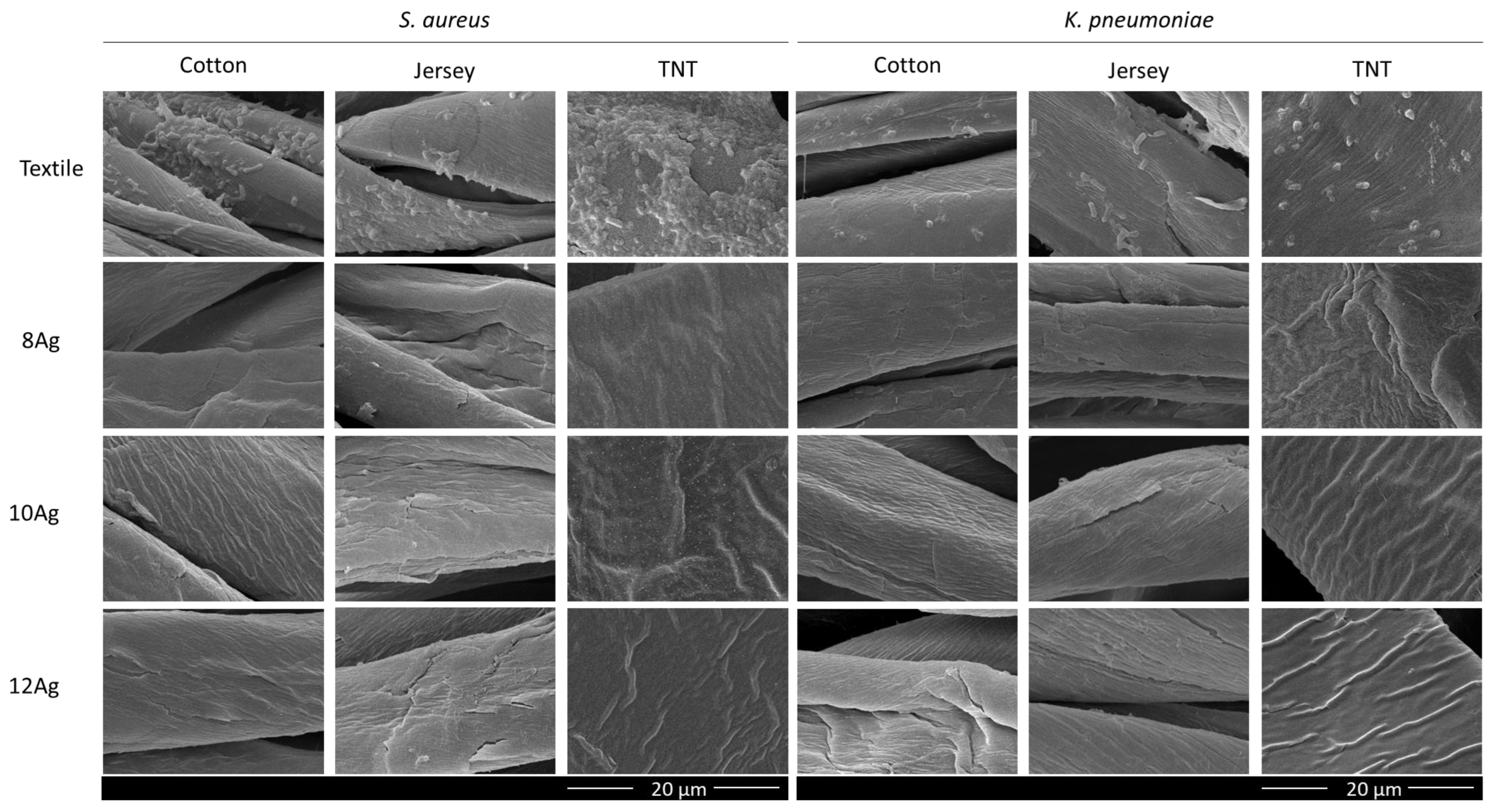
Since the inhibition zones were not very clear in some samples, it was thus not clear if the coatings promoted antibacterial features to the textiles. SEM images were obtained after the inhibition test zone (Figure 4) to clarify the antibacterial activity of coated textiles. It can be seen that the virgin fabrics are colonized by both bacteria, although K. pneumoniae is in less quantity. On the other hand, in the coated textiles, regardless of the type of bacteria and the amount of Ag, there is a complete inhibition of bacterial growth. These images confirm the results of the zone of inhibition and the bioactive nature of Ag.

4. Conclusions

Ag/DLC coatings were deposited on cotton, jersey and TNT, varying the Ag concentration between 0 and 12 at. %. The morphology of the coatings, in general, appeared to have good adhesion to the different fiber types. The use of carbon matrix in the coating was used to provide hydrophobicity to the coatings, which proved to be efficient, even with the introduction of Ag into the matrix, despite variations in the amount of Ag.
The deposited thin films do not represent a risk to the human body since they allow the 3T3 cells to be viable for at least 7 days.
To test the antimicrobial activity of the thin films deposited on the textile, the bacteria were chosen according to the environment where these textiles might be used. Respiratory infections are very common, so gram-negative K. pneumoniae was chosen, and S. aureus is very common on the skin. All Ag coatings inhibited the growth of both bacteria.
Considering that the three types of fabrics showed equivalent results in inhibiting the growth of bacteria and that cotton shows the greatest evolution in hydrophobicity as the amount of Ag increases, it can be concluded that surface modification of cotton fibers is a solution to be considered for the PPE industry, with the next steps in this research being antiviral testing and the wash resistance of the coatings.

Author Contributions

Conceptualization, F.F. and S.M.A.C.; methodology, F.F. and S.M.A.C.; formal analysis, J.A., K.M. and I.C.; investigation, J.A., K.M. and I.C.; resources, F.F. and I.C.; writing—original draft preparation, J.A., K.M., I.C. and S.M.A.C.; writing—review and editing, F.F., S.C. and S.M.A.C.; supervision, F.F. and S.C.; project administration, I.C., F.F. and S.C.; funding acquisition, I.C., F.F., S.C. and S.M.A.C. All authors have read and agreed to the published version of the manuscript.

Funding

This research is sponsored by national funds through FCT—Fundação para a Ciência e a Tecnologia, under the project UIDB/00285/2020, LA/P/0112/2020, UIDB/04469/2020 and LA/P/0029/2020. This research was also sponsored by FEDER funds through the program Centro 2020—Programa Operacional Regional do Centro under project CENTRO-01-0145-FEDER-181221, ActiveStent.

Institutional Review Board Statement

Not applicable.

Informed Consent Statement

Not applicable.

Data Availability Statement

Not applicable.

Conflicts of Interest

The authors declare no conflict of interest.

References

  1. Suetens, C.; Latour, K.; Kärki, T.; Ricchizzi, E.; Kinross, P.; Moro, M.L.; Jans, B.; Hopkins, S.; Hansen, S.; Lyytikäinen, O.; et al. Prevalence of healthcare-associated infections, estimated incidence and composite antimicrobial resistance index in acute care hospitals and long-term care facilities: Results from two European point prevalence surveys, 2016 to 2017. Eurosurveillance 2018, 23, 1800516. [Google Scholar] [CrossRef] [PubMed]
  2. Cassini, A.; Högberg, L.D.; Plachouras, D.; Quattrocchi, A.; Hoxha, A.; Simonsen, G.S.; Colomb-Cotinat, M.; Kretzschmar, M.E.; Devleesschauwer, B.; Cecchini, M.; et al. Attributable deaths and disability-adjusted life-years caused by infections with antibiotic-resistant bacteria in the EU and the European Economic Area in 2015: A population-level modelling analysis. Lancet Infect Dis. 2019, 19, 56–66. [Google Scholar] [CrossRef]
  3. Chua, M.H.; Cheng, W.; Goh, S.S.; Kong, J.; Li, B.; Lim, J.Y.C.; Mao, L.; Wang, S.; Xue, K.; Yang, L.; et al. Face Masks in the New COVID-19 Normal: Materials, Testing, and Perspectives. Research 2020, 2020, 7286735. [Google Scholar] [CrossRef] [PubMed]
  4. Karim, N.; Afroj, S.; Lloyd, K.; Oaten, L.C.; Andreeva, D.V.; Carr, C.; Farmery, A.D.; Kim, I.D.; Novoselov, K.S. Sustainable Personal Protective Clothing for Healthcare Applications: A Review. ACS Nano 2020, 14, 12313–12340. [Google Scholar] [CrossRef]
  5. O’Dowd, K.; Nair, K.M.; Forouzandeh, P.; Mathew, S.; Grant, J.; Moran, R.; Bartlett, J.; Bird, J.; Pillai, S.C. Face Masks and Respirators in the Fight against the COVID-19 Pandemic: A Review of Current Materials, Advances and Future Perspectives. Materials 2020, 13, 3363. [Google Scholar] [CrossRef]
  6. Morais, D.; Guedes, R.; Lopes, M. Antimicrobial Approaches for Textiles: From Research to Market. Materials 2016, 9, 498. [Google Scholar] [CrossRef]
  7. Guo, Z.D.; Wang, Z.Y.; Zhang, S.F.; Li, X.; Li, L.; Li, C.; Cui, Y.; Fu, R.B.; Dong, Y.Z.; Chi, X.Y.; et al. Aerosol and Surface Distribution of Severe Acute Respiratory Syndrome Coronavirus 2 in Hospital Wards, Wuhan, China, 2020. Emerg. Infect. Dis. 2020, 56, 1586. [Google Scholar] [CrossRef]
  8. Bshena, O.; Heunis, T.; Dicks, L.; Klumperman, B. Antimicrobial fibers: Therapeutic possibilities and recent advances. Future Med. Chem. 2011, 3, 1821–1847. [Google Scholar] [CrossRef]
  9. Windler, L.; Height, M.; Nowack, B. Comparative evaluation of antimicrobials for textile applications. Environ. Int. 2013, 53, 62–73. [Google Scholar] [CrossRef]
  10. Bhandari, V.; Jose, S.; Badanayak, P.; Sankaran, A.; Anandan, V. Antimicrobial Finishing of Metals, Metal Oxides, and Metal Composites on Textiles: A Systematic Review. Ind. Eng. Chem. Res. 2022, 61, 86–101. [Google Scholar] [CrossRef]
  11. Antunes, J.; Moreira, I.; Gomes, F.; Cunha, F.; Henriques, M.; Fangueiro, R. Recent Trends in Protective Textiles against Biological Threats: A Focus on Biological Warfare Agents. Polymers 2022, 14, 1599. [Google Scholar] [CrossRef] [PubMed]
  12. Andra, S.; Balu, S.; Jeevanandam, J.; Muthalagu, M. Emerging nanomaterials for antibacterial textile fabrication. Naunyn-Schmiedebergs Arch. Pharmacol. 2021, 394, 1355–1382. [Google Scholar] [CrossRef] [PubMed]
  13. Raza, Z.; Taqi, M.; Tariq, M. Antibacterial agents applied as antivirals in textile-based PPE: A narrative review. J. Text. Inst. 2022, 113, 515–526. [Google Scholar] [CrossRef]
  14. Li, G.; Liu, H.; Li, T.; Wang, J. Surface modification and functionalization of silk fibroin fibers/fabric toward high performance applications. Mater. Sci. Eng. C-Mater. Biol. Appl. 2012, 32, 627–636. [Google Scholar] [CrossRef]
  15. Wang, H.; Wei, Q.; Gao, W. Sputter Deposition of Antibacterial Nano-Silver on PLA Nonwoven Medical Dressings. AATCC Rev. 2009, 9, 34–36. [Google Scholar]
  16. Abd Jelil, R. A review of low-temperature plasma treatment of textile materials. J. Mater. Sci. 2015, 50, 5913–5943. [Google Scholar] [CrossRef]
  17. Shahidi, S.; Moazzenchi, B.; Ghoranneviss, M. A review-application of physical vapor deposition (PVD) and related methods in the textile industry. Eur. Phys. J. Appl. Phys. 2015, 71, 31302. [Google Scholar] [CrossRef]
  18. Tan, X.; Liu, J.; Niu, J.; Tian, J. Recent Progress in Magnetron Sputtering Technology Used on Fabrics. Materials 2018, 11, 1953. [Google Scholar] [CrossRef]
  19. Liu, Y.; Leng, J.; Wu, Q.; Zhang, S.; Teng, X. Investigation on the properties of nano copper matrix composite via vacuum arc melting method. Mater. Res. Express 2017, 4, 106512. [Google Scholar] [CrossRef]
  20. Scholz, J.; Nocke, G.; Hollstein, F.; Weissbach, A. Investigations on fabrics coated with precious metals using the magnetron sputter technique with regard to their anti-microbial properties. Surf. Coat. Technol. 2005, 192, 252–256. [Google Scholar] [CrossRef]
  21. Wei, Q.; Xu, Y.; Wang, Y. 3–Textile surface functionalization by physical vapor deposition (PVD). In Surface Modification of Textiles; Wei, Q., Ed.; Woodhead Publishing: Sawston, UK, 2009; pp. 58–90. [Google Scholar]
  22. Al-Jumaili, A.; Alancherry, S.; Bazaka, K.; Jacob, M. Review on the Antimicrobial Properties of Carbon Nanostructures. Materials 2017, 10, 1066. [Google Scholar] [CrossRef] [PubMed]
  23. Rahmati, M.; Mozafari, M. Biological Response to Carbon-Family Nanomaterials: Interactions at the Nano-Bio Interface. Front. Bioeng. Biotechnol. 2019, 7, 4. [Google Scholar] [CrossRef]
  24. Hauert, R. An overview on the tribological behavior of diamond-like carbon in technical and medical applications. Tribol. Int. 2004, 37, 991–1003. [Google Scholar] [CrossRef]
  25. Narayan, R.J. Laser processing of diamondlike carbon thin films for medical prostheses. Int. Mater. Rev. 2006, 51, 127–143. [Google Scholar] [CrossRef]
  26. Shim, J.W.; Bae, I.H.; Jeong, M.H.; Park, D.S.; Lim, K.S.; Kim, J.U.; Kim, M.K.; Kim, J.H.; Sim, D.S. Effects of a Titanium Dioxide Thin Film for Improving the Biocompatibility of Diamond-Like Coated Coronary Stents. Met. Mater. Int. 2020, 26, 1455–1462. [Google Scholar] [CrossRef]
  27. Bito, K.; Hasebe, T.; Maegawa, S.; Maeda, T.; Matsumoto, T.; Suzuki, T.; Hotta, A. In vitro basic fibroblast growth factor (bFGF) delivery using an antithrombogenic 2-methacryloyloxyethyl phosphorylcholine (MPC) polymer coated with a micropatterned diamond-like carbon (DLC) film. J. Biomed. Mater. Res. Part A 2017, 105, 3384–3391. [Google Scholar] [CrossRef]
  28. Carvalho, I.; Curado, M.; Palacio, C.; Carvalho, S.; Cavaleiro, A. Ag release from sputtered Ag/a:C nanocomposite films after immersion in pure water and NaCl solution. Thin Solid Film. 2019, 671, 85–94. [Google Scholar] [CrossRef]
  29. Manninen, N.; Calderon, S.; Carvalho, I.; Henriques, M.; Cavaleiro, A.; Carvalho, S. Antibacterial Ag/a-C nanocomposite coatings: The influence of nano-galvanic a-C and Ag couples on Ag ionization rates. Appl. Surf. Sci. 2016, 377, 283–291. [Google Scholar] [CrossRef]
  30. Virk, R.K.; Ramaswamy, G.N.; Bourham, M.; Bures, B.L. Plasma and Antimicrobial Treatment of Nonwoven Fabrics for Surgical Gowns. Text. Res. J. 2004, 74, 1073–1079. [Google Scholar] [CrossRef]
  31. Manninen, N.; Galindo, R.; Benito, N.; Figueiredo, N.; Cavaleiro, A.; Palacio, C.; Carvalho, S. Ag-Ti(C, N)-based coatings for biomedical applications: Influence of silver content on the structural properties. J. Phys. D-Appl. Phys. 2011, 44, 375501. [Google Scholar] [CrossRef]
  32. Manninen, N.K.; Ribeiro, F.; Escudeiro, A.; Polcar, T.; Carvalho, S.; Cavaleiro, A. Influence of Ag content on mechanical and tribological behavior of DLC coatings. Surf. Coat. Technol. 2013, 232, 440–446. [Google Scholar] [CrossRef] [Green Version]
  33. Manninen, N.K.; Galindo, R.E.; Carvalho, S.; Cavaleiro, A. Silver surface segregation in Ag-DLC nanocomposite coatings. Surf. Coat. Technol. 2015, 267, 90–97. [Google Scholar] [CrossRef]
  34. Calderon, S.; Ferreri, I.; Henriques, M.; De Hosson, J.; Cavaleiro, A.; Carvalho, S. Nano-galvanic coupling for enhanced Ag+ release in ZrCN-Ag films: Antibacterial application. Surf. Coat. Technol. 2016, 298, 1–6. [Google Scholar] [CrossRef]
  35. Carvalho, I.; Faraji, M.; Ramalho, A.; Carvalho, A.; Carvalho, S.; Cavaleiro, A. Ex-vivo studies on friction behaviour of ureteral stent coated with Ag clusters incorporated in a:C matrix. Diam. Relat. Mater. 2018, 86, 1–7. [Google Scholar] [CrossRef]
  36. Carvalho, I.; Dias, N.; Henriques, M.; Calderon, V.; Ferreira, P.; Cavaleiro, A.; Carvalho, S. Antibacterial Effects of Bimetallic Clusters Incorporated in Amorphous Carbon for Stent Application. ACS Appl. Mater. Interfaces 2020, 12, 24555–24563. [Google Scholar] [CrossRef] [PubMed]
  37. Carvalho, I.; Rodrigues, L.; Lima, M.J.; Carvalho, S.; Cruz, S.M.A. Overview on the Antimicrobial Activity and Biocompatibility of Sputtered Carbon-Based Coatings. Processes 2021, 9, 1428. [Google Scholar] [CrossRef]
  38. Marques, S.; Carvalho, I.; Leite, T.; Henriques, M.; Carvalho, S. Antimicrobial TiN-Ag Coatings in Leather Insole for Diabetic Foot. Materials 2022, 15, 2009. [Google Scholar] [CrossRef]
  39. Pokhrel, L.; Jacobs, Z.; Dikin, D.; Akula, S. Five nanometer size highly positive silver nanoparticles are bactericidal targeting cell wall and adherent fimbriae expression. Sci. Rep. 2022, 12, 1–11. [Google Scholar] [CrossRef]
  40. Ferdous, Z.; Nemmar, A. Health Impact of Silver Nanoparticles: A Review of the Biodistribution and Toxicity Following Various Routes of Exposure. Int. J. Mol. Sci. 2020, 21, 2375. [Google Scholar] [CrossRef]
  41. Hadrup, N.; Sharma, A.; Loeschner, K. Toxicity of silver ions, metallic silver, and silver nanoparticle materials after in vivo dermal and mucosal surface exposure: A review. Regul. Toxicol. Pharmacol. 2018, 98, 257–267. [Google Scholar] [CrossRef] [Green Version]
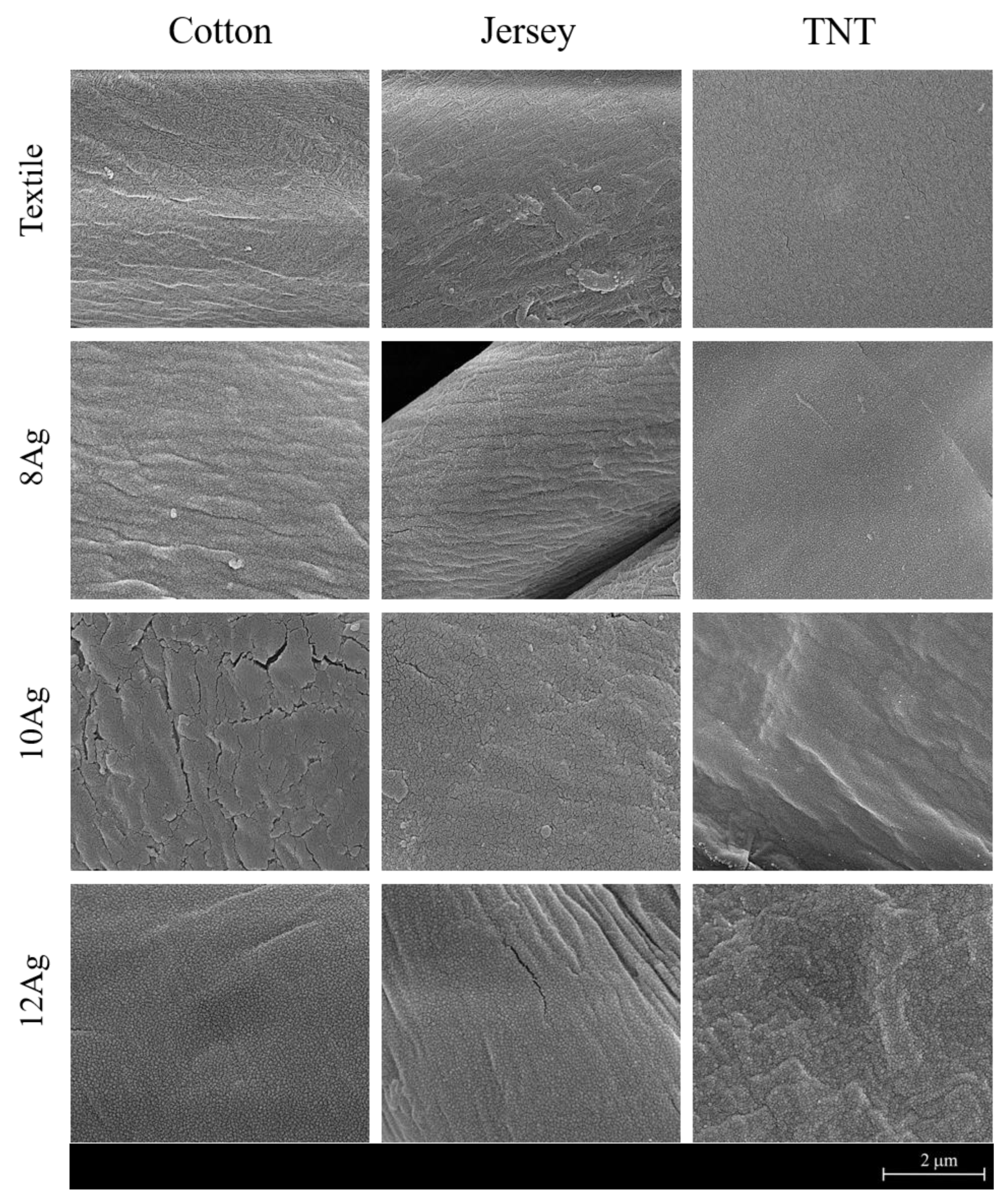
Figure 1. SEM images of the coated and uncoated substrates at a magnification of 50 kx, acquired at 10 kV.
Coatings 12 01399 g001
Figure 2. Cell viability evaluated by indirect contact test, after 168 h of AS contact with uncoated and coated fabrics. 3T3 cells are the control. The red line indicates the limit percentage of viable cells at which the material, in this case the coating, is considered non-toxic.
Coatings 12 01399 g002
Figure 3. SEM images of the coated and uncoated fabrics after the zone of inhibition testing, a red line highlights the ZoI around the fabrics.
Coatings 12 01399 g003
Figure 4. SEM images of coated and uncoated fabrics after ZoI, at a magnification of 5 kx, acquired at 10 kV.
Coatings 12 01399 g004
Table 1. Experimental details of Ag/DLC films deposition, prepared by PVD.
SampleJC of C (W/mm2)JC of Ag (W/mm2)Deposition Time (min)Deposition Rate (nm/min)Ag (at. %)
0Ag67 × 10−3-3.0170
8Ag3 × 10−32.7198
10Ag5 × 10−32.71910
12Ag7 × 10−32.71912
Table 2. Contact Angles (θ) results for the coated and uncoated substrates using water as the reference liquid.
SampleSubstrate/Water Contact Angle (θ)
CottonJerseyTNT
Textile62° (±7)128° (±3)126° (±4)
0Ag117° (±5)127° (±4)120° (±2)
8Ag108° (±5)108° (±14)99° (±4)
10Ag122° (±2)95° (±2)110° (±2)
12Ag126° (±3)120° (±10)121° (±3)
Publisher’s Note: MDPI stays neutral with regard to jurisdictional claims in published maps and institutional affiliations.