Article

Preparation of Silver Antibacterial Agents with Different Forms and Their Effects on the Properties of Water-Based Primer on Tilia europaea Surface

1
Co-Innovation Center of Efficient Processing and Utilization of Forest Resources, Nanjing Forestry University, Nanjing 210037, China
2
College of Furnishings and Industrial Design, Nanjing Forestry University, Nanjing 210037, China
*
Author to whom correspondence should be addressed.
Coatings 2021, 11(9), 1066; https://doi.org/10.3390/coatings11091066
Submission received: 11 August 2021 / Revised: 30 August 2021 / Accepted: 31 August 2021 / Published: 3 September 2021

Abstract

:
Micron silver particles prepared by the chemical reduction method, urea formaldehyde resin-coated nano-silver solution microcapsules, and nano-silver solution were used as three kinds of antibacterial agents. These were added to a water-based primer on the surface of Tilia europaea in contents of 1.0%, 4.0%, 7.0%, 10.0%, 13.0%, and 16.0%. In order to achieve the best comprehensive performance of the water-based primer, the mechanical, optical, and antibacterial properties of the three antibacterial coatings with different contents of silver antibacterial agents were explored. It was concluded that when the antibacterial agent content was 4.0%, the color difference, impact resistance, adhesion, and gloss of water-based primer on the Tilia europaea surface were better. When the antibacterial agent content added was 16.0%, the antibacterial properties of the three groups of coatings improved to 94.89%, 81.75%, and 83.98%, respectively. The results provide a research idea for the preparation of antibacterial coatings on the surface of wood furniture.

1. Introduction

Wooden furniture is frequently used in daily life and is often eroded by microorganisms [1,2,3]. Because wood is porous and contains nutrients such as protein and cellulose, it promotes the breeding and spread of harmful microorganisms such as bacteria and molds. This leads to the problem that wooden furniture is prone to mildew and decay. Therefore, it is necessary to carry out antibacterial treatment and protection on the surface of wood products, to prevent the microbial biodegradation of wood and achieve the antibacterial effect [4,5]. The solvent of the water-based coating is water and low-molecular alcohol, which does not contain organic volatiles, and has excellent properties such as environmental protection and safety [6,7]. The antibacterial agent is directly added to the water-based primer to inactivate bacteria, microorganisms, etc. or to destroy their reproductive capacity, achieving the antibacterial effect without changing the structure of the wood substrate itself, and the process is simple [8]. Therefore, it has great application potential on the surface of wood furniture.
The nano-silver antibacterial agent has good antibacterial effects and high activity [9]. Bechtold et al. [10] studied the properties of silver nanoparticles in a water-soluble polyurethane coating composition. It was concluded that silver nanoparticles have no effect on the gloss of paint film, and they have good protection and drug resistance to bacteria. Auclair et al. [11] found that nanomaterials can be modified by various coatings to regulate their behavior, bioavailability, and toxicity in the environment. Cruz-Pacheco et al. [12] used the wet chemical method to prepare polymer matrix polyether ether ketone (PEEK) coated with silver nanoparticles (AgNPs) at room temperature. The antibacterial activity of the silver coating in Escherichia coli and other strains was evaluated. It was proved that silver concentration was very important for bacterial cell replication. Lok et al. [13] synthesized spherical nano-silver (average diameter of 9.3 nm) particles by the borohydride reduction method, studied the antibacterial action mode of Escherichia coli, and proved that nano-silver is an efficient and broad-spectrum antibacterial agent. Lou et al. [14] proposed a green method of antibacterial nano-silver synthesized by using chitosan and silver nitrate as raw materials and using ultraviolet radiation to increase the reaction rate, which has antibacterial activity against Escherichia coli. Derakhshi et al. [15] found that the antibacterial activity largely depended on the shape of silver nanoparticles. Triangular silver nanoparticles were found to have the highest antibacterial activity against Gram-negative Escherichia coli and Gram-positive Staphylococcus aureus bacteria. Bhullar et al. [16] encapsulated silver nanoparticles as an antibacterial agent in the PCL microfiber structure, and the prepared coatings had inhibitory effects on Staphylococcus aureus and Escherichia coli and had high antibacterial activity. Le et al. [17] prepared a SiO2-Ag composite antibacterial agent and dispersed it into water-based acrylic paint, which not only significantly improved the adhesion, wear resistance, and thermal stability of the paint, but also found that it had good antibacterial activity against Escherichia coli. Zhang et al. [18] synthesized an antibacterial coating composed of silver nanoparticles and waterborne polyurethane. The nano-silver particles were evenly dispersed in waterborne polyurethane, and the adhesion reached grade 4. The antibacterial rate against Escherichia coli was 99.99%, and the antibacterial rate of Staphylococcus aureus was 87.5%. Cheng et al. [19] prepared a waterborne polyurethane coating of nano-cellulose crystal and nano-silver composite suitable for wood, which has compatibility and antibacterial properties. According to relevant literature reports, although nano-silver has excellent antibacterial properties, it has high requirements for equipment and operation, and the small particle size is easy to agglomerate when added into aqueous coatings, resulting in the decline in coating properties [20,21]. Furthermore, the nano-silver is black, and the addition of nano-silver to the water-based coating on the wood surface will cover the texture of the wood itself, affect the transparency of the paint film, and reduce the added value of the wood [22]. Compared with nano-silver, the micron silver powder has a larger particle size and easier preparation [23]. Commercially available nano-silver solution is colorless and transparent, which can be directly mixed with water-based paints without affecting the transparency of the paint film. However, the nano-silver solution contains a high amount of solvent, which may affect the performance of the paint film. Microcapsules are small particles containing active ingredients or core substances surrounded by a covering layer or shell [24]. Therefore, it is hoped that the nano-silver solution coated with the shell can have an antibacterial effect without changing the original properties of the coating.
In this paper, the micron silver particle antibacterial agents prepared by the silver–ammonia complex ion reduction method, the commercially available nano-silver solution with a content of 100 ppm, and the microcapsules of urea formaldehyde resin-coated nano-silver solution were used as three kinds of silver-based antibacterial agents. According to different addition amounts, the water-based primer added with the silver antibacterial agents was coated on the Tilia europaea surface to prepare the bacteria-resistant coating. The mechanical, optical, and antibacterial properties of the three kinds of antibacterial coatings were compared and analyzed. The purpose was to improve the antibacterial effect without losing the performance of the original coating, to provide a basis for the application of the antibacterial coating on wood surface.

2. Experimental Materials and Methods

2.1. Experimental Materials

The materials required for the experiment are shown in Table 1, including Tlia europaea (90 mm × 65 mm × 4 mm, pre-processed, the water content reaches the local equilibrium water content of about 14.9%, and the surface color is uniform), nano-silver solution (concentration of 100 ppm, silver with particle size of 5–7 nm), nutrient broth (NB, 250 g), eluent (NaCl, 0.85%), Staphylococcus aureus AS1.89 (National strain collection and management center), and Dulux water-based primer (aqueous acrylic acid copolymer dispersion (90.0%), matting agent (2.0%), additive (2.0%), and water (6.0%)).

2.2. Preparation of Micron Silver Particles

A mass of 1.88 g of AgNO3 was dissolved in 11.0 mL of distilled water, and then the amount of concentrated aqueous ammonia (25 wt.%) was slowly dropped (1.0–1.5 mL/min) into the stirred AgNO3 solution at 600 rpm. The gray-brown precipitate appeared at the beginning of the dropwise addition, until the precipitate disappeared, and a transparent [Ag(NH3)2]+ solution was obtained. The resulting solution was diluted to 22.0 mL, and the cup was sealed with plastic wrap for later use. An amount of 30 mL of C2H5OH and 4.3 g of 38% formaldehyde solution were mixed, and 23.28 g of absolute ethanol was added to the above solution. The beaker was heated in a water bath to 60 °C and the reaction was 30 min. Subsequently, the transparent [Ag(NH3)2]+ solution was added dropwise. After 1 h of reaction, the black precipitate began to precipitate. Then, the mixture was continuously stirred at a speed of 600 rpm using a HH-1J water bath magnetic stirrer (Changzhou Enpei Instrument Manufacturing Co., Ltd., Changzhou, China) for 1 h. The obtained black precipitate and mother liquor were centrifuged with a MC-45 centrifuge (Ningbo Yinzhou Qun’an Experimental Instrument Co., Ltd., Ningbo, China) at 4000 rpm. The precipitate was air-dried first at room temperature and then placed in an oven at 60 °C for 4 h [25].

2.3. Preparation of the Microcapsule of Urea Formaldehyde Resin-Coated Nano-Silver Solution

(1) Preparation of wall material of urea-formaldehyde prepolymer: The urea and 37.0% formaldehyde solution were added into a beaker at a mass ratio of 1:1.85. According to the best ratio, the 20.0 g of urea and 27.0 g of formaldehyde were weighed. A magnetic rotor was added to the beaker and stirred thoroughly to make the two evenly mixed. The cup was sealed with plastic wrap to prevent degradation of the prepolymer. The temperature was kept at 30 °C and the speed was controlled at 600 rpm until the urea and formaldehyde in the beaker completely reacted to form a colorless transparent solution. Then, 2.0 mL of triethanolamine was slowly added to adjust the pH value to 8–9. The mixture was heated at 70 °C and continuously stirred for 1 h at 600 rpm, and the transparent urea-formaldehyde prepolymer was naturally cooled at room temperature.
(2) Preparation of core material: The 17.5 g of nano-silver solution and 87.5 g of absolute alcohol were mixed and dissolved by 1:5. Then, 174.24 mL of distilled water and 1.76 g of sodium dodecyl benzene sulfonate white powder were mixed to obtain a 1.0% emulsifier solution. The emulsifier solution was added to the diluted nano-silver solution and stirred at a speed of 600 rpm for 30 min, and the homogeneous core emulsion was obtained.
(3) Preparation of urea formaldehyde resin-coated nano-silver solution microcapsules: At the speed of 600 rpm, the wall material of urea-formaldehyde prepolymer was dripped into the core emulsion, and the amount of citric acid monohydrate was added to the above mixture to adjust the pH value to 2.5–3.0. The beaker continued to react in the water bath for 3 h. After being cooled naturally, the microcapsule particles were filtered, and the distilled water and absolute ethanol were used to wash the microcapsule particles. The microcapsule particles were dried at 60 °C for 4 h [26].

2.4. Preparation of Waterborne Wood Antibacterial Paint Film

The micron silver particles, commercial nano-silver solution with a content of 100 ppm, and urea formaldehyde resin-coated nano-silver solution microcapsules were used as three kinds of silver antibacterial agents, which were added into the Dulux transparent coating according to the mass fractions of 1.0%, 4.0%, 7.0%, 10.0%, 13.0%, and 16.0%, respectively (Table 2). For the nano-silver solution, the mass of nano-silver solution ranged from 0.03 to 0.48 g. The mixture was evenly mixed at room temperature and uniformly coated on the Tilia europaea. The coated Tilia europaea was dried at 60 °C for 1 h until a dry and smooth paint film was formed, and then lightly sanded with sandpaper. The above procedure was repeated three times, and the dried film thickness was about 60 μm.

2.5. Testing and Characterization

According to the standard of HG/T 3950-2007 [27], the viable bacteria count (colony counter) was used to test the antibacterial rate of the paint film against Staphylococcus aureus. As the coating has a high degree of surface activity against Staphylococcus aureus [28], it can be evaluated by testing the number of colonies.
The Staphylococcus aureus were removed from the refrigerator and warmed to room temperature. The inoculation loop was dipped with Staphylococcus aureus and was then drawn in a continuous curve on a petri dish. The prepared live Staphylococcus aureus were cultured in a constant-temperature and -humidity incubator at 36 °C for 24 h. The fresh bacteria were scraped into the 50 mL nutrient broth by the inoculation loop. The concentration obtained by 1:100 dilution was (5.0–10.0) × 105 CFU/mL of bacterial solution as the suspension. Then, 0.4–0.5 mL of Staphylococcus aureus solution was dropped on the surface of the coating with and without silver antibacterial agents. Tweezers were used to clamp out the polyethylene film, and then the polyethylene films were covered on the coatings. The polyethylene film was pressed downward to make the suspension flat with no bubbles. Under the conditions of 35–38 °C and relative humidity >90.0%, the bacterial solution was cultured for 24 h. After 24 h of cultivation, the coated samples and their covering films were removed and put into the sterilized 100 mL beaker, respectively. Then, 20 mL eluent was added, and the coated samples and covering films were repeatedly washed. After shaking, the suspension with a dilution of 100 was obtained. Then, 30 g of nutrient agar and 400 mL of distilled water were heated and dissolved and then used when cooled to 45 °C. The suspension and the nutrient agar were mixed in a petri dish. After agar solidification, an agar plate containing bacteria was made. The colonies were obtained after the agar plate was cultured in a constant-temperature and -humidity incubator at 36 °C for 24 h. The test area was based on the 1/3 size standard of an agar plate. Finally, the number of colonies was calculated by a colony counter. The antibacterial rate was calculated with Formula (1):
R = (B − C)/B × 100%
where R is the anti-bacterial rate (%), B is the average number of bacteria recovered after 24 h of the coating without silver antibacterial agents (CFU), and C is the average number of bacteria recovered after 24 h of the antibacterial coating (CFU).
The chromaticity value of water-based paint film on the Tilia europaea surface was measured by a SEGT-J portable color difference instrument (Shenyang Guoti precision testing instrument Co., Ltd., Shenyang, China). L is the brightness, and a and b are the trichromatic coordinates [29]. L1, a1, and b1 at one point on the surface of the paint film were measured with a colorimeter, and L2, a2, and b2 at another point on the surface of the paint film for the same sample were then measured, too [30]. The color difference (ΔE) was calculated according to the following formula:
ΔE = [(L1 − L2)2 + (a1 − a2)2 + (b1 − b2)2]1/2
A HG268 intelligent glossmeter (Qingdao Runlu Electromechanical Equipment Co., Ltd., Qingdao, China) was used to test the gloss of water-based paint film at a 60° incident angle. The adhesive force of the paint film was tested by a QFH-HG600 film gridding instrument (Dongguan Huaguo Precision Instrument Co., Ltd., Dongguan, China), and the damage degree of the paint film was observed by a magnifying glass to determine the adhesion level of the paint film. The impact strength of paint film was tested by a QCJ (Product model) paint film impactor tester (Shanghai Qigong Instrument Co., Ltd., Shanghai, China). The morphology of the microcapsules and the coatings was characterized by a Zeiss Axio Scope A1 biological microscope (OM) (Beijing Hugetall Sciences & Technology Co. Ltd., Beijing, China) and Zeiss Sigma 300 scanning electron microscope (SEM) (Dongguan Sanben Precision Instrument Co., Ltd., Dongguan, China) with an energy-dispersive X-ray spectrometer (EDS). EDS is an analytical technique used for elemental analysis or chemical characterization of the samples. It depends on the interaction between the X-ray excitation source and the sample. Its characterization ability is largely due to the basic principle that each element has a unique atomic structure, allowing a unique set of peaks in its X-ray emission spectrum. The chemical composition of the microcapsules and the coatings was measured by a VERTEX 80v infrared spectrometer (Guangzhou Taoyi Trading Co., Ltd., Guangzhou, China). All tests were repeated 4 times, and the error was less than 5%.

3. Results and Discussion

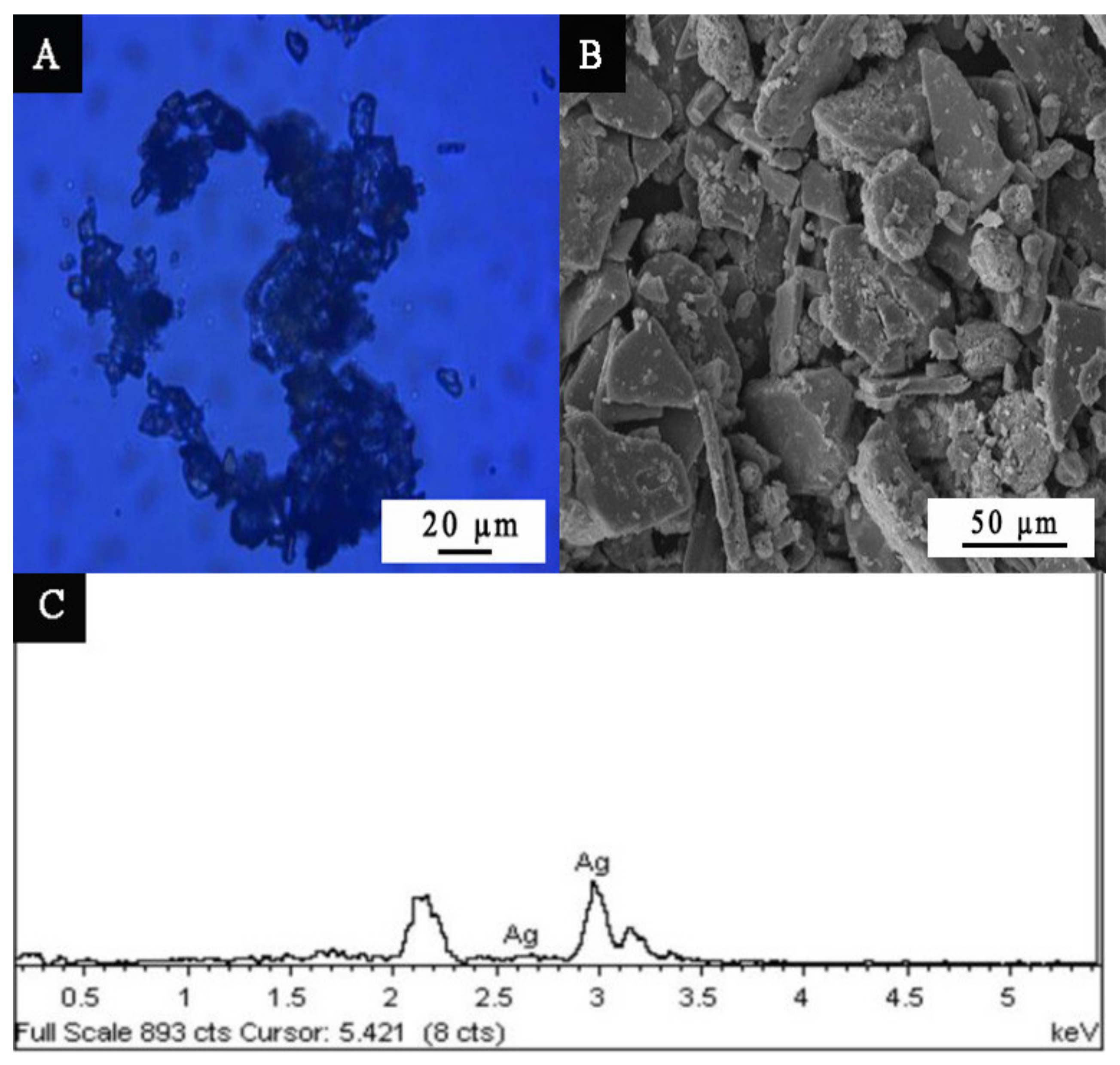
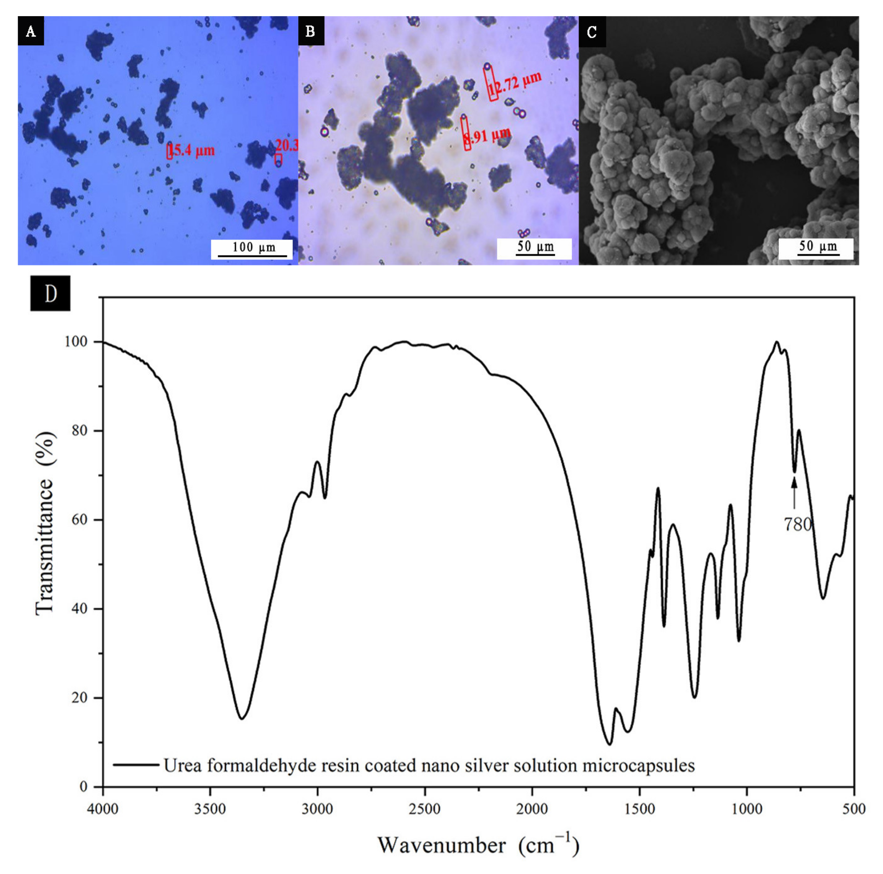
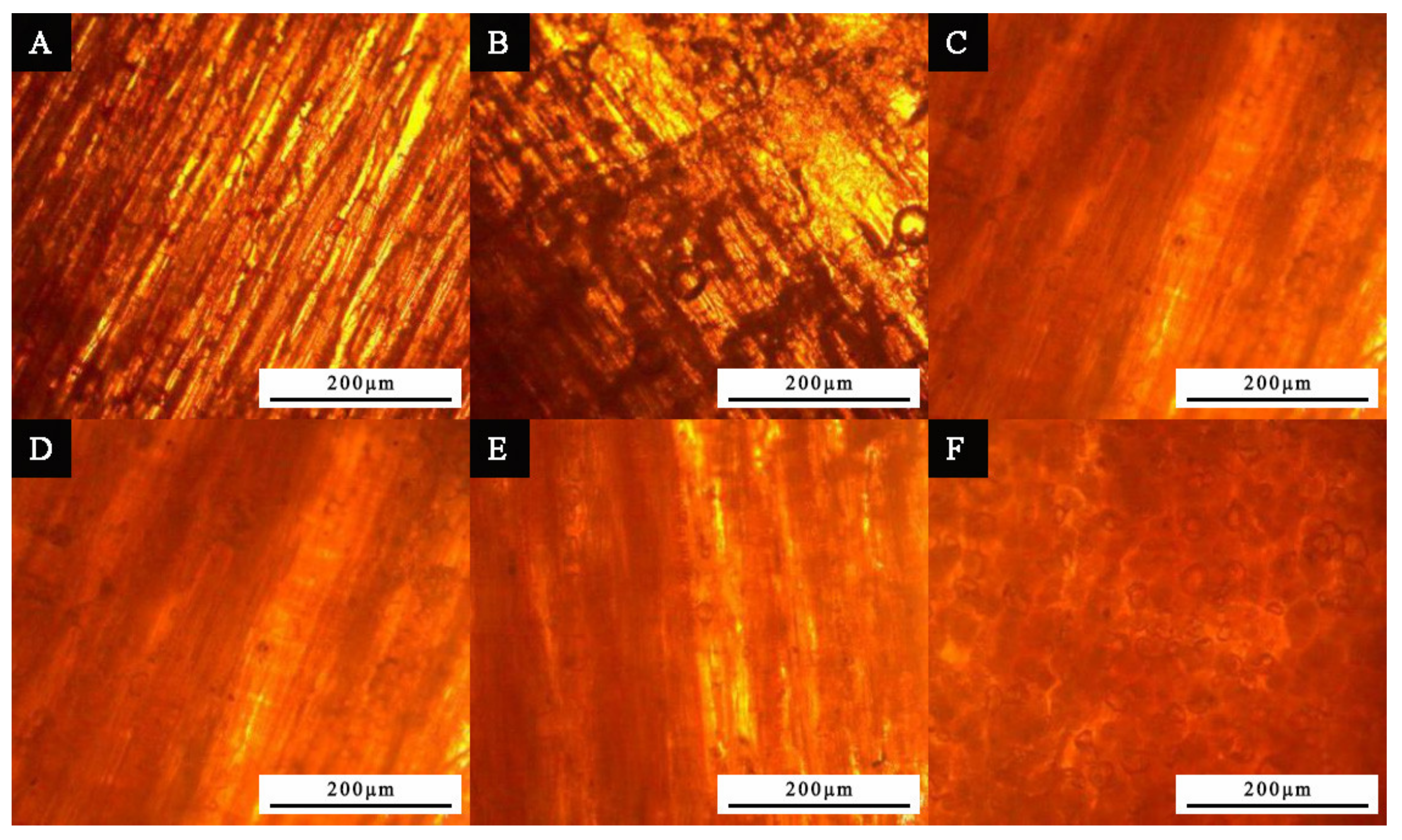
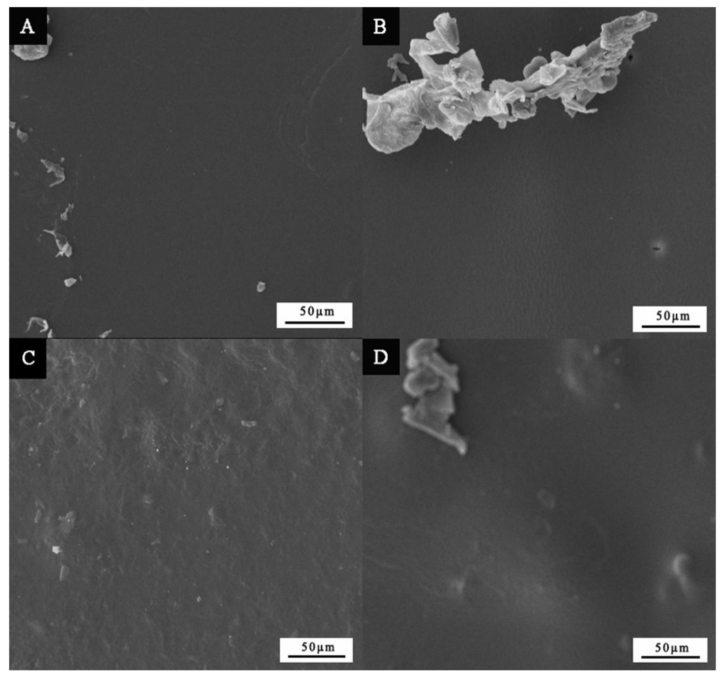
3.1. Microstructure Analysis

The microstructure analysis of micron silver particles is shown in Figure 1. As shown in Figure 1A,B, the micron silver particles were mainly spherical, flake, and agglomerated long rods. EDS (as shown in Figure 1C) was used to observe the energy spectrum of micron silver particles. It can be seen that the self-made micron silver particles were the pure Ag, and there were no diffraction peaks of other impurities.
The microscopic morphology of the urea formaldehyde resin-coated nano-silver solution microcapsules is shown in Figure 2. The particle size of urea formaldehyde resin-coated nano-silver solution microcapsules was about 8.0–20.0 μm. Figure 2C shows the SEM image of the urea-formaldehyde resin-coated nano-silver solution microcapsules. The microcapsules had a relatively uniform size and smooth surface. Figure 2D shows the infrared spectrum of the urea formaldehyde resin-coated nano-silver solution microcapsules. By observing the chemical composition of the nano-silver microcapsules, it can be seen that there was a strong absorption peak at 780 cm1, where the strong absorption peak indicated a small amount of hemiacetal C-O-C asymmetric stretching vibration from the urea formaldehyde resin. This shows that the nano-silver solution was successfully coated.

3.2. Color Difference, Gloss, Adhesion, and Impact Resistance of Antibacterial Coatings

Table 3, Table 4 and Table 5 show the results of the color difference, gloss, adhesion, and impact resistance of water-based primer with three kinds of antibacterial agents. As the content of the urea formaldehyde resin-coated nano-silver solution microcapsules increased, the color difference value also increased. When the content of the urea formaldehyde resin-coated nano-silver solution microcapsules was below 10.0%, the color difference of the paint film increased slowly. When the content of the urea formaldehyde resin-coated nano-silver solution microcapsule was less than or equal to 10.0%, the color of the coating film was more uniform. At the same incident angle and the same addition content, the gloss of the paint film added with nano-silver solution was better. However, with the increase in the addition amount, the gloss decreased. This was due to the increase in the number of particles on the surface of the coating, resulting in the enhancement of the diffuse reflection on the surface of the coating, thus reducing the gloss. Compared with the addition of micron silver particles and urea formaldehyde resin-coated nano-silver solution microcapsules, the gloss of the paint film with the nano-silver solution was higher. When the addition content was 0–5.0%, the gloss of the paint film was better. With the increase in the content of nano-silver solution microcapsules, the nano-silver solution, and micron silver particles, the impact resistance of the paint film was greater when the content of antibacterial agent was 16.0%. At the same addition content, the impact resistance of the paint film with the micron silver particles was best, and the impact force was poor when the nano-silver solution was added to the water-based primer. When the nano-silver solution and micron silver particles were added, the adhesion of paint film was better.
When the content of the three antibacterial agents was 0–7.0%, the color difference and impact resistance of the paint film increased slowly, and the adhesion and gloss decreased continuously. When the added content was 10.0%–16.0%, the color difference value increased most, indicating that the addition of antibacterial agents had the greatest impact on the color difference of the paint film. The impact force values of the three kinds of silver antibacterial agents reached the maximum when the content was 16.0%, indicating that the addition of antibacterial agents played a certain role in improving the mechanical properties of the coatings. When the content of the silver antibacterial agent was 1.0%–10.0%, the color difference, adhesion, and gloss properties of the paint film with nano-silver solution were slightly higher than those of urea formaldehyde resin-coated nano-silver solution microcapsules. When the addition content was 10.0%, the impact resistance of the paint film with micron silver particles was the same as that with urea formaldehyde resin-coated nano-silver solution microcapsules, which was 8.0 kg∙cm. Tables S1–S4 show that there is a significant difference between the measured data in color dif-ference, gloss, adhesion and impact resistance. When the addition content was 4.0%, the optical and mechanical properties, gloss, adhesion, and impact resistance of the film were higher, and the color difference was smaller.

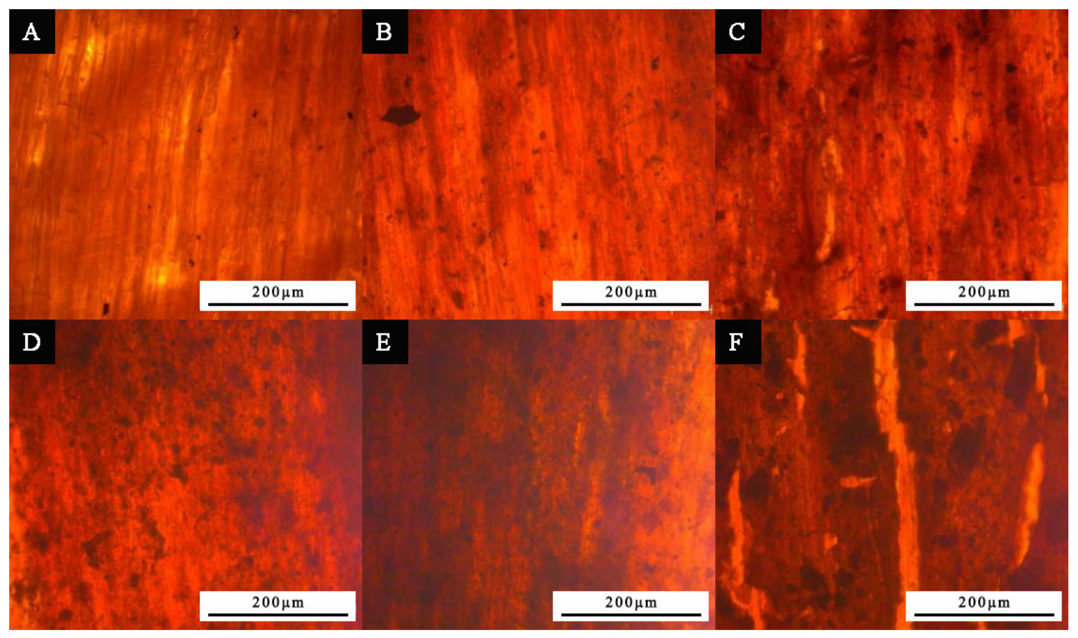
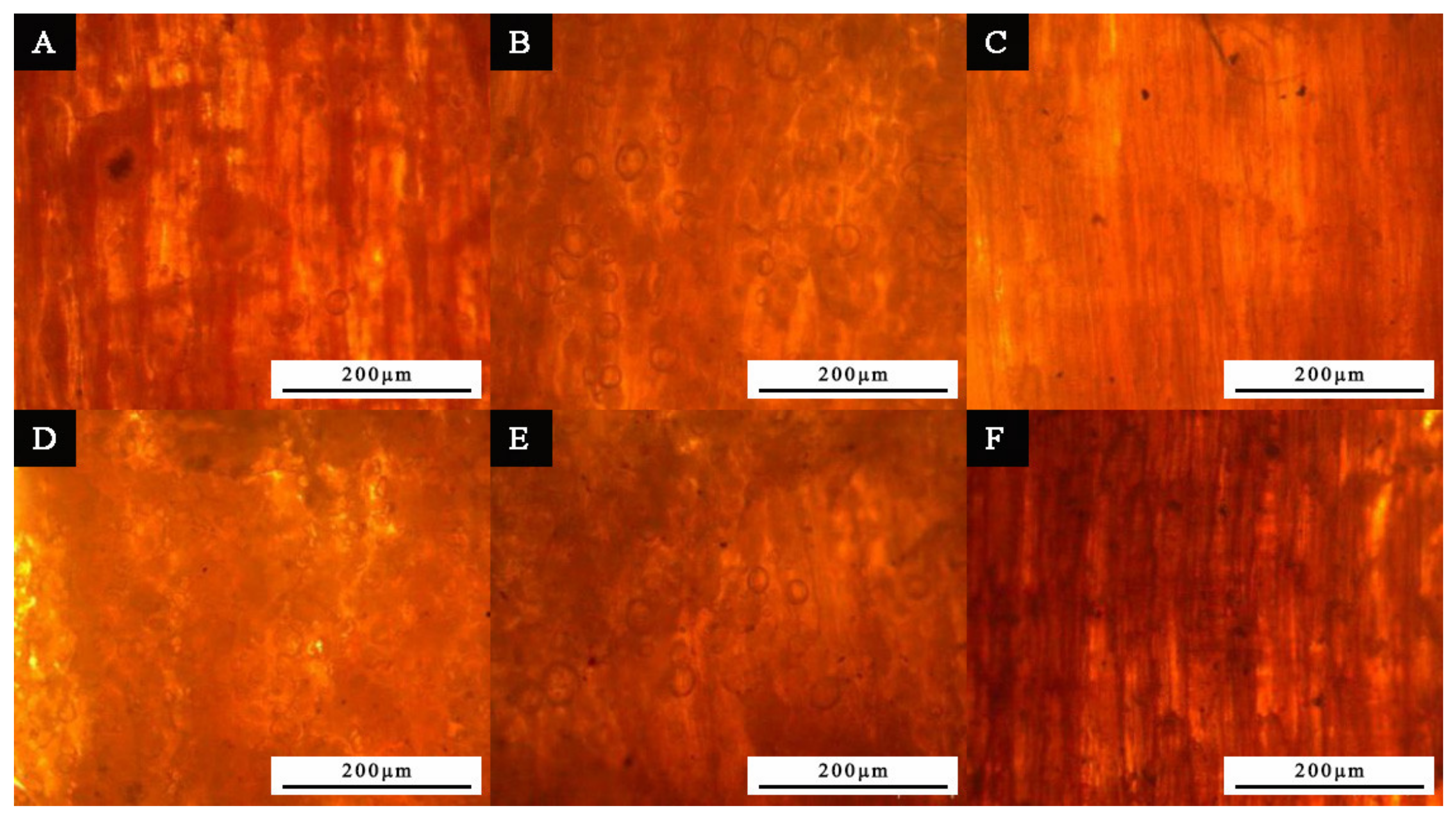
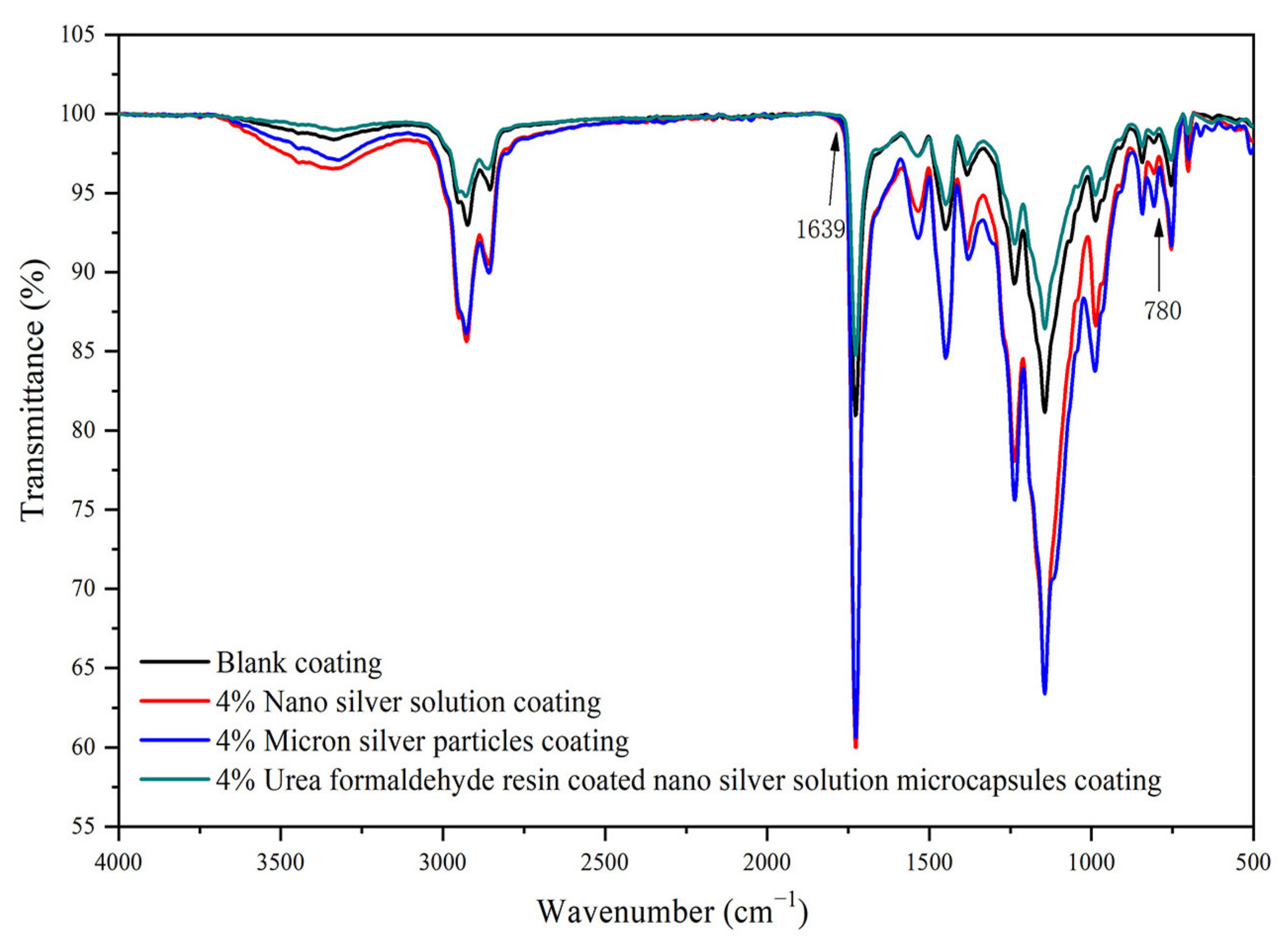
3.3. Microstructure and Composition Analysis of Paint Film

As the content of nano-silver solution in water-based primer increased, the clarity of wood grains showed a downward trend (Figure 3). This is because the number of particles increased with the content of nano-silver solution. The paint film showed an opaque state, which affects the appearance effect of wood grain.
Unlike the paint film added with nano-silver solution, the paint film particles were obvious after adding micron silver particles (Figure 4). The surface of the Tilia europaea substrate was already black during the coating process, and the wood texture was not clear with the increase in micron silver particles. When the addition amount of micron silver particles was 16.0%, slight cracking occurred on the surface. A possible reason is that the paint film cracked due to the reduction in paint content during oven drying.
Figure 5 shows the microstructure of the primer added with the urea formaldehyde resin-coated nano-silver solution microcapsules. As the prepared microcapsules were the white powder, with the increase in the addition amount, the white microcapsules gradually covered the wood grain, resulting in a decrease in visibility.
From Figure 6, it can be found that when the three kinds of silver antibacterial agents were added at 4.0%, the transparency of the paint film was not affected. Compared with the coating films added with the micron silver particles and the urea formaldehyde resin-coated nano-silver solution microcapsules, the surface of the coating film with nano-silver solution was rougher and the particles were easy to agglomerate.
Figure 7 shows the infrared spectrum of the paint film sample without silver antibacterial agents, with 4.0% of nano-silver solution, with 4.0% of urea formaldehyde resin-coated nano-silver solution microcapsules, and with 4.0% of micron silver particles. The 780 cm−1 was the hemiacetal C-O-C asymmetric stretching vibration in the nano-silver solution, and the 1639 cm−1 was the C=O stretching vibration and attributed to the urea-formaldehyde resin. When the urea formaldehyde resin-coated nano-silver solution microcapsules were added into the primer, the transmittance decreased significantly. The reason is that microencapsulated urea formaldehyde resin affects the curing and film-forming reaction of paint film [31].

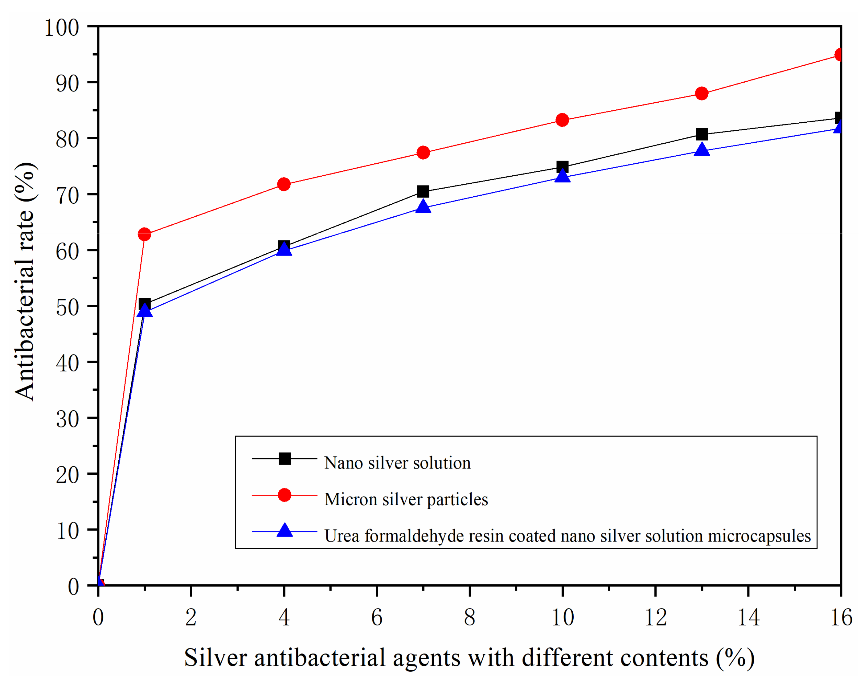
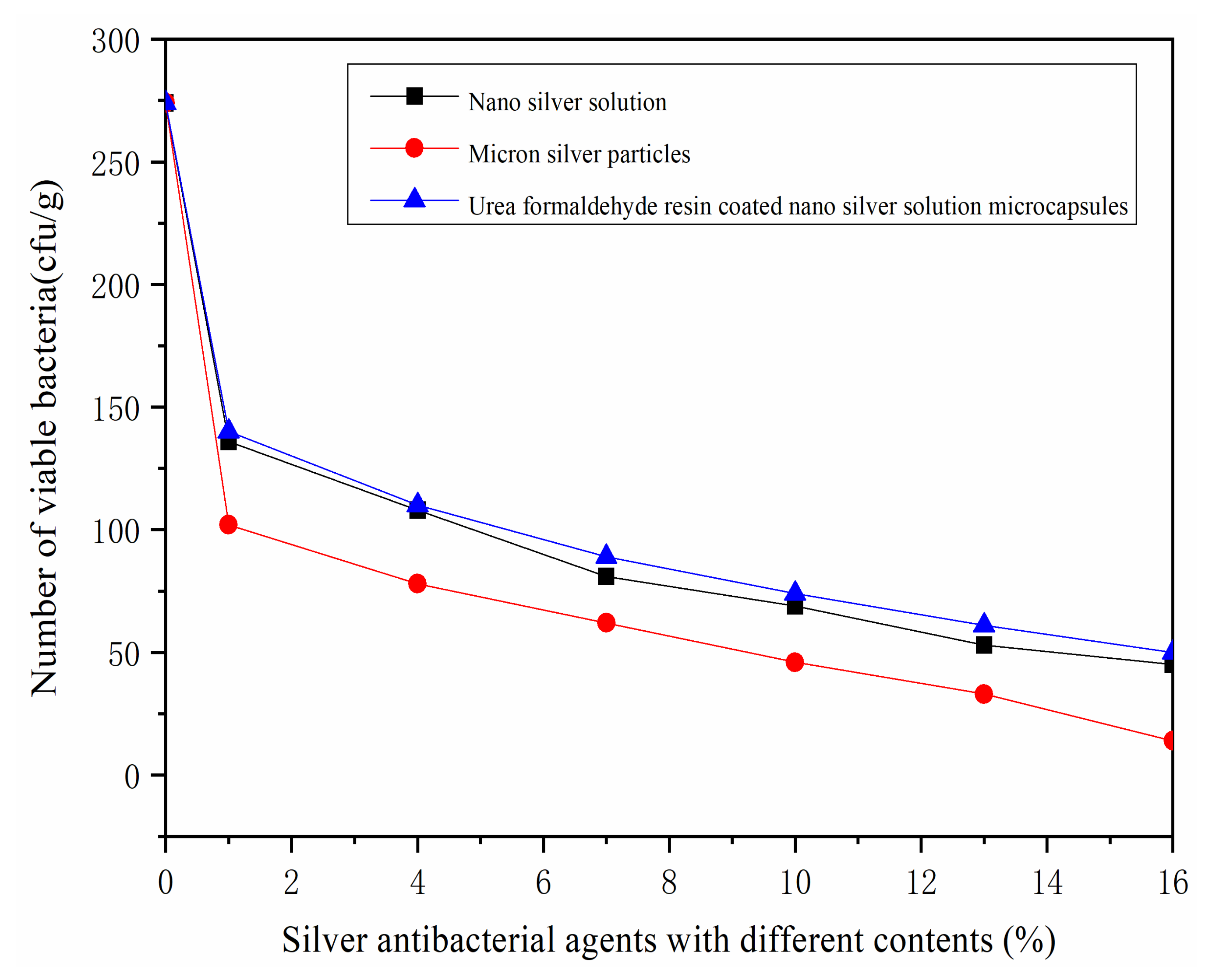
3.4. Analysis of Antibacterial Rate

It can be seen from Figure 8 that the number of viable colonies decreased significantly with the number of silver antibacterial agents. This shows that the antibacterial effect of paint film increased. The antibacterial rate of the paint film of the three kinds of silver antibacterial agents is shown in Figure 9. The antibacterial rate of the film formed by the addition of micron silver particles to the primer was significantly higher than those of the nano-silver solution and the nano-silver solution microcapsule coated with urea formaldehyde resin. When the content of added micron silver particles was 7.0%–16.0%, with the increase in the addition amount, the antibacterial effect showed a more obvious growth trend. The three kinds of silver antibacterial agents reached the maximum antibacterial rate when the addition content was 16.0%. Among them, the antibacterial effect of the paint film added with micron silver particles was better, and the antibacterial rate reached 94.89%. However, at this time, the film also showed an obvious black color and covered the wood grain, so the addition amount did not continue to increase. The antibacterial property of the paint film formed by the nano-silver solution was higher than that of urea formaldehyde resin-coated nano-silver solution microcapsules. However, when the content of silver antibacterial agents was 1.0–7.0%, the antibacterial rate of urea formaldehyde resin-coated nano-silver solution microcapsules was almost the same as that of the paint film formed by adding nano-silver solution, and when the content was 4.0%, the antibacterial rate was 56.57%. Therefore, the microcapsule prepared by the nano-silver solution coated with urea formaldehyde resin as the wall material will not have a great impact on the antibacterial property of the core material.
The effective antibacterial component of silver antibacterial agents is metal silver, which was added into the water-based coating to form an antibacterial layer. Due to the trace metal silver released by the antibacterial agent, the protein in the Staphylococcus aureus is solidified to achieve the effect of bacteriostasis [32,33]. The toxicity of the studied silver compounds was not evaluated and could not be applied to all types of wood products at any concentration. It will be improved in future research.

4. Conclusions

Three kinds of silver antibacterial agents, containing micron silver particles, nano-silver solution, and urea formaldehyde resin-coated nano-silver solution microcapsules, were added into the water-based primer with different contents. Nano-silver solution was successfully coated in urea formaldehyde resin to form microcapsules. With the increase in the content of the three kinds of silver antibacterial agents, the color difference of the paint film increased and the gloss decreased. When the content of antibacterial agent was 16.0%, the impact resistance of the paint film formed by the three antibacterial agents was better. However, with the increase in the amount of antibacterial agent, the adhesion of the paint film decreased. Even a small number of silver particles slowed down the growth of Staphylococcus aureus bacteria, and micron silver particles showed the greater effectiveness. The water-based primer with a micron silver particle content of 4.0% had good comprehensive properties on the surface of Tilia europaea. The results provide a reference for the application of silver antibacterial agents in antibacterial wood coatings.

Supplementary Materials

The following are available online at https://www.mdpi.com/article/10.3390/coatings11091066/s1, Table S1: Significance analysis of color difference, Table S2: Significance analysis of gloss, Table S3: Significance analysis of adhesion; Table S4: Significance analysis of impact resistance.

Author Contributions

Conceptualization, methodology, validation, resources, data management, supervision, X.Y.; formal analysis, investigation, writing—review and editing, P.P. All authors have read and agreed to the published version of the manuscript.

Funding

This project was partly supported by the Natural Science Foundation of Jiangsu Province (Grant No. BK20201386).

Institutional Review Board Statement

Not applicable.

Informed Consent Statement

Not applicable.

Data Availability Statement

Not applicable.

Conflicts of Interest

The authors declare that there is no conflict of interest.

References

  1. Duan, X.P.; Liu, S.M.; Huang, E.T.; Shen, X.Y.; Wang, Z.; Li, S.; Jin, C.D. Superhydrophobic and antibacterial wood enabled by polydopamine-assisted decoration of copper nanoparticles. Colloid. Surface. A. 2020, 602, 125145. [Google Scholar] [CrossRef]
  2. Xu, W.; Fang, X.Y.; Han, J.T.; Wu, Z.H.; Zhang, J.L. Effect of coating thickness on sound absorption property of four wood species commonly used for piano soundboards. Wood Fiber Sci. 2020, 52, 28–43. [Google Scholar] [CrossRef] [Green Version]
  3. Liu, Q.Q.; Gao, D.; Xu, W. Influence of the bottom color modification and material color modification process on the perfor-mance of modified Poplar. Coatings 2021, 11, 660. [Google Scholar] [CrossRef]
  4. Singh, A.P.; Kim, Y.S.; Chavan, R.R. Relationship of wood cell wall ultrastructure to bacterial degradation of wood. IAWA J. 2019, 40, 845–870. [Google Scholar] [CrossRef]
  5. Wu, S.S.; Tao, X.; Xu, W. Thermal conductivity of Poplar wood veneer impregnated with graphene/polyvinyl alcohol. Forests 2021, 12, 777. [Google Scholar] [CrossRef]
  6. Zafar, F.; Ghosal, A.; Sharmin, E.; Chaturvedi, R.; Nishat, N. A review on cleaner production of polymeric and nanocomposite coatings based on waterborne polyurethane dispersions from seed oils. Prog. Org. Coat. 2019, 131, 259–275. [Google Scholar] [CrossRef]
  7. Liu, Q.Q.; Gao, D.; Xu, W. Effect of sanding processes on the surface properties of modified Poplar coated by primer compared with Mahogany. Coatings 2020, 10, 856. [Google Scholar] [CrossRef]
  8. Jiang, G.F.; Li, X.F.; Che, Y.L.; Lv, Y.; Liu, F.; Wang, Y.Q.; Zhao, C.C.; Wang, X.J. Antibacterial and anticorrosive properties of CuZnO@RGO waterborne polyurethane coating in circulating cooling water. Environ. Sci. Pollut. Res. 2019, 26, 9027–9040. [Google Scholar] [CrossRef]
  9. Tang, S.H.; Zheng, J. Antibacterial activity of silver nanoparticles: structural effects. Adv. Healthc. Mater. 2018, 7, 1701503. [Google Scholar] [CrossRef]
  10. Bechtold, M.; Valerio, A.; de Souza, A.A.U.; de Oliveira, D.; Franco, C.V.; Serafim, R.; Souza, S.M.A.G.U. Synthesis and application of silver nanoparticles as biocidal agent in polyurethane coating. J. Coat. Technol. Res. 2020, 17, 613–620. [Google Scholar] [CrossRef]
  11. Auclair, J.; Turcotte, P.; Gagnon, C.; Peyrot, C.; Wilkinson, K.J.; Gagne, F. The influence of surface coatings of silver nanoparticles on the bioavailability and toxicity to elliptio complanata mussels. J. Nanomater. 2019, 2019, 7843025. [Google Scholar] [CrossRef]
  12. Cruz-Pacheco, A.F.; Munoz-Castiblanco, D.T.; Cuaspud, J.A.G.; Paredes-Madrid, L.; Vargas, C.A.P.; Zambrano, J.J.M.; Gomez, C.A.P. Coating of polyetheretherketone films with silver nanoparticles by a simple chemical reduction method and their antibacterial activity. Coatings 2019, 9, 91. [Google Scholar] [CrossRef] [Green Version]
  13. Lok, C.N.; Ho, C.M.; Chen, R.; He, Q.Y.; Yu, W.Y.; Sun, H.Z.; Tam, P.K.H.; Chiu, J.F.; Che, C.M. Proteomic analysis of the mode of antibacterial action of silver nanoparticles. J. Proteome Res. 2006, 5, 916–924. [Google Scholar] [CrossRef] [PubMed]
  14. Lou, C.W.; Chen, A.P.; Lic, T.T.; Lin, J.H. Antimicrobial activity of UV-induced chitosan capped silver nanoparticles. Mater. Lett. 2014, 128, 248–252. [Google Scholar] [CrossRef]
  15. Derakhshi, M.; Ashkarran, A.A.; Bahari, A.; Bonakdar, S. Shape selective silver nanostructures decorated amine-functionalized graphene: A promising antibacterial platform. Colloids Surf. A. 2018, 545, 101–109. [Google Scholar] [CrossRef]
  16. Bhullar, S.K.; Ruzgar, D.G.; Fortunato, G.; Aneja, G.K.; Orhan, M.; Saber-Samandari, S.; Sadighi, M.; Ahadian, S.; Ramalingam, M.A. Facile method for controlled fabrication of hybrid silver nanoparticle-poly (epsilon-caprolactone) fibrous constructs with antimicrobial properties. J. Nanosci. Nanotechnol. 2019, 19, 6949–6955. [Google Scholar] [CrossRef] [Green Version]
  17. Le, T.T.; Nguyen, T.V.; Nguye, T.A.; Nguye, T.T.H.; Thai, H.; Tran, D.L.; Dinh, D.A.; Nguyen, T.M.; Lu, L.T. Thermal, mechanical and antibacterial properties of water-based acrylic polymer/SiO2-Ag nanocomposite coating. Mater. Chem. Phys. 2019, 232, 362–366. [Google Scholar] [CrossRef]
  18. Zhang, X.H.; Wang, W.; Yu, D. Synthesis of waterborne polyurethane-silver nanoparticle antibacterial coating for synthetic leather. J. Coat. Technol. Res. 2018, 15, 415–423. [Google Scholar] [CrossRef]
  19. Cheng, L.S.; Ren, S.B.; Lu, X.N. Application of eco-friendly waterborne polyurethane composite coating incorporated with nano cellulose crystalline and silver nano particles on wood antibacterial board. Polymers 2020, 12, 407. [Google Scholar] [CrossRef] [Green Version]
  20. Ichimaru, H.; Harada, A.; Yoshimoto, S.; Miyazawa, Y.; Mizoguchi, D.; Kyaw, K.; Ono, K.; Tsutsuki, H.; Sawa, T.; Niidome, T. Gold coating of silver nanoplates for enhanced dispersion stability and efficient antimicrobial activity against intracellular bacteria. Langmuir 2018, 34, 10413–10418. [Google Scholar] [CrossRef]
  21. Guo, L.Y.; Yuan, W.Y.; Lu, Z.S.; Li, C.M. Polymer/nanosilver composite coatings for antibacterial applications. Colloids Surf. A. 2013, 439, 69–83. [Google Scholar] [CrossRef]
  22. Yang, X.F.; Liu, J.; Wu, Y.F.; Liu, J.L.; Cheng, F.; Jiao, X.J.; Lai, G.Q. Fabrication of UV-curable solvent-free epoxy modified silicone resin coating with high transparency and low volume shrinkage. Prog. Org. Coat. 2019, 129, 96–100. [Google Scholar] [CrossRef]
  23. Maidaniuc, A.; Miculescu, M.; Voicu, S.I.; Ciocan, L.T.; Niculescu, M.; Corobea, M.C.; Rada, M.E.; Miculescu, F. Effect of micron sized silver particles concentration on the adhesion induced by sintering and antibacterial properties of hydroxyapatite microcomposites. J. Aahes. Sci. Technol. 2016, 30, 1829–1841. [Google Scholar] [CrossRef]
  24. Yan, X.X.; Zhao, W.T.; Qian, X.Y. Effect of water-based emulsion core microcapsules on aging resistance and self-repairing properties of water-based coatings on Linden. Appl. Sci. 2021, 11, 4662. [Google Scholar] [CrossRef]
  25. Yan, X.X.; Xu, G.Y. Effect of surface modification of Cu with Ag by ball-milling on the corrosion resistance of low infrared emissivity coating. Mater. Sci. Eng. B Adv. 2010, 166, 152–157. [Google Scholar] [CrossRef]
  26. Yan, X.X.; Wang, L.; Qian, X.Y. Effect of microcapsules with different core-wall ratios on properties of waterborne primer coating for European Linden. Coatings 2020, 10, 826. [Google Scholar] [CrossRef]
  27. HG/T 3950-2007 Antibacterial Coatings; National Development and Reform Commission of the People’s Republic of China: Beijing, China, 2007; pp. 1–9. (In Chinese)
  28. Cheng, Q.L.; Guo, X.W.; Hao, X.J.; Shi, Z.S.; Zhu, S.; Cui, Z.C. Fabrication of robust antibacterial coatings based on an organic-inorganic hybrid system. ACS Appl. Mater. Inter. 2019, 11, 42607–42615. [Google Scholar] [CrossRef]
  29. Galus, S.; Mikus, M.; Ciurzyńska, A.; Domian, E.; Kowalska, J.; Marzec, A.; Kowalska, H. The effect of whey protein-based edible coatings incorporated with lemon and lemongrass essential oils on the quality attributes of fresh-cut pears during storage. Coatings 2021, 11, 745. [Google Scholar] [CrossRef]
  30. Yan, X.X.; Peng, W.W.; Qian, X.Y. Effect of water-based acrylic acid microcapsules on the properties of paint film for furniture surface. Appl. Sci. 2021, 11, 7586. [Google Scholar] [CrossRef]
  31. Li, J.; Zhang, Y.F. Morphology and crystallinity of urea-formaldehyde resin adhesives with different molar ratios. Polymers 2021, 13, 673. [Google Scholar] [CrossRef]
  32. Niu, J.F.; Tang, G.; Tang, J.Y.; Yang, J.L.; Zhou, Z.Y.; Gao, Y.H.; Chen, X.; Tian, Y.Y.; Li, Y.; Li, J.Q.; et al. Functionalized silver nanocapsules with improved antibacterial activity using silica shells modified with quaternary ammonium polyethyleneimine as a bacterial cell-targeting agent. J. Agric. Food Chem. 2021, 69, 6485–6494. [Google Scholar] [CrossRef] [PubMed]
  33. Panicker, S.; Ahmady, I.M.; Han, C.; Chehimi, M.; Mohamed, A.A. On demand release of ionic silver from gold-silver alloy nanoparticles: fundamental antibacterial mechanisms study. Mater. Today Chem. 2020, 16, 100237. [Google Scholar] [CrossRef]
Figure 1. (A) OM image of micron silver particles, (B) SEM microscopic morphology of micron silver particles, and (C) EDS of micron silver particles.
Coatings 11 01066 g001
Figure 2. OM of urea formaldehyde resin-coated nano-silver solution microcapsules: (A) under 10× objective lens and (B) under 20× objective lens. (C) SEM microscopic view of urea formaldehyde resin-coated nano-silver solution microcapsules and (D) infrared spectrum of urea-formaldehyde resin-coated nano-silver solution microcapsules.
Coatings 11 01066 g002
Figure 3. Microstructure of paint film with different contents of nano-silver solution: (A) 1.0%, (B) 4.0%, (C) 7.0%, (D) 10.0%, (E) 13.0%, and (F) 16.0%.
Coatings 11 01066 g003
Figure 4. Microstructure of paint film with different contents of micron silver particles: (A) 1.0%, (B) 4.0%, (C) 7.0%, (D) 10.0%, (E) 13.0%, and (F) 16.0%.
Coatings 11 01066 g004
Figure 5. Microstructure of paint film with different contents of urea formaldehyde resin-coated nano-silver solution microcapsules: (A) 1.0%, (B) 4.0%, (C) 7.0%, (D) 10.0%, (E) 13.0%, and (F) 16.0%.
Coatings 11 01066 g005
Figure 6. SEM image of paint film: (A) without silver antibacterial agents, (B) with 4.0% of nano-silver solution, (C) with 4.0% of micron silver particles, and (D) with 4.0% of urea formaldehyde coated nano-silver solution microcapsules.
Coatings 11 01066 g006
Figure 7. Infrared spectrum of paint film with 4.0% micron silver particles, 4.0% nano-silver solution, and 4.0% urea formaldehyde resin coated nano-silver solution microcapsules.
Coatings 11 01066 g007
Figure 8. Number of surviving colonies in the coating with micron silver particles, nano-silver solution, and urea formaldehyde resin-coated nano-silver solution microcapsules.
Coatings 11 01066 g008
Figure 9. Antibacterial rate of the coating added with micron silver particles, nano-silver solution, and urea formaldehyde resin-coated nano-silver solution microcapsules.
Coatings 11 01066 g009
Table 1. List of experimental materials.
MaterialMolecular FormulaMW (g/mol)CAS No.ConcentrationProducer
silver nitrate solid powderAgNO3169.877761-88-899.9%Nanjing Chemical Reagent Co., Ltd., Nanjing, China
concentrated ammoniaNH3∙H2O35.001336-21-625%Tianjin Fuchen Chemical Reagent Factory, Tianjin, China
nano-silver solution---100 ppmLuoyang Oulun Environmental Protection Technology Co., Ltd., Luoyang, China
formaldehyde solutionCH2O30.0350-00-037.0%Tianjin University Mao Chemical Reagent Factory, Tianjin, China
ureaCH4N2O60.0657-13-699.9%Tianjin Beichen Fangzheng Chemical Reagent Factory, Tianjin, China
triethanolamineC6H15NO3149.19102-71-699.9%Xilong Science Co., Ltd., Wuxi, China
absolute ethanolC2H6O46.0764-17-599.9%Wuxi Yasheng Chemical Co., Ltd., Wuxi, China
sodium dodecyl benzene sulfonateC18H29NaO3S348.4825155-30-099.9%Wuxi Yatai United Chemical Co., Ltd., Wuxi, China
waterborne acrylic resin--9003-01-4-Dulux Paint Co., Ltd., Shanghai, China
citric acid monohydrateC6H10O8210.145949-29-199.9Kunshan Southeast Chemical Materials Co., Ltd., Kunshan, China
Tilia europaea----Shanghai Puhui industry and Trade Co., Ltd., Shanghai, China
nutrient broth----Guangdong huankai Microbial Technology Co., Ltd. Guangzhou, China
eluentNaCl58.44287647-14-50.85%Sichuan Kelun Pharmaceutical Co., Ltd., Chengdu, China
staphylococcus aureus AS1.89----Beijing Baocang Biotechnology Co., Ltd., Beijing, China
nutrient agar----Hangzhou microbial Reagent Co., Ltd., Hangzhou, China
Table 2. Composition of antibacterial coating.
Percentage (%)Mass of Primer (g)Mass of Nano-Silver Solution and Other Antibacterial Agent (g)
0 3.000
1.02.970.03
4.02.880.12
7.02.790.21
10.02.700.30
13.02.610.39
16.02.520.48
Table 3. Performance of primer film added with different concentrations of urea formaldehyde resin-coated nano-silver solution microcapsules.
Microcapsule Content (%)Color DifferenceGloss (%)Adhesion (Level)Impact Resistance (kg·cm)
00.7330.515.0
1.01.9229.127.0
4.02.458.027.0
7.02.783.638.0
10.02.892.738.0
13.03.642.649.0
16.04.222.549.0
Table 4. Performance of primer film added with different concentrations of nano-silver solution.
Nano-Silver Solution Content (%)Color DifferenceGloss (%)Adhesion (Level)Impact Resistance (kg·cm)
00.7330.515.0
1.01.2729.716.0
4.01.3726.427.0
7.01.5720.837.0
10.01.6017.538.0
13.02.0914.738.0
16.02.1111.238.0
Table 5. Performance of primer film with different concentrations of micron silver particles.
Micron Silver Particle Content (%)Color DifferenceGloss (%)Adhesion (Level)Impact Resistance (kg·cm)
00.7330.515.0
1.03.6414.117.0
4.04.087.2210.0
7.05.324.4211.0
10.05.922.8312.0
13.07.091.6313.0
16.08.911.2314.0
Publisher’s Note: MDPI stays neutral with regard to jurisdictional claims in published maps and institutional affiliations.

