Antimicrobial Properties of Strontium Functionalized Titanium Surfaces for Oral Applications, A Systematic Review

 ,
,  ,
,
Abstract
:1. Introduction
2. Materials and Methods
2.1. Protocol Development
2.2. Search Strategy for Identification of Studies
2.3. Study Selection Criteria
2.4. Data Extraction
2.5. Risk of Bias and Relevance Assessment
3. Results
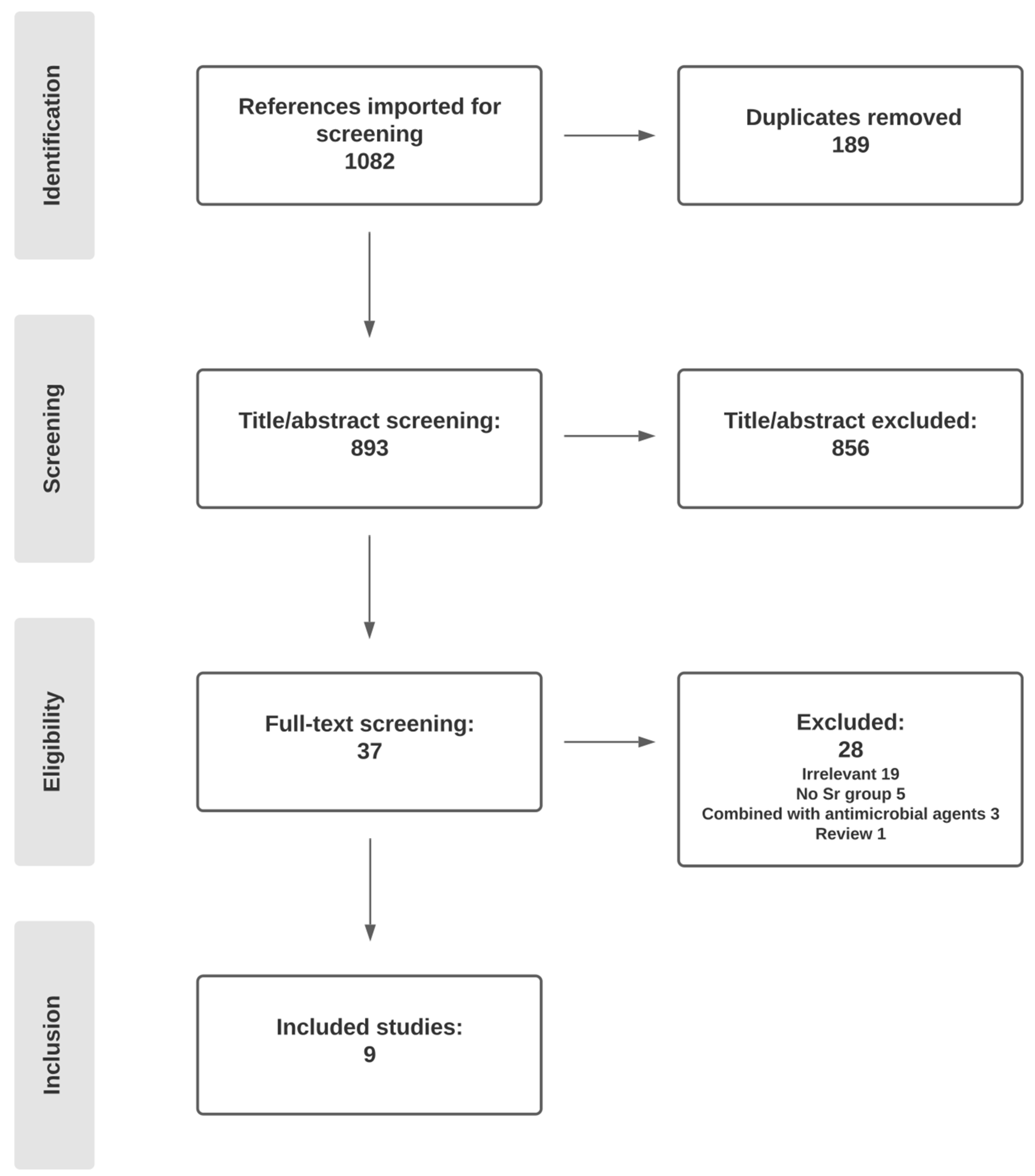
3.1. Search Results
3.2. Surface Substrates and Application Methods
3.3. Bacterial Strains and Methods of Antimicrobial Analysis
3.4. Antimicrobial Effect of Sr
3.5. Sr Ion Release
3.6. Risk of Bias and Relevance Assessment
4. Discussion
5. Conclusions
Author Contributions
Funding
Institutional Review Board Statement
Informed Consent Statement
Data Availability Statement
Conflicts of Interest
References
- Klinge, B.; Klinge, A.; Bertl, K.; Stavropoulos, A. Peri-implant diseases. Eur. J. Oral Sci. 2018, 126 (Suppl. S1), 88–94. [Google Scholar] [CrossRef] [PubMed] [Green Version]
- Heitz-Mayfield, L.J.; Salvi, G.E. Peri-implant mucositis. J. Clin. Periodontol. 2018, 45, S237–S245. [Google Scholar] [CrossRef] [Green Version]
- Schwarz, F.; Derks, J.; Monje, A.; Wang, H.L. Peri-implantitis. J. Periodontol. 2018, 89 (Suppl. S1), S267–S290. [Google Scholar] [CrossRef] [PubMed]
- Ramanauskaite, A.; Becker, K.; Schwarz, F. Clinical characteristics of peri-implant mucositis and peri-implantitis. Clin. Oral Implant. Res. 2018, 29, 551–556. [Google Scholar] [CrossRef]
- Karoussis, I.K.; Salvi, G.E.; Heitz-Mayfield, L.J.; Brägger, U.; Hämmerle, C.H.; Lang, N.P. Long-term implant prognosis in patients with and without a history of chronic periodontitis: A 10-year prospective cohort study of the ITI dental implant system. Clin. Oral Implant. Res. 2003, 14, 329–339. [Google Scholar] [CrossRef] [PubMed]
- Oh, S.L.; Shiau, H.J.; Reynolds, M.A. Survival of dental implants at sites after implant failure: A systematic review. J. Prosthet. Dent. 2020, 123, 54–60. [Google Scholar] [CrossRef] [Green Version]
- Derks, J.; Tomasi, C. Peri-implant health and disease. A systematic review of current epidemiology. J. Clin. Periodontol. 2015, 42 (Suppl. S16), S158–S171. [Google Scholar] [CrossRef]
- Dreyer, H.; Grischke, J.; Tiede, C.; Eberhard, J.; Schweitzer, A.; Toikkanen, S.E.; Glöckner, S.; Krause, G.; Stiesch, M. Epidemiology and risk factors of peri-implantitis: A systematic review. J. Periodontal Res. 2018, 53, 657–681. [Google Scholar] [CrossRef]
- Khoury, F.; Keeve, P.L.; Ramanauskaite, A.; Schwarz, F.; Koo, K.T.; Sculean, A.; Romanos, G. Surgical treatment of peri-implantitis—Consensus report of working group 4. Int. Dent. J. 2019, 69 (Suppl. S2), 18–22. [Google Scholar] [CrossRef] [Green Version]
- van Winkelhoff, A.J. Antibiotics in the treatment of peri-implantitis. Eur. J. Oral Implantol. 2012, 5, S43–S50. [Google Scholar]
- Carcuac, O.; Derks, J.; Abrahamsson, I.; Wennström, J.L.; Berglundh, T. Risk for recurrence of disease following surgical therapy of peri-implantitis—A prospective longitudinal study. Clin. Oral Implant. Res. 2020, 31, 1072–1077. [Google Scholar] [CrossRef]
- Xue, T.; Attarilar, S.; Liu, S.; Liu, J.; Song, X.; Li, L.; Zhao, B.; Tang, Y. Surface modification techniques of titanium and its alloys to functionally optimize their biomedical properties: Thematic review. Front. Bioeng. Biotechnol. 2020, 8, 603072. [Google Scholar] [CrossRef] [PubMed]
- Chouirfa, H.; Bouloussa, H.; Migonney, V.; Falentin-Daudré, C. Review of titanium surface modification techniques and coatings for antibacterial applications. Acta Biomater. 2019, 83, 37–54. [Google Scholar] [CrossRef]
- Carinci, F.; Lauritano, D.; Bignozzi, C.A.; Pazzi, D.; Candotto, V.; Santos de Oliveira, P.; Scarano, A. A New strategy against peri-implantitis: Antibacterial internal coating. Int. J. Mol. Sci. 2019, 20, 3897. [Google Scholar] [CrossRef] [PubMed] [Green Version]
- Grischke, J.; Eberhard, J.; Stiesch, M. Antimicrobial dental implant functionalization strategies—A systematic review. Dent. Mater. J. 2016, 35, 545–558. [Google Scholar] [CrossRef] [Green Version]
- Souza, J.G.S.; Bertolini, M.M.; Costa, R.C.; Nagay, B.E.; Dongari-Bagtzoglou, A.; Barão, V.A.R. Targeting implant-associated infections: Titanium surface loaded with antimicrobial. iScience 2021, 24, 102008. [Google Scholar] [CrossRef] [PubMed]
- Norowski, P.A.; Bumgardner, J.D. Biomaterial and antibiotic strategies for peri-implantitis: A review. J. Biomed. Mater. Res. B Appl. Biomater. 2009, 88, 530–543. [Google Scholar] [CrossRef] [PubMed]
- Bumgardner, J.D.; Adatrow, P.; Haggard, W.O.; Norowski, P.A. Emerging antibacterial biomaterial strategies for the prevention of peri-implant inflammatory diseases. Int. J. Oral Maxillofac. Implant. 2011, 26, 553–560. [Google Scholar]
- Choi, S.H.; Jang, Y.S.; Jang, J.H.; Bae, T.S.; Lee, S.J.; Lee, M.H. Enhanced antibacterial activity of titanium by surface modification with polydopamine and silver for dental implant application. J. Appl. Biomater. Funct. Mater. 2019, 17, 2280800019847067. [Google Scholar] [CrossRef]
- Luo, Q.; Cao, H.; Wang, L.; Ma, X.; Liu, X. ZnO@ZnS nanorod-array coated titanium: Good to fibroblasts but bad to bacteria. J. Colloid Interface Sci. 2020, 579, 50–60. [Google Scholar] [CrossRef] [PubMed]
- Zhou, J.; Wang, X.; Zhao, L. Antibacterial, angiogenic, and osteogenic activities of Ca, P, Co, F, and Sr compound doped titania coatings with different Sr content. Sci. Rep. 2019, 9, 14203. [Google Scholar] [CrossRef] [PubMed]
- Kazachenko, A.; Legler, A.; Per’yanova, O.V.; Vstavskaya, Y.A. Synthesis and antimicrobial activity of silver complexes with histidine and tryptophan. Pharm. Chem. J. 2000, 34, 257–258. [Google Scholar] [CrossRef]
- Rai, M.; Yadav, A.; Gade, A. Silver nanoparticles as a new generation of antimicrobials. Biotechnol. Adv. 2009, 27, 76–83. [Google Scholar] [CrossRef]
- Roy, M.; Fielding, G.A.; Beyenal, H.; Bandyopadhyay, A.; Bose, S. Mechanical, in vitro antimicrobial, and biological properties of plasma-sprayed silver-doped hydroxyapatite coating. ACS Appl. Mater. Interfaces 2012, 4, 1341–1349. [Google Scholar] [CrossRef] [PubMed] [Green Version]
- Jin, G.; Qin, H.; Cao, H.; Qian, S.; Zhao, Y.; Peng, X.; Zhang, X.; Liu, X.; Chu, P.K. Synergistic effects of dual Zn/Ag ion implantation in osteogenic activity and antibacterial ability of titanium. Biomaterials 2014, 35, 7699–7713. [Google Scholar] [CrossRef] [PubMed]
- He, X.; Zhang, X.; Bai, L.; Hang, R.; Huang, X.; Qin, L.; Yao, X.; Tang, B. Antibacterial ability and osteogenic activity of porous Sr/Ag-containing TiO2 coatings. Biomed. Mater. 2016, 11, 045008. [Google Scholar] [CrossRef]
- Zhang, Y.Y.; Zhu, Y.; Lu, D.Z.; Dong, W.; Bi, W.J.; Feng, X.J.; Wen, L.M.; Sun, H.; Qi, M.C. Evaluation of osteogenic and antibacterial properties of strontium/silver-containing porous TiO. J. Biomed. Mater. Res. B Appl. Biomater. 2021, 109, 505–516. [Google Scholar] [CrossRef]
- Alshammari, H.; Neilands, J.; Svensäter, G.; Stavropoulos, A. Antimicrobial potential of strontium hydroxide on bacteria associated with peri-implantitis. Antibiotics 2021, 10, 150. [Google Scholar] [CrossRef]
- Farag, M.M.; Abd-Allah, W.M.; Ahmed, H.Y.A. Study of the dual effect of gamma irradiation and strontium substitution on bioactivity, cytotoxicity, and antimicrobial properties of 45S5 bioglass. J. Biomed. Mater. Res. A 2017, 105, 1646–1655. [Google Scholar] [CrossRef]
- Liu, J.; Rawlinson, S.C.; Hill, R.G.; Fortune, F. Strontium-substituted bioactive glasses in vitro osteogenic and antibacterial effects. Dent. Mater. 2016, 32, 412–422. [Google Scholar] [CrossRef]
- Zhang, W.; Cao, H.; Zhang, X.; Li, G.; Chang, Q.; Zhao, J.; Qiao, Y.; Ding, X.; Yang, G.; Liu, X.; et al. A strontium-incorporated nanoporous titanium implant surface for rapid osseointegration. Nanoscale 2016, 8, 5291–5301. [Google Scholar] [CrossRef]
- Dang, Y.; Zhang, L.; Song, W.; Chang, B.; Han, T.; Zhang, Y.; Zhao, L. In vivo osseointegration of Ti implants with a strontium-containing nanotubular coating. Int. J. Nanomed. 2016, 11, 1003–1011. [Google Scholar] [CrossRef] [Green Version]
- Aroni, M.A.T.; de Oliveira, G.J.P.L.; Spolidório, L.C.; Andersen, O.Z.; Foss, M.; Marcantonio, R.A.C.; Stavropoulos, A. Loading deproteinized bovine bone with strontium enhances bone regeneration in rat calvarial critical size defects. Clin. Oral Investig. 2019, 23, 1605–1614. [Google Scholar] [CrossRef] [PubMed] [Green Version]
- O’Donnell, S.; Cranney, A.; Wells, G.A.; Adachi, J.D.; Reginster, J.Y. Strontium ranelate for preventing and treating postmenopausal osteoporosis. Cochrane Database Syst. Rev. 2006, 3, CD005326. [Google Scholar] [CrossRef]
- Przedlacki, J. Strontium ranelate in post-menopausal osteoporosis. Endokrynol. Pol. 2011, 62, 65–72. [Google Scholar] [PubMed]
- Reginster, J.Y.; Felsenberg, D.; Boonen, S.; Diez-Perez, A.; Rizzoli, R.; Brandi, M.L.; Spector, T.D.; Brixen, K.; Goemaere, S.; Cormier, C. Effects of long-term strontium ranelate treatment on the risk of nonvertebral and vertebral fractures in postmenopausal osteoporosis: Results of a five-year, randomized, placebo-controlled trial. Arthritis Rheum. Off. J. Am. Coll. Rheumatol. 2008, 58, 1687–1695. [Google Scholar] [CrossRef]
- Moher, D.; Liberati, A.; Tetzlaff, J.; Altman, D.G.; Group, P. Preferred reporting items for systematic reviews and meta-analyses: The PRISMA statement. BMJ 2009, 339, b2535. [Google Scholar] [CrossRef] [Green Version]
- Gravius, S.; Wirtz, D.C. [Antimicrobial prosthesis coatings]. Orthopade. 2015, 44, 952, 954–960. [Google Scholar] [CrossRef]
- Zhou, J.; Zhao, L. Multifunction Sr, Co and F co-doped microporous coating on titanium of antibacterial, angiogenic and osteogenic activities. Sci. Rep. 2016, 6, 29069. [Google Scholar] [CrossRef]
- Zhao, Q.; Yi, L.; Jiang, L.; Ma, Y.; Lin, H.; Dong, J. Surface functionalization of titanium with zinc/strontium-doped titanium dioxide microporous coating via microarc oxidation. Nanomedicine 2019, 16, 149–161. [Google Scholar] [CrossRef]
- Geng, Z.; Cui, Z.; Li, Z.; Zhu, S.; Liang, Y.; Liu, Y.; Li, X.; He, X.; Yu, X.; Wang, R.; et al. Strontium incorporation to optimize the antibacterial and biological characteristics of silver-substituted hydroxyapatite coating. Mater. Sci Eng. C Mater. Biol. Appl. 2016, 58, 467–477. [Google Scholar] [CrossRef] [PubMed]
- Geng, Z.; Wang, R.; Zhuo, X.; Li, Z.; Huang, Y.; Ma, L.; Cui, Z.; Zhu, S.; Liang, Y.; Liu, Y.; et al. Incorporation of silver and strontium in hydroxyapatite coating on titanium surface for enhanced antibacterial and biological properties. Mater. Sci. Eng. C Mater. Biol. Appl. 2017, 71, 852–861. [Google Scholar] [CrossRef]
- Fielding, G.A.; Roy, M.; Bandyopadhyay, A.; Bose, S. Antibacterial and biological characteristics of silver containing and strontium doped plasma sprayed hydroxyapatite coatings. Acta Biomater. 2012, 8, 3144–3152. [Google Scholar] [CrossRef] [PubMed] [Green Version]
- O’Sullivan, C.; O’Hare, P.; O’Leary, N.D.; Crean, A.M.; Ryan, K.; Dobson, A.D.; O’Neill, L. Deposition of substituted apatites with anticolonizing properties onto titanium surfaces using a novel blasting process. J. Biomed. Mater. Res. B Appl. Biomater. 2010, 95, 141–149. [Google Scholar] [CrossRef]
- Masamoto, K.; Fujibayashi, S.; Yamaguchi, S.; Otsuki, B.; Okuzu, Y.; Kawata, T.; Goto, K.; Shimizu, T.; Shimizu, Y.; Kawai, T.; et al. Bioactivity and antibacterial activity of strontium and silver ion releasing titanium. J. Biomed. Mater. Res. B Appl. Biomater. 2021, 109, 238–245. [Google Scholar] [CrossRef] [PubMed]
- Okuzu, Y.; Fujibayashi, S.; Yamaguchi, S.; Masamoto, K.; Otsuki, B.; Goto, K.; Kawai, T.; Shimizu, T.; Morizane, K.; Kawata, T.; et al. Study of antibacterial and osteogenic activity of titanium metal releasing strontium and silver ions. J. Biomater. Appl. 2021, 35, 670–680. [Google Scholar] [CrossRef]
- Veerachamy, S.; Yarlagadda, T.; Manivasagam, G.; Yarlagadda, P.K. Bacterial adherence and biofilm formation on medical implants: A review. Proc. Inst. Mech. Eng. H 2014, 228, 1083–1099. [Google Scholar] [CrossRef] [PubMed]
- Persson, G.R.; Renvert, S. Cluster of bacteria associated with peri-implantitis. Clin. Implant. Dent. Relat Res. 2014, 16, 783–793. [Google Scholar] [CrossRef]
- Han, A.; Tsoi, J.K.; Rodrigues, F.P.; Leprince, J.G.; Palin, W.M. Bacterial adhesion mechanisms on dental implant surfaces and the influencing factors. Int. J. Adhes. Adhes. 2016, 69, 58–71. [Google Scholar] [CrossRef] [Green Version]
- Souza, J.G.S.; Costa Oliveira, B.E.; Bertolini, M.; Lima, C.V.; Retamal-Valdes, B.; de Faveri, M.; Feres, M.; Barão, V.A.R. Titanium particles and ions favor dysbiosis in oral biofilms. J. Periodontal Res. 2020, 55, 258–266. [Google Scholar] [CrossRef]
- Rakic, M.; Grusovin, M.G.; Canullo, L. The microbiologic profile associated with peri-implantitis in humans: A systematic review. Int. J. Oral Maxillofac. Implant. 2016, 31, 359–368. [Google Scholar] [CrossRef] [PubMed]
- Stokman, M.A.; van Winkelhoff, A.J.; Vissink, A.; Spijkervet, F.K.; Raghoebar, G.M. Bacterial colonization of the peri-implant sulcus in dentate patients: A prospective observational study. Clin. Oral Investig. 2017, 21, 717–724. [Google Scholar] [CrossRef] [PubMed] [Green Version]
- Ting, M.; Craig, J.; Balkin, B.E.; Suzuki, J.B. Peri-implantitis: A comprehensive overview of systematic reviews. J. Oral Implantol. 2018, 44, 225–247. [Google Scholar] [CrossRef] [PubMed]
- Bürgers, R.; Morsczeck, C.; Felthaus, O.; Gosau, M.; Beck, H.C.; Reichert, T.E. Correction to: Induced surface proteins of Staphylococcus epidermidis adhering to titanium implant substrata. Clin. Oral Investig. 2019, 23, 3139. [Google Scholar] [CrossRef] [Green Version]
- Lafaurie, G.I.; Sabogal, M.A.; Castillo, D.M.; Rincon, M.V.; Gomez, L.A.; Lesmes, Y.A.; Chambrone, L. Microbiome and microbial biofilm profiles of peri-implantitis: A systematic review. J. Periodontol. 2017, 88, 1066–1089. [Google Scholar] [CrossRef]
- Leonhardt, A.; Dahlén, G.; Renvert, S. Five-year clinical, microbiological, and radiological outcome following treatment of peri-implantitis in man. J. Periodontol. 2003, 74, 1415–1422. [Google Scholar] [CrossRef] [PubMed]
- Stewart, E.J.; Payne, D.E.; Ma, T.M.; VanEpps, J.S.; Boles, B.R.; Younger, J.G.; Solomon, M.J. Effect of antimicrobial and physical treatments on growth of multispecies staphylococcal biofilms. Appl. Environ. Microbiol. 2017, 83. [Google Scholar] [CrossRef] [Green Version]
- Koutroulis, A.; Kuehne, S.A.; Cooper, P.R.; Camilleri, J. The role of calcium ion release on biocompatibility and antimicrobial properties of hydraulic cements. Sci. Rep. 2019, 9, 19019. [Google Scholar] [CrossRef] [Green Version]
- Wu, X.; Li, J.; Wang, L.; Huang, D.; Zuo, Y.; Li, Y. The release properties of silver ions from Ag-nHA/TiO2/PA66 antimicrobial composite scaffolds. Biomed. Mater. 2010, 5, 044105. [Google Scholar] [CrossRef]
- Zhang, Z.; Wang, Z.; Nong, J.; Nix, C.A.; Ji, H.F.; Zhong, Y. Metal ion-assisted self-assembly of complexes for controlled and sustained release of minocycline for biomedical applications. Biofabrication 2015, 7, 015006. [Google Scholar] [CrossRef] [Green Version]
- de Castro, D.T.; Valente, M.L.D.C.; Aires, C.P.; Alves, O.L.; Dos Reis, A.C. Elemental ion release and cytotoxicity of antimicrobial acrylic resins incorporated with nanomaterial. Gerodontology 2017, 34, 320–325. [Google Scholar] [CrossRef]
- Szmyd, R.; Goralczyk, A.G.; Skalniak, L.; Cierniak, A.; Lipert, B.; Filon, F.L.; Crosera, M.; Borowczyk, J.; Laczna, E.; Drukala, J.; et al. Effect of silver nanoparticles on human primary keratinocytes. Biol. Chem. 2013, 394, 113–123. [Google Scholar] [CrossRef] [PubMed]
- Sengstock, C.; Diendorf, J.; Epple, M.; Schildhauer, T.A.; Köller, M. Effect of silver nanoparticles on human mesenchymal stem cell differentiation. Beilstein J. Nanotechnol. 2014, 5, 2058–2069. [Google Scholar] [CrossRef] [PubMed] [Green Version]
- Xue, W.; Tao, S.; Liu, X.; Zheng, X.; Ding, C. In vivo evaluation of plasma sprayed hydroxyapatite coatings having different crystallinity. Biomaterials 2004, 25, 415–421. [Google Scholar] [CrossRef]
- Liu, W.; Cheng, M.; Wahafu, T.; Zhao, Y.; Qin, H.; Wang, J.; Zhang, X.; Wang, L. The in vitro and in vivo performance of a strontium-containing coating on the low-modulus Ti35Nb2Ta3Zr alloy formed by micro-arc oxidation. J. Mater. Sci. Mater. Med. 2015, 26, 203. [Google Scholar] [CrossRef] [PubMed]
- Sobolev, A.; Wolicki, I.; Kossenko, A.; Zinigrad, M.; Borodianskiy, K. Coating formation on Ti-6Al-4V alloy by micro arc oxidation in molten salt. Materials 2018, 11, 1611. [Google Scholar] [CrossRef] [Green Version]
- O’Hare, P.; Meenan, B.J.; Burke, G.A.; Byrne, G.; Dowling, D.; Hunt, J.A. Biological responses to hydroxyapatite surfaces deposited via a co-incident microblasting technique. Biomaterials 2010, 31, 515–522. [Google Scholar] [CrossRef]
- Patil, D.; Wasson, M.K.; Aravindan, S.; Vivekanandan, P.; Rao, P.V. Antibacterial and cytocompatibility study of modified Ti6Al4V surfaces through thermal annealing. Mater. Sci. Eng. C Mater. Biol. Appl. 2019, 99, 1007–1020. [Google Scholar] [CrossRef]
- Zhang, X.; Wu, H.; Geng, Z.; Huang, X.; Hang, R.; Ma, Y.; Yao, X.; Tang, B. Microstructure and cytotoxicity evaluation of duplex-treated silver-containing antibacterial TiO₂ coatings. Mater. Sci. Eng. C Mater. Biol. Appl. 2014, 45, 402–410. [Google Scholar] [CrossRef]
- Hadrup, N.; Sharma, A.K.; Loeschner, K. Toxicity of silver ions, metallic silver, and silver nanoparticle materials after in vivo dermal and mucosal surface exposure: A review. Regul. Toxicol. Pharmacol. 2018, 98, 257–267. [Google Scholar] [CrossRef] [Green Version]
- Gallacher, S.J.; Dixon, T. Impact of treatments for postmenopausal osteoporosis (bisphosphonates, parathyroid hormone, strontium ranelate, and denosumab) on bone quality: A systematic review. Calcif. Tissue Int. 2010, 87, 469–484. [Google Scholar] [CrossRef]
- Stevenson, M.; Davis, S.; Lloyd-Jones, M.; Beverley, C. The clinical effectiveness and cost-effectiveness of strontium ranelate for the prevention of osteoporotic fragility fractures in postmenopausal women. Health Technol. Assess. 2007, 11, 1–134. [Google Scholar] [CrossRef] [PubMed] [Green Version]
- Mardas, N.; Dereka, X.; Stavropoulos, A.; Patel, M.; Donos, N. The role of strontium ranelate and guided bone regeneration in osteoporotic and healthy conditions. J. Periodontal Res. 2021, 56, 330–338. [Google Scholar] [CrossRef] [PubMed]
- Scardueli, C.R.; Bizelli-Silveira, C.; Marcantonio, R.A.C.; Marcantonio, E.; Stavropoulos, A.; Spin-Neto, R. Systemic administration of strontium ranelate to enhance the osseointegration of implants: Systematic review of animal studies. Int. J. Implant. Dent. 2018, 4, 21. [Google Scholar] [CrossRef] [PubMed]
- Lu, W.; Zhou, Y.; Yang, H.; Cheng, Z.; He, F. Efficacy of strontium supplementation on implant osseointegration under osteoporotic conditions: A systematic review. J. Prosthet. Dent. 2021, in press. [Google Scholar] [CrossRef] [PubMed]
- López-Valverde, N.; Muriel-Fernández, J.; Gómez de Diego, R.; Ramírez, J.M.; López-Valverde, A. Effect of strontium-coated titanium implants on osseointegration in animal models: A literature systematic review. Int. J. Oral Maxillofac. Implant. 2019, 34, 1389–1396. [Google Scholar] [CrossRef]


| Author/Year | Type of Investigation | Substrate | Material | Tested Ions | Method of Application |
|---|---|---|---|---|---|
| He, et al. 2016 [26] | In vitro | Ti wafers | cp Ti | Sr/Ag | Magnetron sputtering and micro-arc oxidation |
| Geng, et al. 2016 [41] | In vitro | Ti plates | Grade 5 Ti alloy | Sr/Ag/HA coating | Hydrothermal method |
| Geng, et al. 2017 [42] | In vitro | Ti discs | cp Ti | Sr/Ag/HA coating | Hydrothermal method |
| Fielding, et al. 2012 [43] | In vitro | Ti substrate | Grade 2 cp Ti | Sr/Ag in HA coating | Plasma spray |
| Masamoto, et al. 2021 [45] | In vitro * | Ti disks | cp Ti | CaSr/CaSrAg | Alkali-and-heat treatment |
| Okuzu, et al. 2021 [46] | In vitro | Ti disks | cp Ti | CaSr/CaSrAg | Alkali-and-heat treatment |
| Zhao, et al. 2019 [40] | In vitro | Ti specimens | cp Ti | MT-Zn/Sr | Micro-arc oxidation |
| O’Sullivan, et al. 2010 [44] | In vitro | Ti coupons | Grade 5 Ti alloy | HA- Sr/Ag/Zn | CoBlast technology |
| Zhou, et al. 2016 [39] | In vitro | Ti disks | cp Ti | Sr, Ca, Co, P, F | Micro-arc oxidation |
| Author | Active Agent | Bacteria Tested | Method of Analysis/Time of Experiment | Intergroup Comparison * | Antimicrobial Significance of Sr-Related Samples Compared with Non-Functionalized Controls ** | |
|---|---|---|---|---|---|---|
| He, et al. [26] | Ti/Sr/Ag | S. aureus E. coli | Plate counting method (6 h) | Ti = Sr < Sr/Ag 0.40 ≈ Sr/Ag 0.83 | Sr: no antimicrobial effect observed | |
| Immersion and culturing (1, 7, 14, 21, and 28 days) | Sr < Sr/Ag 0.40 < Sr/Ag 0.83 | Sr: no antimicrobial effect observed | ||||
| Bacteria viability assay (24 h) (S. aureus only) | Ti = Sr < Sr/Ag 0.40 ≈ Sr/Ag 0.83 | Sr: no antimicrobial effect observed | ||||
| Geng, et al. [41] | Sr/Ag/HA | S. aureus E. coli | Agar diffusion test (24 h) | HA = Sr < Ag0.3 < 10Sr Ag0.3 = Ag0.1 = Ag0.5 | Sr: no zone of inhibition observed | |
| Geng, et al. [42] | Sr/Ag/HA | S. aureus E. coli | Plate counting method (24 h) Agar diffusion test (24 h) | Ti = HA = 10 Sr * < Sr/Ag = Ag0.1 Ti = HA = 10 Sr * < Sr/Ag = Ag 0.1 | 10Sr: no antimicrobial effect observed 10Sr: no zone of inhibition observed | |
| Fielding, et al. [43] | Sr/Ag/HA | P. aeruginosa | Bacterial viability assay (24 h) | HA = Sr-HA < Sr/Ag-HA = Ag-HA | Sr-HA: limited antimicrobial effect | |
| Masamoto, et al. [45] | CaSr/CaSrAg | S. aureus | Colony forming unit assay (24 h) | Ti < CaSr < CaSrAg | CaSr: limited antimicrobial effect | |
| Okuzu, et al. [46] | CaSr/CaSrAg | S. aureus E. coli | Colony forming unit assay (24 h) | S. aureus | Attached: Ti < CaSr < CaSrAg Planktonic: Ti < CaSr < CaSrAg | CaSr: limited antimicrobial effect CaSr: limited antimicrobial effect |
| E. coli | Attached: Ti = CaSr < CaSrAg Planktonic: CaSr < Ti < CaSrAg | CaSr: no antimicrobial effect observed CaSr: no antimicrobial effect observed | ||||
| Bacterial viability assay (24 h) | S. aureus E. coli | Ti ≈ CaSr < CaSrAg Ti ≈ CaSr < CaSrAg | CaSr: no antimicrobial effect observed CaSr: no antimicrobial effect observed | |||
| Zhao, et al. [40] | MT-Zn/Sr | S. aureus | Plate counting method (24 h) | Ti < MT < MT-Sr < MT-Zn/Sr < MT-Zn | MT-Sr: antimicrobial rate ca. 55% | |
| O’Sullivan, et al. [44] | HA-Sr/Ag/Zn | S. aureus | Modified ASTM, immediately or after 30 days of incubation for ion release. | Immediately After 30 days | Zn-HA < Sr-HA ≈ AgA Zn-HA < Ag-HA < Sr-HA | Sr-HA: antimicrobial rate = 49% Sr-HA: antimicrobial rate ca. 49% |
| Anticolonization activity (1, 7, and 14 days) | Day 1 Day 7 Day 14 | Zn-HA ≈ Sr-HA ≈ Ag-HA Zn-HA ≈ Sr-HA < Ag-HA Zn-HA ≈ Sr-HA < Ag-HA | Sr-HA: biofilm inhibition ca. 40% Sr-HA: biofilm inhibition ca. 40% Sr-HA: biofilm inhibition ca.10% | |||
| Zhou, et al. [39] | Sr, Ca, Co, P, F | S. aureus E. coli | Plate counting method (24 h) | Ti = TiCaP = Sr-TiCaP < SrCo-TiCaP < SrCoF-TiCaP | Sr-TiCaP: no antimicrobial effect observed SrCo-TiCaP: antimicrobial rate ca. 40%, (p < 0.01) | |
| Study | Two Antimicrobial Tests | Different Time Points for Assessment | Multi-Species Consortia | Risk of Bias * | – | Gram+ & Gram− Bacteria | Bacteria Common in Peri-Implantitis | Testing Against Biofilm | Relevance Assessment * | – | Overall Assessment ** |
| He, et al. [26] | ✓ | ✓ | ✕ | ✓ | ✓ | ✓ | |||||
| Geng, et al. [41] | ✓ | ✕ | ✕ | ✓ | ✓ | ✕ | |||||
| Geng, et al. [42] | ✓ | ✕ | ✕ | ✓ | ✓ | ✕ | |||||
| Fielding, et al. [43] | ✕ | ✕ | ✕ | ✕ | ✕ | ✕ | |||||
| Masamoto, et al. [45] | ✓ | ✕ | ✕ | ✕ | ✓ | ✕ | |||||
| Okuzu, et al. [46] | ✓ | ✕ | ✕ | ✓ | ✓ | ✓ | |||||
| Zhao, et al. [40] | ✓ | ✕ | ✕ | ✕ | ✓ | ✕ | |||||
| O’Sullivan, et al. [44] | ✓ | ✓ | ✕ | ✕ | ✓ | ✓ | |||||
| Zhou, et al. [39] | ✓ | ✕ | ✕ | ✓ | ✓ | ✕ |
Publisher’s Note: MDPI stays neutral with regard to jurisdictional claims in published maps and institutional affiliations. |
© 2021 by the authors. Licensee MDPI, Basel, Switzerland. This article is an open access article distributed under the terms and conditions of the Creative Commons Attribution (CC BY) license (https://creativecommons.org/licenses/by/4.0/).
Share and Cite
Alshammari, H.; Bakitian, F.; Neilands, J.; Andersen, O.Z.; Stavropoulos, A. Antimicrobial Properties of Strontium Functionalized Titanium Surfaces for Oral Applications, A Systematic Review. Coatings 2021, 11, 810. https://doi.org/10.3390/coatings11070810
Alshammari H, Bakitian F, Neilands J, Andersen OZ, Stavropoulos A. Antimicrobial Properties of Strontium Functionalized Titanium Surfaces for Oral Applications, A Systematic Review. Coatings. 2021; 11(7):810. https://doi.org/10.3390/coatings11070810
Chicago/Turabian StyleAlshammari, Hatem, Fahad Bakitian, Jessica Neilands, Ole Zoffmann Andersen, and Andreas Stavropoulos. 2021. "Antimicrobial Properties of Strontium Functionalized Titanium Surfaces for Oral Applications, A Systematic Review" Coatings 11, no. 7: 810. https://doi.org/10.3390/coatings11070810
APA StyleAlshammari, H., Bakitian, F., Neilands, J., Andersen, O. Z., & Stavropoulos, A. (2021). Antimicrobial Properties of Strontium Functionalized Titanium Surfaces for Oral Applications, A Systematic Review. Coatings, 11(7), 810. https://doi.org/10.3390/coatings11070810