1. Introduction
From conservation of cultural artefacts to protection of reflective optics, the slowing down or altogether stopping corrosion of silver is an important technological problem. Being the metal with highest reflectivity in visible and infrared wavelength ranges, lowest emissivity and polarization splitting and highest electrical conductivity [
1], silver has many uses, but it is not very stable in the environment as it tarnishes easily in contact with acidic vapors, halogens, sulfuric compounds, ozone etc. The usual approach for protection of silver surface is a deposition of thin transparent overcoat film, which isolates the silver from the atmosphere. Depending on the specific application area and expected environment for use, the requirements for protective film will vary.
Front surface mirrors are important parts of telescope designs. For extending from visible to infrared range in telescopes, either silver or aluminum can be used. Aluminum is far more resistant to aging and corrosion, but inferior to silver for all wavelengths, except ultraviolet. To minimize the losses and maximize signal-to-noise ratio, silver would be the preferred material, but until recently its use was severely hampered by its sensitivity to oxidation. For use in space optics, especially on low Earth orbit (LEO), where the most abundant source of corrosion is atomic oxygen (AO) [
2], the main properties required from protective coatings are resistance to oxidation, minimal influence on mirror reflectance over the whole range of interest and the preservation of silver minimal emissivity.
Currently, a number of different materials are used for the thin film protective coatings, including Al
2O
3, TiO
2, HfO
2, Y
2O
3, Nb
2O
5, Ta
2O
5, SiO
2, SiON
x and Si
3N
4, as single films or in stacks [
3,
4,
5,
6,
7,
8,
9,
10,
11,
12,
13,
14,
15,
16]. Most of them are magnetron sputtered, but also ion-beam sputtered, evaporated with ion assistance and deposited with atomic layer deposition (ALD). To best of our knowledge, the protection with SiN
x films grown by plasma-enhanced chemical vapor deposition (PECVD) has never been previously reported, while this deposition technique can provide high quality layers and is used in flat panel display industry on a scale of 10 m
2 [
17]. This article attempts to fill this gap by studying the behavior of mirrors with PECVD-deposited SiN
x protective overcoats under exposure to high intensity oxygen plasma and comparing these results to our previous studies of the efficiency of ALD-deposited AlO
x films [
16]. While PECVD has more limitations than sputtering, CVD or ALD in terms of uniformity and conformality of the deposition, it has some advantages, such as easily adjustable refractive index of deposited films (using simple flow control) and relatively low deposition temperature, as well as the possibility to do surface pretreatment in situ by Ar or H
2 plasma. PECVD is also a much faster deposition rate technique than ALD. In PECVD, gaseous precursors are used for depositing films and chemical reactions are driven by non-equilibrium plasma. Depending on the requirements, plasma can be generated either by radiofrequency electric fields in capacitive or inductive plasma sources, or by microwaves. In the latter case, the technology to use is the electron-cyclotron resonance (ECR) PECVD at low pressure (0.5–8.0 mTorr range) [
18]. Such ECR-plasma produces large flux of low energy ions, which do not damage surfaces while helping to densify growing film.
Sputtering, apparently, is a leading technology for both silver layer and protective coatings deposition, although it has only limited value in reactive sputtering of oxides over silver, since ionized oxygen will damage the silver layer. Sputtering produces dense films without columnar structure, but that are still susceptible to dust falling onto the wafer, resulting in pinholes, as the configuration with the mirror facing down is not always possible. ALD is very new to the field and is in an active testing period right now. The great advantage of ALD is the virtual absence of pinholes, due to the capability of this technology to fill very narrow spaces, effectively assuring the deposition under the particles of dust.
2. Materials and Methods
The preparation of silicon wafer and the conditions for RF sputtering and AlO
x film deposition were described in detail elsewhere [
16] and here we will recall them only briefly. After stripping native oxide from 4 inch silicon (100) wafers in 5% HF, the high-quality silver layers with a thickness of 200 nm were sputtered onto the silicon wafers at 250 °C. Total sputtering time was 8 min. Before and after the deposition of protective coatings, all mirrors were characterized by spectroscopic ellipsometry Uvisel 1 system (from HORIBA FRANCE, Palaiseau, France) and AFM microscope Dimension 5000 (from Veeco/Digital Instruments, USA) with cantilever ARROW-NCR (NanoWorld, Neuchâtel, Switzerland) in a tapping mode. Reflectance measurements were performed on a Perkin Elmer spectrophotometer Lambda 950 (Waltham, MA, USA) in 200 nm to 2600 nm wavelength range at an 8 degree angle of incidence.
Protective SiN
x layers were deposited in a custom-built 2.45 GHz electron cyclotron resonance (ECR) PECVD system at room temperature. The deposition system was described in detail earlier and here only exact deposition conditions will be given for brevity [
19]. Pure silane and nitrogen were used as the precursor gases. Silane was injected near the substrate through the gas distribution ring, whereas nitrogen was injected via radial openings at the top of plasma chamber. The deposition power was kept at 1 kW and the pressure in the chamber was 2 mTorr, with SiH
4 and N
2 flows being 10 and 80 sccm, respectively. At those conditions, the deposition rate was 0.6 nm per second. Deposition time varied from 3 to 30 s resulting in SiN
x protective layers with thickness ranging from 1.5 nm to 17 nm. In comparison, the deposition of equivalent film thickness by ALD required a time from 2.5 min to 25 min due to the N
2 purges required after each precursor injection pulse. For silicon nitride characterization by spectroscopic ellipsometry, films of 60–70 nm were deposited onto crystalline silicon wafers, with native oxide removed by the immersion in 5% HF solution for 30 s.
In our prior study, the protective films were deposited in a commercial ALD system R200 Advanced (by Picosun Oy, Espoo, Finland) at 150 °C from trimethilaluminum (TMA) and water, with a thickness ranging from 3 nm to 15 nm [
16]. In the current study, we used the same conditions for the silver sputtering and oxygen plasma treatment with only the protection film being different.
Silicon wafers with the mirrors were then quartered to do further tests. The same ECR plasma system was used for oxygen plasma exposures in the following conditions: O2 flow 40 sccm, pressure 2 mTorr and MW power 1 kW. Treatment time was usually 1 min, but for several tests the time was increased up to 40 min.
Pulsed RF GD-OES measurements of the selected samples were done using GD Profiler-2 system (from HORIBA FRANCE, Palaiseau, France) in order to establish the degree of silver layer degradation [
20]. Some samples were also examined with SEM S4800 (Hitachi High-Tech, Tokyo, Japan) and with the optical microscope BX40 (from Olympus, Hamburg, Germany) with 100× objective.
3. Results
3.1. Material Properties
3.1.1. Silver
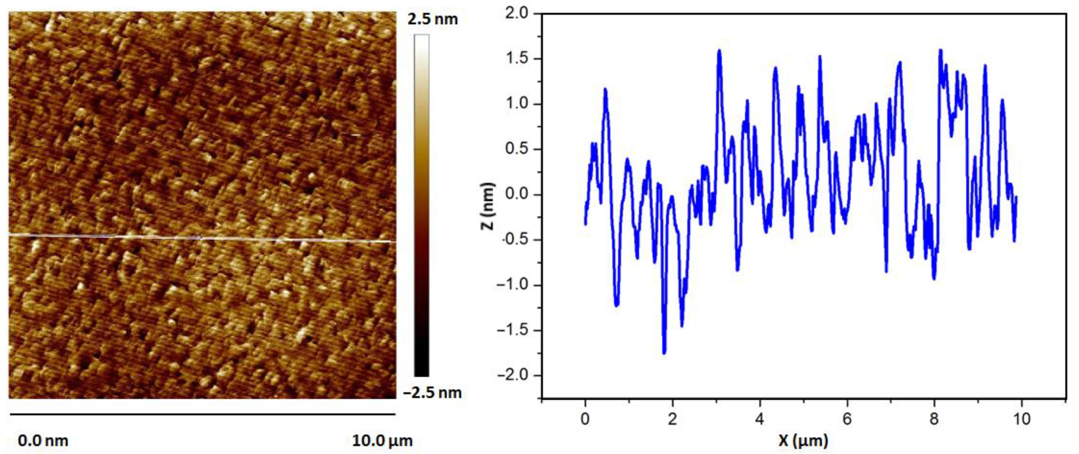
The ellipsometric analysis of the deposited silver mirrors and the AFM scans over its surface show that the silver layer is of high quality. The direct inversion of the experimental ellipsometric data provides the refractive index that is better than the refractive index from Palik’s database of optical properties, as is shown in
Figure 1 [
21]. RMS roughness of the silver layers is also very small, below 1 nm for freshly sputtered silver layers, as is evident from
Figure 2.
3.1.2. Silicon Nitride
Silicon nitride, deposited at an ambient temperature and with an excess of N2 flow, is a non-absorbing material in a visible wavelength range and the uniformity of a layer deposited on a 100 mm wafer is about 5%. As far as absorption in the infrared part of the spectrum is concerned, the presence of Si–H, Si–N and N–H bonds in the material should not create problems, given the strength of oscillators and the small thickness of a layer.
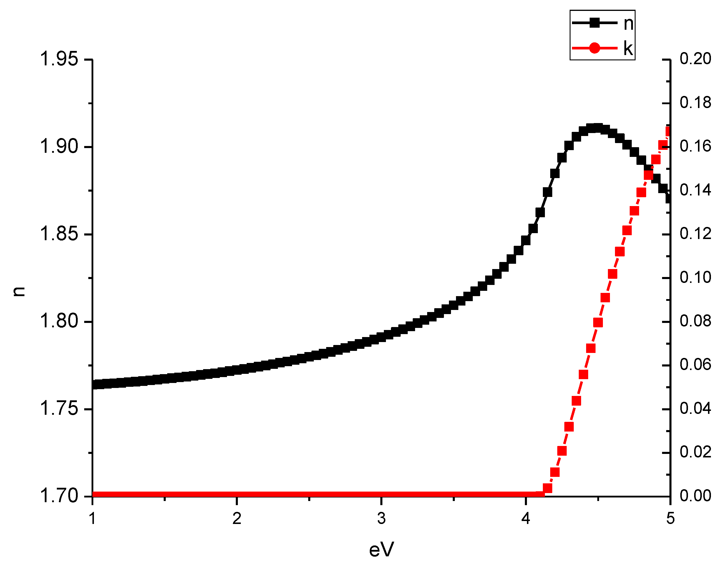
The complex refractive index of SiN
x is shown in
Figure 3 with the corresponding parameters of Tauc–Lorentz dispersion formula given in
Table 1 [
22]. With a bandgap above 4 eV, this silicon nitride cannot affect the reflectance values of the silver mirrors.
3.2. Silver Mirrors
The reflectance of the fabricated silver mirrors (
Figure 4) is 96% at 400 nm and above 98% in the visible range from 460 nm, exceeding 99.5% in the infrared range. It is not affected by the SiN
x layers in the measured range.
The non-protected silver layer oxidizes quickly (full thickness of 200 nm with less than 1 min at test conditions) and at just 1.5 nanometers already slows the process noticeably. The reflectivity of the mirror with a 4.5-nanometer-thick coating does not show any degradation of reflection after 1 min of exposure to O
2 plasma. A 40 min long exposure, however, leads to about 10% reflectivity drop across all spectrums, as can be seen in
Figure 4. Examination of the surface of the coating reveals two distinct kinds of defects on the surface, seen below in optical and electron microscopy images.
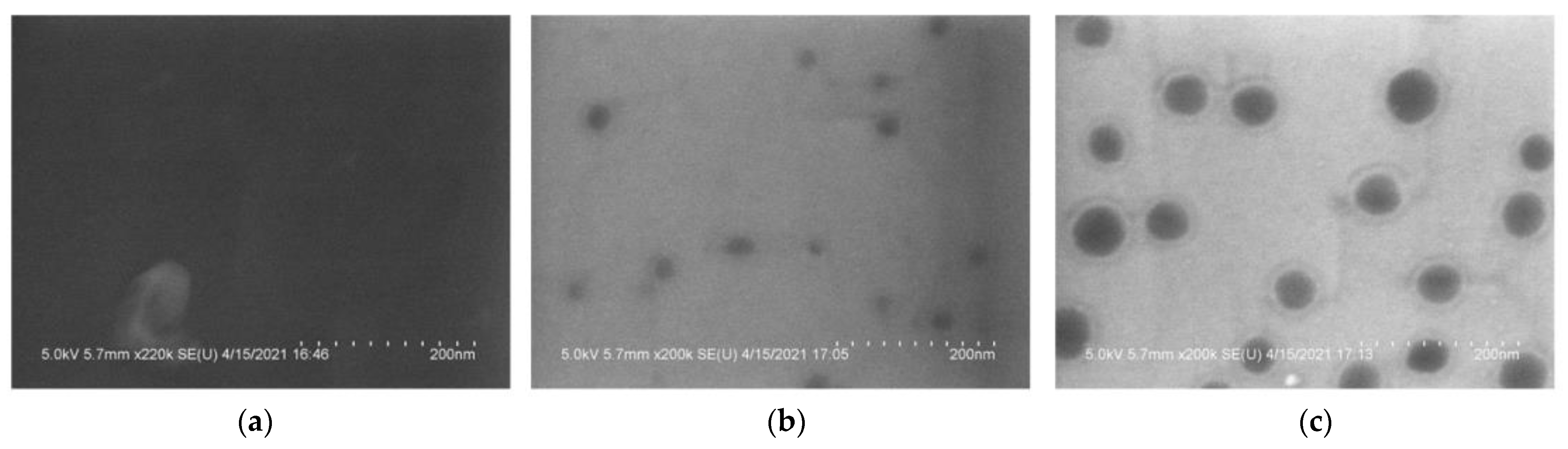
The scanning electron microscopy images (
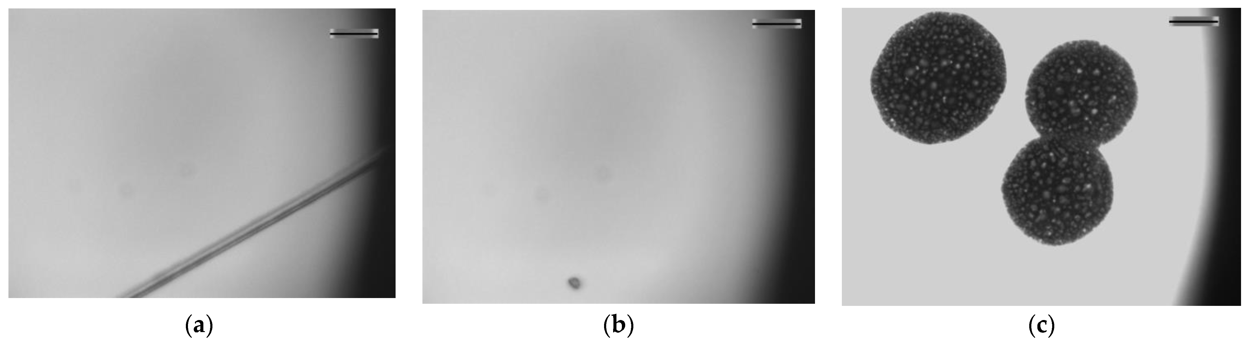
Figure 5) and the optical microscopy images (
Figure 6) reveal the extensive damage for 4.5 nm layers, with the film exposed during 40 min to oxygen plasma demonstrating a halo around the holes resembling an undercut etch defect. Gradual brightening of the images most likely can be explained by the slow decrease of the thickness of a dielectric layer. The left image shows one of the rare fabrication defects, but otherwise its surface looks very uniform. Two other images display gradual deterioration of the surface quality.
The defect types seen in the electron microscopy images of the O
2 plasma-treated mirrors suggest that these defects are rather the result of the erosion of SiN
x films, most likely the spots with a high amount of the Si–H and N–H bonds that are present in this material [
23].
On the other hand, the optical microscopy images show large-scale defects that look like deep craters with no silver left at the bottom (
Figure 6, right panel). The origin of such craters is not yet well understood; however, the pinholes due to the particulate contamination are unlikely to be the reason, as all defects of this kind have a nearly perfect circular shape. Again, the left image of the non-treated with O
2 plasma mirror is only displaying a small scratch, as otherwise it was impossible to find the focus of the microscope, due to the perfect mirror surface.
3.3. GD-OES Studies
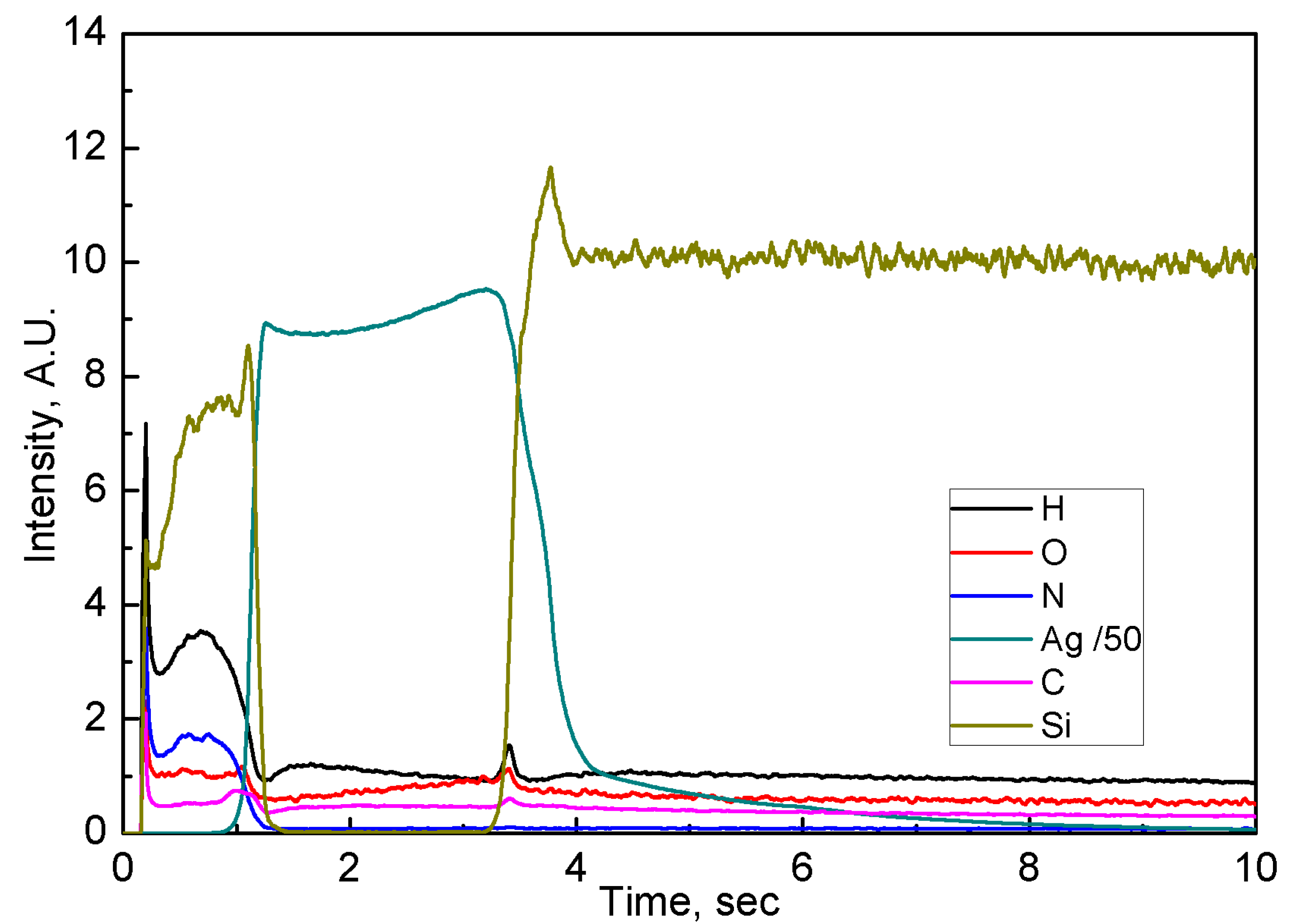
The protected mirrors that are non-treated with the oxygen plasma show distinct layer structure, as can be seen in
Figure 7 for a 12 nm layer of SiN
x on top of 200 nm silver layer deposited on a silicon wafer. The silver emission signal, the strongest by far, was scaled down by a factor of 50 for the sake of the visibility of other elements.
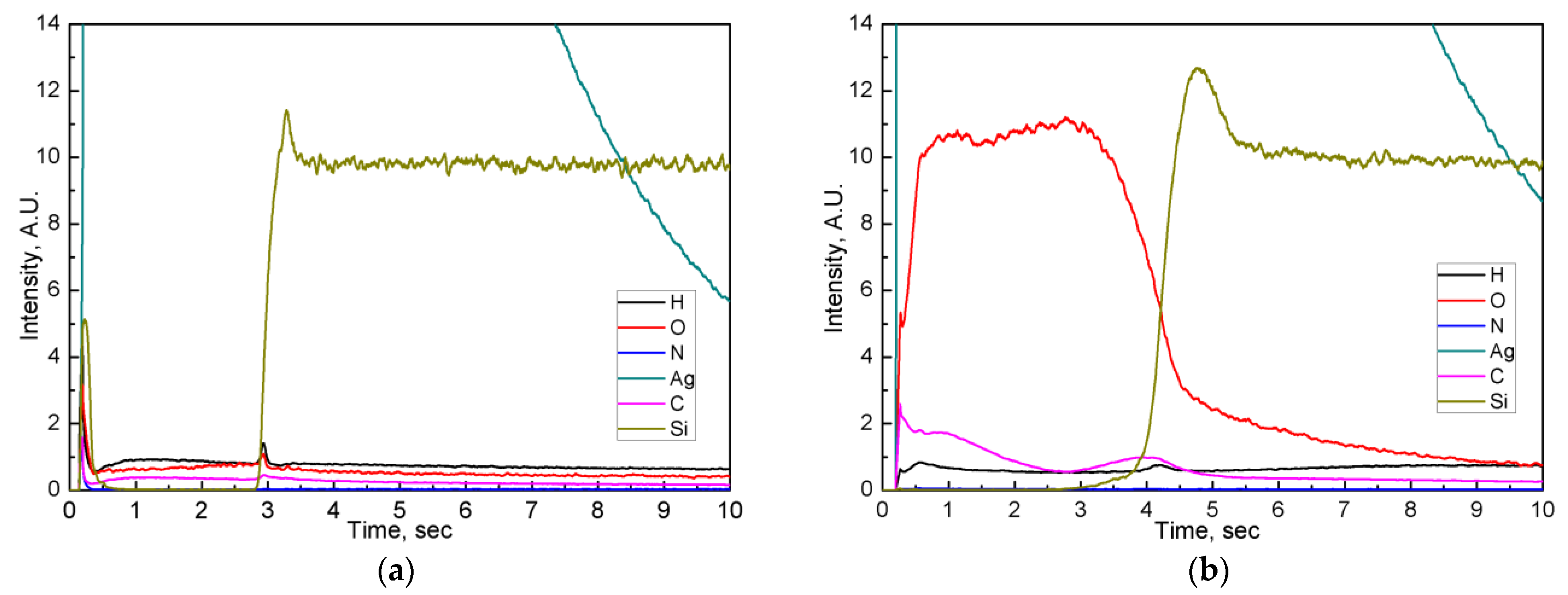
Figure 8 presents the spectra of the non-treated and 1 min oxygen-treated silver mirrors covered with a 1.5 nm SiN
x layer. The initial stages of the GD-OES sputtering clearly demonstrate that for a non-treated sample, both Si and N signals start before the silver signal, indicating the full coverage of the silver layer by the protective coating. After 1 min of oxygen plasma exposure, the protective layer is destroyed and the silver signal starts at the very beginning. Thus, GD-OES analysis allows verifying the state of the coating after the plasma treatment. A strong oxygen signal within the silver layer unambiguously confirms oxidation of a mirror.
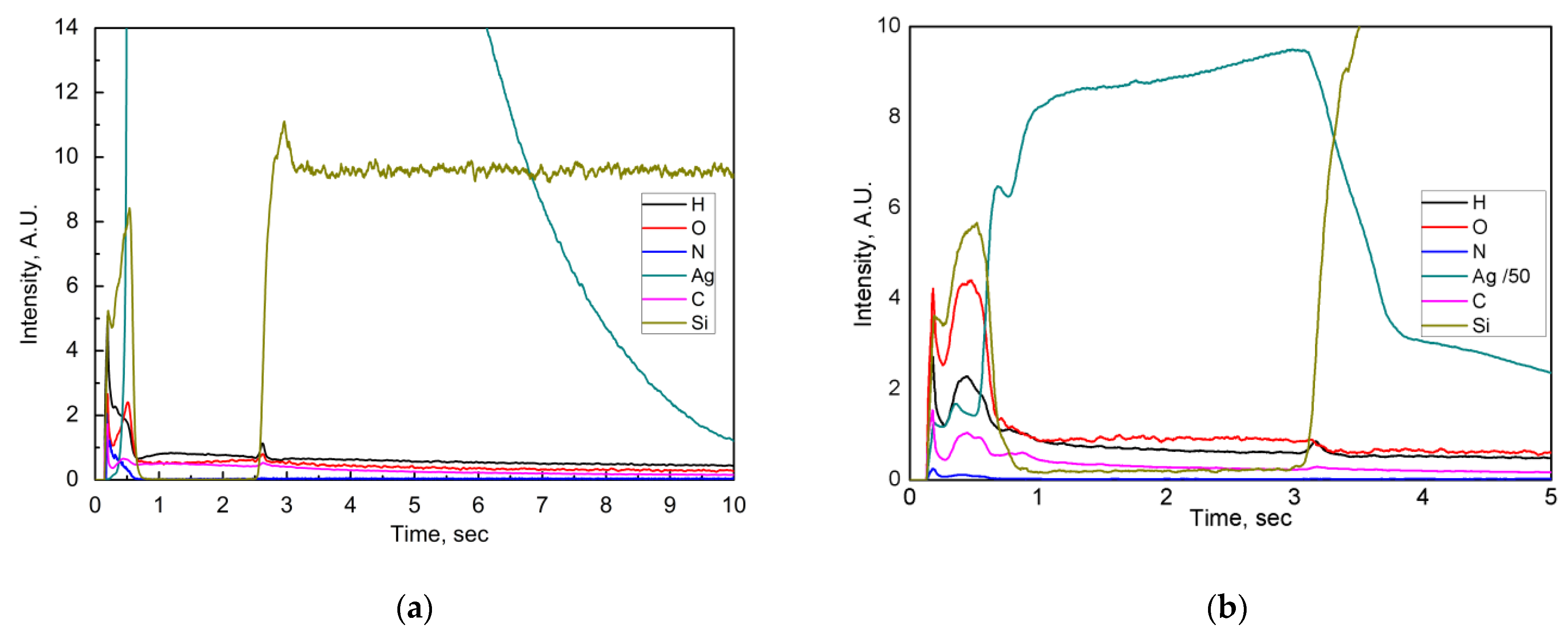
As for the mirrors with a 4.5 nm SiN
x protective layer, the GDOES profiles presented in
Figure 9 demonstrate that 1 min of O
2 exposure at first glance does not lead to the change of a protective layer and, consequently, to the degradation in the reflectance of the mirror. However, the SEM image shown above (
Figure 5, central panel) demonstrates that the damage was already done and a small oxygen peak at the interface between the SiN
x layer and the silver layer demonstrates that (
Figure 9, left panel). After 40 min of O
2 plasma exposure, a silver peak appears from the very beginning and oxygen has already penetrated a silver layer through the small and large defects, created in the SiN
x protective layer, as seen in the right panel of
Figure 9.
4. Discussion
We have undertaken the study of the PECVD silicon nitride for the protective layers on front surface silver mirrors and compared their efficiency with our previous work on AlO
x protective layers. We kept the testing and analysis methodology the same as in a prior study [
16]. PECVD layers of SiN
x provide the effective protection of a silver mirror during 1 min of exposure to oxygen plasma at smaller layer thickness compared to the AlO
x layer (4.5 nm for SiN
x versus 15 nm for AlO
x). However, the types of defects created in the nitride layer even after 1 min of oxygen plasma treatment and the gradual erosion of the protective film and underlying silver during longer oxygen plasma treatment, with the appearance of a micrometric-scale defects, does not allow us to consider PECVD silicon nitride as a viable material for such applications at the moment. Comparison data are presented in
Table 2.
The degradation of a silver layer under the silicon nitride protection during the exposure to atomic and ionized oxygen proceeds very differently compared to the degradation process of AlOx-protected silver. If AlOx layers deposited by ALD completely stop the oxidation and withstand considerably longer exposure times after reaching the layer thickness of 15 nm, the erosion of SiNx protection layer proceeds along two different lines, in specific points at nanoscale, most likely along the hydrogen-rich columnar areas in SiNx, and on a scale of tens of micrometers, the origin of which needs to be further investigated, but at first glance looks like blistering at the interfaces.
Further investigation into the origin of both small-scale and large-scale defects will be required in order to evaluate the potential of the PECVD-deposited silicon nitride to serve as a protection layer for a silver front surface mirror.
5. Conclusions
We fabricated and tested the protected front surface silver mirrors for their sensitivity to the atomic oxygen environment with a protection coating made of PECVD-deposited silicon nitride. We also compared the findings to our prior study on ALD-deposited AlOx-protected silver mirrors. Apparently, for a short-term exposure to atomic oxygen, the SiNx films show reasonable protection at very small (below 5 nm) thickness. However, even a short-term exposure to oxygen produces the small defects in the coating that later lead to slow degradation of the silver mirror reflection, thus proving that this erosion process, while slow, shows that the PECVD-deposited SiNx films are vulnerable to the aggressive environment.
The thin layers of silicon nitride deposited in HD-PECVD system at low substrate temperature can provide reasonable short-term protection of a silver surface against corrosion by active oxygen. These layers also do not noticeably affect the reflection properties of a mirror in all its useful range. The strong points of using SiNx PECVD-deposited layers for the protection of silver mirrors are the existence of the industrially-proven large area deposition technology, the ability to fine-tune the refractive index of the material and the compatibility of PECVD technique with the sensitive silver surface. However, it is too early to suggest that it can be viable addition to the list of PVD and ALD deposited films already used in the protected mirrors technology. Clearly, more research is needed to particularly determine the origins of the defects that develop in a protective film during O2 plasma exposure.
Author Contributions
Conceptualization, P.B. and N.K.; methodology, P.B. and P.C.; validation, P.B.; formal analysis, P.B. and P.C.; investigation, P.B., P.C., G.Z. and D.D.; resources, P.B. and P.C.; data curation, P.B.; writing—original draft preparation, P.B.; writing—review and editing, P.B. and P.C.; visualization, P.B.; supervision, P.B.; project administration, P.B.; funding acquisition, P.B. All authors have read and agreed to the published version of the manuscript.
Funding
This activity was partially funded through the Total-LPICM ANR Industrial Chair “PISTOL” (Contract ANR-17-CHIN-0002-01).
Institutional Review Board Statement
Not Applicable.
Informed Consent Statement
Not Applicable.
Data Availability Statement
Data underlying the results presented in this paper are not publicly available at this time but may be obtained from the authors upon reasonable request.
Conflicts of Interest
The authors declare no conflict of interest.
References
- Paquin, R.A. Properties of Metals. In Handbook of Optics, Optical Society of America, 2nd ed.; Bass, M., Ed.; McGraw-Hill: New York, NY, USA, 2001; Volume 2, Chapter 35; pp. 35.1–35.78. [Google Scholar]
- Garoli, D.; De Marcos, L.V.R.; Larruquert, J.I.; Corso, A.J.; Zaccaria, R.P.; Pelizzo, M.G. Mirrors for space telescopes: Degradation issues. Appl. Sci. 2020, 10, 7538. [Google Scholar] [CrossRef]
- Sytchkova, A.; Schwinde, S.; Protopapa, M.L.; Palmisano, M.; Piegari, A.; Schurman, M.; Kaiser, N. Optical characterization of silver mirrors protected with transparent overcoats. Adv. Opt. Thin Films VI Proc. SPIE 2018, 10691, 106910N. [Google Scholar]
- Fuqua, P.D.; Barrie, J.D. Optical properties and corrosion resistance of durable silver coatings. Mat. Res. Soc. Symp. Proc. 1999, 555, 85–90. [Google Scholar] [CrossRef]
- Xu, X.; He, W.; Wang, C.; Wei, M.; Li, B. SiNx thickness dependence of spectral properties and durability of protected silver mirrors. Surf. Coat. 2017, 324, 175–181. [Google Scholar] [CrossRef]
- Boccas, M.; Vucina, T.; Araya, C.; Vera, E.; Ahhee, C. Protected-silver coatings for the 8-m Gemini telescope mirrors. Thin Solid Films 2006, 502, 275–280. [Google Scholar] [CrossRef]
- Phillips, A.C.; Fryauf, D.M.; Kobayashi, N.P.; Dupraw, B.; Cheleden, S.; Ratliff, C.; Bolte, M.J.; Cowley, D. Update on UCO’s Advanced Coating Lab Development of Silver-Based Mirror Coatings. In Advances in Optical and Mechanical Technologies for Telescopes and Instrumentation II; SPIE-International Society of Optical Engineering: Bellingham, WA, USA, 2016; Volume 9912, p. 99122G. [Google Scholar]
- Fryauf, D.M.; Diaz Leon, J.J.; Phillips, A.C.; Kobayashi, N.P. Silver Film Surface Modification by Ion Bombardment Decreases Surface Plasmon Resonance Absorption. ACS Appl. Mater. Interfaces 2017, 9, 15841–15847. [Google Scholar] [CrossRef] [PubMed]
- Chu, C.-T.; Fuqua, P.D.; Barrie, J.D. Corrosion Characterization of Durable Silver Coatings by Electrochemical Impedance Spectroscopy and Accelerated Environmental Testing. Appl. Opt. 2006, 45, 1583–1593. [Google Scholar] [CrossRef] [PubMed]
- Phillips, A.C.; Fryauf, D.M.; Kobayashi, N.P.; Bolte, M.J.; Dupraw, B.; Ratliff, C.; Pfister, T.; Cowley, D. Progress and new techniques for protected-silver coatings. Proc. SPIE 2014, 9151, 91511B. [Google Scholar]
- Fryauf, D.M.; Phillips, A.C.; Kobayashi, N.P. Corrosion Barriers for Silver-Based Telescope Mirrors: Comparative Study of Plasma-Enhanced Atomic Layer Deposition and Reactive Evaporation of Aluminum Oxide. J. Astron. Telesc. Instrum. Syst. 2015, 1, 044002. [Google Scholar] [CrossRef]
- Fryauf, D.M.; Phillips, A.C.; Bolte, M.J.; Feldman, A.; Tompa, G.S.; Kobayashi, N.P. Testing low-temperature atomic layer deposition of aluminum oxide. 36 Proc. SPIE 2018, 8, 10725. [Google Scholar]
- Fryauf, D.M.; Phillips, A.C.; Bolte, M.J.; Feldman, A.; Tompa, G.S.; Kobayashi, N.P. Scaling Atomic Layer Deposition to Astronomical Optic Sizes: Low-Temperature Aluminum Oxide in a Meter-Sized Chamber. ACS Appl. Mater. Interfaces 2018, 10, 41678–41689. [Google Scholar] [CrossRef]
- Schwinde, S.; Schürmann, M.; Kaiser, N.; Tünnermann, A. Investigation of SiO2-Al2O3 nanolaminates for protection of silver reflectors. Appl. Opt. 2017, 56, C41–C46. [Google Scholar]
- Schwinde, S.; Schürman, M.; Schlegel, R.; Kinast, J.; Dorn, R.J.; Lizon, J.L.; Tordo, S.; Kaiser, N. Protected silver coatings for reflectors. Proc. SPIE 2018, 11180, 111804M. [Google Scholar] [CrossRef] [Green Version]
- Bulkin, P.; Gaiaschi, S.; Chapon, P.; Daineka, D.; Kundikova, N. Protective coatings for front surface silver mirrors by atomic layer deposition. Opt. Exp. 2020, 28, 15753. [Google Scholar] [CrossRef]
- Hoffmann, W.; Pellkofer, T. Thin films in photovoltaics: Technologies and perspectives. Thin Solid Films 2012, 520, 4094–4100. [Google Scholar] [CrossRef]
- Hofrichter, A.; Bulkin, P.; Drevillon, B. Plasma enhanced chemical vapour deposition of SiOxNy in an integrated distributed electron cyclotron resonance reactor. Appl. Surf. Sci. 1999, 142, 447–450. [Google Scholar] [CrossRef]
- Daineka, D.; Bulkin, P.; Girard, G.; Bourée, J.-E.; Drévillon, B. High density plasma enhanced chemical vapor deposition of optical thin films. Eur. Phys. J. Appl. Phys. 2004, 26, 3–9. [Google Scholar] [CrossRef]
- Nelis, T.; Payling, R. Glow Discharge Optical Emission Spectrometry. In Surface Analysis Methods in Materials Science; Springer Series in Surface Sciences; O’Connor, D.J., Sexton, B.A., Smart, R.S.C., Eds.; Springer: Berlin/Heidelberg, Germany, 2003; Volume 23. [Google Scholar]
- Lynch, D.W.; Hunter, W.R. Comments on the Optical Constants of Metals and an Introduction to the Data for Several Metals. In Handbook of Optical Constants of Solids; Palik, E.D., Ed.; Academic Press: Cambridge, MA, USA, 1985; Volume 1, pp. 275–367. [Google Scholar]
- Jellison, J.G.; Modine, F.; Doshi, P.; Rohatgi, A. Spectroscopic Ellipsometry Characterization of Thin-Film Silicon Nitride. Thin Solid Films 1998, 313–314, 193–197. [Google Scholar] [CrossRef] [Green Version]
- Akiki, G.; Frégnaux, M.; Florea, I.; Bulkin, P.; Daineka, D.; Filonovich, S.; Bouttemy, M.; Johnson, E.V. Origin of Area Selective Plasma Enhanced Chemical Vapor Deposition of Microcrystalline Silicon. J. Vac. Sci. Technol. A 2021, 39, 013201. [Google Scholar] [CrossRef]
| Publisher’s Note: MDPI stays neutral with regard to jurisdictional claims in published maps and institutional affiliations. |
© 2021 by the authors. Licensee MDPI, Basel, Switzerland. This article is an open access article distributed under the terms and conditions of the Creative Commons Attribution (CC BY) license (https://creativecommons.org/licenses/by/4.0/).


