Article

N+ Irradiation and Substrate-Induced Variability in the Metamagnetic Phase Transition of FeRh Films

1
United States Naval Research Laboratory, Materials Science and Technology Division, 4555 Overlook Ave., SW, Washington, DC 20375, USA
2
United States Naval Research Laboratory, Electronics Science and Technology Division, 4555 Overlook Ave., SW, Washington, DC 20375, USA
3
United States Naval Research Laboratory, Optical Sciences Division, 4555 Overlook Ave., SW, Washington, DC 20375, USA
*
Author to whom correspondence should be addressed.
Coatings 2021, 11(6), 661; https://doi.org/10.3390/coatings11060661
Submission received: 29 April 2021 / Revised: 25 May 2021 / Accepted: 27 May 2021 / Published: 31 May 2021
(This article belongs to the Special Issue Epitaxial Thin Films: Properties and Applications)

Abstract

:
Metamagnetic FeRh has been the focus of numerous studies for its highly unique antiferromagnetic (AF) to ferromagnetic (FM) metamagnetic transition. While this phase transition usually occurs above room temperature (often Tc > 400 K), both ion irradiation and strained epitaxial growth have been used to bring it to applicable temperatures. Nevertheless, cross sample variability is pervasive in these studies. Here we explore the optical and magnetic properties of 35 nm thick FeRh grown by magnetron sputter deposition simultaneously on two different single crystal substrates: epitaxially on MgO (001) and highly strained with large lattice mismatch on Al2O3 (1000). We then irradiate the epitaxial film with 5 keV N+ ions to introduce disorder (and to a lesser extent, modify chemical composition) without effecting the surface morphology. We find that the phase-transitional properties of both films are strikingly different due to the large lattice mismatch, despite being grown in tandem with nominally identical growth conditions including Fe/Rh stoichiometry, pressure, and temperature. We observe that N+ implantation lowers Tc by ~60 K, yielding a sample with nominally the same transition temperature as the non-epitaxial film on sapphire, yet with a significantly increased magnetic moment, a larger magnetization change and a more abrupt transition profile. We attribute these differences to the Volmer-Weber type growth mode induced by the sapphire substrate and the resulting rougher surface morphology.

1. Introduction

Interest in phase transitional materials has grown over recent years. A significant driver for this change has been an increased need for more robust, lower power and higher speed memory electronics [1,2]. While significant efforts have been underway in memristor development, there are obvious downfalls to the memristors of today including a high error rate and a low average bit lifespan. These are especially troublesome as emerging types of neuromorphic computing elements rely on the unique characteristics of memristors to mimic the spike timing and continuously variable weights of neurons in our human brains [3,4,5]. The memristive capabilities of phase change materials such as the metal insulator transitional VO2 and V2O3 are prime candidates for this next generation [6,7,8]. However, they are hindered by the same endurance issues associated with classical memristors due to electric field oxygen migration [9]. Therein lies the interest in the metamagnetic transition in metallic FeRh whose phase change is rooted in a change in magnetic ordering from antiferromagnetic (AF) to ferromagnetic (FM) along with a volumetric realignment [10,11,12,13,14,15]. This allows for phase change devices with an added magnetic order degree of freedom, as the low temperature AF phase of FeRh can be manipulated at very high speeds using techniques unearthed in the new search for antiferromagnetic electronics [16,17,18]. As interest in FeRh increases, it is important to understand the various factors that have driven significant variability of the metamagnetic transition in literature [10,11,12,19]. Here we compare FeRh films grown in tandem on two different crystalline substrates, MgO (100) and sapphire or Al2O3 (0001), and analyze the impact of N+ irradiation and surface morphology on the metamagnetic transition.
Significant work has already gone into controlling the FeRh phase transition using doping [20,21], pressure [22], irradiation [23,24,25], nanoscale confinement [26], substrate strain [27], and piezoelectric strain [28,29]. A common observation among these studies is the major role strain plays in the metamagnetic transition. This strain-led variation of the exchange integral leads to substantial variations in the transition temperature. Here we consider this by growing FeRh on two different crystalline substrates and observe a sizeable shift (>60 K) in the transition temperature (Tc). We then irradiate the epitaxial film having the higher Tc with 5 keV N+ (approximately 1 × 10¹⁴ N+/cm2) to introduce disorder and observe a similar reduction in Tc that also exceeds 60 K. To investigate the nature of these Tc reductions we examine the strain and its impact on the morphology of the films via XRD and AFM. Additionally, we interrogate the transition temperature with SQUID magnetometry, the magneto-optic Kerr effect (MOKE), and optical reflectance. By growing on both substrates in tandem we have shown for the first time the clear differences between morphological versus irradiative control of the metamagnetic transition in FeRh without questions of cross sample thickness and compositional variability.

2. Experiment

All FeRh films in this study were grown by sample-facing-sputter method in an AJA magnetron sputter system in tandem on a 4 ″rotating sample holder. The substrates were placed within a ~3.5 cm diameter central region of the heating plate to ensure maximum growth condition uniformity between the two samples and then loaded via a loadlock into the chamber with a base pressure of ~10−8 Torr. The samples were then heated to the growth temperature of 630 °C in a 10-mTorr Ar atmosphere and sputter cleaned with a 10-W RF plasma for 5 min before growth. FeRh was grown from an equiatomic 2″ sputter gun with magnets configured for a magnetic target. The films were grown with 75-W DC power then post annealed at 730 °C in HiVac for 1 h.
Since the films grown on sapphire are not epitaxial it is important to quantify the degree of preferred orientation to the substrate. To do this, a quantitative estimate of the preferred orientation can be obtained from the March-Dollase method using our XRD data from Figure 1a [30,31]. Here, the so-called March parameter, r, is related to the fraction of crystallites which exhibit a preferred orientation along a crystalline plane, and is calculated using Equation (1).
r = [sin2α/((κ/κp)2/3 − cos2α)]1/3
where α is the angle between the plane of preferred orientation and a comparison plane, κ is the observed intensity ratio of the diffraction peaks of these planes (in the film), and κp is the corresponding intensity ratio of the two planes from the random powder spectrum. A complete preferred orientation will give r  =  0, while a fully random orientation will give r  =  1. The degree of preferred orientation, η, may be obtained from r using Equation (2).
η = 100% [(1 − r)3/(1 − r3)]1/2
To calculate η for the (110) plane we used the (100) plane as a comparison plane. The angle α between (110) and (100) is calculated from the cubic symmetry to be 45 degrees. The intensity ratio κp is found from the calculated powder spectrum given in the JCPDS card. For the FeRh film on sapphire, r  =  0.5 and η  =  38%, indicating a polycrystalline growth.
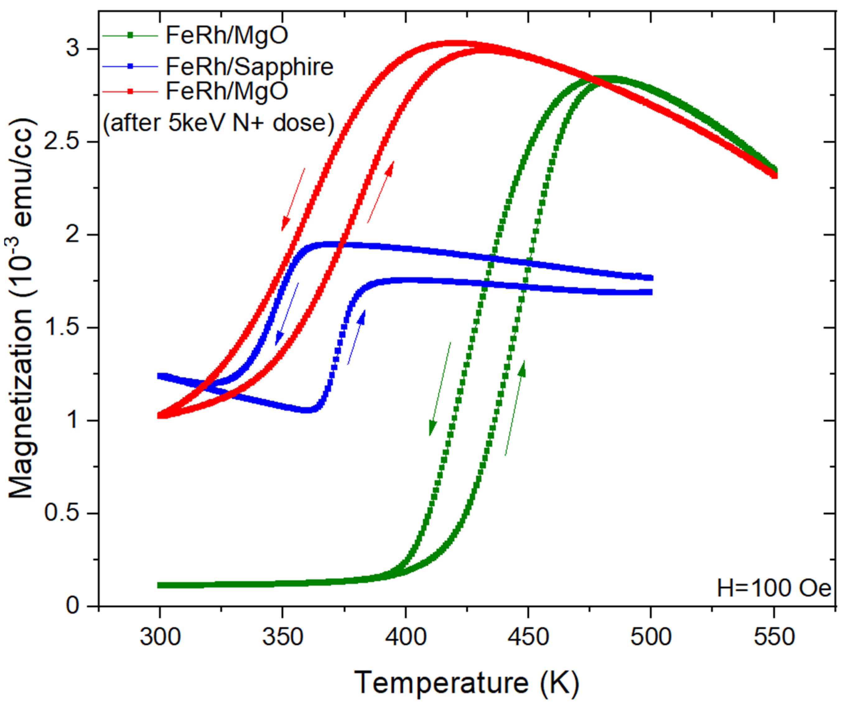
Temperature-dependent magnetization data of the FeRh films was obtained using a Quantum Design SQUID MPMS3 system. Initially, the samples were saturated in a 2 kOe magnetic field at 550 K, then the applied field was removed and the temperature was reduced to 300 K. A 100 Oe field was then applied and measurements made while heating from 300 K to 550 K and back. It is immediately evident that even though the sapphire and MgO samples were grown in tandem, their metamagnetic transitions have very different qualities.
The change in magnetization is only 15% for the sapphire sample as compared to the epitaxial MgO sample. The width of the temperature hysteresis is twice that of the FeRh on sapphire sample. The sapphire sample also shows an increase in magnetization at 300 K at the end of the temperature cycle. This is most likely due to some FM domains not transitioning back to AFM along the super-cooling branch of the first-order transition.
We ascribe these differences in properties to the polycrystalline growth of the FeRh on sapphire versus the epitaxial growth on MgO. It is noteworthy that even without an epitaxial match the sapphire sample has a transition large enough for a range of applications, which aids device integration where epitaxy is not an option.
Figure 2 also shows that the transition temperature can be reduced by dosing the sample with 5 keV N ions. The transition of the undosed sample, the green curve in Figure 2, is ~425 K whereas the dosed transition, the red curve, is ~350 K. This reduction is due to disorder introduced into the FeRh lattice by the high energy ion bombardment as discussed in our previous works [23,24].
It is important to note that even though the N+ dosed sample on MgO and the polycrystalline sample have similar transition temperatures (~350 K), the polycrystalline sample shows a significant loss of magnetic moment, broadening of hysteresis width, and a lowered magnetization change compare to the N+ dosed sample. This is the result of the Volmer-Weber type polycrystalline growth on the mismatched sapphire.
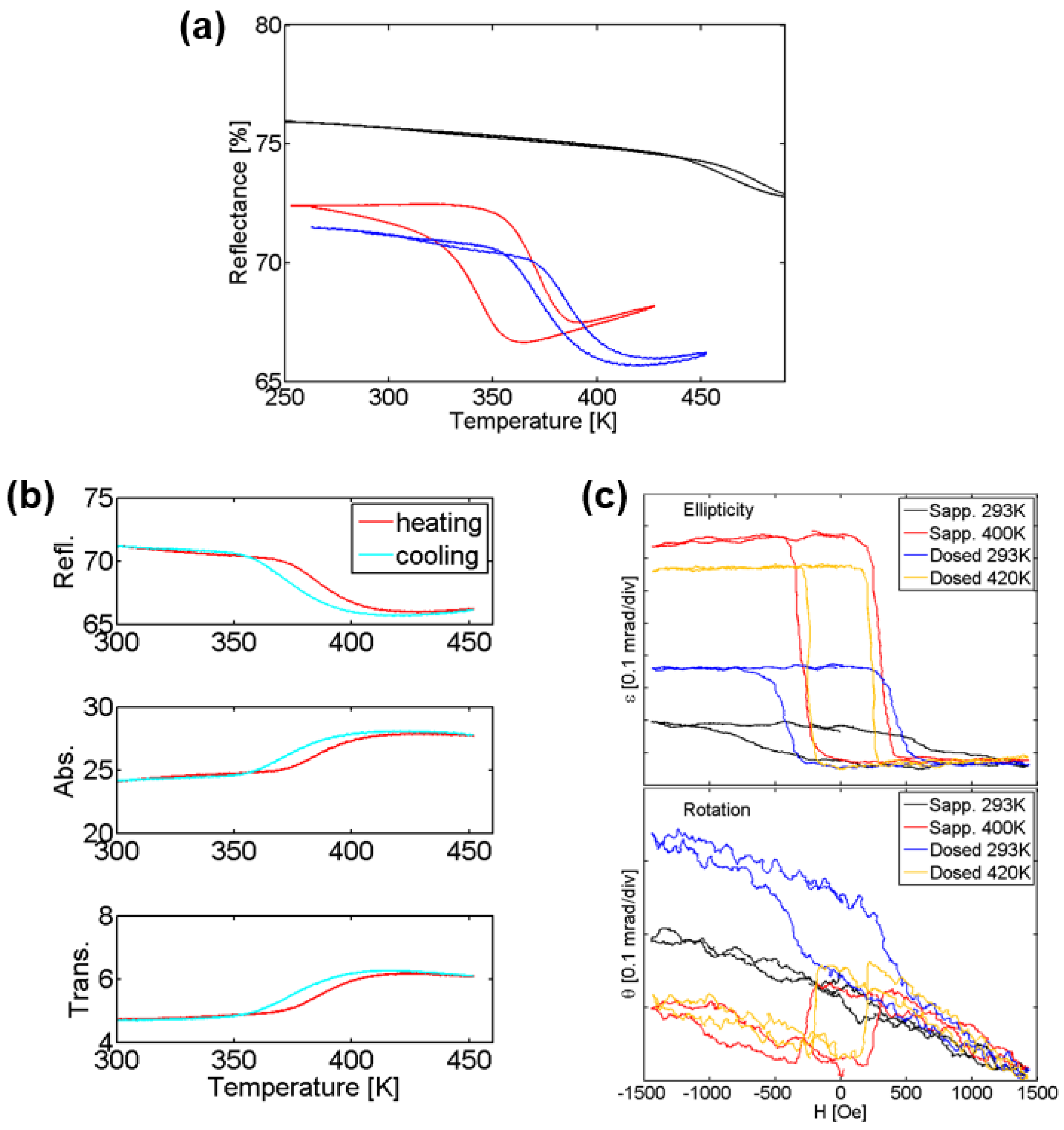
Next, we used optical reflectance to study the metamagnetic transition. The optical reflectance data at 1550 nm shows a decrease in reflectance for both samples with increasing temperature. This change in reflectance is characteristic of the metamagnetic transition, as previously shown [32]. The FeRh on sapphire sample shows a transition temperature upon heating (cooling) of 369 K (337 K). Not only is this a low transition temperature for FeRh, but also the thermal transition hysteresis is very wide (30 K).
For the undosed FeRh/MgO sample the transition temperature is higher than the 500 K temperature limit of our experimental setup. The reflectance and optical hysteresis are small because we can only observe the onset of the transition.
The MgO substrate was polished on both sides which enabled optical transmittance measurements of the FeRh/MgO sample, as shown in Figure 3b. While the transmittance is small (~5%) its trends are opposite to the reflectance, i.e., increases with increasing temperature. The combination of reflectance and transmittance enables calculation of the absorption of the film (since there is a negligible amount of scattering and absorption in the MgO and optical windows at 1550 nm).
Figure 3b shows the optical absorption at 1500 nm of the N+ dosed FeRh increases during the transition. We note that the optical absorption shows the same temperature hysteresis as the transmission, which has interesting scientific and technological implications.
Finally, the magneto-optic properties of the FeRh films are evaluated using MOKE, with the longitudinal signal measured with a 650 nm laser. The MOKE rotation and ellipticity to applied magnetic fields up to 1500 Oe were measured at two temperatures: 293 K and 400 K for the FeRh/Al2O3 film and at 293 K and 420 K for the N+ dosed film on MgO (Figure 3c). The un-dosed FeRh/MgO film did not show any detectable FM characteristics below 450 K, which is the temperature limit on our MOKE setup.
The data for the sapphire substrate sample at 400 K shows a sharp change in polarization rotation and ellipticity for a coercive field of ~300 Oe. However, at 293 K there is still a noticeable yet small and broad change in ellipticity. This is because the samples do not fully recover until they are cooled below 300 K. Once this is done, the weak MOKE signal disappears. This is especially interesting since devices which seek to harness the metamagnetic transition in FeRh will operate around room temperature and a magnetic hysteresis provides an inherent memory effect [33].
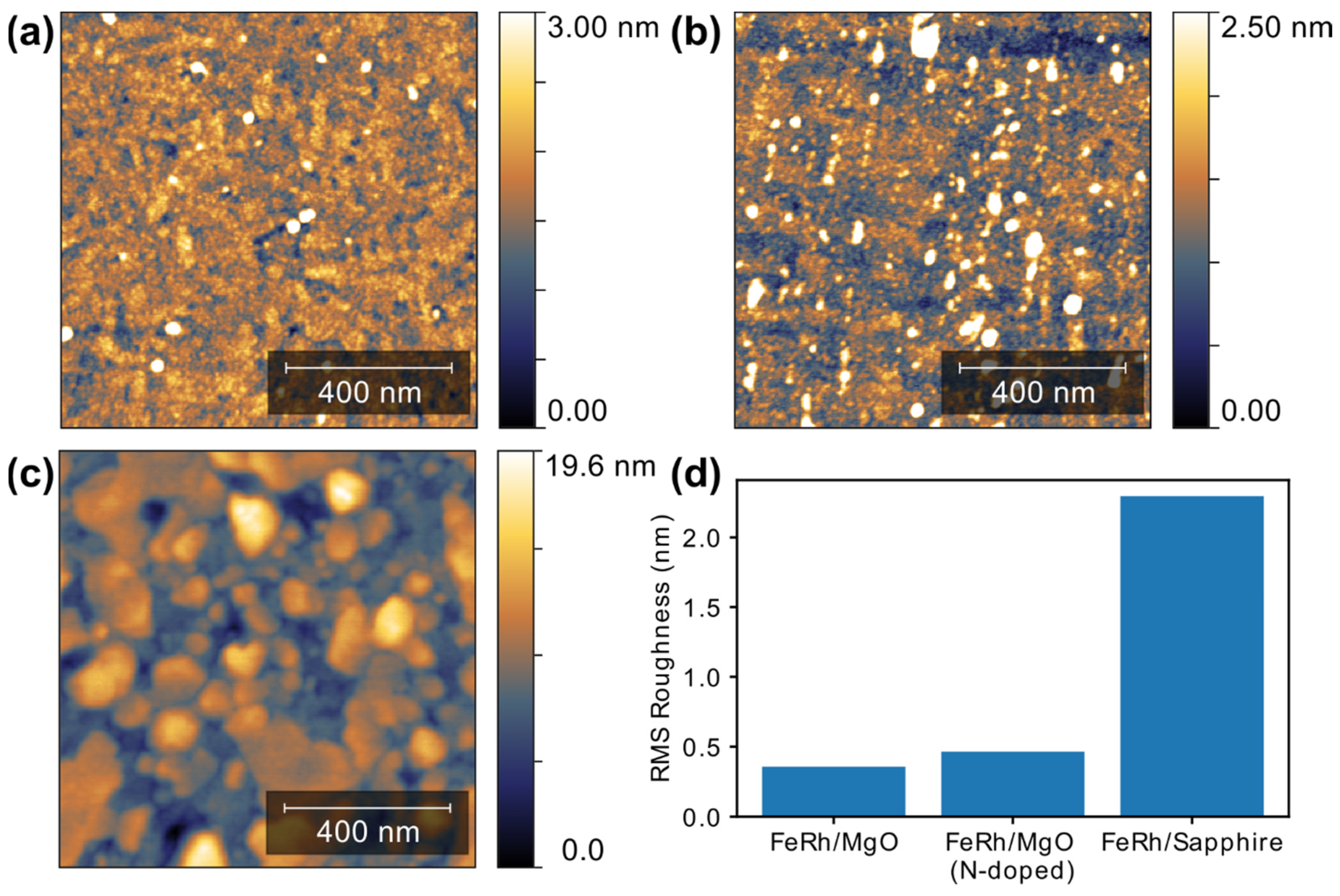
Lastly we use atomic force microscopy (AFM) to image the surface microstructure of the samples and deduce the surface morphology of the as-grown and irradiated films. From Figure 4. it is immediately apparent that the surface roughness of the FeRh/MgO and FeRh/MgO with N+ irradiation is very close with ~0.4–0.5 nm RMS roughness. However, the sample grown with off-epitaxy sputtering on the sapphire crystal shows a significant increase in roughness and larger crystal sizes (RMS ~2.2 nm), which is consistent with our results from XRD analysis and SQUID magnetometry.

3. Conclusions

Here we have investigated the difference between controlling the metamagnetic transition temperature in FeRh films with off-epitaxy sputtering and N+ ion dosing. Samples were grown in tandem to keep growth properties and stoichiometry uniform between all samples, enabling the unambiguous comparison of their metamagnetic properties. Thermomagnetic characterization results show a reduced transition of ~350 K for the sapphire substrate grown film compared to >400 K transition temperature of the un-dosed epitaxial film. We attribute these differences to the Volmer-Weber type growth mode induced by the sapphire substrate and the resulting rougher morphology.
The transition temperature of the 5 keV N+ dosed epitaxial FeRh film is close to that of the polycrystalline film on sapphire; however, the quality of the transition in the sapphire-grown case is much lower than the ion-dosed epitaxial sample. These results show how FeRh can be grown on substrates with epitaxial mismatch and still retain their metamagnetic transition. However, they also demonstrate clearly that using ion irradiation to control the transition temperature of an epitaxially grown film can result in a much higher quality metamagnetic transition.

Author Contributions

All Authors contributed equally to this research. All authors have read and agreed to the published version of the manuscript.

Funding

We acknowledge support by core programs at the U.S. Naval Research Laboratory Nanoscience Institute funded by the Office of Naval Research.

Institutional Review Board Statement

Not applicable.

Informed Consent Statement

Not applicable.

Acknowledgments

This work was supported by the U.S. Office of Naval Research through the Naval Research Laboratory’s basic research program.

Conflicts of Interest

The authors declare no conflict of interest.

References

  1. Masanet, E.; Shehabi, A.; Lei, N.; Smith, S.; Koomey, J. Recalibrating global data center energy-use estimates: Growth in energy use has slowed owing to efficiency gains that smart policies can help maintain in the near term. Science 2020, 367, 984–986. [Google Scholar] [CrossRef]
  2. Jones, N. How to stop data centres from gobbling up the world’s electricity. Nature 2018, 561, 163–166. [Google Scholar] [CrossRef]
  3. Bae, S.H.; Lee, S.; Koo, H.; Lin, L.; Jo, B.H.; Park, C.; Wang, Z.L. The memristive properties of a single VO2 nanowire with switching controlled by self-heating. Adv. Mater. 2013, 25, 5098–5103. [Google Scholar] [CrossRef]
  4. Weiher, M.; Herzig, M.; Tetzlaff, R.; Ascoli, A.; Mikolajick, T.; Slesazeck, S. Improved Vertex Coloring with NbOx Memristor-Based Oscillatory Networks. IEEE Trans. Circuits Syst. I Regul. Pap. 2021, 68, 2082–2095. [Google Scholar] [CrossRef]
  5. Yao, P.; Wu, H.; Gao, B.; Tang, J.; Zhang, Q.; Zhang, W.; Yang, J.J.; Qian, H. Fully hardware-implemented memristor convolutional neural network. Nature 2020, 577, 641–645. [Google Scholar] [CrossRef]
  6. Zimmers, A.; Aigouy, L.; Mortier, M.; Sharoni, A.; Wang, S.; West, K.G.; Ramirez, J.G.; Schuller, I.K. Role of thermal heating on the voltage induced insulator-metal transition in VO2. Phys. Rev. Lett. 2013, 110, 056601. [Google Scholar] [CrossRef] [PubMed] [Green Version]
  7. Mariette, C.; Lorenc, M.; Cailleau, H.; Collet, E.; Guérin, L.; Volte, A.; Trzop, E.; Bertoni, R.; Dong, X.; Lépine, B.; et al. Strain wave pathway to semiconductor-to-metal transition revealed by time-resolved X-ray powder diffraction. Nat. Commun. 2021, 12, 1239. [Google Scholar] [CrossRef]
  8. Sakai, J.; Zaghrioui, M.; Matsushima, M.; Funakubo, H.; Okimura, K. Impact of thermal expansion of substrates on phase transition temperature of VO2 films. J. Appl. Phys. 2014, 116, 123510. [Google Scholar] [CrossRef]
  9. Di Martino, G.; Demetriadou, A.; Li, W.; Kos, D.; Zhu, B.; Wang, X.; de Nijs, B.; Wang, H.; MacManus-Driscoll, J.; Baumberg, J.J. Real-time in situ optical tracking of oxygen vacancy migration in memristors. Nat. Electron. 2020, 3, 687–693. [Google Scholar] [CrossRef]
  10. Kouvel, J.S.; Hartelius, C.C. Anomalous magnetic moments and transformations in the ordered alloy FeRh. J. Appl. Phys. 1962, 33, 1343–1344. [Google Scholar] [CrossRef]
  11. Lommel, J.M. Magnetic and electrical properties of FeRh thin films. J. Appl. Phys. 1966, 37, 1483–1484. [Google Scholar] [CrossRef]
  12. Lommel, J.M.; Kouvel, J.S. Effects of mechanical and thermal treatment on the structure and magnetic transitions in FeRh. J. Appl. Phys. 1967, 38, 1263–1264. [Google Scholar] [CrossRef]
  13. Han, G.C.; Qiu, J.J.; Yap, Q.J.; Luo, P.; Laughlin, D.E.; Zhu, J.G.; Kanbe, T.; Shige, T. Magnetic stability of ultrathin FeRh films. J. Appl. Phys. 2013, 113, 17C107. [Google Scholar] [CrossRef] [Green Version]
  14. Keavney, D.J.; Choi, Y.; Holt, M.V.; Uhlíř, V.; Arena, D.; Fullerton, E.E.; Ryan, P.J.; Kim, J.W. Phase coexistence and kinetic arrest in the magnetostructural transition of the ordered alloy FeRh. Sci. Rep. 2018, 8, 1778. [Google Scholar] [CrossRef] [PubMed] [Green Version]
  15. Fan, R.; Kinane, C.J.; Charlton, T.R.; Dorner, R.; Ali, M.; De Vries, M.A.; Brydson, R.M.D.; Marrows, C.H.; Hickey, B.J.; Arena, D.A.; et al. Ferromagnetism at the interfaces of antiferromagnetic FeRh epilayers. Phys. Rev. B Condens. Matter Mater. Phys. 2010, 82, 184418. [Google Scholar] [CrossRef] [Green Version]
  16. Jungwirth, T.; Marti, X.; Wadley, P.; Wunderlich, J. Antiferromagnetic spintronics. Nat. Nanotechnol. 2016, 11, 231–241. [Google Scholar] [CrossRef]
  17. Němec, P.; Fiebig, M.; Kampfrath, T.; Kimel, A.V. Antiferromagnetic opto-spintronics. Nat. Phys. 2018, 14, 229–241. [Google Scholar] [CrossRef]
  18. Baltz, V.; Manchon, A.; Tsoi, M.; Moriyama, T.; Ono, T.; Tserkovnyak, Y. Antiferromagnetic spintronics. Rev. Mod. Phys. 2018, 90, 015005. [Google Scholar] [CrossRef] [Green Version]
  19. Barton, C.W.; Ostler, T.A.; Huskisson, D.; Kinane, C.J.; Haigh, S.J.; Hrkac, G.; Thomson, T. Substrate Induced Strain Field in FeRh Epilayers Grown on Single Crystal MgO (001) Substrates. Sci. Rep. 2017, 7, 44397. [Google Scholar] [CrossRef] [PubMed] [Green Version]
  20. Barua, R.; Jiménez-Villacorta, F.; Lewis, L.H. Predicting magnetostructural trends in FeRh-based ternary systems. Appl. Phys. Lett. 2013, 103, 102407. [Google Scholar] [CrossRef]
  21. Bennett, S.P.; Ambaye, H.; Lee, H.; LeClair, P.; Mankey, G.J.; Lauter, V. Direct Evidence of Anomalous Interfacial Magnetization in Metamagnetic Pd doped FeRh Thin Films. Sci. Rep. 2015, 5, 9142. [Google Scholar] [CrossRef] [Green Version]
  22. Urban, C.; Bennett, S.P.; Schuller, I.K. Hydrostatic pressure mapping of barium titanate phase transitions with quenched FeRh. Sci. Rep. 2020, 10, 6312. [Google Scholar] [CrossRef]
  23. Cress, C.D.; Wickramaratne, D.; Rosenberger, M.R.; Hennighausen, Z.; Callahan, P.G.; LaGasse, S.W.; Bernstein, N.; van’t Erve, O.M.; Jonker, B.T.; Qadri, S.B.; et al. Direct-Write of Nanoscale Domains with Tunable Metamagnetic Order in FeRh Thin Films. ACS Appl. Mater. Interfaces 2021, 13, 836–847. [Google Scholar] [CrossRef]
  24. Bennett, S.P.; Herklotz, A.; Cress, C.D.; Ievlev, A.; Rouleau, C.M.; Mazin, I.I.; Lauter, V. Magnetic order multilayering in FeRh thin films by the He-ion irradiation. Mater. Res. Lett. 2018, 6, 106–112. [Google Scholar] [CrossRef]
  25. Heidarian, A.; Bali, R.; Grenzer, J.; Wilhelm, R.A.; Heller, R.; Yildirim, O.; Lindner, J.; Potzger, K. Tuning the antiferromagnetic to ferromagnetic phase transition in FeRh thin films by means of low-energy/low fluence ion irradiation. Nucl. Instrum. Methods Phys. Res. Sect. B Beam Interact. Mater. Atoms 2015, 358, 251–254. [Google Scholar] [CrossRef]
  26. Uhlíř, V.; Arregi, J.A.; Fullerton, E.E. Colossal magnetic phase transition asymmetry in mesoscale FeRh stripes. Nat. Commun. 2016, 7, 13113. [Google Scholar] [CrossRef] [Green Version]
  27. Ostler, T.A.; Barton, C.; Thomson, T.; Hrkac, G. Modeling the thickness dependence of the magnetic phase transition temperature in thin FeRh films. Phys. Rev. B 2017, 95, 064415. [Google Scholar] [CrossRef] [Green Version]
  28. Cherifi, R.O.; Ivanovskaya, V.; Phillips, L.C.; Zobelli, A.; Infante, I.C.; Jacquet, E.; Garcia, V.; Fusil, S.; Briddon, P.R.; Guiblin, N.; et al. Electric-field control of magnetic order above room temperature. Nat. Mater. 2014, 13, 345–351. [Google Scholar] [CrossRef] [Green Version]
  29. Bennett, S.P.; Wong, A.T.; Glavic, A.; Herklotz, A.; Urban, C.; Valmianski, I.; Biegalski, M.D.; Christen, H.M.; Ward, T.Z.; Lauter, V. Giant Controllable Magnetization Changes Induced by Structural Phase Transitions in a Metamagnetic Artificial Multiferroic. Sci. Rep. 2016, 6, 22708. [Google Scholar] [CrossRef] [PubMed] [Green Version]
  30. Zolotoyabko, E. Determination of the degree of preferred orientation within the March-Dollase approach. J. Appl. Crystallogr. 2009, 42, 513–518. [Google Scholar] [CrossRef]
  31. Dollase, W.A. Correction of intensities of preferred orientation in powder diffractometry: Application of the march model. J. Appl. Crystallogr. 1986, 19, 267–272. [Google Scholar] [CrossRef]
  32. Bennett, S.P.; Currie, M.; van’t Erve, O.M.J.; Mazin, I.I. Spectral reflectivity crossover at the metamagnetic transition in FeRh thin films. Opt. Mater. Express 2019, 7, 2870. [Google Scholar] [CrossRef]
  33. Gray, I.; Stiehl, G.M.; Heron, J.T.; Mei, A.B.; Schlom, D.G.; Ramesh, R.; Ralph, D.C.; Fuchs, G.D. Imaging uncompensated moments and exchange-biased emergent ferromagnetism in FeRh thin films. Phys. Rev. Mater. 2019, 3, 124407. [Google Scholar] [CrossRef] [Green Version]
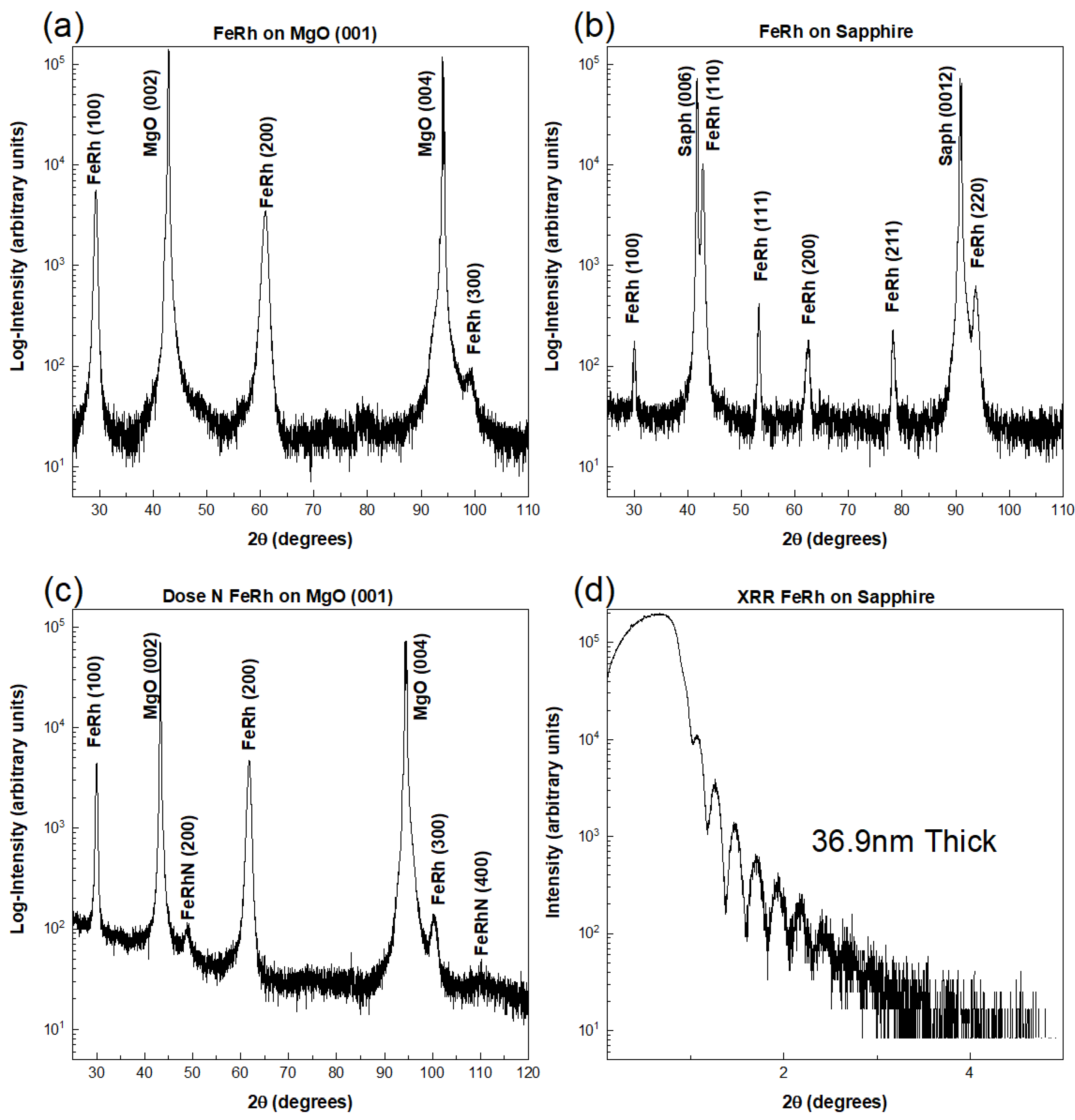
Figure 1. XRD data from FeRh thin film samples. (a) XRD theta-2theta curve of FeRh on MgO. (b) XRD theta-2theta curve of FeRh on Sapphire. (c) XRD theta-2theta curve of FeRh on MgO after dosing with N+. (d) X-ray reflectivity curve showing interference fringes for FeRh on sapphire sample with thickness of 36.9 nm.
Coatings 11 00661 g001
Figure 2. SQUID magnetometry data of the three samples (FeRh on sapphire (blue), FeRh on MgO (green), FeRh on MgO with a 5kV N+ dose (red). Arrows indicate sweep direction of temperature with all measurements starting at 300 K.
Coatings 11 00661 g002
Figure 3. Optical properties of FeRh films. (a) reflectance at 1550 nm wavelength of FeRh films on MgO (black) and Al2O3 (red) substrates and N-dose FeRh on MgO (blue). (b) optical reflectance, transmittance, and absorptance (calculated) of N-dosed FeRh films on MgO substrate at 1550-nm wavelength. (c) MOKE ellipticity (top) and rotation (bottom) of FeRh/Al2O3 at 293 K and 400 K and dosed FeRh/MgO at 293 K and 420 K. For the FeRh/Al2O3 the presence of a small MOKE signal at room temperature is due to the thermal hysteresis which does not recover, as seen in (a), until the sample is brought below 270 K.
Coatings 11 00661 g003
Figure 4. Atomic force microscopy (AFM) contour maps showing the topography of the FeRh films. (a) FeRh/MgO sample (b) FeRh/MgO with 5 keV N+ irradiation (c) FeRh/Sapphire sample (d) Histogram showing RMS roughness for the 3 samples calculated as a average 5 scanned sites on each sample.
Coatings 11 00661 g004
Publisher’s Note: MDPI stays neutral with regard to jurisdictional claims in published maps and institutional affiliations.