Article

Application of Bone Morphogenetic Protein 7 Enhanced the Osteogenic Differentiation and Mineralization of Bone Marrow-Derived Stem Cells Cultured on Deproteinized Bovine Bone

1
Department of Periodontics, College of Medicine, Catholic University of Korea, Seoul 06591, Korea
2
Ebiogen, #405, Sungsu A1 Center, 48 Ttukseom-ro 17-ga-gil, Seongdong-gu, Seoul 04784, Korea
*
Author to whom correspondence should be addressed.
Hyun-Jin Lee and Sae-Kyung Min contributed equally.
Coatings 2021, 11(6), 642; https://doi.org/10.3390/coatings11060642
Submission received: 7 May 2021 / Revised: 22 May 2021 / Accepted: 25 May 2021 / Published: 27 May 2021

Abstract

:
The growth of bone morphogenetic protein 7 (BMP-7) has been applied for tissue regeneration due to its osteoinductive properties. The aim of this research is to analyze the enhancing effects of BMP-7 on the osteogenic differentiation and mineralization of human bone marrow-derived stem cells cultured on the bovine bone particle. After the stem cells were loaded onto the bone graft material, their morphology was observed on day 7. Viability assays based on the application of fluorescent stains were used for qualitative analyses. Alkaline phosphatase activity assays and Alizarin red staining were used for the assessment of osteogenic differentiation on days 7 and 14. Next-generation mRNA sequencing was applied to evaluate global gene expression. Gene ontology and pathway analysis was used to propose the underlying mechanism. Fibroblast-like morphology was attained with the stem cells. The cells were shown to be firmly attached to the bone particle. Most of the stem cells produced an intense green fluorescence. The relative cellular viability assay values for BMP-7 groups at 0, 10, and 100 ng/mL on day 7 were 0.295 ± 0.003, 0.250 ± 0.002, and 0.240 ± 0.003, respectively (p < 0.05). Alkaline phosphatase activity was significantly higher in BMP-7 groups at concentration of 100 ng/mL compared to the control on days 7 and 14 (p < 0.05). The results of the mineralization assay showed significantly higher values for BMP-7 groups at 100 ng/mL concentration when compared with the control (p < 0.05). The expression of RUNX2 was increased with application of BMP-7 and mitogen-activated protein kinase pathway was associated with the target genes. Overall, this study shows that in vitro application of BMP-7 increases alkaline phosphorylase activity and mineralization of stem cells culture on deproteinized bovine bone mineral. The study suggests that combining stem cells with osteoinductive growth factors with scaffolds can have synergy effects on osteogenic differentiation.

1. Introduction

As expected, in the first half of the 21st century, the number of people over 65 years of age dramatically increased [1]. Moreover, there might be a demographic shift within the elderly population in the coming decades in which disease incidences and mortality increase [2]. Most adults suffer from periodontal disease, which is the second most-seen oral disease along with dental caries [3]. Periodontal disease is a major cause of tooth loss in the elderly population [4]. The economic burden and quality of life for the elderly tend to drop significantly due to the need for immediate and continuous treatment of periodontitis [5]. Therefore, it is necessary to regenerate lost tissue and restore it to its functioning state [6].
In more recent years, the application of stem cells has been introduced to the tissue regeneration field [7]. Different medication treatments have been widely studied to enhance the tissue regeneration [8]. Moreover, research on the growth factors has been actively conducted and has contributed to the development of tissue regeneration concepts [9]. One of the most widely applied growth factors for tissue regeneration is bone morphogenetic proteins (BMPs) [10]. One of the widely noted BMPs, BMP-7, was applied in various studies due to its osteoinductive property [11,12,13,14,15]. The effects of the BMP-7 stimulation on osteoblasts cultured on various biomaterials was recently tested, and the loading osteoblasts on bovine bone mineral with rhBMP-7 led to a higher cell proliferation and collagen synthesis [11]. BMPs carried in a matrix was suggested to be able to serve as a substitute for the autologous bone graft in the critical size defects [12]. In a previous study, the reconstruction of the mandibular defect was done by local application of BMP-7 and bone grafting materials including xenogenic bone mineral [13]. The addition of BMP-7 resulted in the significantly stimulated bone formation in sinus augmentation procedures [14]. BMP-7 has been combined with the stem cells for tissue engineering purposes [15]. The purpose of this study is to evaluate the bone regeneration ability through the simultaneous application of BMP-7 and stem cells cultured on deproteinized bovine bone.

2. Materials and Methods

2.1. Culturing of Bone Marrow-Derived Mesenchymal Stem Cells on Bone

The Institutional Review Board of Seoul St. Mary’s Hospital approved the present study (KC18SESI0199 and KC20SISE0582; approval date: 25 April 2018). Human bone marrow-derived mesenchymal stem cells (hBMSCs; Catholic MASTER cells) were obtained from the Catholic Institute of Cell Therapy (CIC, Seoul, Korea) [16]. CIC verified that cells showed >90% positive CD 73, >90% positive CD 90 expression, >90% negative CD 31, >90% negative CD 34, and >90% negative CD 45 expression.
The quantity of bone particle used per culture was 0.08 g/well. Cell-seeding density used for individual assays was 1.0 × 10⁴. The hBMSCs were loaded onto the deproteinized bovine bone particle (granule size of 0.25–1 mm; Geistlich Bio-Oss®, Geistlich Pharma AG, Wolhusen, Switzerland) and incubated for one day in growth media to make the hBMSCs grow on the bone particle. After that, the bone graft was moved to a new culture plate. The stem cell-loaded graft was cultured in osteogenic media. Along with this, the graft was also incubated with the BMP-7 at the final concentrations of 0 (=control), 10 and 100 ng/mL.

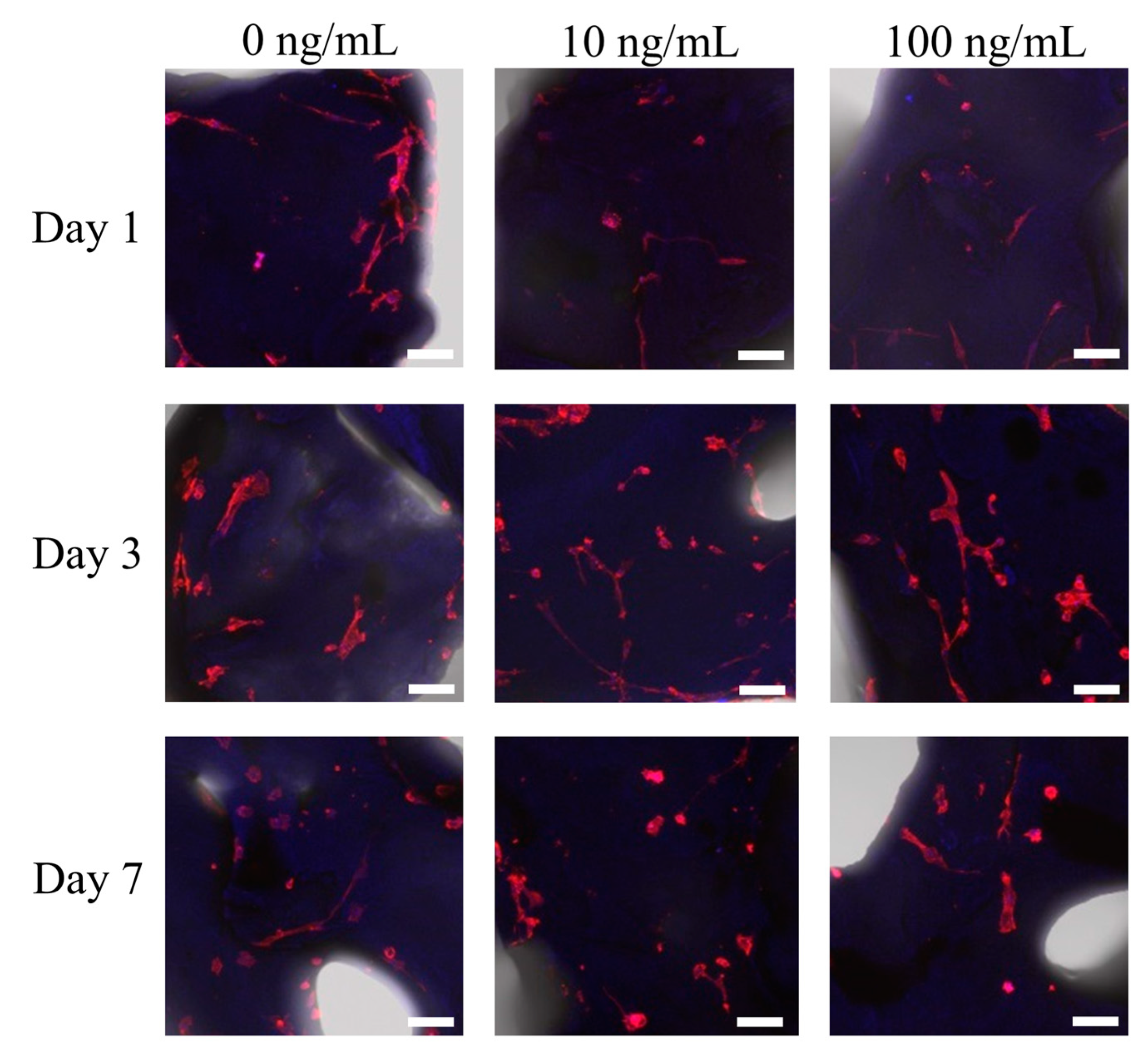
2.2. Morphologic Evaluation of Stem Cells

The morphology of the hBMSCs grown on the bone graft material was perceived on day 7, using a confocal laser microscope (LSM800 w/Airyscan; Carl Zeiss, Zena, Oberkochen, Germany). The culture media was removed and the stem cell-bone graft was meticulously washed with phosphate-buffered saline. The hBMSC-bone graft was fixed with 4% paraformaldehyde (P2031, Biosesang, Seongnam-si, Korea) at room temperature for 20 min. Then hBMSC-bone particle was washed with Dulbecco’s phosphate-buffered saline solution three times. Cells were then stained using a 1× Phalloidin conjugate working solution (ab235138, Abcam, Cambridge, UK) for 60 min at room temperature. The hBMSC-bone graft was washed and mounted with small drop volumes (approximately 25 μL) of VECTASHIELD Mounting Medium with DAPI (H-1200, VECTOR laboratories). The samples were stored at 4 °C, protected from all light.

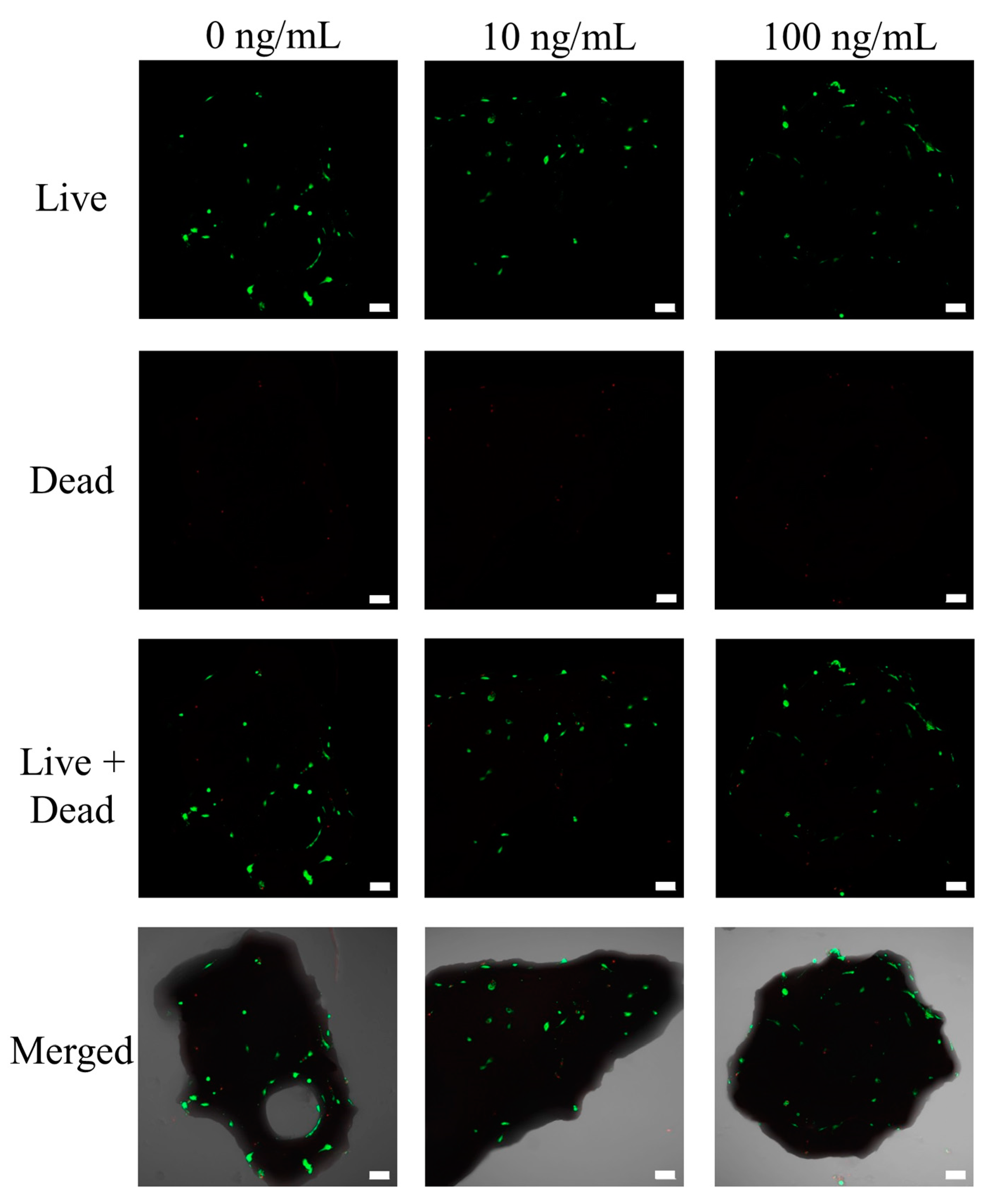
2.3. Determination of Cellular Viability

Viability assays based on the application of fluorescent stains (Live/Dead Kit assay, Molecular Probes, Eugene, OR, USA) was used for qualitative analysis of the hBMSCs cultured on the bone particle on day 7. The hBMSC-bone particle was treated with working solution for one hour at room temperature following the manufacturer’s recommendation. The hBMSC-bone particle was viewed using a confocal laser microscope (LSM800 w/Airyscan; Carl Zeiss, Jena, Germany). Cell Counting Kit-8 (Dojindo, Tokyo, Japan) was used for the evaluation of quantitative cellular viability at 37 °C for 1 h. Three independent cultures were used for the evaluation.

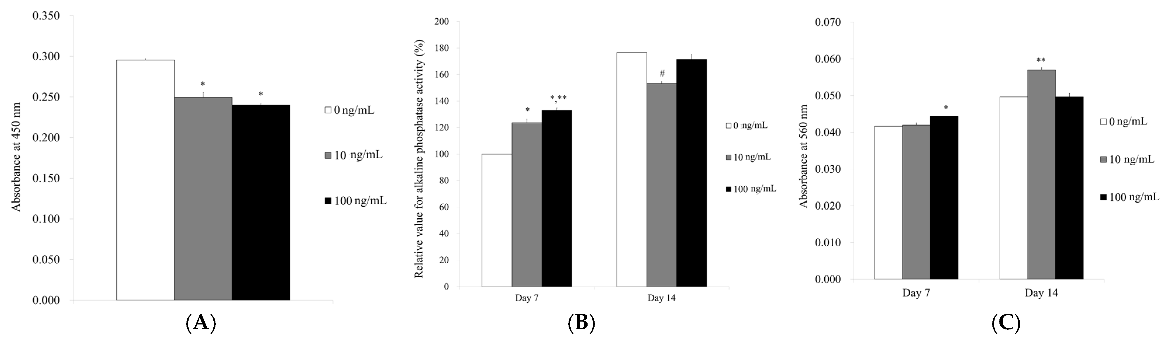
2.4. Level of Alkaline Phosphatase Activity and Calcium Deposition

Alkaline phosphatase activity level was used to assess the osteogenic differentiation on days 7 and 14 (K412-500, BioVision, Inc., Milpitas, CA, USA) by obtaining hBMSCs grown on bone graft material with osteogenic media. The hBMSCs on the deproteinized bovine bone were re-suspended with an assay buffer, then decomposed with triton X-100. Insoluble material was removed, and the supernatant was mixed with p-nitrophenylphosphate substrate. The incubation of the mixture was continued for 60 min at 4 °C. We measured the spectrophotometric absorbance of the samples at 405 nm.
To assess the degree of mineralization, Alizarin red staining was used to evaluate calcium deposition. The hBMSCs on the bone particle was stained with 2% Alizarin-Red Solution (Cat. No. 0223; ScienCell Research Laboratories, Inc., Carlsbad, CA, USA) for 30 min at room temperature. The bound dye was solubilized by applying 10% cetylpyridinium chloride (Cat. No. C0732-100G; Sigma-Aldrich Co., St. Louis, MO, USA) solution for 15 min. The quantification of the bound dyes was performed on days 7 and 14 at 560 nm.

2.5. Extraction of RNA and Sequencing of mRNA

Isolation of total RNA was performed using Trizol reagent (Invitrogen Corp., Carlsbad, CA, USA) and RNA quantification was performed using a spectrophotometer (ND-2000, Thermo Fisher Scientific Inc., Waltham, MA, USA). Library of control and test RNAs were built using SMARTer Stranded RNA-Seq Kit (Clontech Laboratories, Inc., Palo Alto, CA, USA) according to the manufacturer’s instructions [17].
The alignment file was gained by mapping mRNA-Seq reads tool using TopHat software tool [18]. Differentially expressed genes were determined by counts from unique and multiple alignments [19]. The read count data were processed by quantile–quantile normalization method [20]. Transcripts were assembled by cufflinks and the expression level of the gene regions was evaluated using fragments per kilobase of exon per million fragments method. Data mining and graphic visualization were performed using ExDEGA (ebiogen Inc., Seoul, Korea).

2.6. Statistical Analysis

We presented the data as means ± standard deviations of the experiments. We conducted the tests of normality and equal of variances. We tested the differences among groups by applying one-way analysis of variance with Tukey’s post hoc test (SPSS 12 for Windows, SPSS Inc., Chicago, IL, USA; p < 0.05).

3. Results

3.1. Cell Attachment of Stem Cells to the Deproteinized Bovine Bone Mineral

Fibroblast-like morphology was attained with hBMSC. The cells were shown to be firmly attached to the bone particle on day 7 (Figure 1). Addition of BMP-7 did not produce significant changes. However, the number of cells presented on deproteinized bovine bone mineral particles seemed to decrease with addition of BMP-7 (Figure 1).

3.2. Determination of Cellular Viability

Most of the stem cells produced an intense green fluorescence on day 7 (Figure 2). Minimal red fluorescence was noted, indicating there were a very low number of dead cells. The absorbance values at 450 nm on days 1, 3, and 7 are shown in Figure 3A. The relative cellular viability assay values for the BMP-7 groups at 0, 10, and 100 ng/mL on day 7 were 0.295 ± 0.003, 0.250 ± 0.002, and 0.240 ± 0.003, respectively (p < 0.05). The application of BMP-7 showed a statistical decrease in the cellular viability on day 7.

3.3. Level of Alkaline Phosphatase Activity and Calcium Deposition

The alkaline phosphatase activity assays on days 7 and 14 are shown in Figure 3B. The absorbance values at 405 nm on day 7 for BMP-7 at 0, 10, and 100 ng/mL were 0.074 ± 0.002, 0.077 ± 0.001, and 0.080 ± 0.001, respectively (p < 0.05). Significantly higher values were noted for BMP-7 groups at 100 ng/mL concentration when compared with the control group (p < 0.05). The results on day 14 were 0.076 ± 0.001, 0.074 ± 0.002, and 0.081 ± 0.001 for BMP-7 groups at 0, 10, and 100 ng/mL, respectively (p < 0.05). Statistically higher values were seen in BMP-7 groups at 100 ng/mL concentration when compared with the 0 ng/mL group (p < 0.05).
The results of the mineralization assay at days 7 and 14 are shown in Figure 3C. The absorbance values at 560 nm on day 7 were 0.042 ± 0.001, 0.042 ± 0.000, and 0.044 ± 0.001 for BMP-7 at 0, 10, and 100 ng/mL groups, respectively (p < 0.05). The quantification results showed significantly higher values for BMP-7 groups at 100 ng/mL concentration when compared with the 0 ng/mL group (p < 0.05). The results on day 14 were 0.050 ± 0.001, 0.057 ± 0.001, and 0.050 ± 0.002 for BMP-7 groups at 0, 10, and 100 ng/mL, respectively (p < 0.05). Statistically higher values were noted for BMP-7 groups at 10 ng/mL concentration when compared with the 0 ng/mL group (p < 0.05).

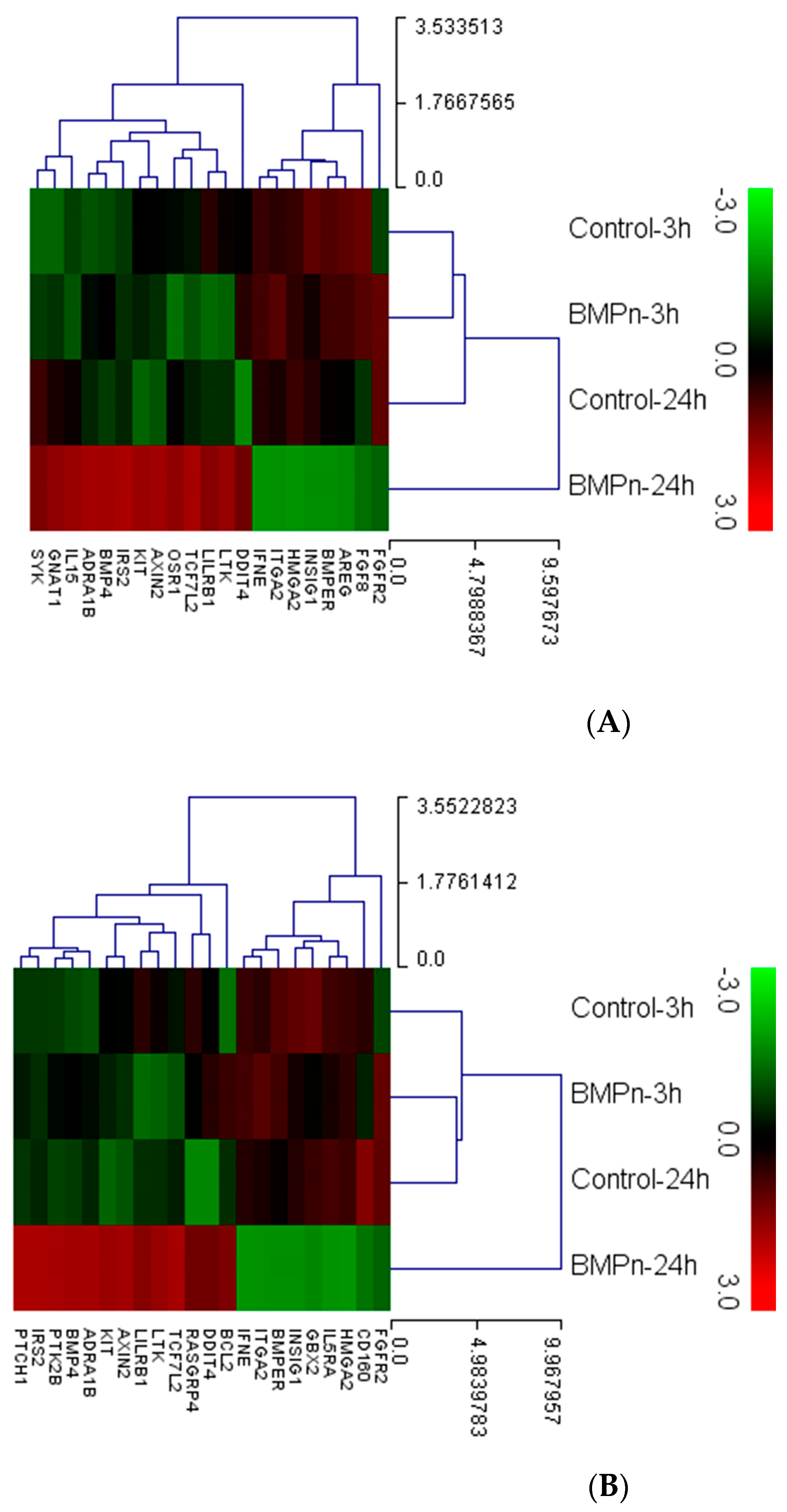
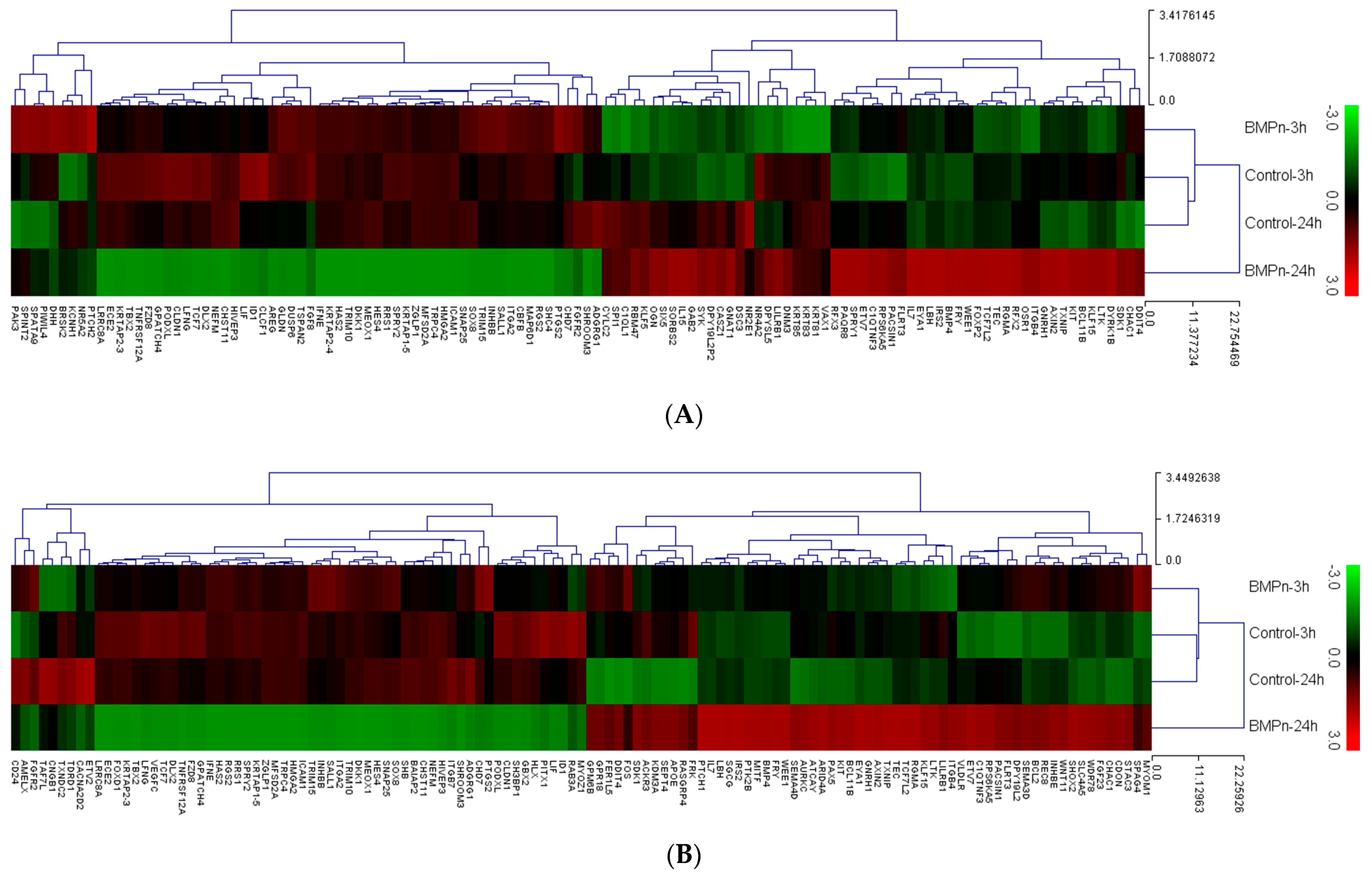
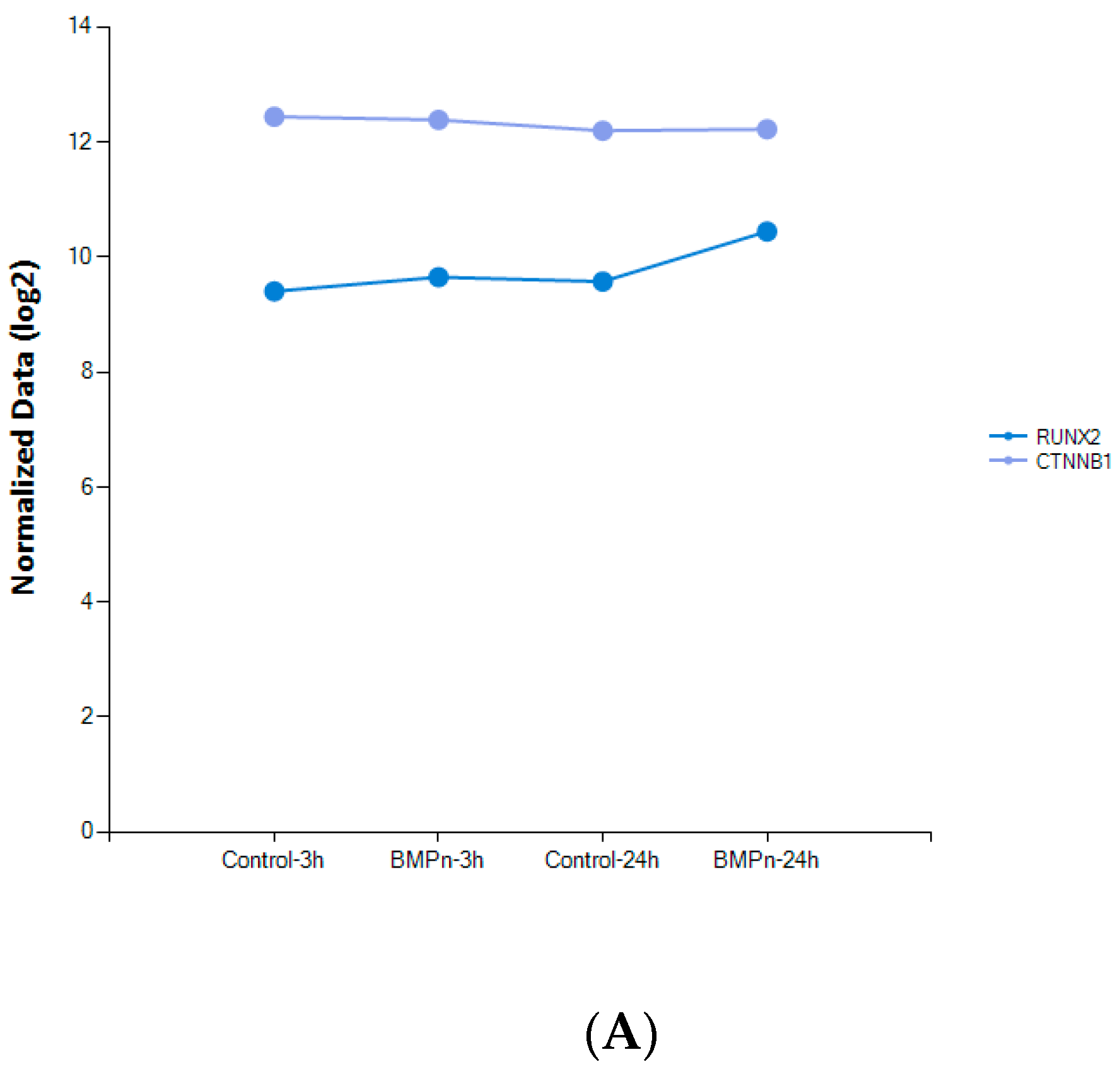
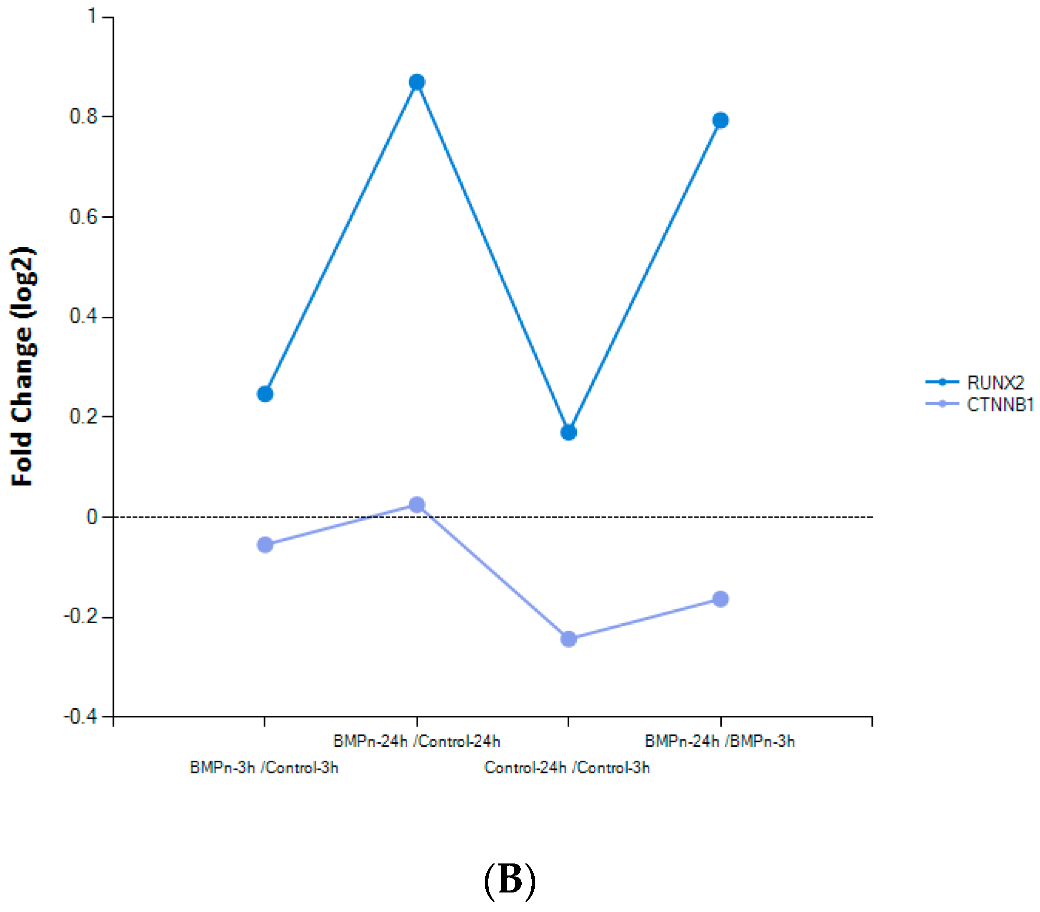
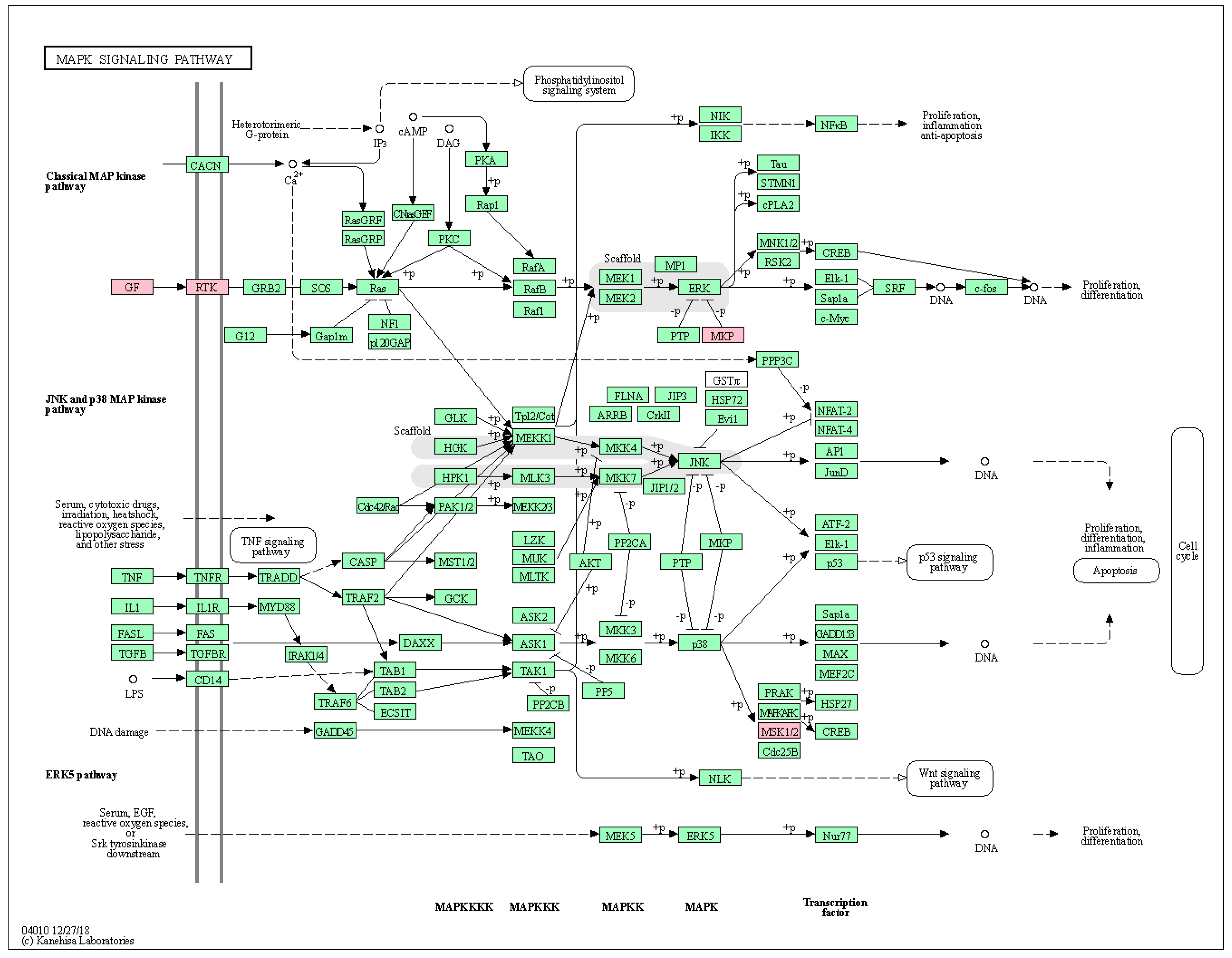
3.4. Evaluation of Gene Ontology and Pathway Analysis

Differentially expressed mRNA related to cell proliferation is shown in Table 1 (fold change of greater than 2, log2 normalized read counts >5 were selected). When comparing gene expression value between samples, the down-regulated gene was shown in green and up-regulated is in red. Comparison of the BMP-7 at 24 h versus control at 24 h revealed that 10 mRNAs were upregulated and 4 were downregulated. Comparison of the BMP-7 at 24 h versus BMP-7 at 3 h revealed that 10 mRNAs were upregulated and 5 were downregulated.
Clustering analysis of differentially expressed mRNA related to cell proliferation is shown in Figure 4.
Differentially expressed mRNA related to cell differentiation is shown in Table 2 (fold change of greater than 2, log2 normalized read counts >5 were selected). Comparison of the BMP-7 at 24 h versus control at 24 h revealed that 41 mRNAs were upregulated and 48 were downregulated. Comparison of the BMP-7 at 24 h versus BMP-7 at 3 h revealed that 38 mRNAs were upregulated and 50 were downregulated. Clustering analysis of differentially expressed mRNA related to cell differentiation is shown in Figure 5.
The changes in expression of RUNX2 and CTNNB1 are shown in Figure 6A. Expression of RUNX2 was increased in the BMP-7 group at both 3 h and 24 h when compared with the control group of each time point. The expression of RUNX2 in BMP-7 groups was higher with longer incubation of 24 h (Figure 6B). However, the expression of CTNNB1 was decreased both at 3 and 24 h when compared with the control group of each time point. The expression of CTNNB1 in BMP-7 groups was lower at 24 h. The signaling pathway associated with the target genes related to cell proliferation and cell differentiation was mitogen-activated protein kinase pathway (Figure 7).

4. Discussion

In this report, we evaluated the effects of BMP-7 on stem cells cultured on deproteinized bovine bone at the concentration of 10 and 100 ng/mL. Applying BMP-7 caused an increased alkaline phosphatase activity and mineralization of the hBMSCs.
BMP-7 is considered anabolic and anti-catabolic growth factor on cell homeostasis [21]. In a previous report, BMP-7 distinctly stimulated chondrocyte proliferation and inhibited chondrocyte hypertrophy [22]. It should be also noted that even though BMP-7 is potent bone anabolic substances, but it may also act as a booster for catabolism, and this may lead to resorption [23]. A previous report showed that BMP-7 upregulated the genes involved in osteogenic differentiation of gingiva-derived stem cells [24]. Moreover, BMP-7 was involved in the induction of cementogenic differentiation of stem cells derived from periodontal ligament [25]. In this report, BMP-7 enhanced the osteogenic differentiation of hBMSCs. However, 50 and 100 ng/mL of BMP-7 induced proliferation of stem cells derived from periodontal ligament, contrary to this study [25] and this may be due to the differences in culture condition, passage and origin of the stem cells [26].
The concentration of the BMP-7 may affect the results. Local injection of a single dose of 200 µg of rhBMP-7 enhanced bone density during distraction osteogenesis in the rabbit mandible model [27]. Six hundred micrograms of BMP-7 per 8 mL xenogenic bone mineral in the block was used for mandibular reconstruction in a miniature pig model [28]. The use of xenograft bone mineral granules with 1000 µg BMP-7 per gram can be applied for bone regeneration in a minipig model [29]. For the adult chimpanzee model, 2.5 mg BMP-7 per gram collagen matrix was used [14]. Upregulation of osteocalcin and osteopontin expression was seen in periodontal ligament-derived stem cells with BMP-7 at 100 ng/mL [30]. Previous report showed that BMP-7 at 100 ng/mL increased the expression of target genes associated with osteoblast differentiation [24]. Similarly, this report showed a significantly higher osteogenic differentiation was noted for BMP-7 groups at 10 and 100 ng/mL concentration when compared with the control group.
There has been a great level of interest in stem cell application for many years [7]. More recently, stem cells are being actively applied to tissue regeneration approaches [31]. When the tissue is damaged, the stem cells are activated by the substances secreted in situ [6]. The activated stem cells receive additional signals from the tissue, and they proliferate or differentiate into mature cells [32]. Newly formed mature cells migrate to the damaged tissue and contribute to regenerating the tissue [33]. Based on the role of the stem cells listed above, stem cells are applied for tissue regeneration purposes [34]. The application of stem cells needs to be done very carefully because they can dislocate or penetrate into blood vessels and form ectopic or fibrotic tissue [35]. This study showed that the application of BMP-7 led to increased expression of RUNX2, which was similar to the previous reports [36,37]. However, in this study, the expression of CTNNB1 was not increased in BMP-7 applied groups. This study also mitogen-activated protein kinase pathway was associated with the target genes related to cell proliferation and cell differentiation.
Moreover, stem cells have been used as growth factor-producing devices [38]. Stem cells are known to secrete various growth factors, including platelet-derived growth factor, vascular endothelial growth factor, and hepatocyte growth factor, to name a few [39,40]. The use of scaffolds may allow and facilitate the application of stem cells exactly to the desired area and their maintenance at the area [41]. It is possible that the type of carrier/scaffold used may influence the morphology and the amount of newly formed bone [42]. In another report, the application of BMP on an allograft, xenograft or cell binding peptide-loaded xenograft enhanced osteogenic differentiation when compared with non-loaded group, but the type of scaffold did not produce significant differences [11]. In this report, we used one of the most widely used biomaterial for bone augmentation namely deproteinized bovine bone [43].
However, there are several things to consider when applying growth factors in stem cell therapy. One of the main strengths of this study is that next-generation mRNA sequencing was used to evaluate the global gene expression [44]. Pathway analysis performed in this study suggested the underlying signaling pathway [45]. A limitation of this study is that further analysis of in vivo models and of human participants may be needed for wider application [46]. When the growth factor is applied, an initial burst phenomenon occurs in the early stage [47]. The half-life of the growth factor is then reduced due to an enzymatic action [48]. The applied growth factor may be washed away by the blood flow [49]. For the desired effect of growth factors, it is necessary to consider the high cost and various side effects when applying a high dose [50]. Though doing this has led to a conflict between the effectiveness and side effects of the growth factors [50]. Modification of surface of titanium surface with BMP-7 led to increased adhesion and differentiation of stem cells [51]. It was also suggested that mesenchymal stem cells, which overexpress BMP-7, may be applied for the enhanced bone healing [52]. Attempts were made to apply a combination of growth factors for optimal results [53,54]. The co-application of BMP-7 and PDGF-BB has been applied for the differentiation of stem cells [53]. Heterodimeric BMP-2/7 was also used for the osteogenic differentiation of adipose-derived stem cells [54]. Moreover, other BMPs have been applied for bone tissue regeneration as these are emerging BMPs for orthotopic bone regeneration [55,56,57].

5. Conclusions

This study shows that the application of BMP-7 increases alkaline phosphatase activity and the mineralization of stem cells culture on deproteinized bovine bone mineral. This report suggests the combined use of stem cells and osteoinductive growth factors with a scaffold may have synergistic effects on osteogenesis.

Author Contributions

Conceptualization, H.-J.L., S.-K.M., Y.-H.P. and J.-B.P.; methodology, H.-J.L., S.-K.M., Y.-H.P. and J.-B.P.; validation, H.-J.L., S.-K.M., Y.-H.P. and J.-B.P.; formal analysis, H.-J.L., S.-K.M., Y.-H.P. and J.-B.P.; writing—original draft preparation, H.-J.L., S.-K.M., Y.-H.P. and J.-B.P.; and writing—review and editing, H.-J.L., S.-K.M., Y.-H.P. and J.-B.P. All authors have read and agreed to the published version of the manuscript.

Funding

This research was supported by the National Research Foundation of Korea (NRF) grant funded by the Korean government (MSIT) (No. 2020R1A2C4001624).

Institutional Review Board Statement

The study was conducted according to the guidelines of the Declaration of Helsinki, and approved by the Institutional Review Board of Seoul St. Mary’s Hospital approved the present study (KC18SESI0199 and KC20SISE0582; approval date: 25 April 2018).

Informed Consent Statement

Informed consent was obtained from all subjects involved in the study.

Data Availability Statement

Data is contained within the article.

Acknowledgments

The Catholic MASTER Cells supplied by Catholic Institute of Cell Therapy (CIC, Seoul, Korea) were derived from human bone marrow donated by healthy donors after informed consent. Geistlich Bio-Oss® used in this study was provided by Geistlich Pharma AG Wolhusen, Switzerland.

Conflicts of Interest

The authors declare no conflict of interest.

References

  1. Benjamin, B.J. Normal aging and the multicultural population. Semin. Speech Lang. 1997, 18, 127–134. [Google Scholar] [CrossRef]
  2. Bouwknegt, M.; van Pelt, W.; Havelaar, A.H. Scoping the impact of changes in population age-structure on the future burden of foodborne disease in the Netherlands, 2020-2060. Int. J. Environ. Res. Public Health 2013, 10, 2888–2896. [Google Scholar] [CrossRef] [PubMed]
  3. Frencken, J.E.; Sharma, P.; Stenhouse, L.; Green, D.; Laverty, D.; Dietrich, T. Global epidemiology of dental caries and severe periodontitis—a comprehensive review. J. Clin. Periodontol. 2017, 44 (Suppl. S18), S94–S105. [Google Scholar] [CrossRef]
  4. Benjamin, R.M. Oral health: The silent epidemic. Public Health Rep. 2010, 125, 158–159. [Google Scholar] [CrossRef] [PubMed] [Green Version]
  5. Vennedey, V.; Derman, S.H.; Hiligsmann, M.; Civello, D.; Schwalm, A.; Seidl, A.; Scheibler, F.; Stock, S.; Noack, M.J.; Danner, M. Patients’ preferences in periodontal disease treatment elicited alongside an IQWiG benefit assessment: A feasibility study. Patient Prefer. Adherence 2018, 12, 2437–2447. [Google Scholar] [CrossRef] [Green Version]
  6. Krafts, K.P. Tissue repair: The hidden drama. Organogenesis 2010, 6, 225–233. [Google Scholar] [CrossRef] [PubMed] [Green Version]
  7. Seetharaman, R.; Mahmood, A.; Kshatriya, P.; Patel, D.; Srivastava, A. An Overview on Stem Cells in Tissue Regeneration. Curr. Pharm. Des. 2019, 25, 2086–2098. [Google Scholar] [CrossRef] [PubMed]
  8. Mao, A.S.; Mooney, D.J. Regenerative medicine: Current therapies and future directions. Proc. Natl. Acad. Sci. USA 2015, 112, 14452–14459. [Google Scholar] [CrossRef] [Green Version]
  9. Amini, A.R.; Laurencin, C.T.; Nukavarapu, S.P. Bone tissue engineering: Recent advances and challenges. Crit. Rev. Biomed. Eng. 2012, 40, 363–408. [Google Scholar] [CrossRef] [PubMed] [Green Version]
  10. Lo, K.W.; Ulery, B.D.; Ashe, K.M.; Laurencin, C.T. Studies of bone morphogenetic protein-based surgical repair. Adv. Drug Deliv. Rev. 2012, 64, 1277–1291. [Google Scholar] [CrossRef] [Green Version]
  11. Acil, Y.; Springer, I.N.; Broek, V.; Terheyden, H.; Jepsen, S. Effects of bone morphogenetic protein-7 stimulation on osteoblasts cultured on different biomaterials. J. Cell. Biochem. 2002, 86, 90–98. [Google Scholar] [CrossRef] [PubMed]
  12. Roldan, J.C.; Jepsen, S.; Miller, J.; Freitag, S.; Rueger, D.C.; Acil, Y.; Terheyden, H. Bone formation in the presence of platelet-rich plasma vs. bone morphogenetic protein-7. Bone 2004, 34, 80–90. [Google Scholar] [CrossRef] [PubMed]
  13. Terheyden, H.; Knak, C.; Jepsen, S.; Palmie, S.; Rueger, D.R. Mandibular reconstruction with a prefabricated vascularized bone graft using recombinant human osteogenic protein-1: An experimental study in miniature pigs. Part I: Prefabrication. Int. J. Oral Maxillofac. Surg. 2001, 30, 373–379. [Google Scholar] [CrossRef] [PubMed]
  14. McAllister, B.S.; Margolin, M.D.; Cogan, A.G.; Taylor, M.; Wollins, J. Residual lateral wall defects following sinus grafting with recombinant human osteogenic protein-1 or Bio-Oss in the chimpanzee. Int. J. Periodontics Restor. Dent. 1998, 18, 227–239. [Google Scholar]
  15. Min, Q.; Liu, J.; Zhang, Y.; Yang, B.; Wan, Y.; Wu, J. Dual Network Hydrogels Incorporated with Bone Morphogenic Protein-7-Loaded Hyaluronic Acid Complex Nanoparticles for Inducing Chondrogenic Differentiation of Synovium-Derived Mesenchymal Stem Cells. Pharmaceutics 2020, 12, 613. [Google Scholar] [CrossRef]
  16. Jeong, C.H.; Kim, S.M.; Lim, J.Y.; Ryu, C.H.; Jun, J.A.; Jeun, S.S. Mesenchymal stem cells expressing brain-derived neurotrophic factor enhance endogenous neurogenesis in an ischemic stroke model. BioMed Res. Int. 2014, 2014, 129145. [Google Scholar] [CrossRef] [PubMed] [Green Version]
  17. Lee, H.; Kim, M.; Park, Y.H.; Park, J.B. Dexamethasone downregulates SIRT1 and IL6 and upregulates EDN1 genes in stem cells derived from gingivae via the AGE/RAGE pathway. Biotechnol. Lett. 2018, 40, 509–519. [Google Scholar] [CrossRef] [PubMed]
  18. Trapnell, C.; Pachter, L.; Salzberg, S.L. TopHat: Discovering splice junctions with RNA-Seq. Bioinformatics 2009, 25, 1105–1111. [Google Scholar] [CrossRef]
  19. Quinlan, A.R.; Hall, I.M. BEDTools: A flexible suite of utilities for comparing genomic features. Bioinformatics 2010, 26, 841–842. [Google Scholar] [CrossRef] [PubMed] [Green Version]
  20. Gentleman, R.C.; Carey, V.J.; Bates, D.M.; Bolstad, B.; Dettling, M.; Dudoit, S.; Ellis, B.; Gautier, L.; Ge, Y.; Gentry, J.; et al. Bioconductor: Open software development for computational biology and bioinformatics. Genome Biol. 2004, 5, R80. [Google Scholar] [CrossRef] [Green Version]
  21. Ellman, M.B.; Kim, J.; An, H.S.; Chen, D.; Kc, R.; Li, X.; Xiao, G.; Yan, D.; Suh, J.; van Wijnen, A.J.; et al. Lactoferricin enhances BMP7-stimulated anabolic pathways in intervertebral disc cells. Gene 2013, 524, 282–291. [Google Scholar] [CrossRef] [PubMed] [Green Version]
  22. Huang, X.; Zhong, L.; Post, J.N.; Karperien, M. Co-treatment of TGF-β3 and BMP7 is superior in stimulating chondrocyte redifferentiation in both hypoxia and normoxia compared to single treatments. Sci. Rep. 2018, 8, 10251. [Google Scholar] [CrossRef]
  23. Harding, A.K.; Aspenberg, P.; Kataoka, M.; Bylski, D.; Tägil, M. Manipulating the anabolic and catabolic response in bone graft remodeling: Synergism by a combination of local BMP-7 and a single systemic dosis of zoledronate. J. Orthop. Res. 2008, 26, 1245–1249. [Google Scholar] [CrossRef] [PubMed]
  24. Lee, H.; Min, S.K.; Song, Y.; Park, Y.H.; Park, J.B. Bone morphogenetic protein-7 upregulates genes associated with osteoblast differentiation, including collagen I, Sp7 and IBSP in gingiva-derived stem cells. Exp. Ther. Med. 2019, 18, 2867–2876. [Google Scholar] [CrossRef]
  25. Torii, D.; Tsutsui, T.W.; Watanabe, N.; Konishi, K. Bone morphogenetic protein 7 induces cementogenic differentiation of human periodontal ligament-derived mesenchymal stem cells. Odontology 2016, 104, 1–9. [Google Scholar] [CrossRef]
  26. Kim, B.B.; Tae, J.Y.; Ko, Y.; Park, J.B. Lovastatin increases the proliferation and osteoblastic differentiation of human gingiva-derived stem cells in three-dimensional cultures. Exp. Ther. Med. 2019, 18, 3425–3430. [Google Scholar] [CrossRef] [Green Version]
  27. Zakhary, K.; Motakis, D.; Hamdy, R.H.; Campisi, P.; Amar, Y.; Lessard, M.L. Effect of recombinant human bone morphogenetic protein 7 on bone density during distraction osteogenesis of the rabbit mandible. J. Otolaryngol. 2005, 34, 407–414. [Google Scholar] [CrossRef] [PubMed]
  28. Terheyden, H.; Warnke, P.; Dunsche, A.; Jepsen, S.; Brenner, W.; Palmie, S.; Toth, C.; Rueger, D.R. Mandibular reconstruction with prefabricated vascularized bone grafts using recombinant human osteogenic protein-1: An experimental study in miniature pigs. Part II: Transplantation. Int. J. Oral Maxillofac. Surg. 2001, 30, 469–478. [Google Scholar] [CrossRef] [PubMed]
  29. Terheyden, H.; Menzel, C.; Wang, H.; Springer, I.N.; Rueger, D.R.; Acil, Y. Prefabrication of vascularized bone grafts using recombinant human osteogenic protein-1-part 3: Dosage of rhOP-1, the use of external and internal scaffolds. Int. J. Oral Maxillofac. Surg. 2004, 33, 164–172. [Google Scholar] [CrossRef]
  30. Hakki, S.S.; Bozkurt, B.; Hakki, E.E.; Kayis, S.A.; Turac, G.; Yilmaz, I.; Karaoz, E. Bone morphogenetic protein-2, -6, and -7 differently regulate osteogenic differentiation of human periodontal ligament stem cells. J. Biomed Mater. Res. B Appl. Biomater. 2014, 102, 119–130. [Google Scholar] [CrossRef] [PubMed]
  31. Loebel, C.; Burdick, J.A. Engineering Stem and Stromal Cell Therapies for Musculoskeletal Tissue Repair. Cell Stem Cell 2018, 22, 325–339. [Google Scholar] [CrossRef] [PubMed] [Green Version]
  32. Amcheslavsky, A.; Jiang, J.; Ip, Y.T. Tissue damage-induced intestinal stem cell division in Drosophila. Cell Stem Cell 2009, 4, 49–61. [Google Scholar] [CrossRef] [PubMed] [Green Version]
  33. Iismaa, S.E.; Kaidonis, X.; Nicks, A.M.; Bogush, N.; Kikuchi, K.; Naqvi, N.; Harvey, R.P.; Husain, A.; Graham, R.M. Comparative regenerative mechanisms across different mammalian tissues. NPJ Regen. Med. 2018, 3, 6. [Google Scholar] [CrossRef] [PubMed]
  34. Zheng, C.; Chen, J.; Liu, S.; Jin, Y. Stem cell-based bone and dental regeneration: A view of microenvironmental modulation. Int. J. Oral Sci. 2019, 11, 23. [Google Scholar] [CrossRef] [Green Version]
  35. Zakrzewski, W.; Dobrzyński, M.; Szymonowicz, M.; Rybak, Z. Stem cells: Past, present, and future. Stem Cell Res. Ther. 2019, 10, 68. [Google Scholar] [CrossRef]
  36. Zhu, L.; Ma, J.; Mu, R.; Zhu, R.; Chen, F.; Wei, X.; Shi, X.; Zang, S.; Jin, L. Bone morphogenetic protein 7 promotes odontogenic differentiation of dental pulp stem cells in vitro. Life Sci. 2018, 202, 175–181. [Google Scholar] [CrossRef] [PubMed]
  37. Schwarting, T.; Benölken, M.; Ruchholtz, S.; Frink, M.; Lechler, P. Bone morphogenetic protein-7 enhances bone-tendon integration in a murine in vitro co-culture model. Int. Orthop. 2015, 39, 799–805. [Google Scholar] [CrossRef]
  38. Tollemar, V.; Collier, Z.J.; Mohammed, M.K.; Lee, M.J.; Ameer, G.A.; Reid, R.R. Stem cells, growth factors and scaffolds in craniofacial regenerative medicine. Genes Dis. 2016, 3, 56–71. [Google Scholar] [CrossRef] [PubMed] [Green Version]
  39. Nie, W.B.; Zhang, D.; Wang, L.S. Growth Factor Gene-Modified Mesenchymal Stem Cells in Tissue Regeneration. Drug Des. Devel. Ther. 2020, 14, 1241–1256. [Google Scholar] [CrossRef] [Green Version]
  40. Nakamura, T.; Mizuno, S. The discovery of hepatocyte growth factor (HGF) and its significance for cell biology, life sciences and clinical medicine. Proc. Jpn. Acad. Ser. B Phys. Biol. Sci. 2010, 86, 588–610. [Google Scholar] [CrossRef] [Green Version]
  41. Biehl, J.K.; Russell, B. Introduction to stem cell therapy. J. Cardiovasc. Nurs. 2009, 24, 98–103; quiz 104–105. [Google Scholar] [CrossRef] [PubMed] [Green Version]
  42. Terheyden, H.; Jepsen, S.; Vogeler, S.; Tucker, M.; Rueger, D.C. Recombinant human osteogenic protein 1 in the rat mandibular augmentation model: Differences in morphology of the newly formed bone are dependent on the type of carrier. Mund Kiefer Gesichtschir. 1997, 1, 272–275. [Google Scholar] [CrossRef]
  43. Khojasteh, A.; Ghahremani, M.H.; Ostad, S.N.; Eslami, M.; Motahhary, P.; Morad, G.; Shidfar, S. The effect of deproteinized bovine bone mineral on saos-2 cell proliferation. Iran. Endod. J. 2013, 8, 118–122. [Google Scholar]
  44. Kim, B.B.; Kim, M.; Park, Y.H.; Ko, Y.; Park, J.B. Short-term application of dexamethasone on stem cells derived from human gingiva reduces the expression of RUNX2 and β-catenin. J. Int. Med. Res. 2017, 45, 993–1006. [Google Scholar] [CrossRef] [PubMed]
  45. Su, D.; Yi, L.; Guan, L.; Li, Q.; Shi, C.; Ma, X. Sequencing Analysis of mRNA Profile in Endothelial Cells in Response to ox-LDL. Biochem. Genet. 2021, 59, 767–780. [Google Scholar] [CrossRef]
  46. Kang, S.H.; Park, J.B.; Kim, I.; Lee, W.; Kim, H. Assessment of stem cell viability in the initial healing period in rabbits with a cranial bone defect according to the type and form of scaffold. J. Periodontal Implant Sci. 2019, 49, 258–267. [Google Scholar] [CrossRef] [PubMed]
  47. Turner, P.A.; Thiele, J.S.; Stegemann, J.P. Growth factor sequestration and enzyme-mediated release from genipin-crosslinked gelatin microspheres. J. Biomater. Sci. Polym. Ed. 2017, 28, 1826–1846. [Google Scholar] [CrossRef]
  48. Mitchell, A.C.; Briquez, P.S.; Hubbell, J.A.; Cochran, J.R. Engineering growth factors for regenerative medicine applications. Acta Biomater. 2016, 30, 1–12. [Google Scholar] [CrossRef] [PubMed]
  49. Zhang, J.; Ding, L.; Zhao, Y.; Sun, W.; Chen, B.; Lin, H.; Wang, X.; Zhang, L.; Xu, B.; Dai, J. Collagen-targeting vascular endothelial growth factor improves cardiac performance after myocardial infarction. Circulation 2009, 119, 1776–1784. [Google Scholar] [CrossRef] [PubMed]
  50. Barrientos, S.; Brem, H.; Stojadinovic, O.; Tomic-Canic, M. Clinical application of growth factors and cytokines in wound healing. Wound Repair Regen. 2014, 22, 569–578. [Google Scholar] [CrossRef] [PubMed] [Green Version]
  51. Kämmerer, P.W.; Pabst, A.M.; Dau, M.; Staedt, H.; Al-Nawas, B.; Heller, M. Immobilization of BMP-2, BMP-7 and alendronic acid on titanium surfaces: Adhesion, proliferation and differentiation of bone marrow-derived stem cells. J. Biomed. Mater. Res. A 2020, 108, 212–220. [Google Scholar] [CrossRef] [PubMed] [Green Version]
  52. Yan, X.; Zhou, Z.; Guo, L.; Zeng, Z.; Guo, Z.; Shao, Q.; Xu, W. BMP7-overexpressing bone marrow-derived mesenchymal stem cells (BMSCs) are more effective than wild-type BMSCs in healing fractures. Exp. Ther. Med. 2018, 16, 1381–1388. [Google Scholar] [CrossRef] [PubMed] [Green Version]
  53. Wada, K.; Gerbaudo, V.H.; Spector, M. Effects of PDGF-BB and OP-1 on mesenchymal stem cells in a porous mineral block. Int. J. Periodontics Restor. Dent. 2013, 33, e72–e78. [Google Scholar] [CrossRef] [PubMed]
  54. Zhang, X.; Guo, J.; Wu, G.; Zhou, Y. Effects of heterodimeric bone morphogenetic protein-2/7 on osteogenesis of human adipose-derived stem cells. Cell Prolif. 2015, 48, 650–660. [Google Scholar] [CrossRef] [PubMed]
  55. Gaihre, B.; Unagolla, J.M.; Liu, J.; Ebraheim, N.A.; Jayasuriya, A.C. Thermoresponsive Injectable Microparticle-Gel Composites with Recombinant BMP-9 and VEGF Enhance Bone Formation in Rats. ACS Biomater. Sci. Eng. 2019, 5, 4587–4600. [Google Scholar] [CrossRef] [PubMed]
  56. Hettiaratchi, M.H.; Krishnan, L.; Rouse, T.; Chou, C.; McDevitt, T.C.; Guldberg, R.E. Heparin-mediated delivery of bone morphogenetic protein-2 improves spatial localization of bone regeneration. Sci. Adv. 2020, 6, eaay1240. [Google Scholar] [CrossRef] [PubMed] [Green Version]
  57. Chiari, C.; Grgurevic, L.; Bordukalo-Niksic, T.; Oppermann, H.; Valentinitsch, A.; Nemecek, E.; Staats, K.; Schreiner, M.; Trost, C.; Kolb, A.; et al. Recombinant Human BMP6 Applied Within Autologous Blood Coagulum Accelerates Bone Healing: Randomized Controlled Trial in High Tibial Osteotomy Patients. J. Bone Miner. Res. 2020, 35, 1893–1903. [Google Scholar] [CrossRef]
Figure 1. Stem cell attachment to the deproteinized bovine bone mineral. Scale bar indicates 100 μm (original magnification: ×50).
Coatings 11 00642 g001
Figure 2. Live, dead, and merged images of stem cells cultured on the deproteinized bovine bone mineral. Scale bar indicates 100 μm (original magnification: ×50).
Coatings 11 00642 g002
Figure 3. Cellular viability and osteogenic differentiation. (A) Cellular viability using a Cell Counting Kit-8 on day 7. * Statistically significant differences were shown when compared with 0 ng/mL (p < 0.05). (B) Alkaline phosphatase activity on days 7 and 14. * Significant increases were noted when compared with 0 ng/mL at day 7 (p < 0.05). ** Statistically significances were noted when compared with 10 ng/mL at day 7 (p < 0.05). # Significant differences were seen when compared with 0 ng/mL at day 14 (p < 0.05). (C) Results of Alizarin Red S staining on days 7 and 14. * Significant increases were noted were noted when compared with 0 ng/mL at day 7 (p < 0.05). ** Statistically significances were noted when compared with 0 ng/mL at day 14 (p < 0.05).
Coatings 11 00642 g003
Figure 4. Clustering analysis of differentially expressed mRNA related to cell proliferation (A) BMP-7 at 24 h/control at 24 h and (B) BMP-7 at 24 h/BMP-7 at 3 h (fold change fold change of greater than 2, log2 normalized read counts >4 were selected).
Coatings 11 00642 g004
Figure 5. Clustering analysis of differentially expressed mRNA related to cell differentiation (A) BMP-7 at 24 h/control at 24 h and (B) BMP-7 at 24 h/BMP-7 at 3 h (fold change fold change of greater than 2, log2 normalized read counts >4 were selected).
Coatings 11 00642 g005
Figure 6. (A) Log2-normalized read counts regarding the expression of RUNX2 and CTNNB1 (for β-catenin expression) and (B) Log2 fold change regarding the expression of RUNX2 and CTNNB1.
Coatings 11 00642 g006aCoatings 11 00642 g006b
Figure 7. Signaling pathway.
Coatings 11 00642 g007
Table 1. Differentially expressed mRNA related to cell proliferation (fold change of greater than 2, log2 normalized data >5 were selected). When comparing gene expression value between samples, the down-regulated gene was shown in green and up-regulated is in red.
Gene SymbolBMP-7 24 h/Control 24 hGene SymbolBMP-7 24 h/BMP-7 3 h
INSIG10.329ITGA20.338
HMGA20.391INSIG10.358
ITGA20.428BMPER0.401
BMPER0.494HMGA20.410
LTK2.141FGF80.497
PTCH12.172BMP42.073
TCF7L22.304OSR12.129
PTK2B2.408DDIT42.157
ADRA1B2.441ADRA1B2.174
IRS22.604IL152.510
BMP42.805LTK2.628
AXIN23.781IRS22.696
KIT15.821TCF7L22.890
DDIT420.173AXIN23.073
--KIT7.478
Table 2. Differentially expressed mRNA related to cell differentiation (fold change of greater than 2, log2 normalized data >5 were selected). When comparing gene expression value between samples, the down-regulated gene was shown in green and up-regulated is in red.
Gene SymbolBMP-7 24 h/Control 24 hGene SymbolBMP-7 24 h/BMP-7 3 h
PTPN220.039DHH0.034
RAB3A0.065MEOX10.045
NR4A20.125TRIM100.053
PITX10.159TRIM150.110
VAX10.184DKK10.117
PODXL0.197FZD80.166
SNAI10.209KRTAP2-30.222
HCLS10.220PTGS20.238
CLDN10.236HES40.248
LFNG0.237LFNG0.261
ANKRD10.238MFSD2A0.273
MYOZ10.250INHBB0.281
LIF0.256ADGRG10.294
CLCF10.274LIF0.298
ETV40.279TRPC40.298
DNAH10.314PTCH20.320
MECOM0.323CHD70.324
HSF40.340KRTAP1-50.330
SEMA3D0.365ICAM10.334
CEP2900.365ITGA20.338
EGR10.370RRS10.343
TET10.371KCNH10.344
MAK0.376TNFRSF12A0.346
REC80.380PODXL0.350
IGSF100.391CLDN10.353
SYNE20.404RGS20.354
CHAC10.408SALL10.360
JUP0.411NEFM0.380
PTCH20.412HAS20.382
BATF20.414CHST110.400
HRNR0.418HMGA20.410
PRDM160.419DLX20.410
DHH0.420ECE20.421
KCNH10.428TBX20.423
-0.429CBFB0.427
-0.435SHC40.427
-0.443TCF70.429
-0.444SNAP250.430
-0.447HIVEP30.432
-0.451SPRY20.448
-0.457MAP6D10.448
-0.457ID10.449
-0.464PIWIL40.453
-0.479CLCF10.463
-0.488GPATCH40.464
-0.489GLDN0.476
-0.491DUSP60.476
-0.494SHROOM30.478
-2.063FGF80.497
-2.141LRRC8A0.497
-2.165RBM472.004
-2.169ETV72.023
-2.172DYRK1B2.047
-2.174DPY19L2P22.060
-2.184BMP42.073
-2.206CYLC22.095
-2.304WEE12.122
-2.319KLF52.123
-2.346OSR12.129
-2.350PAQR82.135
-2.368KRT812.138
-2.408DDIT42.157
-2.417SIX52.190
-2.449DNM32.193
-2.554CHAC12.294
-2.604RFX32.303
-2.609FOXP22.421
-2.625EYA12.464
-2.661IL152.510
-2.682LBH2.558
-2.684GAB22.560
-2.744SPRY12.594
-2.788LTK2.628
-2.805TXNIP2.662
-2.830RFX22.675
-2.893NR4A22.683
-2.973IRS22.696
-3.227RPS6KA52.745
-3.297GNRH12.798
-3.428TCF7L22.890
-3.455DPYSL52.985
-3.547FRY3.013
-3.781AXIN23.073
-3.846TEC3.268
-4.084SORBS23.564
-5.012OGN3.994
-11.467KLF156.356
-15.821KIT7.478
-20.173--
Publisher’s Note: MDPI stays neutral with regard to jurisdictional claims in published maps and institutional affiliations.

