
Shoulder Implant Manufacturer Detection by Using Deep Learning: Proposed Channel Selection Layer


Abstract
1. Introduction
Related Works
2. Materials and Methods
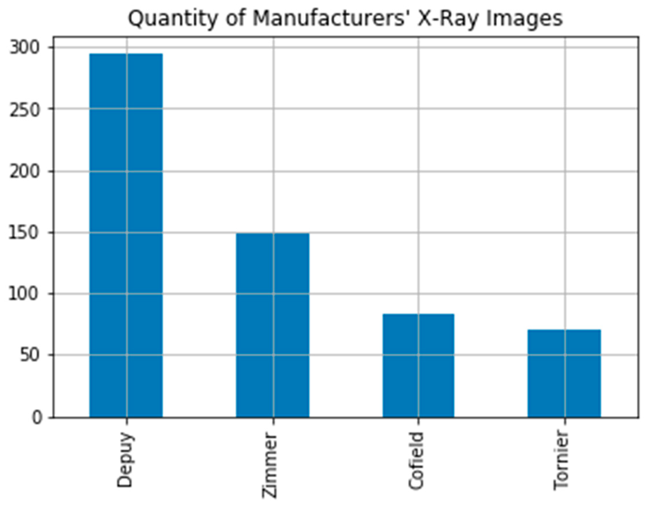
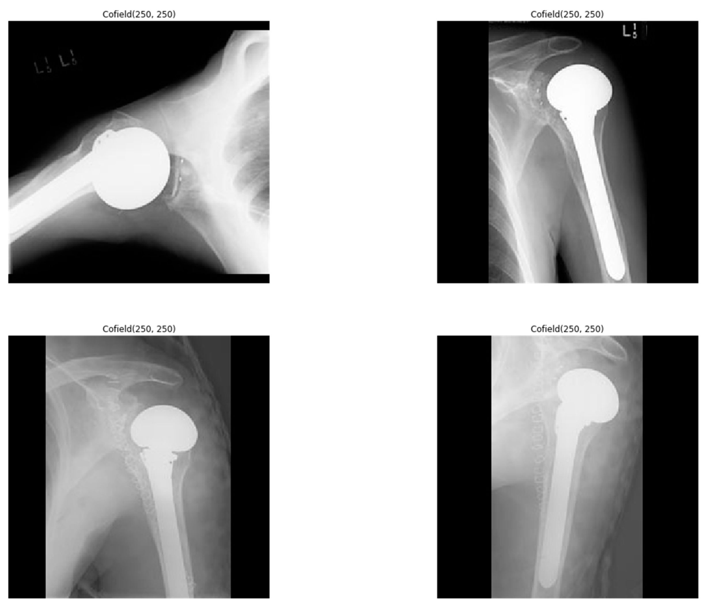
2.1. Dataset
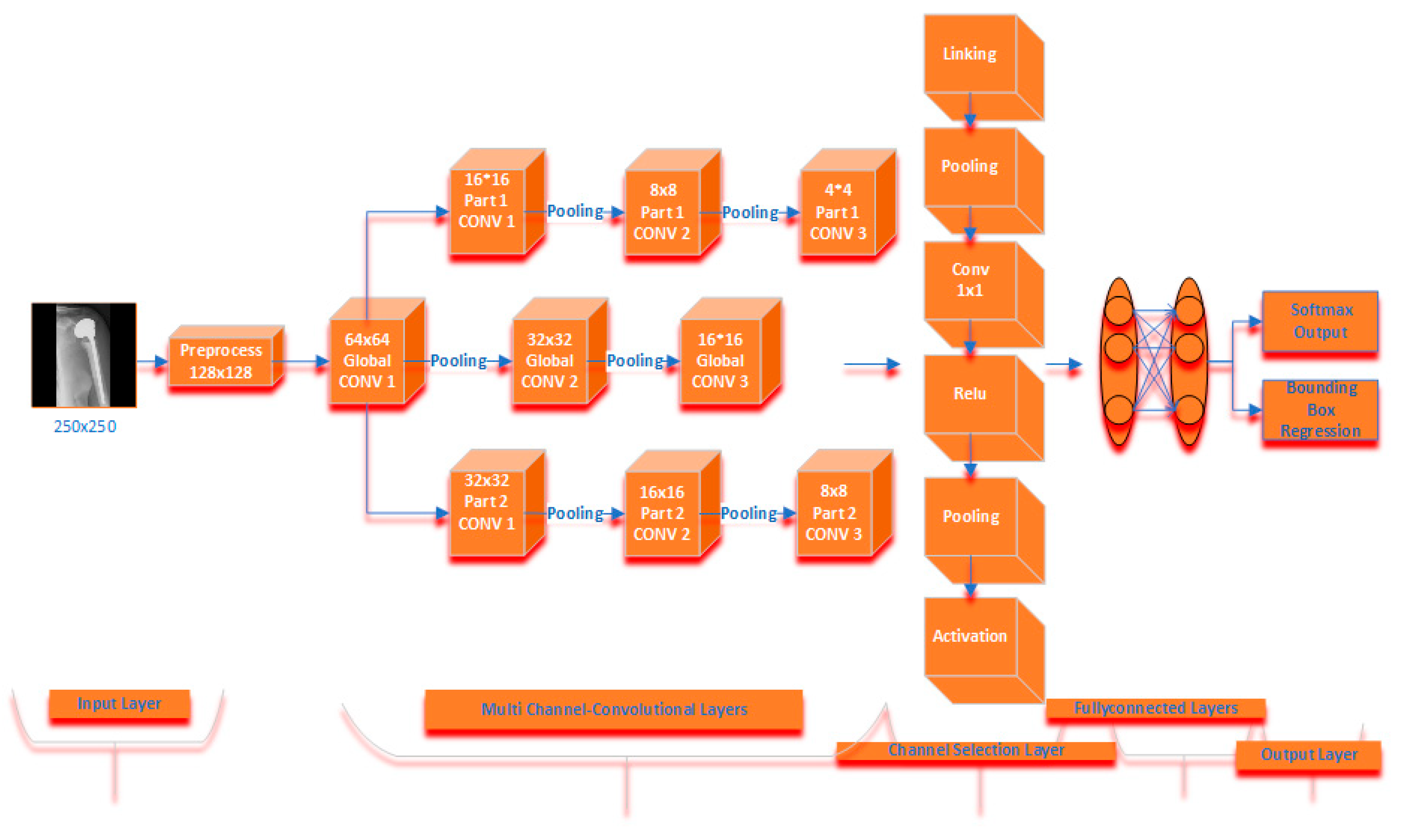
2.2. Deep Learning
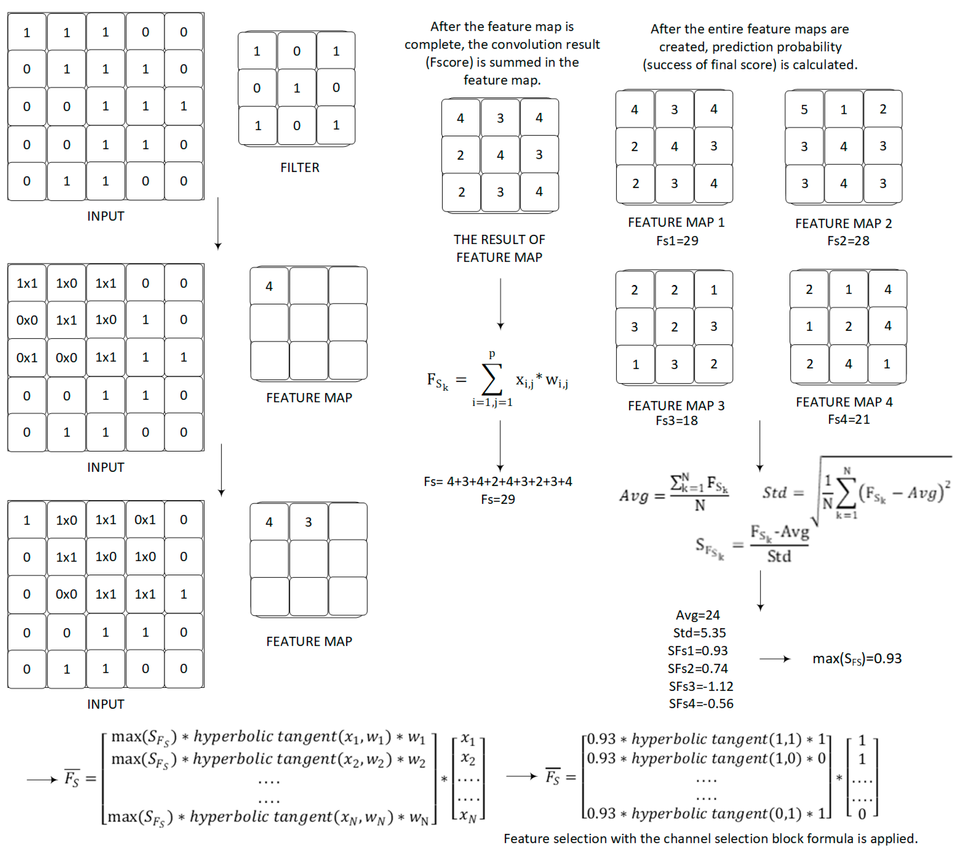
2.3. Proposed Channel Selection Formula
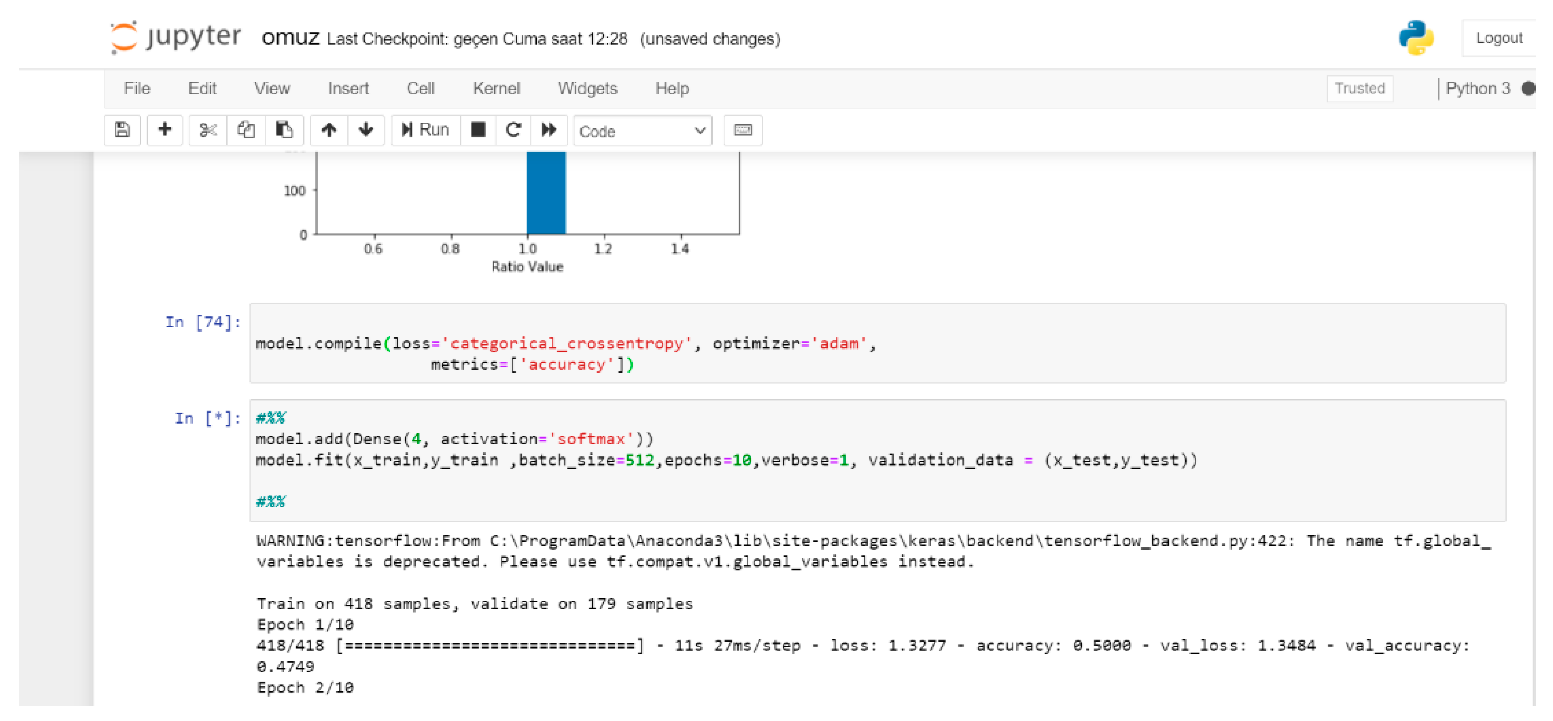
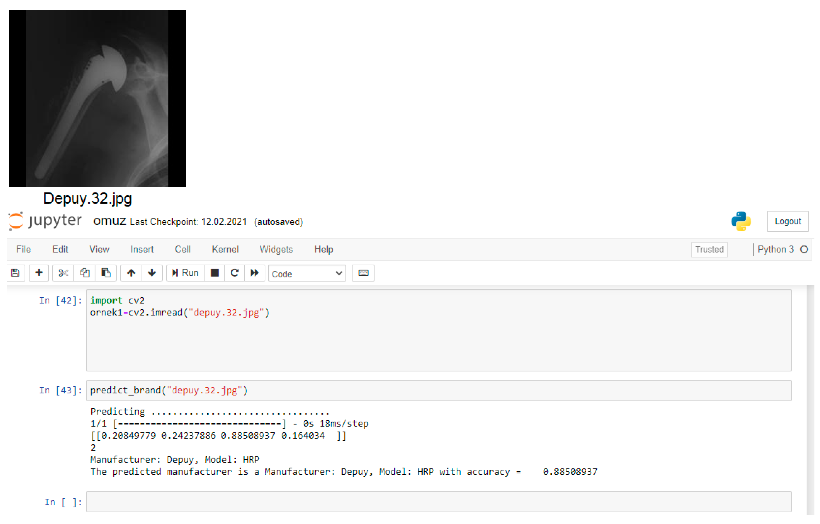
2.4. Experimental Studies
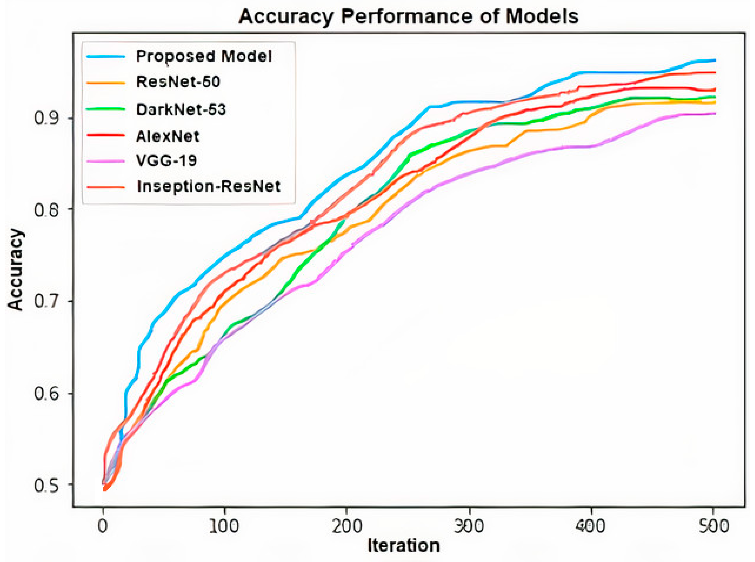
3. Results
4. Discussion
5. Conclusions
Author Contributions
Funding
Informed Consent Statement
Data Availability Statement
Acknowledgments
Conflicts of Interest
References
- Barrett, W.P.; Franklin, J.L.; Jackins, S.E.; Wyss, C.R.; Matsen, F.A. Total shoulder arthroplasty. J. Bone Jt. Surg. 1987, 69, 865–872. [Google Scholar] [CrossRef]
- Hawkins, R.J.; Bell, R.H.; Jallay, B. Total shoulder arthroplasty. Clin. Orthop. Relat. Res. 1989, 242, 188–194. [Google Scholar] [CrossRef]
- Bohsali, K.I.; Wirth, M.A.; Rockwood, C.A., Jr. Complications of total shoulder arthroplasty. J. Bone Jt. Surg. 2006, 88, 2279–2292. [Google Scholar] [CrossRef]
- Cofield, R.H. Total shoulder arthroplasty with the neer prosthesis. J. Bone Jt. Surg. 1984, 66, 899–906. [Google Scholar] [CrossRef]
- Lunati, M.P.; Wilson, J.M.; Farley, K.X.; Gottschalk, M.B.; Wagner, E.R. Preoperative depression is a risk factor for complication and increased health care utilization following total shoulder arthroplasty. J. Shoulder Elb. Surg. 2021, 30, 89–96. [Google Scholar] [CrossRef]
- Urban, G.; Porhemmat, S.; Stark, M.; Feeley, B.; Okada, K.; Baldi, P. Classifying shoulder implants in X-ray images using deep learning. Comput. Struct. Biotechnol. J. 2020, 18, 967–972. [Google Scholar] [CrossRef]
- UCI Machine Learning Repository, Shoulder Implant X-ray Manufacturer Classification Dataset. Available online: https://archive.ics.uci.edu/mL/datasets/Shoulder+Implant+X-ray+Manufacturer+Classification (accessed on 20 February 2021).
- Stark, M.B.C.G. Automatic Detection and Segmentation of Shoulder Implants in X-ray Images. Master’s Thesis, San Francisco State University, San Francisco, CA, USA, 2018. [Google Scholar]
- Lindsey, R.; Daluiski, A.; Chopra, S.; Lachapelle, A.; Mozer, M.; Sicular, S.; Hanel, D.; Gardner, M.; Gupta, A.; Hotchkiss, R. Deep neural network improves fracture detection by clinicians. Proc. Natl. Acad. Sci. USA 2018, 115, 11591–11596. [Google Scholar] [CrossRef] [PubMed]
- Bredow, J.; Wenk, B.; Westphal, R.; Wahl, F.; Budde, S.; Eysel, P.; Oppermann, J. Software-based matching of X-ray images and 3d models of knee prostheses. Technol. Health Care 2014, 22, 895–900. [Google Scholar] [CrossRef] [PubMed]
- Nachimuthu, U.; Geethalakshmi, S.N. Multiple classification system for fracture detection in human bone X-ray images. In Proceedings of the Third International Conference on Computing, Communication and Networking Technologies (ICCCNT’12), Karur, India, 26–28 July 2012; IEEE: Piscataway, NJ, USA, 2012; pp. 1–8. [Google Scholar]
- Takahashi, T.; Nozaki, K.; Gonda, T.; Mameno, T.; Wada, M.; Ikebe, K. Identification of dental implants using deep learning—pilot study. Int. J. Implant Dent. 2020, 6, 53. [Google Scholar] [CrossRef] [PubMed]
- Kim, J.E.; Nam, N.E.; Shim, J.S.; Jung, Y.H.; Cho, B.H.; Hwang, J.J. Transfer learning via deep neural networks for implant fixture system classification using periapical radiographs. J. Clin. Med. 2020, 9, 1117. [Google Scholar] [CrossRef] [PubMed]
- Sukegawa, S.; Yoshii, K.; Hara, T.; Yamashita, K.; Nakano, K.; Yamamato, N.; Nagatsuka, H.; Furuki, Y. Deep neural networks for dental implant system classification. Biomolecules 2020, 10, 984. [Google Scholar] [CrossRef] [PubMed]
- Sharma, M.; Malhotra, H.; Kumar, N.; Yadav, J. Deep learning with convolutional neural network for controlling human arm prosthesis using sEMG signals. In Proceedings of the IEEE International Conference on Computing, Power and Communication Technologies (GUCON), New Delhi, India, 27–28 September 2019. [Google Scholar]
- Yi, P.Y.; Kim, T.K.; Wei, J.W.; Li, X.; Hager, G.H.; Sair, H.I.; Fritz, J. Automated detection and classification of shoulder arthroplasty models using deep learning. Skelet. Radiol. 2020, 49, 1623–1632. [Google Scholar] [CrossRef] [PubMed]
- Gowd, A.K.; Agarwalla, A.; Amin, N.H.; Nicholson, G.P.; Verma, N.N.; Liu, J.N. Construct validation of machine learning in the prediction of short-term postoperative complications following total shoulder arthroplasty. J. Shoulder Elbow Surg. 2019, 28, 410–421. [Google Scholar] [CrossRef] [PubMed]
- Borjali, A.; Chen, A.F.; Bedair, H.S.; Melnic, C.M.; Muratoglu, O.K.; Morid, M.A.; Varadarajan, K.M. Comparing performance of deep convolutional neural network with orthopaedic surgeons on identification of total hip prosthesis design from plain radiographs. MedRxiv 2020. [Google Scholar] [CrossRef]
- The University of Washington Shoulder Site. Available online: http://faculty.washington.edu/alexbert/Shoulder/ (accessed on 11 March 2021).
- Yılmaz, A. Yapay Zeka, 7th ed.; Kodlab Press: İstanbul, Turkey, 2017. [Google Scholar]
- Yılmaz, A.; Kaya, U. Derin Öğrenme, 2nd ed.; Kodlab Press: İstanbul, Turkey, 2019. [Google Scholar]
- Çelik, A.; Arıca, N. Enhancing face pose normalization with deep learning. Turk. J. Electr. Eng. Comput. Sci. 2019, 27, 3699–3712. [Google Scholar] [CrossRef]
- Yan, Z.; Xu, Z.; Dai, J. The big data analysis on the camera-based face image in surveillance cameras. Autosoft J. Intell. Autom. Soft Comput. 2019, 24, 123–132. [Google Scholar] [CrossRef]
- Adem, K.; Közkurt, C. Defect detection of seals in multilayer aseptic packages using deep learning. Turk. J. Electr. Eng. Comput. Sci. 2019, 27, 4220–4230. [Google Scholar] [CrossRef]
- Alqudah, A.M.; Hiam Alquraan, H.; Qasmieh, I.A.; Alqudah, A.; Al-Sharu, W. Brain tumor classification using deep learning technique—A comparison between cropped, uncropped, and segmented lesion images with different sizes. Int. J. Adv. Trends Comput. Sci. Eng. 2019, 8, 3684–3691. [Google Scholar] [CrossRef]
- Jinsakul, N.; Tsai, C.; Tsai, C.; Wu, P. Enhancement of deep learning in image classification performance using xception with the swish activation function for colorectal polyp preliminary screening. Mathematics 2019, 7, 1170. [Google Scholar] [CrossRef]
- Yılmaz, A. Çok kanallı cnn mimarisi ile X-ray görüntülerinden Covid-19 tanısı. Gazi Üniversitesi Mühendislik Mimar. Fakültesi Derg. 2021. (In press)


| Criteria | Proposed Model | ResNet-50 | Dark-Net-53 | AlexNet | VGG-19 | Inseption-ResNet |
|---|---|---|---|---|---|---|
| Accuracy | 97.20 | 92.73 | 93.85 | 94.41 | 91.06 | 95.53 |
| Sensitivity | 0.98 | 0.95 | 0.965 | 0.959 | 0.95 | 0.965 |
| Specificity | 0.019 | 0.05 | 0.034 | 0.041 | 0.048 | 0.034 |
| Precision | 0.986 | 0.956 | 0.958 | 0.972 | 0.937 | 0.979 |
| RMSE | 1.32 | 3.93 | 3.09 | 2.44 | 4.87 | 1.93 |
| F1 | 0.983 | 0.953 | 0.962 | 0.965 | 0.944 | 0.972 |
| Training Time (s) | 46,723 | 45,752 | 45,250 | 44,213 | 45,182 | 45,923 |
| Testing Time (s) | 17,412 | 16,823 | 16,532 | 15,883 | 16,351 | 16,938 |
| Criteria | Proposed Model | ResNet-50 | Dark-Net-53 | AlexNet | VGG-19 | Inseption-ResNet |
|---|---|---|---|---|---|---|
| Accuracy | 96.31 | 88.69 | 90.95 | 93.88 | 86.51 | 93.13 |
| Sensitivity | 0.97 | 0.92 | 0.95 | 0.96 | 0.93 | 0.95 |
| Specificity | 0.021 | 0.07 | 0.047 | 0.036 | 0.066 | 0.044 |
| Precision | 0.974 | 0.923 | 0.927 | 0.955 | 0.891 | 0.957 |
| RMSE | 1.46 | 6.12 | 5.07 | 3.02 | 8.47 | 3.46 |
| F1 | 0.976 | 0.926 | 0.939 | 0.959 | 0.91 | 0.956 |
| Training Time (s) | 81,792 | 80,103 | 79,275 | 77,432 | 79,112 | 80,392 |
| Testing Time (s) | 26,215 | 25,291 | 24,821 | 23,947 | 24,672 | 25,493 |
| Criteria | LR | SVM | NB | RF | XGBoost |
|---|---|---|---|---|---|
| Accuracy | 70.43 | 77.38 | 74.62 | 81.74 | 83.91 |
| Sensitivity | 0.79 | 0.87 | 0.85 | 0.91 | 0.92 |
| Specificity | 0.191 | 0.114 | 0.128 | 0.08 | 0.07 |
| Precision | 0.75 | 0.804 | 0.75 | 0.823 | 0.876 |
| F1 | 0.772 | 0.838 | 0.798 | 0.865 | 0.898 |
| Reference Study | Classifier | Problem | Accuracy (Best%) |
|---|---|---|---|
| Urban et al. [6] | NasNET-CNN | Shoulder Implants | 80.4 |
| Stark [8] | Hough Transform | Shoulder Implants | 94 |
| Lindsey et al. [9] | CNN | Fractures | 90.1 |
| Bredow et al. [10] | CNN | Knee Prosthesis | 90 |
| Takahashi et al. [12] | Yolov3-CNN | Dental Implants | 72 |
| Kim et al. [13] | CNN | Implant Fixture | 97 |
| Sukegawa et al. [14] | CNN | Dental Implants | 90.7 |
| Yi et al. [16] | ResNET-CNN | Shoulder Arthroplasty | 97 |
| Proposed Model | CNN | Shoulder Implants | 97.20 |
| Proposed Model (with data augmentation) | CNN | Shoulder Implants | 96.31 |
Publisher’s Note: MDPI stays neutral with regard to jurisdictional claims in published maps and institutional affiliations. |
© 2021 by the author. Licensee MDPI, Basel, Switzerland. This article is an open access article distributed under the terms and conditions of the Creative Commons Attribution (CC BY) license (http://creativecommons.org/licenses/by/4.0/).
Share and Cite
Yılmaz, A. Shoulder Implant Manufacturer Detection by Using Deep Learning: Proposed Channel Selection Layer. Coatings 2021, 11, 346. https://doi.org/10.3390/coatings11030346
Yılmaz A. Shoulder Implant Manufacturer Detection by Using Deep Learning: Proposed Channel Selection Layer. Coatings. 2021; 11(3):346. https://doi.org/10.3390/coatings11030346
Chicago/Turabian StyleYılmaz, Atınç. 2021. "Shoulder Implant Manufacturer Detection by Using Deep Learning: Proposed Channel Selection Layer" Coatings 11, no. 3: 346. https://doi.org/10.3390/coatings11030346
APA StyleYılmaz, A. (2021). Shoulder Implant Manufacturer Detection by Using Deep Learning: Proposed Channel Selection Layer. Coatings, 11(3), 346. https://doi.org/10.3390/coatings11030346