Multifunctional Nanocrystalline Cu–Ti Thin Films Enhance Survival and Induce Proliferation of Mouse Fibroblasts In Vitro
Abstract
1. Introduction
2. Materials and Methods
2.1. Cell Culture Conditions
2.2. Biological Evaluation of a Nanocrystalline Cu25Ti75 Thin Film
2.3. Preparation of Extracts of the Thin Film Material (Indirect Contact)
2.4. Cell Morphology
2.5. Cytotoxicity of Thin Films Based on Cu and Ti (MTT (3-[4,5-dimethylthiazol-2-yl]-2,5-diphenyl Bromide) Test)
2.6. Clonogenic Test
2.7. In Vitro Scarring Test
2.8. Cell Cycle Phase
2.9. Type of Cell Death
2.10. Statistical Analysis
3. Results
3.1. Physicochemical Properties of Thin Films Based on Cu and Ti
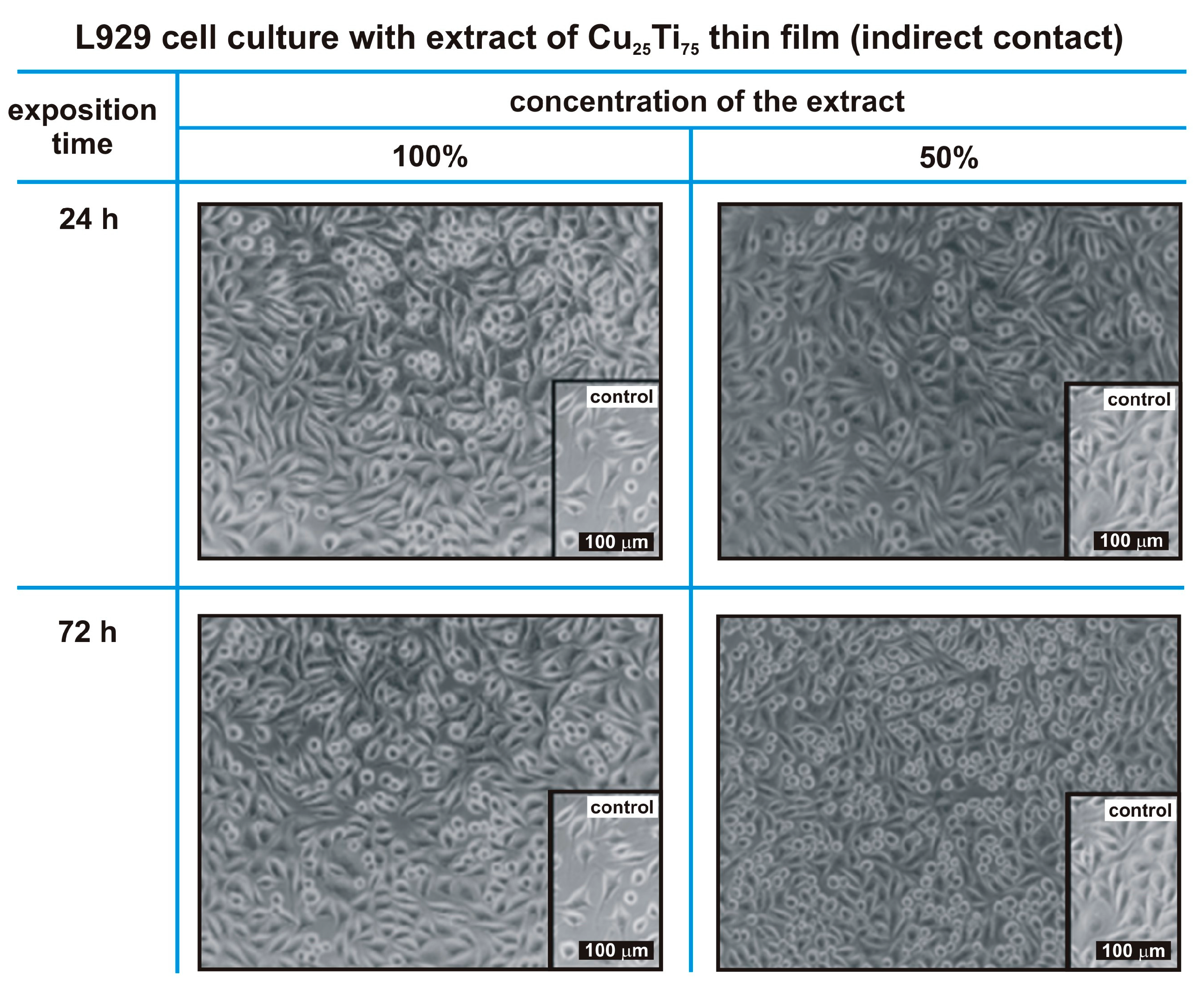
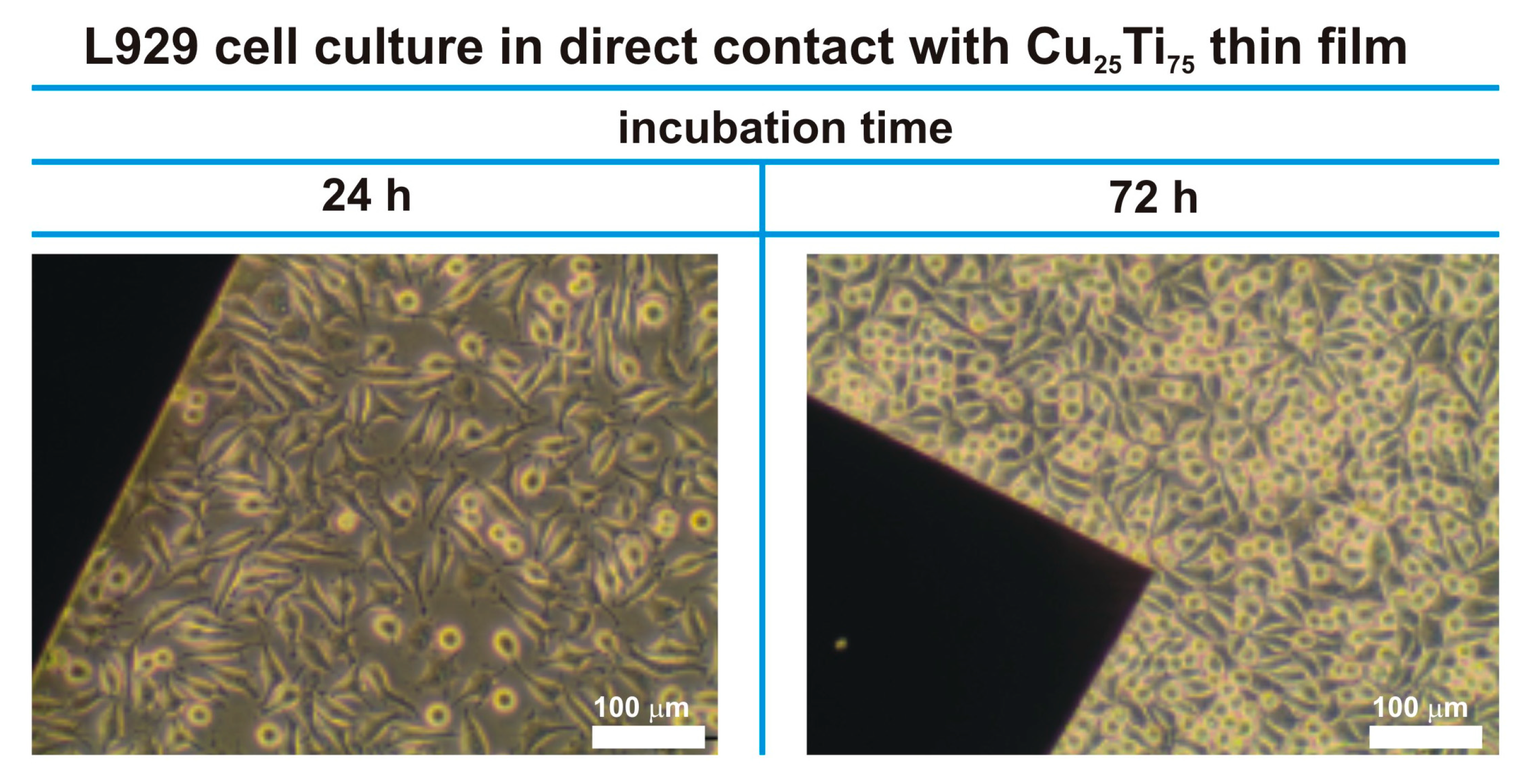
3.2. Morphological Evaluation of L929 Fibroblasts in Contact with a Nanocrystalline Cu25Ti75 Thin Film
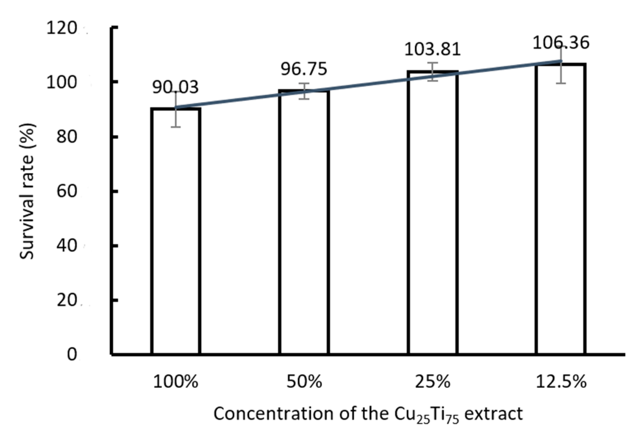
3.3. Metabolic Activity
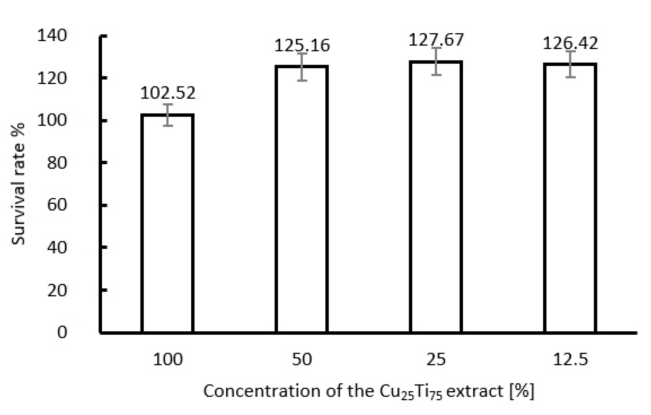
3.4. L929 Fibroblasts Proliferate When Exposed to Cu25Ti75 Thin Films
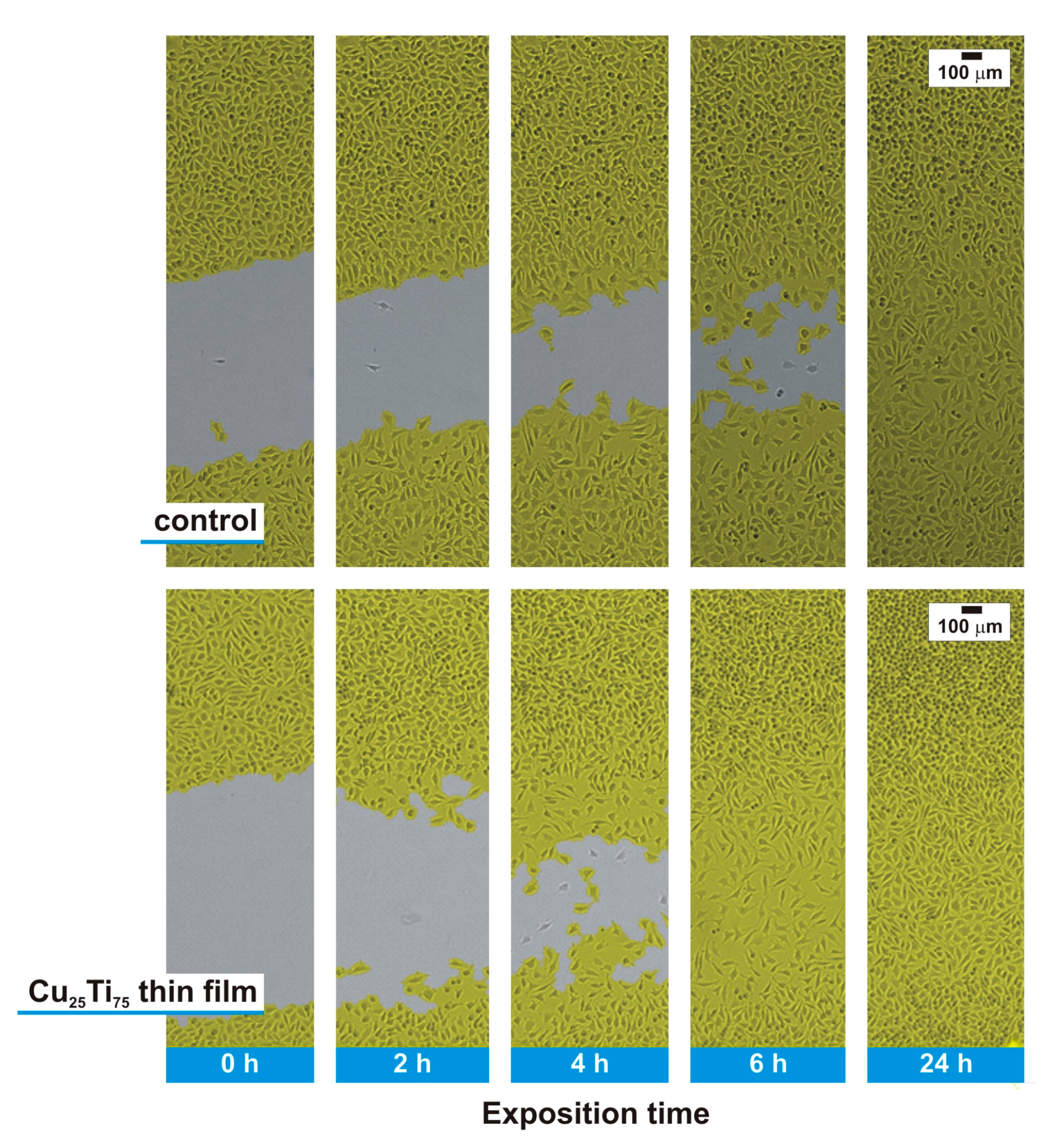
3.5. In Vitro Scarring Test
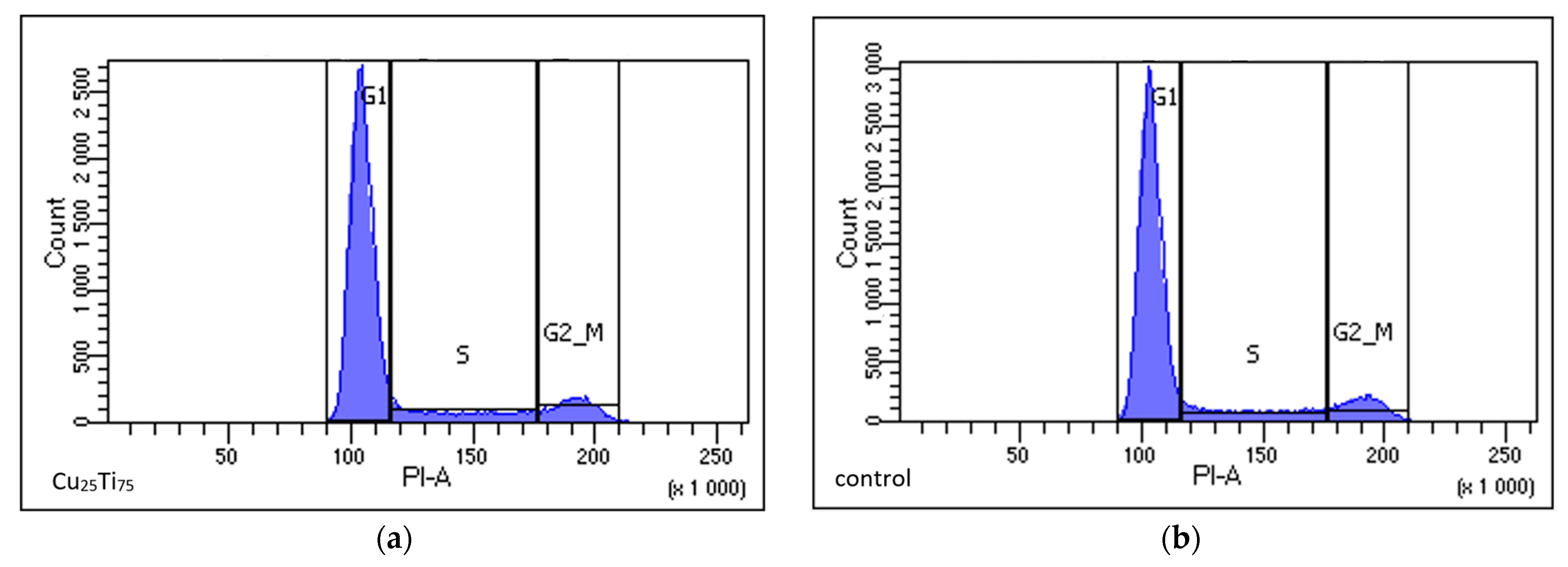
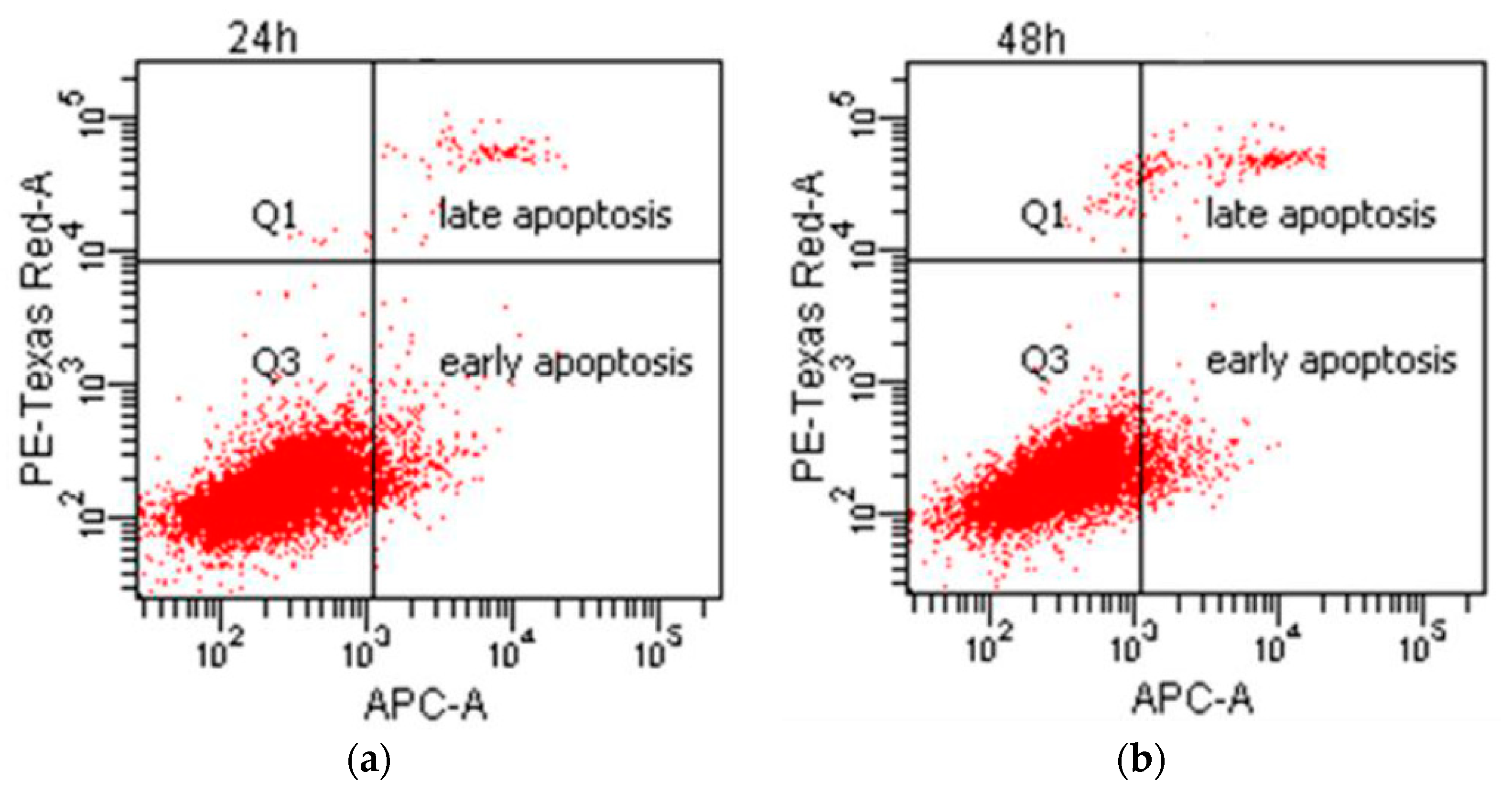
3.6. Cell Cycle Phase and Type of Cell Death (Cytometric Test)
4. Discussion
5. Conclusions
Supplementary Materials
Author Contributions
Funding
Institutional Review Board Statement
Informed Consent Statement
Data Availability Statement
Conflicts of Interest
References
- Murr, L.E. Strategies for creating living, additively manufactured, open-cellular metal and alloy implants by promoting osseointegration, osteoinduction and vascularization: An overview. J. Mater. Sci. Technol. 2019, 35, 231–241. [Google Scholar] [CrossRef]
- Kohn, D.H. Porous coatings in orthopedics. In Comprehensive Biomaterials; Ducheyne, P., Ed.; Elsevier Science: Amsterdam, The Netherlands, 2011. [Google Scholar] [CrossRef]
- Wilson, J. Metallic biomaterials: State of the art and new challenges. In Fundamental Biomaterials: Metals; Balakrishnan, P., Ed.; Woodhead Publishing Series in Biomaterials; Woodhead Publishing: Shaston, UK, 2018; pp. 1–33. [Google Scholar] [CrossRef]
- Kaur, M.; Singh, K. Review on titanium and titanium based alloys as biomaterials for orthopaedic applications. Mater. Sci. Eng. C 2019, 102, 844–862. [Google Scholar] [CrossRef]
- Kikuchi, M.; Takahashi, M.; Okabe, T.; Okuno, O. Grindability of dental cast Ti–Ag and Ti–Cu alloys. Dent. Mater. J. 2003, 22, 191–205. [Google Scholar] [CrossRef]
- Takahashi, M.; Kikuchi, M.; Takada, Y.; Okuno, O. Grindability and mechanical properties of experimental Ti-Au, Ti-Ag and Ti-Cu alloys. Int. Congr. Ser. 2005, 1284, 326–327. [Google Scholar] [CrossRef]
- Liu, R.; Memarzadeh, K.; Chang, B.; Zhang, Y.; Ma, Z.; Allaker, R.P.; Ren, L.; Yang, K. Antibacterial effect of copper-bearing titanium alloy (Ti-Cu) against Streptococcus mutans and Porphyromonas gingivalis. Sci. Rep. 2016, 6, 29985. [Google Scholar] [CrossRef]
- Mei, S.; Wang, H.; Wang, W.; Tong, L.; Pan, H.; Ruan, C.; Ma, Q.; Liu, M.; Yang, H.; Zhang, L. Antibacterial effects and biocompatibility of titanium surfaces with graded silver incorporation in titania nanotubes. Biomaterials 2014, 35, 4255–4265. [Google Scholar] [CrossRef]
- Niinomi, M. Recent metallic materials for biomedical applications. Metall Mat. Trans. A 2002, 33, 477–486. [Google Scholar] [CrossRef]
- Wojcieszak, D.; Mazur, M.; Kaczmarek, D.; Szponar, B.; Grobelny, M.; Kalisz, M.; Pelczarska, A.; Szczygieł, I.; Poniedziałek, A.; Osękowska, M. Structural and surface properties of semitransparent and antibacterial (Cu,Ti,Nb)Ox coating. Appl. Surf. Sci. 2016, 380, 159–164. [Google Scholar] [CrossRef]
- Wojcieszak, D.; Mazur, M.; Kaczmarek, D.; Mazur, P.; Szponar, B.; Domaradzki, J.; Kepinski, L. Influence of the surface properties on bactericidal and fungicidal activity of magnetron sputtered Ti-Ag and Nb-Ag thin films. Mater. Sci. Eng. C 2016, 62, 86–95. [Google Scholar] [CrossRef] [PubMed]
- Park, Y.J.; Song, Y.H.; An, J.H.; Song, H.J.; Anusavice, K.J. Cytocompatibility of pure metals and experimental binary titanium alloys for implant materials. J. Dent. 2013, 41, 1251–1258. [Google Scholar] [CrossRef]
- Shedle, A.; Samorapoompichit, P.; Rausch-Fan, X.H.; Franz, A.; Fureder, W.; Sperr, W.R.; Sperr, W.; Ellinger, A.; Slavicek, R.; Boltz-Nitulescu, G.; et al. Response of L929 fibroblasts, human gingival fibroblasts, and human tissue mast cells to various metal cations. J. Dent. Res. 1995, 74, 1513–1520. [Google Scholar] [CrossRef]
- Yamamoto, A.; Honma, R.; Sumita, M. Cytotoxicity evaluation of 43 metal salts using murine fibroblasts and osteoblastic cells. J. Biomed. Mater. Res. 1998, 39, 331–340. [Google Scholar] [CrossRef]
- Wojcieszak, D.; Osękowska, M.; Kaczmarek, D.; Szponar, B.; Mazur, M.; Mazur, P.; Obstarczyk, A. Influence of material composition on structure, surface properties and biological activity of nanocrystalline coatings based on Cu and Ti. Coatings 2020, 10, 343. [Google Scholar] [CrossRef]
- Wojcieszak, D.; Kaczmarek, D.; Antosiak, A.; Mazur, M.; Rybak, Z.; Rusak, A.; Batycka, M.; Poniedzialek, A.; Domaradzki, J.; Gamian, A.; et al. Antimicrobial activity and impact on living cells of Cu–Ti thin films in relation to their surface properties. Mater. Sci. Eng. C Mater. Biol. Appl. 2015, 56, 48–56. [Google Scholar] [CrossRef]
- Wojcieszak, D.; Mazur, M.; Kaczmarek, D.; Poniedziałek, A.; Osękowska, M. An impact of the copper additive on photocatalytic and bactericidal properties of TiO2 thin films. Mater. Sci. Pol. 2017, 35, 421–426. [Google Scholar] [CrossRef]
- Adamiak, B.; Wiatrowski, A.; Domaradzki, J.; Kaczmarek, D.; Wojcieszak, D.; Mazur, M. Preparation of multicomponent thin films by magnetron co-sputtering method: The Cu–Ti case study. Vacuum 2019, 161, 419–428. [Google Scholar] [CrossRef]
- Lüthen, F.; Bergemann, C.; Bulnheim, U.; Prinz, C.; Neumann, H.; Podbielski, A.; Bader, R.; Rychly, J. A dual role of copper on the surface of bone implants. Mater. Sci. Forum 2010, 638–642, 600–605. [Google Scholar] [CrossRef]
- Banci, L.; Bertini, I.; Cantini, F.; Ciofi-Baffoni, S. Cellular copper distribution: A mechanistic systems biology approach. Cell. Mol. Life Sci. 2010, 67, 2563–2589. [Google Scholar] [CrossRef]
- Prohaska, J.R.; Heller, L.J. Mechanical properties of the copper-deficient rat heart. J. Nutr. 1982, 112, 2142–2150. [Google Scholar] [CrossRef]
- Arredondo, M.; González, M.; Olivares, M.; Pizarro, F.; Araya, M. Ceruloplasmin, an indicator of copper status. Biol. Trace Elem. Res. 2008, 123, 261–269. [Google Scholar] [CrossRef]
- Chambers, A.; Krewski, D.; Birkett, N.; Plunkett, L.; Hertzberg, R.; Danzeisen, R.; Aggett, P.J.; Starr, T.B.; Baker, S.; Dourson, M.; et al. An exposure-response curve for copper excess and deficiency. J. Toxicol. Environ. Health B Crit. Rev. 2010, 13, 546–578. [Google Scholar] [CrossRef]
- Huffman, D.L.; O’Halloran, T.V. Function, structure, and mechanism of intracellular copper trafficking proteins. Annu. Rev. Biochem. 2001, 70, 677–701. [Google Scholar] [CrossRef] [PubMed]
- Failla, M.L. Roles of trace metals in the maturation, activation and effector functions of immune cells. Bibl. Nutr. Dieta. 1998, 54, 103–111. [Google Scholar] [CrossRef]
- Sen, C.K.; Khanna, S.; Venojarvi, M.; Trikha, P.; Ellison, E.C.; Hunt, T.K.; Roy, S. Copper-induced vascular endothelial growth factor expression and wound healing. Am. J. Physiol. Heart Circ. Physiol. 2002, 282, H1821–H1827. [Google Scholar] [CrossRef] [PubMed]
- Harris, E.D. Basic and clinical aspects of copper. Crit. Rev. Clin. Lab. Sci. 2003, 40, 547–586. [Google Scholar] [CrossRef] [PubMed]
- Kobayashi, H.; Ishii, M.; Chanoki, M.; Yashiro, N.; Fushida, H.; Fukai, K.; Kono, T.; Hamada, T.; Wakasaki, H.; Ooshima, A. Immunohistochemical localization of lysyl oxidase in normal human skin. Br. J. Dermatol. 1994, 131, 325–330. [Google Scholar] [CrossRef]
- Liu, Y.; Tay, J.-H. Detachment forces and their influence on the structure and metabolic behaviour of biofilms. World J. Microb. Biot. 2001, 17, 111–117. [Google Scholar] [CrossRef]
- Heidenau, F.; Mittelmeier, W.; Detsch, R.; Haenle, M.; Stenzel, F.; Ziegler, G.; Gollwitzer, H. A novel antibacterial titania coating: Metal ion toxicity and in vitro surface colonization. J. Mater. Sci. Mater. Med. 2005, 16, 883–888. [Google Scholar] [CrossRef]
- Chen, X.; Hu, J.G.; Huang, Y.Z.; Li, S.; Li, S.F.; Wang, M.; Xia, H.W.; Li-Ling, J.; Xie, H.Q. Copper promotes the migration of bone marrow mesenchymal stem cells via Rnd3-dependent cytoskeleton remodeling. J. Cell Physiol. 2020, 235, 221–231. [Google Scholar] [CrossRef]
- Jeney, F.; Bazsó-Dombi, E.; Oravecz, K.; Szabó, J.; Nagy, I.Z. Cytochemical studies on the fibroblast-preadipocyte relationships in cultured fibroblast cell lines. Acta Histochem. 2000, 102, 381–389. [Google Scholar] [CrossRef]
- Yu, L.; Jin, G.; Ouyang, L.; Wang, D.; Qiao, Y.; Liu, X. Antibacterial activity, osteogenic and angiogenic behaviors of copper-bearing titanium synthesized by PIII&D. J. Mater. Chem. B 2016, 4, 1296–1309. [Google Scholar] [CrossRef] [PubMed]
- Zheng, K.; Dai, X.; Lu, M.; Hüser, N.; Taccardi, N.; Boccaccini, A.R. Synthesis of copper-containing bioactive glass nanoparticles using a modified Stöber method for biomedical applications. Colloids Surf. B Biointerfaces. 2017, 150, 159–167. [Google Scholar] [CrossRef] [PubMed]
- Ma, Z.X.; Ren, L.; Liu, R.; Yang, K.; Zhang, Y.; Liao, Z.; Liu, W.; Qi, M.H.; Misra, R.D. Effect of heat treatment on Cu distribution, antibacterial performance and cytotoxicity of Ti–6Al–4V–5Cu alloy. J. Mater. Sci. Technol. 2015, 31, 723–732. [Google Scholar] [CrossRef]
- Saphier, M.; Silberstein, E.; Shotland, Y.; Popov, S.; Saphier, O. Prevalence of monovalent copper over divalent in killing Escherichia coli and Staphylococcus aureus. Curr. Microbiol. 2018, 75, 426–430. [Google Scholar] [CrossRef] [PubMed]

| Crystal Phase | Cu3Ti4 | |
|---|---|---|
| Size of crystallites-D (nm) | 9.9 | |
| Percentage of copper ions on the surface (%) | Cu0,1+ | 58.2 |
| Cu2+ | 41.8 | |
| Amount of copper ions released ((ppb/mm2) per day) | 0.003 | |
Publisher’s Note: MDPI stays neutral with regard to jurisdictional claims in published maps and institutional affiliations. |
© 2021 by the authors. Licensee MDPI, Basel, Switzerland. This article is an open access article distributed under the terms and conditions of the Creative Commons Attribution (CC BY) license (http://creativecommons.org/licenses/by/4.0/).
Share and Cite
Osękowska, M.; Wojcieszak, D.; Kaczmarek, D.; Mazur, M.; Obstarczyk, A.; Szponar, B. Multifunctional Nanocrystalline Cu–Ti Thin Films Enhance Survival and Induce Proliferation of Mouse Fibroblasts In Vitro. Coatings 2021, 11, 300. https://doi.org/10.3390/coatings11030300
Osękowska M, Wojcieszak D, Kaczmarek D, Mazur M, Obstarczyk A, Szponar B. Multifunctional Nanocrystalline Cu–Ti Thin Films Enhance Survival and Induce Proliferation of Mouse Fibroblasts In Vitro. Coatings. 2021; 11(3):300. https://doi.org/10.3390/coatings11030300
Chicago/Turabian StyleOsękowska, Małgorzata, Damian Wojcieszak, Danuta Kaczmarek, Michał Mazur, Agata Obstarczyk, and Bogumiła Szponar. 2021. "Multifunctional Nanocrystalline Cu–Ti Thin Films Enhance Survival and Induce Proliferation of Mouse Fibroblasts In Vitro" Coatings 11, no. 3: 300. https://doi.org/10.3390/coatings11030300
APA StyleOsękowska, M., Wojcieszak, D., Kaczmarek, D., Mazur, M., Obstarczyk, A., & Szponar, B. (2021). Multifunctional Nanocrystalline Cu–Ti Thin Films Enhance Survival and Induce Proliferation of Mouse Fibroblasts In Vitro. Coatings, 11(3), 300. https://doi.org/10.3390/coatings11030300


