Article

A Novel Colorimetric Chemosensor Based on Ferene-S-Conjugated Silver Nanoparticles for Selective Recognition of Fe2+

1
School of Materials Science & Engineering, Northwestern Polytechnical University, Xi’an 710072, China
2
Department of Chemistry, University of Karachi, Karachi 75270, Pakistan
3
Department of Chemistry, Adul Wali Khan University, Mardan 23200, Pakistan
4
Department of Basic Sciences, DHA Suffa University, Off Khayaban-e-Tufail, Phase VII Ext. DHA, Karachi 75500, Pakistan
5
Department of Chemistry, Shaheed Benazir Bhutto Women University, Peshawar 25000, Pakistan
6
Department of Chemistry, University of Malakand, Chakdara 18800, Pakistan
7
Department of Chemistry, College of Science, Taif University, P.O. Box 11099, Taif 21944, Saudi Arabia
*
Authors to whom correspondence should be addressed.
Coatings 2021, 11(11), 1293; https://doi.org/10.3390/coatings11111293
Submission received: 3 September 2021 / Revised: 21 October 2021 / Accepted: 22 October 2021 / Published: 25 October 2021

Abstract

:
Ferene is the most commonly used chromogenic agent for the determination of serum iron in blood. In this work we have successfully synthesized Ferene-S-conjugated silver nanoparticles (Ferene-S-AgNPs) for the first time characterized by UV-visible, Fourier-Transform Infrared Spectroscopy (FTIR), and Matrix-Assisted Laser Desorption/Ionization-Time Of Flight (MALDI-TOF) mass spectrometry techniques. Particle size of the synthesized nanoparticles was determined by atomic-force microscopy and scanning electron microscopy techniques with size ranges from 10–90 nm in diameter. Ferene-S-AgNPs were explored for their chemosensing potential with various metal ions such as Sb3+, Pb2+, Ca2+, Fe2+, K+, Co2+, Ba2+, V5+, Cu+, Cd2+, Hg2+, Ni2+, Cu2+, Fe3+, Mg2+, Mn2+, Al3+, and Cr3+. Ferene-S-AgNPs were found to show selective quenching effects and slight bathochromic shifts to the surface plasmon resonance absorption band after treatment with Fe2+. Furthermore, the developed chemosensor also exhibited substantial selectivity towards Fe2+ in the presence of other competitive ions. We observed that Ferene-S-AgNPs mimic the selectivity of the parent compound of Ferene towards Fe2+. The system obeyed Beer’s law over concentration ranges of 110–190 nM. The detection limit was found to be 110 nM.

1. Introduction

Surface plasmon resonance (SPR) of metallic nanoparticles [1] give rise to interesting colors when interacting with electromagnetic radiation that is different from those observed in bulk materials [2]. Metals that have free electrons including Au, Ag, Cu, and alkali metals shows SPR absorption in the visible range [3]. Metallic nanoparticles have a wide variety of potential applications in many research fields, including chemosensing [4], biomedical [5,6], optical [7], and electronic fields [8]. Ag and Au nanoparticles show a strong surface plasmon resonance at wavelengths in the range of 400–420 nm [9] and 520–530 nm [10], respectively. Ag nanoparticles have been used as an alternative chemosensor to Au nanoparticles due to lower costs and higher extinction coefficients. Ferene-S (3-(2-pyridyl)-5, 6-bis-(2-(5-furyl-sulfonic acid acid)-1,2,4-triazine disodium salt) has been used for several years as a chromogenic reagent for the determination of copper and iron [11]. It has also been used for the determination of serum iron in blood samples since 1979. In this work, we have successfully synthesized Ag nanoparticles (AgNPs) of Ferene-S, and explored its chemosensing potential, which selectively senses iron (Fe2+) in the presence of other interfering metal ions [11,12,13,14].
Iron is involved in several biological processes, such as respiration, oxygen transport, signal transduction, and enzymatic reactions that involve the production of reactive oxygen species (ROS). Under physiologically normal conditions, the production and consumption rate of ROS is sternly controlled to exploit the beneficial aspects of ROS [15]. Dysfunction of the iron homeostasis process could seriously affect the balanced ROS metabolism, leading to the aberrant production of ROS, resulting in cell damage and death. In particular, the Fenton reaction has been known as one of the most harmful reactions in a biological system, where Fe2+ acts as an activator to generate highly toxic hydroxyl radicals that can induce DNA damage and the peroxidation of lipids [16]. Thus, an excess of iron has been related to various diseases, such as cancer and neurodegenerative diseases including Alzheimer’s and Parkinson’s, where excess iron is often observed in patients [17].
Different analytical approaches, routinely used for the determination of iron in any biological or environmental samples, includes inductively coupled plasma mass spectrometry (ICP-MS) [18], inductively coupled plasma atomic emission spectrometry (ICP-AES) [19], and atomic absorption (AA) spectrometry [20]. However, these analytical techniques are expensive and time-consuming. Therefore, the development of a simple and novel chemosensor that can selectively sense iron ion (Fe2+) in any environmental and biological sample has attracted considerable attention [21,22,23,24]. Functionalized silver nanoparticles have recently gained considerable attention for the colorimetric detection of various metal ions [25,26,27]. Ferene-S-conjugated silver nanoparticles, which selectively recognize Fe2+ in the presence of other metal ions, have been synthesized. To our knowledge, the nano-formulation of Ferene-S has not been reported in the literature. This novel synthetic approach for Ferene-S-AgNPs uses a simple and robust method, showing higher selectivity for Fe2+ detection and mimicking the selectivity of the parent compound.

2. Materials and Methods

2.1. Material and Instruments

Silver nitrate and sodium borohydride were purchased from Merck, and Ferene-S was purchased from MP Biomedicals (Shanghai, China). UV-visible spectra were recorded with a Schimadzu UV-240; Hitachi U-3200 Spectrometer (Kyoto, Japan) and IR spectra were recorded using Schimadzu IR-460 (Kyoto, Japan) after making a KBr pellet. A digital pH meter Model 510 (Oakton Instruments, Vernon Hills, IL, USA) equipped with a glass working electrode and a reference Ag/AgCl electrode was used. A MALDI-TOF spectrum was recorded on a Bruker Ultra flex TOF-TOF operated in reflection mode (by Bruker Corporation, Billerica, MA, USA). The matrix used for sampling was 4-Hydroxy-2-cyanocinnamic acid (purchased from Merck & Co, New Jersey, NJ, USA). A topographical image was obtained with an atomic-force microscope, Agilent 5500 by Agilent Technologies, Santa Clara, CA, USA operated in tapping mode. The size of Ferene-S-conjugated silver nanoparticles was determined under a scanning electron microscope (SEM) model JSM5910 manufactured by JEOL, Tokyo, Japan. All solutions were prepared in deionized water and stored at room temperature.

2.2. Synthesis of Ferene-S-Conjugated AgNPs

Silver nanoparticles were synthesized by mixing 0.1 mM solution of silver nitrate with 0.1 mM solution of Ferene-S in 10:1 mole ratio and stirring. After 30 min, 0.1 mL of 4 mM solution of NaBH4 was added dropwise (Figure S1). The color of the solution changed to yellow, indicating the formation of AgNPs. The solution continued to be stirred for 3 h at room temperature to ensure the complete reduction of Ag ions. Ferene-S-conjugated AgNPs were collected in solid form after freeze drying [28].

3. Results and Discussion

3.1. UV-Visible Spectral Analysis of Ferene-S-Conjugated AgNPs

The reduction of silver ions and the formation of Ag nanoparticles was monitored using UV-visible spectral analysis. The SPR absorption spectrum of silver nanoparticles was observed at 400 nm (Figure 1a) to have a yellow color and was stable for 4–5 days with only a minor change in SPR absorption intensity [29].
The stability of Ferene-S-conjugated AgNPs was studied with various concentrations of sodium chloride. Ferene-S-conjugated AgNPs were stable up to a 50 mM solution of NaCl, and they are highly unstable above this concentration. At higher concentrations of NaCl, the decrease in SPR absorption is due to the aggregation phenomenon promoted by chloride ions (Figure S2). The effect of temperature on the stability of Ferene-S-conjugated AgNPs was also investigated at 100 °C. After cooling to room temperature, the absorption spectra were recorded, which shows that temperature has no significant effect on the absorption maxima of the silver nanoparticles. However, a slight shift in the absorption maxima of the surface plasmon peak was observed without any aggregate formation [30] (Figure S3).
To further study the stability of Ferene-S-conjugated AgNPs, we assessed the silver nanoparticles in different pH ranges of 1–12. It was found that in slightly acidic and slightly basic conditions (4–10), an enhancement in the absorbance of Ferene-S-conjugated AgNPs occurs, which may be due to a decrease in the particle size of AgNPs, and no aggregate formation takes place in this pH range. When pH (>10) increases, absorbance decreases, which may be due to the hydroxylation of silver and the formation of Ag(OH). Under acidic conditions (<4), protonation of Ferene-S-AgNPs leads to the aggregation of Ag nanoparticles and an increase in the particle size occurs (Figure S4) [30].

3.2. Comparative FTIR of Ferene-S and Ferene-S-Conjugated AgNPs

FTIR spectra of Ferene-S and Ferene-S-conjugated AgNPs are shown in Figure 1b. Ferene-S shows an absorption band at 3483, 1571, 1474, 1216, and 1048 cm−1. A broad absorption band appears at 3483 cm−1, which can be assigned to H–C=C and H–C=N. It is very high, due to the inductive effect of nitrogen present in the six-member aromatic ring. Medium absorption bands appear at 1571 cm−1 and 1474 cm−1 due to aromatic –C=C–. A sharp absorption band at 1216 cm−1 is observed due to the presence of C–O–C, i.e., of ether functionality. The absorption band at 1048 cm−1 appears due to the S=O in sulfonic acid. There is a prominent change in the intensity of all absorption bands in the spectra of Ferene-S after the formation of silver nanoparticles [31].

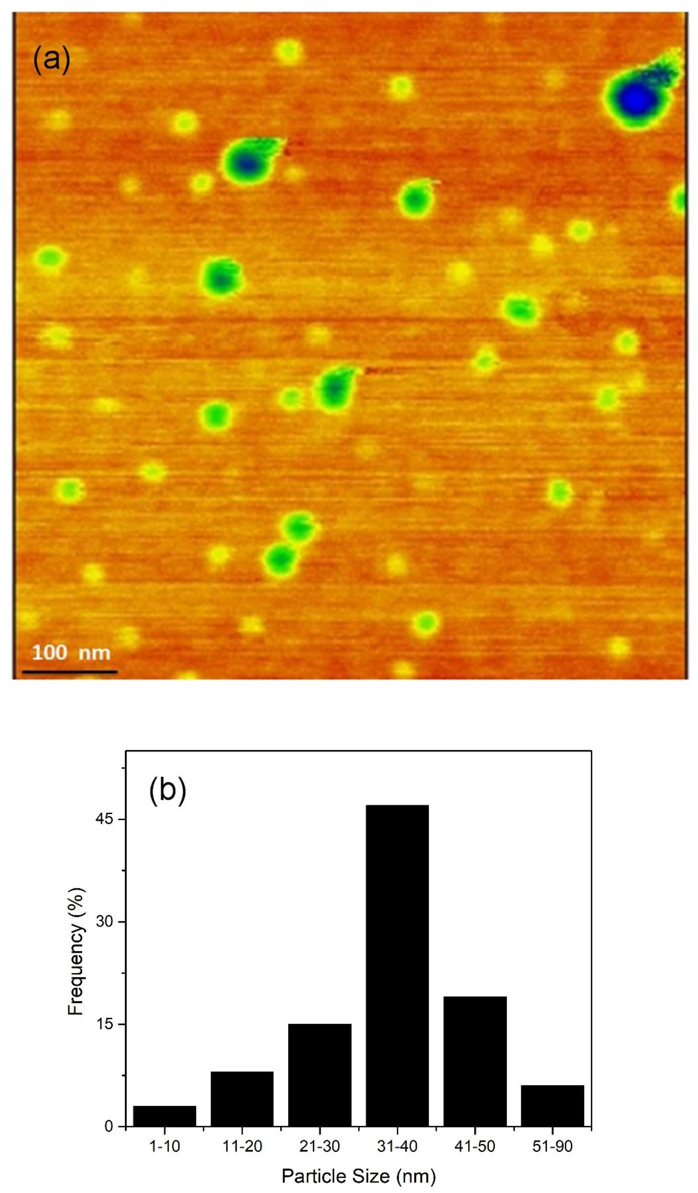
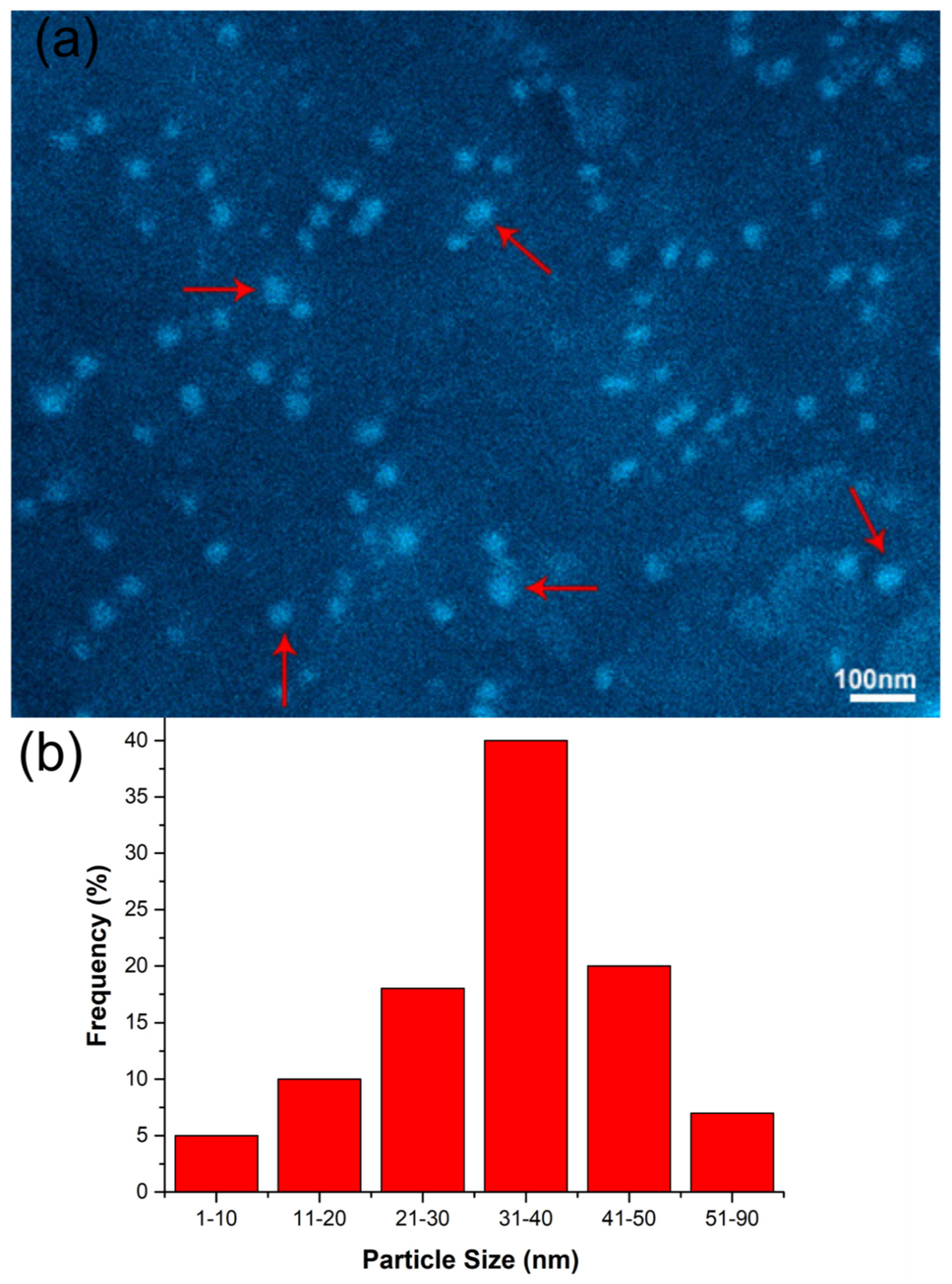
3.3. Particle-Size Determination through AFM, SEM, and Analysis of MALDI-TOF Spectrometry

The morphology and size of Ferene-S-conjugated AgNPs was determined using AFM. AFM analysis shows that these silver nanoparticles were polydispersed and irregular in shape. The sizes range from 10–90 nm in diameter, with average size around 50 nm (Figure 2). The SEM results also showed that these Ferene-S-conjugated AgNPs were polydispersed and irregular in shape. The sizes range from 10–90 nm in diameter with average size around 40 nm (Figure 3). In the MALDI-TOF spectrum, a regular series of fragment ions with increasing number of silver ions to ligand (Ferene-S) are obtained. These fragment ions are in the order of [Ag1L]+, [Ag2L]+, [Ag3L]+, [Ag1L2 × H2O]+, [Ag3L2]+, [Ag4L2]+, and [Ag5L2]+. The fragment ion [Ag1L]+ (556.9) show the highest intensity, which is a base peak in the spectra. This indicates that Ferene-S-conjugated AgNPs are most stable in 1:1 mole ratio of metal to ligand (Figure S5) [30].

3.4. Ferene-S-Conjugated AgNPs as a Potential Chemosensing Probe

The chemosensing study was performed by treating various metals ion with Ferene-S-AgNPs followed by recording their UV-visible spectra. 500-µM stock solutions of various metal ions such as Sb3+, Pb2+, Ca2+, Fe2+, K+, Co2+, Ba2+, V5+, Cu+, Cd2+, Hg2+, Ni2+, Cu2+, Fe3+, Mg2+, Mn2+, Al3+, and Cr3+ were prepared and independently mixed with Ferene-S-AgNPs. It was observed that the Ferene-S-AgNPs showed no effect towards other metal ions with respect to SPR. However, when treated with Fe2+, significant quenching and a minor bathochromic effect was observed in the SPR absorption of the Ferene-S-AgNPs (Figure 4). The selectivity of Ferene-S-AgNPs towards Fe2+ mimics the selectivity of the parent compound, Ferene-S, which also showed specificity towards Fe2+. Ferene-S is largely applied as one of the most sensitive colorimetric reagents used for the determination of serum iron in blood. This new addition of Ferene-S-AgNPs in nano-formulation establishes an alternative approach to the commonly used Ferene-S for the determination of serum iron [11].

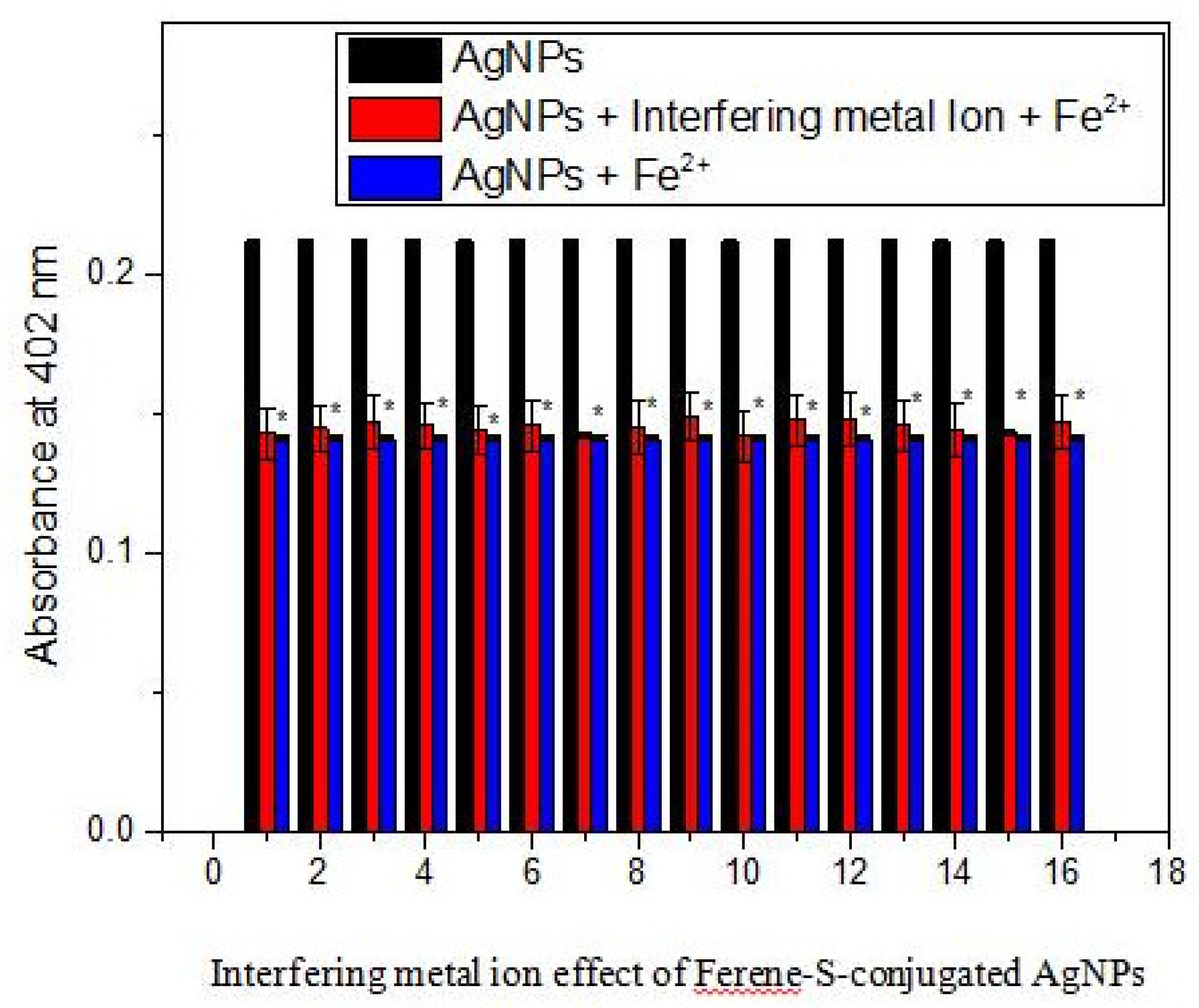
3.5. Interference Study in Real Samples and Binding Study of Ferene-S-AgNPs with Fe2+

To further observe the practical capability of the Ferene-S-AgNPs as a probe displaying high selectivity for Fe2+, the potential interfering effect of different metal ions was investigated in real samples. Different metal ions were spiked in real water samples along with Fe2+, and its UV-visible spectra were recorded. It was observed that the presence of these interfering ions showed no effect on Fe2+ selectivity using Ferene-S-AgNPs, even in real samples. Furthermore, the interaction of these metals with AgNPs in the presence of Fe2+ does not largely affect the surface SPR absorption of Ferene-S-conjugated AgNPs, indicating the selectivity of these AgNPs towards Fe2+ (Figure 5).
To determine the binding stoichiometry between Ferene-S-conjugated AgNPs and Fe2+, the Job plot method was used. We observed a change in the absorption intensity by continually changing the mole fraction between Ferene-S-AgNPs and Fe2+. Binding assays using the Job plot method showed that a 3:1 mole ratio is required for complex formations between Ferene-S-AgNPs and Fe2+ (Figure S6).

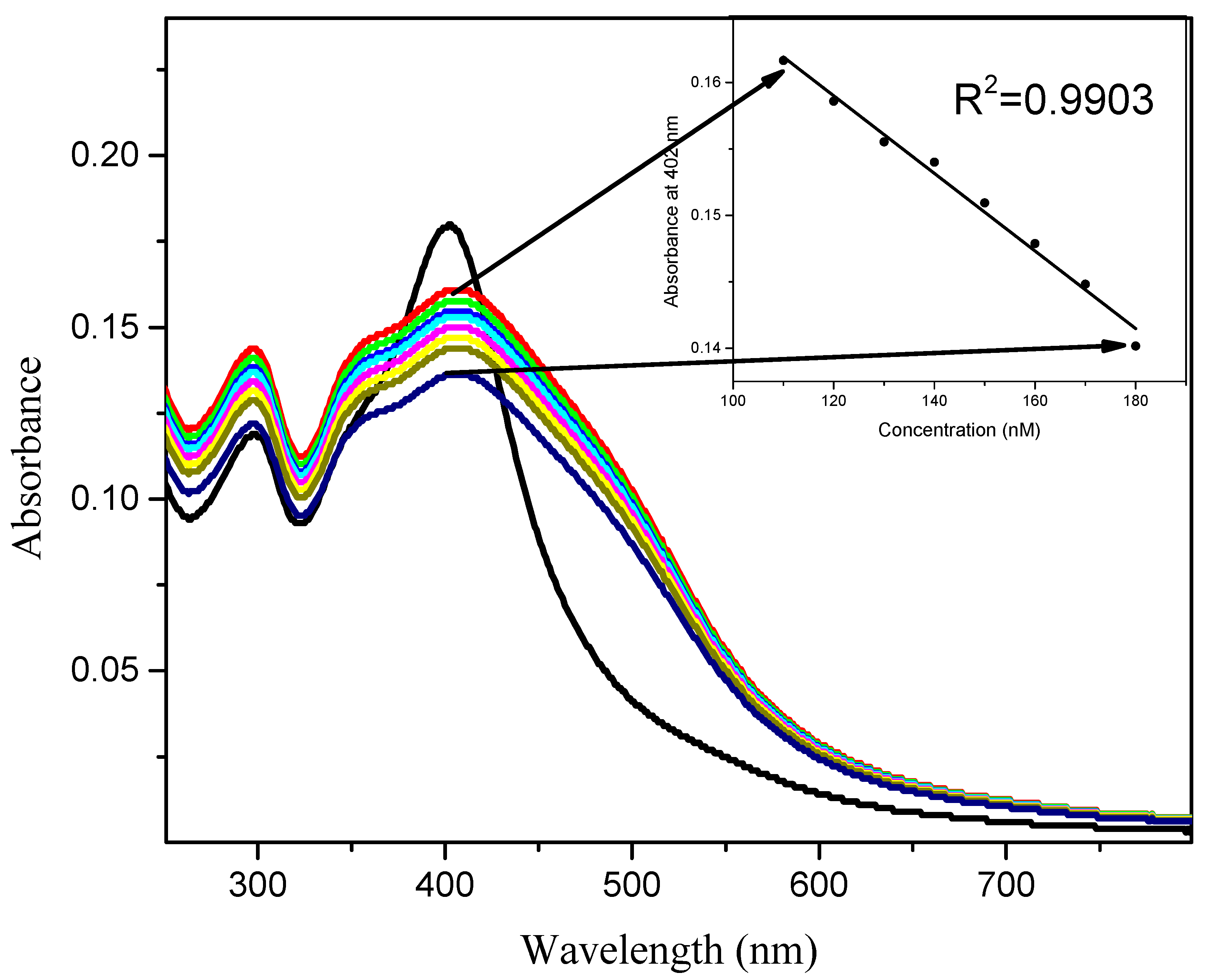
3.6. Detection Limit of the Probe and Effect of pH on the Ferene-S-AgNPs–Fe2+ Complex

The analytical capability of Ferene-S-conjugated AgNPs was explored using different concentrations of Fe2+ (Figure 6), and showed that SPR absorption increases gradually as the concentration of Fe2+ decreases. A linear relationship was found in the concentration range of 110–190 nM with a linear regression of R2 = 0.9903. The limit of detection was found to be 110 nM. Further investigation of the complexation behavior of Ferene-S-conjugated AgNPs with Fe2+ at different levels of pH was evaluated. The Ferene-S-AgNPs–Fe2+ complex was found to be stable in slightly acidic and basic conditions in the pH range of 4–9, and highly unstable in highly acidic and basic media (Figure S7).

4. Conclusions

We have successfully synthesized Ferene-S-conjugated silver nanoparticles, characterized using FTIR, UV-visible, AFM, and MALDI-TOFF spectrometry. Ferene-S-AgNPs were explored for their chemosensing potential towards different metal ions. Ferene-S-AgNPs selectively detect Fe2+ in the presence of other competitive metal ions, and maintain the selectivity of the parent compound, Ferene-S. This novel approach of using Ferene-S as a capping agent for AgNPs and selectivity towards Fe2+ is a relatively new technique to detect trace amounts of Fe2+ in ultrasensitive bioanalysis.

Supplementary Materials

The following are available online at https://www.mdpi.com/article/10.3390/coatings11111293/s1, Figure S1: Synthesis of Ferene-S-conjugated silver nanoparticles. Figure S2: UV-visible spectra of AgNPs after treating with NaCl. Figure S3: UV-visible spectra of AgNPs before and after heating. Figure S4: UV-visible spectra of AgNPs in different pH. Figure S5: MALDI-TOF mass spectrum of the synthesized Ferene-S-AgNPs. Figure S6: Job’s method for the stoichiometry of binding event of both ligand and metal. Figure S7: Efficiency of Ferene-S-AgNPs for the detection of Fe2+ at different pH levels.

Author Contributions

Conceptualization, A.U., P.F. and M.A.; methodology, M.T.M., G.R. and M.R.; software, M.R., M.S.K., G.R. and M.T.M.; formal analysis, M.S.K., O.A.A.A. and D.I.S.; investigation, and resources, M.S.K., O.A.A.A. and D.I.S.; writing—original draft preparation, P.F., M.A. and R.K. and editing A.U., M.A. and R.K. All authors have read and agreed to the published version of the manuscript.

Funding

This work was supported by Taif University Researchers Supporting Project number (TURSP-2020/220), Taif University, Taif, Saudi Arabia. and Higher Education Commission (HEC) of Pakistan.

Institutional Review Board Statement

Not Applicable.

Informed Consent Statement

Not Applicable.

Data Availability Statement

Data is contained within the article or Supplementary Material.

Acknowledgments

The authors are thankful to Taif University Researchers Supporting Project number (TURSP-2020/220), Taif University, Taif, Saudi Arabia and Higher Education Commission (HEC) of Pakistan.

Conflicts of Interest

We as corresponding authors on behalf of all authors in this paper retain the rights for all authors and their representatives. All authors are agreed for the submission of this manuscript and there is no conflict of interest.

References

  1. Willets, K.A.; van Duyne, R.P. Localized surface plasmon resonance spectroscopy and sensing. Annu. Rev. Phys. Chem. 2007, 58, 267–297. [Google Scholar] [CrossRef] [Green Version]
  2. Fulekar, M. Nanotechnology: Importance and Applications; IK International Pvt Ltd.: Delhi, India, 2010. [Google Scholar]
  3. Liz-Marzán, L.M.; Touriño, I.L. Reduction and Stabilization of Silver Nanoparticles in Ethanol by Nonionic Surfactants. Langmuir 1996, 12, 3585–3589. [Google Scholar] [CrossRef]
  4. Jung, J.H.; Lee, J.H.; Shinkai, S. Functionalized magnetic nanoparticles as chemosensors and adsorbents for toxic metal ions in environmental and biological fields. Chem. Soc. Rev. 2011, 40, 4464–4474. [Google Scholar] [CrossRef]
  5. Neuberger, T.; Schöpf, B.; Hofmann, H.; Hofmann, M.; von Rechenberg, B. Superparamagnetic nanoparticles for biomedical applications: Possibilities and limitations of a new drug delivery system. J. Magn. Magn. Mater. 2005, 293, 483–496. [Google Scholar] [CrossRef]
  6. Mirza, S.; Ahmad, M.S.; Shah, M.I.A.; Ateeq, M. Magnetic Nanoparticles: Drug Delivery and Bioimaging Applications. In Metal Nanoparticles for Drug Delivery and Diagnostic Applications; Elsevier: Amsterdam, The Netherlands, 2020; pp. 189–213. [Google Scholar]
  7. Biju, V.; Itoh, T.; Anas, A.; Sujith, A.; Ishikawa, M. Semiconductor quantum dots and metal nanoparticles: Syntheses, optical properties, and biological applications. Anal. Bioanal. Chem. 2008, 391, 2469–2495. [Google Scholar] [CrossRef]
  8. Sweatlock, L.A.; Maier, S.; Atwater, H.A.; Penninkhof, J.J.; Polman, A. Highly confined electromagnetic fields in arrays of strongly coupled Ag nanoparticles. Phys. Rev. B 2005, 71, 235408. [Google Scholar] [CrossRef] [Green Version]
  9. Lok, C.-N.; Ho, C.-M.; Chen, R.; He, Q.-Y.; Yu, W.-Y.; Sun, H.; Tam, P.K.-H.; Chiu, J.-F.; Che, C.-M. Silver nanoparticles: Partial oxidation and antibacterial activities. JBIC J. Biol. Inorg. Chem. 2007, 12, 527–534. [Google Scholar] [CrossRef] [PubMed]
  10. El-Sayed, I.H.; Huang, X.; El-Sayed, M.A. Surface Plasmon Resonance Scattering and Absorption of anti-EGFR Antibody Conjugated Gold Nanoparticles in Cancer Diagnostics: Applications in Oral Cancer. Nano Lett. 2005, 5, 829–834. [Google Scholar] [CrossRef] [PubMed]
  11. Zak, B.; Ressler, N. Simultaneous Microdetermination of Copper and Iron Using Mixed Phenanthrolines. Anal. Chem. 1956, 28, 1158–1161. [Google Scholar] [CrossRef]
  12. Prosposito, P.; Burratti, L.; Venditti, I. Silver Nanoparticles as Colorimetric Sensors for Water Pollutants. Chemosensors 2020, 8, 26. [Google Scholar] [CrossRef] [Green Version]
  13. Sabela, M.; Balme, S.; Bechelany, M.; Janot, J.-M.; Bisetty, K. A Review of Gold and Silver Nanoparticle-Based Colorimetric Sensing Assays. Adv. Eng. Mater. 2017, 19, 1700270. [Google Scholar] [CrossRef]
  14. Firdaus, M.L.; Aprian, A.; Meileza, N.; Hitsmi, M.; Elvia, R.; Rahmidar, L.; Khaydarov, R. Smartphone Coupled with a Paper-Based Colorimetric Device for Sensitive and Portable Mercury Ion Sensing. Chemosensors 2019, 7, 25. [Google Scholar] [CrossRef] [Green Version]
  15. Dixon, S.J.; Stockwell, B.R. The role of iron and reactive oxygen species in cell death. Nat. Chem. Biol. 2014, 10, 9–17. [Google Scholar] [CrossRef]
  16. Enami, S.; Sakamoto, Y.; Colussi, A.J. Fenton chemistry at aqueous interfaces. Proc. Natl. Acad. Sci. USA 2014, 111, 623–628. [Google Scholar] [CrossRef] [Green Version]
  17. Torti, S.V.; Torti, F.M. Iron and cancer: More ore to be mined. Nat. Rev. Cancer 2013, 13, 342–355. [Google Scholar] [CrossRef] [Green Version]
  18. Andersen, J.E.T. A novel method for the filterless preconcentration of iron. Analyst 2005, 130, 385–390. [Google Scholar] [CrossRef] [PubMed]
  19. Busto, M.E.D.C.; Montes-Bayón, M.; Blanco-González, E.; Meija, J.; Sanz-Medel, A. Strategies To Study Human Serum Transferrin Isoforms Using Integrated Liquid Chromatography ICPMS, MALDI-TOF, and ESI-Q-TOF Detection: Application to Chronic Alcohol Abuse. Anal. Chem. 2005, 77, 5615–5621. [Google Scholar] [CrossRef] [PubMed]
  20. Pomazal, K.; Prohaska, C.; Steffan, I.; Reich, G.; Huber, J.F.K. Determination of Cu, Fe, Mn, and Zn in blood fractions by SEC-HPLC-ICP-AES coupling. Analyst 1999, 124, 657–663. [Google Scholar] [CrossRef] [PubMed]
  21. David, C.I.; Bhuvanesh, N.; Jayaraj, H.; Thamilselvan, A.; Devi, D.P.; Abiram, A.; Prabhu, J.; Nandhakumar, R. Experimental and Theoretical Studies on a Simple S–S-Bridged Dimeric Schiff Base: Selective Chromo-Fluorogenic Chemosensor for Nanomolar Detection of Fe2+ & Al3+ Ions and Its Varied Applications. ACS Omega 2020, 5, 3055–3072. [Google Scholar]
  22. Shakya, S.; Khan, I.M.; Ahmad, M. Charge transfer complex based real-time colorimetric chemosensor for rapid recognition of dinitrobenzene and discriminative detection of Fe2+ ions in aqueous media and human hemoglobin. J. Photochem. Photobiol. A Chem. 2020, 392, 112402. [Google Scholar] [CrossRef]
  23. Gong, X.; Zhang, H.; Jiang, N.; Wang, L.; Wang, G. Oxadiazole-based ‘on-off’ fluorescence chemosensor for rapid recognition and detection of Fe2+ and Fe3+ in aqueous solution and in living cells. Microchem. J. 2019, 145, 435–443. [Google Scholar] [CrossRef]
  24. Ravichandiran, P.; Boguszewska-Czubara, A.; Masłyk, M.; Bella, A.P.; Subramaniyan, S.A.; Johnson, P.M.; Shim, K.S.; Kim, H.G.; Yoo, D.J. Naphthoquinone-Based Colorimetric and Fluorometric Dual-Channel Chemosensor for the Detection of Fe2+ Ion and Its Application in Bio-Imaging of Live Cells and Zebrafish. ACS Sustain. Chem. Eng. 2019, 7, 17210–17219. [Google Scholar] [CrossRef]
  25. Kumar, V.V.; Anbarasan, S.; Christena, L.R.; SaiSubramanian, N.; Anthony, S.P. Bio-functionalized silver nanoparticles for selective colorimetric sensing of toxic metal ions and antimicrobial studies. Spectrochim. Acta Part A Mol. Biomol. Spectrosc. 2014, 129, 35–42. [Google Scholar] [CrossRef] [PubMed]
  26. Roto, R.; Mellisani, B.; Kuncaka, A.; Mudasir, M.; Suratman, A. Colorimetric Sensing of Pb2+ Ion by Using Ag Nanoparticles in the Presence of Dithizone. Chemosensors 2019, 7, 28. [Google Scholar] [CrossRef] [Green Version]
  27. Zhu, X.; Xiao, Y.; Jiang, X.; Li, J.; Qin, H.; Huang, H.; Zhang, Y.; He, X.; Wang, K. A ratiometric nanosensor based on conjugated polyelectrolyte-stabilized AgNPs for ultrasensitive fluorescent and colorimetric sensing of melamine. Talanta 2016, 151, 68–74. [Google Scholar] [CrossRef]
  28. Shah, M.R.; Ali, S.; Ateeq, M.; Perveen, S.; Ahmed, S.; Bertino, M.F.; Ali, M. Morphological analysis of the antimicrobial action of silver and gold nanoparticles stabilized with ceftriaxone on Escherichia coli using atomic force microscopy. New J. Chem. 2014, 38, 5633–5640. [Google Scholar] [CrossRef]
  29. Ullah, A.; Ateeq, M.; Shah, M.R.; Muhammad, M.T.; Shah, F.; Khan, I.; Fazil, P.; Khalique, A.; Khan, M. Highly selective colorimetric naked-eye Cu2+ detection using new bispyrazolone silver nanoparticle-based chemosensor. Int. J. Environ. Anal. Chem. 2018, 98, 977–985. [Google Scholar] [CrossRef]
  30. Ateeq, M.; Shah, M.R.; Ain, N.U.; Bano, S.; Anis, I.; Lubna; Faizi, S.; Bertino, M.F.; Naz, S.S. Green synthesis and molecular recognition ability of patuletin coated gold nanoparticles. Biosens. Bioelectron. 2015, 63, 499–505. [Google Scholar] [CrossRef]
  31. Ateeq, M.; Shah, M.R.; Ali, H.; Kabir, N.; Khan, A.; Nadeem, S. Hepatoprotective and urease inhibitory activities of garlic conjugated gold nanoparticles. New J. Chem. 2015, 39, 5003–5007. [Google Scholar] [CrossRef]
Figure 1. (a) UV-visible absorption spectrum of Ferene-S-AgNPs (b) Comparative FTIR spectra of Ferene-S and Ferene-S-AgNPs.
Coatings 11 01293 g001
Figure 2. (a) AFM image of Ferene-S-AgNPs and (b) Particle-size distribution histogram of Ferene-S-AgNPs.
Coatings 11 01293 g002
Figure 3. (a) SEM image of Ferene-S-AgNPs and (b) Particle-size distribution histogram of Ferene-S-AgNPs.
Coatings 11 01293 g003
Figure 4. Individual metal ion effect on Ferene-S-conjugated silver nanoparticles.
Coatings 11 01293 g004
Figure 5. Interfering studies with different metals 1: Sb 3+, 2: Pb2+, 3: Ca2+, 4: Fe3+, 5: Hg2+, 6: Co2+, 7: Ba2+, 8: Cu+, 9: Cd2+, 10: Ni2+, 11: Cu2+, 12: V5+, 13: Mg2+, 14: Mn2+, 15: Al3+, 16: Cr3+, * p < 0.01.
Coatings 11 01293 g005
Figure 6. UV-visible spectra of Ferene-S-AgNPs in the presence of different concentrations of Fe2+.
Coatings 11 01293 g006
Publisher’s Note: MDPI stays neutral with regard to jurisdictional claims in published maps and institutional affiliations.

