Article

In Vitro Corrosion and Tribocorrosion Performance of Biocompatible Carbide Coatings

1
Department for Advanced Surface Processing and Analysis by Vacuum Technologies, National Institute of Research and Development for Optoelectronics-INOE 2000, 409 Atomistilor St., 077125 Magurele, Romania
2
Physical Materials Science and Composite Materials Centre, National Research Tomsk Polytechnic University, Lenin Avenue 43, 634050 Tomsk, Russia
3
Faculty of Material Sciences and Engineering, Gheorghe Asachi Technical University of Iasi, 41 Prof.dr.doc. D. Mangeron St., 700050 Iasi, Romania
4
Faculty of Material Science and Engineering, University Politehnica of Bucharest, 313 Spl. Independentei, 060042 Bucharest, Romania
*
Author to whom correspondence should be addressed.
Coatings 2020, 10(7), 654; https://doi.org/10.3390/coatings10070654
Submission received: 3 June 2020 / Revised: 29 June 2020 / Accepted: 5 July 2020 / Published: 7 July 2020
(This article belongs to the Special Issue Surface Modification of Medical Implants)

Abstract

:
The present study aims to explain the corrosion and the tribocorrosion performance in simulated conditions of the human body by the level of stress, adhesion of coating to substrate, roughness, and hardness. The coatings were synthesized by the cathodic arc evaporation method on 316L stainless steel substrates to be used for load bearing implants. Structure, elemental, and phase compositions were studied by means of energy dispersive spectrometry and X-ray diffraction, respectively. The grain size and strain of the coatings were determined by the Williamson–Hall plot method. Tests on hardness, adhesion, roughness, and electrochemical behavior in 0.9% NaCl solution at 37 ± 0.5 °C were carried out. Tribocorrosion performances, evaluated by measuring the friction coefficient and wear rate, were conducted in 0.9% NaCl solution using the pin on disc method at 37 ± 0.5 °C. TiC and ZrC exhibited a (111) preferred orientation, while TiNbC had a (200) orientation and the smallest crystallite size (8.1 nm). TiC was rougher than ZrC and TiNbC; the lowest roughness was found for TiNbC coatings. The highest hardness and adhesion values were found for TiNbC, followed by TiC and the ZrC. All coatings improved the corrosion resistance of 316L steels, but TiNbC showed the best corrosion behavior. TiNbC had the lowest friction coefficient (1.6) and wear rate (0.99 × 10⁻⁵ mm3·N−1∙m−1) values, indicating the best tribocorrosive performance in 0.9% NaCl at 37 ± 0.5 °C.

Graphical Abstract

1. Introduction

Biomedical fields have used various materials with wide characteristics. The main quality of these materials that need to be well accepted within the human body is considered to be biocompatibility. In medicine, many materials have been used: Ti and Ti-based alloys, stainless steel, Co–Cr alloys etc. [1,2,3,4,5,6]. Each group is characterized by advantages and disadvantages. Over the years, researchers have tried to turn the disadvantages into advantages, which has been a challenging task. The most important problem of common metallic biomaterials is related to the corrosion process, which occurs after insertion in the human body. On corrosion of the metallic alloys, the release of metallic ions takes place, which is toxic for the body. Also, this corrosion involves many other side effects, which lead to a rejection of implants [1,3,6,7,8]. A more practical solution to this problem has been to coat the surface of the alloys with different biocompatible coatings. Over time, many types of coatings have been produced for coating implant surfaces such as oxides, phosphates, carbides, oxynitrides, nitrides etc. [9,10,11,12,13]. All these coatings can be considered useful for biomedical applications, but they depend on the position of implant inside the human body. For example, the oxides are not well accepted for load bearing and fixation devices, due to the disadvantage of being brittle and having a high friction coefficient. Phosphates have high osseointegration abilities, but they are mechanically brittle and weak in the amorphous state with high degradation rate and they are still not as good as natural bone matrix [8,14,15]. Regarding carbides, these coatings are known to be suitable in the medical field, especially for load bearing implants, due to their high inertness to body fluid, excellent biocompatibility, and superior tribological properties [16,17]. Zhang et al. proposed TiCuN for coatings to improve the antibacterial properties of the implants along with the mechanical corrosion resistance and the tribological properties [18]. Du et al. also reported TaC as protective coatings for load bearing in total hip joint replacement due to their high tribological performance in Simulated Body Fluid (SBF) and their good biocompatibility [10]. Among various carbides, TiC and ZrC have been the most investigated as possible candidates in the medical field due to their superior biocompatibility [10,17,18,19]. For example, Kumar et al. showed that TiC and ZrC, produced by magnetron sputtering, with crystallite sizes around of 5–9 nm, are capable to inhibit P. aeruginosa adhesion and biofilm formation and they have high protein adsorption (five times higher than that of uncoated 316L) and high corrosion resistance in Artificial Blood Plasma (ABP) solution containing 1% and 10% H2O2 [20]. Nevertheless, the anticorrosive properties and the biocompatibility of TiC and ZrC coatings are not fully understood. Consequently, it is worth investigating the corrosion, biomineralization, degradation rate, tribological performance, protein adsorption, cell attachment, and antibacterial properties of TiC and ZrC coatings, which are crucial properties in biomedical applications.
The aim of the present paper was to study three types of carbide coatings (TiC, ZrC, and TiNbC) as possible candidates for load bearing implants. For this study, the deposition and the characterization of TiC, ZrC, and TiNbC coatings were performed on 316L stainless steel substrates. This steel was selected because it is easy to produce, it is cost-effective and it has a biocompatible nature [21]. The coatings were prepared by the cathodic arc evaporation (CAE) technique. The CAE method is a valuable plasma-based technology used to coat different types of substrates for various applications in the biomedical field. The plasma generated by the cathodic arc during the deposition process is fully ionized, being capable of providing highly adherent and dense films [22]. Other relevant deposition techniques, for coatings used in the biomedical field, are represented by the following: electrodeposition, plasma-spray, sol-gel, pulsed laser deposition, plasma electrolytic oxidation, and magnetron sputtering etc. Compared to other techniques, in CAE the condensing atoms have high deposition energy, which is crucial for coating growth with a columnar structure. Moreover, due to the high deposition rate, a coating with thickness of 2 µm can be obtained in a short time, meaning that this technique has good ratio cost/productivity.
The coatings were investigated in terms of microstructural properties, corrosion resistance, and mechanical properties. Special attention was devoted to the tribological performance in corrosive 0.9% NaCl solution using the pin on disc method at 37 ± 0.5 °C. Ti, Zr, and Nb were selected for this study as they are considered to be the most biocompatible transition metals [23]. The combination of Ti and Nb as the third carbide was chosen because it consisted of non-toxic elements with high biocompatibility capacities [24,25]. In this study, the investigated carbide coatings exhibit a non- cytotoxic effect, the inherent risks of releasing harmful metallic ions into the human body being further reduced than in the case of using pure steel substrates [20,26]. Nevertheless, in biomedical applications, the development of a protective coating combined with high mechanical properties, high corrosion resistance, good tribocorrosion performance in the corrosive solution of the human body, high biocompatibility, and antibacterial abilities, becomes necessary. The acceptance or the rejection of the metallic implant by the human body depend on many factors [27]. Thus, in this paper we aim to show if the proposed thin films are capable of withstanding the conditions, which can be found in the human body.

2. Materials and Methods

2.1. Coatings Preparation

The coatings were prepared simultaneously on both 316L steel discs and Si wafer substrates, depending on the envisaged investigation. The cathodic arc deposition technique was selected and the coating equipment was equipped with three cathodes made of Zr (99.99% purity), Ti (99.99% purity), and Ti85 at.%–Nb15 at.%, used independently, and depending on the nature of the coating. A scheme for the deposition system may be found in Ref. [28].
The 316L steel discs were first mechanically polished up to a roughness Ra of about 50 ± 2 nm. Before deposition, the substrates were ultrasonically cleaned in isopropanol for 10 min, then rinsed with dry nitrogen and finally positioned on a rotating holder in the deposition system. Prior to coating, the samples were biased at −850 V for 10 min. in an Ar atmosphere at a pressure of 0.2 Pa to clean the surface of the samples. As the reactive gas, CH4 (99.999% purity, Linde, Dublin, Ireland) was used. The deposition conditions are presented in Table 1. The deposition was carried out at the substrate temperature of 330 °C. The criteria for selecting the values of arc current and substrate bias were based on the results of research studies over recent years [29,30,31]. More specifically, our previous research proved that the TiSiN and TiSiON coatings, prepared at substrate bias voltage of −150 V, revealed the highest hardness and adhesion strength, superior corrosion resistance in artificial saliva, good biocompatibility, and the necessary tribological properties for biomedical applications [32,33,34]. All the parameters were adjusted to obtain coatings with thicknesses of 1.3 ± 0.078 µm.

2.2. Coatings Characterization

Elemental composition and morphology were analyzed using an electronic microscope (SEM, Hitachi TM3030Plus, Tokyo, Japan) equipped with an energy dispersive spectrometry module (EDS, Quantax70, Bruker, Billerica, MA, USA). Phase composition was evaluated using the X-ray diffraction technique using a SmartLab diffractometer (XRD, SmartLab diffractometer, Rigaku, Tokyo, Japan) with Cu Kα radiation from 20° to 80° with a step size of 0.02°/min.
A Dektak 150 surface profiler (Bruker, Billerica, MA, USA) was used for the determination of film thickness (EN 1071-1:2003) and surface roughness at significant distance (4 mm) and resolution of 0.222 × 10⁻³ µm/sample, using a diamond stylus with radius of 2.5 µm. Hardness (H) was measured with an FM-700 Digital Microhardness Tester (0.1 N load) (Future-Tech, Tokyo, Japan). Scratch tests were performed to estimate the adhesion strength of the coatings to the substrate using the UMT-TriboLab platform (Bruker, Madison, WI, USA). The testing parameters were as follows: indenter—0.2 mm radius diamond tip, load—continuous increase from 0 to 100 N, scratching speed—10 mm/min., scratching distance—10 mm, in good accordance with EN1071-3:2005 standard [35]. The critical load (Lc), at which failure of the coating occurs, was identified by optical microscopic observation and by an acoustic sensor [35].
The in vitro electrochemical behavior was completed using a PARSTAT 4000 Potentiostat/Galvanostat (Princeton Applied Research, Oak-Ridge, TN, USA) coupled with a Low Current Interface (VersaSTAT LC, Princeton Applied Research, Oak-Ridge, TN, USA). The tests were performed in 0.9% NaCl solution (pH = 7.4) at 37 ± 0.5 °C. Each sample was placed in a Teflon sample holder with exactly 1 cm2 exposed to the corrosive media. A platinum electrode was used as the counter electrode (CE) and a saturated calomel electrode (SCE) as the reference electrode (RE). All measurements were achieved according to the ASTM G102-89 standard [36] at a scanning rate of 0.167 mV/s. The open circuit potential (EOC) was monitored for 1 h, starting right after the immersion of the sample in the NaCl solution and the potentiodynamic curves were recorded at ± 1.5 V vs. EOC.
Tribocorrosive tests were carried out in 0.9% NaCl solution using the pin on disc method at 37 ± 0.5 °C. The evaluation of the tribocorrosive performance was evaluated by measuring the friction coefficient in time and the wear rate at the end of the test. The conditions of the test were as follows: contour piece—safire ball of 6 mm diameter; applied load—5 N; rotating speed—15 cm/s; sliding distance—250 m. The wear rate (k) was calculated by normalizing the worn volume (V) over the normal load (F) and the sliding distance (d): k = V/F × d, in good agreement with EN 1071-13:2010 standard [37]. The worn volume was calculated by determining the cross-sectional areas of the wear scar at 5 points on each wear track.

3. Results

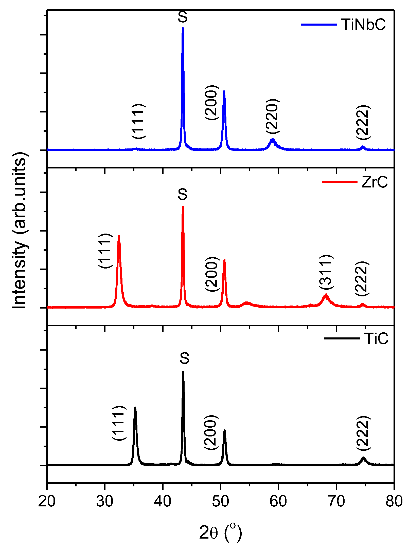
3.1. Elemental and Phase Composition

The elemental compositions of coatings investigated by EDS are presented in Table 2. One can see that the coatings are almost stoichiometric, the C/ΣMe ratio is close to 1. The amount of Fe content originates from the 316L steel substrates.
The XRD diffractions patterns of the coatings are shown in Figure 1. All coatings exhibited typical diffraction patterns of Me-based carbide solid solutions, with (111), (200), (220), and (222) reflections being detected with positions close to those given in JCPDS 65-7994 (TiC), JCPDS74-1221 (ZrC), and JCPDS 010-0181 (NbC) standards. The texture was quantified by calculating the texture coefficients T (hkl) using the formula: T (hkl) = I (hkl)/ΣI (hkl), where I (hkl) is the intensity of line (hkl) and ΣI (hkl) is the sum of the intensities of all detected diffraction peaks. The texture coefficients calculated on each detected peak can be found in Table 2. According to these results, one can see that TiC and ZrC are textured in the (111) direction, while TiNbC exhibited a strong (200) preferred orientation. The (111) orientation is specific to coatings deposited by the cathodic arc method [38,39,40]. The (111) preferred texture is frequently reported in the case of coatings deposited with intense ion bombardment. Usually, the intense energetic ion bombardment leads to an increase of undesired residual stress. For the TiC coatings, it can be seen that the value of the texture coefficient of (111) and (200) crystallographic planes is much closer, indicating that the preferred orientation is controlled by the competition between the strain energy and the surface free energy. The resulting (111) preferred orientation suggests that the strain energy was dominant, indicating that, in the re-nucleation process, the strain energy exceeded the surface energy [41,42,43].
Crystallite size and strain of the coatings were determined by the Williamson–Hall plot method [44,45] and the obtained values are presented in Table 2. Close values of crystallite size were revealed for both TiC and ZrC coatings, while the smaller value corresponding to the TiNbC coating indicated a finer crystal structure. TiC coatings exhibited high strain, indicating that this coating was stressed. The TiNbC coating had a small strain value, meaning a less stressed coating. This finding is in good agreement with the statement that coating textured by the (111) plane is more stressed than that of the (200) direction.

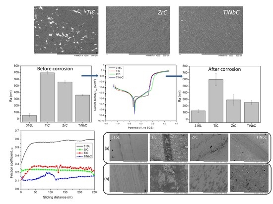
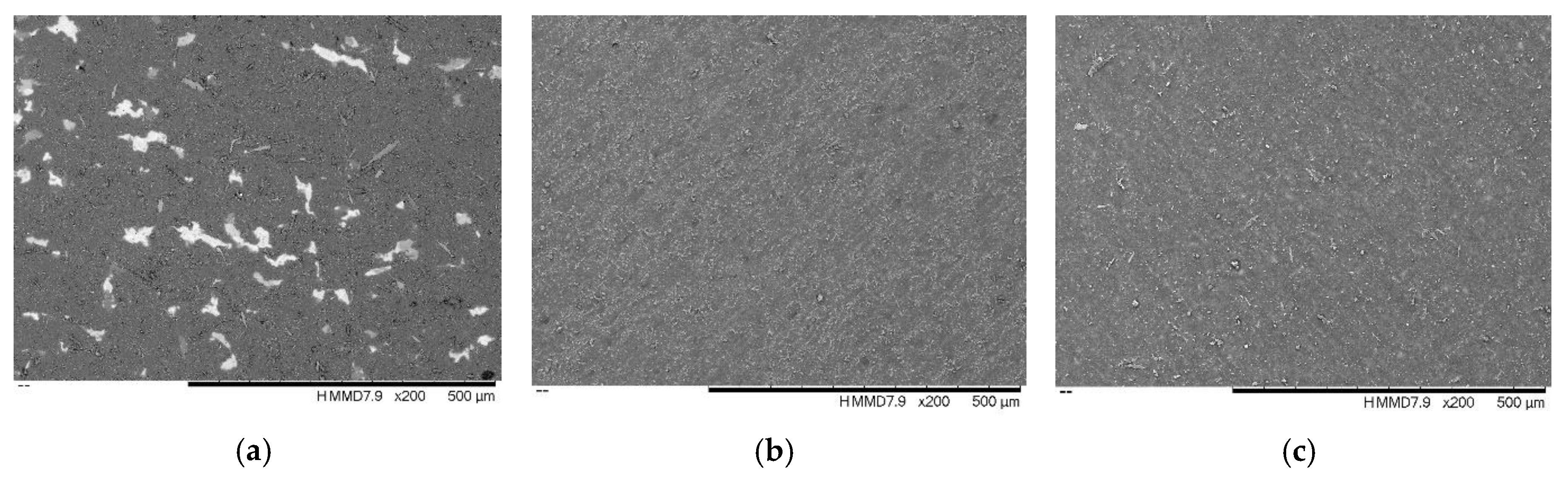
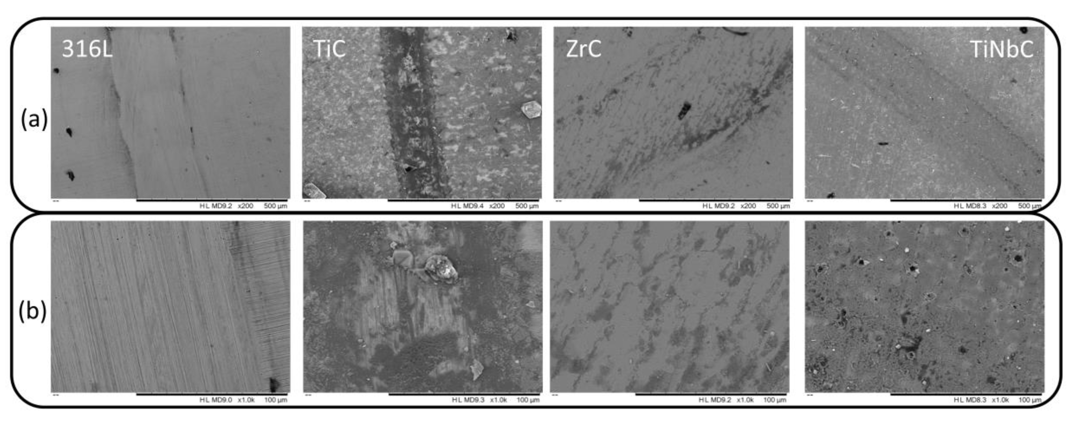
3.2. Morphology and Roughness

The morphology of coatings was investigated by SEM and typical images are presented in Figure 2. As can be seen, there are various microdroplets on the surface of all coatings, which are expected due to the deposition technique used for the preparation of the coatings. These droplets are ejected by the cathode due to the local melting of the cathode material and they lead to an increase of surface roughness. Munz reported that a cathode material with a low melting temperature produces many droplets with larger size [46]. However, on the TiC surface a high number of droplets were observed, because Ti has the lowest melting temperature compared to Zr or Nb. Moreover, on the TiC surface some white areas could be seen, which are related to uncoated substrate, indicating that this coating exhibited an exfoliation process. No significant differences were found in the case of the ZrC and TiNbC surfaces.
The roughness was evaluated by the determination of the arithmetical mean of the absolute values of the profile deviations from the mean line of the roughness profile (Ra). Figure 3 shows the average Ra values determined in five different areas of each specimen (4 mm length). As already mentioned, the roughness of 316L steel discs was around 50 nm. In terms of deposited coatings, TiC was rougher than ZrC and TiNbC. The latter exhibited a low roughness, but it was still higher than that of the uncoated steel, indicating the presence of a large number of droplets.

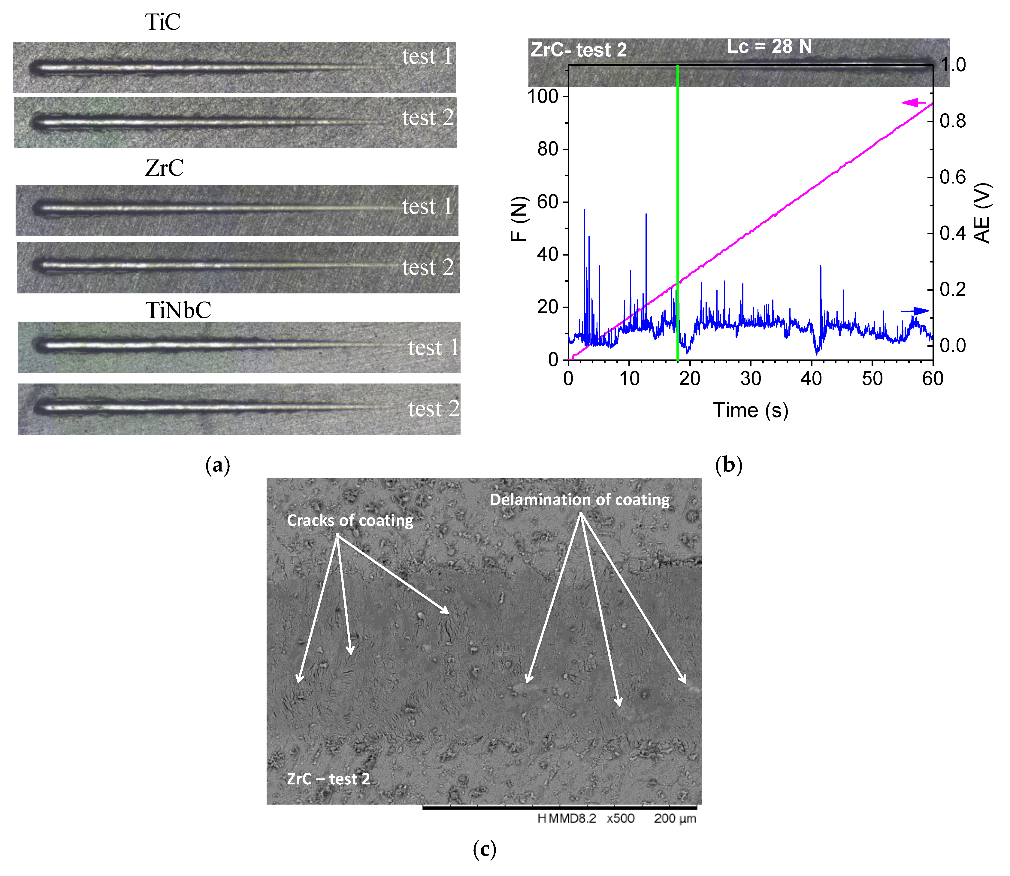
3.3. Hardness and Adhesion

The average hardness and adhesion (Lc) values were determined at different locations over the sample’s surface (Table 3).
As depicted in Figure 4a, the scratch tracks were performed in two different areas of each sample. As an example, in Figure 4b, the evolution of the signal generated by the acoustic sensor during the applied force in the case of the ZrC coating (test 2 in Figure 4a) is presented. One can see that the hardness of 316L steel can be increased by the used coatings. The highest hardness value was found for TiNbC, followed by those of TiC and ZrC coatings. According to Figure 4b, for ZrC coatings a drop in the signal, caused by the acoustic sensor, appeared around a force of 26 N. In Figure 4c, a detailed SEM image can be observed in the area of cracks/delamination of the ZrC coating. By corroborating optical images and the values of acoustic sensor signal during the adhesion tests, one may conclude that TiNbC has a high value compared to the other two coatings. However, the TiC coating proved to have a low adhesion to the 316L substrate.

3.4. In Vitro Corrosion Resistance

The electrochemical corrosion activity was investigated in 0.9% NaCl at 37 ± 0.5 °C, by evaluating the evolution of the open circuit potential in time and the potentiodynamic curves (Figure 5). The open circuit potential (EOC) is a parameter, which is related to the stability of the passive film. During the 1 h immersion, the EOC slightly changes its value indicating that steady state conditions have not been reached (Figure 5a). This change of the EOC can be attributed to a weakening of the passive film due to being damaged. Somehow, a passive film is formed and its stability does not seem to be altered during the 1 h of immersion. If we calculate an average value during the whole test (1 h of immersion), we can observe that 316L is nobler than the others (−137.7 ± 1.9 mV) followed by TiC (−208.5 ± 3.2 mV), ZrC (−211.8 ± 4.8 mV) and then by TiNbC (−225.5 ± 3.9 mV). For all coatings, some fluctuations can be observed during the EOC evolution, which can be attributed to the porosity of the surfaces (described below).
Figure 5b presents polarization curves of the investigated surfaces. One can observe that all curves reveal active–passive behavior. For all coatings, in the domain ranging from 0.3 to 0.5 V, an insignificant gradual increase of the current density can be seen (Figure 5b), which could be attributed to the capability of electron transfer through the passive film for the oxidation of water (oxygen evolution) reaction that is strongly governed by the defect state of the passive film growing. For the 316L uncoated substrate, a breakdown potential can be seen of around 320 mV. Note that an important increase in current density can be seen for ZrC of around 439 mV, while for TiC of around 346 mV and for TiNbC of 297 mV, nearby. Based on these values, we can see that the corrosion resistance follows the order: ZrC–TiC–316L–TiNbC. After 0.7 V, all samples exhibited a passive state in the tested corrosive solution.
Based on Tafel extrapolation, the main corrosion parameters were extracted (Table 4), as has been frequently reported [47]. All coatings exhibited more electropositive values of the corrosion potential (Ei = 0) compared to the uncoated substrate, indicating better corrosion resistance. Approximately 10 mV differences were found between the coatings, indicating that the corrosive solution had less influence on their surfaces. The lower icorr and higher Rp values were observed for TiNbC coatings, showing good corrosion resistance. Taking into account the electrochemical parameters, one can say that TiNbC had the best corrosion resistance at 0.9% NaCl, followed by ZrC and then TiC. On the whole, all coatings improved on the corrosion resistance of 316L steel, but the TiNbC ternary coatings were more suitable for biomedical applications from this point of view.
In order to explain the corrosion behavior at the corrosive attack, the porosity (P) and coatings protective efficiency (Pe) were determined based on the corrosion parameters, according to the equations described in Refs. [48,49]. By comparing the Pe values, the TiNbC coating has the highest value, indicating good protection against the NaCl corrosive attack. Regarding the porosity, there are small differences between the values of ZrC and TiNbC. For TiC, the porosity is slightly high compared to the other two coatings. Thus, the poor corrosion resistance of TiC can be also attributed to this porosity. Both ZrC and TiNbC provided a small porosity with similar values, being difficult to differentiate between them. Probably, the better protection against corrosive attack of TiNbC can be ascribed to the small grain size and less defects found on its surface, which can act as an efficient barrier to electrolyte penetration.
To identify the degradation of the surfaces after the corrosion test, the roughness of the corroded surface was investigated (Figure 6) and compared to the results obtained before corrosion (Figure 3). It is obvious that the uncoated 316L steel suffered a high surface degradation and the Ra value increased more than two times after corrosion tests (from about 50 to 126 nm). In the case of the coated surfaces, a decrease of the Ra vales was found, more evident for ZrC. For TiNbC, the Ra decreased from ~360 to 250 nm, indicating that the surface was not too affected by the corrosive solution. Considering this aspect, the results obtained during the corrosion tests were confirmed—the TiNbC coating had the best corrosion behavior.

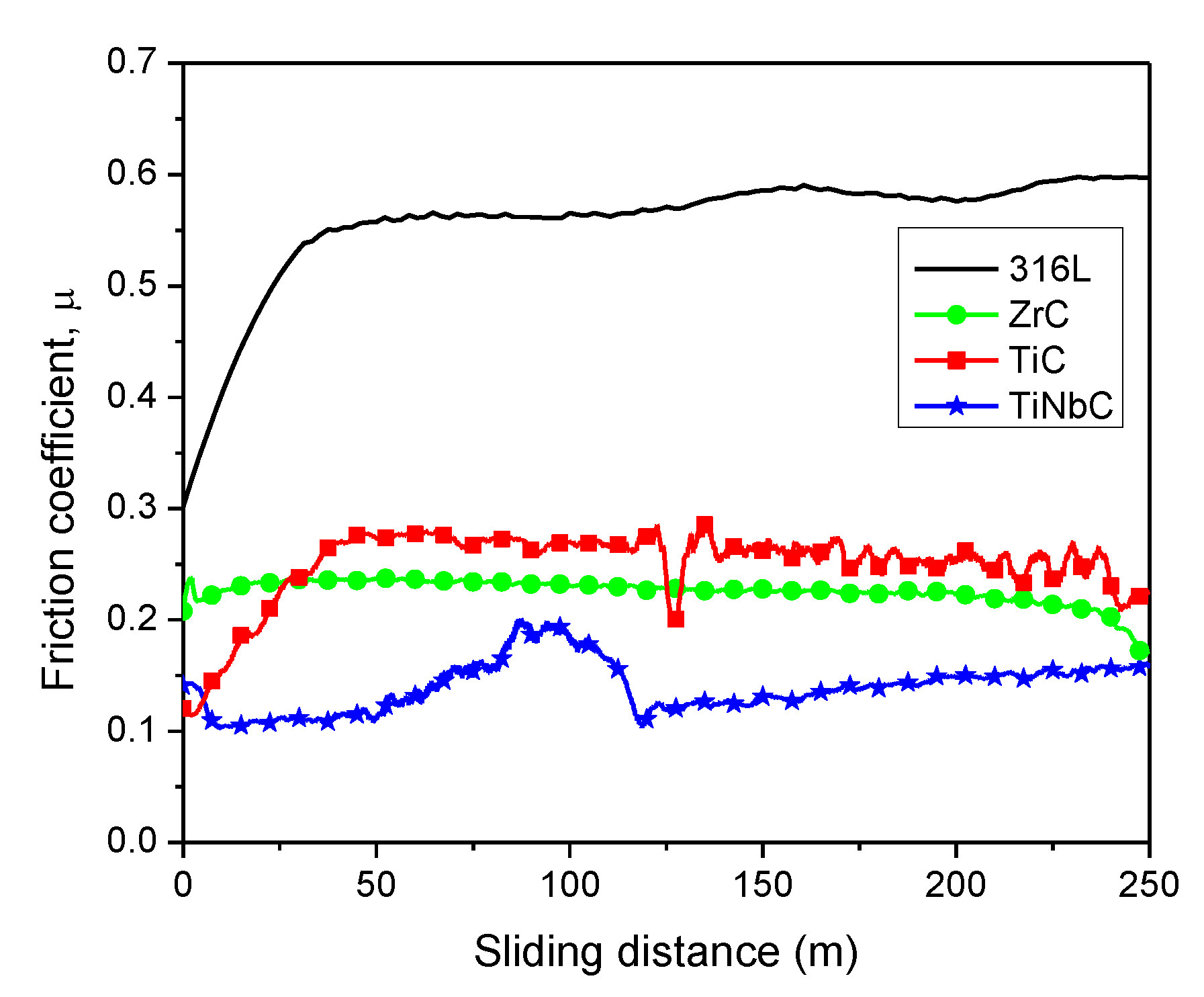
3.5. In Vitro Tribocorrosive Performance

Tribocorrosive behavior was expressed in terms of the change in the friction coefficient (µ) vs. the sliding distance in the corrosive solution and the wear rate (k) at the end of the test. Figure 7 shows the evolution of the coefficients of friction as a function of sliding distance. One can see that the values for uncoated 316 steel and ZrC are fairly constant during the test. TiNbC exhibited some fluctuations, but after 120 m a steady state stage was obtained. TiC showed instabilities during the whole test. All the fluctuations can be explained by the effect of the corrosive solution, which forms the following: (1) a hydrated layer at the interface between coating and counterball which can act as a lubricant over a short distance, but it can be broken at an extended distance; (2) an oxide layer due to oxidation of the surface when the sample surface is in contact with the aqueous solution. Thus, this process can be related to the formation/delamination of these formed layers, which is evident by continuous/variation of the µ values.
Figure 8 presents the wear rate (k), calculated by the determination of the cross-sectional areas of the wear scar at five points on each wear track at the end of the test. Uncoated substrate exhibited high wear rate compared to all coated specimens, indicating that the tribological performance of 316L steel was improved by the coating.
For TiC coatings, the wear rate was higher than the other two, probably due to a more intense oxidation, leading to an intensive damaging effect. Actually, the evolution of wear rates was in good agreement with that of the friction coefficients. TiNbC has the lowest µ and k values, indicating the best tribocorrosive performance, being a proper choice for biomedical applications where a low friction process is required.
Below, the SEM images of wear tracks at the end of the test are presented (Figure 9a). A few detailed SEM images are presented in Figure 9b. On the 316L surface, there are some parallel grooves to the sliding direction, indicating grooving wear or two-body abrasion [50]. This mechanism is associated with the movement of abrasive particles, which are formed during contact between the two surfaces and are fixed to the ball during the process (Figure 9b). The largest wear track was found for the TiC coatings, with many cracks, and delamination of the coatings can be seen in some areas (Figure 9b). In the case of ZrC, the surface seems to be polished during the test, associated with rolling abrasion or three-body abrasion (fine particles rolling over the contact zone). Regarding the TiNbC coatings, a polished area can be seen, similar to those observed for ZrC, but additionally some holes can also be observed, probably the area where microdroplets where pulled out. Some scratches were also observed, attributed to debris activity (Figure 9b).

4. Discussion

It is not possible to say that one material can be used for all types of biomedical applications (such as dental implants, maxillofacial plates, orthopedic prostheses, screws, nails etc.). Thus, the request for performance biomaterials has led to a rapid increase of research in this field in order to find the best solutions to improve the implant material functionality. Thus, this study is focused on improving the functionality of 316L stainless steel substrate by covering its surface with TiC, ZrC, and TiNbC coatings and on investigating if the proposed coatings are capable to withstand the conditions, which can be found in the human body in the case of load bearing implants.
The results related to elemental and phase compositions showed that the developed coatings were those we anticipated, carbide coatings of Ti, Zr, and TiNb, with stoichiometric composition. In the literature, it was reported that the stoichiometric structure is stabler and over long-time successful in the conditions of the human body [51].
Comparing the binary coatings, ZrC has good adhesion to substrates, good corrosion, and tribocorrosion resistance compared to that of TiC. This finding can be related to the low stress level found in ZrC. The low adhesion between TiC coatings and the substrate is probably due to its high stress. This poor adhesion leads also to a decrease of corrosion resistance and tribocorrosive performance. Generally, if a coating has low adhesion to a metallic substrate, delamination of the coating during the corrosion or tribological tests could occur. Probably this is the reason for all the poor properties of the TiC coating. The poor adhesion of TiC can be related to a high number of microdroplets found on the surface, which are generated from the target material during the reactive cathodic arc deposition. Anyway, the presence of microdroplets also results in an increase of roughness of the TiC surface, which can affect the corrosion performance. Usually, when a surface has high roughness, the chloride from the corrosive solution infiltrates and reaches the coating—substrate interface, accelerating the corrosion processes. Moreover, the microdroplets are seen as defects and the corrosive process will be accelerated in that area.
Ternary coatings (TiNbC) exhibited the best corrosion and tribocorrosive performance along with low stress and high hardness compared to those of both binary coatings. This finding was accepted because ternary coatings have been found to be superior to binary ones [52]. The good corrosion resistance of TiNbC can be attributed to low crystalline size and the long paths over the grain boundaries delaying electrolyte penetration. Also, this good behavior can be related to the presence of Nb2O5 oxide, which plays a key role in protecting the surface, known as an oxide with high chemical stability and high corrosion resistance [53].
The roughness can also play an important role in the corrosion behavior. TiC is rougher that ZrC and TiNbC. This high Ra roughness value indicates the formation of holes on the surface and the electrolyte (especially Cl) penetrates through it, where the corrosion process will be accelerated, reaching to the substrate. This situation is less likely to take place for the TiNbC, because its specific Ra value is less than that of the TiC and ZrC coatings.
According to ISO 13779-2:2000 [54], the coatings should have a minimum adhesion strength to the substrate of about 15 MPa [55], which is considered sufficient for the adhesion and the growing of cells on a coating surface. Taking into account this observation, one can conclude that TiNbC will be a good choice for biomedical application because it proved to have the best adhesion to the metallic substrate, followed by ZrC and then by TiC. The findings of this study underline the importance of further investigations using human cells. In the literature, information about the biocompatibility of TiC and ZrC coatings can be found, which has proved to have superior biocompatibility compared to other biocompatible carbide coatings [17]. Little information on the biocompatibility of TiNbC coatings is reported in the literature. Shtansky et al. cultivated epithelial and fibroblasts cells on the TiNbC coatings and showed that they have a good biocompatibility, but the coatings had a modified shape and a disturbed actin cytoskeleton [56].
Among the various carbide coatings, TiC and ZrC possess also superior tribological performance in dry/wet environments [17,19]. TiNbC coatings have also been the subject of many scientific papers, due to their properties being superior to TiC or NbC, such as wear resistance [57].
For example, Shtansky et al. prepared TiNbC with high hardness (~30 GPa) and low friction coefficient (0.12 ÷ 0.22) in dry medium under normal loads of 1 and 5 N with WC +6% Co ball as a counterpart material and under normal loads of 1 and 5 N with 3-mm diameter WC +6% Co ball as a counterpart material, sliding speed of 10 cm/s [56]. No information about the tribocorrosion results was found in the literature. Nevertheless, some results about TiC, ZrC, and TiNbC, used for biomedical applications, can be found in the literature, but the corrosion, the tribology, and the biocompatibility are not fully understood, as there are many factors, which can influence these characteristics. Consequently, the present study aimed to explain the corrosion and the tribocorrosion performance in simulated conditions of the human body by the level of stress, adhesion of the coating to substrate, roughness, and hardness.

5. Conclusions

This study has highlighted the enhanced functionality of 316L stainless steel substrates obtained using covering films such as TiC, ZrC, and TiNbC. This ensures the possibility for fully covered 316L steel substrates to withstand the conditions, which can be found in the human body in the case of load bearing implants. The experimental results also showed:
  • TiC and ZrC have (111) preferred orientation, while TiNbC has a strong (200) orientation, suggesting that the strain energy was dominant in both binary coatings and indicating that in the re-nucleation process, the strain energy exceeds the surface energy;
  • TiNbC exhibited the smallest crystallite size compared to TiC and ZrC coatings;
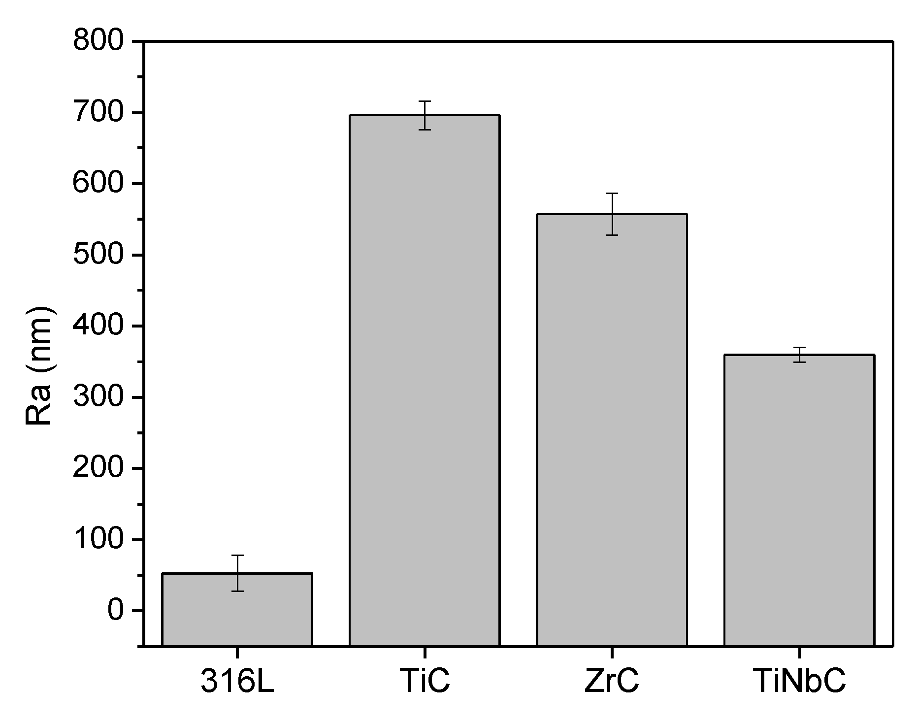
  • TiC was rougher than ZrC and TiNbC; the lowest roughness was found for TiNbC coatings;
  • The highest hardness and adhesion were found for TiNbC, followed by TiC and ZrC;
  • All coatings improved the corrosion resistance of 316L uncoated stainless steel. TiNbC coating showed the best corrosion behavior (lowest icorr = 0.55 µA/cm2, highest Rp = 81.27 kΩ, highest protective efficiency = 57.7%), followed by ZrC (icorr = 0.59 µA/cm2, Rp = 73.61 kΩ, protective efficiency = 54.6%);
  • The TiC coating was more porous than ZrC and TiNbC. There are small differences between the porosity of ZrC and TiNbC;
  • TiNbC has the lowest µ (1.6) and k (0.99 × 10⁻⁵ mm3·N−1∙m−1) values, indicating the best tribocorrosive performance in 0.9% NaCl at 37 ± 0.5 °C.
ZrC and TiNbC coatings possessed superior mechanical, corrosive, and tribocorrosive properties to that of TiC coatings. However, based on the obtained results, TiNbC is a better choice for biomedical applications where a low friction process along with high corrosion resistance are required.

Author Contributions

Conceptualization, A.V. and C.M.C.; methodology, A.V.; investigation, M.D., L.R.C., and I.G.S.; resources, A.V.; data curation, I.P.; writing—original draft preparation, I.P.; writing—review and editing, I.P., C.M.C., and A.V.; supervision, A.V. All authors have read and agreed to the published version of the manuscript.

Funding

The present work was supported under a grant of the Romanian National Authority for Scientific Research, CNCS—UEFISCDI, project number PN-III-P1-1.2-PCCDI-2017-0239/60PCCDI 2018, within PNCDI III. A part of work is also supported by Romanian National Core Program No. 18N/18.02.2019 and PROINSTITUTIO Project No. 19PFE/17.10.2018. The EDS, SEM and XRD results were acquired using the systems purchased by the infrastructure project INOVA-OPTIMA SMIS code 49164, contract No. 658/2014.

Conflicts of Interest

The authors declare no conflict of interest. The funders had no role in the design of the study; in the collection, analyses, or interpretation of data; in the writing of the manuscript, or in the decision to publish the results.

References

  1. Manivasagam, G.; Dhinasekaran, D.; Rajamanickam, A. Biomedical implants: Corrosion and its prevention—A review. Recent Pat. Corros. Sci. 2010, 2, 40–54. [Google Scholar] [CrossRef] [Green Version]
  2. Ibrahim, M.Z.; Sarhan, A.A.D.; Yusuf, F.; Hamdi, M. Biomedical materials and techniques to improve the tribological, mechanical and biomedical properties of orthopedic implants—A review article. J. Alloy. Compd. 2017, 714, 636–667. [Google Scholar] [CrossRef]
  3. Balamurugan, A.; Rajeswari, S.; Balossier, G.; Rebelo, A.H.S.; Ferreira, J.M.F. Corrosion aspects of metallic implants—An overview. Mater. Corros. 2008, 59, 855–869. [Google Scholar] [CrossRef]
  4. Niinomi, M. Advances in Metallic Biomaterials; Niinomi, M., Narushima, T., Nakai, M., Eds.; Springer: Berlin/Heidelberg, Germany, 2015; ISBN 978-3-662-46835-7. [Google Scholar]
  5. Ivanova, E.P.; Bazaka, K.; Crawford, R.J. Metallic biomaterials: Types and advanced applications. In New Functional Biomaterials for Medicine and Healthcare; Woodhead Publishing: Cambridge, UK, 2014; pp. 121–147. [Google Scholar]
  6. Hussein, M.A.; Mohammed, A.S.; Al-Aqeeli, N. Wear characteristics of metallic biomaterials: A review. Materials (Basel) 2015, 8, 2749–2768. [Google Scholar] [CrossRef]
  7. Wang, G.; Zreiqat, H. Functional coatings or films for hard-tissue applications. Materials (Basel) 2010, 9, 3994–4050. [Google Scholar] [CrossRef] [PubMed] [Green Version]
  8. Sargeant, A.; Goswami, T. Hip implants—Paper VI—Ion concentrations. Mater. Des. 2007, 28, 155–171. [Google Scholar] [CrossRef]
  9. Ege, D.; Duru, İ.; Kamali, A.R.; Boccaccini, A.R. Nitride, zirconia, alumina, and carbide coatings on Ti6Al4V femoral heads: Effect of deposition techniques on mechanical and tribological properties. Adv. Eng. Mater. 2017, 19, 1700177. [Google Scholar] [CrossRef]
  10. Du, S.; Zhang, K.; Wen, M.; Qin, Y.; Li, R.; Jin, H.; Bao, X.; Ren, P.; Zheng, W. Optimizing the tribological behavior of tantalum carbide coating for the bearing in total hip joint replacement. Vacuum 2018, 150, 222–231. [Google Scholar] [CrossRef]
  11. Cho, S.M.; Park, J.W.; Han, H.S.; Seok, H.K.; Moon, M.W.; Kim, Y.C. Multifunctional composite coating as a wear-resistant layer for the bearing in total hip joint replacement. ACS Appl. Mater. Interfaces 2013, 5, 395–403. [Google Scholar] [CrossRef]
  12. Yate, L.; Coy, L.E.; Gregurec, D.; Aperador, W.; Moya, S.E.; Wang, G. Nb-C nanocomposite films with enhanced biocompatibility and mechanical properties for hard-tissue implant applications. ACS Appl. Mater. Interfaces 2015, 25, 6351–6358. [Google Scholar] [CrossRef]
  13. Zhang, K.; Wen, M.; Meng, Q.N.; Hu, C.Q.; Li, X.; Liu, C.; Zheng, W.T. Effects of substrate bias voltage on the microstructure, mechanical properties and tribological behavior of reactive sputtered niobium carbide films. Surf. Coat. Technol. 2012, 212, 185–191. [Google Scholar] [CrossRef]
  14. Heimann, R.B. Plasma-Sprayed hydroxylapatite-based coatings: Chemical, mechanical, microstructural, and biomedical properties. J. Therm. Spray Technol. 2016, 25, 827–850. [Google Scholar] [CrossRef] [Green Version]
  15. Daugaard, H.; Bechtold, J.E.; Soballe, K. HA-coated implant: Bone interface in total joint arthroplasty. In Bone-Implant Interface in Orthopedic Surgery: Basic Science to Clinical Applications; Springer: London, UK, 2014; ISBN 9781447154099. [Google Scholar]
  16. Li, L.; Bai, W.; Wang, X.; Gu, C.; Jin, G.; Tu, J. Mechanical properties and in vitro and in vivo biocompatibility of a-C/a-C:Ti nanomultilayer films on Ti6Al4V alloy as medical implants. ACS Appl. Mater. Interfaces 2017, 9, 15933–15942. [Google Scholar] [CrossRef] [PubMed]
  17. Wang, L.; Zhao, X.; Ding, M.H.; Zheng, H.; Zhang, H.S.; Zhang, B.; Li, X.Q.; Wu, G.Y. Surface modification of biomedical AISI 316L stainless steel with zirconium carbonitride coatings. Appl. Surf. Sci. 2015, 340, 113–119. [Google Scholar] [CrossRef]
  18. Zhang, Y.J.; Qin, Y.G.; Qing, Y.A.; Deng, R.P.; Jin, H.; Li, R.Y.; Rehman, J.; Wen, M.; Zhang, K. TiCuN solid solution coating: Excellent wear-resistant biocompatible material to protect artificial joint. Mater. Lett. 2018, 227, 145–148. [Google Scholar] [CrossRef]
  19. Sánchez-López, J.C.; Martínez-Martínez, D.; López-Cartes, C.; Fernández, A. Tribological behaviour of titanium carbide/amorphous carbon nanocomposite coatings: From macro to the micro-scale. Surf. Coat. Technol. 2008, 202, 4011–4018. [Google Scholar] [CrossRef]
  20. Kumar, D.D.; Kaliaraj, G.S.; Kirubaharan, A.M.K.; Alagarsamy, K.; Vishwakarma, V.; Baskaran, R. Biocorrosion and biological properties of sputtered ceramic carbide coatings for biomedical applications. Surf. Coat. Technol. 2019, 374, 569–578. [Google Scholar] [CrossRef]
  21. Finšgar, M.; Uzunalić, A.P.; Stergar, J.; Gradišnik, L.; Maver, U. Novel chitosan/diclofenac coatings on medical grade stainless steel for hip replacement applications. Sci. Rep. 2016, 24, 26653. [Google Scholar] [CrossRef]
  22. Anders, A. Unfiltered and filtered cathodic arc deposition. In Handbook of Deposition Technologies for Films and Coatings; William Andrew: Norwich, NY, USA, 2010; ISBN 9780815520313. [Google Scholar]
  23. Zardiackas, L.D.; Kraay, M.J.; Freese, H.L.; International, A. Titanium, Niobium, Zirconium, and Tantalum for Medical and Surgical Applications; ASTM STP 1471; ASTM: West Conshohocken, PA, USA, 2006; ISBN 9780803134973. [Google Scholar]
  24. Naganawa, T.; Ishihara, Y.; Iwata, T.; Koide, M.; Ohguchi, M.; Ohguchi, Y.; Murase, Y.; Kamei, H.; Sato, N.; Mizuno, M.; et al. In vitro biocompatibility of a new titanium-29niobium-13tantalum-4.6zirconium alloy with osteoblast-like MG63 cells. J. Periodontol. 2004, 75, 1701–1707. [Google Scholar] [CrossRef]
  25. Kolegar, T.; Matousek, M.; Vilemova, M.; Stary, V. Adhesion of biocompatible TiNb coating. Acta Polytech. CTU Proc. 2017, 8, 5–7. [Google Scholar] [CrossRef]
  26. Lin, H.; Gao, S.; Dai, C.; Chen, Y.; Shi, J. A two-dimensional biodegradable niobium carbide (MXene) for photothermal tumor eradication in NIR-I and NIR-II biowindows. J. Am. Chem. Soc. 2017. [Google Scholar] [CrossRef] [PubMed]
  27. Eisenbarth, E.; Velten, D.; Müller, M.; Thull, R.; Breme, J. Biocompatibility of β-stabilizing elements of titanium alloys. Biomaterials 2004, 25, 5705–5713. [Google Scholar] [CrossRef] [PubMed]
  28. Constantin, L.R.; Parau, A.C.; Balaceanu, M.; Dinu, M.; Vladescu, A. Corrosion and tribological behaviour in a 3.5% NaCl solution of vacuum arc deposited ZrCN and Zr–Cr–Si–C–N coatings. Proc. Inst. Mech. Eng. Part J J. Eng. Tribol. 2019, 233, 158–169. [Google Scholar] [CrossRef]
  29. Vladescu, A.; Pruna, V.; Kulesza, S.; Braic, V.; Titorencu, I.; Bramowicz, M.; Gozdziejewska, A.; Parau, A.; Cotrut, C.M.; Pana, I.; et al. Influence of Ti, Zr or Nb carbide adhesion layers on the adhesion, corrosion resistance and cell proliferation of titania doped hydroxyapatite to the Ti6Al4V alloy substrate, utilizable for orthopaedic implants. Ceram. Int. 2019, 45, 1710–1723. [Google Scholar] [CrossRef]
  30. Vladescu, A.; Titorencu, I.; Dekhtyar, Y.; Jinga, V.; Pruna, V.; Balaceanu, M.; Dinu, M.; Pana, I.; Vendina, V.; Braic, M. In vitro biocompatibility of Si alloyed multi-principal element carbide coatings. PLoS ONE 2016, 11. [Google Scholar] [CrossRef] [PubMed]
  31. Dinu, M.; Pana, I.; Braic, V.; Miculescu, F.; Balaceanu, M.; Vladescu, A.; Braic, M. In vitro corrosion resistance of Si containing multi-principal element carbide coatings. Mater. Corros. 2016, 67. [Google Scholar] [CrossRef]
  32. Cotrut, C.M.; Vitelaru, C.; Dinu, M.; Petreus, T.; Vladescu, A. Synthesis and characterization of TiSiON biocompatible thin films used in biomedical applications. Sci. Adv. Mater. 2015, 7, 1351–1360. [Google Scholar] [CrossRef]
  33. Vladescu, A.; Dinu, M.; Braic, M.; Vitelaru, C.; Balaceanu, M.; Tarcolea, M.; Braic, V.; Baciu, F.; Cotrut, C.M. The effect of TiSiN interlayers on the bond strength of ceramic to NiCr and CoCr alloys. Ceram. Int. 2015, 41, 8051–8058. [Google Scholar] [CrossRef]
  34. Dinu, M.; Braic, V.; Colease, G.; Baciu, F.; Cotrut, C.M.; Braic, M.; Tarcolea, M.; Vitelaru, C.; Vladescu, A.; Vranceanu, D.M. TiSiN coatings for improved bond strength of CoCr alloy to dental ceramic. Key Eng. Mater. 2014, 587, 275–281. [Google Scholar] [CrossRef]
  35. EN 1071-3. Advanced Technical Ceramics - Methods of Test for Ceramic Coatings - Part 3 : Determination of Adhesion and Other Mechanical Failure Modes by a Scratch Test; The European Committee for Standardization CEN: Brussels, Belgium, 2004. [Google Scholar]
  36. ASTM G102—89. Standard Practice for Calculation of Corrosion Rates and Related Information from Electrochemical Measurements. In Annual Book of ASTM Standards; ASTM International: West Conshohocken, PA, USA, 2010. [Google Scholar]
  37. EN 1071-3. Advanced Technical Ceramics. Methods of Test for Ceramic Coatings - Part 13 : Determination of Wear Rate by the Pin-on-Disk Method; The European Committee for Standardization CEN: Brussels, Belgium, 2010. [Google Scholar]
  38. Kothari, D.C.; Kale, A.N. Recent trends in surface engineering using cathodic arc technique. Surf. Coat. Technol. 2002, 158–159, 174–179. [Google Scholar] [CrossRef]
  39. Constantin, L.; Braic, M.; Dinu, M.; Balaceanu, M.; Braic, V.; Farcau, C.; Vladescu, A. Effects of Zr, Nb, or Si addition on the microstructural, mechanical, and corrosion resistance of TiCN hard coatings. Mater. Corros. 2016, 67, 929–938. [Google Scholar] [CrossRef]
  40. Pelleg, J.; Zevin, L.Z.; Lungo, S.; Croitoru, N. Reactive-sputter-deposited TiN films on glass substrates. Thin Solid Films 1991, 197, 117–128. [Google Scholar] [CrossRef]
  41. Barna, P.B.; Adamik, M. Fundamental structure forming phenomena of polycrystalline films and the structure zone models. Thin Solid Films 1998, 317, 27–33. [Google Scholar] [CrossRef]
  42. Oh, U.C.; Je, J.H. Effects of strain energy on the preferred orientation of TiN thin films. J. Appl. Phys. 1993, 74, 1692. [Google Scholar] [CrossRef]
  43. Petrov, I.; Barna, P.B.B.; Hultman, L.; Greene, J.E.E. Microstructural evolution during film growth. J. Vac. Sci. Technol. Vac. Surf. Film. 2003, 21, S117. [Google Scholar] [CrossRef]
  44. Khorsand Zak, A.; Abd. Majid, W.H.; Abrishami, M.E.; Yousefi, R. X-ray analysis of ZnO nanoparticles by Williamson-Hall and size-strain plot methods. Solid State Sci. 2011, 13, 251–256. [Google Scholar] [CrossRef]
  45. Ma, Y.; Chang, Y.C.; Yin, J.Z. Evaluation of lattice strain in ZnO thin films based on Williamson-Hall analysis. J. Optoelectron. Adv. Mater. 2019, 21, 702–709. [Google Scholar]
  46. Münz, W.D.; Smith, I.J.; Lewis, D.B.; Creasey, S. Droplet formation on steel substrates during cathodic steered arc metal ion etching. Vacuum 1997, 48, 473–481. [Google Scholar] [CrossRef]
  47. Yate, L.; Coy, L.E.; Aperador, W. Robust tribo-mechanical and hot corrosion resistance of ultra-refractory Ta-Hf-C ternary alloy films. Sci. Rep. 2017, 7, 3080. [Google Scholar] [CrossRef]
  48. Vladescu, A.; Vranceanu, D.M.; Kulesza, S.; Ivanov, A.N.; Bramowicz, M.; Fedonnikov, A.S.; Braic, M.; Norkin, I.A.; Koptyug, A.; Kurtukova, M.O.; et al. Influence of the electrolyte’s pH on the properties of electrochemically deposited hydroxyapatite coating on additively manufactured Ti64 alloy. Sci. Rep. 2017, 7, 16819. [Google Scholar] [CrossRef]
  49. Dinu, M.; Mouele, E.S.M.; Parau, A.C.; Vladescu, A.; Petrik, L.F.; Braic, M. Enhancement of the corrosion resistance of 304 stainless steel by Cr-N and Cr(N,O) coatings. Coatings 2018, 8, 132. [Google Scholar] [CrossRef] [Green Version]
  50. Adachi, K.; Hutchings, I.M. Wear-mode mapping for th micro-scale abrasion test. Wear 2003, 255, 23–29. [Google Scholar] [CrossRef]
  51. Ducheyne, P. Comprehensive Biomaterials, 2nd ed.; Elsevier: Amsterdam, The Netherlands, 2017; ISBN 9780081006924. [Google Scholar]
  52. Mandes, A.; Vladoiu, R.; Prodan, G.; Dinca, V.; Porosnicu, C.; Dinca, P. The properties of binary and Ternary Ti based coatings produced by Thermionic Vacuum Arc (TVA) technology. Coatings 2018, 8, 114. [Google Scholar] [CrossRef] [Green Version]
  53. Mujawar, S.H.; Inamdar, A.I.; Betty, C.A.; Ganesan, V.; Patil, P.S. Effect of post annealing treatment on electrochromic properties of spray deposited niobium oxide thin films. Electrochim. Acta 2007, 52, 4899–4906. [Google Scholar] [CrossRef]
  54. ISO BS. Standards 13779-2: 2000:Implants for Surgery—Hydroxyapatite—Part 2: Coatings of Hydroxyapatite; British Standards Institution: London, UK, 2000. [Google Scholar]
  55. Eliaz, N.; Ritman-Hertz, O.; Aronov, D.; Weinberg, E.; Shenhar, Y.; Rosenman, G.; Weinreb, M.; Ron, E. The effect of surface treatments on the adhesion of electrochemically deposited hydroxyapatite coating to titanium and on its interaction with cells and bacteria. J. Mater. Sci. Mater. Med. 2011, 22, 1741–1752. [Google Scholar] [CrossRef]
  56. Shtansky, D.V.; Gloushankova, N.A.; Sheveiko, A.N.; Kharitonova, M.A.; Moizhess, T.G.; Levashov, E.A.; Rossi, F. Design, characterization and testing of Ti-based multicomponent coatings for load-bearing medical applications. Biomaterials 2005, 26, 2909–2924. [Google Scholar] [CrossRef]
  57. Li, Q.; Lei, Y.; Fu, H. Growth mechanism, distribution characteristics and reinforcing behavior of (Ti, Nb)C particle in laser cladded Fe-based composite coating. Appl. Surf. Sci. 2014, 316, 610–616. [Google Scholar] [CrossRef]
Figure 1. X-ray diffraction (XRD) patterns of the investigated coatings (TiC, ZrC, TiNbC) determined using Cu Kα radiation from 20° to 80° with a step size of 0.02°/min. The “S” are peaks related to substrate.
Coatings 10 00654 g001
Figure 2. Surface of the investigated coatings ((a) TiC, (b) ZrC, (c) TiNbC) determined by SEM method at magnification of ×200.
Coatings 10 00654 g002
Figure 3. The arithmetic average deviation from the mean line of the roughness profile (Ra) of substrate and coatings measured on a distance of 4 mm using surface profilometry.
Coatings 10 00654 g003
Figure 4. (a) Scratch tracks of the investigated samples determined using the following parameters: indenter—0.2 mm radius diamond tip, load—continuous increase from 0 to 100 N, scratching speed—10 mm/min., scratching distance—10 mm [35]; (b) Time-compressed acoustic sensor signal envelopes of the whole scratch test of the ZrC coating (test 2); (c) detailed SEM image in the area of cracks/delamination of the ZrC coating.
Coatings 10 00654 g004
Figure 5. (a) Evolution of open circuit potential versus time in 0.9% NaCl at 37 ± 0.5 °C (immersion 1 h); (b) Potentiodynamic curves of the investigated surfaces in 0.9% NaCl at 37 ± 0.5 °C.
Coatings 10 00654 g005
Figure 6. The arithmetic average deviation from the mean line of the roughness profile (Ra) of substrate and coatings measured on a distance of 4 mm using surface profilometry after the corrosion test in 0.9% NaCl at 37 ± 0.5 °C.
Coatings 10 00654 g006
Figure 7. Evolution of the friction coefficients vs. sliding distance in 0.9% NaCl at 37 ± 0.5 °C of the coated and uncoated 316L steel.
Coatings 10 00654 g007
Figure 8. Wear rate (k) of the coated and uncoated substrates at the end of tribological test in 0.9% NaCl at 37 ± 0.5 °C (values were calculated by determining the cross-sectional areas of the wear scar at 5 points on each wear track).
Coatings 10 00654 g008
Figure 9. SEM images of wear tracks of the coated and uncoated substrates at the end of the tribological test in 0.9% NaCl at 37 ± 0.5 °C: (a) view of the wear track; (b) details of the specific area of the wear track.
Coatings 10 00654 g009
Table 1. Depositions conditions for the developed coatings.
Deposition ParametersTiCZrCNbC
Base pressure2 × 10−3 Pa
Working pressure1 × 10−1 Pa6 × 10−2 Pa1 × 10−1 Pa
CH4 mass flow rate120 sccm
Arc current on each cathode90 A130 A90 A
Substrate bias voltage−150 V
Deposition duration40–50 min
Table 2. Elemental composition measured by EDS, texture coefficient T (hkl), crystallite size d, and strain ε determined by the Williamson–Hall plot method of the investigated coatings.
CoatingElemental Composition (at.%)C/ΣMecoatingT (hkl)d (nm)ε
TiZrNbCFe(200)(111)(220)(222)
TiC48.68 ± 1.2--48.05 ± 1.33.27 ± 0.50.990.4090.437-0.11016.30.023
ZrC-49.51 ± 1.3-48.14 ± 1.22.35 ± 0.40.970.2940.442-0.17313.10.014
TiNbC32.56 ± 1.2-12.97 ± 0.749.00 ± 1.32.47 ± 0.41.070.6590.0270.2770.0348.10.008
Table 3. Hardness (H) and adhesion (Lc) of uncoated and coated samples.
SubstrateCoatingH (GPa)Lc (N)
316L-4.3 ± 0.2-
TiC39.4 ± 0.318 ± 1 ÷ 20 ± 1
ZrC32.2 ± 0.226 ± 1 ÷ 28 ± 1
TiNbC40.3 ± 0.328 ± 1 ÷ 30 ± 1
Table 4. The main corrosion parameters of uncoated and coated samples: Ei = 0—corrosion potential; icorr—corrosion current density; Rp—polarization resistance; P—porosity; Pe—coatings protective efficiency. The values represent the average of measurements performed on 3 different samples for each deposition conditions of coating.
SampleEi = 0 (mV)icorr (µA/cm2)Rp (kΩ)PPe (%)
316L−336 ± 231.3 ± 0.32.54 ± 1.1--
TiC−268 ± 110.70 ± 0.262.75 ± 3.80.0307 ± 0.001146.2 ± 0.3
ZrC−251 ± 180.59 ± 0.273.61 ± 4.60.02449 ± 0.001454.6 ± 0.2
TiNbC−273 ± 160.55 ± 0.281.27 ± 4.70.02424 ± 0.001357.7 ± 0.2

