Article

Bismuth Nanoantibiotics Display Anticandidal Activity and Disrupt the Biofilm and Cell Morphology of the Emergent Pathogenic Yeast Candida auris

1
Department of Biology, and South Texas Center for Emerging Infectious Diseases, The University of Texas at San Antonio, San Antonio, TX 78249, USA
2
School of Engineering, The University of Texas at Austin, Austin, TX 78712, USA
*
Authors to whom correspondence should be addressed.
Antibiotics 2020, 9(8), 461; https://doi.org/10.3390/antibiotics9080461
Submission received: 19 June 2020 / Revised: 27 July 2020 / Accepted: 28 July 2020 / Published: 29 July 2020
(This article belongs to the Special Issue Fungal Pathogens: Resistance and Novel Therapeutics)

Abstract

:
Candida auris is an emergent multidrug-resistant pathogenic yeast, which forms biofilms resistant to antifungals, sanitizing procedures, and harsh environmental conditions. Antimicrobial nanomaterials represent an alternative to reduce the spread of pathogens—including yeasts—regardless of their drug-resistant profile. Here we have assessed the antimicrobial activity of easy-to-synthesize bismuth nanoparticles (BiNPs) against the emergent multidrug-resistant yeast Candida auris, under both planktonic and biofilm growing conditions. Additionally, we have examined the effect of these BiNPs on cell morphology and biofilm structure. Under planktonic conditions, BiNPs MIC values ranged from 1 to 4 µg mL−1 against multiple C. auris strains tested, including representatives of all different clades. Regarding the inhibition of biofilm formation, the calculated BiNPs IC50 values ranged from 5.1 to 113.1 µg mL−1. Scanning electron microscopy (SEM) observations indicated that BiNPs disrupted the C. auris cell morphology and the structure of the biofilms. In conclusion, BiNPs displayed strong antifungal activity against all strains of C. auris under planktonic conditions, but moderate activity against biofilm growth. BiNPs may potentially contribute to reducing the spread of C. auris strains at healthcare facilities, as sanitizers and future potential treatments. More research on the antimicrobial activity of BiNPs is warranted.

1. Introduction

Infectious diseases are among the first causes of death worldwide [1] and pose a burden for global health and economy, as they negatively impact major social aspects of everyday life [2]. The main threats associated with communicable diseases are the rise of drug-resistance, the limited number and diversity of available treatments, and the emergence of new pathogens [3,4,5], some of them with pandemic potential. The Candida genus is the main cause of fungal diseases [6,7]. Every year, more than 250,000 people worldwide are affected by invasive candidiasis, leading to more than 50,000 deaths, as the mortality can be as high as 40%, even when patients receive antifungal therapy [6]. Candida albicans is the main cause of Candida-related diseases; however, other Candida species also represent a health risk [8]. Among them, C. auris, an emergent pathogenic yeast, has risen as a clinical concern worldwide, as it spreads in healthcare-related facilities and has caused several outbreaks around the world [9,10].
C. auris is an ovoid-shaped yeast which has been described to be non-dimorphic, yet, some strains can display the pseudohyphae shape [11]. This yeast has been classified as an urgent threat by the CDC, as it has displayed resistance to different antifungal drug classes (multidrug-resistant) [3,12]. C. auris causes invasive infections with high mortality rates, particularly for inpatient care for patients that stay hospitalized for a long time or who are subject to surgery or intubation procedures [11,13]. Since its first description in 2009, it has rapidly spread around the globe. The different C. auris strains are classified in four geographic clades that may have emerged independently, and almost simultaneously, in different regions: South Asian (clade I), East Asian (clade II), South African (clade III), and South American (clade IV) [10,14]. Recently, in Iran, a potential fifth clade has been identified [15]. Each clade displays distinctive genotypic (genetic polymorphisms) and phenotypic traits (morphology, metabolism, and pathogenicity, among others), although the differences between the clades are minimal [10]. Additionally, there are differences regarding their drug-susceptibility profile [16] and behavior, as some can form aggregates [17].
C. auris inherent multidrug-resistance and its ability to form very resistant biofilms challenge our ability to reduce its spread [9,18]. This yeast is known to be resistant to sanitizing procedures [19] and to the main classes of antifungal drugs, particularly to fluconazole [16,20], which is among the most used anticandidal drugs, alongside with amphotericin B and echinocandins [21]. The biofilm stage allows C. auris to colonize and survive in different surfaces in healthcare facilities for long periods [13], under unfavorable environments, such as high temperature and salinity [22,23]. Furthermore, Candida biofilms can colonize implantable biomedical devices [24] and other medical instrumentation, which increases the risk in healthcare facilities worldwide. As C. auris displays enhanced resistance to current antifungal drugs and sanitizers, novel treatments are needed to effectively combat this pathogenic yeast. Currently, new anticandidal treatments are being researched, such as antifungal peptides [25], drug repurposing [26], and new antifungal drugs [27]. Recently, nanotechnology has been studied to combat C. auris [28,29].
Nanomaterials are an emergent alternative to combat infectious diseases. While numerous nanotechnology-based drugs (nanopharmaceuticals) are currently under preclinical and clinical development [30,31], many are now available for use in healthcare, such as vaccines and nanoantibiotics [32,33,34]. Nanomaterials display a wide range of inhibitory activities against a broad diversity of pathogens—including viruses, bacteria, fungi, and protozoa—and can even overcome their resistance to current antimicrobial drugs [35,36,37,38,39]. The use of antimicrobial nanomaterials (nanoantibiotics) is a novel approach to combat C. auris, due to the possibility of synthesizing cost-effective, potent disinfectants and treatments. The antifungal activity of nanoantibiotics on C. auris is virtually unknown; however, their anticandidal effect has been widely demonstrated in the literature [40,41,42,43,44,45,46]. More recently, our group demonstrated that AgNPs are effective against C. auris biofilms [29,47].
For this study, we evaluated the inhibitory activity of bismuth nanoparticles (BiNPs) against C. auris. Bismuth compounds have been historically used as antimicrobial agents [48], and some bismuth-based compounds and BiNPs exhibit anticandidal activity against C. albicans [49,50]. In a recent report, we described a new method for producing PVP-coated BiNPs with potential antimicrobial activity [51] and subsequently demonstrated their antibacterial and antifungal activity against Staphylococcus aureus and C. albicans, respectively [52]. In this work, we hypothesized that BiNPs display anticandidal activity against the different strains of C. auris. Our aims were to determine the inhibitory effect on the planktonic and the biofilm stages on different strains from the known clades of C. auris and then to assess the influence of nanoparticles on the phenotype and structural organization of the biofilms. In Appendix A, concepts such as planktonic and biofilm stages, and pseudohyphae are described.

2. Results and Discussion

The antifungal activity of the BiNPs was determined on the planktonic and biofilms stages of different strains representing multiple clades of C. auris. In subsequent experiments we assessed the impact of the BiNPs on the cell morphology and biofilm structure of C. auris.

2.1. BiNPs Displayed Strong Antifungal Activity Against all C. auris Strains under Planktonic Growing Conditions

We performed a susceptibility assay to assess the anticandidal activity of the BiNPs against the planktonic stage of multiple strains of C. auris, mostly following CLSI methodologies [53] with minor modifications.
We found that most C. auris strains exhibited similar susceptibility to the BiNPs, as the MIC values ranged from 1 to 4 µg mL−1 (Table 1), with a geometric mean of 2 µg ± 0.7 mL−1. The overall anticandidal activity of BiNPs was very similar between the clades, although we noted that two strains from Clade I exhibited slight variations in their susceptibility to BiNPs. The MIC values for the Clade I strains AR no. 0382 and no. 0389 were 1 and 4 µg mL−1, respectively; which are within one-step concentration dilution value from the MIC values observed in the other strains (2 µg mL−1). As far as the authors know, there are no previous reports on the antifungal activity of BiNPs against C. auris. BiNPs displayed strong anticandidal activity against the planktonic stage of all strains, irrespective of their origin, clade, susceptibility profiles against conventional antifungals, and phenotypical and genotypical differences [10,16,17].
Our results also show that the BiNP anticandidal activity under planktonic conditions parallels silver nanoparticles (AgNPs), which usually display MIC values within the 1 to 10 µg mL−1 range [36]. This parallelism is relevant because AgNPs are among the most potent antimicrobial nanomaterials [54]. Also, the anticandidal activity of BiNPs is comparable to that of repositionable compounds, such as miltefosine and iodoquinol (MIC = 4 µg mL−1), as reported previously by our group [26]. Additionally, the activity of BiNPs matches the proposed activity of the main antifungal drugs. Currently, there are not established antifungal MIC breakpoints for C. auris; however, the CDC has proposed the following MIC values to indicate resistance: >32 µg mL−1 for fluconazole and >2 µg mL−1 for both amphotericin B and caspofungin [16]. Fluconazole is one of the most used antifungal agents against Candida infections [21], but C. auris is resistant to it; however, amphotericin B and caspofungin display stronger antifungal activity against C. auris.

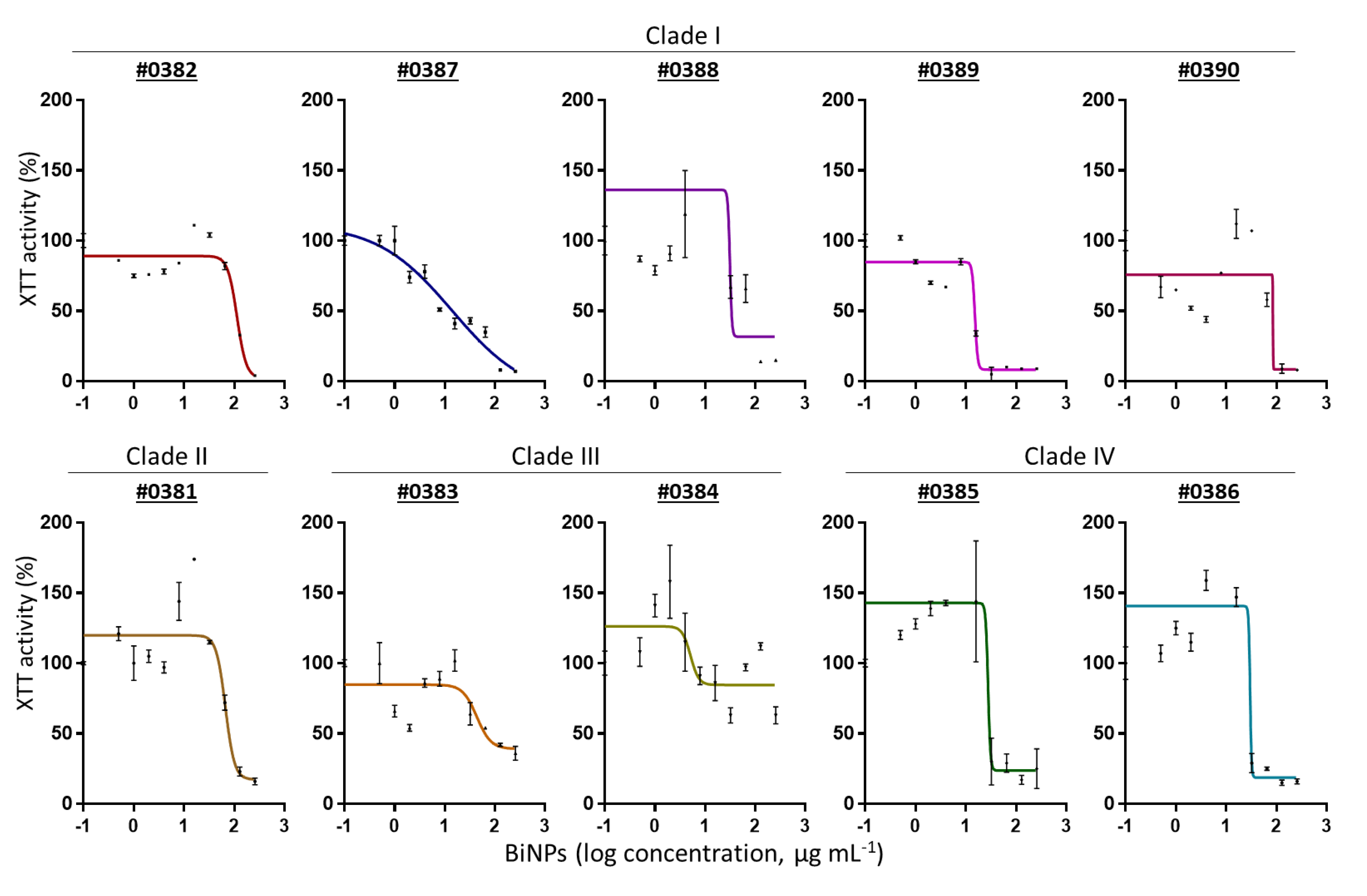
2.2. BiNPs Inhibit Biofilm Formation by C. auris

We evaluated the antibiofilm activity of BiNPs during the biofilm formation stage of C. auris. As biofilms confer resistance to antifungal drugs and sanitizers, it is clinically relevant to assess the ability of nanoparticles to inhibit biofilm formation. The inhibitory activity of BiNPs on the biofilm was measured using an XTT-reduction assay that measures the metabolic activity of sessile cells within the biofilms, as originally described by our group [55].
BiNPs inhibited the biofilm formation in all C. auris strains. The calculated BiNPs IC50 values ranged from 5.1 to 113.1 µg mL−1, with a geometric mean of 26.8 ± 24.5 µg mL−1 (Table 2). The IC50 values indicate that each C. auris strains had a unique susceptibility to BiNPs, as the biofilm activity was different in each strain. However, the strains from Clade IV were the only ones that showed a consistent behavior to the BiNP treatments. In Figure 1 dose–response curves show the antibiofilm activity against a representative C. auris strain from each one of the four clades. The individual dose–response curves for all the C. auris AR strains are shown in Figure A1 (Appendix B). It seems that the susceptibility to BiNPs among the different strains is not related to the clade. As C. auris is an emergent pathogen, the research regarding their particular physiological traits of each strain is still ongoing, particularly regarding the behavior and differences of the biofilm stages [56,57]. Moreover, the biofilm stage has remained poorly understood up to date, even in thoroughly studied Candida species such as C. albicans [58].
We noted that the antibiofilm activity of BiNPs was close to the activity reported for other anticandidal agents during the biofilm formation stage. BiNPs displayed lower antibiofilm potency than that of AgNPs against C. auris biofilms [29,47]. However, the fact that BiNPs display antibiofilm activity should be considered for further research. Regarding current antifungal drugs, BiNPs display greater antibiofilm potency than fluconazole (IC50 > 64 µg mL−1), but lower than caspofungin (IC50 = 5 to 1 µg mL−1) and amphotericin B (IC100 = 1 to >8 µg mL−1), according to Dekkerová et al., for the AR no. 0383, no. 0386, and no. 0390 C. auris strains [59].
An interesting observation is that the biofilm activity –as measured by the XTT absorbance− displayed an irregular behavior in response to the BiNP treatments (Figure A1 (Appendix B)). Some C. auris strains exhibited an increase in the biofilm activity—higher than the untreated control—when treated with sublethal concentrations of BiNPs. After the peak of intensity—up to 2.5 times higher than in the control—the activity declined again as the BiNP concentrations increased.

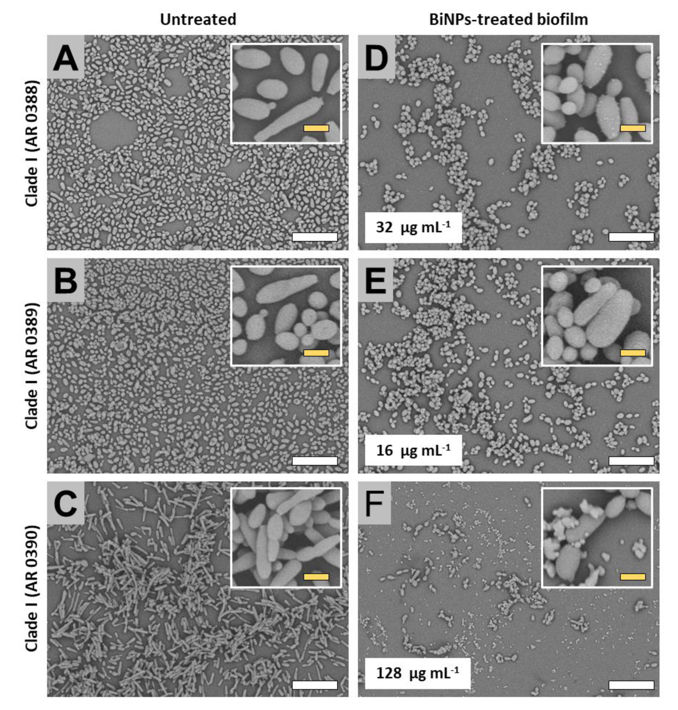
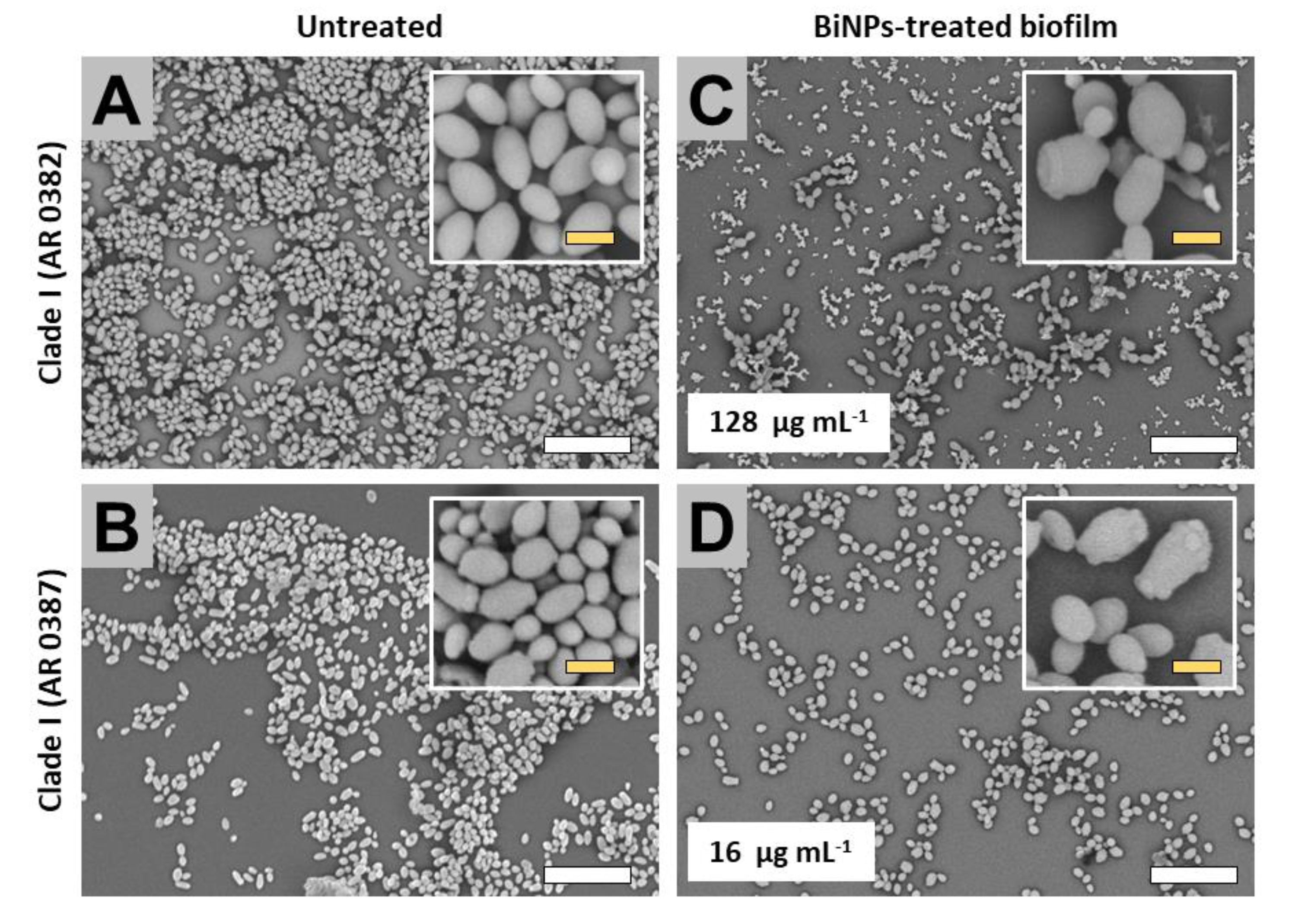
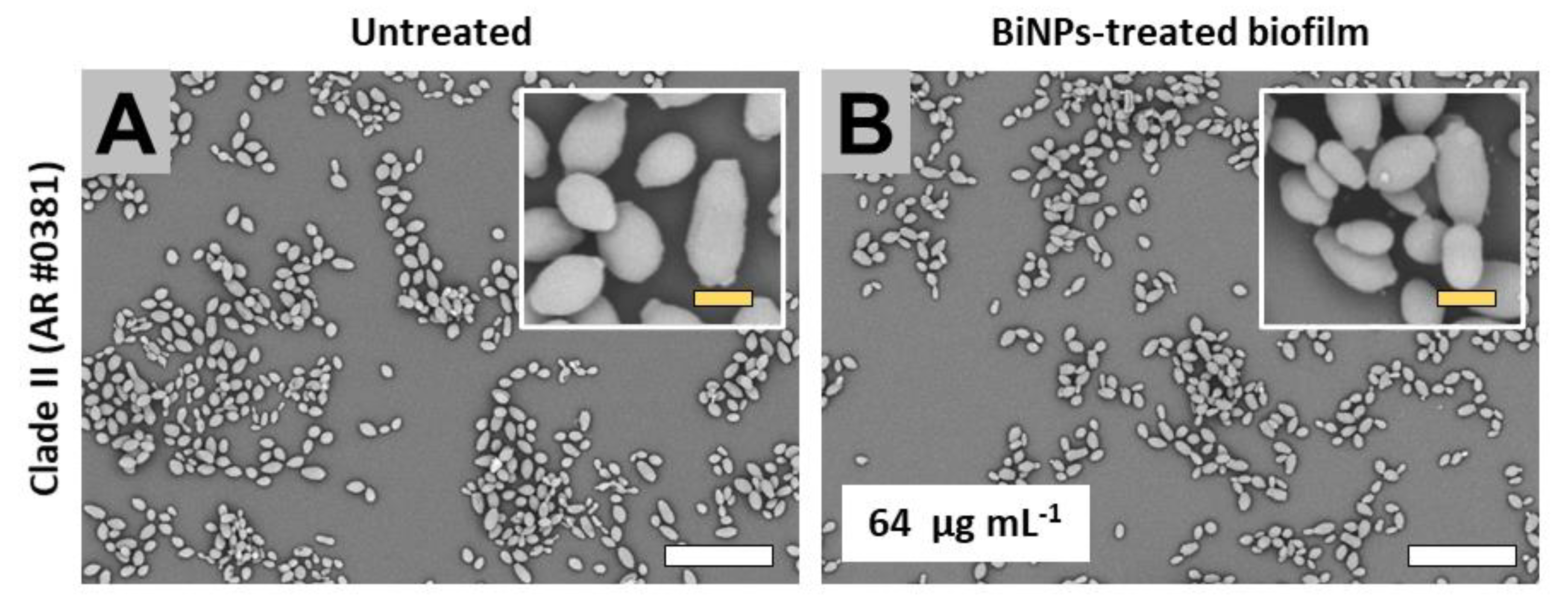
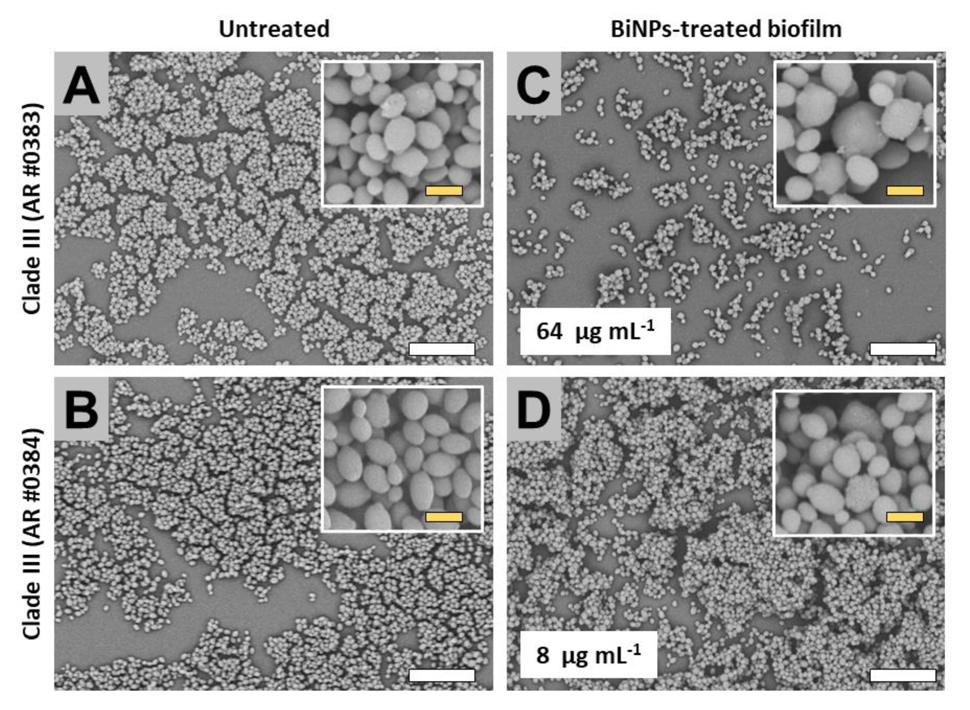
2.3. BiNPs Alter the Cellular Morphology and Structure of C. auris Biofilms

To assess the effects BiNPs on the structural organization of C. auris biofilms and cell morphology, we analyzed the untreated and the BiNP-treated samples via scanning electron microscopy (SEM). All the strains of C. auris, from the four main clades, were exposed to sub-inhibitory concentrations of BiNPs during the biofilm formation phase. Then, we visualized the impact of the nanoparticles on the fungal cell morphology and biofilm structure. As the different strains exhibited variations in their susceptibility to BiNPs, the concentration of BiNPs used for the SEM analysis was selected according to their corresponding calculated IC50 values (Table 2) and the experimental concentration from the antibiofilm assays (Section 2.2). Therefore, for each strain, the selected BiNP subinhibitory treatment was the higher experimental concentration of BiNPs closest to the calculated IC50 values.

2.3.1. Clade I (South Asia Clade)

SEM images revealed that the untreated strains form clade I displayed two major structural behaviors. The untreated samples cells first subgroup (AR strains no. 0388, no. 0389, and no. 0390), exhibited both the yeast and pseudohyphae morphologies within the biofilms, whereas the cells from the second subgroup (strains no. 0382 and no. 0387) only displayed the yeast-like shape. The presence of the pseudohyphae-like phenotype has been also described in other works [17]. Subgroup 1: Untreated samples from this subgroup formed biofilms that extensively covered the surface in the bottom of the well (Figure 2A–C), whereas in the BiNP-treated samples, the covered area by the biofilm was noticeably reduced in all strains, regardless of the BiNP concentration in the different samples (from 16 to 128 µg mL−1) (Figure 2D–F). The dominant shape for the no. 0390 strain is the pseudohyphae (Figure 2A), whereas, for the no. 0388 and no. 0389 strains, the yeast morphology is the most common (Figure 2B,C). On the BiNP-treated, the presence of the pseudohyphae morphology was reduced in all strains (Figure 2D–F). Subgroup 2: Similar to the subgroup 1, untreated samples also form biofilms that extensively cover the surface in the bottom of the well (Figure 3A,B), whereas the BiNP-treated samples exhibited an evident reduction in all strains, regardless of the BiNPs concentration (16 and 128 µg mL−1) (Figure 3C,D). In contrast to the subgroup 1, the cells from the subgroup 2 (strains no. 0382 and no. 0387), only display the yeast-like shape, with no evidence of the pseudohyphae-like shape (Figure 3A,B). The BiNP-treated samples preserved the morphology of the cells, although some alterations on cell size and shape were observed (Figure 3C,D).

2.3.2. Clade II (East Asia Clade)

The AR no. 0381 strain is the only one from this clade in the CDC panel. The biofilms from the untreated samples partly covered the bottom of the well (Figure 4A), and the cells display the typical yeast-like morphology (Figure 4A). BiNP-treated samples (64 µg mL−1) did not reveal a noticeable reduction in the biofilm formation (Figure 4B); moreover, the cells did not show evident alterations on cell morphology (Figure 4B) when compared with the untreated control.

2.3.3. Clade III (Africa Clade)

Untreated samples from the AR no. 0383 and no. 0384 strains formed biofilms that moderately covered the well surface (Figure 5A,B). The cells from both strains exhibited the typical yeast-like shape (Figure 5A,B). Subinhibitory concentrations of the BiNPs inhibited the biofilm formation in the no. 0383 strain, but the lower concentration used for strain no. 0384 (based on initial calculated IC50 values) did not result in significant inhibition of biofilm formation (Figure 5C,D). BiNPs altered the cell morphology in both strains (Figure 5C,D).

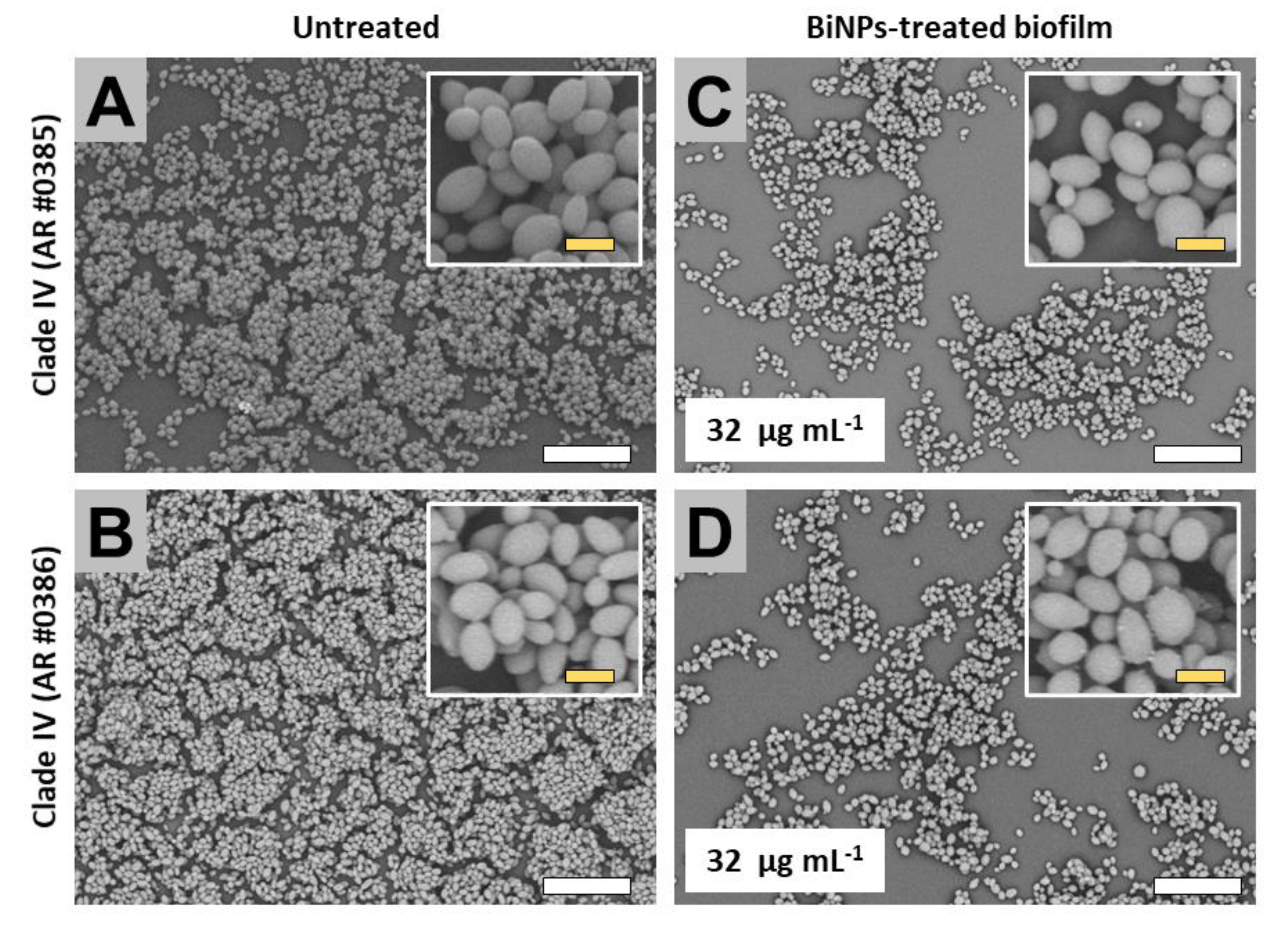
2.3.4. Clade IV (South America Clade)

The untreated biofilms from the AR no. 0385 and no. 0386 strains extensively covered the surface of the well (Figure 6A,B). Also, for both strains, cells within the biofilms displayed the typical yeast morphology (Figure 6A,B). The BiNP-treated samples (BiNPs = 32 µg mL−1 for both strains) exhibited a visible reduction in the covered area (Figure 6C,D); however, the cell morphology was mostly unaltered by the treatments.
We observed that the untreated samples from the different C. auris strains display variations on their phenotypes. This diversity of size and shapes has been described in other works [17,47]. In this work, we expand the current knowledge as we associate the observed specific phenotypes to particular clades.
On the BiNP-treated samples, we observed that nanoparticles altered the shape and size of the yeast cells in some strains. The cause of those alterations is unknown, as this is the first time that the effects of BiNPs are assessed in C. auris. Moreover, although the antimicrobial activity of BiNPs and thiolated bismuth complexes have been studied by different groups [60,61,62], their physicochemical interactions between BiNPs and microbial cells remains largely unknown. Bismuth nanoparticles likely exert anticandidal activity by their own, and also may be releasing antimicrobial bismuth ions and thiolated bismuth compounds, as it has been observed in other metallic nanoparticles [44,63]. Moreover, it has been suggested that metal-containing compounds are a promising alternative for developing novel substances with antibiotic properties [12], and the results from different research groups—including ours—increase the evidence for supporting that premise. Nevertheless, the mode of action of BiNPs and the physicochemical interactions within the nanoparticles–cells–biomolecules complex system remains yet to be addressed.

3. Materials and Methods

3.1. Material and Strains

Menadione, 2,3-Bis(2-methoxy-4-nitro-5-sulfophenyl)-2H-tetrazolium-5-carboxanilide salt (XTT), and phosphate-buffered saline (PBS), from Sigma-Aldrich (MO). Osmium tetroxide (OsO4) and glutaraldehyde were acquired from Ted Pella. 0.74 mM XTT, 10 mM menadione, and 1% OsO4 solutions were prepared in Milli Q water.
Nanoantibiotics: We used PVP-coated bismuth nanoparticles (BiNPs) synthesized by a method previously reported by our group [51]. These PVP-BiNPs were produced via a fast, facile, and cost-effective chemical reduction process. Briefly, bismuth nitrate salts were added to a stirring glycine solution, pre-warmed at 75 °C; then pH was raised to 9 using NaOH. After, dimercaptopropanol and PVP solutions were consecutively added to the warm alkaline bismuth solution, still under constant stirring. Finally, NaBH4 was added dropwise in two stages. The formation of the BiNPs is rapidly evident by the change of color of the suspension, from yellow to black. The suspension was kept on vigorous stirring for 10 min. This synthesis protocol can be easily replicated in non-specialized facilities, and nanoparticles can be produced in less than 1 h. The obtained BiNPs are small spheroids (average diameter of 8 nm), with a negative surface charge.
Strains: We used 10 different Candida auris strains, from the Centers for Disease Control and Prevention (CDC) Antimicrobial Resistance (AR) Isolate Bank stock [64]. The AR strains, organized by clade, are Clade I: South Asia—(AR strains no. 0382, no. 0387, no. 0388, no. 0389, and no. 0390); Clade II: East Asia—(AR strain no. 0381); Clade III: Africa—(AR strains no. 0383 and no. 0384); and Clade IV: South America—(AR strains no. 0385 and no. 0386). Freshly cultured C. auris strains were prepared as follows: a loopful of C. auris cells from frozen glycerol stocks were subcultured onto yeast extract–peptone–dextrose (YPD) (BD Difco, MD), at 30 °C for 48 h. Then, a couple of C. auris colonies were transferred into YPD broth and incubated at 35 °C, overnight, in an orbital shaker. Cells from these fresh cultures were used for the susceptibility assays described in the following sections.

3.2. Susceptibility Tests on Planktonic Cells

To assess the antifungal activity of BiNPs, the CLSI M27 protocol [53] was followed, with slight modifications. Briefly, yeast cells from the overnight cultures were washed twice in PBS and adjusted in RPMI culture media and transferred to a 96 multi-well round-bottom plate. BiNPs were prepared in a two-fold dilution series in RPMI and transferred to the multi-well plates with the yeast cells. The experimental final concentrations of yeast were 2.5 × 10³ cells mL−1, whereas the BiNPs serial dilutions ranged from 0.5 to 256 µg mL−1. Plates with BiNP-treated cells and the controls (untreated cells, BiNPs with no cells, blank RPMI with no cells, and no BiNPs) were cultured for 48 h, at 180 rpm, 35 °C. The Minimal inhibitory concentration (MIC) was set as the concentration where no microbial growth was observed visually, according to the guidelines of the M27 protocol from the CLSI.

3.3. Antibiofilm Activity Assays

The inhibitory effect of BiNPs on the biofilm formation stage of C. auris was determined using a method previously reported by our group [55], with minor modifications. Briefly, the yeast cells from the overnight cultures were washed twice in PBS and the cells were adjusted in RPMI and transferred to flat-bottom 96-multiwell plates. BiNPs were prepared in a two-fold dilution series, then transferred to the multi-well plates with the yeast cells. The experimental final concentration for the biofilm assays was 1 × 10⁶ cells mL−1; whereas the BiNP serial dilutions ranged from 0.5 to 256 µg mL−1. The multi-well plates with the BiNP-treated yeast cells and the controls (untreated cells, BiNPs with no cells, blank RPMI with no cells, and no BiNPs) were cultured stationery, at 37 °C for 24 h, for inducing the biofilm formation phase.
To assess the antibiofilm effect of BiNPs, the XTT absorbance was used as a measure of biofilm activity [55], as follows: post-incubation, biofilms were washed twice with PBS and 100 µL of XTT/menadione solution were added to the plates with the treated and the controls. Immediately, the plates were protected from light and incubated for 2 h at 37 °C. The absorbance of XTT was measured at λ = 490 nm in a Benchmark Microplate Reader (Bio-Rad Inc). From the absorbance readings, dose–response curves were obtained to calculate the IC50, set as the concentration of BiNPs required for reducing the biofilm activity by 50%. The IC50 was calculated by fitting the normalized results to the variable slope Hill equation (to assess the nonlinear dose–response relationship), in the software Prism 8 (GraphPad Software, Inc.).
To ensure the experimental reproducibility, the anticandidal activity of BiNPs was evaluated using different rounds of nanoparticle syntheses, using three independent replicates of the 96-multiwell plates, with two replicates of the treatments within each multiwell plate (n = 6, for each strain), for both the planktonic and the biofilm stages.

3.4. Ultrastructural Analysis

The effect of the BiNPs on the structure of the C. auris biofilms was evaluated in the inhibition of biofilm formation experiments via scanning electron microscopy (SEM). Briefly, biofilms were treated with subinhibitory concentrations of BiNPs, according to the corresponding calculated IC50 values for each C. auris strain. After incubation, the biofilm samples were washed twice with PBS, then fixed with 2% glutaraldehyde for 3 h, and then stained with 1% osmium tetroxide for 30 min. Then, the samples were dehydrated using an ascending concentration series of ethanol, from 30% to 100%. Then, ethanol was completely removed, and the samples were left to dry overnight. The dehydrated samples were coated with gold in a sputter coater SC7620 (Quorum Technologies). To form a uniform and thick gold layer over the samples, the current was set at 25 milliamperes for 3 min. Finally, the samples coated with gold were observed in a TM4000Plus Scanning Electron Microscope (Hitachi Inc.), using the 500× and 2500× magnification, with a voltage of 10 KeV.

4. Conclusions

Overall, our results seem to indicate that bismuth nanoparticles display a strong anticandidal activity against all the multiple C. auris CDC AR strains when tested under planktonic conditions. However, the BiNPs displayed much more modest antibiofilm activity, with also more accused differences among the different strains, which were not necessarily related to their clade. Despite this lower activity, treatment with BiNPs affected the biofilm structure and, in some instances, the cell morphology of cells within biofilms. Our results seem to indicate that the broad anticandidal activity of bismuth nanoparticles may contribute to reducing the spread of the multidrug-resistant C. auris strains in the healthcare-related facilities. Finally, more studies regarding the antimicrobial properties of BiNPs, against different pathogens, will contribute to expanding their future potential applications.

Author Contributions

R.V.-M. and J.L.L.-R. contributed to the study design and manuscript preparation. R.V.-M. and F.D.L. executed the experimental procedures. All authors participated in data analysis. All authors have read and agreed to the published version of the manuscript.

Funding

This research was funded by the Mexican Council of Science and Technology of Mexico (CONACYT, RV-M postdoctoral scholarship). Support in the laboratory was provided by the Margaret Batts Tobin Foundation, San Antonio, TX, USA.

Acknowledgments

We thank the CDC for providing the panel of AR C. auris strains. We thank the South Texas Center for Emerging Infectious Diseases (STCEID) for the purchase of the HITACHI SEM microscope used in this project.

Conflicts of Interest

The funders had no role in the design of the study; in the collection, analyses, or interpretation of data; in the writing of the manuscript, or in the decision to publish the results.

Appendix A

Planktonic stage: Is the state in which the cells are free-living, acting as independent organisms.
Biofilm stage: Is the state when the cells are attached to a surface, forming multicellular communities, protected by an extracellular matrix [65]. Biofilms can transition between both the planktonic and the biofilms stages. Traditionally, it is considered that biofilms undergo a maturity process: from the biofilm formation (adhesion stage) to the mature biofilms (development and maturation stages).
Yeast phenotype: It is the typical single-cell oval-shape structure seen for most Candida species. However, there are several morphological variations across different Candida strains and species, resulting in shapes such as ellipsoidal, elongated, and rectangular with smooth rounded ends, among others.
Pseudohyphae phenotype: Some pleiomorphic fungus, such as C. albicans, can undergo a morphogenesis process for transitioning (forth and back) to a pseudohyphal or hyphal phenotype. [66,67]. Pseudohyphae displays a multicellular organization, as the daughter cells bud elongates and remains attached to the mother cell, after septum formation, forming strings of connected budding cells. These filaments can grow so long that they resemble hyphae. Some strains of C. auris can form elongated cells that resemble pseudohyphae [9].
Hyphae phenotype: The hyphal phenotype is characterized by the extreme elongation of the fungal cells, leading to the formation of long germ tubes that are usually narrower than pseudohyphal cells. The initial germ tubes do not display apparent septa [66], and they exhibit polarized growth. The ‘true’ hyphal stage has not been reported in C. auris.

Appendix B

Figure A1. Susceptibility of the multiple C. auris strains, clustered by clade, exposed to BiNPs during the biofilm formation stage. The dose–response curves (continuous line) show the effect of the BiNPs (as XTT absorbance readings) during biofilm formation in the multiple strains of C. auris, clustered by clade. Within each graph, the individual points show the average absorbance % over the BiNPs concentration, whereas de vertical bars in each point represent the standard deviation (n = 6).
Antibiotics 09 00461 g0a1

References

  1. World Health Organization. The Top 10 Causes of Death; WHO: Geneva, Switzerland, 2018. [Google Scholar]
  2. Smith, R.; Coast, J. The true cost of antimicrobial resistance. BMJ 2013, 346, f1493. [Google Scholar] [CrossRef] [PubMed] [Green Version]
  3. Centers for Disease Control and Prevention. Antibiotic Resistance Threats in the United States, 2019; CDC: Atlanta, GA, USA, 2019.
  4. Frieri, M.; Kumar, K.; Boutin, A. Antibiotic resistance. J. Infect. Public Health 2017, 10, 369–378. [Google Scholar] [CrossRef] [PubMed] [Green Version]
  5. Khameneh, B.; Diab, R.; Ghazvini, K.; Fazly Bazzaz, B.S. Breakthroughs in bacterial resistance mechanisms and the potential ways to combat them. Microb. Pathog. 2016, 95, 32–42. [Google Scholar] [CrossRef] [PubMed]
  6. Kullberg, B.J.; Arendrup, M.C. Invasive Candidiasis. N. Engl. J. Med. 2015, 373, 1445–1456. [Google Scholar] [CrossRef] [Green Version]
  7. Silva, S.; Rodrigues, C.; Araújo, D.; Rodrigues, M.; Henriques, M. Candida Species Biofilms’ Antifungal Resistance. J. Fungi 2017, 3, 8. [Google Scholar] [CrossRef] [Green Version]
  8. Pappas, P.G.; Lionakis, M.S.; Arendrup, M.C.; Ostrosky-Zeichner, L.; Kullberg, B.J. Invasive candidiasis. Nat. Rev. Dis. Prim. 2018, 4, 1–20. [Google Scholar] [CrossRef]
  9. Sherry, L.; Ramage, G.; Kean, R.; Borman, A.; Johnson, E.M.; Richardson, M.D.; Rautemaa-Richardson, R. Biofilm-Forming Capability of Highly Virulent, Multidrug-Resistant Candida auris. Emerg. Infect. Dis. 2017, 23, 328–331. [Google Scholar] [CrossRef] [Green Version]
  10. Jeffery-Smith, A.; Taori, S.K.; Schelenz, S.; Jeffery, K.; Johnson, E.M.; Borman, A.; Manuel, R.; Brown, C.S. Candida auris: A Review of the Literature. Clin. Microbiol. Rev. 2017, 31. [Google Scholar] [CrossRef] [Green Version]
  11. Forsberg, K.; Woodworth, K.; Walters, M.; Berkow, E.L.; Jackson, B.; Chiller, T.; Vallabhaneni, S. Candida auris: The recent emergence of a multidrug-resistant fungal pathogen. Med. Mycol. 2019, 57, 1–12. [Google Scholar] [CrossRef] [Green Version]
  12. Arendrup, M.C.; Patterson, T.F. Multidrug-resistant candida: Epidemiology, molecular mechanisms, and treatment. J. Infect. Dis. 2017, 216, S445–S451. [Google Scholar] [CrossRef] [Green Version]
  13. Sears, D.; Schwartz, B.S. Candida auris: An emerging multidrug-resistant pathogen. Int. J. Infect. Dis. 2017, 63, 95–98. [Google Scholar] [CrossRef] [PubMed] [Green Version]
  14. Rhodes, J.; Fisher, M.C. Global epidemiology of emerging Candida auris. Curr. Opin. Microbiol. 2019, 52, 84–89. [Google Scholar] [CrossRef] [PubMed]
  15. Chow, N.A.; De Groot, T.; Badali, H.; Abastabar, M.; Chiller, T.M.; Meis, J.F. Potential fifth clade of Candida auris, Iran, 2018. Emerg. Infect. Dis. 2019, 25, 1780–1781. [Google Scholar] [CrossRef] [Green Version]
  16. CDC Antifungal Susceptibility Testing and Interpretation | Candida auris. Available online: https://www.cdc.gov/fungal/candida-auris/c-auris-antifungal.html (accessed on 15 January 2020).
  17. Szekely, A.; Borman, A.M.; Johnsona, E.M. Candida auris isolates of the southern asian and south african lineages exhibit different phenotypic and antifungal susceptibility profiles in vitro. J. Clin. Microbiol. 2019, 57. [Google Scholar] [CrossRef] [PubMed] [Green Version]
  18. Kean, R.; Delaney, C.; Sherry, L.; Borman, A.; Johnson, E.M.; Richardson, M.D.; Rautemaa-Richardson, R.; Williams, C.; Ramage, G. Transcriptome Assembly and Profiling of Candida auris Reveals Novel Insights into Biofilm-Mediated Resistance. mSphere 2018, 3, 1–12. [Google Scholar] [CrossRef] [Green Version]
  19. Ku, T.S.N.; Walraven, C.J.; Lee, S.A. Candida auris: Disinfectants and Implications for Infection Control. Front. Microbiol. 2018, 9. [Google Scholar] [CrossRef]
  20. Adams, E.; Quinn, M.; Tsay, S.; Poirot, E.; Chaturvedi, S.; Southwick, K.; Greenko, J.; Fernandez, R.; Kallen, A.; Vallabhaneni, S.; et al. Candida auris in healthcare facilities, New York, USA, 2013–2017. Emerg. Infect. Dis. 2018, 24, 1816–1824. [Google Scholar] [CrossRef] [Green Version]
  21. Campoy, S.; Adrio, J.L. Antifungals. Biochem. Pharmacol. 2017, 133, 86–96. [Google Scholar] [CrossRef]
  22. Casadevall, A.; Kontoyiannis, D.P.; Robert, V. On the emergence of candida auris: Climate change, azoles, swamps, and birds. MBio 2019, 10, e01397-19. [Google Scholar] [CrossRef] [Green Version]
  23. Welsh, R.M.; Bentz, M.L.; Shams, A.; Houston, H.; Lyons, A.; Rose, L.J.; Litvintseva, A.P. Survival, Persistence, and Isolation of the Emerging Multidrug-Resistant Pathogenic Yeast Candida auris on a Plastic Health Care Surface. J. Clin. Microbiol. 2017, 55, 2996–3005. [Google Scholar] [CrossRef] [Green Version]
  24. Ramage, G.; Martínez, J.P.; López-Ribot, J.L. Candida biofilms on implanted biomaterials: A clinically significant problem. FEMS Yeast Res. 2006, 6, 979–986. [Google Scholar] [CrossRef] [Green Version]
  25. Pathirana, R.U.; Friedman, J.; Norris, H.L.; Salvatori, O.; McCall, A.D.; Kay, J.; Edgerton, M. Fluconazole-Resistant Candida auris Is Susceptible to Salivary Histatin 5 Killing and to Intrinsic Host Defenses. Antimicrob. Agents Chemother. 2018, 62. [Google Scholar] [CrossRef] [PubMed] [Green Version]
  26. Wall, G.; Herrera, N.; Lopez-Ribot, J.L. Repositionable compounds with antifungal activity against multidrug resistant candida auris identified in the medicines for malaria venture’s pathogen box. J. Fungi 2019, 5, 92. [Google Scholar] [CrossRef] [PubMed] [Green Version]
  27. Nicola, A.M.; Albuquerque, P.; Paes, H.C.; Fernandes, L.; Costa, F.F.; Kioshima, E.S.; Abadio, A.K.R.; Bocca, A.L.; Felipe, M.S. Antifungal drugs: New insights in research & development. Pharmacol. Ther. 2019, 195, 21–38. [Google Scholar] [PubMed]
  28. Taneja, N.K.; Tyagi, J.S. Jaya Sivaswami Tya Resazurin reduction assays for screening of anti-tubercular compounds against dormant and actively growing Mycobacterium tuberculosis, Mycobacterium bovis BCG and Mycobacterium smegmatis. J. Antimicrob. Chemother. 2007, 60, 288–293. [Google Scholar] [CrossRef] [PubMed]
  29. Lara, H.H.; Ixtepan-Turrent, L.; Jose Yacaman, M.; Lopez-Ribot, J. Inhibition of Candida auris Biofilm Formation on Medical and Environmental Surfaces by Silver Nanoparticles. ACS Appl. Mater. Interfaces 2020, 12, 21183–21191. [Google Scholar] [CrossRef]
  30. Weissig, V.; Guzman-Villanueva, D. Nanopharmaceuticals (part 2): Products in the pipeline. Int. J. Nanomed. 2015, 10, 1245–1257. [Google Scholar] [CrossRef] [Green Version]
  31. Çalış, S.; Öztürk Atar, K.; Arslan, F.B.; Eroğlu, H.; Çapan, Y. Nanopharmaceuticals as Drug-Delivery Systems. In Nanocarriers for Drug Delivery; Elsevier: Amsterdam, The Netherlands, 2019; pp. 133–154. [Google Scholar]
  32. Murdock, N. Nanopharmaceuticals (part 1): Products on the market. Int. J. Nanomed. 2014, 9, 4357–4373. [Google Scholar]
  33. Singh, L.; Kruger, H.G.; Maguire, G.E.M.; Govender, T.; Parboosing, R. The role of nanotechnology in the treatment of viral infections. Ther. Adv. Infect. Dis. 2017, 4, 105–131. [Google Scholar] [CrossRef]
  34. Ventola, C.L. Progress in Nanomedicine: Approved and Investigational Nanodrugs. Pharm. Ther. 2017, 42, 742–755. [Google Scholar]
  35. Lara, H.H.; Guisbiers, G.; Mendoza, J.; Mimun, L.C.; Vincent, B.A.; Lopez-Ribot, J.L.; Nash, K.L. Synergistic antifungal effect of chitosan-stabilized selenium nanoparticles synthesized by pulsed laser ablation in liquids against Candida albicans biofilms. Int. J. Nanomed. 2018, 13, 2697–2708. [Google Scholar] [CrossRef] [PubMed] [Green Version]
  36. Vazquez-Muñoz, R.; Borrego, B.; Juárez-Moreno, K.; García-García, M.; Mota Morales, J.D.; Bogdanchikova, N.; Huerta-Saquero, A. Toxicity of silver nanoparticles in biological systems: Does the complexity of biological systems matter? Toxicol. Lett. 2017, 276, 11–20. [Google Scholar] [CrossRef] [PubMed]
  37. Montes, M.; Pierce, C.G.; Lopez-Ribot, J.L.; Bhalla, A.S.; Guo, R. Properties of silver and copper nanoparticle containing aqueous suspensions and evaluation of their in vitro activity against candida albicans and staphylococcus aureus biofilms. J. Nano Res. 2016, 37, 109–121. [Google Scholar] [CrossRef]
  38. Jaime-Acuña, O.E.; Meza-Villezcas, A.; Vasquez-Peña, M.; Raymond-Herrera, O.; Villavicencio-García, H.; Petranovskii, V.; Vazquez-Duhalt, R.; Huerta-Saquero, A. Synthesis and complete antimicrobial characterization of CEOBACTER, an Ag-Based nanocomposite. PLoS ONE 2016, 11, e0166205. [Google Scholar] [CrossRef]
  39. Lee, N.-Y.; Ko, W.-C.; Hsueh, P.-R. Nanoparticles in the Treatment of Infections Caused by Multidrug-Resistant Organisms. Front. Pharmacol. 2019, 10, 1153. [Google Scholar] [CrossRef] [Green Version]
  40. Lara, H.H.; Romero-Urbina, D.G.; Pierce, C.; Lopez-Ribot, J.L.; Arellano-Jiménez, M.J.; Jose-Yacaman, M. Effect of silver nanoparticles on Candida albicans biofilms: An ultrastructural study. J. Nanobiotechnol. 2015, 13, 1–12. [Google Scholar] [CrossRef] [Green Version]
  41. Vazquez-Muñoz, R.; Arellano-Jimenez, M.J.; Lopez, F.D.; Lopez-Ribot, J.L. Protocol optimization for a fast, simple and economical chemical reduction synthesis of antimicrobial silver nanoparticles in non-specialized facilities. BMC Res. Notes 2019, 12, 773. [Google Scholar] [CrossRef]
  42. Vazquez-Muñoz, R.; Avalos-Borja, M.; Castro-Longoria, E. Ultrastructural Analysis of Candida albicans When Exposed to Silver Nanoparticles. PLoS ONE 2014, 9, e108876. [Google Scholar] [CrossRef]
  43. Zazo, H.; Colino, C.I.; Lanao, J.M. Current applications of nanoparticles in infectious diseases. J. Control. Release 2016, 224, 86–102. [Google Scholar] [CrossRef]
  44. Raza, M.A.; Kanwal, Z.; Rauf, A.; Sabri, A.N.; Riaz, S.; Naseem, S. Size- and Shape-Dependent Antibacterial Studies of Silver Nanoparticles Synthesized by Wet Chemical Routes. Nanomaterials 2016, 6, 74. [Google Scholar] [CrossRef] [Green Version]
  45. Ahmad, T.; Wani, I.A.; Lone, I.H.; Ganguly, A.; Manzoor, N.; Ahmad, A.; Ahmed, J.; Al-Shihri, A.S. Antifungal activity of gold nanoparticles prepared by solvothermal method. Mater. Res. Bull. 2013, 48, 12–20. [Google Scholar] [CrossRef]
  46. Ramyadevi, J.; Jeyasubramanian, K.; Marikani, A.; Rajakumar, G.; Rahuman, A.A. Synthesis and antimicrobial activity of copper nanoparticles. Mater. Lett. 2012, 71, 114–116. [Google Scholar] [CrossRef]
  47. Vazquez-Munoz, R.; Lopez, F.D.; Lopez-Ribot, J.L. Silver nanoantibiotics display strong antifungal activity against the emergent multidrug-resistant yeast Candida auris under both planktonic and biofilm growing conditions. Front. Microbiol. 2020, 11, 1673. [Google Scholar] [CrossRef]
  48. Goldman, R.D. Bismuth salicylate for diarrhea in children. Can. Fam. Physician 2013, 59, 843–844. [Google Scholar] [PubMed]
  49. Ferraz, K.S.O.; Reis, D.C.; Da Silva, J.G.; Souza-Fagundes, E.M.; Baran, E.J.; Beraldo, H. Investigation on the bioactivities of clioquinol and its bismuth(III) and platinum(II, IV) complexes. Polyhedron 2013, 63, 28–35. [Google Scholar] [CrossRef]
  50. Badireddy, A.R.; Hernandez-Delgadillo, R.; Sánchez-Nájera, R.I.; Chellam, S.; Cabral-Romero, C. Synthesis and characterization of lipophilic bismuth dimercaptopropanol nanoparticles and their effects on oral microorganisms growth and biofilm formation. J. Nanoparticle Res. 2014, 16, 2456. [Google Scholar] [CrossRef]
  51. Vazquez-Munoz, R.; Arellano-Jimenez, M.J.; Lopez-Ribot, J.L. Fast, facile synthesis method for BAL-mediated PVP-bismuth nanoparticles. MethodsX 2020, 7, 100894. [Google Scholar] [CrossRef]
  52. Vazquez-Munoz, R.; Arellano-Jimenez, M.J.; Lopez-Ribot, J. Bismuth nanoparticles obtained by a facile synthesis method exhibit antimicrobial activity against Staphylococcus aureus and Candida albicans. bioRxiv 2020. [Google Scholar] [CrossRef]
  53. CLSI. M27. Reference Method for Broth Dilution Antifungal Susceptibility Testing of Yeasts, 4th ed.; Alexander, B.D., Ed.; Clinical Laboratory Standards Institute: Wayne, PA, USA, 2017; ISBN 1-56238-826-6. [Google Scholar]
  54. Vaidya, M.Y.; McBain, A.J.; Butler, J.A.; Banks, C.E.; Whitehead, K.A. Antimicrobial Efficacy and Synergy of Metal Ions against Enterococcus faecium, Klebsiella pneumoniae and Acinetobacter baumannii in Planktonic and Biofilm Phenotypes. Sci. Rep. 2017, 7, 1–9. [Google Scholar] [CrossRef]
  55. Pierce, C.G.; Uppuluri, P.; Tristan, A.R.; Wormley, F.L.; Mowat, E.; Ramage, G.; Lopez-Ribot, J.L. A simple and reproducible 96-well plate-based method for the formation of fungal biofilms and its application to antifungal susceptibility testing. Nat. Protoc. 2008, 3, 1494–1500. [Google Scholar] [CrossRef]
  56. Larkin, E.; Hager, C.; Chandra, J.; Mukherjee, P.K.; Retuerto, M.; Salem, I.; Long, L.; Isham, N.; Kovanda, L.; Borroto-Esoda, K.; et al. The Emerging Pathogen Candida auris: Growth Phenotype, Virulence Factors, Activity of Antifungals, and Effect of SCY-078, a Novel Glucan Synthesis Inhibitor, on Growth Morphology and Biofilm Formation. Antimicrob. Agents Chemother. 2017, 61. [Google Scholar] [CrossRef] [PubMed] [Green Version]
  57. Dominguez, E.G.; Zarnowski, R.; Choy, H.L.; Zhao, M.; Sanchez, H.; Nett, J.E.; Andes, D.R. Conserved Role for Biofilm Matrix Polysaccharides in Candida auris Drug Resistance. mSphere 2019, 4. [Google Scholar] [CrossRef] [PubMed] [Green Version]
  58. Chaabane, F.; Graf, A.; Jequier, L.; Coste, A.T. Review on Antifungal Resistance Mechanisms in the Emerging Pathogen Candida auris. Front. Microbiol. 2019, 10, 2788. [Google Scholar] [CrossRef] [PubMed]
  59. Dekkerová, J.; Lopez-Ribot, J.L.; Bujdáková, H. Activity of anti-CR3-RP polyclonal antibody against biofilms formed by Candida auris, a multidrug-resistant emerging fungal pathogen. Eur. J. Clin. Microbiol. Infect. Dis. 2019, 38, 101–108. [Google Scholar] [CrossRef] [PubMed]
  60. Mahony, D.E.; Lim-Morrison, S.; Bryden, L.; Faulkner, G.; Hoffman, P.S.; Agocs, L.; Briand, G.G.; Burford, N.; Maguire, H. Antimicrobial activities of synthetic bismuth compounds against Clostridium difficile. Antimicrob. Agents Chemother. 1999, 43, 582–588. [Google Scholar] [CrossRef] [Green Version]
  61. Varposhti, M.; Abdi Ali, A.; Mohammadi, P. Synergistic effects of bismuth thiols and various antibiotics against Pseudomonas aeruginosa Biofilm. Jundishapur J. Microbiol. 2014, 7. [Google Scholar] [CrossRef] [Green Version]
  62. Yang, Y.; Ouyang, R.; Xu, L.; Guo, N.; Li, W.; Feng, K.; Ouyang, L.; Yang, Z.; Zhou, S.; Miao, Y. Review: Bismuth complexes: Synthesis and applications in biomedicine. J. Coord. Chem. 2015, 68, 379–397. [Google Scholar] [CrossRef]
  63. Tamboli, D.P.; Lee, D.S. Mechanistic antimicrobial approach of extracellularly synthesized silver nanoparticles against gram positive and gram negative bacteria. J. Hazard. Mater. 2013, 260, 878–884. [Google Scholar] [CrossRef] [PubMed]
  64. CDC FDA-CDC Antimicrobial Resistance Isolate Bank. Available online: https://wwwn.cdc.gov/arisolatebank/ (accessed on 3 September 2019).
  65. Ramage, G.; Mowat, E.; Jones, B.; Williams, C.; Lopez-Ribot, J. Our Current Understanding of Fungal Biofilms Fungal biofilms. Crit. Rev. Microbiol. 2009, 35, 340–355. [Google Scholar] [CrossRef] [PubMed]
  66. Nadeem, S.G.; Shafiq, A.; Hakim, S.T.; Anjum, Y.; Kazm, S.U. Effect of Growth Media, pH and Temperature on Yeast to Hyphal Transition in Candida albicans. Open J. Med. Microbiol. 2013, 3, 185–192. [Google Scholar] [CrossRef] [Green Version]
  67. Sardi, J.C.O.; Scorzoni, L.; Bernardi, T.; Fusco-Almeida, A.M.; Mendes Giannini, M.J.S. Candida species: Current epidemiology, pathogenicity, biofilm formation, natural antifungal products and new therapeutic options. J. Med. Microbiol. 2013, 62, 10–24. [Google Scholar] [CrossRef] [PubMed]
Figure 1. BiNPs reduce the ability of C. auris to form biofilms during the biofilm formation stage. The dose–response curves (continuous line) show the effect of the BiNPs (as XTT absorbance readings) during biofilm formation in representative strains from the four clades. The individual points show the average absorbance % over the BiNP concentration, whereas de vertical bars in each point represent the standard deviation (n = 6).
Antibiotics 09 00461 g001
Figure 2. SEM observations on the BiNP antibiofilm activity on the C. auris strains from the Clade I (subgroup 1). BiNPs display a reduced biofilm formation—when compared to the untreated control—on the C. auris strains no. 0388 (A,D), no. 0389 (B,E), and no. 0390 (C,F). The cell morphology is also affected by the BiNPs (A–F). Scale bar: yellow 2 µm, white = 20 µm.
Antibiotics 09 00461 g002
Figure 3. SEM observations on the BiNP antibiofilm activity on the C. auris strains from the Clade I (subgroup 2). BiNPs displayed an evident biofilm reduction—when compared to the untreated control—on the C. auris strains no. 0382 (A,C) and no. 0387 (B,D). The cell morphology also exhibited changes by the BiNPs (A–D). Scale bar: yellow 2 µm, white = 20 µm.
Antibiotics 09 00461 g003
Figure 4. SEM observations on the BiNP antibiofilm activity on the C. auris strains from the Clade II. The subinhibitory concentrations of the BiNPs mildly reduced the biofilm formation (B) when compared with the control (A) on the C. auris strains no. 0381. The cell morphology remained mostly unaltered by the BiNP treatment (A,B). Scale bar: yellow 2 µm, white = 20 µm.
Antibiotics 09 00461 g004
Figure 5. SEM observations on the BiNPs antibiofilm activity on the C. auris strains from the Clade III. The BiNP-treated strain displayed a reduced biofilm formation when compared to the untreated control no. 0383 (A,C), in contrast, the strain no. 0384 biofilms were not noticeably reduced (B,D). The cell morphology was affected by the BiNPs in both strains, no. 0383 (A,C) and no. 0384 (B,D). Scale bar: yellow 2 µm, white = 20 µm.
Antibiotics 09 00461 g005
Figure 6. SEM observations on the BiNP antibiofilm activity on the C. auris strains from the Clade IV. When compared to the untreated control, BiNP-treated samples exhibited a reduction in the biofilm formation on both strains no. 0385 (A,C), and no. 0386 (B,D). No effect on cell morphology was observed on BiNP-treated samples (B,D). Scale bar: yellow 2 µm, white = 20 µm.
Antibiotics 09 00461 g006
Table 1. Planktonic MIC values (µg mL−1) of BiNPs against C. auris strains from the different clades.
CladeStrainsMICGeometric Mean
I038212
03872
03882
03894
03902
II038122
III038322
03842
IV038522
03862
Table 2. Calculated IC50 (µg mL−1) for the biofilm inhibitory activity of BiNPs against C. auris strains from the different clades.
CladeStrainsIC50Geometric Mean
I0382113.127.5
038714.1
038831.4
038915.2
039085.2
II038166.566.5
III038343.614.9
03845.1
IV038527.728.8
038629.9

