Review

Short Cationic Peptidomimetic Antimicrobials

1
School of Chemistry, University of New South Wales, Sydney, NSW 2052, Australia
2
School of Optometry and Vision Science, University of New South Wales, Sydney, NSW 2052, Australia
*
Author to whom correspondence should be addressed.
Antibiotics 2019, 8(2), 44; https://doi.org/10.3390/antibiotics8020044
Submission received: 29 March 2019 / Revised: 15 April 2019 / Accepted: 15 April 2019 / Published: 18 April 2019

Abstract

:
The rapid growth of antimicrobial resistance against several frontline antibiotics has encouraged scientists worldwide to develop new alternatives with unique mechanisms of action. Antimicrobial peptides (AMPs) have attracted considerable interest due to their rapid killing and broad-spectrum activity. Peptidomimetics overcome some of the obstacles of AMPs such as high cost of synthesis, short half-life in vivo due to their susceptibility to proteolytic degradation, and issues with toxicity. This review will examine the development of short cationic peptidomimetics as antimicrobials.

1. Antimicrobial Peptides

Endogenous, usually cationic, peptides are produced by all organisms to defend themselves against potential pathogens [1]. This group of peptides are given the general name of antimicrobial peptides (AMPs). More than 3000 AMPs have been isolated from six kingdoms.
Naturally occurring AMPs are generally composed of fewer than 50 amino acids mostly in their L-configuration. Most AMPs are cationic [2,3] but anionic AMPs also known [4]. AMPs have been classified into groups based on their structures such as β-sheets, α-helices, and extended peptides (http://aps.unmc.edu/AP/main.php; Table 1). The α-helical peptides are the largest group of AMPs and have notable amphipathic characteristics. These possess a tertiary structure with a hinge in the middle [5]. They can be unstructured in aqueous solutions but fold into an α-helix upon binding to lipids in membranes [6]. They often contain the helix stabilizing amino acids alanine, leucine, or lysine [6], as well as the positively charged amino acids lysine and arginine. Cationic β-sheet peptides contain one to five disulfide bridges that help stabilise the peptides into conformationally restricted β-sheets [7]. Their antibacterial activity can be greatly impacted by the number of disulfide bridges in the overall structure [8,9]. Cationic linear peptides have unusual biases in amino acids such as containing large amounts of proline, arginine, or tryptophan [10]. One example for such peptide is histatin which is rich in the amino acid histidine [11,12]. Cationic loop peptides are rich in proline and arginine and cannot form amphipathic structures because of the overexpression of proline residues [13,14]. These peptides adopt a loop formation with one disulfide bridge [15]. There are also cyclic AMPs such as gramicidin S [16].

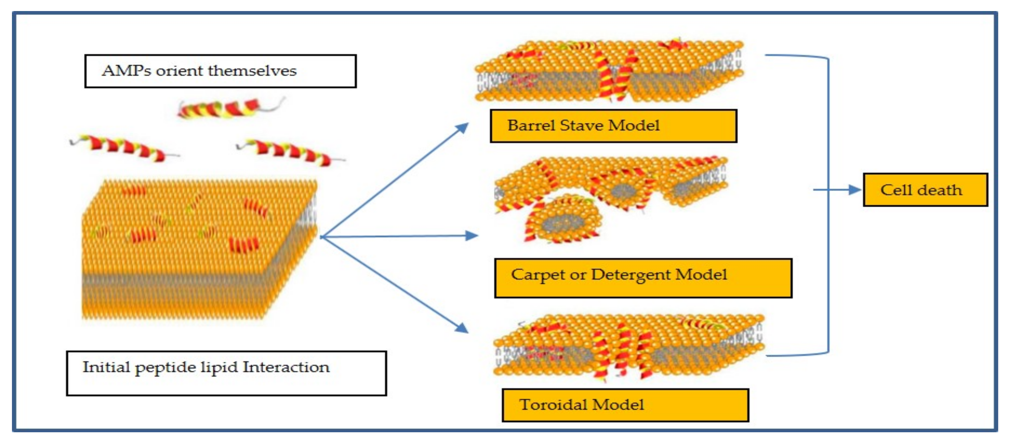
2. Mechanism of Action

AMPs principally target the negatively charged phospholipids in bacterial membranes. The cationic AMPs can outcompete native Mg2+ and Ca2+ ions bound to lipopolysaccharides in the outer membrane of Gram-negative bacteria, resulting in destabilised regions through which the peptides can pass [17]. Once through the outer membrane, the peptides interact with the cytoplasmic membrane causing depolarization and pore formation [18]. Commonly, these activities are bactericidal [19], but some peptides also interact with internal substances to cause cell death [20,21]. In Gram-positive bacteria, the interaction of cationic AMPs with lipoteichoic acid (LTA) in the cell wall occurs initially [22]. This may lead to activation of autolysins that then go on to cause cell death [23], or the peptides may directly cause death through actions of the cytoplasmic membrane [24]. The outer membrane of eukaryotic cells is generally neutral, which results in reduced affinity for the cationic peptides. Most of the membrane active AMPs are amphipathic, i.e., they contain hydrophobic and hydrophilic groups. After their initial electrostatic interaction with negatively charged membranes, the peptides aggregate at the membrane surface and the hydrophobic part helps insertion into the bacterial membrane [25].
The mechanism of action of membrane active peptides is not fully understood [26]. Their activity and cell selectivity depend on physicochemical parameters of peptides and also on the target membranes [27]. A number of models (Figure 1) have been proposed by which AMPs disrupt membranes [28] (Figure 1). In the barrel-stave-drilling model, AMPs orient themselves perpendicularly to the plane of the membrane bilayer to form pores [29,30]. During this time, AMPs with defined secondary structure undergo orientation in which their hydrophobic groups interacting with membrane lipids and their hydrophilic moieties lining up in the lumen of the pore they create [31]. The AMPs alamethicin, protegrins, and pardaxin act in this barrel-stave fashion [28,32,33].
In the carpet model, AMPs accumulate on the bilayer surface [35]. As the concentration of AMPs increases, the membrane is weakened by unfavourable energetics and AMPs are inserted into the membrane in a detergent-like fashion causing the membrane to break into micelles. This mechanism of action does not involve pore formation and, hence, the concentration of AMPs need to be relatively high to cover the bacteria like a carpet [36]. Some examples of AMPs acting by the carpet model are cecropin [37], aurein 1.2 [38], and LL-37 [18]. The toroidal model–wormhole model combines the actions of the barrel-stave and carpet models [39]. The hydrophobic regions of the AMPs associate with the central part of the lipid bilayer with their hydrophilic regions facing the pore [24]. The toroidal model differs from the barrel model in that the peptides are always associated with the lipid head groups even when they are perpendicularly inserted in the lipid bilayer [40]. Some examples of AMPs acting by toroidal model are magainin 2 [41], aurein 2.2 [42], and melittin [40].
Since there may be more than one proposed model applicable for the mode of action of AMPs, it is important to understand the sequential steps leading to and then leading from their initial interactions with membranes [41]. Examples of research in this area to more fully understand the mechanism of action is exemplified by research conducted on gramicidin S. Debate has occurred about whether gramicidin S acts by formation of discrete pores or by less specific membrane disruption similar to detergents [43]. Early studies showed that its mechanism of action was due to pore formation [44]. However, more recent studies using cyclic peptide mimics of gramicidin S showed that the mechanism of action involved only with delocalization of peripheral membrane proteins without forming pores [45].

3. Mechanism of Resistance

AMPs have been suggested as good alternatives to conventional antibiotics due to their relatively non-specific mechanism of action on bacterial membranes and the rapidity [46] of their antimicrobial action. This has been thought to decrease the chance of resistance development. However, resistance has been demonstrated, and this can be either constitutive, inducible, or acquired.
Inducible resistance to AMPs occurs when the bacteria recognises that AMPs are interacting with it and genes are activated that substitute [47], modify [48], or acylate [49] membrane lipids to reduce their interactions with AMPs. This type of resistance can also include the activation of proteolytic enzymes [50], efflux pumps [51], and modification of intracellular targets [52]. Constitutive resistance to AMPs occurs due to inherent properties of a bacterium that confers resistance and the bacteria express the resistance even in the absence of exposure to peptides. Examples of constitutive resistance include electrostatic shielding of membranes [53], changes in membrane potential during different stages of cell growth [54], and biofilm formation [55]. Acquired resistance to AMPs has been described by horizontal transfer of genes between bacteria [56]. Polymyxin resistance due to the plasmid-mediated mcr-1 gene was first identified in China in an Escherichia. coli strain [57]. Several variants of this gene, mcr-1-9, [58] that can confer resistance to polymyxin antibiotics have been identified. However, this gene seems unable to also confer resistance to AMPs such as human cathelicidin LL-37, α-defensin 5 (HD5), or β-defensin 3 (HDB3) [59].
Although bacteria can be or become resistant to AMPs, it is thought that this often requires a high expenditure of energy or substantial changes to the lipid bilayer making resistance development unfavourable [60]. A high level of expression of mcr-1 decreases cell growth rate and viability, results in degradation of cell membranes and cytoplasmic structures, and so reduces the overall fitness of cell carrying this gene [61]. Also, the relatively low level of resistance to AMPs of bacteria in their natural environments could be due to the combination of the interaction of AMPs with the cell membrane and their ability to act at multiple targets.

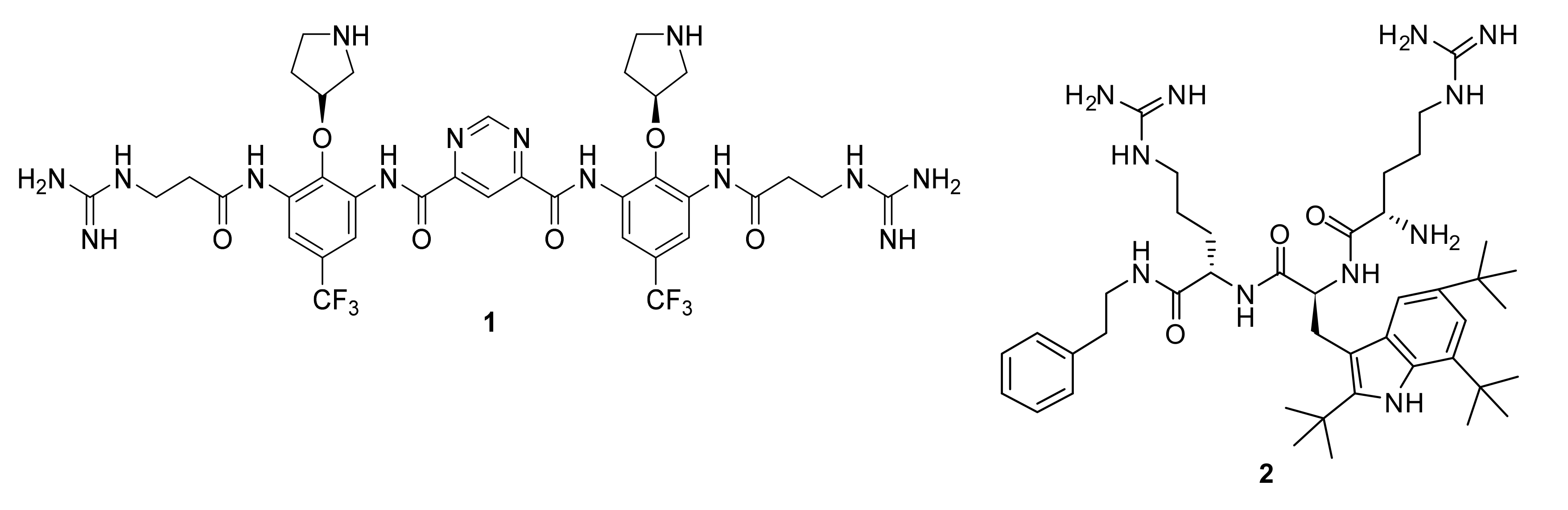
4. Development of Peptidomimetics

Although AMPs have many favourable qualities, few AMPs have been approved for clinical use, often due to failure to demonstrate efficacy over existing treatments [62,63]. The exception is polymyxins, cationic lipopeptides which are used to treat multi-drug-resistant Pseudomonas. aeruginosa infections as a last resort drug [64]. However, polymyxins suffer from significant toxicity problems [65].
The high cost of peptide synthesis and their short half-lives in vivo as a result of their susceptibility to proteolytic enzymes, and issues with toxicity has led to interest in the development of peptidomimetics. Peptidomimetics can be defined as molecules derived from the existing peptides that mimic the biological effect of that peptide. They may have secondary structures and other features to mimic the original peptide and are able to mimic the properties or biological activity of the peptide. The last definition indicates the importance of similar function rather than the similar structure. In this review, most of the peptidomimetic compounds mimic the properties or biological activity of the peptide.
Short cationic peptidomimetics can overcome some of the challenges currently faced with natural AMPs such as ease of synthesis, increased stability (resistant to the action of proteases/peptidases), and reduced toxicity. Indeed, the peptidomimetics compounds (Figure 2) Brilacidin (1) and LTX-109 (2) have successfully completed Phase-II clinical trials for skin infection and impetigo [63].
Peptidomimetics such as α-peptides, β3-peptides, peptoids, β-peptoids, α/β-peptides, peptide/peptoid hybrids, α-AA peptides, γ-AA peptides, α-oligoacyllysines, and β-oligoacyllysines have been well discussed in a review by Molchanova et. al. [66]. Small-molecule peptidomimetics and glycopeptide antibiotics have been reviewed by Ghosh and Haldar [67,68] including a recent review on bacteriophages as alternatives for conventional antibiotics [69]. Abdel Monaim et al. recently published a review on cyclic AMPs [16] as effective antibacterials. From the key structural features of natural AMPs, peptidomimetic molecules possess an amphiphilic backbone and net positive charge that helps to mimic the biological function of AMPs. Table 2 summarises the research on cationic peptidomimetic antimicrobials by several research groups. In this review, the design of short cationic peptidomimetics as antimicrobials by Australian researchers will be presented according to the theme of this special edition of Antimicrobials “From the Southern Hemisphere: research on resistance, antibiotics and treatments”.

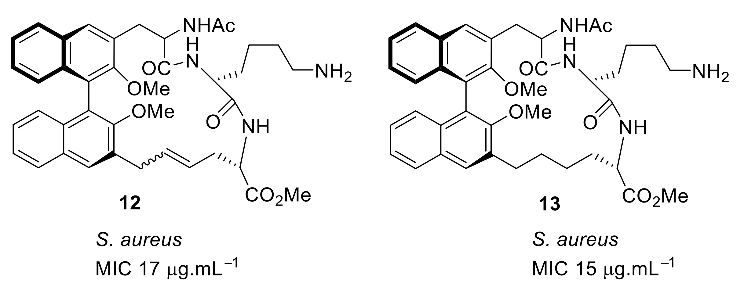
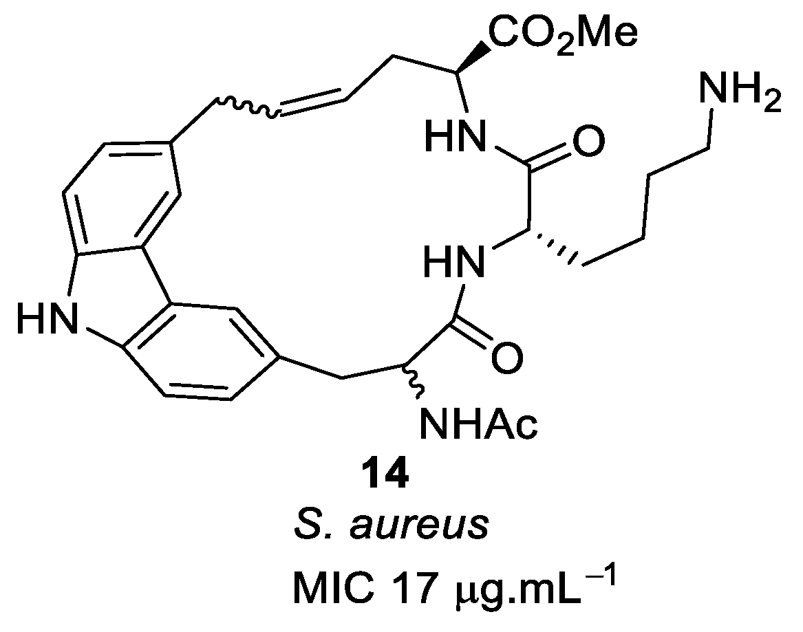
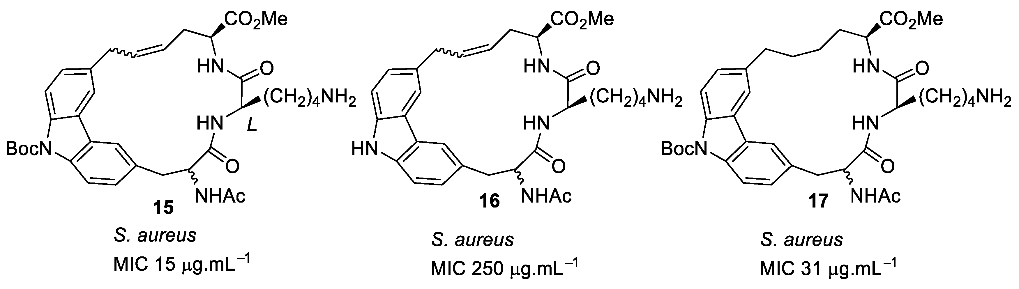
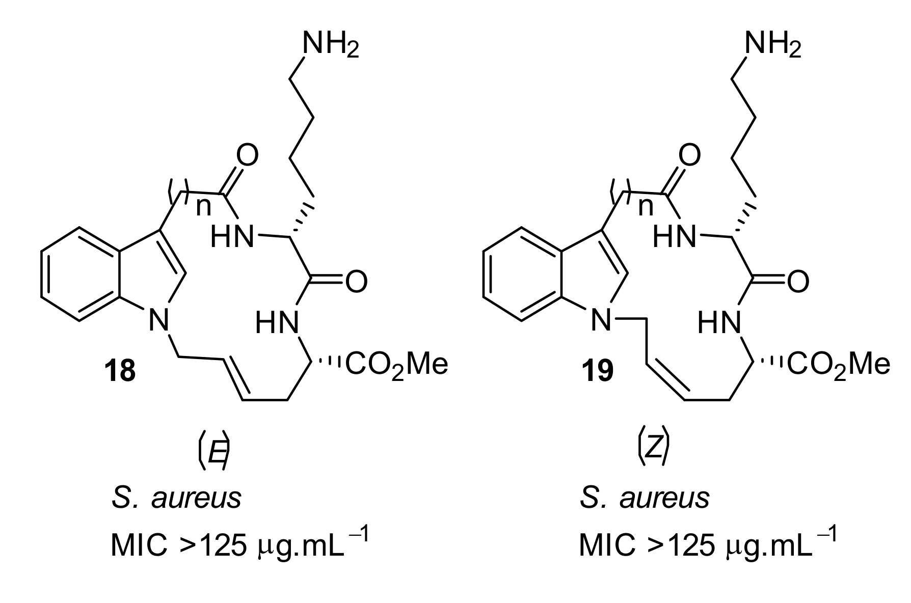
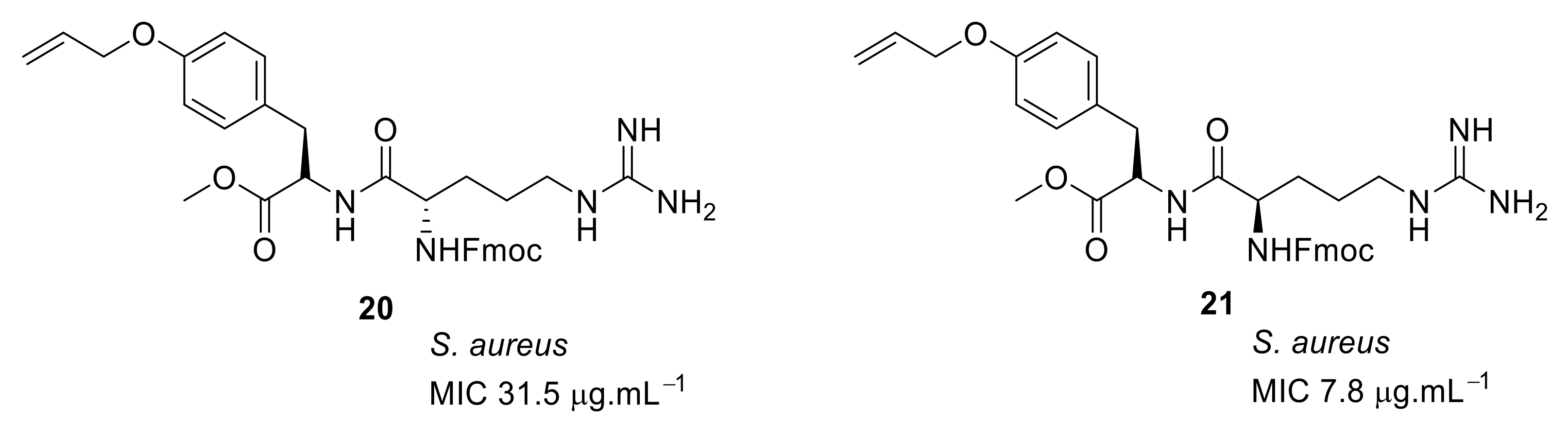
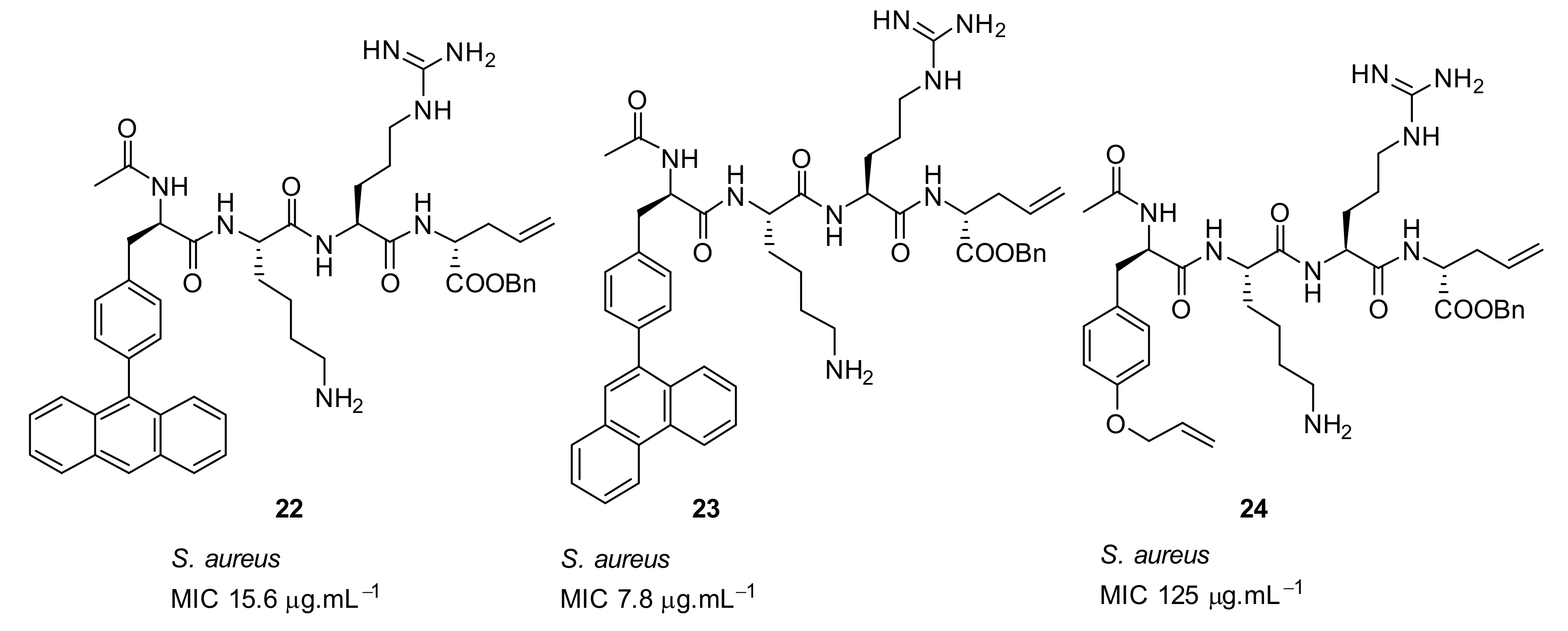
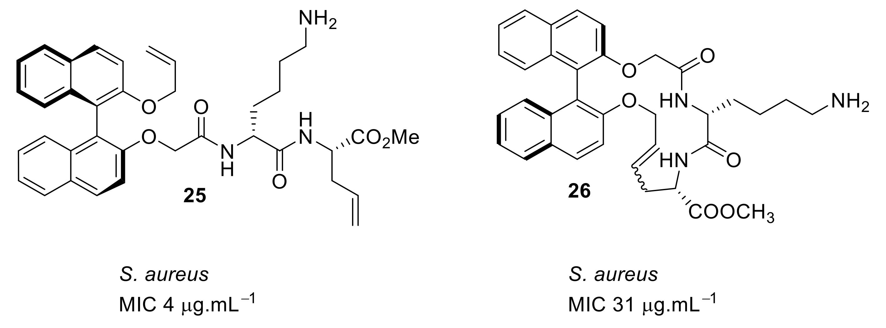
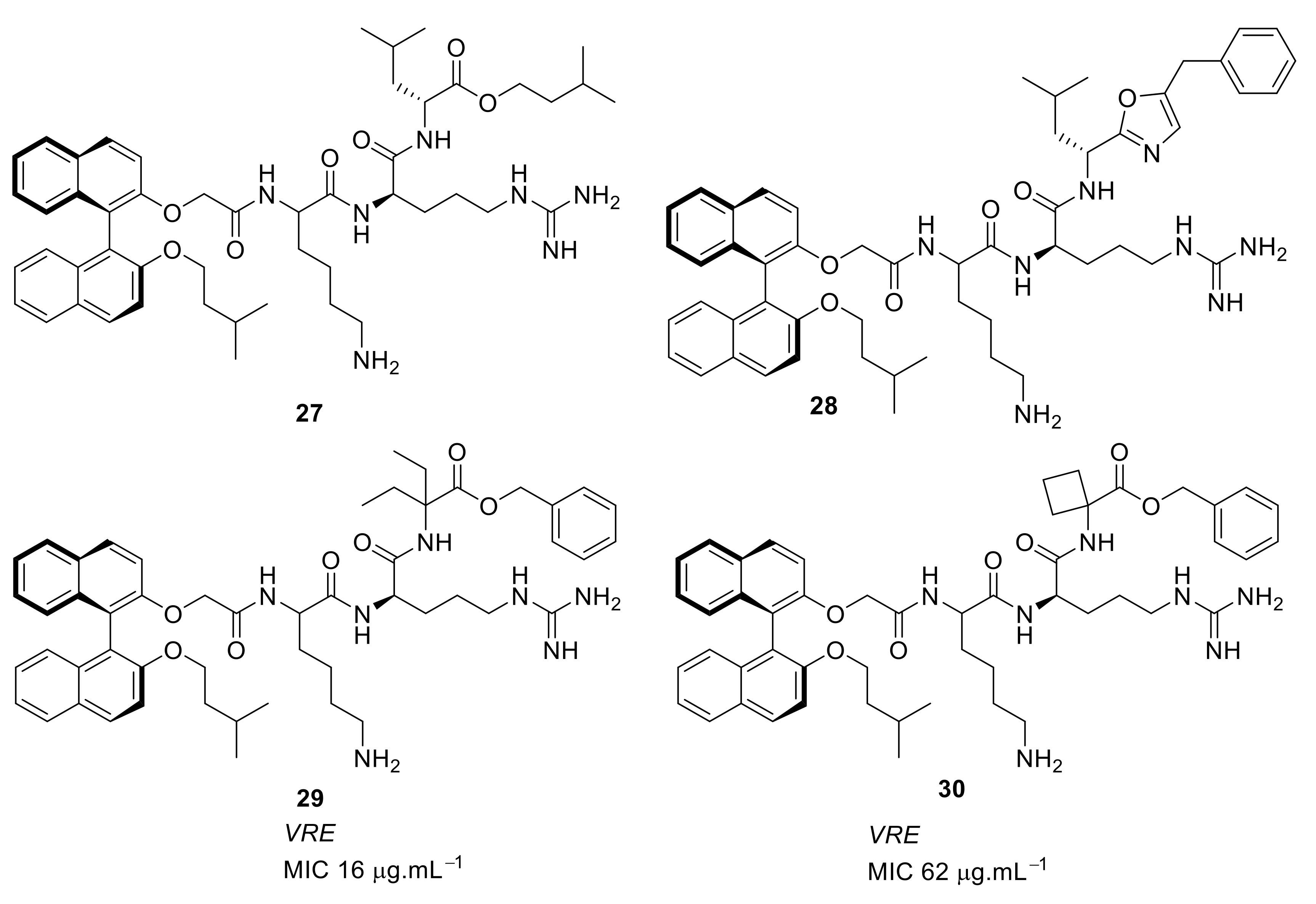
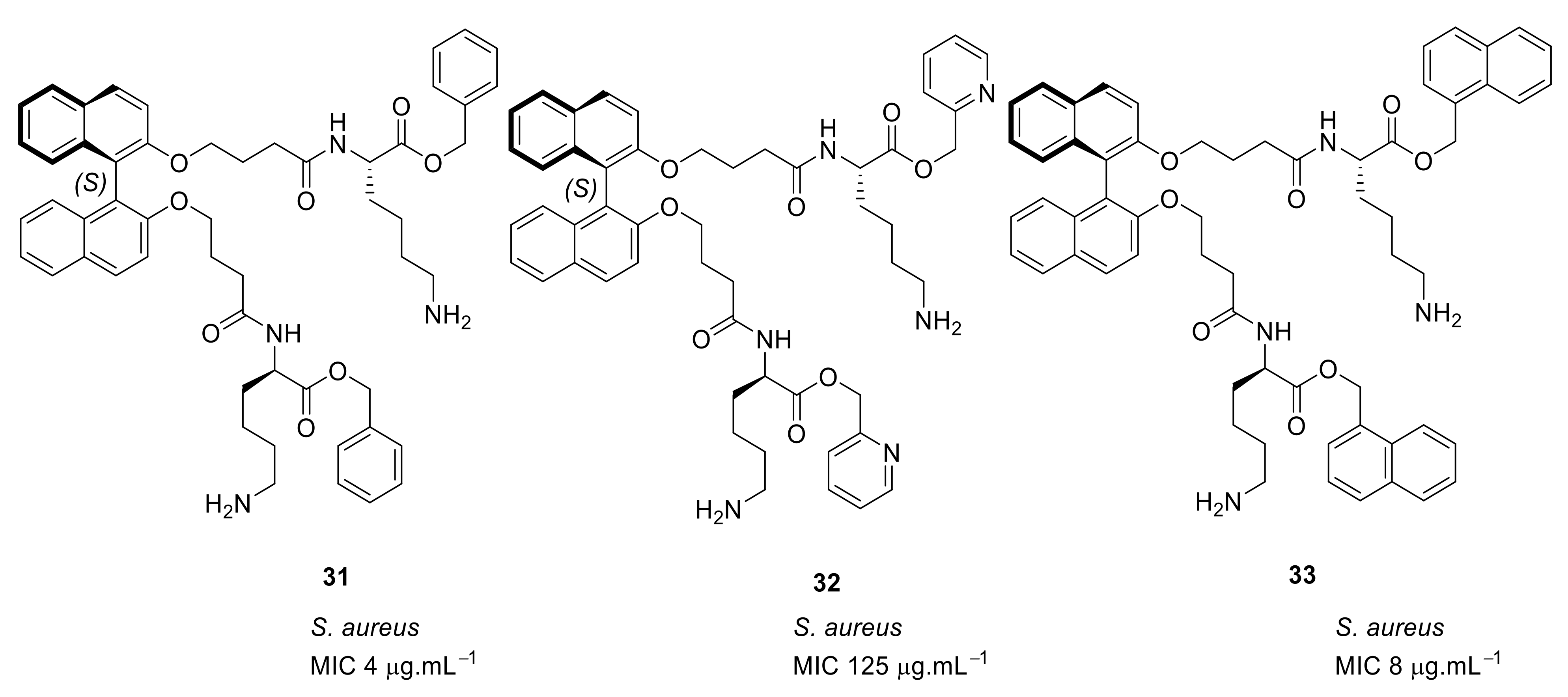
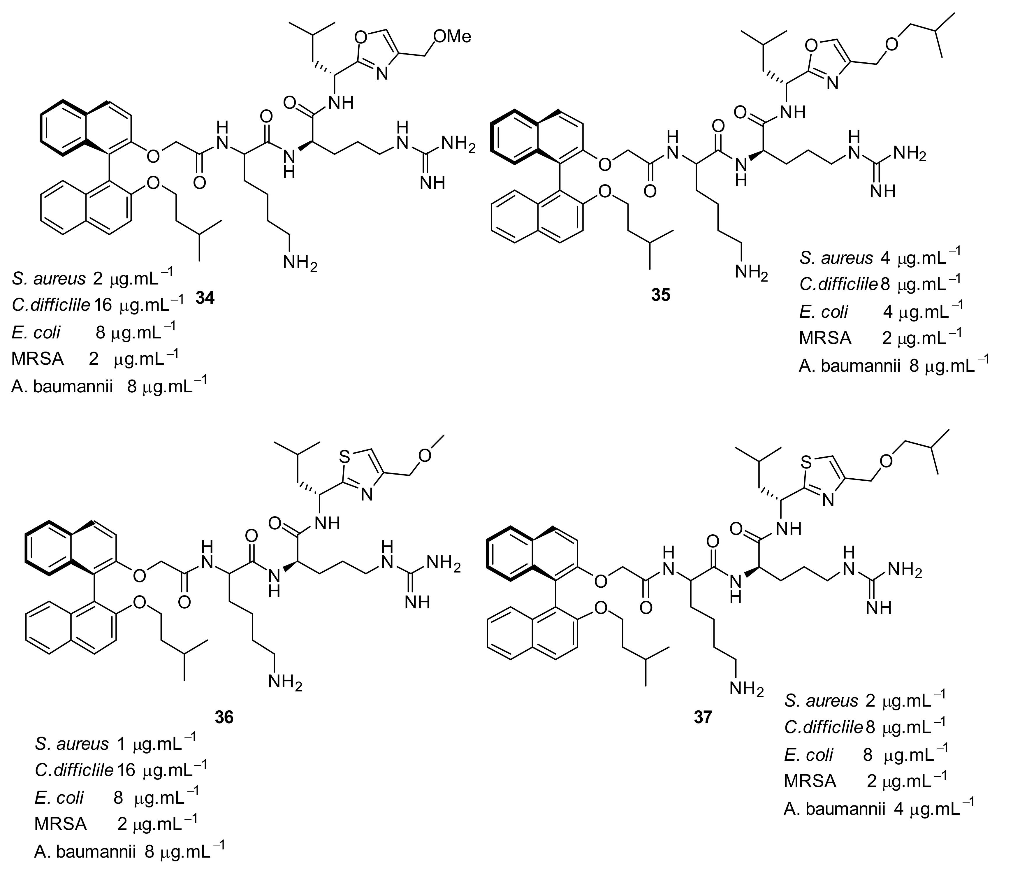
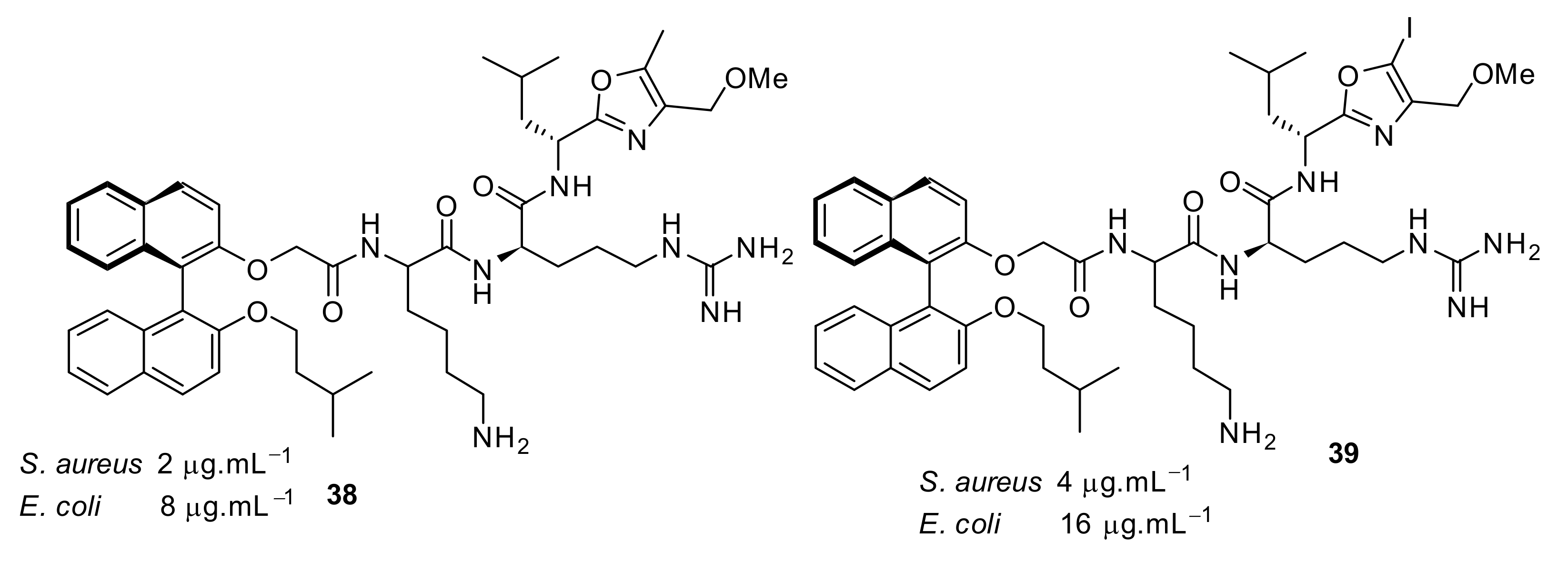
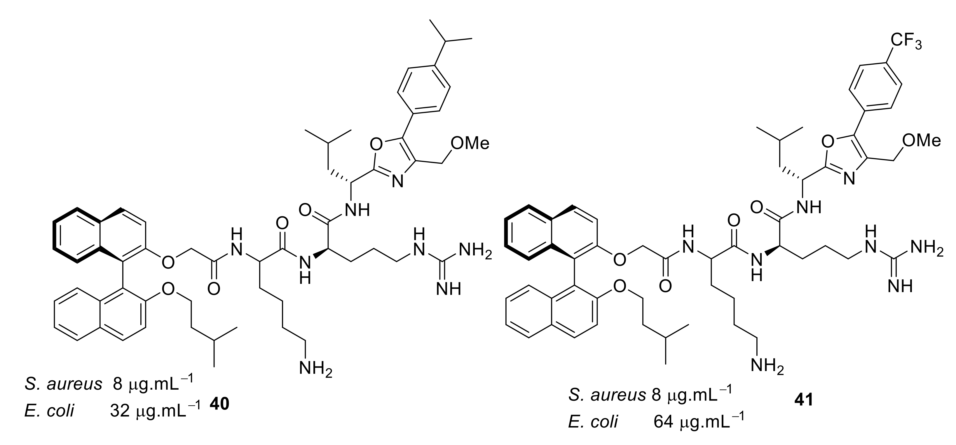
4.1. Binaphthyl Peptidomimetics

The group of Bremner, Keller, and Pyne [79] designed a peptidomimetic to mimic the action of vancomycin. Vancomycin disrupts bacterial cell wall synthesis by inhibiting the formation of the strengthening crosslinks in peptidoglycan, which contains terminal D-alanine [80]. However, in vancomycin-resistant bacteria such as vancomycin-resistant enterococci (VRE), the cross-linking peptide is terminated with D-lactate (L-Lys-D-Ala-D-Lac), which results in reduction in binding by vancomycin and, hence, its inability to prevent cross-linking in the bacterial cell wall.
At first, in order to increase binding interactions, cyclic peptide 12 (Figure 3) based upon a 1,1′-binaphthyl scaffold [79] with tripeptide bridge was investigated. The structure–activity relationship (SAR) was developed by utilizing structural features such as the lysine basic residue for the electrostatic interaction with the carboxy group of terminal D-Ala or D-Lac, a tripeptide for H-bonding interactions with D-Ala and D-Lac of VRE and hydrophobic 1,1′-binaphthyl to control the conformation of peptide unit by employing R or S-enantiomers [79]. The major and minor diastereomer 12 had minimum inhibitory concentrations (MICs) of 17 µg·mL−1 and µg·mL−1, respectively, against Staphylococcus. aureus. However, the hydrogenolysis of a double bond gave compound 13 (Figure 3), which showed MIC against S. aureus of 15 µg·mL−1. The E or the Z olefin in 12 was not important for antibacterial activity [79]. The initial binding studies were done using mass spectrometry. The compound 12 indicated a greater affinity to model vancomycin-resistant bacterial cell wall precursor peptide Gly-Ala-Ala-D-Ala-D-Lac with an unbound to bound ratio of 3.5:1. The cytotoxicity of the compounds were not reported.
In another study, a carbazole system was introduced in place of the binaphthyl hydrophobic moiety to improve the antibacterial activity of compound 12. The carbazole peptide 14 (Figure 4) was synthesised using key ring-closing metathesis to attach different sugar residues through the carbazole NH to mimic the mechanism of action of vancomycin [81]. The carbazole system did not improve the antibacterial activity but gave the same MIC as the binaphthyl system (17 µg·mL−1).
In subsequent structure–activity relationship (SAR) studies, novel carbazole-linked cyclic and acyclic peptides were synthesised [82] with lysine (D and L) and arginine (D and L) being used as the basic groups. The corresponding dihydro derivatives were also synthesised by hydrogenolysis of alkene moiety. The SAR study showed that the extra hydrophobicity provided by N-Boc compound 15 (Figure 5) increased the antibacterial activity by 16-fold compared to compound 16 (Figure 5). The effect of hydrophobicity was consistent with the N-Boc-L-lysine dihydro compound 17 (Figure 5). The basic residue lysine showed better antibacterial activity compared to arginine. However, the antibacterial activities were relatively poor (>250 µg·mL−1) for the fully protected basic residues, which emphasises the importance of basic groups. The antibacterial activity results suggested that L-lysine and a large hydrophobic group were required for maximum antibacterial activity. There was no report about the cytotoxicity and mechanism of action.
Another cyclic peptide based on an indole scaffold was explored to study the effect of a smaller rigid scaffold compared to binaphthyl and carbazole scaffold. The indole-based scaffold 1819 (Figure 6) did not show any antibacterial activity, even at 125 µg·mL−1. This result suggested that the binaphthyl scaffold might contain a degree of flexibility, thus giving it improved antimicrobial function, however the carbazole and indole scaffolds may be quite rigid and so reduce antimicrobial function.
In order to improve the antibacterial activity, a series of cyclic and acyclic peptide compounds were developed with a simple tyrosine system [83] as the hydrophobic group and the cationic residues from lysine or arginine. In this design, the linear chain showed better antibacterial activity than the corresponding macrocyclic compound. The SAR showed that there was no profound effect on antibacterial activity by changing the lysine cationic residue to arginine. However, the linear chain with an arginine residue in the D-configuration of compound 21 (Figure 7) showed good activity against S. aureus with an MIC of 7.8 µg·mL−1 compared to L-configuration of arginine compound 20 with an MIC of 31.5 µg·mL−1. The activity of the peptide mimic could be attributed to the presence of the more hydrophobic Fmoc group compared to the tyrosine residue, which is more isosteric to the binaphthyl ring system. This design showed that macrocyclic tyrosine was not necessary for antibacterial activity. The active compounds were not subjected to cytotoxicity or mode of action studies.
Short cationic peptides using aryl phenylalanine [84] as the hydrophobic group attached with lysine and arginine as cationic groups have been developed. The increase in hydrophobicity with 9-pheneanthrenyl compound 23 (Figure 8) along with two cationic groups increased the antibacterial activity giving an MIC of 8 µg·mL−1 against S. aureus compared to the less hydrophobic O-allyl tyrosine compound 24 (MIC = 125 µg·mL−1; Figure 8). These biological results indicate that two hydrophobic and two cationic groups might be important for antibacterial activity. The active compounds were not subjected to cytotoxicity and mode of action studies.
Based on the results from the tyrosine system, the binaphthyl hydrophobic cores were reinvestigated. In this design [85], peptides were attached at the 2 and 2′ positions of the binaphthyl system, rather than at the 3 and 3′ positions [79]. The SAR was compared with the cyclic and acyclic counterparts. An acyclic peptide compound 25 with lysine showed excellent antibacterial activity (MIC = 4 µg·mL−1) against S. aureus ATCC6538 compared to the cyclic cationic peptide 26 (Figure 9). The acyclic cationic peptide 25 (Figure 9) can be easily prepared compared to the more complex cyclic peptide 26. The active compounds have not been subjected to cytotoxicity and mode of action studies.
The group of Bremner, Keller, and Pyne examined several cyclic peptide scaffolds based on 1,1′-binaphthyl [79], carbazole [81], indole [86], and tyrosine [83], which all contain cationic amino acid residues to interact with the altered peptidoglycan cell wall of vancomycin-resistant S. aureus (VRSE) and vancomycin-resistant enterococci (VRE). Using selective modifications on the 1,1′-binaphthyl [87] hydrophobic scaffold, the Lys-Arg-containing acyclic peptide was found to be effective against S. aureus and with further modification to the end chain with isopentyl ester and oxazole the effective compounds 27 (MIC = 4 µg·mL−1) and 28 (MIC = 4 µg·mL−1) were produced (Figure 10; Table 3). Further modification of the end chain hydrophobicity with cyclohexyl substituent [88] in the dicationic tripeptide [87] showed good activity against several gram-positive strains (Table 3). The incorporation of hydrophobic alkyl ring resulted in good activity against S. aureus and S. epidermidis, but greater variation against enterococcal strains. The conformationally less-restricted diethyl-substituted compound 29 showed better activity against vancomycin-resistant enterococci (VRE) compared to constrained cycloalkyl compound 30 (Figure 10).
The mode of action of compound 27 was studied using cell-wall model peptide sequences of vancomycin-resistant and vancomycin-sensitive S. aureus. Electrospray ionization mass spectrometry (ESI-MS) showed that the vancomycin complexed only with the terminal Ala-sequence, whereas 27 showed 1:1 complex with model peptide sequences together with separate peaks for the individual components. This confirmed that the mode of action of 27 was probably due to inhibition of cell-wall synthesis. The compounds showed bactericidal activity within 2 h at a concentration of 8 µg·mL−1, indicating that these compounds may have dual actions. The observed in vitro activity was taken further into in vivo studies. Compound 28 was dosed systematically in an animal model which measured the bacterial growth of methicillin-resistant S. aureus (MRSA) in the spleens of infected mice and the number of viable bacteria was reduced after four days of treatment. The control (DMSO vehicle) showed 128 cfu per spleen, while for the compound 28, only 11 cfu per spleen was observed. The activity of compound 28 was also established topically by using a mouse nasal decolonization model. A single administration of 5 wt% of compound 28 was as active as 2% mupirocin. Furthermore, compound 28 was stable up to 4 h in human plasma.
Binaphthyl [89] hydrophobic scaffolds with cationic amino group substitutions on both 2,2′-oxy positions were further investigated. The SAR was developed by positioning the amino acids sequentially pendant from one of the naphthyl unit hydrophobic scaffold. The different compounds 31–33 (Figure 11) demonstrated that the terminal hydrophobic steric bulk decreases antibacterial activity and extra length of the cationic side chains had no profound effect on antibacterial activity. It was concluded that the compounds with one side chain on the C2-symmetric scaffold [87] would be sufficient for improved antibacterial activity.
The second generation of binaphthyl C2-symmetirc scaffolds were developed with oxazole and thiazole [90] functionalities at the end chain of a dicationic peptide and showed that this produced the most potent binaphthyl peptidomimetics 3437 (Figure 12) against various bacterial strains.
Increasing the hydrophobicity in the oxazole ring 3841 (Figure 13 and Figure 14) retained antibacterial activity against S. aureus but decreased the activity against Gram-negative bacteria. Antibacterial activity decreased up to 8-fold with phenyl substitution. The active compounds 34 and 35 (Figure 12) exhibited <4% hemolysis of sheep erythrocytes at 5.6 µM. However, at approximately 185 µM, they gave >70% hemolysis. Compounds 34 and 35 were not toxic up to their MIC values. The mode of action of these compounds has not been studied.
Table 3 summarises the best antibacterial compounds derived from binaphthyl series by Bremner, Keller, Pyne et al.

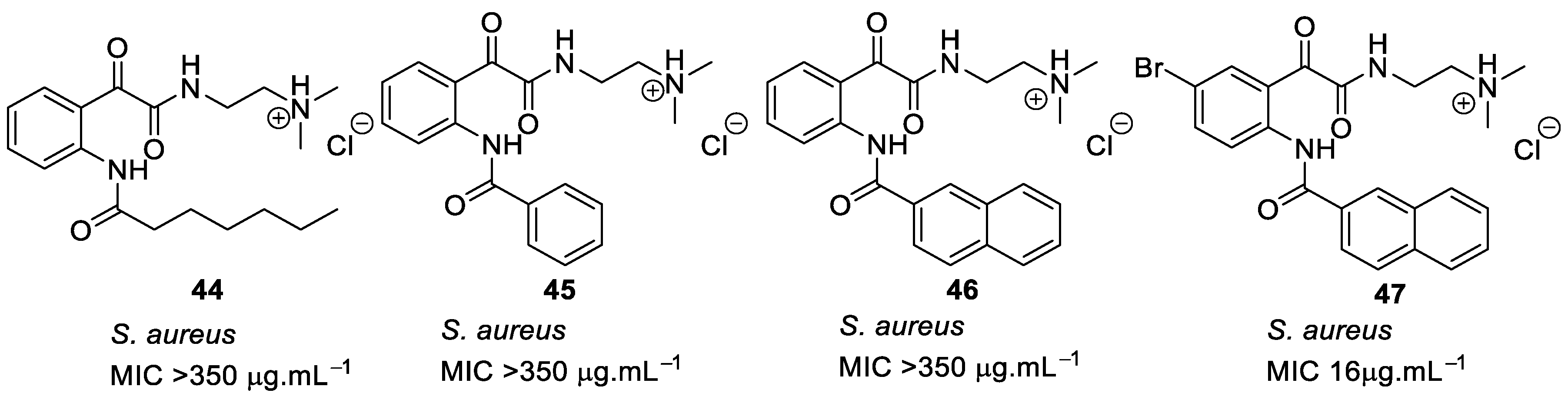
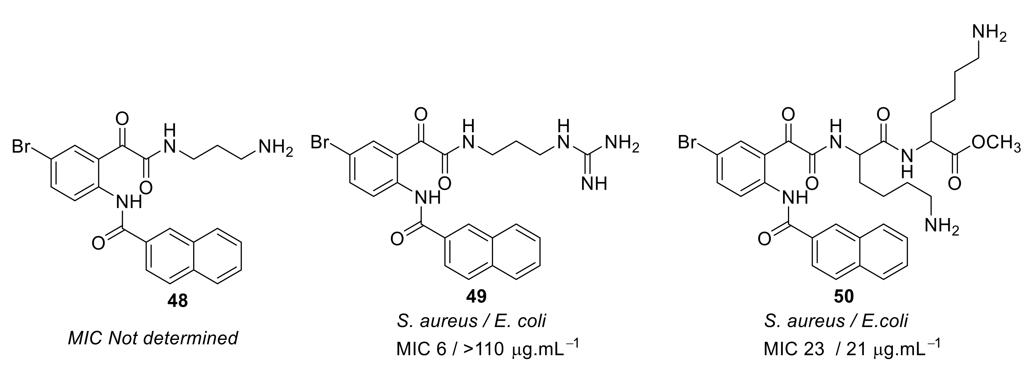
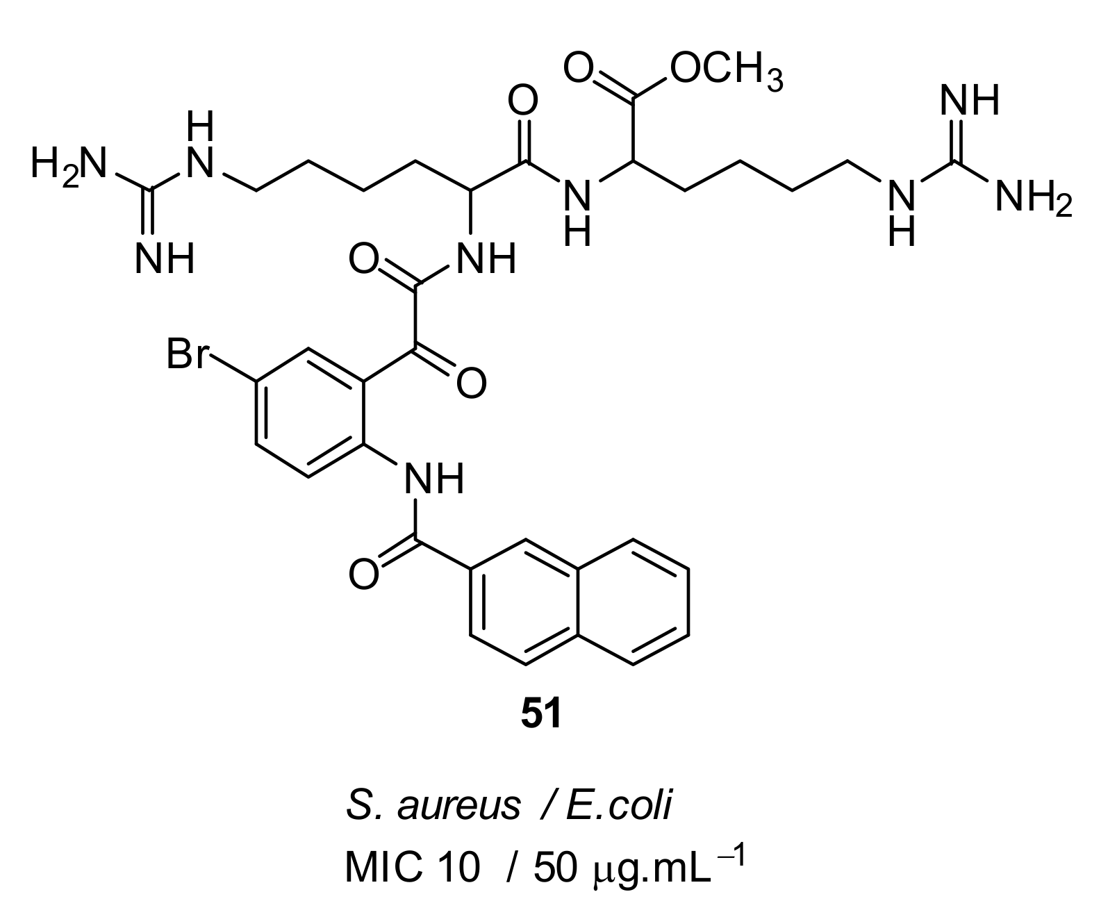
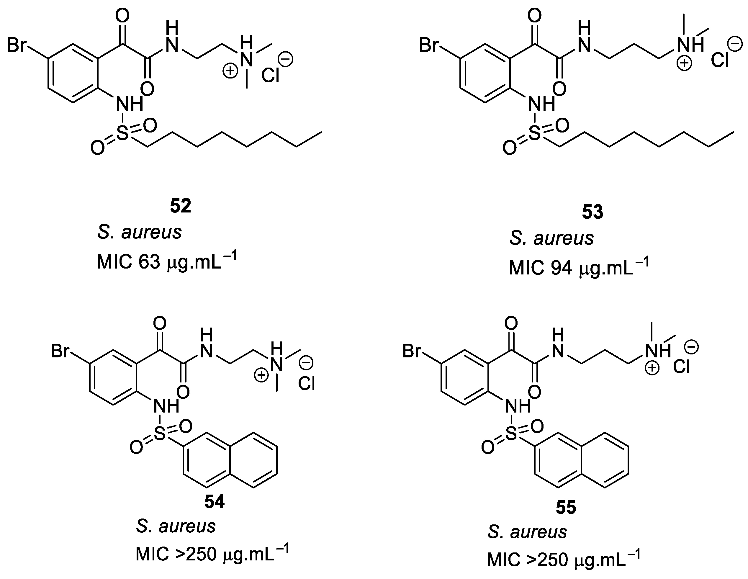
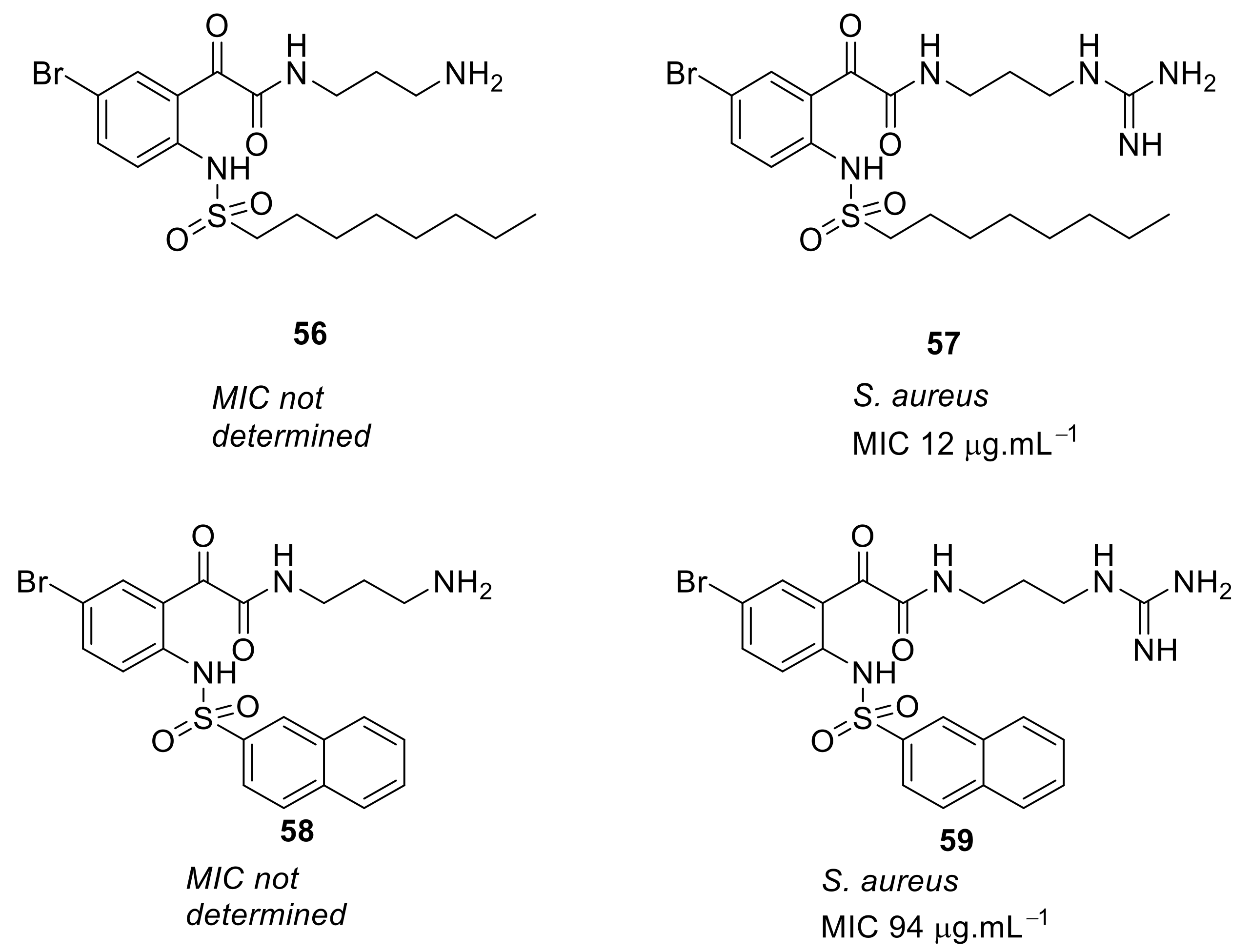
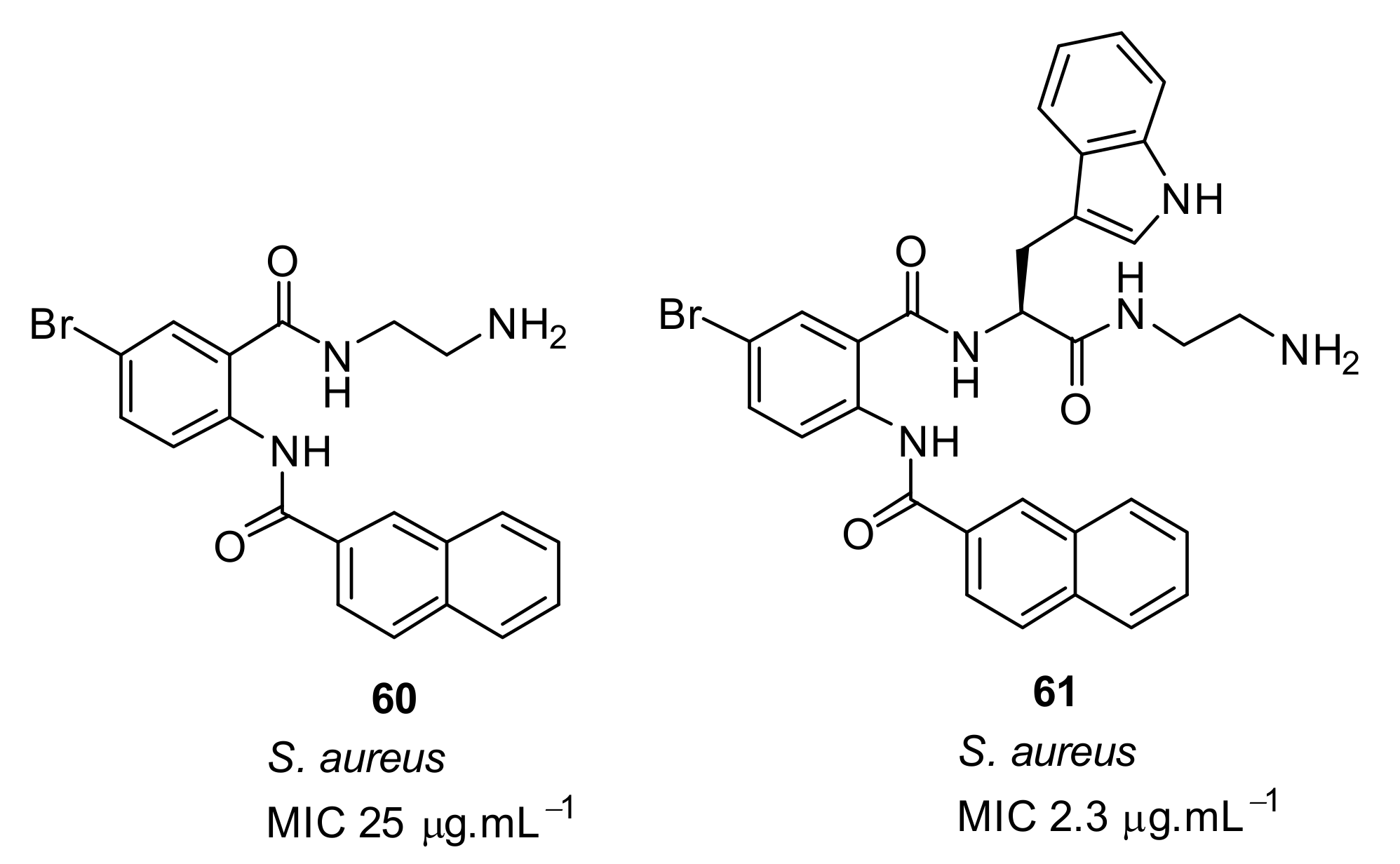
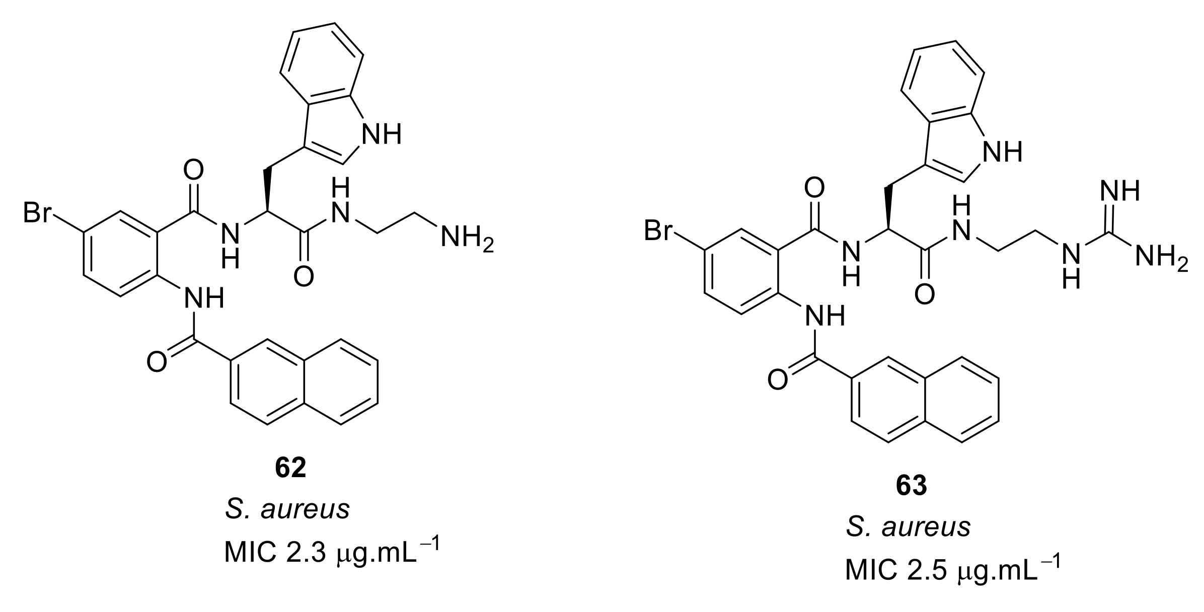
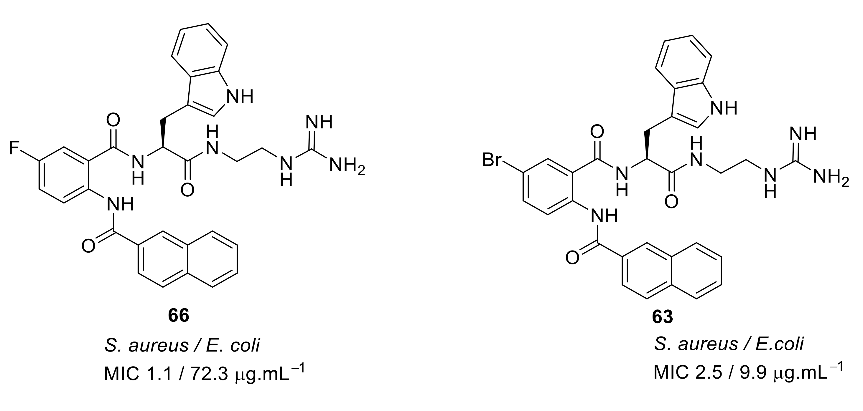
4.2. Glyoxamides and Carboxamide Peptidomimetics

Kumar et al. have developed peptidomimetics based on glyoxamide and carboxamide scaffolds. Mono- and bis-glyoxamide derivatives and dendrimeric peptide mimics were synthesised by a simple and efficient ring-opening reaction of N-acylisatins with a range of amino acids [91,92]. However, the compounds’ antibacterial activity was not reported.
The short peptidomimetic compounds [93] based on novel N-naphthoyl-phenylglyoxamides were designed (Figure 15). The design investigated the antibacterial effect of cationic tertiary and quaternary salts with different hydrophobic groups. SAR studies demonstrated that the hydrophobicity given by naphthoyl was superior to other hydrophobic groups. A bromo-substituted naphthoyl derivative 47 showed greater antibacterial activity compared to Cl, F, and CH3 substituents. Tertiary salts were preferable for antibacterial activity. However, the quaternary ammonium iodide salts (>400 µM) were less toxic to mammalian cells compared to the hydrochloride salts (<40 µM). A tethered bilayer lipid membrane (tBLM) study of the active compound 47 suggested that the antibacterial activity was unlikely to be as a result of membrane rupture.
To improve the antibacterial activity, the cationic groups were replaced with guanidine [94] or lysine to form dipeptide compounds with increased net charge (Figure 16). The guanidine cationic group compound 49 gave better antibacterial activity (MIC = 6 µg·mL−1) compared with the lysine dipeptide compound 50 (MIC = 23 µg·mL−1) against S. aureus. Similar to previous results [93], bromo-substituted compounds showed better antibacterial activity compared to H, Cl, F, and CH3 substituents. The increase in net charge of bromo-substituted compound 50 showed moderate activity against Gram-negative bacteria E. coli (MIC = 21 µg·mL−1).
The dicationic arginine compound 51 (Figure 17) did not improve antibacterial activity over 50. Hence, it is notable that simple guanidine with a naphthoyl hydrophobic group is sufficient for antibacterial activity against S. aureus. The active compounds 49 and 51 showed low toxicity of 750 µM and 224 µM against human cells. Compounds 49 and 51 were not toxic against mammalian cells. The membrane rupture mechanism of action of compound 49 was investigated using tBLMs. The result showed that the compound did not increase the membrane conduction in bacterial-like negatively charged lipid palmitoyl-oleoyl-phosphatidylglycerol (POPG) and so cell lysis may not occur via the formation of membrane pores. The mechanism of cell death with compound 49 has yet to be established.
In a further advancement, the naphthoyl hydrophobic group [93] was replaced with different sulfonyl groups [95], and tertiary ammonium, quaternary ammonium, or guanidinium cationic groups were used to improve the antibacterial activity of glyoxamide compounds (Figure 18). SAR revealed that the bromo-substituted compound 53 with octane sulfonyl hydrophobic group and tertiary ammonium cationic group showed moderate antibacterial activity but was not as effective as the naphthoyl-containing compound 47. When a naphthoyl sulfonyl hydrophobic group replaced the octane sulfonyl group 5455, the antibacterial activity was lost.
The bromo-substituted octane sulfonyl derivative 57 (Figure 19) with a guanidine cationic group had good antibacterial activity against S. aureus (MIC = 12 µg·mL−1), and compounds with other cationic groups were not as effective as those with guanidine. The naphthoyl sulfonyl hydrophobic group 59 (Figure 19) improved the antibacterial activity compared to the respective naphthoyl carboxamide hydrophobic-substituted compound 49. These results suggested that the bromo-substituted scaffold with simple naphthoyl or octane sulfonyl groups and guanidine cationic groups can give improved antimicrobial activity when used in peptidomimetics. The active octane sulfonyl derivative 57 was not toxic (178 µM) against human cells and the mode of action using tBLMs suggests that they may act via membrane disruption.
The glyoxamides with naphthoyl hydrophobic groups in combination with different cationic groups showed good antibacterial activity, and hence a study of the effect of glyoxamides and carboxamides on antibacterial activity was conducted (Figure 20). The carboxamide peptidomimetics [96], in which one tryptophan inserted is in compound 60, dramatically improved the antibacterial activity of compound 61 against gram-positive S. aureus (MIC = 2.3 µg·mL−1)
The SAR was developed using different cationic groups such as amines, guanidines, tertiary, and quaternary ammonium iodides (Figure 21). The halogen-substituted compounds with a naphthoyl hydrophobic group and amine cationic groups showed good antibacterial activities ranging in MICs 4.1–2.3 µg·mL−1. Compound 65 gave the least antibacterial activity but compounds with an amino 62 or 63 with a guanidine group showed excellent antibacterial activity against S. aureus [96].
The fluoro-substituted guanidine derivative 66 (Figure 22) exhibited excellent antibacterial activity against S. aureus but was relatively inactive against E. coli. However, the bromo-substituted compound 63 (Figure 22) was active against S. aureus and E. coli. Although the amine cationic groups showed good antibacterial activity against S. aureus, they showed toxicity towards mammalian cells. However, the bromo-substituted guanidine 63 was not toxic even at 100 µM concentration [96].
Using bacterial cells loaded with a membrane impermeable dye, addition of the compound 63 caused an increase in fluorescence demonstrating the ability of the compounds to permeate the membrane and cause bacterial cell death [96]. Table 4 summarises the list of best antibacterial compounds derived from glyoxamide and carboxamide peptidomimetics.

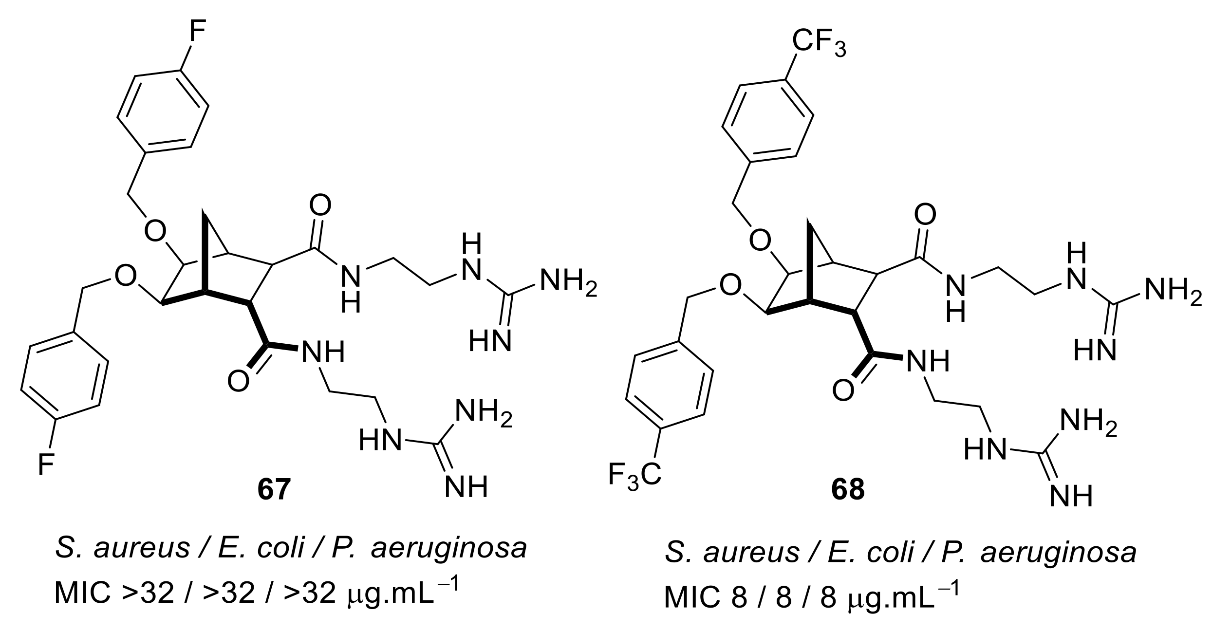
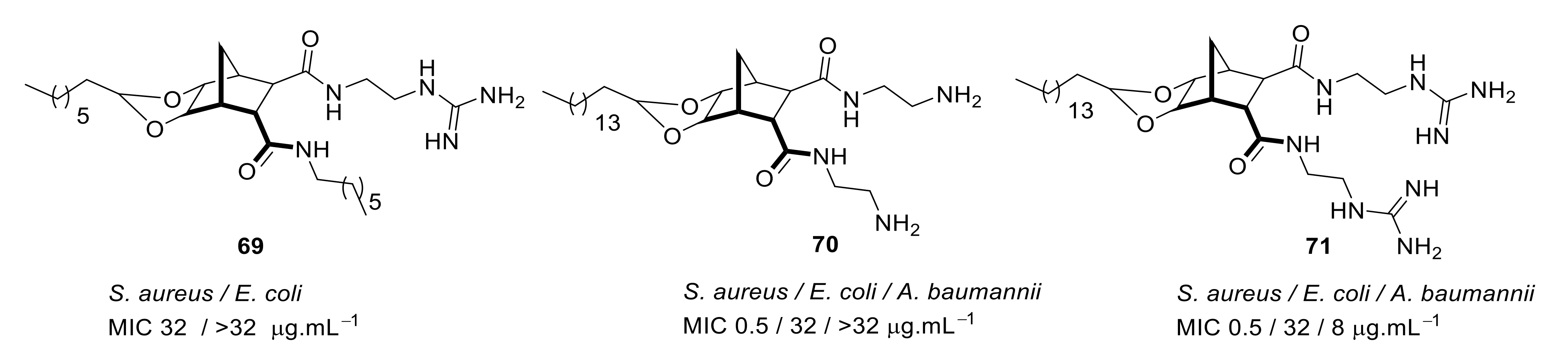
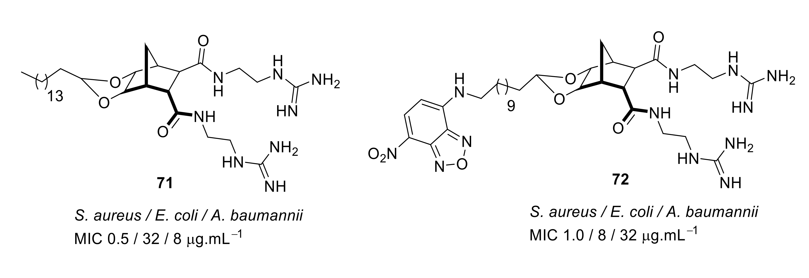
4.3. Norbornane Peptidomimetics

Pfeffer et al. developed peptidomimetic compounds using norbornane scaffolds [97,98], and investigated SAR for norbornane bisether diguanidines [99] (Figure 23). Compound 67 with smaller substitutions fluoro on benzyl ethers were not antibacterial, however the compound 68 with larger substituents such as CF3 on benzyl ether showed good antibacterial activity (MIC = 8 µg·mL−1). Compounds with higher c log P (i.e., higher hydrophobicity) values showed higher antibacterial activity. However, compound 68 at 100 µM concentration exhibited moderate cytotoxicity with 43% cell survival against human embryonic kidney cells (HEK293) after 24 h. Compound 68 was toxic at a higher concentration than its MIC.
The SAR of norbornanes with different hydrophobic and cationic groups, as well as some neutral anion recognition groups such as thioureas and squaramides, was determined [98]. The dicationic guandinium compound 71 (Figure 24) with a hexadecyl hydrophobic group showed MICs of 0.5–2.0 µg·mL−1 against various methicillin-resistant S. aureus (MRSA) and vancomycin-intermediate S. aureus (VISA) strains. The singularly charged norbornane guanidine compound 69 (Figure 24) showed modest activity compared to the dicationic norbornane 71, thus demonstrating the importance of net cationic charge. However, the active compound 71 showed toxicity against human embryonic kidney cells (HEK293) at a concentration (6 µg·mL−1) approximately equal to its MIC (0.5–32 µg·mL−1).
The mechanism of action of the norbornane antibacterial compound was studied by attaching fluorophores [100] (Figure 25). Molecular modelling and fluorescence microscopy studies showed that the compounds 71 and 72 aggregated prior to interacting with cell membranes and fluorescence studies confirmed that these peptidomimetics penetrated into S. aureus. Compound 72 was not toxic against mammalian cells.
Table 5 summarises the best antibacterial compounds derived from norbornane cationic peptidomimetics.

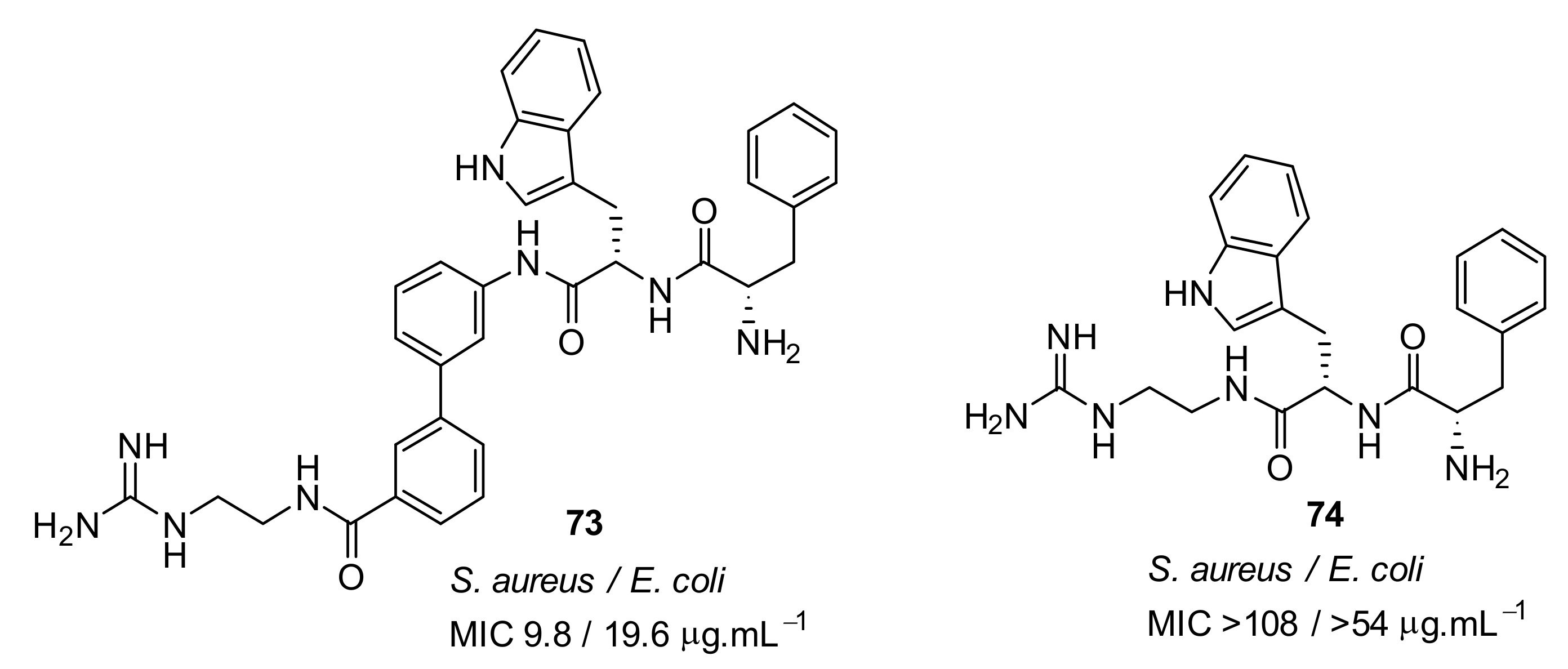
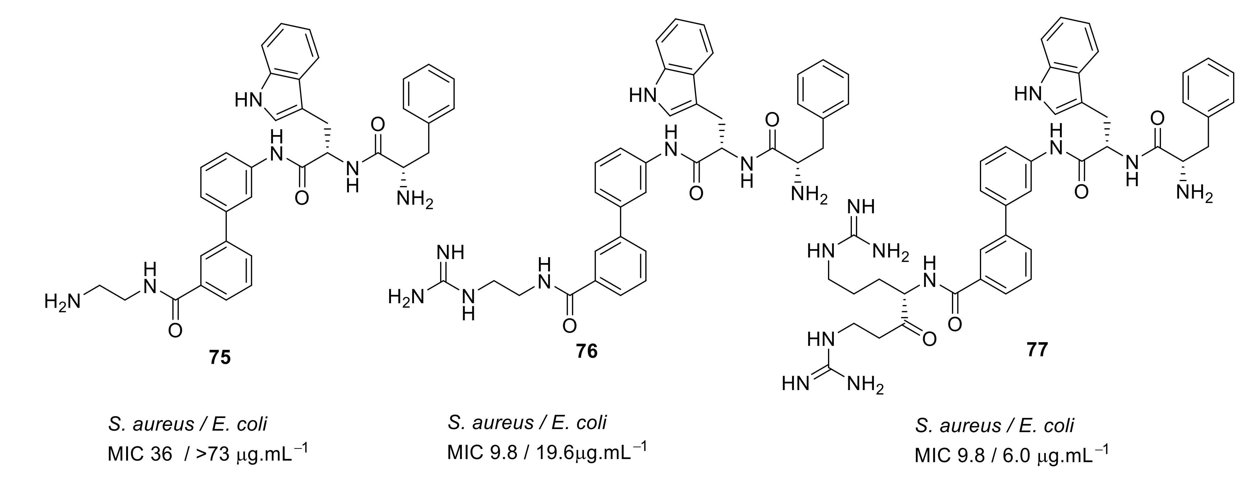
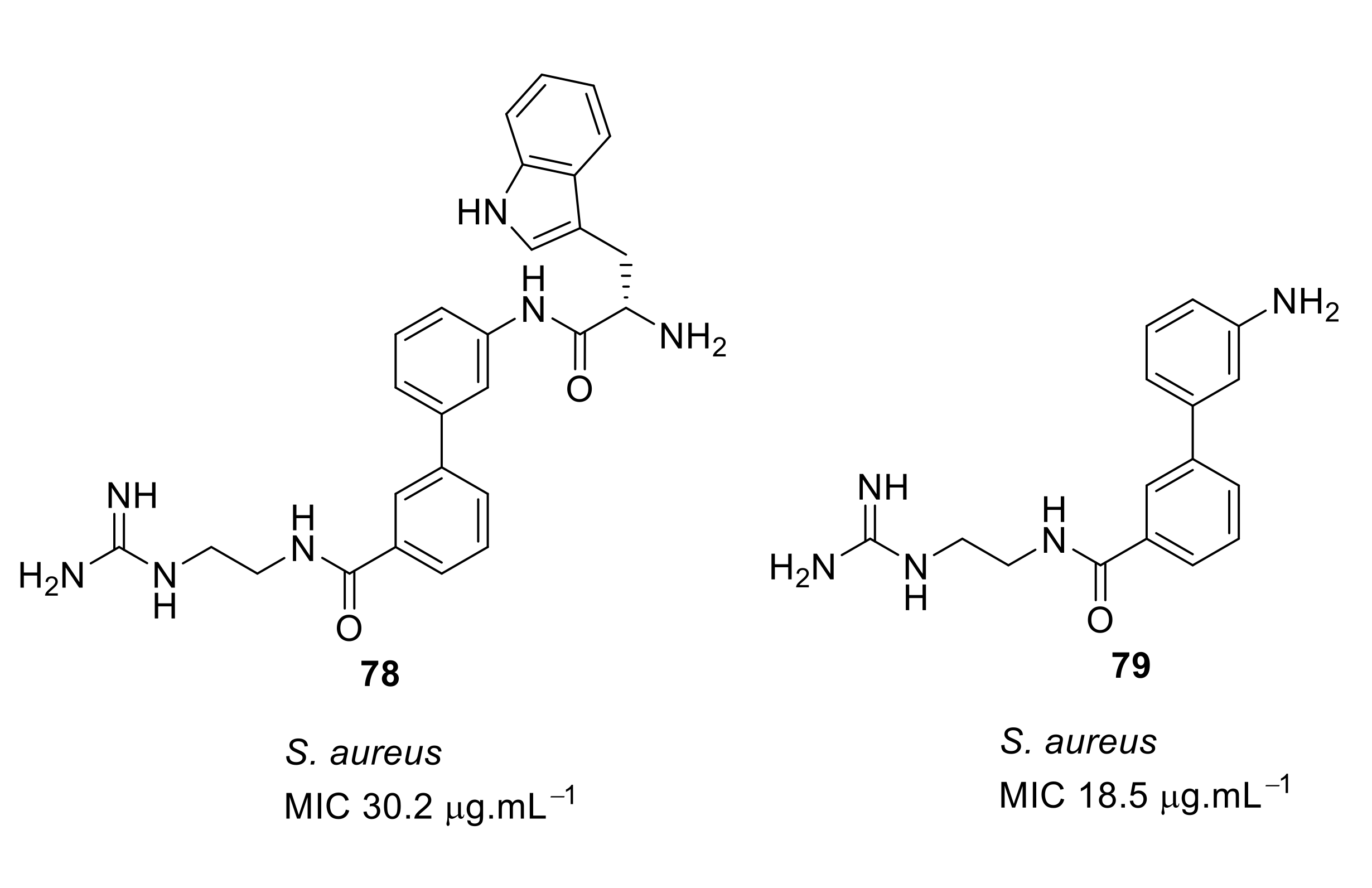
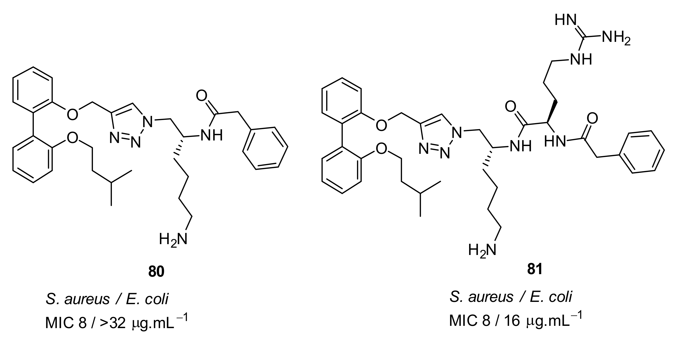
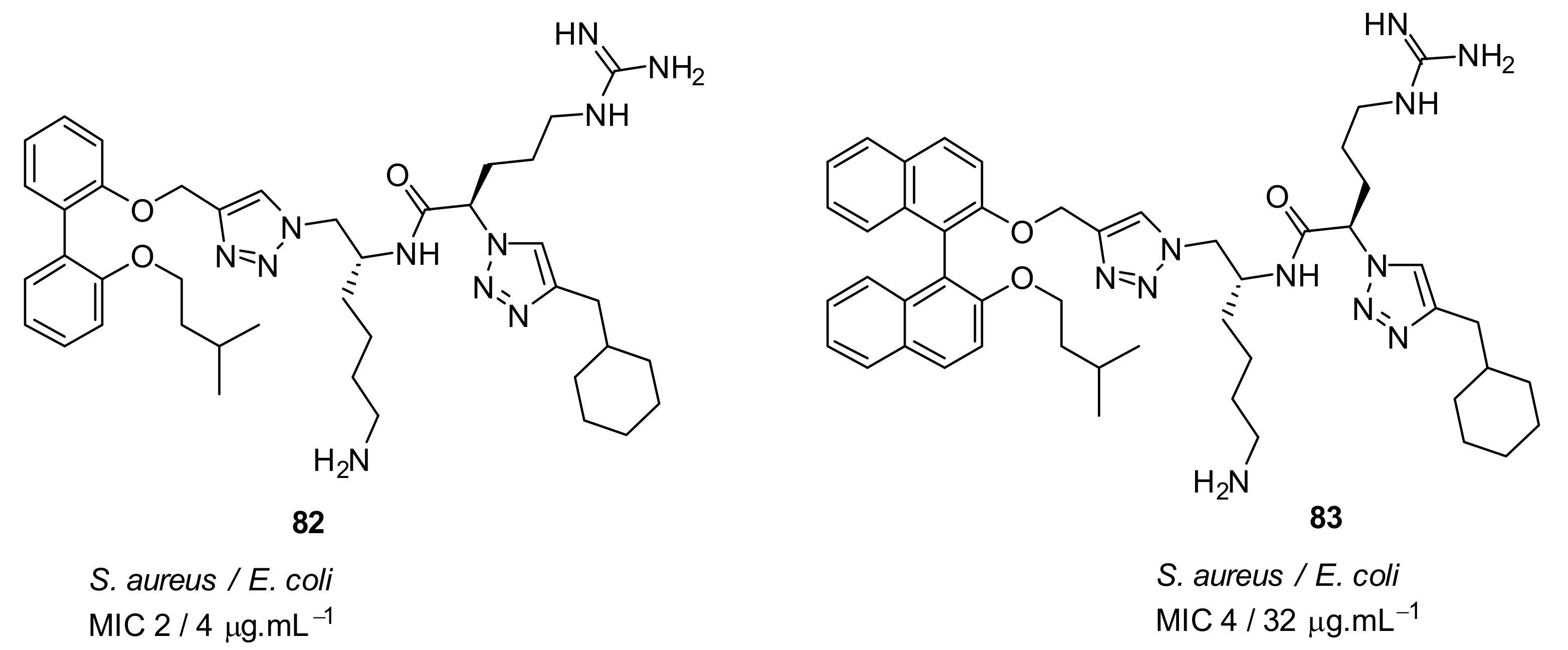
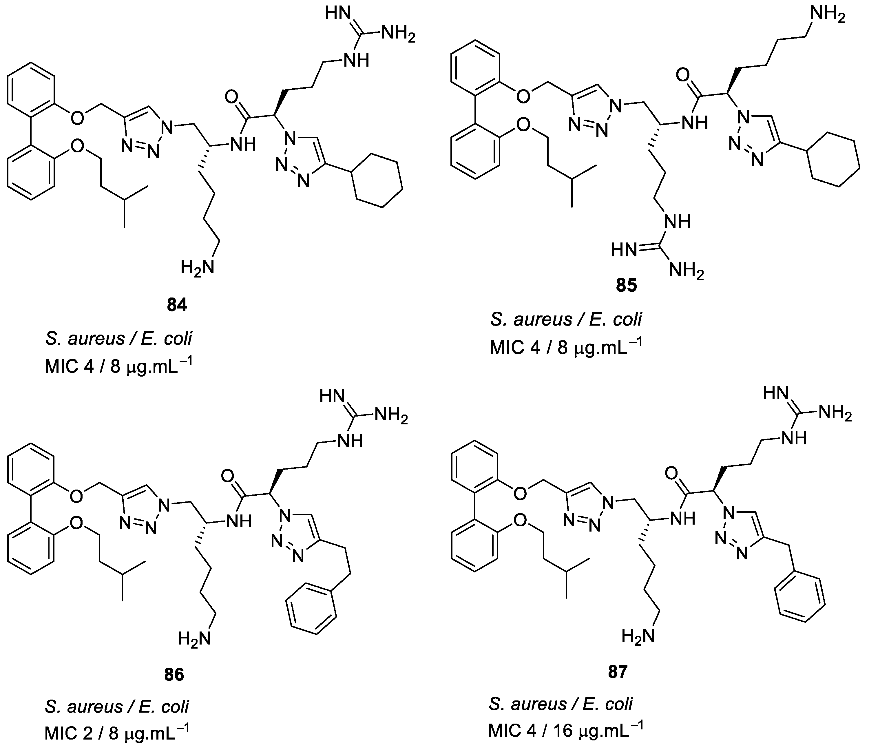
4.4. Biaryl (Biphenyl) Peptidomimetics

Kuppusamy et. al utilised the hydrophobic biphenyl backbone [101] for their peptidomimetic compounds (Figure 26). A SAR was developed by tuning the hydrophobic (Trp/Phe) and cationic groups (Lys/Arg). Upon removing the biphenyl from the active compound 73, the antibacterial activity of the compound 74 was lost. Thus, the amphipathic nature of the compound was shown to be very important for antibacterial activity [101].
A simple diaminoethane substituent 75 (Figure 27) was enough to mimic the lysine cationic residue and the guanidine substituent of 76 (Figure 27) was able to mimic an arginine residue. Increasing the net charge of compound 77 increased the antibacterial activity against gram-negative bacteria. These active compounds 7577 (Figure 27) were not toxic to mammalian cells even at a concentration of 400 µM [101].
The compound’s ability to interact with lipid bilayers was tested using tBLMs and AC electrical impedance spectroscopy [102,103]. The change in conductance of biphenyl compounds 7779 (Figure 28) with negatively charged membranes demonstrated that the effectiveness of these compounds was not so much related to their overall cationic charge but rather to their ability to insert into a lipid bilayer [102,103]. This means that tryptophan played a crucial role in membrane permeability. Using bacterial cells loaded with a membrane impermeable dye, addition of the biphenyl compound 77 caused an increase in fluorescence demonstrating the ability of the compounds to permeate the membrane and cause bacterial cell death.
Tague et al. have developed biphenyl peptidomimetic compounds [104] based on their most active binaphthyl peptidomimetic compound 38, but this compound showed toxicity against mammalian cell. Cytotoxicity is the major problem for the further development of some peptidomimetics into antimicrobial drugs. The SAR was developed by comparing the pharmacophores binaphthyl and biphenyl to improve the cytotoxicity [104].
The antibacterial activity against gram-negative bacteria of biphenyl pharmacophore compound 81 (Figure 29) with net charge of +2 was improved compared to the compound 80 with a net charge of +1 [104].
The incorporation of a second bioisostere triazole in dicationic derivatives of biphenyl 82 gave an 8-fold increase in activity against E. coli compared to binaphthyl derivative 83 [105] (Figure 30).
The antibacterial potency varied with different hydrophobic terminal residues. Flexible hydrophobic groups such as phenethyl 86 and cyclohexyl methyl 82 at the terminus were slightly more potent against S. aureus and E. coli compared to compounds with cyclohexyl 84 and benzyl termini 87 [104] (Figure 31).
Changing the dicationic group Arg-Lys to Lys-Arg of compound 85 to 84 (Figure 31) did not impact the antibacterial efficacy. The cytotoxicity of biphenyl derivative 82 (42.4%), measured as a percentage of hemolysis, was less compared to the binaphthyl compound 83 (88.8%) which was haemolytic at 66 and 58 µM. Compound 82 was not toxic to mammalian cells at concentrations greater than its MIC value (2–4 µg·mL−1).
Using bacterial cells loaded with non-permeable dye (propidium iodide), addition of compound 82 caused a 600% increase in fluorescence confirming that the membrane has been disturbed, and hence the dye enters the cytoplasm and stains the DNA which causes the increase in fluorescence.
Table 6 summarises the best antibacterial compounds derived from biaryl cationic peptidomimetics.

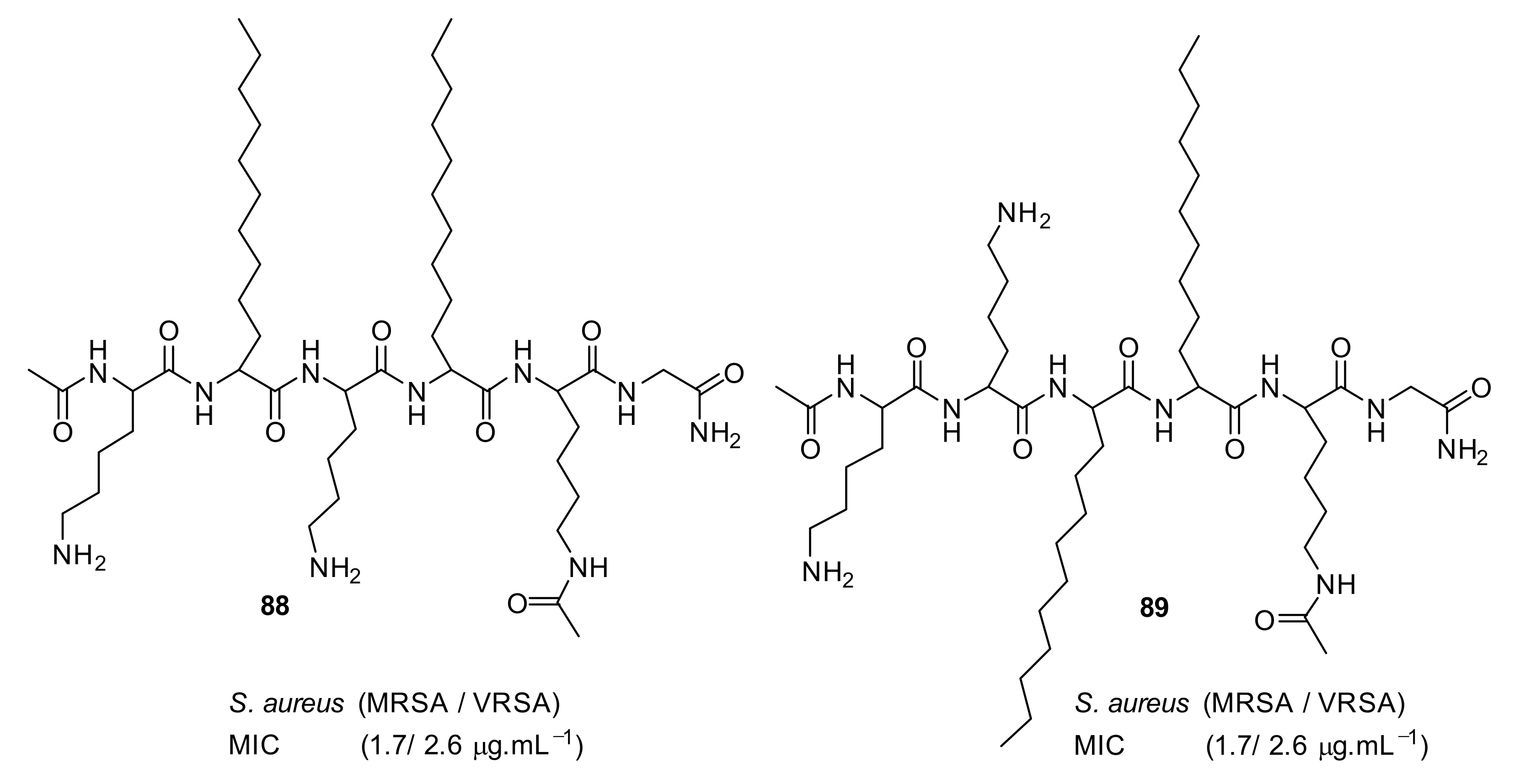
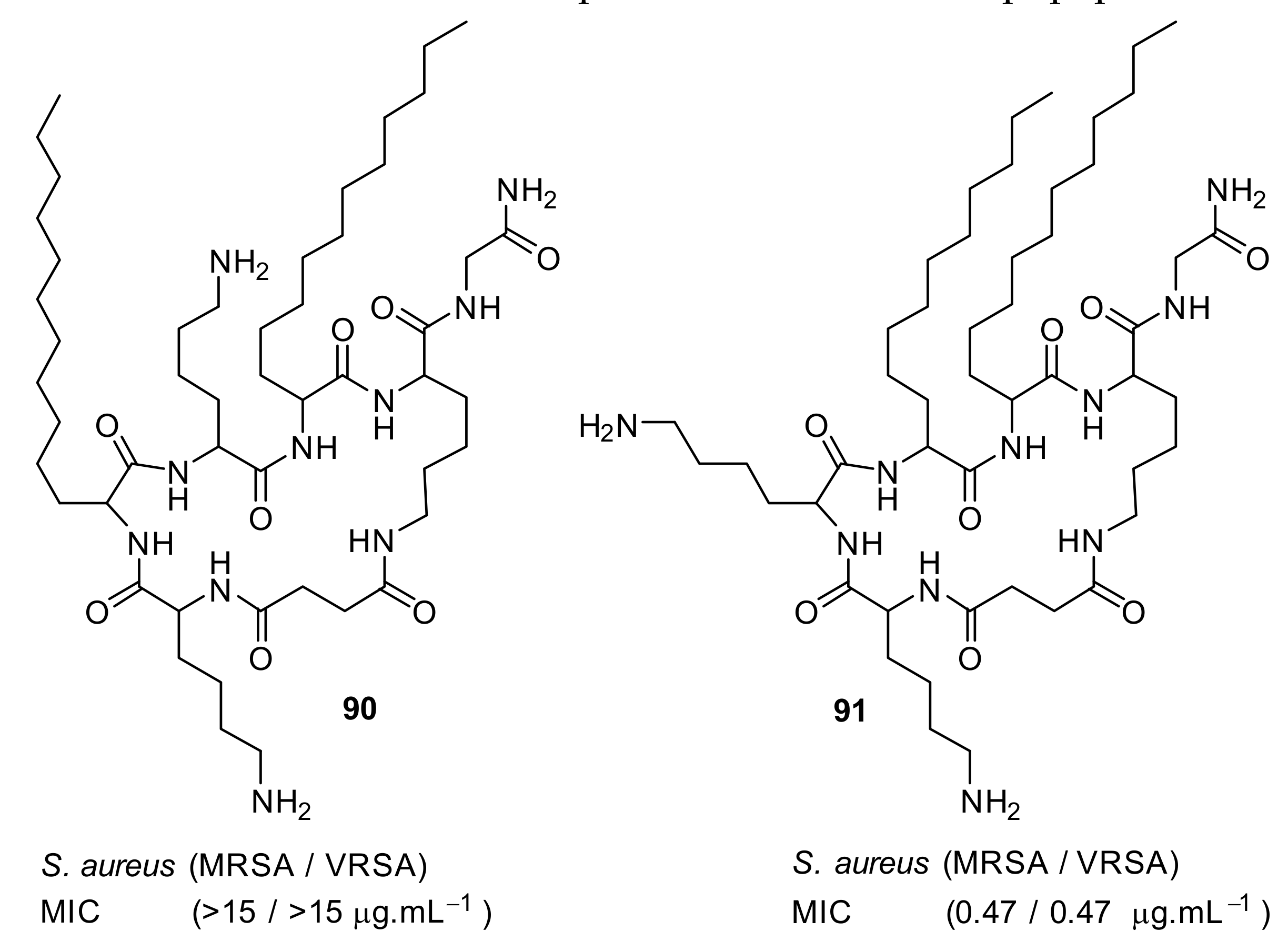
4.5. Short Cationic Lipopeptides

Short cationic peptidomimetic compounds (Figure 32) based on lipopeptides have been developed by Azmi et al. [106] The lipophilicity and the cationic charge of compounds is driven by C12 lipoamino acids (D, L-dodecanoic acid) and lysine residues. The SAR was studied by positioning the lysine residues in lipopeptides.
The lysine and C12-lipoamino acid (C12-LAA) arranged alternatively in compound 88 and the lysine and C12-LAA residues grouped in distinct segments in compound 89 almost showed similar antibacterial activity against several S. aureus strains [106].
The cyclic derivative 90 (Figure 33) with lysine and C12-LAA arranged alternatively lost antibacterial activity compared to the distinct segment pattern of the cyclic derivative 91. The lipopeptides 88 and 91 showed negligible hemolytic activity at 100 µM concentration against mammalian cells. [106] These compounds can self-assemble and form nanoparticles in aqueous environments. Due to this, the cationic charge density is increased, and this may lead to greater bacterial cell selectivity over mammalian cells [106,107]. The mechanism of action was not reported. Table 7 summarises the best antibacterial compounds derived from lipopeptide mimics.

5. Conclusions

The increasing emergence of antibiotic resistance has led to the search for alternatives. Short cationic peptidomimetic compounds can mimic the mechanism of action of AMPs and it is possible to quickly synthesise large quantities of them. The examples shown throughout this review illustrate how different scaffolds may be utilised for development of antibacterials. Generally, all these examples contain the concept of specific hydrophobic and cationic groups. The backbone (hydrophobic) has often been shown to be important for the antibacterial activity; for example the removal of biphenyl group led to the loss of antibacterial activity. Structural diversity has been created by modifying the backbone utilizing bioisosteres and heterocycles. Substitution in the backbone also affects the antibacterial activity. For example, bromo-substituted glyoxamides and carboxamides enhanced the antibacterial activity over other halides. The cationic groups lysine and arginine can be replaced with simple amine and guanidine to retain the antibacterial activity. The amphiphilicity of compounds is important and the overall charge and hydrophobicity must be balanced to achieve optimal activity.
There is evidence that AMPs act by multiple mechanisms, but not all studies have undertaken mechanistic studies of peptidomimetic compounds. AMPs can show synergy with conventional antibiotics, but synergistism of short cationic peptidomimetics remains largely unexplored. Considering the success of brilacidin and LTX-109 in Phase-II clinical trials, this class of compounds with different backbones, unnatural amino acids, can be exploited as potential antibacterials.

Author Contributions

Writing—original draft preparation, R.K., M.W., D.B., and N.K.; writing—review and editing, R.K., M.W., D.B., and N.K.

Funding

This research was funded by Discovery Project from Australian Research Council grant (DP 180100845).

Acknowledgments

Rajesh Kuppusamy is thankful to the University of New South Wales for a Tuition Fee Scholarship (TFS) and to Naresh Kumar for a Living Allowance Scholarship.

Conflicts of Interest

The authors declare no conflict of interest.

References

  1. Lehrer, R.I.; Ganz, T. Antimicrobial peptides in mammalian and insect host defence. Curr. Opin. Immunol. 1999, 11, 23–27. [Google Scholar] [CrossRef]
  2. Brown, K.L.; Hancock, R.E. Cationic host defense (antimicrobial) peptides. Curr. Opin. Immunol. 2006, 18, 24–30. [Google Scholar] [CrossRef] [PubMed]
  3. Bradshaw, J. Cationic antimicrobial peptides: issues for potential clinical use. BioDrugs 2003, 17, 233–240. [Google Scholar] [CrossRef] [PubMed]
  4. Harris, F.; Dennison, S.R.; Phoenix, D.A. Anionic antimicrobial peptides from eukaryotic organisms. Curr. Protein Pept. Sci. 2009, 10, 585–606. [Google Scholar] [CrossRef] [PubMed]
  5. Tossi, A.; Sandri, L.; Giangaspero, A. Amphipathic, α-helical antimicrobial peptides. Biopolymers 2000, 55, 4–30. [Google Scholar] [CrossRef]
  6. Park, C.B.; Yi, K.S.; Matsuzaki, K.; Kim, M.S.; Kim, S.C. Structure–activity analysis of buforin II, a histone H2A-derived antimicrobial peptide: The proline hinge is responsible for the cell-penetrating ability of buforin II. Proc. Natl. Acad. Sci. USA 2000, 97, 8245–8250. [Google Scholar] [CrossRef]
  7. Lay, F.T.; Anderson, M.A. Defensins—Components of the innate immune system in plants. Curr. Protein Pept. Sci. 2005, 6, 85–101. [Google Scholar] [CrossRef] [PubMed]
  8. Matsuzaki, K. Molecular action mechanisms and membrane recognition of membrane-acting antimicrobial peptides. Yakugaku Zasshi 1997, 117, 253–264. [Google Scholar] [CrossRef]
  9. Matsuzaki, K.; Yoneyama, S.; Fujii, N.; Miyajima, K.; Yamada, K.; Kirino, Y.; Anzai, K. Membrane permeabilization mechanisms of a cyclic antimicrobial peptide, tachyplesin I, and its linear analog. Biochemistry 1997, 36, 9799–9806. [Google Scholar] [CrossRef] [PubMed]
  10. Chan, D.I.; Prenner, E.J.; Vogel, H.J. Tryptophan- and arginine-rich antimicrobial peptides: structures and mechanisms of action. Biochim. Biophys. Acta 2006, 1758, 1184–1202. [Google Scholar] [CrossRef]
  11. Oppenheim, F.G.; Xu, T.; McMillian, F.M.; Levitz, S.M.; Diamond, R.D.; Offner, G.D.; Troxler, R.F. Histatins, a novel family of histidine-rich proteins in human parotid secretion. Isolation, characterization, primary structure, and fungistatic effects on Candida albicans. J. Biol. Chem. 1988, 263, 7472–7477. [Google Scholar]
  12. Huq, N.L.; Cross, K.J.; Ung, M.; Myroforidis, H.; Veith, P.D.; Chen, D.; Stanton, D.; He, H.; Ward, B.R.; Reynolds, E.C. A review of the salivary proteome and peptidome and saliva-derived peptide therapeutics. Int. J. Pept. Res. Ther. 2007, 13, 547–564. [Google Scholar] [CrossRef]
  13. Cabiaux, V.; Agerberth, B.; Johansson, J.; Homble, F.; Goormaghtigh, E.; Ruysschaert, J.M. Secondary structure and membrane interaction of PR-39, a Pro+Arg-rich antibacterial peptide. Eur. J. Biochem. 1994, 224, 1019–1027. [Google Scholar] [CrossRef] [PubMed]
  14. Conti, S.; Radicioni, G.; Ciociola, T.; Longhi, R.; Polonelli, L.; Gatti, R.; Cabras, T.; Messana, I.; Castagnola, M.; Vitali, A. Structural and functional studies on a proline-rich peptide isolated from swine saliva endowed with antifungal activity towards Cryptococcus neoformans. Biochim. Biophys. Acta 2013, 1828, 1066–1074. [Google Scholar] [CrossRef] [PubMed]
  15. Xu, R.; Zhao, X.-Y.; Guo, S.; Yang, Q. Effect of loop structure of bovine lactoferricin on Escherichia Coli. AFSE 2018, 2. [Google Scholar] [CrossRef]
  16. Abdel Monaim, S.A.H.; Somboro, A.M.; El-Faham, A.; de la Torre, B.G.; Albericio, F. Bacteria hunt bacteria through an intriguing cyclic peptide. Chem. Med. Chem. 2019, 14, 24–51. [Google Scholar] [CrossRef] [PubMed]
  17. Clifton, L.A.; Skoda, M.W.A.; Le Brun, A.P.; Ciesielski, F.; Kuzmenko, I.; Holt, S.A.; Lakey, J.H. Effect of divalent cation removal on the structure of Gram-negative bacterial outer membrane models. Langmuir 2015, 31, 404–412. [Google Scholar] [CrossRef] [PubMed]
  18. Shai, Y. Mode of action of membrane active antimicrobial peptides. Biopolymers 2002, 66, 236–248. [Google Scholar] [CrossRef] [PubMed]
  19. Mohan, K.V.; Rao, S.S.; Atreya, C.D. Evaluation of antimicrobial peptides as novel bactericidal agents for room temperature-stored platelets. Transfusion 2010, 50, 166–173. [Google Scholar] [CrossRef]
  20. Straus, S.K.; Hancock, R.E. Mode of action of the new antibiotic for Gram-positive pathogens daptomycin: comparison with cationic antimicrobial peptides and lipopeptides. Biochim. Biophys. Acta 2006, 1758, 1215–1223. [Google Scholar] [CrossRef]
  21. Sang, Y.; Blecha, F. Antimicrobial peptides and bacteriocins: alternatives to traditional antibiotics. Anim. Health Res. Rev. 2008, 9, 227–235. [Google Scholar] [CrossRef] [PubMed]
  22. Malanovic, N.; Lohner, K. Gram-positive bacterial cell envelopes: The impact on the activity of antimicrobial peptides. Biochim. Biophys. Acta 2016, 1858, 936–946. [Google Scholar] [CrossRef]
  23. Biswas, R.; Martinez, R.E.; Gohring, N.; Schlag, M.; Josten, M.; Xia, G.; Hegler, F.; Gekeler, C.; Gleske, A.K.; Gotz, F.; et al. Proton-binding capacity of Staphylococcus aureus wall teichoic acid and its role in controlling autolysin activity. PLoS ONE 2012, 7, e41415. [Google Scholar] [CrossRef] [PubMed]
  24. Wu, M.; Maier, E.; Benz, R.; Hancock, R.E. Mechanism of interaction of different classes of cationic antimicrobial peptides with planar bilayers and with the cytoplasmic membrane of Escherichia coli. Biochemistry 1999, 38, 7235–7242. [Google Scholar] [CrossRef]
  25. Mensa, B.; Howell, G.L.; Scott, R.; DeGrado, W.F. Comparative mechanistic studies of brilacidin, daptomycin, and the antimicrobial peptide LL16. Antimicrob. Agents Chemother. 2014, 58, 5136–5145. [Google Scholar] [CrossRef] [PubMed]
  26. Shai, Y. Mechanism of the binding, insertion and destabilization of phospholipid bilayer membranes by alpha-helical antimicrobial and cell non-selective membrane-lytic peptides. Biochim. Biophys. Acta 1999, 1462, 55–70. [Google Scholar] [CrossRef]
  27. Toke, O. Antimicrobial peptides: New candidates in the fight against bacterial infections. Biopolymers 2005, 80, 717–735. [Google Scholar] [CrossRef] [PubMed]
  28. Brogden, K.A. Antimicrobial peptides: pore formers or metabolic inhibitors in bacteria? Nat. Rev. Microbiol. 2005, 3, 238–250. [Google Scholar] [CrossRef]
  29. Zhang, L.; Rozek, A.; Hancock, R.E. Interaction of cationic antimicrobial peptides with model membranes. J. Biol. Chem. 2001, 276, 35714–35722. [Google Scholar] [CrossRef]
  30. Shimazaki, K.; Tazume, T.; Uji, K.; Tanaka, M.; Kumura, H.; Mikawa, K.; Shimo-Oka, T. Properties of a heparin-binding peptide derived from bovine lactoferrin. J. Dairy Sci. 1998, 81, 2841–2849. [Google Scholar] [CrossRef]
  31. Epand, R.M.; Shai, Y.; Segrest, J.P.; Anantharamaiah, G.M. Mechanisms for the modulation of membrane bilayer properties by amphipathic helical peptides. Biopolymers 1995, 37, 319–338. [Google Scholar] [CrossRef] [PubMed]
  32. Spaar, A.; Munster, C.; Salditt, T. Conformation of peptides in lipid membranes studied by x-ray grazing incidence scattering. Biophys. J. 2004, 87, 396–407. [Google Scholar] [CrossRef]
  33. Vad, B.S.; Bertelsen, K.; Johansen, C.H.; Pedersen, J.M.; Skrydstrup, T.; Nielsen, N.C.; Otzen, D.E. Pardaxin permeabilizes vesicles more efficiently by pore formation than by disruption. Biophys. J. 2010, 98, 576–585. [Google Scholar] [CrossRef] [PubMed]
  34. Mojsoska, B.; Jenssen, H. Peptides and peptidomimetics for antimicrobial drug design. Pharmaceuticals 2015, 8, 366–415. [Google Scholar] [CrossRef] [PubMed]
  35. Pouny, Y.; Rapaport, D.; Mor, A.; Nicolas, P.; Shai, Y. Interaction of antimicrobial dermaseptin and its fluorescently labeled analogs with phospholipid-membranes. Biochemistry 1992, 31, 12416–12423. [Google Scholar] [CrossRef]
  36. Bechinger, B. The structure, dynamics and orientation of antimicrobial peptides in membranes by multidimensional solid-state NMR spectroscopy. Biochim. Biophys. Acta 1999, 1462, 157–183. [Google Scholar] [CrossRef]
  37. Sitaram, N.; Nagaraj, R. Interaction of antimicrobial peptides with biological and model membranes: structural and charge requirements for activity. Biochim. Biophys. Acta 1999, 1462, 29–54. [Google Scholar] [CrossRef]
  38. Fernandez, D.I.; Le Brun, A.P.; Whitwell, T.C.; Sani, M.A.; James, M.; Separovic, F. The antimicrobial peptide aurein 1.2 disrupts model membranes via the carpet mechanism. Phys. Chem. Chem. Phys. 2012, 14, 15739–15751. [Google Scholar] [CrossRef]
  39. Hancock, R.E.; Chapple, D.S. Peptide antibiotics. Antimicrob. Agents Chemother. 1999, 43, 1317–1323. [Google Scholar] [CrossRef]
  40. Yang, L.; Harroun, T.A.; Weiss, T.M.; Ding, L.; Huang, H.W. Barrel-stave model or toroidal model? A case study on melittin pores. Biophys. J. 2001, 81, 1475–1485. [Google Scholar] [CrossRef]
  41. Lee, T.H.; Hall, K.N.; Aguilar, M.I. Antimicrobial peptide structure and mechanism of action: A focus on the role of membrane structure. Curr. Top. Med. Chem. 2016, 16, 25–39. [Google Scholar] [CrossRef] [PubMed]
  42. Cheng, J.T.; Hale, J.D.; Elliot, M.; Hancock, R.E.; Straus, S.K. Effect of membrane composition on antimicrobial peptides aurein 2.2 and 2.3 from Australian southern bell frogs. Biophys. J. 2009, 96, 552–565. [Google Scholar] [CrossRef]
  43. Guan, Q.; Huang, S.; Jin, Y.; Campagne, R.; Alezra, V.; Wan, Y. Recent advances in the exploration of therapeutic analogues of Gramicidin S, an old but still potent antimicrobial peptide. J. Med. Chem. 2019. [Google Scholar] [CrossRef] [PubMed]
  44. Katsu, T.; Imamura, T.; Komagoe, K.; Masuda, K.; Mizushima, T. Simultaneous measurements of K+ and calcein release from liposomes and the determination of pore size formed in a membrane. Anal. Sci. 2007, 23, 517–522. [Google Scholar] [CrossRef] [PubMed]
  45. Wenzel, M.; Rautenbach, M.; Vosloo, J.A.; Siersma, T.; Aisenbrey, C.H.M.; Zaitseva, E.; Laubscher, W.E.; van Rensburg, W.; Behrends, J.C.; Bechinger, B.; et al. The multifaceted antibacterial mechanisms of the pioneering peptide antibiotics tyrocidine and gramicidin S. mBio 2018, 9, e00802–e00818. [Google Scholar] [CrossRef]
  46. Li, J.; Koh, J.J.; Liu, S.; Lakshminarayanan, R.; Verma, C.S.; Beuerman, R.W. Membrane active antimicrobial peptides: Translating mechanistic insights to design. Front. Neurosci. 2017, 11, 73. [Google Scholar] [CrossRef]
  47. Lewis, L.A.; Choudhury, B.; Balthazar, J.T.; Martin, L.E.; Ram, S.; Rice, P.A.; Stephens, D.S.; Carlson, R.; Shafer, W.M. Phosphoethanolamine substitution of lipid A and resistance of Neisseria gonorrhoeae to cationic antimicrobial peptides and complement-mediated killing by normal human serum. Infect. Immun. 2009, 77, 1112–1120. [Google Scholar] [CrossRef] [PubMed]
  48. Gunn, J.S. Bacterial modification of LPS and resistance to antimicrobial peptides. J. Endotoxin Res. 2001, 7, 57–62. [Google Scholar] [CrossRef] [PubMed]
  49. Guo, L.; Lim, K.B.; Poduje, C.M.; Daniel, M.; Gunn, J.S.; Hackett, M.; Miller, S.I. Lipid A acylation and bacterial resistance against vertebrate antimicrobial peptides. Cell 1998, 95, 189–198. [Google Scholar] [CrossRef]
  50. Guina, T.; Yi, E.C.; Wang, H.; Hackett, M.; Miller, S.I. A PhoP-regulated outer membrane protease of Salmonella enterica serovar typhimurium promotes resistance to alpha-helical antimicrobial peptides. J. Bacteriol. 2000, 182, 4077–4086. [Google Scholar] [CrossRef]
  51. Shafer, W.M.; Qu, X.D.; Waring, A.J.; Lehrer, R.I. Modulation of Neisseria gonorrhoeae susceptibility to vertebrate antibacterial peptides due to a, member of the resistance/nodulation/division efflux pump family. Proc. Natl. Acad. Sci. USA 1998, 95, 1829–1833. [Google Scholar] [CrossRef] [PubMed]
  52. Del Castillo, F.J.; del Castillo, I.; Moreno, F. Construction and characterization of mutations at codon 751 of the Escherichia coli gyrB gene that confer resistance to the antimicrobial peptide microcin B17 and alter the activity of DNA gyrase. J. Bacteriol. 2001, 183, 2137–2140. [Google Scholar] [CrossRef] [PubMed]
  53. Friedrich, C.; Scott, M.G.; Karunaratne, N.; Yan, H.; Hancock, R.E. Salt-resistant alpha-helical cationic antimicrobial peptides. Antimicrob. Agents Chemother. 1999, 43, 1542–1548. [Google Scholar] [CrossRef]
  54. Yeaman, M.R.; Bayer, A.S.; Koo, S.P.; Foss, W.; Sullam, P.M. Platelet microbicidal proteins and neutrophil defensin disrupt the Staphylococcus aureus cytoplasmic membrane by distinct mechanisms of action. J. Clin. Investig. 1998, 101, 178–187. [Google Scholar] [CrossRef] [PubMed]
  55. Rabin, N.; Zheng, Y.; Opoku-Temeng, C.; Du, Y.; Bonsu, E.; Sintim, H.O. Biofilm formation mechanisms and targets for developing antibiofilm agents. Future Med. Chem. 2015, 7. [Google Scholar] [CrossRef]
  56. Van Hoek, A.H.; Mevius, D.; Guerra, B.; Mullany, P.; Roberts, A.P.; Aarts, H.J. Acquired antibiotic resistance genes: an overview. Front. Microbiol. 2011, 2, 203. [Google Scholar] [CrossRef] [PubMed]
  57. Liu, Y.Y.; Wang, Y.; Walsh, T.R.; Yi, L.X.; Zhang, R.; Spencer, J.; Doi, Y.; Tian, G.; Dong, B.; Huang, X.; et al. Emergence of plasmid-mediated colistin resistance mechanism MCR-1 in animals and human beings in China: a microbiological and molecular biological study. Lancet Infect. Dis. 2016, 16, 161–168. [Google Scholar] [CrossRef]
  58. Liu, H.; Zhu, B.; Liang, B.; Xu, X.; Qiu, S.; Jia, L.; Li, P.; Yang, L.; Li, Y.; Xiang, Y.; et al. A novel mcr-1 variant carried by an IncI2-type plasmid identified from a multidrug resistant enterotoxigenic Escherichia coli. Front. Microbiol. 2018, 9, 815. [Google Scholar] [CrossRef]
  59. Dobias, J.; Poirel, L.; Nordmann, P. Cross-resistance to human cationic antimicrobial peptides and to polymyxins mediated by the plasmid-encoded MCR-1? Clin. Microbiol. Infect. 2017, 23. [Google Scholar] [CrossRef]
  60. Yeung, A.T.; Gellatly, S.L.; Hancock, R.E. Multifunctional cationic host defence peptides and their clinical applications. Cell Mol. Life Sci. 2011, 68, 2161–2176. [Google Scholar] [CrossRef]
  61. Yang, Q.; Li, M.; Spiller, O.B.; Andrey, D.O.; Hinchliffe, P.; Li, H.; MacLean, C.; Niumsup, P.; Powell, L.; Pritchard, M.; et al. Balancing mcr-1 expression and bacterial survival is a delicate equilibrium between essential cellular defence mechanisms. Nat. Commun. 2017, 8, 2054. [Google Scholar] [CrossRef] [PubMed]
  62. Andres, E. Cationic antimicrobial peptides in clinical development, with special focus on thanatin and heliomicin. Eur J. Clin. Microbiol. Infect. Dis 2012, 31, 881–888. [Google Scholar] [CrossRef] [PubMed]
  63. Ciumac, D.; Gong, H.; Hu, X.; Lu, J.R. Membrane targeting cationic antimicrobial peptides. J. Colloid Interface Sci. 2019, 537, 163–185. [Google Scholar] [CrossRef] [PubMed]
  64. Huttner, B.; Jones, M.; Rubin, M.A.; Neuhauser, M.M.; Gundlapalli, A.; Samore, M. Drugs of last resort? The use of polymyxins and tigecycline at US Veterans affairs medical centers, 2005-2010. PLoS ONE 2012, 7, e36649. [Google Scholar] [CrossRef]
  65. Gai, Z.; Samodelov, S.L.; Kullak-Ublick, G.A.; Visentin, M. Molecular mechanisms of colistin-induced nephrotoxicity. Molecules 2019, 24, 653. [Google Scholar] [CrossRef]
  66. Molchanova, N.; Hansen, P.R.; Franzyk, H. Advances in development of antimicrobial peptidomimetics as potential drugs. Molecules 2017, 22, 1430. [Google Scholar] [CrossRef]
  67. Ghosh, C.; Haldar, J. Membrane-active small molecules: Designs inspired by antimicrobial peptides. Chem. Med. Chem. 2015, 10, 1606–1624. [Google Scholar] [CrossRef]
  68. Sarkar, P.; Yarlagadda, V.; Ghosh, C.; Haldar, J. A review on cell wall synthesis inhibitors with an emphasis on glycopeptide antibiotics. Medchemcomm 2017, 8, 516–533. [Google Scholar] [CrossRef]
  69. Ghosh, C.; Sarkar, P.; Issa, R.; Haldar, J. Alternatives to conventional antibiotics in the era of antimicrobial resistance. Trends Microbiol. 2019, 27, 323–338. [Google Scholar] [CrossRef]
  70. Isaksson, J.; Brandsdal, B.O.; Engqvist, M.; Flaten, G.E.; Svendsen, J.S.; Stensen, W. A synthetic antimicrobial peptidomimetic (LTX 109): stereochemical impact on membrane disruption. J. Med. Chem. 2011, 54, 5786–5795. [Google Scholar] [CrossRef]
  71. Choi, S.; Isaacs, A.; Clements, D.; Liu, D.; Kim, H.; Scott, R.W.; Winkler, J.D.; DeGrado, W.F. De novo design and in vivo activity of conformationally restrained antimicrobial arylamide foldamers. Proc. Natl. Acad. Sci. USA 2009, 106, 6968–6973. [Google Scholar] [CrossRef]
  72. Koh, J.J.; Lin, S.; Aung, T.T.; Lim, F.; Zou, H.; Bai, Y.; Li, J.; Lin, H.; Pang, L.M.; Koh, W.L.; et al. Amino acid modified xanthone derivatives: novel, highly promising membrane-active antimicrobials for multidrug-resistant Gram-positive bacterial infections. J. Med. Chem. 2015, 58, 739–752. [Google Scholar] [CrossRef]
  73. Bucki, R.; Niemirowicz, K.; Wnorowska, U.; Byfield, F.J.; Piktel, E.; Watek, M.; Janmey, P.A.; Savage, P.B. Bactericidal activity of ceragenin CSA-13 in cell culture and in an animal model of peritoneal infection. Antimicrob. Agents Chemother. 2015, 59, 6274–6282. [Google Scholar] [CrossRef]
  74. Konai, M.M.; Samaddar, S.; Bocchinfuso, G.; Santucci, V.; Stella, L.; Haldar, J. Selectively targeting bacteria by tuning the molecular design of membrane-active peptidomimetic amphiphiles. Chem. Commun. (Camb) 2018, 54, 4943–4946. [Google Scholar] [CrossRef]
  75. Gunasekaran, P.; Rajasekaran, G.; Han, E.H.; Chung, Y.H.; Choi, Y.J.; Yang, Y.J.; Lee, J.E.; Kim, H.N.; Lee, K.; Kim, J.S.; et al. Cationic amphipathic triazines with potent anti-bacterial, anti-inflammatory and anti-atopic dermatitis properties. Sci. Rep. 2019, 9, 1292. [Google Scholar] [CrossRef]
  76. Ahn, M.; Gunasekaran, P.; Rajasekaran, G.; Kim, E.Y.; Lee, S.J.; Bang, G.; Cho, K.; Hyun, J.K.; Lee, H.J.; Jeon, Y.H.; et al. Pyrazole derived ultra-short antimicrobial peptidomimetics with potent anti-biofilm activity. Eur. J. Med. Chem. 2017, 125, 551–564. [Google Scholar] [CrossRef]
  77. Vooturi, S.K.; Dewal, M.B.; Firestine, S.M. Examination of a synthetic benzophenone membrane-targeted antibiotic. Org. Biomol. Chem. 2011, 9, 6367–6372. [Google Scholar] [CrossRef] [PubMed]
  78. Teng, P.; Huo, D.; Nimmagadda, A.; Wu, J.; She, F.; Su, M.; Lin, X.; Yan, J.; Cao, A.; Xi, C.; et al. Small antimicrobial agents based on acylated reduced amide scaffold. J. Med. Chem. 2016, 59, 7877–7887. [Google Scholar] [CrossRef]
  79. Bremner, J.B.; Coates, J.A.; Coghlan, D.R.; David, D.M.; Keller, P.A.; Pyne, S.G. The synthesis of a novel binaphthyl-based cyclic peptoid with anti-bacterial activity. New J. Chem. 2002, 26, 1549–1551. [Google Scholar] [CrossRef]
  80. Williams, D.H.; Bardsley, B. The vancomycin group of antibiotics and the fight against resistant bacteria. Angew. Chem. Int. Ed. Engl. 1999, 38, 1172–1193. [Google Scholar] [CrossRef]
  81. Bremner, J.B.; Coates, J.A.; Keller, P.A.; Pyne, S.G.; Witchard, H.M. The synthesis of a novel carbazole-linked cyclic peptoid with antibacterial activity. Synlett 2002, 219–222. [Google Scholar] [CrossRef]
  82. Bremner, J.B.; Coates, J.A.; Keller, P.A.; Pyne, S.G.; Witchard, H.M. Synthesis of carbazole-linked cyclic and acyclic peptoids with antibacterial activity. Tetrahedron 2003, 59, 8741–8755. [Google Scholar] [CrossRef]
  83. Boyle, T.P.; Bremner, J.B.; Coates, J.; Deadman, J.; Keller, P.A.; Pyne, S.G.; Rhodes, D.I. New cyclic peptides via ring-closing metathesis reactions and their anti-bacterial activities. Tetrahedron 2008, 64, 11270–11290. [Google Scholar] [CrossRef]
  84. Boyle, T.P.; Bremner, J.B.; Coates, J.A.; Deadman, J.; Keller, P.A.; Pyne, S.G.; Somphol, K. Synthesis of novel N-protected hydrophobic phenylalanines and their application in potential antibacterials. Eur. J. Med. Chem. 2009, 44, 1001–1009. [Google Scholar] [CrossRef]
  85. Garas, A.; Bremner, J.B.; Coates, J.; Deadman, J.; Keller, P.A.; Pyne, S.G.; Rhodes, D.I. Binaphthyl scaffolded peptoids via ring-closing metathesis reactions and their anti-bacterial activities. Bioorg. Med. Chem. Lett. 2009, 19, 3010–3013. [Google Scholar] [CrossRef]
  86. Au, V.S.; Bremner, J.B.; Coates, J.; Keller, P.A.; Pyne, S.G. Synthesis of some cyclic indolic peptoids as potential antibacterials. Tetrahedron 2006, 62, 9373–9382. [Google Scholar] [CrossRef]
  87. Bremner, J.B.; Keller, P.A.; Pyne, S.G.; Boyle, T.P.; Brkic, Z.; David, D.M.; Garas, A.; Morgan, J.; Robertson, M.; Somphol, K.; et al. Binaphthyl-based dicationic peptoids with therapeutic potential. Angew. Chem. Int. Ed. 2010, 49, 537. [Google Scholar] [CrossRef]
  88. Bremner, J.B.; Keller, P.A.; Pyne, S.G.; Robertson, M.J.; Sakthivel, K.; Somphol, K.; Baylis, D.; Coates, J.A.; Deadman, J.; Jeevarajah, D.; et al. Binaphthyl-anchored antibacterial tripeptide derivatives with hydrophobic C-terminal amino acid variations. Beilstein J. Org. Chem. 2012, 8, 1265–1270. [Google Scholar] [CrossRef]
  89. Robertson, M.; Bremner, J.B.; Coates, J.; Deadman, J.; Keller, P.A.; Pyne, S.G.; Somphol, K.; Rhodes, D.I. Synthesis and antibacterial activity of C2-symmetric binaphthyl scaffolded amino acid derivatives. Eur. J. Med. Chem. 2011, 46, 46–4201. [Google Scholar] [CrossRef]
  90. Wales, S.M.; Hammer, K.A.; Somphol, K.; Kemker, I.; Schroder, D.C.; Tague, A.J.; Brkic, Z.; King, A.M.; Lyras, D.; Riley, T.V.; et al. Synthesis and antimicrobial activity of binaphthyl-based, functionalized oxazole and thiazole peptidomimetics. Org. Biomol. Chem. 2015, 13, 10813–10824. [Google Scholar] [CrossRef]
  91. Cheah, W.C.; Black, D.S.; Goh, W.K.; Kumar, N. Synthesis of anti-bacterial peptidomimetics derived from N-acylisatins. Tetrahedron Lett. 2008, 49, 2965–2968. [Google Scholar] [CrossRef]
  92. Le, T.; Cheah, W.C.; Wood, K.; Black, D.S.C.; Willcox, M.D.; Kumar, N. Synthesis of dendrimeric N-glyoxylamide peptide mimics. Tetrahedron Lett. 2011, 52, 3645–3647. [Google Scholar] [CrossRef]
  93. Nizalapur, S.; Ho, K.K.K.; Kimyon, O.; Yee, E.; Berry, T.; Manefield, M.; Cranfield, C.G.; Willcox, M.; Black, D.S.; Kumar, N. Synthesis and biological evaluation of N-naphthoyl-phenylglyoxamide-based small molecular antimicrobial peptide mimics as novel antimicrobial agents and biofilm inhibitors. Org. Biomol. Chem. 2016, 14, 3623–3637. [Google Scholar] [CrossRef]
  94. Nizalapur, S.; Kimyon, O.; Yee, E.; Ho, K.; Berry, T.; Manefield, M.; Cranfield, C.G.; Willcox, M.; Black, D.S.; Kumar, N. Amphipathic guanidine-embedded glyoxamide-based peptidomimetics as novel antibacterial agents and biofilm disruptors. Org. Biomol. Chem. 2017, 15, 2033–2051. [Google Scholar] [CrossRef]
  95. Yu, T.T.; Nizalapur, S.; Ho, K.K.K.; Yee, E.; Berry, T.; Cranfield, C.G.; Willcox, M.; Black, D.S.; Kumar, N. Design, synthesis and biological evaluation of N-Sulfonylphenyl glyoxamide-based antimicrobial peptide mimics as novel antimicrobial agents. ChemistrySelect 2017, 2, 3452–3461. [Google Scholar] [CrossRef]
  96. Kuppusamy, R.; Yasir, M.; Yee, E.; Willcox, M.; Black, D.S.; Kumar, N. Guanidine functionalized anthranilamides as effective antibacterials with biofilm disruption activity. Org. Biomol. Chem. 2018, 16, 5871–5888. [Google Scholar] [CrossRef]
  97. Henderson, L.C.; Li, J.; Nation, R.L.; Velkov, T.; Pfeffer, F.M. Developing an anion host for lipid A binding and antibacterial activity. Chem. Commun. 2010, 46, 3197–3199. [Google Scholar] [CrossRef]
  98. Hickey, S.M.; Ashton, T.D.; Khosa, S.K.; Robson, R.N.; White, J.M.; Li, J.; Nation, R.L.; Yu, H.Y.; Elliott, A.G.; Butler, M.S.; et al. Synthesis and evaluation of cationic norbornanes as peptidomimetic antibacterial agents. Org. Biomol. Chem. 2015, 13, 6225–6241. [Google Scholar] [CrossRef]
  99. Hickey, S.M.; Ashton, T.D.; White, J.M.; Li, J.; Nation, R.L.; Yu, H.Y.; Elliott, A.G.; Butler, M.S.; Huang, J.X.; Cooper, M.A.; et al. Synthesis of norbornane bisether antibiotics via silver-mediated alkylation. RSC Adv. 2015, 5, 28582–28596. [Google Scholar] [CrossRef]
  100. Hickey, S.M.; Ashton, T.D.; Boer, G.; Bader, C.A.; Thomas, M.; Elliott, A.G.; Schmuck, C.; Yu, H.Y.; Li, J.; Nation, R.L.; et al. Norbornane-based cationic antimicrobial peptidomimetics targeting the bacterial membrane. Eur. J. Med. Chem. 2018, 160, 9–22. [Google Scholar] [CrossRef]
  101. Kuppusamy, R.; Yasir, M.; Berry, T.; Cranfield, C.G.; Nizalapur, S.; Yee, E.; Kimyon, O.; Taunk, A.; Ho, K.K.K.; Cornell, B.; et al. Design and synthesis of short amphiphilic cationic peptidomimetics based on biphenyl backbone as antibacterial agents. Eur. J. Med. Chem. 2018, 143, 1702–1722. [Google Scholar] [CrossRef]
  102. Cranfield, C.G.; Cornell, B.A.; Grage, S.L.; Duckworth, P.; Carne, S.; Ulrich, A.S.; Martinac, B. Transient potential gradients and impedance measures of tethered bilayer lipid membranes: Pore-forming peptide insertion and the effect of electroporation. Biophys. J. 2014, 106, 182–189. [Google Scholar] [CrossRef]
  103. Alghalayini, A.; Garcia, A.; Berry, T.; Cranfield, C.G. The use of tethered bilayer lipid membranes to identify the mechanisms of antimicrobial peptide Iinteractions with lipid bilayers. Antibiotics (Basel) 2019, 8. [Google Scholar] [CrossRef]
  104. Tague, A.J.; Putsathit, P.; Hammer, K.A.; Wales, S.M.; Knight, D.R.; Riley, T.V.; Keller, P.A.; Pyne, S.G. Cationic biaryl 1,2,3-triazolyl peptidomimetic amphiphiles: synthesis, antibacterial evaluation and preliminary mechanism of action studies. Eur. J. Med. Chem. 2019, 168, 386–404. [Google Scholar] [CrossRef]
  105. Wales, S.M.; Hammer, K.A.; King, A.M.; Tague, A.J.; Lyras, D.; Riley, T.V.; Keller, P.A.; Pyne, S.G. Binaphthyl-1,2,3-triazole peptidomimetics with activity against Clostridium difficile and other pathogenic bacteria. Org. Biomol. Chem. 2015, 13, 5743–5756. [Google Scholar] [CrossRef] [PubMed]
  106. Azmi, F.; Elliott, A.G.; Khalil, Z.G.; Hussein, W.M.; Kavanagh, A.; Huang, J.X.; Quezada, M.; Blaskovich, M.A.T.; Capon, R.J.; Cooper, M.A.; et al. Self-assembling lipopeptides with a potent activity against Gram-positive bacteria, including multidrug resistant strains. Nanomedicine 2015, 10, 3359–3371. [Google Scholar] [CrossRef]
  107. Liu, L.H.; Xu, K.J.; Wang, H.Y.; Tan, P.K.J.; Fan, W.M.; Venkatraman, S.S.; Li, L.J.; Yang, Y.Y. Self-assembled cationic peptide nanoparticles as an efficient antimicrobial agent. Nat. Nanotechnol. 2009, 4, 457–463. [Google Scholar] [CrossRef]
Figure 1. Different models for the action mechanisms of membrane-active AMPs. (modified from Biljana Mojsoska and Havard Jenssen 2015 [34]).
Antibiotics 08 00044 g001
Figure 2. Peptidomimetic compounds in clinical trials.
Antibiotics 08 00044 g002
Figure 3. Binaphthyl cyclic peptides.
Antibiotics 08 00044 g003
Figure 4. Carbazole peptide.
Antibiotics 08 00044 g004
Figure 5. Carbazole-based peptides.
Antibiotics 08 00044 g005
Figure 6. Indole-based peptides.
Antibiotics 08 00044 g006
Figure 7. Tyrosine-based peptides.
Antibiotics 08 00044 g007
Figure 8. Aryl phenyl alanine and tyrosine-based peptides.
Antibiotics 08 00044 g008
Figure 9. 2, 2′-Binaphthyl-based peptides.
Antibiotics 08 00044 g009
Figure 10. End-chain-modified binaphthyl peptides.
Antibiotics 08 00044 g010
Figure 11. Increased steric bulk binaphthyl peptides.
Antibiotics 08 00044 g011
Figure 12. Binaphthyl peptides with oxazole and thiazole substituents.
Antibiotics 08 00044 g012
Figure 13. Binaphthyl peptides with oxazole and thiazole substituents.
Antibiotics 08 00044 g013
Figure 14. Binaphthyl peptides with increased hydrophobicity in oxazole and thiazole rings.
Antibiotics 08 00044 g014
Figure 15. Glyoxamide peptidomimetics with quaternary cationic groups.
Antibiotics 08 00044 g015
Figure 16. Glyoxamide peptidomimetics with amino and guanidine cationic groups.
Antibiotics 08 00044 g016
Figure 17. Glyoxamide peptidomimetics with dicationic groups.
Antibiotics 08 00044 g017
Figure 18. Glyoxamide peptidomimetics with sulfonyl hydrophobic groups.
Antibiotics 08 00044 g018
Figure 19. Glyoxamide peptidomimetics with sulfonyl hydrophobic and guanidine cationic groups.
Antibiotics 08 00044 g019
Figure 20. Carboxamide peptidomimetic compounds.
Antibiotics 08 00044 g020
Figure 21. Carboxamide peptidomimetics with different cationic groups.
Antibiotics 08 00044 g021
Figure 22. Carboxamide peptidomimetics with guanidine cationic groups.
Antibiotics 08 00044 g022
Figure 23. Norbornane peptidomimetic compounds.
Antibiotics 08 00044 g023
Figure 24. Norbornane peptidomimetics with dicationic groups.
Antibiotics 08 00044 g024
Figure 25. Norbornane peptidomimetic compound attached with fluorophore.
Antibiotics 08 00044 g025
Figure 26. Biphenyl peptidomimetic compound.
Antibiotics 08 00044 g026
Figure 27. Biphenyl peptidomimetic compounds with different cationic groups.
Antibiotics 08 00044 g027
Figure 28. Biphenyl peptidomimetic compound without tryptophan hydrophobic group.
Antibiotics 08 00044 g028
Figure 29. Biphenyl peptidomimetic compound without tryptophan hydrophobic group.
Antibiotics 08 00044 g029
Figure 30. Biphenyl peptidomimetic compound with two triazole bioisostere for amide.
Antibiotics 08 00044 g030
Figure 31. Biphenyl peptidomimetic compounds with different hydrophobic terminal residues.
Antibiotics 08 00044 g031
Figure 32. Lipopeptide compounds with alternate and distinct lysine cationic groups.
Antibiotics 08 00044 g032
Figure 33. Cyclic lipopeptide compounds with alternate and distinct lysine cationic groups.
Antibiotics 08 00044 g033
Table 1. Structural Statistics of 3058 antimicrobial peptides (AMPs) in the Antimicrobial Peptide Database (APD) database.
Structural ClassNumber of AMPsPercentage
α-helix46515.2%
β-structure822.68%
Mix of α-helix and β-sheet1063.46%
Extended1003.27%
Disulfide bridge49316.12%
Unknown178958.5%
Table 2. Peptidomimetic compounds produced using different scaffolds and their minimum inhibitory concentrations (MIC) to various bacteria.
StructureMIC µg·mL−1Ref
Antibiotics 08 00044 i001S. aureus (3)
E. coli (8)
P. aeruginosa (5)
Methicillin-resistant S. aureus (3)
Methicillin-resistant S. epidermidis (1)
glycopeptide-intermediate S. aureus (3)
Isaksson et al. [70]
Antibiotics 08 00044 i002S. aureus (0.045)
E. coli
(0.4)
K. pneumoniae (2)
P. aeruginosa (2)
Choi et al. [71]
Antibiotics 08 00044 i003S. aureus (0.5)
E. coli (1)
Isaksson et al. [70]
Antibiotics 08 00044 i004S. aureus (0.5)
Bacillus cereus (2)
Methicillin-resistant S. aureus (2)
Koh et al. [72]
Antibiotics 08 00044 i005S. aureus (0.5)
E. coli (2)
Bucki et al. [73]
Antibiotics 08 00044 i006S. aureus (2)
E. coli (3)
P. aeruginosa (3)
Methicillin-resistant S. aureus (3)
Konai et al. [74]
Antibiotics 08 00044 i007S. aureus (3)
E. coli (5)
P. aeruginosa (5)
Gunasekaran et al. [75]
Antibiotics 08 00044 i008S. aureus (4)
E. coli (32)
P. aeruginosa (8)
Methicillin-resistant S. aureus (8)
Vancomycin-resistant E. faecalis (8)
Ahn et al. [76]
Antibiotics 08 00044 i009Methicillin-resistant S. aureus (1)
Vancomycin-resistant S. aureus (0.5)
S. aureus (4)
Vooturi et al. [77]
Antibiotics 08 00044 i010Methicillin-resistant S. aureus (2)
Vancomycin-resistant enterococci (6)
Methicillin-resistant S. epidermidis (3)
E. coli (3)
P. aeruginosa (6)
Teng et al. [78]
Table 3. In vitro antibacterial activities of binaphthyl series compounds against Gram-positive and Gram-negative isolates.
StructureMIC µg·mL−1Ref
Antibiotics 08 00044 i011S. aureus (17)Bremner et al. [79]
Antibiotics 08 00044 i012S. aureus (15)Bremner et al. [81]
Antibiotics 08 00044 i013S. aureus (>125)Au et al. [86]
Antibiotics 08 00044 i014S. aureus (8)Boyle et al. [84]
Antibiotics 08 00044 i015Methicillin-resistant S. aureus (2–4)
Methicillin-sensitive S. aureus (4)
Vancomycin-resistant S. aureus (2)
S. epidermidis (4)
Vancomyin-resistant enterococci (4)
A. baumannii (4)
E. coli (16)
Bremner et al. [87]
Antibiotics 08 00044 i016Methicillin-resistant S. aureus (4)
Methicillin-sensitive S. aureus (4)
Vancomycin-resistant S. aureus (4)
S. epidermidis (4)
Vancomyin-resistant enterococci (4)
A. baumannii (8)
E. coli (32)
Bremner et al. [87]
Antibiotics 08 00044 i017Methicillin-sensitive S. aureus (4)
Methicillin-resistant S. aureus (4)
Vancomycin-intermediate S. aureus (4)
S. epidermidis (2)
Bremner et al. [88]
Antibiotics 08 00044 i018S. aureus (4/8) (Bn (31)/Nap (33))
Vancomycin-resistant S. aureus (83/>125) (Bn (31)/Nap (33))
Robertson et al. [89]
Antibiotics 08 00044 i019S. aureus (1)
MRSA (2)
E. faecalis (2)
S. pneumoniae (2)
E. coli (4)
A. baumannii (4)
C. difficile (8)
Wales et al. [90]
Table 4. Antibacterial activity of glyoxamide and carboxamide compounds against Gram-positive and Gram-negative isolates.
StructureMIC µg·mL−1Ref
Antibiotics 08 00044 i020S. aureus (6)
E. coli (>110)
Nizalapur et al. [94]
Antibiotics 08 00044 i021S. aureus (12) Yu et al. [95]
Antibiotics 08 00044 i022S. aureus (3)
E. coli (10)
Kuppusamy et al. [96]
Table 5. Antibacterial activity of norbornane cationic compounds against Gram-positive and Gram-negative isolates.
StructureMIC µg mL−1Ref
Antibiotics 08 00044 i023S. aureus (0.5)
E. coli (32)
Henderson et al.
Hickey et al. [97,98]
Antibiotics 08 00044 i024S. aureus (1)
E. coli (8)
S. pneumoniae (1)
Hickey et al. [100]
Table 6. Antibacterial activity of biaryl cationic compounds against Gram-positive and Gram-negative isolates.
StructureMIC µg mL−1Ref
Antibiotics 08 00044 i025S. aureus (10)
E. coli (6)
Kuppusamy et al. [101]
Antibiotics 08 00044 i026S. aureus (4)
E. coli (32)
Vancomyin-resistant enterococci (4)
C. difficile (4)
A. baumannii (4)
Wales et al. [105]
Antibiotics 08 00044 i027MRSA (2)
E. coli (4)
C. difficile (4)
A. baumannii (8)
Tague et al. [104]
Table 7. Antibacterial activity of lipopeptides against Gram-positive and Gram-negative isolates.
StructureMIC µg·mL−1Ref
Antibiotics 08 00044 i028methicillin-resistant S. aureus (1.7)
Glycopeptide-intermediate S. aureus (7.5)
Vancomycin-resistant S. aureus (2.6)
S. pneumoniae (5.6)
Azmi et al. [106]
Antibiotics 08 00044 i029methicillin-resistant S. aureus (0.47)
Glycopeptide-intermediate S. aureus (7.5)
Vancomycin-resistant S. aureus (0.47)
S. pneumoniae (2.8)
Azmi et al. [106]

