Review

NETs and CF Lung Disease: Current Status and Future Prospects

by
Robert D. Gray
,
Brian N. McCullagh
and
Paul B. McCray, Jr.
*
Department of Pediatrics, Carver College of Medicine, University of Iowa, Iowa City, IA 52242, USA
*
Author to whom correspondence should be addressed.
These authors contributed equally to this work.
Antibiotics 2015, 4(1), 62-75; https://doi.org/10.3390/antibiotics4010062
Submission received: 30 October 2014 / Accepted: 5 January 2015 / Published: 15 January 2015
(This article belongs to the Special Issue Antimicrobial Peptides)

Abstract

:
Cystic Fibrosis (CF) is the most common fatal monogenic disease among Caucasians. While CF affects multiple organ systems, the principle morbidity arises from progressive destruction of lung architecture due to chronic bacterial infection and inflammation. It is characterized by an innate immune defect that results in colonization of the airways with bacteria such as Staphylococcus aureus and Pseudomonas aeruginosa from an early age. Within the airway microenvironment the innate immune cells including epithelial cells, neutrophils, and macrophages have all been implicated in the host defense defect. The neutrophil, however, is the principal effector cell facilitating bacterial killing, but also participates in lung damage. This is evidenced by a disproportionately elevated neutrophil burden in the airways and increased neutrophil products capable of tissue degradation, such as neutrophil elastase. The CF airways also contain an abundance of nuclear material that may be originating from neutrophils. Neutrophil extracellular traps (NETs) are the product of a novel neutrophil death process that involves the expulsion of nuclear material embedded with histones, proteases, and antimicrobial proteins and peptides. NETs have been postulated to contribute to the bacterial killing capacity of neutrophils, however they also function as a source of proteases and other neutrophil products that may contribute to lung injury. Targeting nuclear material with inhaled DNase therapy improves lung function and reduces exacerbations in CF and some of these effects may be due to the degradation of NETs. We critically discuss the evidence for an antimicrobial function of NETs and their potential to cause lung damage and inflammation. We propose that CF animal models that recapitulate the human CF phenotype such as the CFTR−/− pig may be useful in further elucidating a role for NETs.

1. Introduction

Cystic fibrosis is a multisystem disease caused by mutations in the cystic fibrosis transmembrane conductance regulator (CFTR) gene, which codes for a phosphorylation and nucleotide activated anion channel, resulting in altered transcellular chloride and bicarbonate transport. In excess of 85% of people with CF will die prematurely from respiratory complications, in spite of recent therapeutic advances [1]. The exact mechanisms leading to lung destruction and progressive respiratory failure have not been fully elucidated but are in part the result of a host defense defect that impairs bacterial killing, abnormal mucociliary transport, and a dysfunctional innate immune response to infection [2]. These mechanisms contribute to airway colonization with bacteria such as Staphylococcus aureus and Haemophilus influenzae early in life and with Pseudomonas aeruginosa through later childhood and adolescence [3].

2. Investigating Innate Immunity in CF Using Animal Models

The lung innate immune system includes airway epithelial cells, neutrophils, macrophages, and a vast array of proteins and peptides produced by these cells. Understanding how innate immunity is altered in CF is key to understanding the onset and progression of lung disease in early childhood. Progress has been hampered by the lack of an animal model that mirrors the lung disease features of people with CF. For example, cftr−/− mice developed following the discovery of CFTR lack key features of lung disease seen in the human [4]. The advent of the CF pig has facilitated advancements in our understanding of CF lung disease as, like man, it develops a spontaneous lung disease phenotype with spontaneous bacterial colonization and associated innate immune dysfunction when CFTR function is lost [5,6]. CF pigs also display defective mucociliary transport and abnormal mucous properties [7,8], both features of early CF lung disease in humans. Furthermore, ferrets with loss of CFTR function also develop lung disease with similarities to the human disease [9].

3. Airways Surface Liquid (ASL) Antimicrobial Peptides in CF

ASL is a first line of defense against potential pathogens and contains secreted products of surface and submucosal gland epithelia. ASL is comprised of a mucous layer that traps inhaled or aspirated organisms and an aqueous layer in which cilia beat. ASL acts as a physical barrier to microorganisms but also contains numerous antimicrobial proteins and peptides (AMPs) to combat potentially harmful bacteria, fungi, and viruses [10]. These include lysozyme, lactoferrin, cathelicidins, β-defensins, secretory leukocyte protease inhibitor (SLPI), and the collectins surfactant proteins A and D (SP-A and SP-D). These multiple AMPs interact in an additive or synergistic fashion to inactivate pathogens [11]. ASL pH is in part regulated by HCO3 secretion via CFTR; loss of CFTR function leads to acidification of ASL, a feature reported in primary cell culture of epithelial cells [12] and submucosal glands [13], as well as airways breath condensate from CF patients [14], and more recently in the CF pig [15]. Low (acidic) pH impairs the function of several antimicrobial peptides such as lysozyme, lactoferrin, cathelicidin LL37, SP-A and -D, and SLPI [10]. ASL acidification in the CF pig impairs bacterial killing at birth, and raising ASL pH in CF pig airways reverses this defect. Conversely, the antimicrobial activity of ASL of non-CF pigs was impaired by lowering the pH. In vitro studies with individual AMPs, including lactoferrin and lysozyme, demonstrated that the factors antibacterial properties were pH dependent. Together, these studies provide in vivo evidence of a primary bacterial killing defect due to impaired AMP function in a low pH environment [15].

4. Neutrophils and Antimicrobial Defense in CF
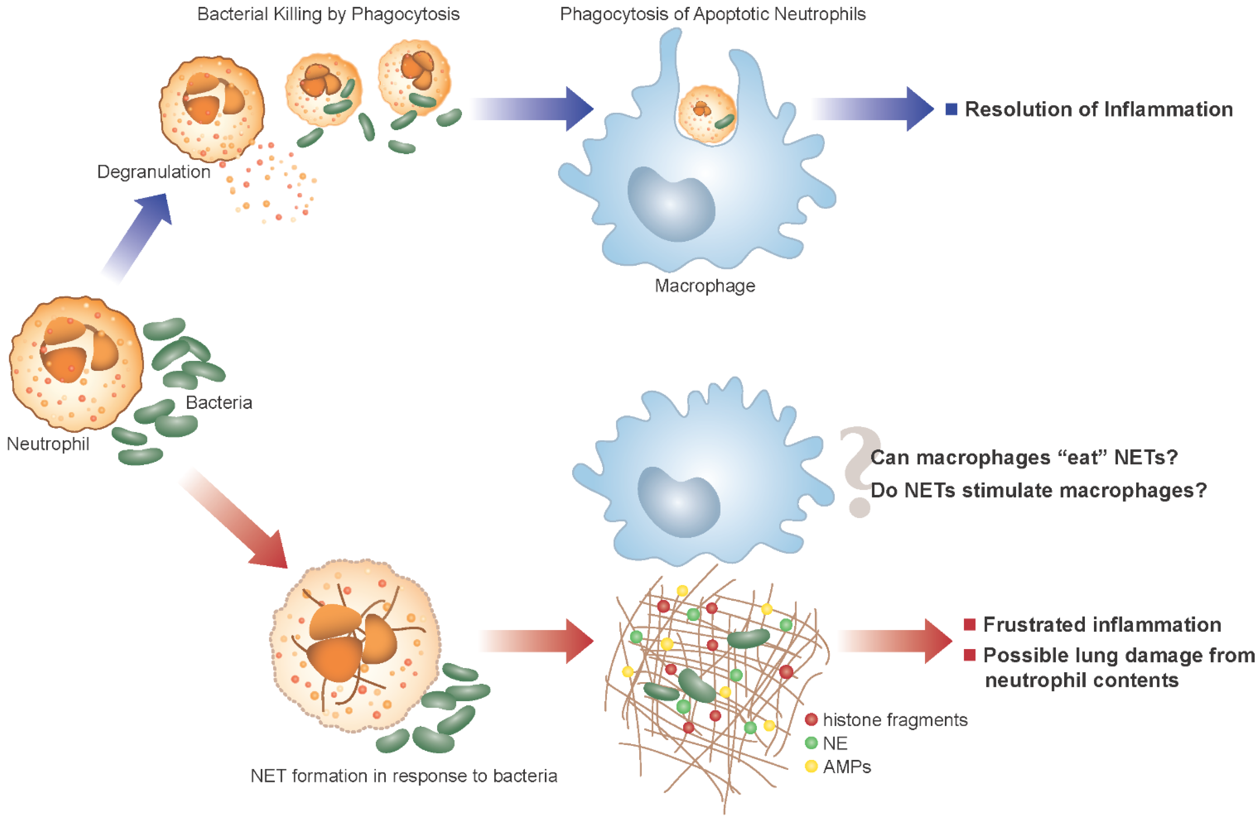
ASL also contains resident immune cells that act as a first line of host defense against inhaled pathogens, with the neutrophil being of primary importance as a phagocyte. Neutrophils are recruited to the lungs as part of the innate immune response to microbes (Figure 1). The airways of infants and children with CF are characterized by a disproportionately elevated neutrophil burden either attributed to early microbial challenge [16,17,18] or, in the absence of microbes, to dysfunctional CFTR trafficking [19,20,21]. In spite of this response, bacteria such as Staphylococcus aureus, Haemophilus influenzae, and Pseudomonas aeruginosa persist in the airways and establish chronic infection. Neutrophils have granules that contain a wide array of products such as proteolytic enzymes, AMPs, myeloperoxidase (MPO), and peptides that degrade bacteria following phagocytosis. These products may also be released by degranulation to assist in the extracellular killing of organisms [22]. Phagocytosis is a primary function of neutrophils and is composed of three processes: (1) receptor mediated pathogen uptake into vacuoles (phagosomes); (2) production of reactive oxygen species in the vacuole (later utilized by MPO to form hypochlorous acid); and (3) the fusion of granules containing proteases and antimicrobial mediators to these vacuoles (the phagolysosome) resulting in destruction and digestion of pathogens [22]. There is evidence to suggest that microbicidal function of neutrophils is impaired in CF as a result of reduced chloride anion availability for the production of hypochlorous acid, an essential mediator in the destruction of pathogens in the phagolysosome [23]. Phagocytosis typically accelerates neutrophil apoptosis; in turn, macrophages recognize and phagocytose the aging neutrophil which ultimately promotes an orderly resolution of inflammation [24,25]. Neutrophils also release proteases and AMPs by degranulation with extracellular levels of these proteins increasing as the neutrophil burden increases in the lung [26]. AMPs produced by neutrophils include lysozyme, phospholipase A2, bacterial permeability increasing protein (BPI), lactoferrin, cathelicidins such as LL37, and the α-defensins or human neutrophil defense peptides 1–4 (HNP). In CF the proteolytic degradation of AMPs can further reduce their effectiveness and exacerbate local tissue damage [27,28].
Neutrophil proteases have the potential to damage the host if present extracellularly instead of targeted to the phagolysosome. Neutrophil elastase (NE), a serine protease contained in azurophilic (primary) granules, can degrade nearly all structural proteins of the lung [29], as well as reduce ciliary motility and facilitate bacterial colonization [16]. NE is present early on in the airways of infants and young children with CF, and serves as a predictor of disease progression and is associated with lung function decline, as reported in the ARREST CF study [30]. The quantity of NE released into CF airways overwhelms the anti-protease capacity, leading to detectable NE activity and local architectural destruction. Therefore, a paradox exists in CF whereby an abundance of resident antimicrobial material, including AMPs and proteases, has the potential to damage the host. Moreover, in spite of the abundance of antimicrobial products, chronic lung infection is the characteristic phenotype in people with CF.
Figure 1. Alternative fates of Neutrophils in the Cystic Fibrosis (CF) lung. In the normal lung (purple arrows/top panel), neutrophils encounter and phagocytose bacteria. Following phagocytosis neutrophils rapidly undergo apoptosis and clearance by macrophages thus promoting resolution of inflammation. Alternatively, the excess of bacteria in the CF airway (red arrows/bottom panel) may lead to neutrophils forming neutrophil extracellular traps (NETs) in addition to normal phagocytosis. NETs may contribute to host defense, but also allow the release of toxic components into the airway that can damage the host lung. We can postulate that NETs contribute to the failed resolution of inflammation in the CF lung, and the clearance of NETs by macrophages may not be as anti-inflammatory as the clearance of apoptotic neutrophils.
Antibiotics 04 00062 g001

5. Neutrophil Extracellular Traps and CF

Neutrophil extracellular trap (NET) formation was described in 2004 by Brinkmann et al. [31] and has rapidly gained prominence in the literature as an antibacterial defense mechanism employed by neutrophils to kill microorganisms (by a mechanism distinct from phagocytosis or degranulation). Now termed NETosis, the process of NET formation is characterized by neutrophils undergoing an oxidative burst and releasing webs of decondensed DNA complexed to peptides and proteins, accompanied by cell death [32]. NETs have been described in CF sputum by a number of investigators [33,34,35,36], and recently it has been suggested that the majority of extracellular DNA in the CF lung is derived from NETs [33]. Oxidative burst and NADPH oxidase activation are thought to be central mechanisms in NET formation [32] and both MPO and NE are deemed essential co-factors [37,38]. The oxidative burst ultimately leads to downstream activation of peptidyl dearginase 4 (PAD4) which translocates to the nucleus and modifies histones (H3 and H4) by (hyper)citrinulation which contributes to nuclear decondensation and NET formation [39]. This is supported by evidence that neutrophils from PAD4−/− mice do not make NETs, and furthermore PAD4−/− mice are more susceptible to challenge with bacteria [40]. Neutrophil elastase complexes are involved in the decondensation of chromatin and consequently NET formation can be inhibited by neutrophil elastase inhibitors [38]. Furthermore, recent evidence suggests that MPO is an important co-factor in elastase release from granules and its proteolytic activity during NET formation [41]. Additional evidence implicates autophagic activity in NETosis as an essential partner to the oxidative burst demonstrating that inhibition of either system stopped NETosis. Indeed the promotion and inhbition of autophagy have now been demonstrated to alter NET formation in a number of studies [42,43]. The normal fate of a neutrophil is to perform its role in host defense and die by apoptosis. Why neutrophils (responding to infection) may favor a radically different form of cell death than apoptosis is intriguing and an additional hypothesis proposes that CF neutrophils do not undergo apoptosis or NET formation but rather switch to an alternate “hyperexocytosing” phenotype and release their primary granule content including MPO and NE into the airway lumen by enhanced exocytosis [44,45,46]. Therefore, further investigation of the process and relevance of NET formation in CF is required.
Initial studies demonstrated NE, MPO, and histones as the main proteins associated with NETs [31,32,47]. Further proteomic studies have revealed a major presence of AMPs on NETs with lysozyme, lactoferrin, HNPs, and calprotectin all demonstrated as NET constituents [48,49], adding to the hypothesis that NETs play a role in bacterial killing. Histones are also believed to have a major role as an antimicrobial on NETs [47]. Cathelicidins, although not identified in proteomic studies of NET formation have nevertheless been described as NET proteins [50,51]. Therefore, NETs could be a rich source of AMPs in the CF airway, but the functional significance of this has yet to be elucidated, particularly in the context of chronic infection.

6. NET Formation and Killing in Response to Bacteria

A number of groups have demonstrated the ability of Pseudomonas aeruginosa to induce NET formation, with reference to CF lung disease [33,36,52,53,54]. Interestingly, two groups independently demonstrated that NET induction using clinical isolates of Pseudomonas results in a divergent response, namely isolates from early disease induce NETs but isolates from the same patients in later disease do not [36,53]. Furthermore pyocyanin, a bacterial toxin produced by Pseudomonas can induce NET formation [55] but can also induce neutrophil apoptosis [56]. These observations require further investigation to help us understand the temporal relationship between Pseudomonas and NET production and why certain toxin challenges such as pyocyanin can result in disparate outcomes for neutrophils.
The ability of NETs to kill bacteria following their induction remains controversial. Early reports demonstrated that neutrophils, pre-activated with cytokines or phorbol ester to form NETs, could subsequently trap and kill pathogens in a manner independent of phagocytosis [31,57]. Young and colleagues demonstrated that killing of Pseudomonas did occur, even without the pre-activation of neutrophils. However, this NET-mediated killing only accounted for a small proportion of total bacterial killing compared to phagocytosis, unless the experiments were performed with neutrophils in suspension; the effect was lost if neutrophils were cultured under standard conditions [36]. Additionally, they demonstrated that the ability to kill Pseudomonas was lost when patient isolates from later stages in disease were used, suggesting this pathogen develops resistance to NET-mediated killing as CF lung disease advances. This finding was further reinforced by Dwyer and colleagues who demonstrated a decreased ability of NETs to kill mucoid strains of Pseudomonas [33]. Other reports suggest that NETs may not be as potent in killing as initially suggested. Mengazzi et al reported that NETs trap rather than kill microbes, with the majority of NET bound bacteria being live when liberated by DNase treatment [58], which is in keeping with a previous observation that NETs alone were not sufficient to kill bacteria and needed the addition of exogenous hydrogen peroxide to be truly bacteriocidal [59]. Nevertheless the interaction of Pseudomonas and NETs is an area of great interest in CF and whether this process is central to the development of lung disease or indeed involved the pathoadaptation of Pseudomonas in the CF airway will require further investigation as reviewed elsewhere recently [60].
To date, the majority of studies linking NETs to CF have concentrated on the interaction of neutrophils with Pseudomonas, however the ability of S. aureus to induce NET formation has been studied and confirmed [52,61,62,63]. S. aureus is a major pathogen in early CF lung disease, with colonization often pre-dating Pseudomonas acquisition by years. Furthermore, the ability of S. aureus to degrade NETs and escape specific protease activity has been described [61,64]. This could be significant in early CF lung disease if indeed killing of S. aureus by NETs was inefficient. Thus, further work utilizing CF isolates of S. aureus to induce NETs and assess killing capacity are required, particularly in models of early disease.

7. Potential Damaging Consequences of NETs in CF

CF sputum contains large amounts of NETs when measured by biochemical assays, and these are decorated with antimicrobial peptides, neutrophil proteins, and proteases [33]. Therefore, it is somewhat surprising that (in spite of these structures) chronic infection persists. This observation suggests that the innate antimicrobial properties of NETs may be over emphasized in the context of CF lung disease. The presence of large amounts of DNA in the CF airways and its potential contribution to CF pathogenesis was first acknowledged in the 1950s [65]. However, it was not until several decades later that targeting DNA degradation as a therapy was proved to be efficacious in reducing sputum viscosity, improving pulmonary function, and reducing the number of pulmonary exacerbations [66]. These data suggest that clearing NETs from the airway in CF is beneficial and this may account for some of the effects of DNase therapy. As previously stated NE (a key NET-associated protein) is present in high concentrations in CF, contributing to a protease/anti-protease imbalance in the lung. Cathepsin G and proteinase 3 are also released in NETs and likely add to the protease excess in the CF lung and damage tissue [52]. These data suggest that NETs may also represent a potentially deleterious source of proteases in the CF lung.
In addition to possibly contributing to protease-induced lung damage in CF, NETs may also be an important pro-inflammatory stimulus in the CF lung. NETs are a feature of autoimmune diseases characterized by significant levels of inflammation, and are implicated in anti-neutrophil cyoplasmic antibody (ANCA) vasculitis and associated kidney disease [67], and the pathogenesis of systemic lupus erythematosis (SLE). An impairment of DNAse degradation of NETs has been linked with the development of lupus nephritis [68], and further studies have demonstrated this inability to degrade NETs as more prominent in patients with other severe complications such as alopecia [69]. Un-degraded NET fragments activate complement as a central disease mechanism [70] and NETs in association with LL37 have been described as a source of extracellular antigen (acting via TLR-9) in SLE, which may also drive the disease process [71]. The presence of the cathelicidin on NETs has also been shown to promote atherosclerosis in mice by activating plasmacytoid dendritic cells [50] further suggesting that AMPs in association with DNA may promote inflammation. This feature may be highly relevant to CF lung disease, particularly when we consider that DNase is already employed as a successful therapy in CF.

8. Outstanding Questions in CF

Despite the presence of NETs (and associated AMPs, proteases, and other key neutrophil proteins) in the CF airway, there is unremitting bacterial infection. Thus we can infer that either NETs are inefficient in killing bacteria in CF lungs (possibly because the bacteria have evolved mechanisms to evade NET-based killing) or that NETs are simply trapping bugs rather than killing them. In fact, the process of trapping bacteria could be an important factor in stopping bacterial dissemination in CF and may be similar to extracellular trap encapsulation, a process seen in invertebrates, which possess only an innate immune system [72]. A further consideration is that phagocytosis and NET-mediated killing are overwhelmed by the volume of bacteria in the CF lung and simply attempt to hold infection in check. This is an attractive theory, particularly if we consider that low ASL pH, poor mucociliary transport, and other features of the environment of the CF lung could dampen the antimicrobial functions of NET-bound AMPs. Therefore, NETs in the CF lung may predominantly act as a source of damaging neutrophil products while being relatively impotent in antimicrobial activity. A further, as yet unanswered question, is whether the abundance of NETs in the CF lung is related to overproduction by CFTR-deficient neutrophils or a lack of clearance due to the reduced mucociliary transport in CF. Finally, there is a paucity of data on the mechanism of clearance of NETs with some evidence suggesting that NETs can be taken up by macrophages and degraded in the lysosome in an immunologically silent process [73]. However, further work is required to determine the mechanism for macrophage mediated clearance of NETs and how it affects the resolution of inflammation, which is normally driven by the efferocytosis of apoptotic neutrophils [25]. We propose that many of these questions could be answered by carefully designed experiments in appropriate systems and animal models such as the CF pig.

9. NETs Research in the CF Pig Model

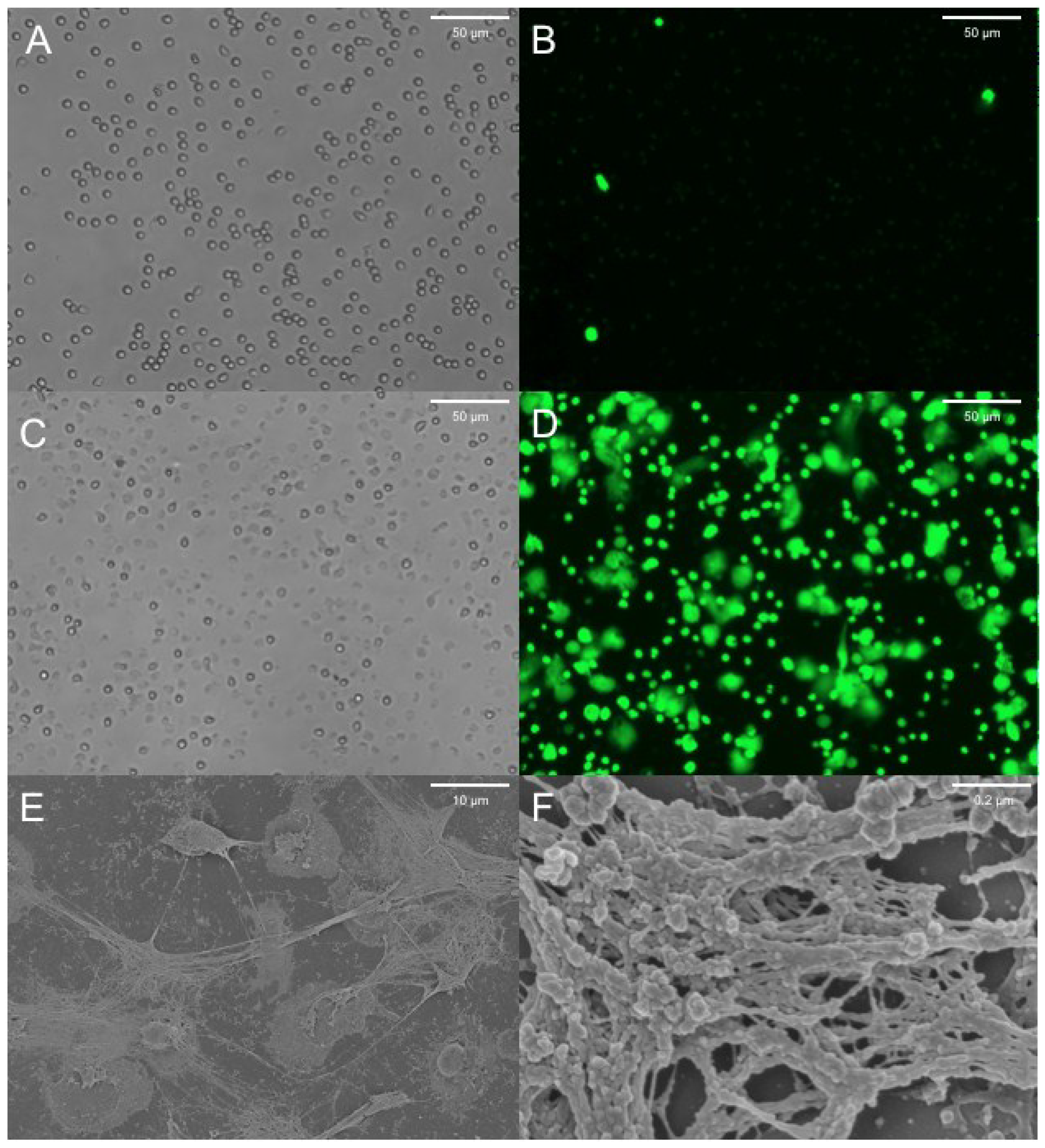
The CF pig, like the human, develops significant gut and lung disease, the latter being characterized by neutrophil infiltration and lung destruction [6]. Human CF blood neutrophils demonstrate no impairment in releasing NETs when compared to non-CF neutrophils [36]. NET formation and the associated release of proteases have been demonstrated in wild type porcine neutrophils following calcium ionophore treatment [74]. We can demonstrate NET formation by neutrophils from CFTR−/− animals following lipopolysaccharide (LPS) treatment (Figure 2). These porcine NETs show characteristic extracellular DNA release in ex vivo cell culture and associated features of NETosis by scanning electron microscopy. Thus, we can study the process of NETosis in neutrophils from a relevant large animal model of CF and potentially address many of the questions posed above. Furthermore by monitoring the development of lung disease in the CF pig model it may be possible to delineate the role of NET formation in this process. By measuring NET abundance in this model at different time points both longitudinally and following challenge with pathogens, we will be able to assess the contribution of NETs to the early pathogenesis of CF lung disease. Furthermore, therapeutic approaches to modulate NET production may be studied in an animal where repeated sampling and biopsy of the airway with standard techniques is possible.
Figure 2. Porcine CFTR−/− neutrophils stimulated with LPS to produce NETs. Cells were plated at a density of 50,000 cells per well in 24 well plates and treated for 6–8 h with LPS 100 μg/mL or control. Cells were then stained with Sytox green, which preferentially stains extracellular DNA and is excluded from live cells. (A) Brightfield image of untreated neutrophils showing normal morphology; (B) Sytox staining of untreated neutrophils demonstrates minimal staining for extracellular DNA; (C) Brightfield image of LPS treated neutrophils showing flattened and activated cells; (D) Sytox staining of LPS treated neutrophils showing characteristic NET structures; (E,F) Scanning electron microscopy (SEM) of fixed preparations at low and high magnification demonstrating characteristic mesh-like structures of NETs following LPS treatment.
Antibiotics 04 00062 g002

10. Conclusions

NET formation is a potentially important mechanism in the development of CF lung disease with results to date showing that it likely occurs in response to bacterial infection. The evidence that NETs are a major antimicrobial mechanism in CF is at present incomplete. Further work is required to determine whether NETs in CF contribute to host defense (which is knowingly impaired in CF) or whether their main contribution is to inflammation and lung damage.

Acknowledgments

Robert D. Gray is a Wellcome Trust Fellow WT 093767. We acknowledge the support of NIH P01 HL-51670, P01 HL-091842, and the Roy J. Carver Charitable Trust. The authors would like to thank Lily Paemka for assistance with porcine neutrophil preparations and Tom Moninger for technical help with SEM. We also thank Jennifer Bartlett for critically reviewing the manuscript.

Author Contributions

Robert D. Gray, Brian N. McCullagh and Paul B. McCray, Jr. conceived and wrote the article.

Conflicts of Interest

The authors declare no conflict of interest.

References

  1. Morgan, W.J.; Butler, S.M.; Johnson, C.A.; Colin, A.A.; FitzSimmons, S.C.; Geller, D.E.; Konstan, M.W.; Light, M.J.; Rabin, H.R.; Regelmann, W.E.; et al. Epidemiologic study of cystic fibrosis: Design and implementation of a prospective, multicenter, observational study of patients with cystic fibrosis in the U.S. and Canada. Pediatr. Pulmonol. 1999, 28, 231–241. [Google Scholar]
  2. Bartlett, J.A.; Fischer, A.J.; McCray, P.B., Jr. Innate immune functions of the airway epithelium. Contrib. Microbiol. 2008, 15, 147–163. [Google Scholar]
  3. Abman, S.H.; Ogle, J.W.; Harbeck, R.J.; Butler-Simon, N.; Hammond, K.B.; Accurso, F.J. Early bacteriologic, immunologic, and clinical courses of young infants with cystic fibrosis identified by neonatal screening. J. Pediatr. 1991, 119, 211–217. [Google Scholar] [CrossRef] [PubMed]
  4. Davidson, D.J.; Dorin, J.R. The CF mouse: An important tool for studying cystic fibrosis. Expert Rev. Mol. Med. 2001, 2001, 1–27. [Google Scholar] [CrossRef] [PubMed]
  5. Rogers, C.S.; Stoltz, D.A.; Meyerholz, D.K.; Ostedgaard, L.S.; Rokhlina, T.; Taft, P.J.; Rogan, M.P.; Pezzulo, A.A.; Karp, P.H.; Itani, O.A.; et al. Disruption of the cftr gene produces a model of cystic fibrosis in newborn pigs. Science 2008, 321, 1837–1841. [Google Scholar] [CrossRef]
  6. Stoltz, D.A.; Meyerholz, D.K.; Pezzulo, A.A.; Ramachandran, S.; Rogan, M.P.; Davis, G.J.; Hanfland, R.A.; Wohlford-Lenane, C.; Dohrn, C.L.; Bartlett, J.A.; et al. Cystic fibrosis pigs develop lung disease and exhibit defective bacterial eradication at birth. Sci. Transl. Med. 2010, 2, 29ra31. [Google Scholar] [PubMed]
  7. Hoegger, M.J.; Fischer, A.J.; McMenimen, J.D.; Ostedgaard, L.S.; Tucker, A.J.; Awadalla, M.A.; Moninger, T.O.; Michalski, A.S.; Hoffman, E.A.; Zabner, J.; et al. Cystic fibrosis. Impaired mucus detachment disrupts mucociliary transport in a piglet model of cystic fibrosis. Science 2014, 345, 818–822. [Google Scholar] [CrossRef] [PubMed]
  8. Hoegger, M.J.; Awadalla, M.; Namati, E.; Itani, O.A.; Fischer, A.J.; Tucker, A.J.; Adam, R.J.; McLennan, G.; Hoffman, E.A.; Stoltz, D.A.; et al. Assessing mucociliary transport of single particles in vivo shows variable speed and preference for the ventral trachea in newborn pigs. Proc. Natl. Acad. Sci. USA 2014, 111, 2355–2360. [Google Scholar] [CrossRef]
  9. Sun, X.; Sui, H.; Fisher, J.T.; Yan, Z.; Liu, X.; Cho, H.J.; Joo, N.S.; Zhang, Y.; Zhou, W.; Yi, Y.; et al. Disease phenotype of a ferret CFTR-knockout model of cystic fibrosis. J. Clin. Invest. 2010, 120, 3149–3160. [Google Scholar] [CrossRef]
  10. Berkebile, A.R.; McCray, P.B., Jr. Effects of airway surface liquid ph on host defense in cystic fibrosis. Int. J. Biochem. Cell. Biol. 2014, 52, 124–129. [Google Scholar] [CrossRef]
  11. Singh, P.K.; Tack, B.F.; McCray, P.B., Jr.; Welsh, M.J. Synergistic and additive killing by antimicrobial factors found in human airway surface liquid. Am. J. Physiol. Lung Cell Mol. Physiol. 2000, 279, L799–L805. [Google Scholar]
  12. Coakley, R.D.; Grubb, B.R.; Paradiso, A.M.; Gatzy, J.T.; Johnson, L.G.; Kreda, S.M.; OʼNeal, W.K.; Boucher, R.C. Abnormal surface liquid ph regulation by cultured cystic fibrosis bronchial epithelium. Proc. Natl. Acad. Sci. USA 2003, 100, 16083–16088. [Google Scholar] [CrossRef]
  13. Song, Y.; Salinas, D.; Nielson, D.W.; Verkman, A.S. Hyperacidity of secreted fluid from submucosal glands in early cystic fibrosis. Am. J. Physiol. Cell Physiol. 2006, 290, C741–C749. [Google Scholar] [CrossRef] [PubMed]
  14. Tate, S.; MacGregor, G.; Davis, M.; Innes, J.A.; Greening, A.P. Airways in cystic fibrosis are acidified: Detection by exhaled breath condensate. Thorax 2002, 57, 926–929. [Google Scholar] [CrossRef] [PubMed]
  15. Pezzulo, A.A.; Tang, X.X.; Hoegger, M.J.; Abou Alaiwa, M.H.; Ramachandran, S.; Moninger, T.O.; Karp, P.H.; Wohlford-Lenane, C.L.; Haagsman, H.P.; van Eijk, M.; et al. Reduced airway surface PH impairs bacterial killing in the porcine cystic fibrosis lung. Nature 2012, 487, 109–113. [Google Scholar] [CrossRef] [PubMed]
  16. Birrer, P.; McElvaney, N.G.; Rudeberg, A.; Sommer, C.W.; Liechti-Gallati, S.; Kraemer, R.; Hubbard, R.; Crystal, R.G. Protease-antiprotease imbalance in the lungs of children with cystic fibrosis. Am. J. Respir. Crit. Care Med. 1994, 150, 207–213. [Google Scholar] [CrossRef] [PubMed]
  17. Khan, T.Z.; Wagener, J.S.; Bost, T.; Martinez, J.; Accurso, F.J.; Riches, D.W. Early pulmonary inflammation in infants with cystic fibrosis. Am. J. Respir. Crit. Care Med. 1995, 151, 1075–1082. [Google Scholar] [PubMed]
  18. Konstan, M.W.; Hilliard, K.A.; Norvell, T.M.; Berger, M. Bronchoalveolar lavage findings in cystic fibrosis patients with stable, clinically mild lung disease suggest ongoing infection and inflammation. Am. J. Respir. Crit. Care Med. 1994, 150, 448–454. [Google Scholar] [CrossRef] [PubMed]
  19. Sly, P.D.; Brennan, S.; Gangell, C.; de Klerk, N.; Murray, C.; Mott, L.; Stick, S.M.; Robinson, P.J.; Robertson, C.F.; Ranganathan, S.C.; et al. Lung disease at diagnosis in infants with cystic fibrosis detected by newborn screening. Am. J. Respir. Crit. Care Med. 2009, 180, 146–152. [Google Scholar] [CrossRef]
  20. Martino, M.E.; Olsen, J.C.; Fulcher, N.B.; Wolfgang, M.C.; OʼNeal, W.K.; Ribeiro, C.M. Airway epithelial inflammation-induced endoplasmic reticulum Ca2+ store expansion is mediated by X-box binding protein-1. J. Biol. Chem. 2009, 284, 14904–14913. [Google Scholar] [CrossRef]
  21. Weber, A.J.; Soong, G.; Bryan, R.; Saba, S.; Prince, A. Activation of NF-kappaB in airway epithelial cells is dependent on cftr trafficking and Cl channel function. Am. J. Physiol. Lung Cell Mol. Physiol. 2001, 281, L71–L78. [Google Scholar] [PubMed]
  22. Mayadas, T.N.; Cullere, X.; Lowell, C.A. The multifaceted functions of neutrophils. Annu. Rev. Pathol. 2014, 9, 181–218. [Google Scholar] [CrossRef] [PubMed]
  23. Painter, R.G.; Bonvillain, R.W.; Valentine, V.G.; Lombard, G.A.; LaPlace, S.G.; Nauseef, W.M.; Wang, G. The role of chloride anion and CFTR in killing of Pseudomonas aeruginosa by normal and CF neutrophils. J. Leukoc. Biol. 2008, 83, 1345–1353. [Google Scholar] [CrossRef] [PubMed]
  24. Kennedy, A.D.; DeLeo, F.R. Neutrophil apoptosis and the resolution of infection. Immunol. Res. 2009, 43, 25–61. [Google Scholar] [CrossRef] [PubMed]
  25. Savill, J.; Dransfield, I.; Gregory, C.; Haslett, C. A blast from the past: Clearance of apoptotic cells regulates immune responses. Nat. Rev. Immunol. 2002, 2, 965–975. [Google Scholar] [CrossRef] [PubMed]
  26. Sagel, S.D.; Sontag, M.K.; Accurso, F.J. Relationship between antimicrobial proteins and airway inflammation and infection in cystic fibrosis. Pediatr. Pulmonol. 2009, 44, 402–409. [Google Scholar] [CrossRef] [PubMed]
  27. Bergsson, G.; Reeves, E.P.; McNally, P.; Chotirmall, S.H.; Greene, C.M.; Greally, P.; Murphy, P.; OʼNeill, S.J.; McElvaney, N.G. Ll-37 complexation with glycosaminoglycans in cystic fibrosis lungs inhibits antimicrobial activity, which can be restored by hypertonic saline. J. Immunol. 2009, 183, 543–551. [Google Scholar] [CrossRef] [PubMed]
  28. Rogan, M.P.; Taggart, C.C.; Greene, C.M.; Murphy, P.G.; OʼNeill, S.J.; McElvaney, N.G. Loss of microbicidal activity and increased formation of biofilm due to decreased lactoferrin activity in patients with cystic fibrosis. J. Infect. Dis. 2004, 190, 1245–1253. [Google Scholar] [CrossRef] [PubMed]
  29. Quinn, D.J.; Weldon, S.; Taggart, C.C. Antiproteases as therapeutics to target inflammation in cystic fibrosis. Open Respir. Med. J. 2010, 4, 20–31. [Google Scholar] [PubMed]
  30. Sly, P.D.; Gangell, C.L.; Chen, L.; Ware, R.S.; Ranganathan, S.; Mott, L.S.; Murray, C.P.; Stick, S.M.; Investigators, A.C. Risk factors for bronchiectasis in children with cystic fibrosis. N. Engl. J. Med. 2013, 368, 1963–1970. [Google Scholar] [CrossRef] [PubMed]
  31. Brinkmann, V.; Reichard, U.; Goosmann, C.; Fauler, B.; Uhlemann, Y.; Weiss, D.S.; Weinrauch, Y.; Zychlinsky, A. Neutrophil extracellular traps kill bacteria. Science 2004, 303, 1532–1535. [Google Scholar] [CrossRef] [PubMed]
  32. Fuchs, T.A.; Abed, U.; Goosmann, C.; Hurwitz, R.; Schulze, I.; Wahn, V.; Weinrauch, Y.; Brinkmann, V.; Zychlinsky, A. Novel cell death program leads to neutrophil extracellular traps. J. Cell Biol. 2007, 176, 231–241. [Google Scholar] [CrossRef] [PubMed]
  33. Dwyer, M.; Shan, Q.; DʼOrtona, S.; Maurer, R.; Mitchell, R.; Olesen, H.; Thiel, S.; Huebner, J.; Gadjeva, M. Cystic fibrosis sputum DNA has netosis characteristics and neutrophil extracellular trap release is regulated by macrophage migration-inhibitory factor. J. Innate Immun. 2014, 6, 765–779. [Google Scholar] [CrossRef]
  34. Manzenreiter, R.; Kienberger, F.; Marcos, V.; Schilcher, K.; Krautgartner, W.D.; Obermayer, A.; Huml, M.; Stoiber, W.; Hector, A.; Griese, M.; et al. Ultrastructural characterization of cystic fibrosis sputum using atomic force and scanning electron microscopy. J. Cyst. Fibros. 2012, 11, 84–92. [Google Scholar] [CrossRef]
  35. Papayannopoulos, V.; Staab, D.; Zychlinsky, A. Neutrophil elastase enhances sputum solubilization in cystic fibrosis patients receiving DNase therapy. PLOS ONE 2011, 6, e28526. [Google Scholar] [CrossRef] [PubMed]
  36. Young, R.L.; Malcolm, K.C.; Kret, J.E.; Caceres, S.M.; Poch, K.R.; Nichols, D.P.; Taylor-Cousar, J.L.; Saavedra, M.T.; Randell, S.H.; Vasil, M.L.; et al. Neutrophil extracellular trap (net)-mediated killing of Pseudomonas aeruginosa: Evidence of acquired resistance within the CF airway, independent of CFTR. PLOS ONE 2011, 6, e23637. [Google Scholar] [CrossRef]
  37. Metzler, K.D.; Fuchs, T.A.; Nauseef, W.M.; Reumaux, D.; Roesler, J.; Schulze, I.; Wahn, V.; Papayannopoulos, V.; Zychlinsky, A. Myeloperoxidase is required for neutrophil extracellular trap formation: Implications for innate immunity. Blood 2011, 117, 953–959. [Google Scholar] [CrossRef] [PubMed]
  38. Papayannopoulos, V.; Metzler, K.D.; Hakkim, A.; Zychlinsky, A. Neutrophil elastase and myeloperoxidase regulate the formation of neutrophil extracellular traps. J. Cell Biol. 2010, 191, 677–691. [Google Scholar] [CrossRef] [PubMed]
  39. Wang, Y.; Li, M.; Stadler, S.; Correll, S.; Li, P.; Wang, D.; Hayama, R.; Leonelli, L.; Han, H.; Grigoryev, S.A.; et al. Histone hypercitrullination mediates chromatin decondensation and neutrophil extracellular trap formation. J. Cell Biol. 2009, 184, 205–213. [Google Scholar] [CrossRef] [PubMed]
  40. Li, P.; Li, M.; Lindberg, M.R.; Kennett, M.J.; Xiong, N.; Wang, Y. Pad4 is essential for antibacterial innate immunity mediated by neutrophil extracellular traps. J. Exp. Med. 2010, 207, 1853–1862. [Google Scholar] [CrossRef] [PubMed]
  41. Metzler, K.D.; Goosmann, C.; Lubojemska, A.; Zychlinsky, A.; Papayannopoulos, V. A myeloperoxidase-containing complex regulates neutrophil elastase release and actin dynamics during netosis. Cell Rep. 2014, 8, 883–896. [Google Scholar] [CrossRef] [PubMed]
  42. Itakura, A.; McCarty, O.J. Pivotal role for the mtor pathway in the formation of neutrophil extracellular traps via regulation of autophagy. Am. J. Physiol. Cell Physiol. 2013, 305, C348–C354. [Google Scholar] [CrossRef] [PubMed]
  43. Mitroulis, I.; Kambas, K.; Chrysanthopoulou, A.; Skendros, P.; Apostolidou, E.; Kourtzelis, I.; Drosos, G.I.; Boumpas, D.T.; Ritis, K. Neutrophil extracellular trap formation is associated with IL-1beta and autophagy-related signaling in gout. PLOS ONE 2011, 6, e29318. [Google Scholar] [CrossRef] [PubMed]
  44. Laval, J.; Touhami, J.; Herzenberg, L.A.; Conrad, C.; Taylor, N.; Battini, J.L.; Sitbon, M.; Tirouvanziam, R. Metabolic adaptation of neutrophils in cystic fibrosis airways involves distinct shifts in nutrient transporter expression. J. Immunol. 2013, 190, 6043–6050. [Google Scholar] [CrossRef] [PubMed]
  45. Makam, M.; Diaz, D.; Laval, J.; Gernez, Y.; Conrad, C.K.; Dunn, C.E.; Davies, Z.A.; Moss, R.B.; Herzenberg, L.A.; Herzenberg, L.A.; et al. Activation of critical, host-induced, metabolic and stress pathways marks neutrophil entry into cystic fibrosis lungs. Proc. Natl. Acad. Sci. USA 2009, 106, 5779–5783. [Google Scholar] [CrossRef] [PubMed]
  46. Tirouvanziam, R.; Gernez, Y.; Conrad, C.K.; Moss, R.B.; Schrijver, I.; Dunn, C.E.; Davies, Z.A.; Herzenberg, L.A.; Herzenberg, L.A. Profound functional and signaling changes in viable inflammatory neutrophils homing to cystic fibrosis airways. Proc. Natl. Acad. Sci. USA 2008, 105, 4335–4339. [Google Scholar] [CrossRef] [PubMed]
  47. Brinkmann, V.; Zychlinsky, A. Beneficial suicide: Why neutrophils die to make nets. Nat. Rev. Microbiol. 2007, 5, 577–582. [Google Scholar] [CrossRef] [PubMed]
  48. Khandpur, R.; Carmona-Rivera, C.; Vivekanandan-Giri, A.; Gizinski, A.; Yalavarthi, S.; Knight, J.S.; Friday, S.; Li, S.; Patel, R.M.; Subramanian, V.; et al. Nets are a source of citrullinated autoantigens and stimulate inflammatory responses in rheumatoid arthritis. Sci. Transl. Med. 2013, 5, 178ra140. [Google Scholar] [CrossRef]
  49. Urban, C.F.; Ermert, D.; Schmid, M.; Abu-Abed, U.; Goosmann, C.; Nacken, W.; Brinkmann, V.; Jungblut, P.R.; Zychlinsky, A. Neutrophil extracellular traps contain calprotectin, a cytosolic protein complex involved in host defense against candida albicans. PLOS Pathog. 2009, 5, e1000639. [Google Scholar] [CrossRef] [PubMed]
  50. Doring, Y.; Manthey, H.D.; Drechsler, M.; Lievens, D.; Megens, R.T.; Soehnlein, O.; Busch, M.; Manca, M.; Koenen, R.R.; Pelisek, J.; et al. Auto-antigenic protein-DNA complexes stimulate plasmacytoid dendritic cells to promote atherosclerosis. Circulation 2012, 125, 1673–1683. [Google Scholar] [CrossRef] [PubMed]
  51. Garcia-Romo, G.S.; Caielli, S.; Vega, B.; Connolly, J.; Allantaz, F.; Xu, Z.; Punaro, M.; Baisch, J.; Guiducci, C.; Coffman, R.L.; et al. Netting neutrophils are major inducers of type I IFN production in pediatric systemic lupus erythematosus. Sci. Transl. Med. 2011, 3, 73ra20. [Google Scholar] [CrossRef] [PubMed]
  52. Dubois, A.V.; Gauthier, A.; Brea, D.; Varaigne, F.; Diot, P.; Gauthier, F.; Attucci, S. Influence of DNA on the activities and inhibition of neutrophil serine proteases in cystic fibrosis sputum. Am. J. Respir Cell Mol. Biol. 2012, 47, 80–86. [Google Scholar] [CrossRef] [PubMed]
  53. Yoo, D.G.; Floyd, M.; Winn, M.; Moskowitz, S.M.; Rada, B. Net formation induced by Pseudomonas aeruginosa cystic fibrosis isolates measured as release of myeloperoxidase-DNA and neutrophil elastase-DNA complexes. Immunol. Lett. 2014, 160, 186–194. [Google Scholar] [CrossRef] [PubMed]
  54. Yoo, D.G.; Winn, M.; Pang, L.; Moskowitz, S.M.; Malech, H.L.; Leto, T.L.; Rada, B. Release of cystic fibrosis airway inflammatory markers from pseudomonas aeruginosa-stimulated human neutrophils involves nadph oxidase-dependent extracellular DNA trap formation. J. Immunol. 2010, 192, 4728–4738. [Google Scholar] [CrossRef]
  55. Rada, B.; Jendrysik, M.A.; Pang, L.; Hayes, C.P.; Yoo, D.G.; Park, J.J.; Moskowitz, S.M.; Malech, H.L.; Leto, T.L. Pyocyanin-enhanced neutrophil extracellular trap formation requires the nadph oxidase. PLOS ONE 2013, 8, e54205. [Google Scholar] [CrossRef] [PubMed]
  56. Allen, L.; Dockrell, D.H.; Pattery, T.; Lee, D.G.; Cornelis, P.; Hellewell, P.G.; Whyte, M.K. Pyocyanin production by Pseudomonas aeruginosa induces neutrophil apoptosis and impairs neutrophil-mediated host defenses in vivo. J. Immunol. 2005, 174, 3643–3649. [Google Scholar] [CrossRef] [PubMed]
  57. Urban, C.F.; Reichard, U.; Brinkmann, V.; Zychlinsky, A. Neutrophil extracellular traps capture and kill Candida albicans yeast and hyphal forms. Cell Microbiol. 2006, 8, 668–676. [Google Scholar] [CrossRef] [PubMed]
  58. Menegazzi, R.; Decleva, E.; Dri, P. Killing by neutrophil extracellular traps: Fact or folklore? Blood 2012, 119, 1214–1216. [Google Scholar] [CrossRef] [PubMed]
  59. Parker, H.; Albrett, A.M.; Kettle, A.J.; Winterbourn, C.C. Myeloperoxidase associated with neutrophil extracellular traps is active and mediates bacterial killing in the presence of hydrogen peroxide. J. Leukoc. Biol. 2011, 91, 369–371. [Google Scholar] [CrossRef] [PubMed]
  60. Rahman, S.; Gadjeva, M. Does netosis contribute to the bacterial pathoadaptation in cystic fibrosis? Front. Immunol. 2014, 5, e378. [Google Scholar] [CrossRef]
  61. Berends, E.T.; Horswill, A.R.; Haste, N.M.; Monestier, M.; Nizet, V.; von Kockritz-Blickwede, M. Nuclease expression by Staphylococcus aureus facilitates escape from neutrophil extracellular traps. J. Innate Immun. 2010, 2, 576–586. [Google Scholar] [CrossRef] [PubMed]
  62. Malachowa, N.; Kobayashi, S.D.; Freedman, B.; Dorward, D.W.; DeLeo, F.R. Staphylococcus aureus leukotoxin GH promotes formation of neutrophil extracellular traps. J. Immunol. 2013, 191, 6022–6029. [Google Scholar] [CrossRef] [PubMed]
  63. Pilsczek, F.H.; Salina, D.; Poon, K.K.; Fahey, C.; Yipp, B.G.; Sibley, C.D.; Robbins, S.M.; Green, F.H.; Surette, M.G.; Sugai, M.; et al. A novel mechanism of rapid nuclear neutrophil extracellular trap formation in response to Staphylococcus aureus. J. Immunol. 2010, 185, 7413–7425. [Google Scholar] [CrossRef] [PubMed]
  64. Thammavongsa, V.; Missiakas, D.M.; Schneewind, O. Staphylococcus aureus degrades neutrophil extracellular traps to promote immune cell death. Science 2013, 342, 863–866. [Google Scholar] [CrossRef] [PubMed]
  65. Hodson, M.E. Aerosolized dornase alfa (rhDNase) for therapy of cystic fibrosis. Am. J. Respir. Crit. Care Med. 1995, 151, S70–S74. [Google Scholar] [CrossRef] [PubMed]
  66. Fuchs, H.J.; Borowitz, D.S.; Christiansen, D.H.; Morris, E.M.; Nash, M.L.; Ramsey, B.W.; Rosenstein, B.J.; Smith, A.L.; Wohl, M.E. Effect of aerosolized recombinant human DNase on exacerbations of respiratory symptoms and on pulmonary function in patients with cystic fibrosis. The pulmozyme study group. N. Engl. J. Med. 1994, 331, 637–642. [Google Scholar] [CrossRef] [PubMed]
  67. Kessenbrock, K.; Krumbholz, M.; Schonermarck, U.; Back, W.; Gross, W.L.; Werb, Z.; Grone, H.J.; Brinkmann, V.; Jenne, D.E. Netting neutrophils in autoimmune small-vessel vasculitis. Nat. Med. 2009, 15, 623–625. [Google Scholar] [CrossRef] [PubMed]
  68. Hakkim, A.; Furnrohr, B.G.; Amann, K.; Laube, B.; Abed, U.A.; Brinkmann, V.; Herrmann, M.; Voll, R.E.; Zychlinsky, A. Impairment of neutrophil extracellular trap degradation is associated with lupus nephritis. Proc. Natl. Acad. Sci. USA 2010, 107, 9813–9818. [Google Scholar] [CrossRef] [PubMed]
  69. Leffler, J.; Gullstrand, B.; Jonsen, A.; Nilsson, J.A.; Martin, M.; Blom, A.M.; Bengtsson, A.A. Degradation of neutrophil extracellular traps co-varies with disease activity in patients with systemic lupus erythematosus. Arthritis Res. Ther. 2013, 15, R84. [Google Scholar] [CrossRef] [PubMed]
  70. Leffler, J.; Martin, M.; Gullstrand, B.; Tyden, H.; Lood, C.; Truedsson, L.; Bengtsson, A.A.; Blom, A.M. Neutrophil extracellular traps that are not degraded in systemic lupus erythematosus activate complement exacerbating the disease. J. Immunol. 2012, 188, 3522–3531. [Google Scholar] [CrossRef] [PubMed]
  71. Villanueva, E.; Yalavarthi, S.; Berthier, C.C.; Hodgin, J.B.; Khandpur, R.; Lin, A.M.; Rubin, C.J.; Zhao, W.; Olsen, S.H.; Klinker, M.; et al. Netting neutrophils induce endothelial damage, infiltrate tissues, and expose immunostimulatory molecules in systemic lupus erythematosus. J. Immunol. 2011, 187, 538–552. [Google Scholar] [CrossRef] [PubMed]
  72. Robb, C.T.; Dyrynda, E.A.; Gray, R.D.; Rossi, A.G.; Smith, V.J. Invertebrate extracellular phagocyte traps show that chromatin is an ancient defence weapon. Nat. Commun. 2014, 5, 4627. [Google Scholar] [CrossRef] [PubMed]
  73. Farrera, C.; Fadeel, B. Macrophage clearance of neutrophil extracellular traps is a silent process. J. Immunol. 2013, 191, 2647–2656. [Google Scholar] [CrossRef] [PubMed]
  74. Brea, D.; Meurens, F.; Dubois, A.V.; Gaillard, J.; Chevaleyre, C.; Jourdan, M.L.; Winter, N.; Arbeille, B.; Si-Tahar, M.; Gauthier, F.; et al. The pig as a model for investigating the role of neutrophil serine proteases in human inflammatory lung diseases. Biochem. J. 2012, 447, 363–370. [Google Scholar] [CrossRef] [PubMed]