Article

Antimicrobial and Cytoprotective Effects of Tea Extracts Against Escherichia coli-Producing Colibactin Toxin Infections

by
Wipawadee Teppabut
1,2,
Yingmanee Tragoolpua
1,3 and
Thida Kaewkod
1,3,*
1
Department of Biology, Faculty of Science, Chiang Mai University, Chiang Mai 50200, Thailand
2
Master of Science Program in Applied Microbiology (International Program), Faculty of Science, Chiang Mai University, Chiang Mai 50200, Thailand
3
Natural Extracts and Innovative Products for Alternative Healthcare Research Group, Faculty of Science, Chiang Mai University, Chiang Mai 50200, Thailand
*
Author to whom correspondence should be addressed.
Antibiotics 2025, 14(9), 886; https://doi.org/10.3390/antibiotics14090886
Submission received: 2 August 2025 / Revised: 22 August 2025 / Accepted: 28 August 2025 / Published: 2 September 2025
(This article belongs to the Special Issue Antimicrobial Activity of Extracts from Plants, 2nd Edition)

Abstract

Background/Objectives: Camellia sinensis (L.) Kuntze or tea contains bioactive compounds such as catechin and caffeine, known for their antimicrobial and health-promoting properties. Colibactin-producing Escherichia coli are linked to genotoxicity in colon epithelial cells, potentially contributing to colorectal disease. This study aimed to evaluate the inhibitory effects of tea extracts (green, oolong, and black) and the phytochemicals catechin and caffeine on E. coli pathogenesis mediated by colibactin toxins, including transient infections, DNA damage, and cell cycle alterations in Caco-2 colon cells. Methods: Tea extracts were analyzed by HPLC for phytochemical content. Their antimicrobial activity against colibactin-producing E. coli (ATCC 25922) was assessed. Caco-2 cells were infected with the bacteria and treated with tea extracts or compounds. Cell viability was measured by MTT assay, DNA damage was measured by alkaline comet assay, and the expression of CDK-1, CDK-2, and Ki-67 genes was measurd by qRT-PCR. Results: Tea extracts and catechin inhibited colibactin-producing E. coli and significantly protected Caco-2 cells. Oolong tea showed the highest protection (90.78 ± 2.76%), with others maintaining viability above 80%. DNA damage was markedly reduced, and cell cycle regulation improved. All extracts upregulated CDK-1 and downregulated CDK-2, aiding in cell cycle restoration. Ki-67 expression indicated enhanced cell proliferation during infection. Conclusions: This study highlights new findings showing that tea extracts, including green, oolong, and black tea, as well as the tea compounds catechin and caffeine, can protect against DNA damage and help maintain the normal cell cycle of colon cells infected with E. coli-producing colibactin toxin. These results support their potential role in preventing and mitigating infections caused by such E. coli strains while promoting colon cell health.

Graphical Abstract

1. Introduction

Camellia sinensis (L.) Kuntze (tea; family Theaceae) is the second most widely consumed beverage in the world, following water. It is more popular than coffee, beer, wine, and soft drinks [1,2]. With a history spanning over 2000 years, tea has become an integral part of social culture. Currently, around 70% of the global population consumes approximately 18–20 billion cups of tea each day. To meet the high demand, approximately 2.9 billion tons of tea are produced annually [3]. Tea is made from the buds, young leaves, and tender stalks of the tea plant and is classified into different types based on the degree of fermentation during production: green tea (unfermented), oolong tea (semi-fermented), and black tea (fully fermented) [4]. All tea types originate from the same plant (C. sinensis), with heat treatment playing a key role in enzyme inactivation, particularly in the production of green and oolong tea. During oolong tea fermentation, catechin oxidation levels range from 20% to 80%, while black tea features fully oxidized catechins [5,6]. The chemical makeup of tea varies by type, with flavonoids making up 20–30% of its composition. Green tea contains catechins (flavan-3-ols) such as epigallocatechin-3-gallate (EGCG), epigallocatechin (EGC), epicatechin (EC), and epicatechin-3-gallate (ECG). Black tea, on the other hand, is rich in polymerized catechins known as theaflavins, such as theaflavin-3′-gallate, as well as other theaflavins and thearubigins [7]. Additional major theaflavins found in black tea include theaflavin, theaflavin-3-gallate, and theaflavin-3,3′-gallate [8]. Numerous studies have demonstrated the beneficial effects of different types of tea. Tea produced from C. sinensis has shown activities such as antidiabetic [9], anticancer [10], antibacterial [11], neuroprotective [12], cardioprotective [13], anti-obesity [14], hepatoprotective [15], and antiviral [16] effects. In this study, we focused on the use of tea extracts to inhibit bacterial infection mechanisms.
Pathogenic enteric bacteria often cause diarrhea and associated illnesses, typically leading to secretory or watery diarrhea following an infection [17]. Escherichia coli is the most common bacterial resident of the human gut, and its strains are divided into four primary phylogenetic groups (A, B1, B2, and D), each linked to specific ecological niches [18]. Extraintestinal E. coli (ExPEC), classified under group B2, are found in the intestinal microbiota of both humans and animals. These emerging pathogens are known to cause extraintestinal infections, such as urinary tract infections, neonatal meningitis, sepsis, pneumonia, surgical site infections, and infections at other sites outside the intestine [19,20]. Moreover, antimicrobial resistance (AMR) is a significant concern worldwide, particularly concerning E. coli strains. A study published in 2024 reported that 58% of E. coli isolates exhibited resistance to gentamicin, a commonly used antibiotic [21]. ExPEC strains tend to remain in the colon for longer periods compared to other E. coli groups and account for 30–50% of the strains found in the feces of healthy individuals in high-income countries [22,23]. The genomes of ExPEC strains contain the polyketide synthase (pks) pathogenicity island, known as the pks island, which encodes the 54-kbp colibactin gene cluster responsible for the production of the colibactin toxin [24]. This toxin induces cell cycle arrest at the G2 or M phase, chromosomal aberrations, and double-strand DNA breaks [25]. Cyclin-dependent kinases 1 and 2 (CDK-1 and CDK-2) are essential for controlling cell cycle progression, including transitions from G1 to S, through S, and from G2 to M phases. The activation of CDK-1 and CDK-2 facilitates cell growth and division [26]. Colibactin-induced DNA damage disrupts the cell cycle by causing arrest at the G1/S and G2/M checkpoints, thereby hindering cell growth and regeneration, which supports bacterial colonization. In response to severe DNA damage, cells may undergo apoptosis or develop a senescent phenotype. In cases where DNA repair is incomplete, surviving cells can experience genomic instability, potentially leading to tumor initiation or accelerated tumor progression [27]. Additionally, colibactin-harboring E. coli have been found in higher numbers among colorectal cancer (CRC) patients and have been shown to increase tumor formation in various CRC mouse models [28]. This study evaluated the inhibitory effects of tea extracts, including green tea, oolong tea and black tea, and phytochemical compounds of catechin and caffeine on E. coli pathogenesis mediated by colibactin toxins, including transient infections, colibactin-induced DNA damage, and their influence on the cell cycle, as observed in Caco-2 colon cell culture models.

2. Results

2.1. Phytochemical Compounds in Tea Extracts

The phytochemical contents of tea extracts were determined including catechin, EGCG, caffeine and theaflavin by HPLC assays. The results showed that oolong tea extract contained all chemical compounds with high values at 233.475 ± 13.320, 251.839 ± 8.751, 2.658 ± 0.053 and 2.192 ± 0.297 mg/mL, respectively (Table 1). In addition, green tea extract detected a high caffeine contents as 56.909 ± 0.444 mg/mL, followed by catechin and caffeine. For black tea extract was found high content of EGCG with the value of 269.714 ± 7.860 mg/mL (Table 1).

2.2. Antibacterial Activity by Agar Well Diffusion

Green tea, oolong tea, and black tea extracts were evaluated for their ability to inhibit E. coli ATCC 25922 (a colibactin-producing strain) and E. coli K-12 (a non-colibactin-producing strain) using the agar well diffusion technique. The results showed that oolong tea extract at 500 mg/mL exhibited the greatest inhibition of E. coli ATCC 25922, with an inhibition zone of 18.33 ± 0.47 mm. Green tea and black tea followed with inhibition zones of 16.33 ± 4.50 mm and 13.67 ± 1.25 mm, respectively (Figure S1 and Table 2). Therefore, phytochemical compounds, specifically catechin (5 mg/mL) and caffeine (5 mg/mL), demonstrated inhibitory effects against E. coli ATCC 25922 (Figure S1 and Table 2). For E. coli K-12, the inhibition zones were 15.00 ± 1.73 mm for green tea, 16.00 ± 1.00 mm for oolong tea, and 14.00 ± 1.00 mm for black tea (Figure S1 and Table 2). Additionally, 1 mg/mL gentamycin was used as a positive control for bacterial inhibition, resulting in inhibition zones of 23.76 ± 0.47 mm and 26.33 ± 0.58 mm against E. coli ATCC 25922 and E. coli K-12, respectively (Figure S1 and Table 2).

2.3. Cytotoxicity of Tea Extracts and Phytochemical Compounds on Caco-2 Cells

The cytotoxicity of tea extracts on Caco-2 cells was determined using the MTT assay to identify non-toxic dose concentrations. After treatment with tea extracts for 48 h, green tea extract at 8 µg/mL and 16 µg/mL did not exhibit toxicity toward Caco-2 cells, with cell viability percentages of 85.09 ± 8.93% and 83.46 ± 9.82%, respectively. Additionally, oolong tea at concentrations of 4 µg/mL and 8 µg/mL, and black tea at concentrations of 16 µg/mL and 32 µg/mL, did not show toxicity to Caco-2 cells, as cell viability remained above 80% (Figure 1). For phytochemical compounds, catechin and caffeine at 0.78–3.13 µg/mL did not have the toxicity to Caco-2 cells with the cell viability above 80% (Figure 1).

2.4. Inhibitory Effects of Tea Extracts and Phytochemical Compounds on Transient Infection of Caco-2 Cells by E. coli ATCC 25922

The ability of tea extracts to inhibit transient infection of Caco-2 cells was evaluated by incubating E. coli ATCC 25922, a colibactin toxin-producing strain, with the extracts in cell culture for 4 h, using E. coli K-12 as a negative control. Tea extracts were tested at concentrations that preserved cell viability above 80%, as determined by the MTT assay. Caco-2 cells were subsequently infected with E. coli ATCC 25922 and cocultured with the tea extracts. The results revealed a reduction in the cytopathic effects, such as an increase in cell viability and a decrease in megalocytosis, compared to cells without tea extract treatment. Additionally, all tea extracts increased cell viability while reducing megalocytosis (giant cells) when compared with Caco-2 cells infected with E. coli ATCC 25922 at an MOI of 400 (Figure 2 and Table 3). Specifically, the viability of Caco-2 cells significantly increased after cocultivation with E. coli ATCC 25922 (MOI 400) when treated with 8 µg/mL oolong tea (90.78 ± 2.76%), 32 µg/mL black tea (87.58 ± 9.74%), and 8 µg/mL or 16 µg/mL green tea (86.15 ± 7.03% and 83.13 ± 1.99%), compared to the viability of Caco-2 cells infected with E. coli ATCC 25922 alone (69.74 ± 4.18%). For the phytochemical compounds tested, catechin at concentrations of 1.6 and 3.2 µg/mL significantly protected cell viability against infection, whereas caffeine did not exhibit protective effects. Moreover, E. coli K-12 did not affect transient infection in Caco-2 cells (Figure 2 and Table 3).

2.5. Inhibition of Colibactin-Induced DNA Damage in Eukaryotic Cells by Treatment with Tea Extracts and Phytochemical Compounds

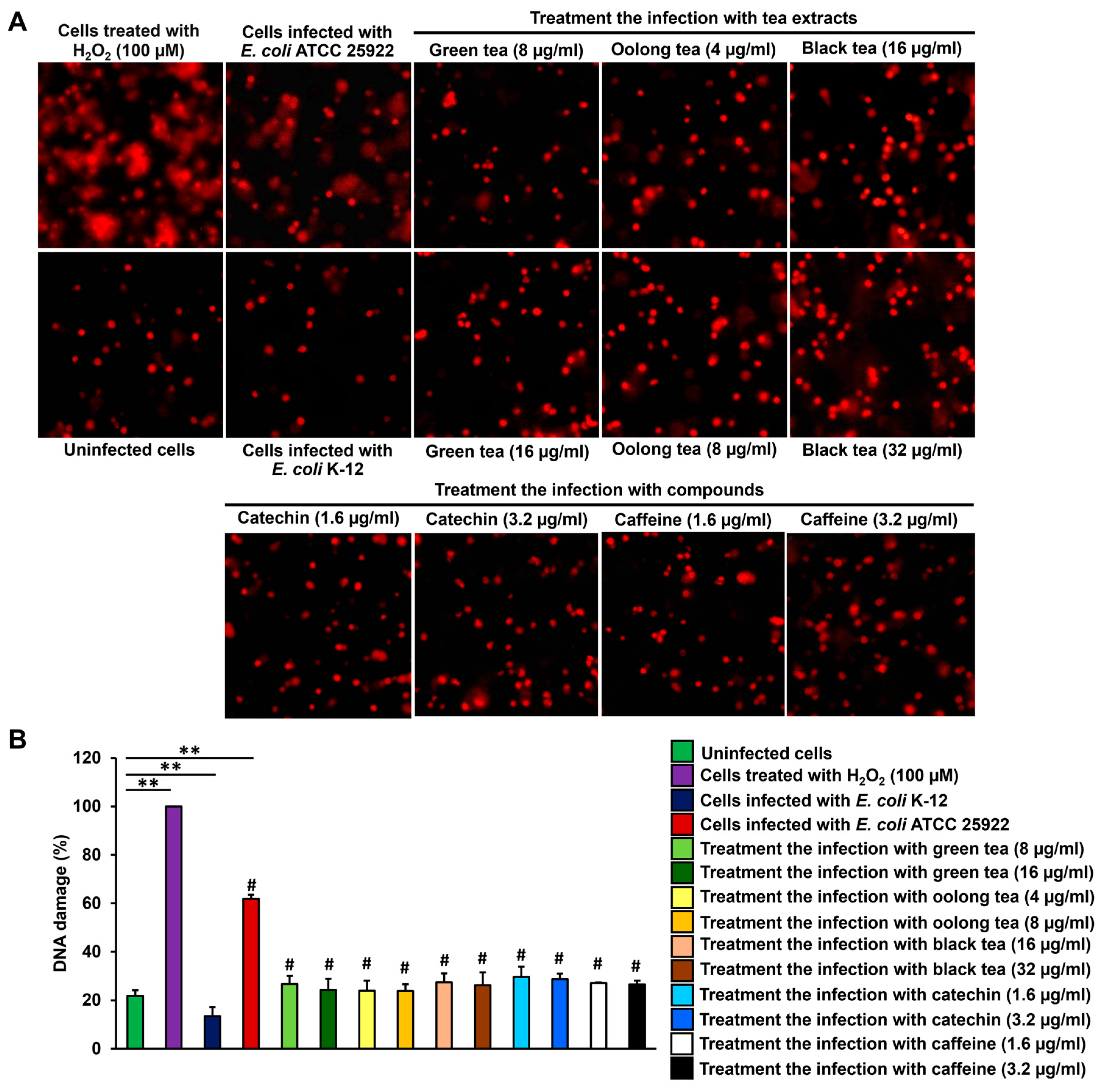
The potential of tea extracts to mitigate DNA damage in Caco-2 cells infected with E. coli ATCC 25922 was examined. Caco-2 cells were exposed to E. coli ATCC 25922 and E. coli K-12 at a MOI of 400 for 4 h. DNA damage was detected in the infected cells through the alkaline comet assay, which revealed DNA tails (Figure 3). The degree of DNA damage in cells infected with E. coli ATCC 25922 was 61.89 ± 1.67%, compared to 21.83 ± 2.25% in uninfected control cells. Treatment with tea extracts reduced DNA damage in the infected cells, with oolong tea (8 µg/mL) showing significant protective effects, resulting in a DNA damage percentage of 23.84 ± 2.81% (Figure 3). Moreover, green tea (8–16 µg/mL) and black tea (16–32 µg/mL) extracts could inhibit the DNA damage of infected cells at 24.24 ± 4.61–26.72 ± 3.33% and 26.21 ± 5.35–27.36 ± 3.71% (Figure 3). In addition, catechin and caffeine compounds (1.6–3.2 µg/mL) also supported to protect the DNA damage from the infection, with showed the percentage of DNA damage of 26.56 ± 1.50–29.66 ± 4.23% (Figure 3). In this study was used the hydrogen peroxide and E. coli K-12 as positive and negative control that represented the DNA damage of 100% and 13.42 ± 3.75%, respectively.
Figure 2. Efficacy of tea extracts and phytochemical compounds for inhibition of transient infection with E. coli ATCC 25922 (A) at a MOI of 400 on Caco-2 cells for 4 h when compared to control infected epithelial cells and transient infection with E. coli K-12 (B).
Antibiotics 14 00886 g002
Figure 3. Comet assay (A) and percentage of DNA damage (B) of Caco-2 cells infected with E. coli ATCC 25922 and treatment with tea extracts and compounds. # Tea extracts and compounds significantly reduce the DNA damage of cells from the infection compared to the E. coli ATCC 25922 infection without the treatments (p < 0.05). Values are mean ± standard deviation; n = 3 samples. ** indicated p < 0.001. All data are used to analyze between two groups using one-way ANOVA and Tukey’s test for multiple comparisons.
Antibiotics 14 00886 g003

2.6. Effect of Colon Cell Cycle After Infection with E. coli-Producing Colibactin Toxin by Treatment with Tea Extracts and Phytochemical Compounds

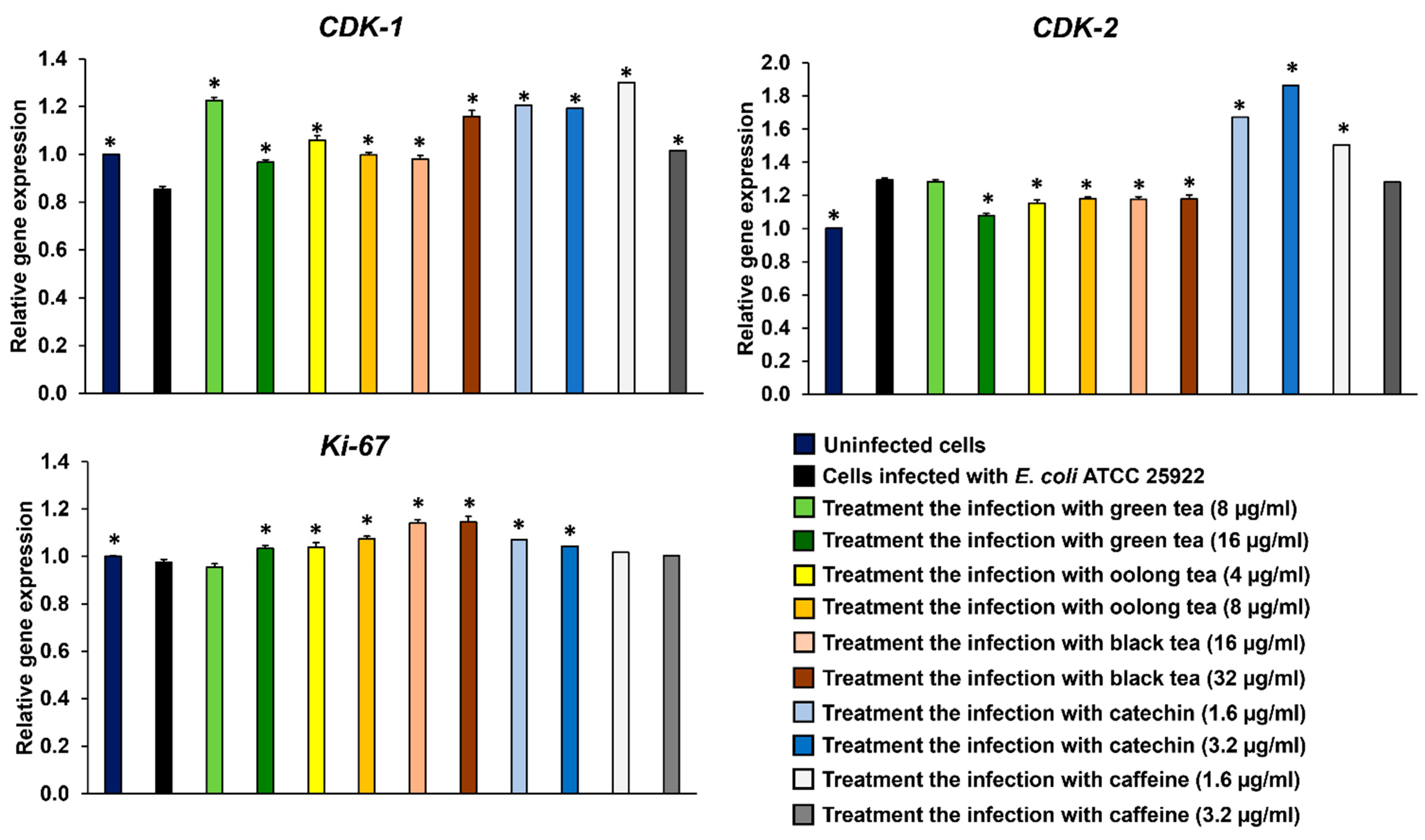
After Caco-2 cells were infected with E. coli ATCC 25922 for 4 h and treated with tea extracts for 3 days, gene expression related to the cell cycle, including CDK-1, CDK-2, and Ki-67, was detected by qRT-PCR. The results revealed that E. coli ATCC 25922 could interfere with the cell cycle arrest of Caco-2 cells by suppressing the gene expression of CDK-1 and upregulating CDK-2 compared to uninfected cells (Figure 4). Moreover, all tea extracts could upregulate CDK-1 gene expression in Caco-2 cells infected with E. coli ATCC 25922. Additionally, green tea (16 µg/mL), oolong tea (4 and 8 µg/mL), and black tea (16 and 32 µg/mL) were able to suppress CDK-2 gene expression compared to Caco-2 cells infected with E. coli ATCC 25922 and uninfected cells. These results were correlated with exogenous compounds, catechin and caffeine, which could upregulate the CDK-1 and CDK-2 genes (Figure 4). However, the expression of Ki-67 in Caco-2 cells infected with E. coli ATCC 25922 did not differ from that in uninfected cells. The extracts of green tea (16 µg/mL), oolong tea (4 and 8 µg/mL) and black tea (16 and 32 µg/mL), and catechin compound (1.6 and 3.2 µg/mL) were able to upregulate Ki-67 gene expression in Caco-2 cells during infection (Figure 4).

3. Discussion

Most therapeutic drugs used to treat bacterial infections are derived from chemical synthetic agents, which are costly, have greater side effects, and may contribute to the development of antibiotic-resistant bacterial strains [29]. This study identifies promising natural compounds as potential drug candidates via protection from infectious diseases using natural products. Tea has been a popular beverage for centuries, especially green tea, oolong tea, and black tea, which are rich in nutraceuticals, dietary supplements, traditional remedies, pharmaceutical intermediates, and ingredients for synthetic drug development [30]. In this study, extracts from C. sinensis leaves, including green tea, oolong tea, and black tea, were prepared using hot water. In the present study, we employed water extraction to investigate the bioactive effects of tea. This approach was chosen because water infusion is the most common and practical method of tea preparation for human consumption, thereby providing results that are more representative of real-life dietary intake. Moreover, water is a non-toxic solvent that ensures the safety and physiological relevance of the extracts [31,32]. Additionally, the extracts were tested against E. coli ATCC 25922, a strain known for producing colibactin toxin. This strain carries colibactin genes and is linked to diarrheal diseases and bacteremia [33,34]. The results showed that all tea leaf extracts were effective in inhibiting E. coli ATCC 25922, as demonstrated by agar well diffusion. Moreover, tea compounds such as catechin and caffeine have also demonstrated inhibitory effects on E. coli that produces colibactin toxin. The antimicrobial activity of tea is due to the presence of catechin polyphenols, which damage the bacterial cell membrane. Epigallocatechin (EGC) and epigallocatechin gallate (EGCG) exhibit the highest antimicrobial effects, with EGCG being the most established in terms of bactericidal activity [35]. In E. coli, EGCG induces the destruction of biofilms by damaging bacterial membranes and degrading exopolysaccharides [36]. Oolong tea extract has been shown to exhibit synergistic antibacterial activity against Streptococcus mutans due to its monomeric polyphenols [37]. Additionally, polyphenols in black tea have been found to reduce the expression of virulence traits in clinical isolates of Shigella dysenteriae and enteropathogenic E. coli (EPEC P2 1265) strains [38]. Green tea has higher levels of catechins compared to oolong, black, and dark teas. Specifically, oolong tea contains about half the amount of EGCG found in green tea, while its polymerized polyphenol content is double that of green tea [39]. Caffeine could inhibit uropathogenic E. coli biofilm formation [40]. Tea compounds such as polyphenols and flavonoids can improve gut health by promoting healthy bacterial growth, protecting against bacterial pathogens, and reducing inflammation [41,42]. Moreover, flavonoids such as naringin, naringenin, genistein, kaempferol, anthocyanins, epigallocatechin-3-gallate, and baicalein have been reported to exhibit therapeutic effects for various diseases [43]. However, the other constituents, such as alkaloids, flavonols, and phenolic acids, may also play important roles in tea bioactivity. Comprehensive profiling of tea samples has been reported previously using advanced analytical methods such as UHPLC-DAD, where 20 major compounds were quantified across 121 samples representing six tea types (black tea, green tea, yellow tea, white tea, oolong tea and dark tea). That study demonstrated that the chemical diversity of tea depends strongly on processing and identified biomarkers such as gallocatechin, EGCG, and epicatechin gallate for tea classification [44]. Not only does the type of tea reveal differing contents of catechins and other compounds, but many factors such as species, season, age of the leaves (plucking position), climate, and horticultural conditions (soil, water, mineral fertilizers, etc.) are also relevant [45].
Several studies have reported on the phytochemical compounds in tea leaves and their inhibitory effects on bacterial pathogens. In this study, we evaluated the ability of tea extracts to protect against infection, DNA damage, and cell cycle alterations in colon cells. This research presents a novel report on the effects of tea extracts and the tea compounds catechin and caffeine on E. coli ATCC 25922 infections in colon cells. E. coli ATCC 25922 produces colibactin, a genotoxin, due to the presence of its clb gene cluster responsible for toxin synthesis [34,46]. These toxins can cause DNA double-strand breaks (DSBs) in both in vitro [24] and in vivo studies [25]. To investigate the effects of this toxin, mammalian colon cells (Caco-2) were used and infected with E. coli ATCC 25922. The impact on these cells was compared with that of E. coli K-12, a strain that does not produce colibactin, at a multiplicity of infection (MOI) of 400 bacteria per cell for 4 h. The morphological changes in the epithelial cells were monitored over a period of 3 days. The results revealed that cytopathic effects on cell morphology, such as megalocytosis (giant cells) and cellular debris, were decreased when treated with tea extracts, catechin and caffeine. Additionally, green tea, oolong tea, black tea, and the tea compounds catechin and caffeine provided significant protection to Caco-2 cells against E. coli ATCC 25922 infection, maintaining cell viability above 80% compared to Caco-2 cells infected with E. coli ATCC 25922 without the addition of tea extracts or compounds. Zhang et al. [47] showed that oral administration of tea polyphenols (400 mg/kg) alleviated oxidative stress and intestinal damage in C57BL/6 mice with bacterial infection. This protective effect was achieved by inhibiting myeloperoxidase activity and MDA production, as well as by boosting the activity of various intestinal antioxidant enzymes. Catechin-mediated photoprotection of human skin against bacterial infection has also been reported [48]. Moreover, tea catechins, especially EGCG, have been shown to inhibit various viral infections, including HIV, Hepatitis B virus, Rotavirus, and SARS-CoV-2, by interfering with binding protein receptors on host cells [49,50,51,52]. In addition, caffeine can significantly reduce the survival and cytotoxicity of bacteria [40].
Therefore, tea extracts, catechin and caffeine could protect against DNA damage in cells caused by bacterial colibactin (E. coli ATCC 25922) infection. These results are supported by both in vitro and in vivo studies. Tea catechins, such as EC, EGC, ECG, and EGCG, can help reduce hydroxyl radical-induced DNA single-strand breaks and base damage by facilitating the rapid chemical repair of DNA radicals. These studies also explain the mechanism through which catechins transfer hydrogen atoms and/or electrons to radical sites on DNA [53,54]. Caffeine can inhibit the DNA damage response (DDR) pathway, which is responsible for repairing DNA breaks and preventing cell division until the damage is resolved [55,56]. Likewise, black tea theaflavins (20 or 50 µM) have been shown to prevent oxidative stress-induced DNA damage in rat normal liver epithelial RL-34 cells [57]. Oral administration of a green tea polyphenol extract equivalent to a human intake of 500 mL of green tea per day for 5 days was found to protect lymphocytes, and to a lesser degree, internal organs like colonocytes and hepatocytes, from oxidative DNA damage in rats [58]. Following infection with bacterial colibactin, CDK-1 gene expression in Caco-2 cells was found to be downregulated compared to uninfected cells. This reduction in CDK-1 expression resulted in changes to cell morphology and an increase in the expression of various differentiation markers. Cells with lower CDK-1 expression exhibited a higher incidence of double-strand breaks (DSBs), failed to activate CHK2 expression, and could not sustain G2/M cell cycle arrest [59,60]. This phenomenon is associated with elevated DNA damage in Caco-2 cells infected with E. coli ATCC 25922. Additionally, the downregulation of CDK-1 led to the accumulation of cells with abnormal numbers of mitotic organelles, chromosomal abnormalities, and polyploidy [60,61].
Additionally, CDK-2 gene expression was upregulated in Caco-2 cells infected with E. coli ATCC 25922. CDK-2 is a core cell-cycle regulator that is active from the late G1 phase throughout the S phase [62]. In addition to promoting cell cycle progression, CDK-2 has been described as playing a positive role in cell cycle arrest during the DNA damage response, particularly at the G2/M checkpoint [63]. Accumulating evidence indicates that CDK-2 promotes hyperproliferation of cells and induces the progression of cancer cells [64]. This study confirms previous research showing that colibactin toxin induces DNA double-strand breaks, chromosome aberrations, and cell cycle arrest at the G2/M phase [24,25]. Moreover, infection with E. coli-producing colibactin toxin has been shown to increase the number of tumors in various colorectal cancer (CRC) mouse models [65,66]. This study highlights new findings that tea extracts, including green tea, oolong tea, and black tea, as well as the tea compounds catechin and caffeine, can maintain the normal cell cycle of colon cells infected with E. coli-producing colibactin toxin through the modulation of CDK-1 and CDK-2. Furthermore, tea extracts and compounds, particularly catechin, were found to promote the proliferation of colon cells infected with E. coli-producing colibactin toxin, as evidenced by the upregulation of Ki-67 gene expression. The expression of Ki-67 is strongly associated with cell proliferation and growth [67]. Overall, the findings suggest that tea extracts and tea compounds exhibit potent antimicrobial and cytoprotective properties, making them promising candidates for preventing or mitigating colibactin-associated infections. Further studies are warranted to elucidate the mechanisms underlying these effects and to explore the clinical applications of tea extracts in combating bacterial infections and related diseases. For safety reasons, consuming tea catechins and caffeine from a typical cup of tea is generally considered safe for most people. However, the safe amount of catechins and caffeine varies among individuals. Exceeding the recommended daily intake of approximately 400 mg is not advised [68].

4. Materials and Methods

4.1. Tea Extraction

Dried tea leaves, including green tea (batch number: 170323), oolong tea (batch number: 200323), and black tea (batch number: 211122), were sourced from the Royal Project Foundation, Chiang Mai, Thailand, a government-supported project that complies with national agricultural regulations. No wild plant collection was performed for this study. The leaves were extracted using hot distilled water (100 °C) at a 1:10 (w/v) ratio for 1 h, with the process repeated three times [69]. The resulting extract was filtered through Whatman No. 1 filter paper, and the filtrate was concentrated using a rotary evaporator (BÜCHI, Flawil, Switzerland) at 50 °C under a reduced pressure of 50 mbar. It was then dried via freeze drying (LABCONCO, Kansas, MO, USA). The dried extracts were stored at −20 °C and dissolved in sterile deionized water at a concentration of 500 mg/mL prior to use. Tea extracts at this high concentration (500 mg/mL) were used in the antibacterial assay to evaluate maximal bacterial inhibition, whereas lower concentrations were used in cell infection experiments to avoid cytotoxicity.

4.2. High Performance Liquid Chromatography (HPLC)

High-performance liquid chromatography (HPLC) was employed to determine the levels of catechin, EGCG, caffeine, and theaflavin in tea extracts. All standard compounds were purchased from Sigma-Aldrich (Darmstadt, Germany). The extracts and standard compounds were filtered through a 0.45 µm sterile microfilter. Subsequently, 20 µL of the filtered sample was injected into the HPLC system (Agilent Technologies 1200 series, Santa Clara, CA, USA). Analysis was conducted using a UV detector set at 276 nm with an Agilent Eclipse XDB-C18 column (4.6 × 150 mm, 5 µm). The mobile phase used for separation consisted of mobile phase A (deionized water) and mobile phase B (methanol, RCl Labscan, Bangkok, Thailand). Gradient elution was performed with varying ratios of mobile phases A and B as follows: 100:0 at 0 min, 50:50 at 15 min, and 0:100 at 30 min. The HPLC conditions included a flow rate of 1.0 mL/min and a total run time of 30 min. The concentrations of catechin, EGCG, caffeine, and theaflavin in the tea extracts were quantified by comparison with the corresponding standard compounds.

4.3. Bacterial Strains

The standard strain of E. coli ATCC 25922 and E. coli K-12 strain BW25113 were procured from Department of Biotechnology, College of Life Sciences, Ritsumeikan University, Kusatsu, Shiga, Japan.

4.4. Agar Well Diffusion Assay

The bacterial inhibition assay was performed using the agar well diffusion method [70]. Bacterial strains were cultured in Mueller–Hinton (MH) broth (HiMedia, Maharashtra, India) and incubated at 37 °C for 18–24 h. The bacterial suspensions were adjusted to a McFarland standard No. 0.5 (108 CFU/mL, OD600 = 0.08–0.1) and spread evenly onto Mueller Hinton agar (MHA). Wells were created using a 10 mm cork borer, and 100 µL of sample was placed in each well. After an incubation period, the inhibition zone diameter was measured. Gentamycin (Bio Basic Inc., Amherst, NY, USA) at a concentration of 1 mg/mL was used as a positive control.

4.5. Cytotoxicity Assay

The cytotoxicity of tea extracts or compounds was evaluated using the MTT assay. Human colon (Caco-2) cells were purchased from the Cell Engineering Division, RIKEN BioResource Research Center (BRC) (Cell No. RCB0988, Ibaraki, Japan) and cultured in Dulbecco’s Modified Eagle Medium (DMEM; Gibco, Grand Island, NY, USA) supplemented with 10% (v/v) heat-inactivated fetal bovine serum (HyClone™, Pittsburgh, PA, USA), 100 Units/mL penicillin, and 100 µg/mL streptomycin (CAISSON, Smithfield, UT, USA). The cells were incubated at 37 °C in a 5% CO2 incubator (SHEL LAB, Cornelius, OR, USA), then washed twice with phosphate-buffered saline (PBS, pH 7.4) and trypsinized using 0.05% (v/v) trypsin-EDTA solution (CAISSON, Smithfield, UT, USA). Caco-2 cells (5 × 10⁴ cells/mL) were seeded into 96-well plates and incubated at 37 °C in a 5% CO2 incubator for 24 h. After incubation, different concentrations of the samples were added to the wells, and the plates were further incubated for 48 h under the same conditions. MTT solution (Bio Basic Inc., Amherst, NY, USA) was then added to each well and incubated for 4 h. The resulting blue formazan crystals were dissolved in dimethyl sulfoxide (DMSO), and absorbance was measured at 540 nm and 630 nm using a microplate reader (EZ Read 2000, Biochrom, Cambridge, UK). Cell viability was calculated as a percentage by comparing the absorbance values of treated cells to those of the control group [71].

4.6. Transient Infection and Treatment

Caco-2 cells (5 × 10⁴ cells/mL) were seeded in a 96-well plate (Corning, New York, NY, USA) and incubated for 24 h. E. coli ATCC 25922 (colibactin-producing) and E. coli K-12 (non-colibactin-producing) were grown in Tryptic Soy Broth (TSB) (HiMedia, Maharashtra, India) for 8–12 h. The bacteria were harvested by centrifuge (Hettich GmbH & Co. KG, Tuttlingen, Germany) at 6000 rpm for 5 min, washed twice with PBS, and resuspended in DMEM at a concentration of 5 × 10⁸ CFU/mL (OD600 = 0.4–0.5). Caco-2 cells were washed with PBS and infected at a multiplicity of infection (MOI) of 400 with the bacterial strains in the presence of tea extracts or compounds. Sample concentrations were selected to maintain cell viability above 80%. The plate was incubated at 37 °C in a 5% CO2 environment for 4 h. After infection, cells were washed with PBS to remove bacteria and further incubated with DMEM supplemented with 10% fetal bovine serum and 200 µg/mL gentamicin for 3–4 days.
Cells were washed with PBS, fixed with 4% formaldehyde (Sigma-Aldrich, Darmstadt, Germany) in PBS for 20 min, and stained with 1% methylene blue (TPC™, Thailand Pharmaceutical Chemical Co., Ltd., Bangkok, Thailand) for 1 h. Morphological observations were conducted using an inverted fluorescence microscope (ECLIPSE Ts2R-FL, Nikon, Tokyo, Japan). Uninfected cells without treatment displayed a confluent monolayer, whereas cells infected with E. coli ATCC 25922 showed a cytopathic phenotype characterized by reduced cell density, megalocytosis, and markers of cell death such as apoptotic bodies and cell debris, as reported by Bossuet-Greif et al. [72]. Cell viability was quantified using the MTT assay following the protocol of Umthong et al. [71].

4.7. DNA Damage by Alkaline Comet Assay

The inhibition of DNA damage in Caco-2 colon cells was evaluated following infection with the colibactin-producing strain E. coli ATCC 25922 and subsequent treatment with tea extracts or compounds. This was analyzed using the alkaline comet assay as described by Kothandapani et al. [73] and Sawant et al. [74]. Caco-2 cells (5 × 10⁴ cells/mL) were cultured in DMEM at 37 °C in 5% CO2 for 24 h. Samples at non-toxic concentrations (maintaining over 80% cell viability) were introduced to the cultures, followed by infection with E. coli at a multiplicity of infection (MOI) of 400. Hydrogen peroxide (100 µM) served as a positive control for DNA damage. Plates were incubated for 4 h, then cells were washed, trypsinized, and collected by centrifugation at 3000 rpm and 4 °C for 5 min.
Cells were resuspended in PBS, counted, and adjusted to 10,000 cells. Each sample was mixed with 1% low melting point agarose, layered onto slides pre-coated with 1% agarose, and topped with 0.5% low melting point agarose. After solidification, slides were lysed in buffer; 2.5 M NaCl (RCl Labscan, Bangkok, Thailand), 10 mM Tris (Research Organics, Cleveland, OH, USA), 100 mM EDTA (VWR, Leuven, Belgium), 1% Triton X-100 (Amresco, Solon, OH, USA), pH 10 for 1 h at 4 °C in the dark, then placed in an electrophoresis tank with alkaline buffer; 50 mM NaOH (RCl Labscan, Bangkok, Thailand), 1 mM EDTA (VWR, Belgium), pH > 12) at 4 °C. Electrophoresis (Cleaver Scientific Ltd., Rugby, Warwickshire, UK) was performed at 0.7 V/cm and 300 mA for 30 min. Afterward, slides were neutralized in 0.4 M Tris-HCl buffer (pH 7.5) for 10 min, stained with propidium iodide (PI, Abcam, Cambridge, MA, Middlesex) for 30 min in the dark, and observed using an inverted fluorescence microscope. DNA comet tails from a minimum of 200 cells per slide were analyzed, and the percentages of DNA damage were calculated and compared to the uninfected control cells.

4.8. Quantitative Real-Time Polymerase Chain Reaction (qRT-PCR)

The mRNA expression of cell cycle-related genes, including CDK-1, CDK-2, and Ki-67, was analyzed following infection and treatment with tea extracts or compounds using qRT-PCR. Caco-2 cells (2 × 10⁵ cells/mL) were cultured in DMEM at 37 °C in a 5% CO2 incubator for 24 h. Tea extracts were introduced to the cell culture, then infected with E. coli ATCC 25922 (a colibactin-producing strain) at a multiplicity of infection (MOI) of 400 for a duration of 4 h. Bacterial growth was then inhibited using gentamicin, and the plates were incubated for 3 days at 37 °C in a 5% CO2 environment. After incubation, cells were harvested and washed three times with 1X PBS prior to RNA extraction. Total mRNA from the Caco-2 cells was extracted using TRIzol™ reagent (Invitrogen, Carlsbad, CA, USA) and converted to cDNA using the ReverTra Ace® qPCR RT Master Mix (TOYOBO, Osaka, Japan). The qPCR reaction mixture included 100 ng of cDNA, 2X SensiFAST SYBR® No-ROX Mix (BIOLINE, London, UK), and primers at a concentration of 400 nM each (forward and reverse). The realtime PCR was conducted in QIAquant real-time PCR cyclers (Qiagen, Venlo, Netherlands). The qPCR conditions were as follows: initial denaturation at 95 °C for 2 min, followed by 40 cycles of denaturation at 95 °C for 5 s and annealing/extension at 60 °C for 30 s. Cycle threshold (Ct) values for the target genes were normalized to the internal control gene GAPDH to determine relative expression levels. Primer sequences for the target genes were designed based on previously published studies [75,76,77]. CDK-1 (forward primer: 5′-GTCCGCAACAGGGAAGAACAG-3′, reverse primer: 5′-CGAAAGCCAAGATAAGCAACTCC-3′), CDK-2 (forward primer: 5′-TATGAGTCAAAAGGCGGGGT-3′, reverse primer: 5′-TGCACTCATCCACCAATCCT-3′) and Ki-67 (forward primer: 5′-GTTGGTCTCGCGTAAGTCAA-3′, reverse primer: 5′-CAGACTCCACGTCTCTTCCC-3′).

4.9. Statistical Analysis

All experiments were conducted independently on three separate occasions. Data were expressed as mean ± SD values of the independent samples. Statistical analysis of the results from both treatment and control groups was performed using T-tests and ANOVA in IBM SPSS Statistics 20.

5. Conclusions

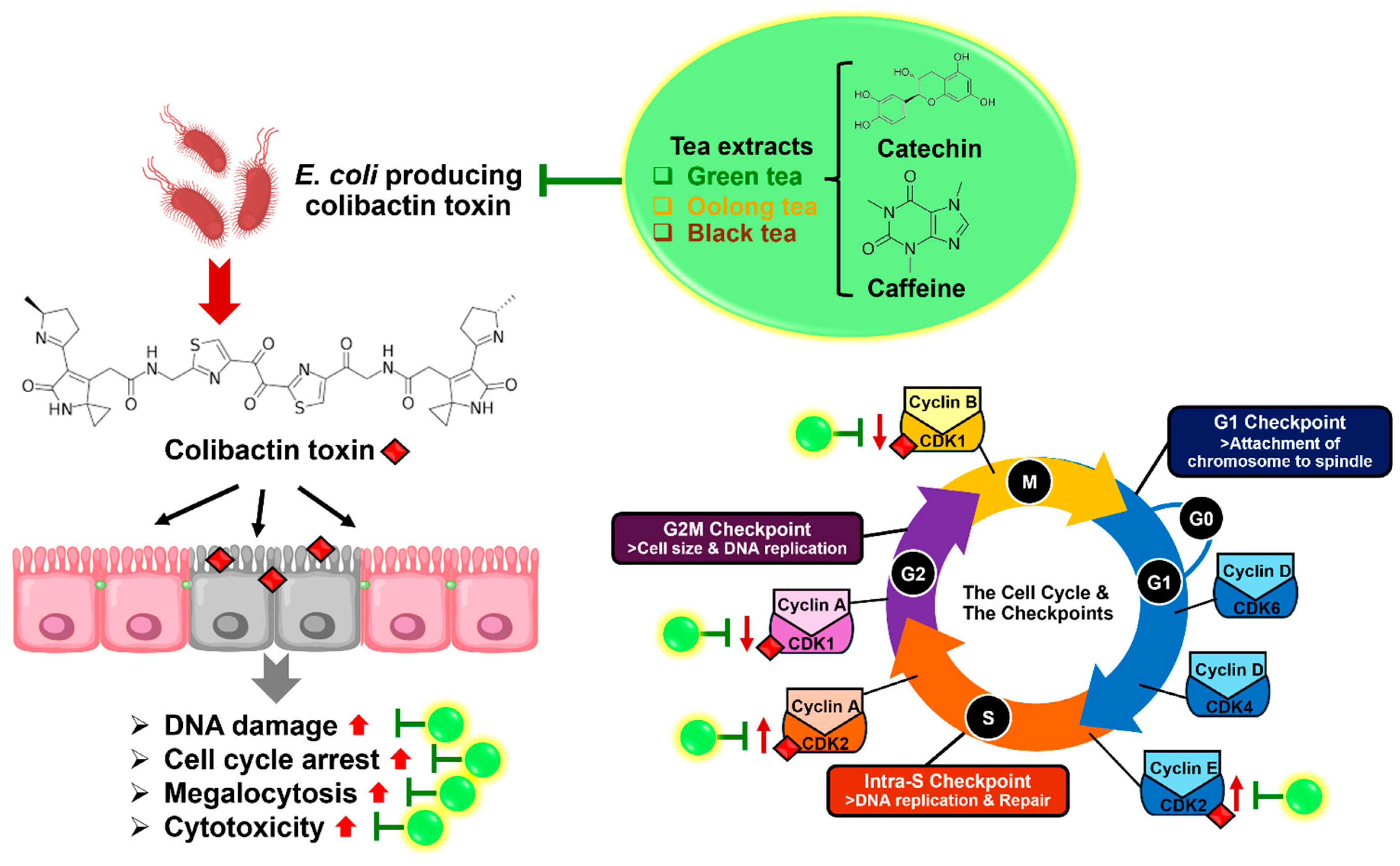
This study highlights the potential of tea extracts, particularly green tea, oolong tea and black tea, and tea compounds (catechin and caffeine), as natural agents for combating bacterial infections, specifically those caused by E. coli ATCC 25922, a colibactin-producing strain. These tea extracts and tea compounds demonstrated significant antimicrobial activity against E. coli-producing colibactin toxins. Furthermore, tea extracts and catechin protected colon cells (Caco-2) from cytopathic effects, including megalocytosis and cell debris, induced by E. coli ATCC 25922 infection. All tea extracts and tea compounds also promoted higher cell viability and reduced DNA damage caused by colibactin-induced double-strand breaks. In terms of the cell cycle, the study revealed that E. coli ATCC 25922 downregulated CDK-1 and upregulated CDK-2 in Caco-2 cells, leading to cell cycle arrest and increased DNA damage. Interestingly, treatment with tea extracts and tea compounds restored normal cell cycle function by upregulating CDK-1 and suppressing CDK-2 expression, while tea extracts and catechin promoted cell proliferation via Ki-67 expression (Figure 5). Overall, this research demonstrates the therapeutic potential of tea extracts as a natural alternative to synthetic antimicrobial agents. They not only inhibit bacterial infection but also protect host cells from the detrimental effects of E. coli-producing colibactin toxin, making them promising candidates for future treatment strategies against bacterial infections and related diseases.

Supplementary Materials

The following supporting information can be downloaded at: https://www.mdpi.com/article/10.3390/antibiotics14090886/s1, Figure S1: Antibacterial activity of tea extracts; green tea (G), oolong tea (O), black tea (B), and compounds; catechin (CC) and caffeine (CF) against E. coli K-12 (A,C) and E. coli ATCC 25922 (B,D) by agar well diffusion assay. Sterile distilled water (N) and gentamycin (P) were used as a negative and positive controls for bacterial inhibition.

Author Contributions

Conceptualization, W.T. and T.K.; methodology, W.T. and T.K.; validation, W.T. and T.K.; formal analysis, W.T. and T.K.; investigation, W.T. and T.K.; writing—original draft preparation, W.T., Y.T. and T.K.; writing—review and editing, W.T., Y.T. and T.K.; visualization, W.T. and T.K.; supervision, Y.T.; funding acquisition, T.K. All authors have read and agreed to the published version of the manuscript.

Funding

This research was financially supported by CMU Junior Research Fellowship Program (Grant No. JRCMU2566R_039).

Data Availability Statement

The datasets used and analyzed are available from the corresponding author upon reasonable request.

Acknowledgments

This research study was supported by CMU Junior Research Fellowship Program, Chiang Mai University, Chiang Mai, Thailand. Department of Biology, Faculty of Science and Natural Extracts and Innovative Products for Alternative Healthcare Research Group, Faculty of Science, Chiang Mai University, Chiang Mai were also acknowledged. We are grateful to Hisaaki Mihara from the Department of Biotechnology, College of Life Sciences, Ritsumeikan University, Kusatsu, Shiga, Japan, for providing the bacterial strains and offering valuable guidance during the experiments.

Conflicts of Interest

The authors declare no conflicts of interest.

References

  1. Cabrera, C.; Artacho, R.; Giménez, R. Beneficial effects of green tea—A review. J. Am. Coll. Nutr. 2006, 25, 79–99. [Google Scholar] [CrossRef]
  2. Pekal, A.; Drozdz, P.; Biesaga, M.; Pyrzynska, K. Evaluation of the antioxidant properties of fruit and flavoured black teas. Eur. J. Nutr. 2011, 50, 681–688. [Google Scholar] [CrossRef] [PubMed]
  3. Koch, W. Handbook of Dietary Phytochemicals; Springer: Singapore, 2020; pp. 1–29. [Google Scholar]
  4. Jiang, H.; Yu, F.; Qin, L.; Zhang, N.; Cao, Q.; Schwab, W.; Li, D.; Song, C. Dynamic change in amino acids, catechins, alkaloids, and gallic acid in six types of tea processed from the same batch of fresh tea (Camellia sinensis L.) leaves. J. Food Compos. Anal. 2019, 77, 28–38. [Google Scholar] [CrossRef]
  5. Dou, J.; Lee, V.S.; Tzen, J.T.; Lee, M.R. Identification and comparison of phenolic compounds in the preparation of oolong tea manufactured by semifermentation and drying processes. J. Agric. Food Chem. 2007, 55, 7462–7468. [Google Scholar] [CrossRef]
  6. Salman, S.; Yılmaz, C.; Gökmen, V.; Özdemir, F. Effects of fermentation time and shooting period on amino acid derivatives and free amino acid profiles of tea. LWT-Food Sci. Technol. 2021, 137, 110481. [Google Scholar] [CrossRef]
  7. McKay, D.; Blumberg, J. The role of tea in human health: An update. J. Am. Coll. Nutr. 2002, 21, 1–13. [Google Scholar] [CrossRef]
  8. Wang, Y.; Ho, C.T. Polyphenolic chemistry of tea and coffee: A century of progress. J. Agric. Food Chem. 2009, 57, 8109–8114. [Google Scholar] [CrossRef]
  9. Sabu, M.C.; Kuttan, R. Antidiabetic activity of medicinal plants and its relationship with antioxidant property. J. Ethnopharmacol. 2002, 81, 155–160. [Google Scholar] [CrossRef]
  10. Khan, N.; Mukhtar, H. Cancer and metastasis: Prevention and treatment by green tea. Cancer Metastasis Rev. 2010, 29, 435–445. [Google Scholar] [CrossRef] [PubMed]
  11. Suzuki, Y.; Miyoshi, N.; Isemura, M. Health-promoting effects of green tea. Proc. Jpn. Acad. Ser. B 2012, 88, 88–101. [Google Scholar] [CrossRef] [PubMed]
  12. Mandel, S.A.; Avramovich-Tirosh, Y.; Reznichenko, L.; Zheng, H.; Weinreb, O.; Amit, T.; Youdim, M.B. Multifunctional activities of green tea catechins in neuroprotection. Neurosignals 2005, 14, 46–60. [Google Scholar] [CrossRef]
  13. Kuriyama, S.; Shimazu, T.; Ohmori, K.; Kikuchi, N.; Nakaya, N.; Nishino, Y.; Tsuji, I.; Tsubono, Y. Green tea consumption and mortality due to cardiovascular disease, cancer, and all causes in Japan: The Ohsaki study. J. Am. Med. Assoc. 2006, 296, 1255–1265. [Google Scholar] [CrossRef] [PubMed]
  14. Thavanesan, N. The putative effects of green tea on body fat: An evaluation of the evidence and a review of the potential mechanisms. Br. J. Nutr. 2011, 106, 1297–1309. [Google Scholar] [CrossRef]
  15. Shareef, S.H.; Ibrahim, I.A.A.; Alzahrani, A.R.; Al-Medhtiy, M.H.; Abdulla, M.A. Hepatoprotective effects of methanolic extract of green tea against thioacetamide-induced liver injury in Sprague Dawley rats. Saudi J. Biol. Sci. 2022, 29, 564–573. [Google Scholar] [CrossRef]
  16. Wang, Y.Q.; Li, Q.S.; Zheng, X.Q.; Lu, J.L.; Liang, Y.R. Antiviral effects of green tea EGCG and its potential application against COVID-19. Molecules 2021, 26, 3962. [Google Scholar] [CrossRef]
  17. Engleberg, N.C.; DiRita, V.J.; Dermody, T.S. Mechanisms of Microbial Disease, 4th ed.; Lippincott Williams & Wilkins: Philadelphia, PA, USA, 2013; pp. 1–864. [Google Scholar]
  18. Clermont, O.; Christenson, J.K.; Denamur, E.; Gordon, D.M. The Clermont Escherichia coli phylotyping method revisited: Improvement of specificity and detection of new phylogroups. Environ. Microbiol. Rep. 2013, 5, 58–65. [Google Scholar] [CrossRef]
  19. Smith, J.L.; Fratamico, P.M.; Gunther, N.W. Extraintestinal pathogenic Escherichia coli. Foodborne Pathog. Dis. 2007, 4, 134–163. [Google Scholar] [CrossRef] [PubMed]
  20. Oshima, K.; Toh, H.; Ogura, Y.; Sasamoto, H.; Morita, H.; Park, S.H.; Ooka, T.; Iyoda, S.; Taylor, T.D.; Hayashi, T.; et al. Complete genome sequence and comparative analysis of the wildtype commensal Escherichia coli strain SE11 isolated from a healthy adult. DNA Res. 2008, 15, 375–386. [Google Scholar] [CrossRef] [PubMed]
  21. Núñez-Samudio, V.; Pimentel-Peralta, G.; De La Cruz, A.; Landires, I. Multidrug-resistant phenotypes of genetically diverse Escherichia coli isolates from healthy domestic cats. Sci. Rep. 2024, 14, 11260. [Google Scholar] [CrossRef] [PubMed]
  22. Escobar-Páramo, P.; Grenet, K.; Le Menac’h, A.; Rode, L.; Salgado, E.; Amorin, C.; Gouriou, S.; Picard, B.; Rahimy, M.C.; Andremont, A.; et al. Large-scale population structure of human commensal Escherichia coli isolates. Appl. Environ. Microbiol. 2004, 70, 5698–5700. [Google Scholar] [CrossRef]
  23. Nowrouzian, F.L.; Wold, A.E.; Adlerberth, I. Escherichia coli strains belonging to phylogenetic group B2 have superior capacity to persist in the intestinal microflora of infants. J. Infect. Dis. 2005, 191, 1078–1083. [Google Scholar] [CrossRef] [PubMed]
  24. Nougayrède, J.P.; Homburg, S.; Taieb, F.; Boury, M.; Brzuszkiewicz, E.; Gottschalk, G.; Buchrieser, C.; Hacker, J.; Dobrindt, U.; Oswald, E. Escherichia coli induces DNA double-strand breaks in eukaryotic cells. Science 2006, 313, 848–851. [Google Scholar] [CrossRef]
  25. Cuevas-Ramos, G.; Petit, C.R.; Marcq, I.; Boury, M.; Oswald, E.; Nougayrède, J.P. Escherichia coli induces DNA damage in vivo and triggers genomic instability in mammalian cells. Proc. Natl. Acad. Sci. USA 2010, 107, 11537–11542. [Google Scholar] [CrossRef]
  26. Sakurikar, N.; Eastman, A. Critical reanalysis of the methods that discriminate the activity of CDK2 from CDK1. Cell Cycle 2016, 15, 1184–1188. [Google Scholar] [CrossRef] [PubMed]
  27. Markelova, N.N.; Semenova, E.F.; Sineva, O.N.; Sadykova, V.S. The role of cyclomodulins and some microbial metabolites in bacterial microecology and macroorganism carcinogenesis. Int. J. Mol. Sci. 2022, 23, 11706. [Google Scholar] [CrossRef]
  28. Tomkovich, S.; Yang, Y.; Winglee, K.; Gauthier, J.; Mühlbauer, M.; Sun, X.; Mohamadzadeh, M.; Liu, X.; Martin, P.; Wang, G.P.; et al. Locoregional effects of microbiota in a preclinical model of colon carcinogenesis. Cancer Res. 2017, 77, 2620–2632. [Google Scholar] [CrossRef] [PubMed]
  29. Muteeb, G.; Rehman, M.T.; Shahwan, M.; Aatif, M. Origin of antibiotics and antibiotic resistance, and their impacts on drug development: A narrative review. Pharmaceuticals 2023, 16, 1615. [Google Scholar] [CrossRef]
  30. Khan, N.; Mukhtar, H. Tea and health: Studies in humans. Curr. Pharm. Des. 2013, 19, 6141–6147. [Google Scholar] [CrossRef]
  31. Farhoosh, R.; Golmovahhed, G.A.; Khodaparast, M.H.H. Antioxidant activity of various extracts of old tea leaves and black tea wastes (Camellia sinensis L.). Food Chem 2007, 100, 231–236. [Google Scholar] [CrossRef]
  32. Piran, F.; Khoshkhoo, Z.; Hosseini, S.E.; Azizi, M.H. Controlling the antioxidant activity of green tea extract through encapsulation in chitosan-citrate nanogel. J. Food Qual. 2020, 2020, 7935420. [Google Scholar] [CrossRef]
  33. Johnson, J.R.; Johnston, B.; Kuskowski, M.A.; Nougayrede, J.P.; Oswald, E. Molecular epidemiology and phylogenetic distribution of the Escherichia coli pks genomic island. J. Clin. Microbiol. 2008, 46, 3906–3911. [Google Scholar] [CrossRef]
  34. Kaewkod, T.; Tobe, R.; Tragoolpua, Y.; Mihara, H. Medicinal plant extracts protect epithelial cells from infection and DNA damage caused by colibactin-producing Escherichia coli, and inhibit the growth of bacteria. J. Appl. Microbiol. 2021, 130, 769–785. [Google Scholar] [CrossRef]
  35. Gopal, J.; Muthu, M.; Paul, D.; Kim, D.H.; Chun, S. Bactericidal activity of green tea extracts: The importance of catechin containing nanoparticles. Sci. Rep. 2016, 6, 19710. [Google Scholar] [CrossRef] [PubMed]
  36. Maeyama, R.; Kwon, I.K.; Mizunoe, Y.; Anderson, J.M.; Tanaka, M.; Matsuda, T. Novel bactericidal surface: Catechin-loaded surface-erodible polymer prevents biofilm formation. J. Biomed. Mater. Res. 2005, 75 Pt A, 146–155. [Google Scholar] [CrossRef] [PubMed]
  37. Sasaki, H.; Matsumoto, M.; Tanaka, T.; Maeda, M.; Nakai, M.; Hamada, S.; Ooshima, T. Antibacterial activity of polyphenol components in oolong tea extract against Streptococcus mutans. Caries Res. 2004, 38, 2–8. [Google Scholar] [CrossRef]
  38. Kiran, S.; Ratho, R.K.; Sharma, P.; Harjai, K.; Capalash, N.; Tiwari, R.P. Effect of black tea (Camellia sinensis) on virulence traits of clinical isolates of Shigella dysenteriae and Escherichia coli EPEC P2 1265 strain. Eur. Food Res. Technol. 2010, 231, 763–770. [Google Scholar] [CrossRef]
  39. Sajilata, M.G.; Bajaj, P.R.; Singhal, R.S. Tea polyphenols as nutraceuticals. Compr. Rev. Food Sci. Food Saf. 2008, 7, 229–254. [Google Scholar] [CrossRef]
  40. Rathi, B.; Gupta, S.; Kumar, P.; Kesarwani, V.; Dhanda, R.S.; Kushwaha, S.K.; Yadav, M. Anti-biofilm activity of caffeine against uropathogenic E. coli is mediated by curli biogenesis. Sci. Rep. 2022, 12, 18903. [Google Scholar] [CrossRef] [PubMed]
  41. Bond, T.; Derbyshire, E. Tea compounds and the gut microbiome: Findings from trials and mechanistic studies. Nutrients 2019, 11, 2364. [Google Scholar] [CrossRef]
  42. Ağagündüz, D.; Cocozza, E.; Cemali, Ö.; Bayazıt, A.D.; Nanì, M.F.; Cerqua, I.; Morgillo, F.; Saygılı, S.K.; Canani, R.B.; Amero, P.; et al. Understanding the role of the gut microbiome in gastrointestinal cancer: A review. Front. Pharmacol. 2023, 14, 1130562. [Google Scholar] [CrossRef]
  43. Açar, Y.; Ağagündüz, D.; De Cicco, P.; Capasso, R. Flavonoids: Their putative neurologic roles, epigenetic changes, and gut microbiota alterations in Parkinson’s disease. Biomed. Pharmacother. 2023, 168, 115788. [Google Scholar] [CrossRef]
  44. Chen, Y.; Lai, L.; You, Y.; Gao, R.; Xiang, J.; Wang, G.; Yu, W. Quantitative analysis of bioactive compounds in commercial teas: Profiling catechin alkaloids, phenolic acids, and flavonols using targeted statistical approaches. Foods 2023, 12, 3098. [Google Scholar] [CrossRef]
  45. Fernandez, P.L.; Fernando, P.; Martin, J.; Gonzalez, A.G. Study of catechin and xanthine tea profiles as geographical tracers. J. Agric. Food Chem. 2002, 50, 1833–1839. [Google Scholar] [CrossRef]
  46. Minogue, T.D.; Daligault, H.A.; Davenport, K.W.; Bishop Lilly, K.A.; Broomall, S.M.; Bruce, D.C.; Chain, P.S.; Chertkov, O.; Coyne, S.R.; Freitas, T.; et al. Complete genome assembly of Escherichia coli ATCC 25922, a serotype O6 reference strain. Genome Announc. 2014, 2, 5. [Google Scholar] [CrossRef]
  47. Zhang, L.; Gui, S.; Wang, J.; Chen, Q.; Zeng, J.; Liu, A.; Chen, Z.; Lu, X. Oral administration of green tea polyphenols (TP) improves ileal injury and intestinal flora disorder in mice with Salmonella typhimurium infection via resisting inflammation, enhancing antioxidant action and preserving tight junction. J. Funct. Foods 2020, 64, 103654. [Google Scholar] [CrossRef]
  48. Hsu, S. Green tea and the skin. J. Am. Acad. Dermatol. 2005, 52, 1049–1059. [Google Scholar] [CrossRef]
  49. Kawai, K.; Tsuno, N.H.; Kitayama, J.; Okaji, Y.; Yazawa, K.; Asakage, M.; Hori, N.; Watanabe, T.; Takahashi, K.; Nagawa, H. Epigallocatechin gallate, the main component of tea polyphenol, binds to CD4 and interferes with gp120 binding. J. Allergy Clin. Immunol. 2003, 112, 951–957. [Google Scholar] [CrossRef]
  50. Li, J.; Zhou, L.; Zhang, Y. Studies on the effects of tea catechins against hepatitis B virus infection. Chin. J. Prev. Med. 2001, 35, 404–407. [Google Scholar]
  51. Apostolides, Z.; Selematsela, M. Anti-Retroviral Agent in Combination with Tea Polyphenol for the Treatment of Viral Infections. PCT International Application WO 2003002126 A1, 9 January 2003. [Google Scholar]
  52. Storozhuk, M.; Lee, S.; Lee, J.I.; Park, J. Green tea consumption and the COVID-19 omicron pandemic era: Pharmacology and epidemiology. Life 2023, 13, 852. [Google Scholar] [CrossRef] [PubMed]
  53. Anderson, R.F.; Fisher, L.J.; Hara, Y.; Harris, T.; Mak, W.B.; Melton, L.D.; Packer, J.E. Green tea catechins partially protect DNA from ·OH radical-induced strand breaks and base damage through fast chemical repair of DNA radicals. Carcinogenesis 2001, 22, 1189–1193. [Google Scholar] [CrossRef] [PubMed]
  54. Truong, V.L.; Jeong, W.S. Cellular defensive mechanisms of tea polyphenols: Structure-activity relationship. Int. J. Mol. Sci. 2021, 22, 9109. [Google Scholar] [CrossRef]
  55. Mercer, J.R.; Gray, K.; Figg, N.; Kumar, S.; Bennett, M.R. The methyl xanthine caffeine inhibits DNA damage signaling and reactive species and reduces atherosclerosis in ApoE−/− mice. Arterioscler. Thromb. Vasc. Biol. 2012, 32, 2461–2467. [Google Scholar] [CrossRef] [PubMed]
  56. Chung, W.H. Pleiotropic effects of caffeine leading to chromosome instability and cytotoxicity in eukaryotic microorganisms. J. Microbiol. Biotechnol. 2021, 31, 171. [Google Scholar] [CrossRef] [PubMed]
  57. Feng, Q.; Torii, Y.; Uchida, K.; Nakamura, Y.; Hara, Y.; Osawa, T. Black tea polyphenols, theaflavins, prevent cellular DNA damage by inhibiting oxidative stress and suppressing cytochrome P450 1A1 in cell cultures. J. Agric. Food Chem. 2002, 50, 213–220. [Google Scholar] [CrossRef] [PubMed]
  58. Kager, N.; Ferk, F.; Kundi, M.; Wagner, K.H.; Mišík, M.; Knasmüller, S. Prevention of oxidative DNA damage in inner organs and lymphocytes of rats by green tea extract. Eur. J. Nutr. 2010, 49, 227–234. [Google Scholar] [CrossRef] [PubMed]
  59. Li, L.; Wang, J.; Hou, J.; Wu, Z.; Zhuang, Y.; Lu, M.; Zhang, Y.; Zhou, X.; Li, Z.; Xiao, W.; et al. CDK1 interplays with Oct4 to repress differentiation of embryonic stem cells into trophectoderm. FEBS Lett. 2012, 586, 4100–4107. [Google Scholar] [CrossRef]
  60. Neganova, I.; Tilgner, K.; Buskin, A.; Paraskevopoulou, I.; Atkinson, S.P.; Peberdy, D.; Passos, J.F.; Lako, M. CDK1 plays an important role in the maintenance of pluripotency and genomic stability in human pluripotent stem cells. Cell Death Dis. 2014, 5, e1508. [Google Scholar] [CrossRef]
  61. Zhang, W.W.; Zhang, X.J.; Liu, H.X.; Chen, J.; Ren, Y.H.; Huang, D.G.; Zou, X.H.; Xiao, W. Cdk1 is required for self-renewal of mouse embryonic stem cells. J. Cell. Biochem. 2011, 112, 942–948. [Google Scholar] [CrossRef]
  62. Zardavas, D.; Pondé, N.; Tryfonidis, K. CDK4/6 blockade in breast cancer: Current experience and future perspectives. Expert Opin. Investig. Drugs 2017, 26, 1357–1372. [Google Scholar] [CrossRef] [PubMed]
  63. Cerqueira, A.; Santamaría, D.; Martínez-Pastor, B.; Cuadrado, M.; Fernández-Capetillo, O.; Barbacid, M. Overall CDK activity modulates the DNA damage response in mammalian cells. J. Cell Biol. 2009, 187, 773–780. [Google Scholar] [CrossRef]
  64. Wang, J.; Yang, T.; Xu, G.; Liu, H.; Ren, C.; Xie, W.; Wang, M. Cyclin-dependent kinase 2 promotes tumor proliferation and induces radio resistance in glioblastoma. Transl. Oncol. 2016, 9, 548–556. [Google Scholar] [CrossRef] [PubMed]
  65. Buc, E.; Dubois, D.; Sauvanet, P.; Raisch, J.; Delmas, J.; Darfeuille-Michaud, A.; Pezet, D.; Bonnet, R.; Battista, J.R. High prevalence of mucosa-associated E. coli producing cyclomodulin and genotoxin in colon cancer. PLoS ONE 2013, 8, e56964. [Google Scholar] [CrossRef] [PubMed]
  66. Prorok-Hamon, M.; Friswell, M.K.; Alswied, A.; Roberts, C.L.; Song, F.; Flanagan, P.K.; Knight, P.; Codling, C.; Marchesi, J.R.; Winstanley, C.; et al. Colonic mucosa-associated diffusely adherent afaC+ Escherichia coli expressing lpfA and pks are increased in inflammatory bowel disease and colon cancer. Gut 2014, 63, 761–770. [Google Scholar] [CrossRef]
  67. Li, L.T.; Jiang, G.; Chen, Q.; Zheng, J.N. Ki67 is a promising molecular target in the diagnosis of cancer (Review). Mol. Med. Rep. 2015, 11, 1566–1572. [Google Scholar] [CrossRef] [PubMed]
  68. Hu, J.; Webster, D.; Cao, J.; Shao, A. The safety of green tea and green tea extract consumption in adults—Results of a systematic review. Regul. Toxicol. Pharmacol. 2018, 95, 412–433. [Google Scholar] [CrossRef]
  69. Eric, W.C.; Eu, Y.S.; Pei, P.T.; Yon, P.L. Antioxidant and antibacterial properties of green, black and herbal teas of Camellia sinensis. J. Pharm. Pharmacogn. Res. 2011, 3, 266–272. [Google Scholar]
  70. Clinical and Laboratory Standards Institute (CLSI). Performance Standards for Antimicrobial Disk Susceptibility Tests, 13th ed.; Clinical and Laboratory Standards Institute: Wayne, PA, USA, 2018. [Google Scholar]
  71. Umthong, S.; Phuwapraisirisan, P.; Puthong, S.; Chanchao, C. In vitro antiproliferative activity of partially purified Trigona laeviceps propolis from Thailand on human cancer cell lines. BMC Complement. Altern. Med. 2011, 11, 37. [Google Scholar] [CrossRef]
  72. Bossuet-Greif, N.; Belloy, M.; Boury, M.; Oswald, E.; Nougayrede, J. Protocol for HeLa cells infection with Escherichia coli strains producing colibactin and quantification of the induced DNA-damage. Bio-protocol 2017, 7, e2325. [Google Scholar] [CrossRef]
  73. Kothandapani, A.; Sawant, A.; Dangeti, V.S.; Sobol, R.W.; Patrick, S.M. Epistatic role of base excision repair and mismatch repair pathways in mediating cisplatin cytotoxicity. Nucleic Acids Res. 2013, 41, 7332–7343. [Google Scholar] [CrossRef]
  74. Sawant, A.; Floyd, A.M.; Dangeti, M.; Lei, W.; Sobol, R.W.; Patrick, S.M. Differential role of base excision repair proteins in mediating cisplatin cytotoxicity. DNA Repair 2017, 51, 46–59. [Google Scholar] [CrossRef]
  75. Chang, H.Y.; Shih, M.H.; Huang, H.C.; Tsai, S.R.; Juan, H.F.; Lee, S.C. Middle infrared radiation induces G2/M cell cycle arrest in A549 lung cancer cells. PLoS ONE 2013, 8, e54117. [Google Scholar] [CrossRef] [PubMed]
  76. Tadesse, S.; Anshabo, A.T.; Portman, N.; Lim, E.; Tilley, W.; Caldon, C.E.; Wang, S. Targeting CDK2 in cancer: Challenges and opportunities for therapy. Drug Discov. Today 2020, 25, 406–413. [Google Scholar] [CrossRef] [PubMed]
  77. Chierico, L.; Rizzello, L.; Guan, L.; Joseph, A.S.; Lewis, A.; Battaglia, G. The role of the two splice variants and extranuclear pathway on Ki-67 regulation in non-cancer and cancer cells. PLoS ONE 2017, 12, e0171815. [Google Scholar] [CrossRef] [PubMed]
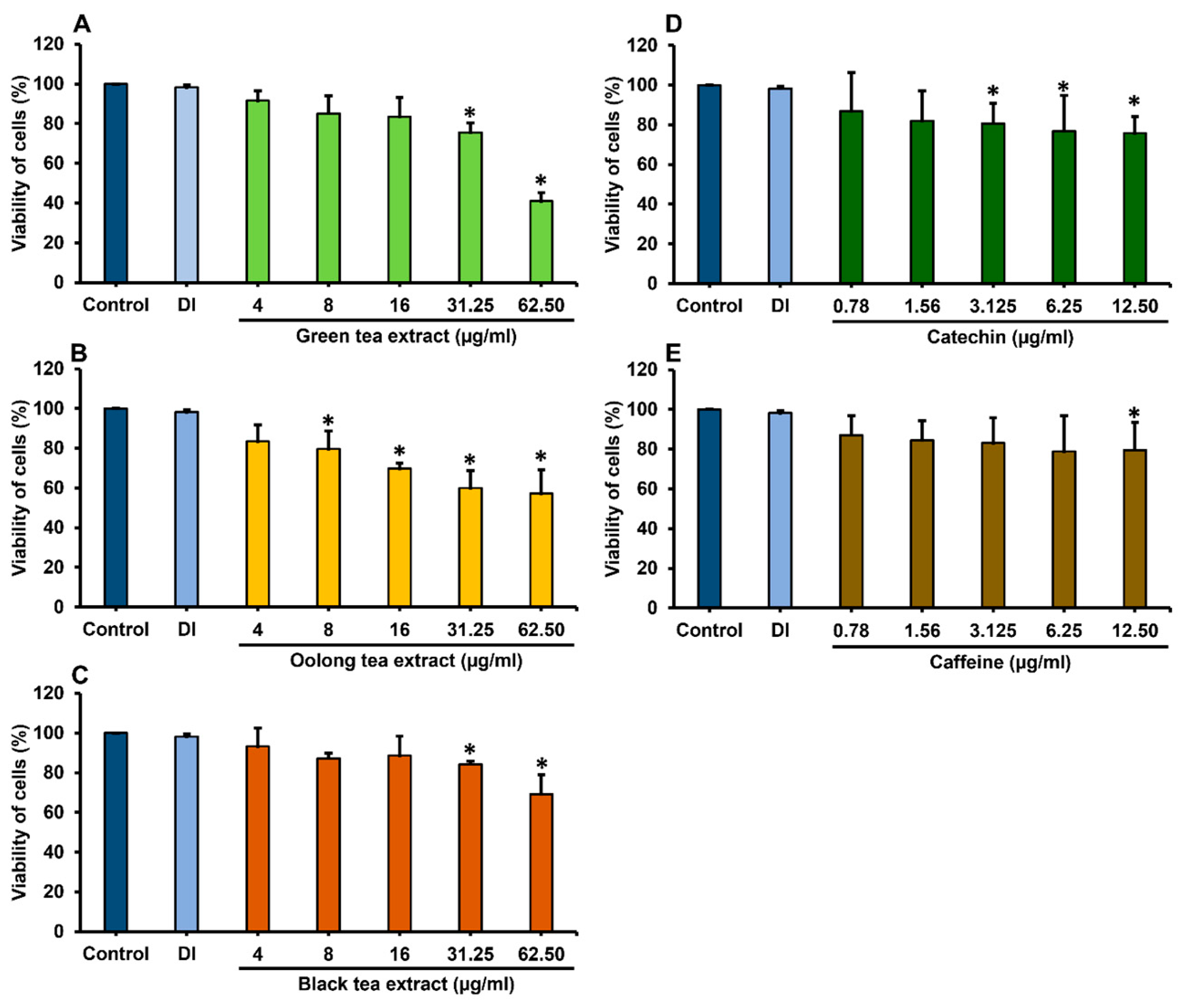
Figure 1. Viability of Caco-2 cells after treatment with green tea (A), oolong tea (B) and black tea (C) extracts and phytochemical compounds of catechin (D) and caffeine (E) for 48 h. * Data are significantly different compared to control cells (p < 0.05). Values are presented as mean ± standard deviation; n = 3 samples. Deionized water (DI) was used as a vehicle control.
Antibiotics 14 00886 g001
Figure 4. Expression of CDK-1, CDK-2, and Ki-67 genes associated with the cell cycle in Caco-2 cells following infection with colibactin-producing E. coli and treatment with tea extracts and compounds. * Data are significantly different compared to cells infected with E. coli ATCC 25922 (p < 0.05). Values are presented as mean ± standard deviation; n = 3 samples. Statistical analysis between groups was performed using one-way ANOVA and Tukey’s test for multiple comparisons.
Antibiotics 14 00886 g004
Figure 5. Proposed mechanism of action by which tea extracts (green, oolong, and black tea) and tea compounds (catechin and caffeine) inhibit E. coli colibactin toxin infection in colon cells. Tea extracts and compounds showed strong antimicrobial activity against colibactin-producing E. coli. They protected Caco-2 cells from cytopathic effects (megalocytosis, cell debris), improved cell viability, and reduced colibactin-induced DNA damage. E. coli infection downregulated CDK-1 and upregulated CDK-2, causing cell cycle arrest and DNA damage. Tea extracts and compounds restored normal cell cycle regulation (↑CDK-1, ↓CDK-2) and catechin promoted cell proliferation via Ki-67 expression.
Antibiotics 14 00886 g005
Table 1. Phytochemical compounds contents in tea extracts.
Tea ExtractsChemical Compound Contents (mg/mL)
CatechinEGCGCaffeineTheaflavin
Green tea12.680 ± 0.019 a37.622 ± 0.163 a56.909 ± 0.444 aND a
Oolong tea233.475 ± 13.320 b251.839 ± 8.751 b2.658 ± 0.053 b2.192 ± 0.297 b
Black teaND c269.714 ± 7.860 cND cND c
The data represented with different superscript letters (a, b, c), are expressed as mean ± SD from three independent experiments and indicate significant differences in the chemical compound contents among the tea extracts (p < 0.05). ND: not detected.
Table 2. Antibacterial activity by agar well diffusion.
SamplesConcentration (mg/mL)Zone of Inhibition (mm) *
E. coli ATCC 25922E. coli K-12
Tea extracts
Green tea50016.3 ± 4.515.0 ± 1.7
Oolong tea50018.3 ± 0.516.0 ± 1.0
Black tea50013.7 ± 1.314.0 ± 1.0
Phytochemical compounds
Catechin5.016.3 ± 0.615.7 ± 0.6
Caffeine5.019.3 ± 0.619.0 ± 0.0
Gentamycin1.023.8 ± 0.526.3 ± 0.6
* The results are presented as mean ± SD of triplicate independent experiments.
Table 3. Viability of cells after treatment with tea extracts during infection of Caco-2 cells with E. coli ATCC 25922 and E. coli K-12 at MOI of 400 for 4 h.
SamplesE. coli ATCC 25922 Infection with Caco-2 CellsE. coli K-12 Infection with Caco-2 Cells
Concentration (µg/mL)Viability of Cells (%) *Concentration (µg/mL)Viability of Cells (%) *
Tea extracts
Green tea886.15 ± 7.03 b880.58 ± 16.05 a
1683.13 ± 1.99 b1686.47 ± 12.59 a
Oolong tea487.93 ± 14.22 a488.01 ± 8.03 a
890.78 ± 2.76 b882.31 ± 11.49 a
Black tea1681.42 ± 9.96 a 1686.98 ± 11.88 a
3287.58 ± 9.74 b3288.28 ± 11.25 a
Phytochemical compounds
Catechin1.681.77 ± 9.47 b1.680.97 ± 7.04 a
3.285.59 ± 9.74 b3.284.49 ± 7.13 a
Caffeine1.676.43 ± 13.64 a1.683.58 ± 11.42 a
3.279.81 ± 6.46 a3.285.29 ± 9.19 a
No treatment-69.74 ± 4.18 a-78.92 ± 0.62 a
* The data represented with different superscript letters (a, b) are expressed as mean ± SD from three independent experiments and indicate significant differences in cell viability percentages after treatment with tea extracts or compounds compared to untreated controls (p < 0.05). Statistical analysis between groups was performed using one-way ANOVA, followed by Tukey’s test for multiple comparisons.
Disclaimer/Publisher’s Note: The statements, opinions and data contained in all publications are solely those of the individual author(s) and contributor(s) and not of MDPI and/or the editor(s). MDPI and/or the editor(s) disclaim responsibility for any injury to people or property resulting from any ideas, methods, instructions or products referred to in the content.