Review

Targeting Bacterial Biofilms on Medical Implants: Current and Emerging Approaches

by
Alessandro Calogero Scalia
and
Ziba Najmi
*
Department of Health Sciences, Center for Translational Research on Autoimmune & Allergic Diseases, CAAD, University of Piemonte Orientale, Corso Trieste 15/A, 28100 Novara, Italy
*
Author to whom correspondence should be addressed.
Antibiotics 2025, 14(8), 802; https://doi.org/10.3390/antibiotics14080802
Submission received: 2 July 2025 / Revised: 30 July 2025 / Accepted: 1 August 2025 / Published: 6 August 2025
(This article belongs to the Special Issue Antibacterial and Antibiofilm Properties of Biomaterial)

Abstract

Biofilms are structured communities of microorganisms encased in a self-produced extracellular matrix, and they represent one of the most widespread forms of microbial life on Earth. Their presence poses serious challenges in both environmental and clinical settings. In natural and industrial systems, biofilms contribute to water contamination, pipeline corrosion, and biofouling. Clinically, biofilm-associated infections are responsible for approximately 80% of all microbial infections, including endocarditis, osteomyelitis, cystic fibrosis, and chronic sinusitis. A particularly critical concern is their colonization of medical devices, where biofilms can lead to chronic infections, implant failure, and increased mortality. Implantable devices, such as orthopedic implants, cardiac pacemakers, cochlear implants, urinary catheters, and hernia meshes, are highly susceptible to microbial attachment and biofilm development. These infections are often recalcitrant to conventional antibiotics and frequently necessitate surgical revision. In the United States, over 500,000 biofilm-related implant infections occur annually, with prosthetic joint infections alone projected to incur revision surgery costs exceeding USD 500 million per year—a figure expected to rise to USD 1.62 billion by 2030. To address these challenges, surface modification of medical devices has emerged as a promising strategy to prevent bacterial adhesion and biofilm formation. This review focuses on recent advances in chemical surface functionalization using non-antibiotic agents, such as enzymes, chelating agents, quorum sensing quenching factors, biosurfactants, oxidizing compounds and nanoparticles, designed to enhance antifouling and mature biofilm eradication properties. These approaches aim not only to prevent device-associated infections but also to reduce dependence on antibiotics and mitigate the development of antimicrobial resistance.

Graphical Abstract

1. Bacterial Biofilm

1.1. Biofilm Formation

Biofilm formation is a multi-step, complex process in which bacteria transition from a free-floating planktonic state to a sessile form. This process is influenced by various external factors, including desiccation, nutrient limitation, extreme temperature or pH, antimicrobial agents, gravitational and hydrodynamic forces, the nature of the colonized surfaces, quorum sensing, secondary messengers, and other signaling molecules [1,2]. Figure 1 illustrates the major stages of biofilm development, typically divided into four main steps:

1.1.1. Initial Attachment

The first stage of biofilm formation is bacterial attachment, initiated by favorable interactions between planktonic cells and a surface. Bacterial transport to these surfaces occurs through Brownian motion, sedimentation, or convection [3]. Upon contact, adhesion is governed by a balance of attractive and repulsive interfacial force. When attractive forces dominate, attachment occurs; conversely, if repulsive forces prevail, adhesion is hindered [4]. This initial, reversible phase relies primarily on non-specific physical interactions, including electrostatic forces, hydrophobic interactions, and Lifshitz–van der Waals forces [5].
Both inert and biological surfaces can support bacterial adhesion, and virtually any material in contact with a bacterial suspension may serve as a substrate for biofilm development [6,7]. Surface properties, such as roughness and topography [7,8], hydrophobicity [8,9], surface charge [8,10], and the presence of conditioning films (layers of adsorbed nutrients and molecules) [11,12], strongly influence the efficiency and speed of bacterial attachment.

1.1.2. Irreversible Adhesion and Aggregation

Irreversible bacterial attachment is governed by short-range interactions, including dipole–dipole forces, hydrogen bonds, ionic and covalent bonding, and hydrophobic interactions, all of which are facilitated by bacterial adhesins. Additionally, adhesion structures, such as flagella, pili/fimbriae, and non-fimbriae adhesins, which extend from the bacterial surface, play crucial roles in establishing initial contact with substrates [5].
Flagella, filamentous structures responsible for bacterial motility, support both swimming (in liquids) and swarming (on moist surfaces), enabling cells to move toward favorable environments and initiate surface attachment [13,14]. A study on four types of Escherichia coli, with and without flagella, demonstrated that flagellated strains exhibited stronger and more consistent attachment to six different types of plastics commonly found in freshwater systems [14]. Furthermore, two flagellar mutant strains of Salmonella enteritidis, SE-ΔflhD and SE-ΔfligE, showed reduced sedimentation rates and lower biofilm biomass compared to wild-type (WT) strains [15].
Pili/fimbriae, another class of filamentous surface appendage, contribute to intercellular interactions and early surface colonization in several bacterial species [16], including Klebsiella pneumoniae [17], Streptococcus agalactiae [18], Clostridium difficile [19], and Acinetobacter baumanii [20]. For instance, Pseudomonas aeruginosa utilizes pili for both surface adhesion and twitching motility [17]. In K. pneumoniae, both type 1 and type 3 fimbriae facilitate adhesion to abiotic surfaces and promote mature biofilm formation. In contrast, in Erwinia amylovora, only type 1 fimbriae are involved in this process; mutant strains lacking type 1 fimbriae exhibit significantly reduced adherence to abiotic surfaces compared to wild-type strains [17]. In addition to physical appendages, pathogens may express adhesins that bind to eukaryotic cell receptors, promoting host invasion. For example, Yersinia pseudotuberculosis and Y. enterocolitica produce invasion proteins that bind β1 integrins on M-cells, facilitating cellular entry [21].
Following irreversible adhesion, bacteria undergo multiplication and division to form microcolonies. This process is initiated by the production of extracellular polymeric substances (EPSs) [22,23,24]. EPS is a key component of the biofilm matrix, promoting cell-to-cell cohesion and surface attachment through hydrophobic and ionic bridging interactions [25,26]. In addition to structural stability, EPS plays diverse roles, including surface adherence, cellular communication, biofilm architecture formation, water retention, signaling, protection, nutrient entrapment, symbiosis, and genetic exchange. The intracellular secondary messenger cyclic di-guanosine monophosphate (c-di-GMP) further promotes the transition to irreversible adhesion by upregulating the synthesis of EPS and surface-associated structures [25]. The composition and functional roles of the extracellular matrix will be described in detail in Section 3.

1.1.3. Biofilm Maturation

The third stage of biofilm formation is maturation, during which microcolonies grow and develop into more complex structures. This phase is facilitated by the secretion of signaling molecules, such as quorum sensing (QS) autoinducers and intercellular communication signals, by the attached bacterial cells, leading to the expression of biofilm-specific genes. The release of EPS plays a pivotal role in stabilizing the biofilm structure and protecting it from antimicrobial agents [26]. Following EPS production, water-filled channels form within the biofilm, functioning like a circulatory system that distributes nutrients to cells and removes waste products [27]. The maturation process typically occurs in two distinct phases: stage I involves cell-to-cell interactions and the synthesis of autoinducer signaling molecules, such as N-acylated homoserine lactone (AHL); stage II is characterized by an increase in the size and thickness of the microcolonies, reaching up to approximately 100 µm, considered a defining feature of established microcolonies [28]. During maturation, bacterial motility is restricted, and the expression of genes involved in surface structure formation is downregulated. Consequently, the gene expression profile of sessile cells within the biofilm differs significantly from that of planktonic cells. For instance, DNA microarray analyses have revealed altered expression of over 70 genes in P. aeruginosa after five days of biofilm growth [29]. Additionally, QS facilitates intra- and interspecies communication through the production and detection of autoinducers (AIs), small signaling molecules that allow bacteria to sense population density and coordinate gene expression accordingly [30]. The mechanisms and types of QS signals will be discussed in detail in Section 4.3.

1.1.4. Dispersal/Detachment

Biofilm detachment, also known as dispersal, represents the terminal stage of biofilm development. It is considered a survival strategy, allowing bacterial cells to exit the biofilm and initiate a new cycle of biofilm formation elsewhere. Dispersal is a complex, regulated process influenced by environmental cues, signal transduction pathways, and effector molecules [31].
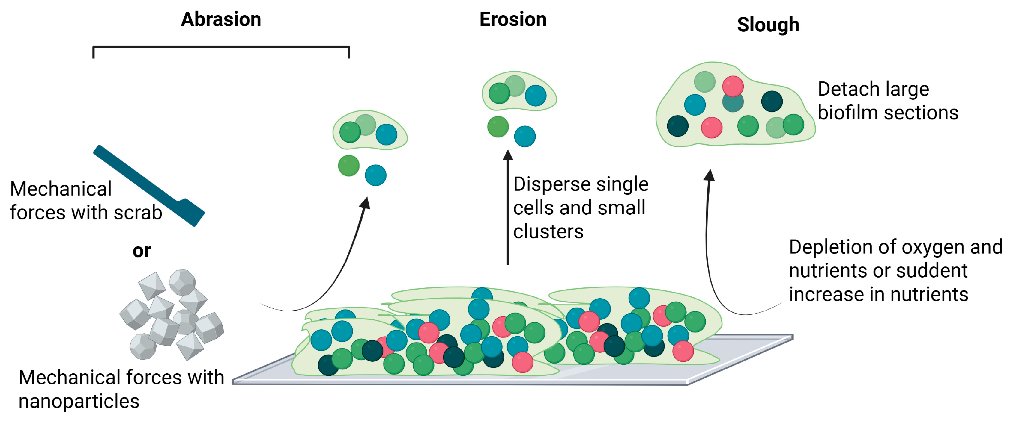
Although dispersal mechanisms vary among bacterial species, the overall process can generally be divided into three key stages: detachment of cells from the microcolonies, movement of cells to a new surface, and reattachment to initiate colonization of the new substrate [31,32]. Detachment can occur via both active and passive mechanisms: (i) active dispersal is initiated by the biofilm-resident cells themselves in response to environmental stressors such as antimicrobial exposure, matrix-degrading enzymes, and nutrient depletion; (ii) passive detachment occurs due to external physical forces such as shear forces or fluid flow [33,34,35]. Active dispersal typically results in the rapid release of planktonic cells or microcolonies, through mechanisms collectively referred to as follows (Figure 2): (i) abrasion is a mechanical loss due to collision with particles in the surrounding liquid [36]; (ii) erosion is a gradual release of individual cells or small clusters from the biofilm matrix [37,38]; (iii) sloughing is the sudden detachment of large biofilm sections, often caused by oxygen or nutrient depletion at the base of the biofilm or abrupt increases in nutrient availability in the surrounding liquid [38].
During active dispersal, bacteria typically upregulate genes involved in motility (e.g., flagella synthesis) and EPS degradation, while downregulating genes responsible for EPS production, surface attachment, and fimbriae synthesis [39]. Another regulatory mechanism involves c-di-GMP signaling. Inhibiting c-di-GMP pathways or reducing intracellular c-di-GMP levels can suppress biofilm formation and stimulate dispersal [31]. For example, oxygen limitation can promote c-di-GMP degradation, triggering detachment; glucose supplementation has been shown to reduce intracellular c-di-GMP, leading to increased flagella expression and enhanced dispersal capacity [35]. In addition to environmental cues, several physicochemical parameters and intrinsic bacterial properties, such as autolysis, also contribute to biofilm dispersal [35,39].

2. Clinical Problem of Biofilm

2.1. Biofilm-Mediated Immune Evasion and Virulence Strategies

A major clinically relevant challenge posed by biofilms is the ability of the bacteria within them to evade the host immune system. Roilides et al. [40] reported that biofilm-associated bacteria achieve immune evasion by reducing their metabolic activity, altering antigen expression, and secreting immunosuppressive molecules. A clear example of this mechanism is provided by the Gram-positive strain Staphylococcus aureus. According to Merino et al. [41], the expression of S. aureus protein A (SpA) enhances bacterial aggregation and biofilm formation. SpA not only promotes aggregation but also contributes to immune evasion by binding to the Fc region of IgG antibodies, thereby inhibiting opsonization and complement activation, two key processes of the innate immune response. In addition, SpA functions as a superantigen, simultaneously activating T and B lymphocytes, which ultimately leads to their depletion. This mechanism impairs the adaptive immune response and prevents the development of protective antibodies [42]. The combined effects of antibody interference and superantigen activity enable S. aureus to evade both the humoral (antibody-mediated) and cellular (T-cell-mediated) branches of the adaptive immune system [42].
Moreover, S. aureus secretes several cytotoxic molecules that contribute to tissue damage and bacterial dissemination. Among these are leucocidins, such as Panton-Valentine leucocidin (PVL), which specifically target and lyse neutrophils, weakening the host’s first line of defense, and hemolysins. S. aureus produces four types of hemolysins—alpha, beta, gamma, and delta—that disrupt host cell membranes, promoting immune evasion and tissue invasion [43,44].
Generally, biofilms are not composed of a single species but are predominantly multi-species communities. Interspecies cooperation within biofilms can enhance virulence by enabling different microbial species to produce toxins or enzymes that they could not generate individually, or by increasing the effectiveness of existing virulence factors [45].

2.2. Multifactorial Antibiotic Resistance in Biofilm Communities

The second major clinically relevant issue associated with biofilms is their strong resistance to antibiotics conferred by physical barriers, physiological factors, and genetic mechanisms.
The extremely low permeability of the EPS, estimated to range between 10−18 and 10−15 m2 [46], creates a physical barrier that impedes or significantly slows the penetration and diffusion of antibiotics within the biofilm.
Moreover, the high bacterial density in biofilms can increase the frequency of genetic mutations and facilitate the horizontal transfer of antibiotic resistance genes through mechanisms such as conjugation. The presence of efflux pumps within the biofilm further contributes to resistance by actively reducing intracellular antibiotic concentrations to sub-lethal levels, thereby promoting the selection and persistence of resistant strains [47].
Physiological heterogeneity within the biofilm also plays a crucial role. Variations in nutrient and oxygen availability lead to metabolic diversification, with some bacteria entering a dormant or low-metabolic state that makes them less susceptible to antibiotic treatment. This metabolic dormancy is accompanied by significantly slower growth rates compared to planktonic (free-floating) bacteria. Since many antibiotics are most effective against actively dividing cells, their efficacy is markedly diminished in the biofilm environment [48]. The presence of these dormant cells poses an additional clinical challenge, as they can survive antibiotic treatment and later reawaken when conditions become favorable, leading to recurrent and persistent infections.
Bacterial biofilms pose a serious clinical problem, accounting for 65–80% of all human infections, both chronic and acute. They are accountable for chronic inflammation, impaired healing, and recurrent infections. Biofilms can develop on both biological tissues and medical devices (e.g., prostheses, catheters, valves), and device-associated infections contribute considerably to disease morbidity, mortality, and healthcare expenses [49].

2.3. Medical Device-Associated Infections

The implantation of a prosthesis creates an opportunity for bacterial infection and the subsequent development of a bacterial biofilm, especially in individuals with an impaired immune system [50]. These infections are relatively common and are associated with a significant increase in morbidity, mortality, and healthcare costs. Stewart et al. indicate that approximately 5% to 10% of orthopedic implants become infected [51]. Staphylococcus aureus and Staphylococcus epidermidis are the predominant causative agents, followed by other Staphylococcal species such as S. hominis and S. haemolyticus, as well as Gram-negative strains like Pseudomonas aeruginosa and Enterococcus faecalis [52,53].
Trampuz and Zimmerli categorized the infections by the time of onset after implantation [54]: (i) early infections (first three months) are generally caused by highly virulent organisms (e.g., S. aureus); (ii) delayed infections (3–24 months post-surgery) are often due to less virulent microbes; (iii) late or secondary infections (>24 months), often stemming from infections in adjacent tissues or organs. These infections can occur at various sites, with the most life-threatening and clinically significant being infections of prosthetic hips; catheter-associated infections; central venous catheter (CVC) infections; ventilator-associated pneumonia; and prosthetic valve infections.

2.3.1. Infections of Prosthetic Hips

Infections of prosthetic hips must be taken seriously because of the potential for serious complications. The incidence has been variously reported to range between 0.5% and 2%, with a 1-year mortality rate estimated at 4.22% according to a meta-analysis [55,56]. Older patients, especially those with pre-existing conditions, like cardiovascular diseases, diabetes mellitus, and cancers, or autoimmune diseases like rheumatoid arthritis, are at increased risk. The treatment of such cases is often complex and may even require the removal of the implant, thereby compromising the success of the intervention. Staphylococcus spp. are always the most frequently identified pathogens in such cases.

2.3.2. Catheter-Associated Infections

Catheter-associated infections are one of the most prevalent device-associated infections. Urinary catheters are used in approximately 25% of hospitalized patients at some point during their stay, making catheter-associated urinary tract infections (CAUTIs) a significant cause of morbidity, mortality, and prolonged hospital stays [57]. According to the International Society for Infectious Diseases, about 5% of patients with CAUTIs develop bacteremia, which carries a mortality rate of around 30% [58]. The causative organisms are typically part of the patient’s endogenous flora or are introduced via the hands of healthcare workers. The most frequently implicated pathogens include Escherichia coli, Pseudomonas spp., Klebsiella spp., Enterobacter spp., and Candida spp.

2.3.3. Central Venous Catheters (CVCs)

Central venous catheters (CVCs), particularly in intensive care units, are a considerable source of hospital-acquired infection [59]. Guggenbichler et al. [60] estimated that nearly 250,000 patients in the United States are infected every year, and the mortality rate associated with this can be as high as 35%. S. epidermidis, S. aureus, Enterococcus spp., Candida spp., P. aeruginosa, and Klebsiella pneumoniae are the most frequently isolated microorganisms in CVC infections.

2.3.4. Ventilator-Associated Pneumonia (VAP)

Ventilator-associated pneumonia (VAP) is one of the most common hospital-acquired infections in intensive care units (ICUs), typically occurring after 48 h of mechanical ventilation via tracheal intubation [61]. It affects 20–36% of ventilated patients and is mainly caused by the colonization of respiratory devices by Gram-negative bacteria from the oral or gut microbiota [62]. VAP is a major contributor to morbidity, prolonged hospital stays, increased healthcare costs, and has a mortality rate ranging from 27% to 76%, especially in surgical ICUs [63]. Late-onset VAP is often associated with multidrug-resistant pathogens, such as methicillin-resistant S. aureus (MRSA), P. aeruginosa, Acinetobacter spp., Enterobacter spp., Klebsiella spp., and Serratia spp. [64]. Patients with invasive respiratory devices, such as endotracheal tubes or tracheostomies, are at particularly high risk.

2.3.5. Prosthetic Valve Infections

Although less frequent, infections of prosthetic heart valves are particularly severe and can be associated with mortality rates reaching 19–50% [65]. These infections, known as prosthetic valve endocarditis, typically arise from microbial colonization of the valve surface, followed by the development of a biofilm. The incidence is estimated at around 4–5% for arterio-venous and femoro-popliteal grafts. Although aortic graft infections are rarer—occurring in about 2% of cases—they are associated with a particularly high mortality rate, reaching up to 75% [66]. The most commonly involved microorganisms include S. epidermidis, S. aureus, Streptococcus species, Gram-negative bacilli, Enterococcus spp., and Candida spp.

3. Impact of Extracellular Polymeric Substance on Biofilm Formation

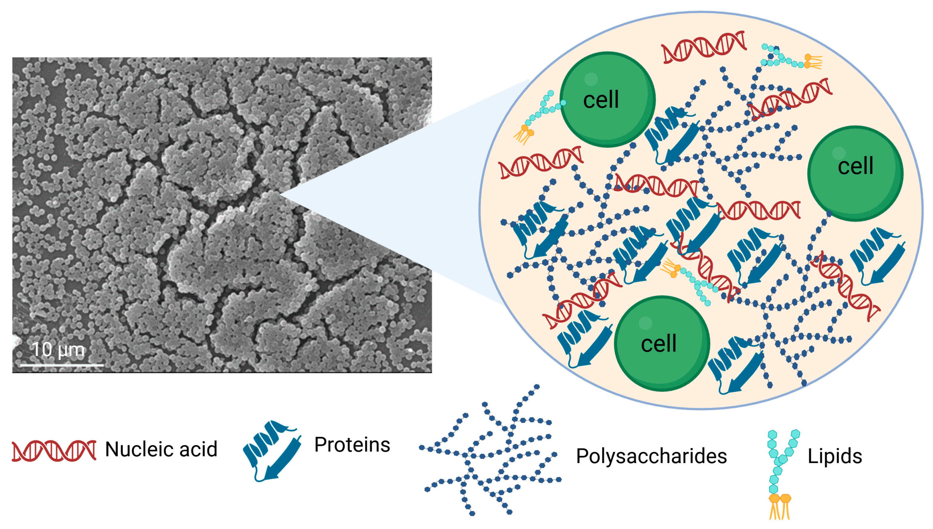
Biofilm is a heterogeneous structure composed primarily of water (85–95%), microbial cells (10–25% of dry mass), and a self-produced EPS matrix (75–90% of dry mass), which includes polysaccharides, proteins, lipids, DNA, and RNA [2]. In this complex architecture, the interstitial voids or water channels within the biofilm are essential for separating microcolonies [67]. The EPS forms a shield that holds the biofilm together, facilitating cell-to-cell communication and providing the adhesion and cohesion necessary for biofilm formation. Moreover, EPS contributes to nutrient cycling, maintains the availability of DNA for horizontal gene transfer, and acts as a protective barrier against oxidizing biocides, antibiotics, ultraviolet radiation, desiccation, and host immune defenses [68].
The main components of EPS can be categorized as follows (Figure 3):

3.1. Water

Water is the largest component of the EPS matrix in biofilms. It maintains hydration and protects the biofilm from desiccation, even under fluctuating environmental moisture conditions. The availability and distribution of water directly influence the flow and maintenance of essential nutrients within the biofilm [69]. Furthermore, water-filled channels enable the transport of nutrients, waste products, signaling molecules, and antibiotics through the biofilm [67].

3.2. Polysaccharides

The majority of biofilms depend on secreted extracellular polysaccharides, which constitute a primary component of the EPS matrix [70]. These polysaccharides can be categorized into homopolysaccharides and heteropolysaccharides based on their monosaccharide composition. Most are heterogeneous; however, some are homogeneous, such as fructan in Actinomycetes naeslundii [71], glucan in Streptococcus mutans, and β-glucan in Lactobacillus spp. [72]. In Escherichia coli, three major heteropolysaccharide ESP components have been identified: poly-N-acetyl-glucosamine (PGA), colanic acid (CA), and cellulose. PGA is considered crucial for biofilm formation and is found in various eubacterial species [73]. Colanic acid, composed of glucose, galactose, fructose, and glucuronic acid, is prevalent among E. coli isolates and is thought to play a role in bacterial survival outside the host [74]. Although cellulose is often the most abundant EPS component in many E. coli strains, it is absent in the K-12 strain due to a single-nucleotide polymorphism in the bcsQ gene [75]. Many clinically significant bacterial pathogens produce a common exopolysaccharide known as partially de-N-acetylated poly β-(1,6)-N-acetyl-d-glucosamine (dPNAG) as a key biofilm matrix component [76]. Both Gram-positive and Gram-negative bacteria produce dPNAG (referred to as polysaccharide intercellular adhesin in bacterial strains), including Staphylococcus aureus, Escherichia coli, Aggregatibacter actinomycetemcomitans, Acinetobacter baumannii, and Klebsiella pneumoniae [76]. In Pseudomonas aeruginosa, three main exopolysaccharides, Pel, Psl, and alginate, play central roles in biofilm formation and architecture. Pel is a cationic polysaccharide containing acetylgalactosamine and acetylglucosamine. It facilitates cell–cell adhesion, surface attachment, DNA cross-linking, and protection against aminoglycoside antibiotics [77,78]. Psl is rich in mannose, glucose, and rhamnose and is involved in both cell–cell interactions and surface attachment [77,79]. Alginate, a mucoid polysaccharide composed of guluronic and mannuronic acids, is commonly found in P. aeruginosa isolates from cystic fibrosis (CF) lung infections. It contributes to biofilm formation and immune evasion but plays a limited role outside the CF lung environment [77,80].

3.3. Proteins

Extracellular proteins in the biofilm matrix include secreted enzymes, structural proteins, subunits of surface appendages (e.g., pili and flagella), surface adhesins, and proteins contained in outer membrane vesicles (OMVs) [70]. These proteins interact with exopolysaccharides and nucleic acids, contributing to matrix stabilization, surface colonization, and the overall structural integrity of the biofilm [81]. Under nutrient-limited conditions, bacteria secrete enzymes capable of degrading EPS components to access alternative energy sources [70]. Examples include glycoside hydrolases such as dispersin B (DspB), which breaks down polysaccharides [82], and DNases, which degrade extracellular nucleic acids [83]. The impact of these enzymes on the EPS matrix will be discussed in more detail in the following sections. Jiao et al. [84] reported a significant difference between the proteome composition of EPS and that of individual cell fractions, identifying a high concentration of protein peptidases, cell wall, polysaccharide metabolism enzymes, disulfide-isomerases, and chaperones such as cold shock and DNA-binding proteins in the EPS matrix. Vo et al. [85] showed that a major part of cytosolic proteins, including anionic ribosomal proteins, is closely bound to poly-(1→6)-β-N-acetylglucosamine (PNAG) in live S. epidermidis biofilms. These proteins are presumably released during cell lysis, which occurs as part of biofilm maturation, and remain bound to the cationic PNAG within the EPS. Notably, they found that the well-known S. epidermidis biofilm protein accumulation-associated protein (Aap) does not directly interact with PNAG. Importantly, they identified elastin-binding protein (EbpS), a microbial surface component involved in adhesion to human elastin peptides, as being highly enriched in a PNAG-dependent manner in S. epidermidis biofilms. Toyofuku et al. [86] demonstrated that approximately 30% of the EPS matrix proteins in P. aeruginosa are membrane-associated and found within OMVs, while other proteins originate from lysed cells or are secreted.

3.4. Nucleic Acids

Extracellular DNA (eDNA) is a key component of the EPS matrix, playing an important role in microbial aggregation within the biofilm. The source of eDNA can vary; however, cell lysis is the primary mechanism in many bacterial species [81]. In Streptococcus gordonii, hydrogen peroxide-induced cell lysis contributes to eDNA release and subsequent EPS formation [87,88]. In S. mutans, eDNA may be released via membrane vesicle extrusion or (auto)lysis [88,89]. In human infections such as cystic fibrosis (CF), the eDNA found in P. aeruginosa biofilms originates primarily from human polymorphonuclear leukocytes (PMNs), which are recruited to the infection site [90]. EndA, a DNA-specific endonuclease, degrades eDNA within P. aeruginosa biofilms, leading to biofilm dispersal [91]. These findings align with recent reports indicating that eDNA is primarily responsible for the viscoelasticity of P. aeruginosa biofilms [92], with the loss of eDNA fiber structures coinciding with biofilm dissolution. However, the exact mechanism by which eDNA contributes to biofilm viscoelasticity and the nature of its supramolecular interactions remain unclear [93].
Extracellular RNA (eRNA) is also present in high concentrations in nucleic acid-containing biofilms and may contribute to eDNA-based viscoelastic networks [93]. Most established knowledge surrounds eDNA, but eRNA is gaining attention for its potential structural and regulatory roles in biofilms. It can be interactions and functional connections between eRNA and eDNA in biofilm matrices, though this is a relatively emerging area of research [94]. They have potential for DNA-RNA hybrid structures, although their stability and role in biofilms are not fully elucidated. Some studies suggest the eRNA can cooperate with eDNA in biofilm matrix stabilization and maintain the viscoelasticity, particularly in P. aeruginosa [93]. Additionally, in S. aureus biofilms, eRNA was reported to serve a structural function by associating with eDNA and stabilizing the polysaccharide-rich matrix [93]. Ribonuclease treatment has been shown to inhibit the formation of eDNA-containing Nontypeable Haemophilus influenzae (NTHI) biofilms [93]. Nevertheless, the lack of robust eRNA extraction protocols and the inherent stability of the molecule [95] have hindered a comprehensive understanding of its precise structural role or its potential interactions with eDNA in biofilm matrices [93].

3.5. Surfactants and Lipids

Lipid A, a key component of lipopolysaccharide (LPS) located in the outer membrane of Gram-negative bacteria, can also be found in the EPS of biofilms. In addition, several lipids with surface-active properties, such as surfactin, emulsan, and viscosin, are present in the EPS matrix. These biosurfactants enhance the availability of hydrophobic substances by dispersing them [96,97]. Ron and Rosenberg [98] reported that biosurfactants contribute to heavy metal binding and the production of virulence factors. Akter et al. [99] evaluated biofilm formation by P. aeruginosa and E. coli on various food-contact surface coupons, particularly aluminum (ALU), silicone rubber (SR), and polyethylene terephthalate (PET). The resulting biofilms exhibited a complex mixture of hydrophilic and hydrophobic components. As they matured, the polysaccharide structures remained stable while the lipid content increased, contributing to enhanced biofilm resilience.

4. Mitigation of Biofilm Formation on Biomaterials via EPS Matrix Disruption

The EPS matrix presents a major challenge for biofilm eradication strategies due to its chemical complexity and strain-specific variability [97]. Nonetheless, recent advances have led to the development of innovative approaches targeting this matrix. The following sections of this review focus on strategies to modify biomaterial surfaces to disrupt the EPS and thereby mitigate biofilm formation. The increasing resistance of bacteria to antibiotics, combined with the limited effectiveness of conventional antimicrobials against biofilm, has driven attention toward the use of non-antibiotic agents to modify surfaces to disturb the EPS matrix as a primary target.

4.1. Biofilm-Dispersing Enzymes

Biofilm-dispersing enzymes have demonstrated effectiveness against both developing and mature biofilms, typically requiring relatively low concentrations to achieve high specificity and efficacy. Unlike small-molecule drugs, which often encounter antibiotic resistance, these enzymes function extracellularly and do not require transport across the bacterial membrane, reducing the likelihood of resistance development. Three major classes of biofilm-disrupting enzymes have been extensively studied, namely glycoside hydrolases, proteases, and deoxyribonucleases (DNases). These enzymes enhance therapeutic outcomes by degrading key components of the EPS matrix, thereby improving antimicrobial penetration and immune cell access [76,100].

4.1.1. Glycoside Hydrolysis Enzyme

Dispersin B (DspB) is a member of glycoside hydrolase family 20 (GH20) and was initially isolated from Aggregatibacter actinomycetemcomitans [76]. DspB hydrolyzes the exopolysaccharide dPNAG in biofilm matrices through both endo- and exo-glycosidic activity [101,102]. This enzyme has been shown to inhibit biofilm and pellicle formation, detach mature biofilms, disaggregate bacterial clusters, and increase the susceptibility of preformed biofilms to enzymatic removal [103]. Ghalsasi et al. [104] engineered E. coli K-12 W3110 to synthesize and secrete DspB, which enables the degradation of poly-N-acetyl glucosamine (PGA) and subsequent disruption of target biofilms. Liu et al. [105] utilized magnetoreceptor (MagR), a magnetic protein responsive to external magnetic fields, as a fusion partner for immobilizing DspB on magnetic Fe3O4@SiO2 core–shell particles, with Fe3O4 as a core and SiO2 as a shell. Pavlukhina et al. [106] developed a biocompatible surface-attached polymer matrix via layer-by-layer (LBL) deposition for DspB delivery. The LbL system was constructed using poly(allylamine hydrochloride) (PAH) and poly(methacrylic acid) (PMAA), followed by glutaraldehyde crosslinking and pH-triggered removal of PMAA, resulting in a stable PAH hydrogel suitable for DspB loading. This system achieved more than 98% biofilm inhibition for two clinical isolates of S. epidermidis compared to mock-loaded multilayers [106]. Although DspB does not possess intrinsic antimicrobial activity, it shows enhanced in vitro efficacy against biofilms when combined with agents such as silver, cefamandole nafate, and ampicillin [107,108]. By degrading PNAG, DspB also prevents biofilm formation by disrupting the structural scaffold of the EPS matrix. Kane Biotech (Winnipeg, MB, Canada), a Canadian company, has trademarked Dispersin B® for the development of novel therapies targeting staphylococcal biofilms [82]. When combined with teicoplanin in catheter lock solutions, DspB dispersed S. aureus biofilms and improved the clearance of bloodstream infections in catheterized sheep [82]. It also showed efficacy in catheter-associated infections in rabbits when combined with triclosan [109]. However, these studies assessed only efficacy, without addressing toxicity or enzyme stability. Research has primarily focused on the use of DspB on Gram-positive bacteria, particularly staphylococci, dominant pathogens in skin and wound infections. Nonetheless, DspB could have broader applications in treating bacterial biofilms, and Kane Biotech (Winnipeg, MB, Canada) is also marketing Dispersin B® as an oral care product for domestic animals.
PgaB is another glycoside hydrolase encoded by the pgaB gene of the PNAG biosynthetic operon. It is capable of degrading PNAG analogues and disrupting PNAG-dependent biofilms formed by Bordetella pertussis, Staphylococcus carnosus, S. epidermidis, and E. coli [110,111]. However, DspB and Pga B differ in their catalytic mechanism: DspB acts as both an exo- and endo-glycosidase, cleaving internal and terminal glycosidic bonds, whereas PgaB is strictly an endo-glycosidase (cleaves within a polysaccharide unit). In S. epidermidis, DspB exhibits a lower half-maximal effective concentration (EC50; 0.24 nM) compared to PgaB, although both enzymes enhance the activity of antimicrobials to a similar extent [110,112].
In addition to dPNAG-targeting enzymes, alginate lyases have proven effective in dispersing mature biofilms [113]. These enzymes, which degrade alginate, have been isolated from various organisms with different substrate specificities, including algae, marine mollusks, marine and terrestrial bacteria, and some viruses and fungi [114]. Alginate lyase treatment reduces viscosity in cultures and cystic fibrosis (CF) sputum, strips biofilms from abiotic surfaces, promotes P. aeruginosa phagocytosis by immune cells, and enhances the efficacy of antipseudomonal antibiotics [113]. Other notable glycoside hydrolases include PslG (GH39 family) and PelA (a periplasmic glycoside hydrolase), which inhibit biofilm formation by clinical and environmental P. aeruginosa isolates and disrupt newly formed biofilms over 24 h [115,116]. Chareza et al. [117] immobilized alginate lyase onto bacterial cellulose (BC) pellicle produced by Komagataeibacter xylinus via physical adsorption, protecting wounds from P. aeruginosa infection and significantly reducing biofilm polysaccharide content. Wan et al. [118] demonstrated pH-responsive drug release using silver nanocomposites co-delivering alginate lyase and ceftazidime, effectively inhibiting and degrading P. aeruginosa PAO1 biofilms. Implanted medical devices, such as central venous catheters, are highly susceptible to microbial colonization and biofilm formation and represent a significant risk factor for nosocomial infections. P. aeruginosa relies on exopolysaccharides like Psl for initial attachment and biofilm maturation. Asker et al. [99] immobilized Psl-specific glycoside hydrolase (PslGh) onto the lumen surfaces of polyethylene, polyurethane, and polydimethylsiloxane (silicone) catheters. Under dynamic in vitro flow conditions, these modified catheters showed a 3-log reduction in bacterial counts over 11 days and a 2-log reduction by day 14, compared to untreated catheters as controls. In an in vivo rat model, PslGh-coated PE-100 catheters demonstrated a ~1.5-log reduction in colonization by the clinical P. aeruginosa ATCC 27,853 strain after 24 h [119]. Table 1 summarizes other well-characterized glycoside hydrolases with established biofilm-dispersing properties.

4.1.2. Proteases

Exoproteins, another major component of the EPS, account for a significant portion of the biomass in most biofilms. They play critical roles in bacterial aggregation, surface adhesion, and maintaining the structural integrity of biofilm matrices [140]. Enzymatic degradation of EPS exoproteins is one of the most effective strategies for biofilm eradication. To date, a variety of proteases with biofilm-dispersing capabilities have been identified and studied. These include Proteinase K, trypsin, pepsin, aureolysin, Spl proteases, and staphopain A and B, which are produced by various microorganisms to degrade biofilms [103,141].
Proteinase K is a broad-spectrum serine protease that cleaves peptide bonds near carboxylic groups of amino acids. It inhibits S. aureus biofilm formation by disrupting early adhesion and is capable of dispersing biofilms aged 24 and 48 h [103]. Although direct covalent attachment of proteinase K to biomaterial surfaces is less commonly reported, its incorporation into coatings or delivery systems has been explored. For instance, Sun et al. [142] developed multifunctional nanohybrids by combining phenylboronic acid-modified carbon dots (PCDs) with proteinase K, enhancing photosensitization to overcome biofilm barriers. In vitro studies demonstrated that PCDs were effective against a variety of bacteria, including multidrug-resistant strains, and exhibited strong affinity for both Gram-positive and Gram-negative species. This resulted in elevated reactive oxygen species (ROS) levels and, when combined with proteinase K, facilitated deep penetration and active biofilm elimination, improving treatment outcomes [142].
Trypsin, a pancreatic serine protease that cleaves peptide bonds on the carboxyl side of lysine and arginine residues, has been used to disrupt biofilms on teeth and wound surfaces [143,144]. Bovine trypsin has demonstrated the ability to degrade mature biofilms of various Gram-positive and Gram-negative bacteria [143]. It can reduce the biomass of 24 h old biofilms of P. aeruginosa and Enterococcus faecalis, though it does not completely eradicate them, regardless of treatment duration or enzyme concentrations [145]. Caro et al. [146] proposed a promising antibiofilm strategy by covalently immobilizing lysozyme and trypsin on stainless-steel oxide surfaces to inhibit biofilm formation.
Pepsin is a broad-specificity endopeptidase that contains a catalytic aspartate residue in its active site and preferentially cleaves at phenylalanine and leucine residues, but its activity is hindered by histidine, lysine, arginine, and proline residues [147]. Like trypsin, pepsin reduces the biomass of 24 h old biofilms of P. aeruginosa and E. faecalis, but it cannot fully remove biofilms from polystyrene surfaces, regardless of enzyme concentrations or exposure time [145]. However, when co-administered with trypsin and carvacrol, it has been shown to effectively eradicate preformed P. aeruginosa and E. faecalis biofilms [148]. Table 1 summarizes other proteolytic enzymes known for their biofilm-dispersing activity.

4.1.3. Deoxyribonucleases

eDNA is a ubiquitous and essential structural component of the EPS, contributing to microbial adhesion, cell signaling, horizontal gene transfer, and biofilm matrix stabilization [149,150]. Although eDNA plays a vital role in biofilms, its importance went largely unrecognized until 2002, when Whitchurch et al. [151] demonstrated that exogenous addition of DNase I could disperse biofilms and enhance the bactericidal activity of antibiotics. Since then, considerable research has focused on using various DNases to target eDNA for biofilm eradication.
DNase I is a widely studied pancreatic endonuclease that specifically digests DNA and has been shown to disrupt biofilm formation in both mono- and polymicrobial communities [22]. However, DNase I is more effective against early-stage biofilms. It can degrade newly formed biofilms (up to 60 h old), whereas mature biofilms (older than 84 h) display considerable resistance to DNase I. This resistance is likely due to the structural reinforcement of the biofilm by other EPS components such as exopolysaccharides and exoproteins. Additionally, mature biofilms may produce proteolytic exoenzymes that locally inactivate DNase I [151]. Recombinant human DNase I (rhDNase) has been clinically applied to reduce the viscosity of purulent sputum in patients with cystic fibrosis [152]. Ye et al. [153] demonstrated that immobilization of DNase I on titanium (Ti) surfaces using dopamine as a crosslinking agent significantly reduced the adhesion and biofilm formation of S. mutans and S. aureus over 24 h. Aktan et al. [154] evaluated the effectiveness of alternating current electrophoretic deposition (AC-EPD) as a novel method to apply DNase I onto titanium surfaces. Their results showed a significant reduction in biofilm formation by S. epidermidis and P. aeruginosa up to 20 h post-treatment. Dornase alfa, a recombinant human deoxyribonuclease I (rhDNase I), has been approved by the Food and Drug Administration (FDA) under the brand name Pulmozyme®. Daily inhalation of Dornase alfa has been shown to reduce the viscoelasticity of sputum, lower the frequency of pulmonary exacerbations, and improve lung function in patients with cystic fibrosis [152]. Table 1 summarizes other deoxyribonuclease enzymes known for their biofilm-dispersing activity.

4.2. Chelating Agents

Metallic cations, like Fe3+, Mg2+, and Ca2+, are essential to microbial adherence, biofilm formation, and bacterial growth [155,156]. Recently, many efforts have been made to utilize high-affinity metal-binding agents to chelate these ions, inhibiting bacterial growth. The most common chelating agents are Ethylenediamine-tetra-acetic acid (EDTA), Trisodium citrate (TSCs), and Ethylene glycol-bis (β-aminoethyl ether)-N, N, N’, N’-tetra-acetic acid (EGTA).
EDTA is a metal chelator normally used in clinics as an anticoagulant agent. However, in the early 2000s, clinicians saw an inhibitory activity of EDTA against methicillin-resistant Staphylococci, Gram-negative bacilli, and Candida species. EDTA can efficiently bind iron, magnesium, and calcium required to stabilize the extracellular biofilm matrix [157]. The biofilm matrix destabilization not only enhances the detachment of bacteria from a surface but may also increase the bacterial cell permeability and help the penetration of antibiotics such as gentamicin or cefotaxime [158,159].
Similar to EDTA, TSC chelates Ca2+ and Mg2+ and inhibits biofilm formation. Chu et al. [160] demonstrated that a 4% TSC solution can prevent biofilm formation by S. aureus, P. aeruginosa, E. coli, and K. pneumoniae. Unlike EDTA, TSC also inhibits extracellular matrix production and quorum sensing.
EGTA is a chelating agent with a much higher affinity for Ca2+ ions than for Mg2+ ions, making it useful for selectively binding calcium in the presence of magnesium. Lyamba et al. [161] demonstrated that low concentrations of EGTA prevent the adhesion of S. aureus to abiotic surfaces and inhibit biofilm development. Furthermore, once established, the biofilm becomes resistant to EGTA, suggesting that EGTA-containing lock solutions should be used to prevent biofilm formation rather than to promote its removal.
In addition to EDTA, TSC, and EGTA, many other chelators are effective in inhibiting bacterial adhesion and biofilm formation. The most recently studied include transferrin, lactoferrin, desferrioxamine (DFO), deferiprone (DFP), and kojic acid [162,163]. DFP and kojic acid have been shown to inhibit P. aeruginosa biofilm formation without affecting bacterial growth [164]. However, higher concentrations of DFO may promote biofilm formation by acting as an iron carrier.

4.3. Quorum Sensing Inhibitors

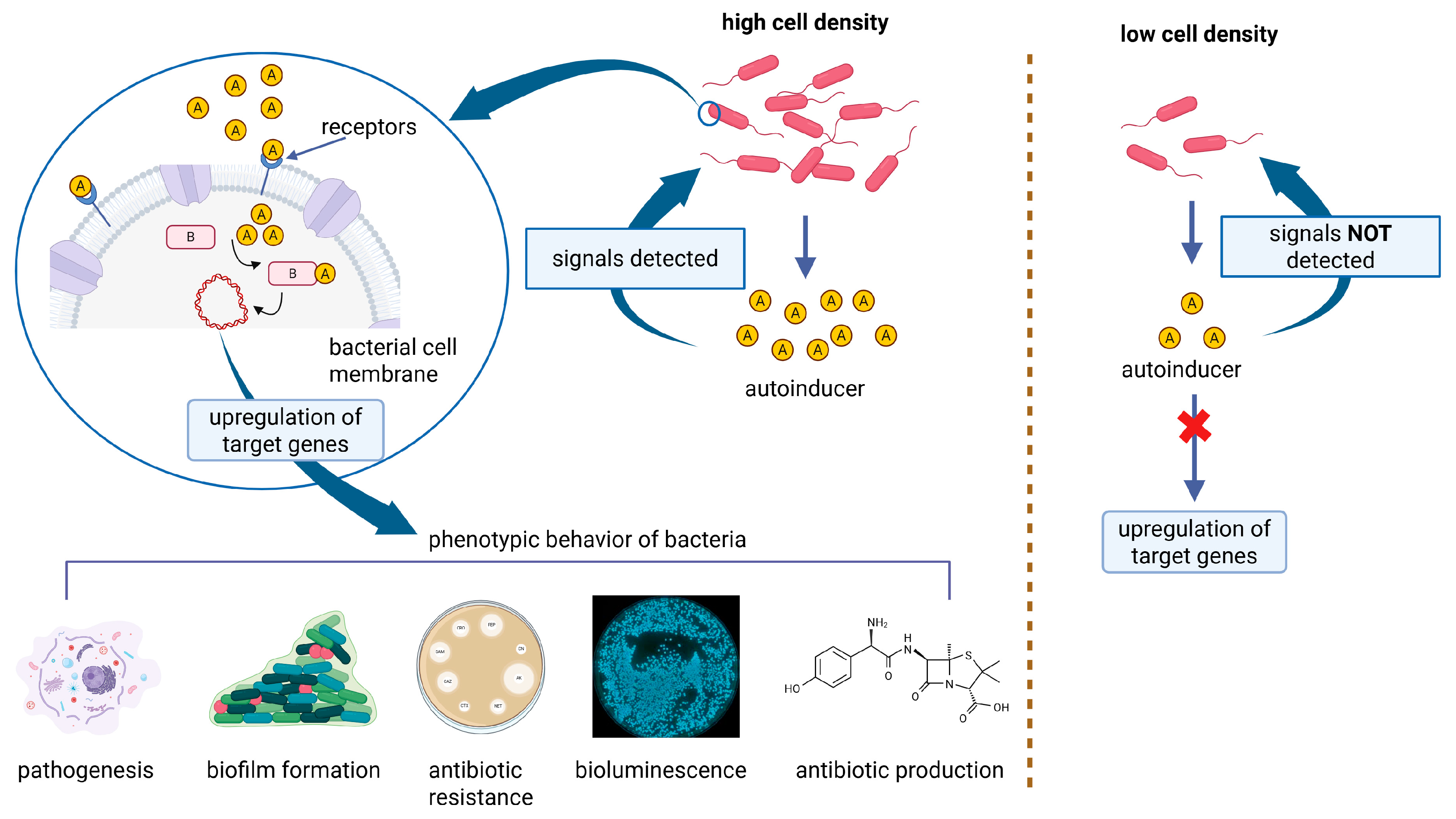
The development of bacterial biofilms involves self-organization, cooperation, and communication among microbial communities as they transition from free-floating planktonic cells to a structured, three-dimensional biofilm lifestyle. Multiple environmental cues, including stress, nutrient limitation, and intercellular communication, influence this transition. To adapt to changing environments, bacteria rely on coordinated gene expression triggered by these external stimuli [165]. A key mechanism facilitating this coordination is quorum sensing (QS), a form of bacterial cell-to-cell communication that regulates gene expression based on population density. QS operates through the release, detection, and uptake of small diffusible signaling molecules known as autoinducers (AIs). These molecules accumulate in the environment and, once a threshold concentration is reached, trigger a synchronized response among bacterial cells. QS can occur within a single species (intraspecies) or between different species (interspecies) and regulates several physiological and pathogenic behaviors, including horizontal gene transfer, expression of virulence factors, synthesis of secondary metabolites (e.g., antibiotics), motility, and biofilm formation (Figure 4) [166]. The QS systems of many Gram-positive and Gram-negative bacteria are well characterized. In Gram-negative species, the primary AIs include N-acyl homoserine lactones (AHL), autoinducer 2 (AI-2), a furanosyl borate diester, and autoinducer 3 (AI-3). QS plays a critical role in various stages of biofilm formation by modulating processes, such as bacterial motility, adhesin production, extracellular polysaccharide synthesis, and the release of eDNA [167]. These factors contribute to both biofilm maturation and cell dispersion, enabling the biofilm to maintain structural integrity while ensuring the ability of cells to detach and colonize new environments. Through these regulatory pathways, QS enhances the adaptability, persistence, and pathogenic potential of bacterial communities [5]. Guendouze et al. reported the effectiveness of the lactonase enzyme SsoPox-W2631 in attenuating the virulence of 51 clinical P. aeruginosa isolates obtained from diabetic foot ulcers. This enzyme significantly reduced the secretion of proteases and pyocyanin, leading to a subsequent decrease in biofilm formation [168]. Vogel et al. immobilized the quorum-quenching enzyme PvdQ, an N-terminal nucleophile acylase encoded within the pyoverdine (pvd) gene cluster, onto polydimethylsiloxane silicone (PDMS) surfaces. This functionalized coating demonstrated quorum-quenching activity and effectively reduced bacterial colonization on indwelling medical devices such as urinary and intravascular catheters [169].

4.4. Biosurfactants

Biosurfactants are biologically synthesized microbial products that have gained worldwide attention owing to their eco-friendly, biodegradable characteristics and their immense effectiveness in bioremediation, pharmaceuticals, agriculture, the food industry, and anti-biofouling [170]. Some studies have revealed that the inhibition of bacterial adhesion and biofilm dispersal properties of the biosurfactants is due to their surface and interfacial tension reduction, as well as solubilized lipids and proteins in the EPS matrix, interruption in the QS signaling system, and an increase in the bacterial membrane permeabilization [171,172,173]. Sophrolipids, produced by Starmerella bombicola (previously classified as Candida bombicola), showed bactericidal properties when compared to conventional antimicrobial agents with bacteriostatic effects [174]. Ceresa et al. [175] used this property of the sophrolipids and evaluated their antimicrobial properties in medical-grade silicon discs against several clinically relevant strains: C. albicans, S. aureus, and P. aeruginosa. The obtained results demonstrated that pre-coating silicone discs with lactonic sophrolipids (SLA) reduced S. aureus and P. aeruginosa by about 70–80% and inhibited bacterial attachment up to 95% [175].
For the production of nanoparticles, it is currently necessary to identify non-toxic technologies. The use of biosurfactants as coupling agents is a recent method for the sustainable, environmentally friendly, and compatible synthesis of bioactive metallic nanoparticles. Top-down strategies using biosurfactants as capping and stabilizing agents have been proposed in many studies, disclosing the fabrication of metallic nanocomposites, including Ag, Au, Cu, Pt, Pd, NiO, and ZnS, to limit or mitigate aggregation after and during the fabrication of the preferred nanocomposite [176]. Microbial and biosurfactant technologies for nanomaterial fabrication are still being developed because they are clean, safe, and environmentally friendly. Additionally, the self-assembly of amphiphilic molecules acting as surfactants is a facile method to produce organic nanoparticles [177] and can be used as stabilizers and help to maintain a regular shape because of the electrostatic attraction forces [178]. Farias et al. [178] prepared silver nanoparticles with a size of 1.13 nm using the biosurfactant of P. aeruginosa acting as a stabilizer. Khalid et al. [179] demonstrated a simple approach for the preparation of rhamnolipid (RL)-coated iron NP and proposed a synergistic antibacterial and antiadhesive mechanism against P. aeruginosa and S. aureus biofilms. Reactive oxygen compounds are mechanically generated by RL-coated iron NP (48 nm), which supports the antibacterial effect.

4.5. Oxidizing Agents

Oxidizing agents, also known as reactive species, are low-molecular-weight molecules that induce oxidation by accepting electrons from other substances. These molecules are highly unstable due to the presence of unpaired or missing electrons. Their accumulation within cells can lead to oxidative stress. When applied to biomaterials, oxidizing agents can help prevent biofilm formation by disrupting the bacterial redox balance and inducing oxidative stress, ultimately damaging or killing bacterial cells. The most common producers of reactive species include metal nanoparticles, graphene oxide coatings, nitric oxide (NO)-releasing biomaterials, ozone-based coatings, and halogens.

4.5.1. Metal Ions and Metal Nanoparticles

Silver, gold, copper, zinc, and gallium-based ions, along with metal and metal oxide nanoparticles such as ZnO and MgO, have been extensively studied for their potent antibacterial activity against both planktonic bacteria and biofilms. These systems exert antimicrobial effects through multiple mechanisms, including disruption of bacterial adhesion, damage to the cell membrane, induction of oxidative stress, interference with QS, and enhancement in antibiotic efficacy via synergistic interactions. Additionally, metal-based nanoparticles release ions that disrupt bacterial membranes and interfere with vital cellular processes, thereby preventing biofilm formation. Moreover, they can contribute to biofilm eradication by activating the host immune response [180,181].
Ferraris et al. developed a titanium alloy coated with a zirconia matrix doped with silver for use in temporary fixation devices. The authors demonstrated that this coating effectively inhibits the initial adhesion of S. aureus, thereby preventing biofilm formation. This anti-adhesive effect is attributed not only to the antimicrobial properties of the zirconia–silver coating but also to the synergistic interaction with the surface nanotopography (<0.2 μm), which limits the availability of anchorage points for bacterial attachment [182].
Qais et al. developed gold nanoparticles (AuNPs-CA) bio-fabricated using an aqueous extract of Capsicum annuum and tested them against P. aeruginosa and Serratia marcescens. The results demonstrated that these nanoparticles not only reduced the virulence factors of P. aeruginosa, including pyocyanin production, elastase activity, and rhamnolipid synthesis, but also inhibited biofilm formation and EPS production in both bacterial species [183].
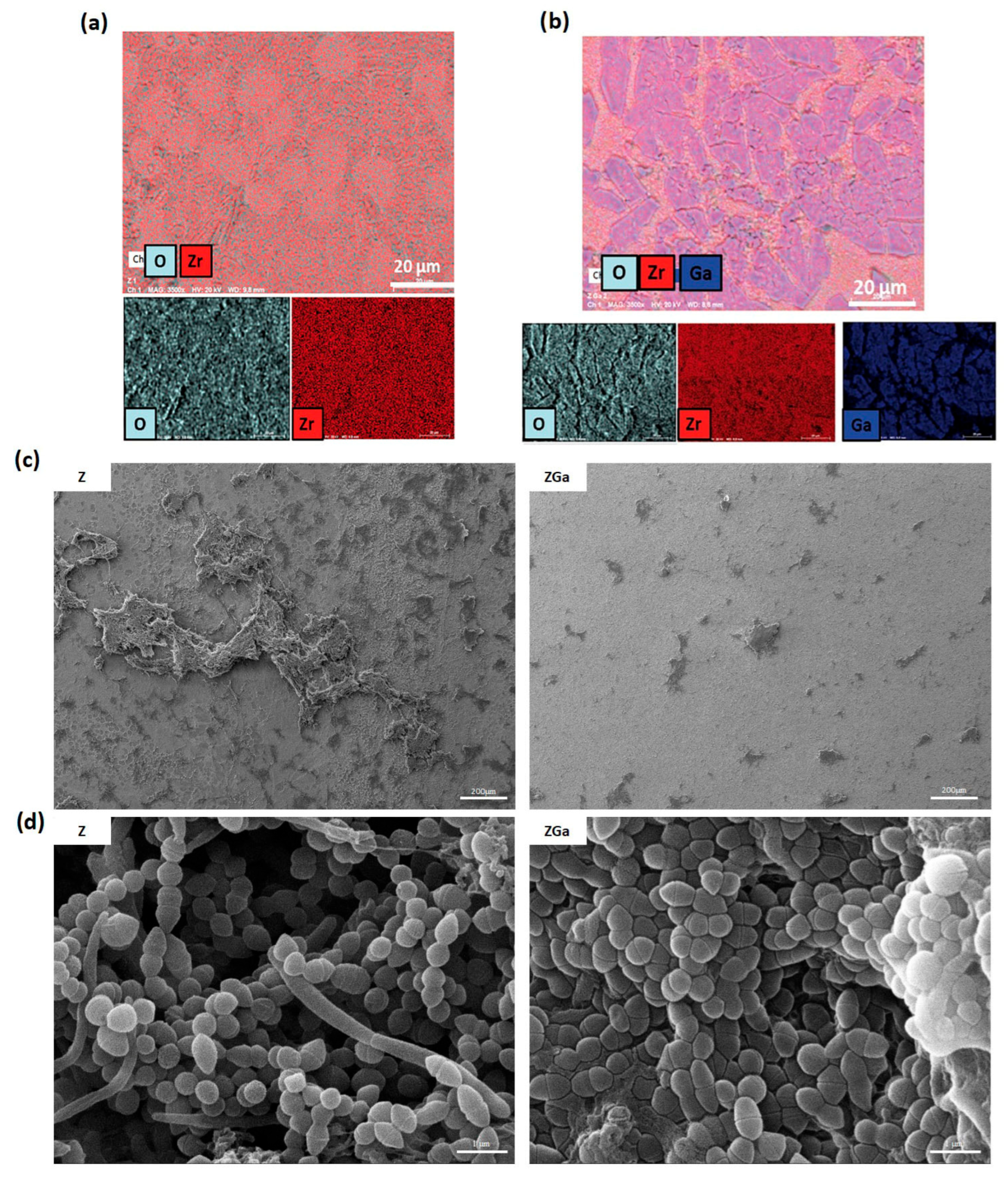
Gallium (Ga) has garnered significant research interest over the past decade. Unlike many other metal ions, gallium is generally considered cytocompatible and exhibits selective antibacterial activity against pathogenic bacteria. In its ionic form (Ga3+), gallium exerts its antimicrobial effects by interfering with bacterial iron metabolism. This “Trojan horse” mechanism exploits bacterial iron uptake pathways to facilitate Ga3+ entry into the cell, where it disrupts iron-dependent enzymatic processes, ultimately inhibiting bacterial growth [184]. Recently, D’Agostino et al. [185] developed a gallium-doped zirconia coating for dental implant surfaces. The results showed that gallium selectively eliminates pathogenic bacteria while preserving commensal microbiota. Additionally, the coating significantly reduced oral plaque adhesion and overall biofilm biomass. Proteomic analysis of plaque samples in contact with the gallium-doped surface revealed a marked reduction in the adhesion of Fusobacterium nucleatum, a Gram-negative bacterium known for its role in oral biofilm stability and for shielding less aerotolerant species, such as Porphyromonas gingivalis, a key pathogen in periodontal disease, from oxidative stress (Figure 5).
The use of nanoparticles still raises several concerns related to toxicity. Their small size and large surface area can promote interactions with biological systems, potentially leading to adverse effects, such as oxidative stress, inflammation, DNA damage, immune system alterations, and organ toxicity [186,187]. To mitigate these risks, multifaceted strategies are required. Jangid et al. [188] highlighted that encapsulation technologies using biodegradable polymers can reduce silver nanoparticle (AgNP) toxicity without compromising their antimicrobial activity. Similarly, Rashid et al. [189] proposed that incorporating AgNPs into polymer matrices to create AgNP–polymer nanocomposites (AgNP–PNCs) allows for a controlled and sustained release of silver ions, thereby reducing the cytotoxicity typically associated with free AgNPs.
Another strategy to minimize nanoparticle-induced toxicity involves modulation of their size and shape. Blanco et al. [190] reported that nanoparticles smaller than 5 nm tend not to accumulate in vital organs and are efficiently cleared by the kidneys.
As with many innovative medical technologies, regulatory barriers and scalability represent major challenges. Nadar et al. [191] emphasized that nanoparticles face significant hurdles in the approval process, primarily due to the absence of standardized manufacturing and characterization protocols, inconsistent preclinical toxicity data, and a limited body of clinical evidence. In addition, the lack of long-term environmental impact assessments further complicates the regulatory approval pathway for nanoparticle-based products.

4.5.2. Graphene Oxide

Graphene and graphene oxide (GO) coatings have emerged as promising antibacterial materials due to their ability to generate reactive oxygen species (ROS), which can damage bacterial cells and inhibit biofilm formation. In a study by Wang et al. [192], graphene was synthesized directly on the surface of a titanium alloy. The resulting graphene-coated titanium significantly reduced the adhesion and biofilm formation of P. aeruginosa, F. nucleatum, and C. albicans. Beyond their intrinsic antimicrobial properties, graphene-based materials also offer advantages as drug delivery platforms, thanks to their large surface area. Sun et al. [193] demonstrated that graphene not only kills pathogenic bacteria and prevents biofilm development but also enables the controlled release of levofloxacin, enhancing its antibacterial activity against S. aureus and E. coli. Moreover, Cheng et al. [194] showed that a graphene oxide coating can efficiently carry and release the antimicrobial peptide NAL-P-113, effectively targeting peri-implant pathogens such as S. mutans and P. gingivalis.

4.5.3. Nitric Oxide

Nitric oxide (NO)-releasing materials can hinder biofilm formation by depleting exopolysaccharides and damaging bacterial membranes. Sadrearhami et al. [195] developed an antibiofilm coating that contains a sulfur-nitroso thiol NO donor and can produce adjustable NO fluxes. The plasma coatings were shown to retain a high percentage of sulfur groups, and their thickness could be predictably adjusted through deposition time. Coatings with a 250 nm film thickness reduced P. aeruginosa biofilm formation by 75% after 6 h and 55% after 24 h of exposure. Increasing the polymer coating thickness to 500 nm improved antibiofilm efficiency, achieving 81% and 52% inhibition of bacterial attachment after 24 and 36 h of incubation in bacterial culture solution, respectively.
Chug et al. [196] developed a portable NO-releasing catheter insert that combines antimicrobial effectiveness, biocompatibility, and ease of activation. The system employs a light-sensitive NO donor, S-nitroso-N-acetylpenicillamine (SNAP), which is covalently bonded to a polydimethylsiloxane (PDMS) matrix. This covalent attachment enhances NO retention and minimizes early leaching. The resulting SNAP-PDMS composite was applied to a side-emitting optical fiber and integrated with a wearable light-emitting module operating at 450 nm, yielding a functional and compact antimicrobial device. The optical fiber can be activated with a single click, triggering controlled NO release for at least 24 h. This light-induced NO delivery achieved over 90% reduction in bacterial viability for S. aureus, S. epidermidis, and Proteus mirabilis, without causing cytotoxic effects on mammalian cells.

4.5.4. Ozone

Ozone is a powerful oxidizing agent that can damage the cell membranes of both Gram-positive and Gram-negative bacteria, leading to cell death. It is commonly used in dental clinics, both as a pre-treatment and post-treatment agent. As a pre-treatment, it prevents bacterial adhesion and effectively kills planktonic bacteria [197]. As a post-treatment, it can penetrate and disrupt the biofilm EPS matrix. This disruption not only leads to the breakdown of the biofilm structure but also increases bacterial susceptibility to antibiotic treatment or mechanical removal [198]. Coating biomaterials with ozone can modify surface properties, such as surface energy and hydrophobicity, making them less conducive to bacterial adhesion. In addition to dental clinics, ozone therapy is also employed in various other fields, including food processing and water treatment.

4.5.5. Halogens

Halogens, such as fluorine, iodine, and chlorine, are well known for their antimicrobial properties. They disrupt bacterial cell membranes, inactivate essential enzymes, and interfere with DNA replication, thereby preventing or inhibiting bacterial growth and biofilm formation. For example, Tang et al. [199] designed a fluorinated alkoxyphosphazene surface that inhibited the adhesion of both Gram-positive and Gram-negative bacteria by 50- to 200-fold compared to untreated surfaces. Similarly, Inoue et al. [200] demonstrated that iodine-coated implants exhibit broad-spectrum antibacterial and antifungal activity. These implants were partially incorporated into the guidelines of the 2018 International Consensus Meeting on Musculoskeletal Infection due to their potential to prevent infections [201]. In addition to preventing bacterial adhesion, surfaces can be engineered to release halogens in a controlled manner. This approach enables a sustained release of the antimicrobial agent, providing long-lasting protection against biofilm formation. Wu et al. [202] developed an N-halamine coating that incorporates chlorine into a polymer matrix, which can be further applied to titanium scaffolds. The authors demonstrated that the coating not only releases chlorine in a controlled manner, providing long-lasting antibacterial efficacy, but can also self-regenerate itself after use.

4.6. Engineered Nanoparticles

Strategies to Penetrate the Biofilm

Implementing biomaterials that prevent bacterial adhesion and growth is the most effective way to combat biofilm-related infections. Biofilms can protect bacteria inside from chemical and physical attacks, making them 10- to 1000-times more resistant than their free-floating counterparts. As mentioned earlier, various methods have been created to design biomaterials that stop bacterial biofilm formation. However, these methods mostly focus on prevention, and if they fail, uncontrolled bacterial growth may occur, often requiring implant removal and additional surgery. These events can greatly reduce patient quality of life and significantly raise healthcare costs.
In 2014, Komnatnyy et al. [203] reported the development of a smart material capable of releasing an antibacterial compound only upon biofilm formation. Specifically, they engineered a coating in which ciprofloxacin is covalently bound to the surface via ester bonds sensitive to bacterial lipase hydrolases. These enzymes, particularly abundant in mature biofilms, act as biological triggers for drug release. Notably, no antibiotic was released in the absence of bacteria or a stable biofilm.
However, the success of antibiotic or particle penetration into biofilms depends on multiple factors, including particle size, shape, surface chemistry, and the physicochemical properties of the biofilm itself. Studies have shown that small particles (5–130 nm) penetrate biofilms more effectively than larger ones [204,205]. Additionally, spherical particles exhibit higher penetration rates compared to elongated forms [206]. Surface properties, such as hydrophobicity and electrostatic charge, also play a crucial role. Li et al. [207] demonstrated that neutral or anionic quantum dots (QDs) cannot penetrate E. coli biofilms, whereas cationic QDs can do so without obstruction. Interestingly, the diffusion behavior of cationic QDs also varies: hydrophilic QDs tend to cluster, while hydrophobic QDs are more evenly distributed within the biofilm, making them more suitable for therapeutic applications.
Over the past decade, bacteriophages, viruses that infect and kill bacteria, have emerged as promising agents for biofilm disruption. They offer several advantages over traditional antibiotics, particularly due to their high specificity and efficacy against antibiotic-resistant strains. Unlike antibiotics, bacteriophages do not harm human cells or disrupt beneficial microbiota, reducing the risk of collateral damage. The antibacterial and antibiofilm efficacy of bacteriophages has been extensively studied. In a rat model of prosthetic joint infection, Morris et al. [208] observed a five-fold reduction in S. aureus when using phages alone and a 22.5-fold reduction when phages were combined with vancomycin. Moreover, Yazdi et al. [209] reported a 5-log reduction in P. mirabilis biofilm on urinary catheters within three hours of phage treatment. When phages were combined with ampicillin, they achieved a 5-log reduction in just one hour and an 85% reduction in biofilm mass.
Mayorga-Ramos et al. [210], in their mini-review, highlighted the potential of encapsulating bacteriophages in liposomes, metallic nanoparticles (e.g., silver, gold, magnesium), and polymeric nanoparticles (e.g., poly(lactic-co-glycolic acid), PLGA). These delivery systems enhance phage stability, improve biofilm penetration, and enable targeted therapeutic delivery.
Despite their considerable therapeutic potential, several critical issues remain to be addressed regarding the use of bacteriophages. As observed with antibiotics, bacteria can develop resistance to bacteriophages. Known resistance mechanisms include mutations in phage receptor proteins, activation of restriction–modification systems, abortive infection strategies—where bacterial cells self-destruct to prevent phage propagation—and the acquisition of CRISPR-Cas-mediated immunity [211]. Such mechanisms may significantly limit the long-term efficacy of phage-based therapies.
An additional concern involves the role of lysogenic phages. As noted by Anomaly [212], lysogenic phages that integrate into bacterial genomes can promote antibiotic resistance in their bacterial hosts. This interaction may lead to a mutualistic relationship between bacteria and phages, potentially enhancing bacterial pathogenicity in humans.
Moreover, the immunogenicity of bacteriophages represents another important limitation. Podlacha et al. [213] reported that bacteriophages, due to their nucleoproteinaceous composition, are readily recognized by the host immune system. They may also modulate the adaptive immune response and induce the production of anti-phage antibodies [214], potentially affecting the efficacy and safety of repeated or systemic phage administration.
Lastly, phage therapy faces several regulatory hurdles due to its unique nature as a biological agent. A major limitation is the scarcity of clinical trials. According to ClinicalTrials.gov, only 37 clinical trials involving phage therapy (search term: bacteriophages) have been registered, of which just 2 have reached Phase 3 and only 1 has progressed to Phase 4 [215]. Other key concerns include issues related to intellectual property, the establishment of quality and safety standards, and the intrinsic variability in bacteriophage preparations [216].
Table 2 provides a summary of the strengths, weaknesses, mitigation strategies, and toxicity profiles of the methods discussed.

5. Future Perspective

This review focuses on the eradication of biofilms from biomaterial surfaces used in medical devices, emphasizing surface modification or functionalization with non-antibiotic agents to address the growing threat of multidrug-resistant bacterial pathogens. It provides comprehensive information on a wide range of EPS disruption strategies that have been investigated to prevent bacterial adhesion and remove established biofilm layers. Future studies should concentrate on the rational design of combinatorial therapies that employ synergistic, multi-target approaches. As most current investigations are limited to in vitro settings, future work can prioritize translational and preclinical in vivo models to assess long-term antifouling and biofilm eradication performance, host tissue compatibility, and immune responses. A deeper understanding of the molecular mechanisms by which these agents disrupt biofilms, along with their potential to influence bacterial resistance development, is essential. OMICs-based approaches (e.g., transcriptomics, proteomics) could be instrumental in elucidating these mechanisms. Furthermore, the absence of standardized protocols to evaluate non-antibiotic antibiofilm treatments limits reproducibility and clinical translation, meaning future efforts, therefore, will aim to develop standardized testing frameworks and clarify regulatory considerations for the application of these agents in medical device coatings and functionalized biomaterials.

Author Contributions

Conceptualization, A.C.S. and Z.N.; writing—original draft preparation, A.C.S. and Z.N.; writing—review and editing, A.C.S. and Z.N.; All authors have read and agreed to the published version of the manuscript.

Funding

This research received no external funding.

Institutional Review Board Statement

Not applicable.

Informed Consent Statement

Not applicable.

Data Availability Statement

The dataset is available on request from the authors.

Conflicts of Interest

The funders had no role in the design of the study; in the collection, analyses, or interpretation of data; in the writing of the manuscript; or in the decision to publish the results.

Abbreviations

The following abbreviations are used in this manuscript:
WTWild type
EPSExtracellular polymeric substances
C-DI-GMPCyclic di-guanosine monophosphate
QSQuorum sensing
AHLN-acylated homoserine lactone
AIAutoinducers
SpAS. Aureus protein a
PVLPanton-Valentine leucocidin
CVCCentral venous catheter
CAUTI- Catheter-associated urinary tract infections
VAPVentilator-associated pneumonia
ICUIntensive care units
MRSAMethicillin-resistant S. Aureus
PGAPoly-N-acetyl-glucosamine (PGA)
CAColanic acid
dPNAGDe-N-acetylated poly β-(1,6)-N-acetyl-d-glucosamine
CFCystic fibrosis
OMV Outer membrane vescicles
DspBDispersin B
PNAGPoly-(1→6)-β-N-acetylglucosamine
Aap Accumulation-associated protein
EbpSElastin-binding protein
eDNAExtracellular DNA
PMNsPolymorphonuclear leukocytes
eRNAExtracellular RNA
NTHINon-typeable Haemophilus influenzae
LPSLipopolysaccaride
ALUAluminium
SRSilicon rubber
PETPolyethylene terephthalate
DNasesDeoxyribonucleases
GH20Glycoside hydrolase family 20
PGAPoly-N-acetyl glucosamine
MagRMagnetoreceptor
LBLLayer by layer
PAHPoly(allylamine hydrochloride)
PMAAPoly(methacrylic acid)
BCBacteria cellulose
PCDPhenylboronic acid-modified carbon dots
ROSReactive oxygen species
RhDNAse IRecombinant human dnase I
Ti Titanium
AC-EPDAlternating current electrophoretic deposition
FDA- Food and Drug Administration
EDTAEthylenediamine-tetra-acetic acid
TSC Trisodium citrate
EGTAEthylene glycol-bis (β-aminoethyl ether)-N,N,N’,N’-tetra-acetic acid
DFODesferrioxamine
DFP Deferiprone
AHLN-acyl homoserine lactones
PVDPyoverdine
PDMSPolydimethylsiloxane
SLASilicone discs with lactonic sophrolipids
RLRhamnolipid
NONitric oxide
AuNPs-Gold nanoparticles
GaGallium
AgNPsSilver nanoparticles
AgNP–PNCsAgnp–polymer nanocomposites
EDSDispersive X-ray Spectroscopy
ZZirconia
ZGaGallium-doped zirconia
GOGraphene oxide
SNAPS-nitroso-N-acetylpenicillamine
QDQuantum dot
PLGAPoly(lactic-co-glycolic acid)

References

  1. Galié, S.; García-Gutiérrez, C.; Miguélez, E.M.; Villar, C.J.; Lombó, F. Biofilms in the Food Industry: Health Aspects and Control Methods. Front. Microbiol. 2018, 9, 898. [Google Scholar] [CrossRef] [PubMed]
  2. Rather, M.A.; Gupta, K.; Mandal, M. Microbial Biofilm: Formation, Architecture, Antibiotic Resistance, and Control Strategies. Braz. J. Microbiol. 2021, 52, 1701–1718. [Google Scholar] [CrossRef]
  3. Mugunthan, S.; Wong, L.L.; Winnerdy, F.R.; Summers, S.; Bin Ismail, M.H.; Foo, Y.H.; Jaggi, T.K.; Meldrum, O.W.; Tiew, P.Y.; Chotirmall, S.H.; et al. RNA Is a Key Component of Extracellular DNA Networks in Pseudomonas aeruginosa Biofilms. Nat. Commun. 2023, 14, 7772. [Google Scholar] [CrossRef] [PubMed]
  4. Carniello, V.; Peterson, B.W.; van der Mei, H.C.; Busscher, H.J. Physico-Chemistry from Initial Bacterial Adhesion to Surface-Programmed Biofilm Growth. Adv. Colloid Interface Sci. 2018, 261, 1–14. [Google Scholar] [CrossRef] [PubMed]
  5. Muhammad, M.H.; Idris, A.L.; Fan, X.; Guo, Y.; Yu, Y.; Jin, X.; Qiu, J.; Guan, X.; Huang, T. Beyond Risk: Bacterial Biofilms and Their Regulating Approaches. Front. Microbiol. 2020, 11, 928. [Google Scholar] [CrossRef]
  6. Riedel, R.; Rani, G.; Sengupta, A. Bacterial Adhesion on Soft Surfaces: The Dual Role of Substrate Stiffness and Bacterial Growth Stage. Microorganisms 2025, 13, 637. [Google Scholar] [CrossRef]
  7. Chinnaraj, S.B.; Jayathilake, P.G.; Dawson, J.; Ammar, Y.; Portoles, J.; Jakubovics, N.; Chen, J. Modelling the Combined Effect of Surface Roughness and Topography on Bacterial Attachment. J. Mater. Sci. Technol. 2021, 81, 151–161. [Google Scholar] [CrossRef]
  8. Zheng, S.; Bawazir, M.; Dhall, A.; Kim, H.-E.; He, L.; Heo, J.; Hwang, G. Implication of Surface Properties, Bacterial Motility, and Hydrodynamic Conditions on Bacterial Surface Sensing and Their Initial Adhesion. Front. Bioeng. Biotechnol. 2021, 9, 643722. [Google Scholar] [CrossRef]
  9. Sharifikolouei, E.; Najmi, Z.; Cochis, A.; Scalia, A.C.; Aliabadi, M.; Perero, S.; Rimondini, L. Generation of Cytocompatible Superhydrophobic Zr–Cu–Ag Metallic Glass Coatings with Antifouling Properties for Medical Textiles. Mater. Today Bio 2021, 12, 100148. [Google Scholar] [CrossRef]
  10. Oh, J.K.; Yegin, Y.; Yang, F.; Zhang, M.; Li, J.; Huang, S.; Verkhoturov, S.V.; Schweikert, E.A.; Perez-Lewis, K.; Scholar, E.A.; et al. The Influence of Surface Chemistry on the Kinetics and Thermodynamics of Bacterial Adhesion. Sci. Rep. 2018, 8, 17247. [Google Scholar] [CrossRef]
  11. Bhagwat, G.; O’Connor, W.; Grainge, I.; Palanisami, T. Understanding the Fundamental Basis for Biofilm Formation on Plastic Surfaces: Role of Conditioning Films. Front. Microbiol. 2021, 12, 687118. [Google Scholar] [CrossRef]
  12. Rummel, C.D.; Lechtenfeld, O.J.; Kallies, R.; Benke, A.; Herzsprung, P.; Rynek, R.; Wagner, S.; Potthoff, A.; Jahnke, A.; Schmitt-Jansen, M. Conditioning Film and Early Biofilm Succession on Plastic Surfaces. Environ. Sci. Technol. 2021, 55, 11006–11018. [Google Scholar] [CrossRef]
  13. Haiko, J.; Westerlund-Wikström, B. The Role of the Bacterial Flagellum in Adhesion and Virulence. Biology 2013, 2, 1242–1267. [Google Scholar] [CrossRef] [PubMed]
  14. Zhang, M.; He, L.; Qin, J.; Wang, S.; Tong, M. Influence of Flagella and Their Property on the Initial Attachment Behaviors of Bacteria onto Plastics. Water Res. 2023, 231, 119656. [Google Scholar] [CrossRef]
  15. Hu, H.; Xu, J.; Chen, J.; Tang, C.; Zhou, T.; Wang, J.; Kang, Z. Influence of Flagella on Salmonella Enteritidis Sedimentation, Biofilm Formation, Disinfectant Resistance, and Interspecies Interactions. Foodborne Pathog. Dis. 2024, 22, 1–18. [Google Scholar] [CrossRef]
  16. Epler Barbercheck, C.R.; Bullitt, E.; Andersson, M. Bacterial Adhesion Pili. In Membrane Protein Complexes: Structure and Function; Springer: Berlin/Heidelberg, Germany, 2018; pp. 1–18. [Google Scholar]
  17. Guerra, M.E.S.; Destro, G.; Vieira, B.; Lima, A.S.; Ferraz, L.F.C.; Hakansson, A.P.; Darrieux, M.; Converso, T.R. Klebsiella Pneumoniae Biofilms and Their Role in Disease Pathogenesis. Front. Cell. Infect. Microbiol. 2022, 12, 877995. [Google Scholar] [CrossRef] [PubMed]
  18. Yadav, P.; Verma, S.; Bauer, R.; Kumari, M.; Dua, M.; Johri, A.K.; Yadav, V.; Spellerberg, B. Deciphering Streptococcal Biofilms. Microorganisms 2020, 8, 1835. [Google Scholar] [CrossRef]
  19. McKee, R.W.; Aleksanyan, N.; Garrett, E.M.; Tamayo, R. Type IV Pili Promote Clostridium Difficile Adherence and Persistence in a Mouse Model of Infection. Infect. Immun. 2018, 86, e00943-17. [Google Scholar] [CrossRef] [PubMed]
  20. Pakharukova, N.; Tuittila, M.; Paavilainen, S.; Malmi, H.; Parilova, O.; Teneberg, S.; Knight, S.D.; Zavialov, A.V. Structural Basis for Acinetobacter Baumannii Biofilm Formation. Proc. Natl. Acad. Sci. USA 2018, 115, 5558–5563. [Google Scholar] [CrossRef]
  21. Seabaugh, J.A.; Anderson, D.M. Pathogenicity and Virulence of Yersinia. Virulence 2024, 15, 2316439. [Google Scholar] [CrossRef]
  22. Sharma, S.; Mohler, J.; Mahajan, S.D.; Schwartz, S.A.; Bruggemann, L.; Aalinkeel, R. Microbial Biofilm: A Review on Formation, Infection, Antibiotic Resistance, Control Measures, and Innovative Treatment. Microorganisms 2023, 11, 1614. [Google Scholar] [CrossRef]
  23. Wu, R.X.; Zhang, Y.; Guo, Z.Q.; Zhao, B.; Guo, J.S. Role of Ca2+ and Mg2+ in Changing Biofilm Structure and Enhancing Biofilm Formation of P. Stutzeri Strain XL-2. Colloids Surf. B Biointerfaces 2022, 220, 112972. [Google Scholar] [CrossRef]
  24. Balducci, E.; Papi, F.; Capialbi, D.E.; Del Bino, L. Polysaccharides’ Structures and Functions in Biofilm Architecture of Antimicrobial-Resistant (AMR) Pathogens. Int. J. Mol. Sci. 2023, 24, 4030. [Google Scholar] [CrossRef]
  25. Vandana; Das, S. Genetic Regulation, Biosynthesis and Applications of Extracellular Polysaccharides of the Biofilm Matrix of Bacteria. Carbohydr. Polym. 2022, 291, 119536. [Google Scholar] [CrossRef] [PubMed]
  26. Markowska, K.; Szymanek-Majchrzak, K.; Pituch, H.; Majewska, A. Understanding Quorum-Sensing and Biofilm Forming in Anaerobic Bacterial Communities. Int. J. Mol. Sci. 2024, 25, 12808. [Google Scholar] [CrossRef] [PubMed]
  27. Agarwal, H.; Gurnani, B.; Pippal, B.; Jain, N. Capturing the Micro-Communities: Insights into Biogenesis and Architecture of Bacterial Biofilms. BBA Adv. 2025, 7, 100133. [Google Scholar] [CrossRef]
  28. Paharik, A.E.; Horswill, A.R. The Staphylococcal Biofilm: Adhesins, Regulation, and Host Response. In Virulence Mechanisms of Bacterial Pathogens; ASM Press: Washington, DC, USA, 2016; pp. 529–566. [Google Scholar]
  29. Tuon, F.F.; Dantas, L.R.; Suss, P.H.; Tasca Ribeiro, V.S. Pathogenesis of the Pseudomonas aeruginosa Biofilm: A Review. Pathogens 2022, 11, 300. [Google Scholar] [CrossRef]
  30. Sahreen, S.; Mukhtar, H.; Imre, K.; Morar, A.; Herman, V.; Sharif, S. Exploring the Function of Quorum Sensing Regulated Biofilms in Biological Wastewater Treatment: A Review. Int. J. Mol. Sci. 2022, 23, 9751. [Google Scholar] [CrossRef]
  31. Kaplan, J.B. Biofilm Dispersal: Mechanisms, Clinical Implications, and Potential Therapeutic Uses. J. Dent. Res. 2010, 89, 205–218. [Google Scholar] [CrossRef] [PubMed]
  32. Shen, D.; Langenheder, S.; Jürgens, K. Dispersal Modifies the Diversity and Composition of Active Bacterial Communities in Response to a Salinity Disturbance. Front. Microbiol. 2018, 9, 2188. [Google Scholar] [CrossRef]
  33. Wille, J.; Coenye, T. Biofilm Dispersion: The Key to Biofilm Eradication or Opening Pandora’s Box? Biofilm 2020, 2, 100027. [Google Scholar] [CrossRef]
  34. Gupta, A.; Srivastava, A.; Agarwal, V. Biofilm Detachment and Its Implication in Spreading Biofilm-Related Infections. In Proceedings of the Conference BioSangam 2022: Emerging Trends in Biotechnology (BIOSANGAM 2022), Prayagrai, India, 10–12 March 2022; Atlantis Press International BV: Dordrecht, The Netherlands, 2023; pp. 3–13. [Google Scholar]
  35. Rumbaugh, K.P.; Sauer, K. Biofilm Dispersion. Nat. Rev. Microbiol. 2020, 18, 571–586. [Google Scholar] [CrossRef]
  36. David, A.; Tahrioui, A.; Tareau, A.-S.; Forge, A.; Gonzalez, M.; Bouffartigues, E.; Lesouhaitier, O.; Chevalier, S. Pseudomonas aeruginosa Biofilm Lifecycle: Involvement of Mechanical Constraints and Timeline of Matrix Production. Antibiotics 2024, 13, 688. [Google Scholar] [CrossRef] [PubMed]
  37. Kaplan, J.B.; Horswill, A.R. Micrococcal Nuclease Regulates Biofilm Formation and Dispersal in Methicillin-Resistant Staphylococcus aureus USA300. mSphere 2024, 9, e00126-24. [Google Scholar] [CrossRef]
  38. Alotaibi, G.F.; Bukhari, M.A. Factors Influencing Bacterial Biofilm Formation and Development. Am. J. Biomed. Sci. Res. 2021, 12, 617–626. [Google Scholar] [CrossRef]
  39. Afrasiabi, S.; Partoazar, A. Targeting Bacterial Biofilm-Related Genes with Nanoparticle-Based Strategies. Front. Microbiol. 2024, 15, 1387114. [Google Scholar] [CrossRef] [PubMed]
  40. Roilides, E.; Simitsopoulou, M.; Katragkou, A.; Walsh, T.J. How Biofilms Evade Host Defenses. Microbiol. Spectr. 2015, 3, 287–300. [Google Scholar] [CrossRef]
  41. Merino, N.; Toledo-Arana, A.; Vergara-Irigaray, M.; Valle, J.; Solano, C.; Calvo, E.; Lopez, J.A.; Foster, T.J.; Penadés, J.R.; Lasa, I. Protein A-Mediated Multicellular Behavior in Staphylococcus aureus. J. Bacteriol. 2009, 191, 832–843. [Google Scholar] [CrossRef]
  42. Bear, A.; Locke, T.; Rowland-Jones, S.; Pecetta, S.; Bagnoli, F.; Darton, T.C. The Immune Evasion Roles of Staphylococcus aureus Protein A and Impact on Vaccine Development. Front. Cell. Infect. Microbiol. 2023, 13, 1242702. [Google Scholar] [CrossRef]
  43. Müller, E.; Monecke, S.; Armengol Porta, M.; Narvaez Encalada, M.V.; Reissig, A.; Rüttiger, L.; Schröttner, P.; Schwede, I.; Söffing, H.-H.; Thürmer, A.; et al. Rapid Detection of Panton–Valentine Leukocidin Production in Clinical Isolates of Staphylococcus aureus from Saxony and Brandenburg and Their Molecular Characterisation. Pathogens 2025, 14, 238. [Google Scholar] [CrossRef]
  44. Divyakolu, S.; Chikkala, R.; Ratnakar, K.S.; Sritharan, V. Hemolysins of Staphylococcus aureus—An Update on Their Biology, Role in Pathogenesis and as Targets for Anti-Virulence Therapy. Adv. Infect. Dis. 2019, 9, 80–104. [Google Scholar] [CrossRef]
  45. Lou, Y.; Liu, Z.; Zhang, Q.; Zhang, L.; Liao, X.; Yang, T.; Liu, D.; Lu, X.; Ahn, J.; Ding, T.; et al. Interspecies and Intraspecies ‘Talk’ Shape the Bacterial Biofilms. Food Qual. Saf. 2025, 9, fyaf008. [Google Scholar] [CrossRef]
  46. Lan, J.; Zou, J.; Xin, H.; Sun, J.; Han, T.; Sun, M.; Niu, M. Nanomedicines as Disruptors or Inhibitors of Biofilms: Opportunities in Addressing Antimicrobial Resistance. J. Control. Release 2025, 381, 113589. [Google Scholar] [CrossRef]
  47. Abebe, G.M. The Role of Bacterial Biofilm in Antibiotic Resistance and Food Contamination. Int. J. Microbiol. 2020, 2020, 1705814. [Google Scholar] [CrossRef]
  48. Uruén, C.; Chopo-Escuin, G.; Tommassen, J.; Mainar-Jaime, R.C.; Arenas, J. Biofilms as Promoters of Bacterial Antibiotic Resistance and Tolerance. Antibiotics 2020, 10, 3. [Google Scholar] [CrossRef]
  49. Mancuso, G.; Trinchera, M.; Midiri, A.; Zummo, S.; Vitale, G.; Biondo, C. Novel Antimicrobial Approaches to Combat Bacterial Biofilms Associated with Urinary Tract Infections. Antibiotics 2024, 13, 154. [Google Scholar] [CrossRef]
  50. Song, Z.; Borgwardt, L.; Høiby, N.; Wu, H.; Sørensen, T.S.; Borgwardt, A. Prosthesis Infections after Orthopedic Joint Replacement: The Possible Role of Bacterial Biofilms. Orthop. Rev. 2013, 5, e14. [Google Scholar] [CrossRef]
  51. Stewart, S.; Barr, S.; Engiles, J.; Hickok, N.J.; Shapiro, I.M.; Richardson, D.W.; Parvizi, J.; Schaer, T.P. Vancomycin-Modified Implant Surface Inhibits Biofilm Formation and Supports Bone-Healing in an Infected Osteotomy Model in Sheep. J. Bone Jt. Surg. 2012, 94, 1406–1415. [Google Scholar] [CrossRef]
  52. Oliveira, W.F.; Silva, P.M.S.; Silva, R.C.S.; Silva, G.M.M.; Machado, G.; Coelho, L.C.B.B.; Correia, M.T.S. Staphylococcus aureus and Staphylococcus epidermidis Infections on Implants. J. Hosp. Infect. 2018, 98, 111–117. [Google Scholar] [CrossRef] [PubMed]
  53. Mussab, R.M.; Khan, S.; Bubak, S.Z.; Madni, A.; Ishaq, U.; Rimsha, S.; Arqam, S.M.; Javed, H. Organisms Causing Postoperative Implant Infection in Orthopedic Patients Presenting at a Tertiary Care Hospital. Cureus 2024, 16, e70821. [Google Scholar] [CrossRef] [PubMed]
  54. Trampuz, A.; Zimmerli, W. Diagnosis and Treatment of Implant-Associated Septic Arthritis and Osteomyelitis. Curr. Infect. Dis. Rep. 2008, 10, 394–403. [Google Scholar] [CrossRef]
  55. The McMaster Arthroplasty Collaborative (MAC). Risk Factors for Periprosthetic Joint Infection Following Primary Total Hip Arthroplasty. J. Bone Jt. Surg. 2020, 102, 503–509. [Google Scholar] [CrossRef]
  56. Natsuhara, K.M.; Shelton, T.J.; Meehan, J.P.; Lum, Z.C. Mortality During Total Hip Periprosthetic Joint Infection. J. Arthroplast. 2019, 34, S337–S342. [Google Scholar] [CrossRef]
  57. Catheter-Associated Urinary Tract Infection Basics. Available online: https://www.cdc.gov/uti/about/cauti-basics.html (accessed on 27 June 2025).
  58. Nicastri, E.; Leone, S. Guide to Infection Control in the Healthcare Setting Hospital-Acquired Urinary Tract Infection; The International Society for Infectious Diseases: Brookline, MA, USA, 2018; pp. 1–13. Available online: https://isid.org/wp-content/uploads/2019/07/ISID_GUIDE_HOSPITAL_ACQUIRED_UTI.pdf (accessed on 27 June 2025).
  59. Alqaissi, N. Nurses’ Knowledge and Behavior in Hospitals Regarding the Prevention of Central Line-Associated Bloodstream Infections: A Systematic Review. SAGE Open Nurs. 2025, 11, 23779608251347119. [Google Scholar] [CrossRef]
  60. Guggenbichler, J.P.; Assadian, O.; Boeswald, M.; Kramer, A. Incidence and Clinical Implication of Nosocomial Infections Associated with Implantable Biomaterials—Catheters, Ventilator-Associated Pneumonia, Urinary Tract Infections. Dtsch. Ges. Für Allg. Und Krankenh.-Hyg. 2012, 6, Doc18. [Google Scholar]
  61. Kenaan, M.; Hyzy, R.C. Mechanical Ventilation and Advanced Respiratory Support in the Cardiac Intensive Care Unit. In Cardiac Intensive Care; Elsevier: Amsterdam, The Netherlands, 2019; pp. 548–557. [Google Scholar]
  62. Howroyd, F.; Chacko, C.; MacDuff, A.; Gautam, N.; Pouchet, B.; Tunnicliffe, B.; Weblin, J.; Gao-Smith, F.; Ahmed, Z.; Duggal, N.A.; et al. Ventilator-Associated Pneumonia: Pathobiological Heterogeneity and Diagnostic Challenges. Nat. Commun. 2024, 15, 6447. [Google Scholar] [CrossRef] [PubMed]
  63. Sadigov, A.; Mamedova, I.; Mammmadov, K. Ventilator-Associated Pneumonia and In-Hospital Mortality: Which Risk Factors May Predict In-Hospital Mortality in Such Patients? J. Lung Health Dis. 2019, 3, 8–12. [Google Scholar] [CrossRef]
  64. Thakur, H.K.; Tarai, B.; Bhargava, A.; Soni, P.; Ojha, A.K.; Kancharla, S.; Kolli, P.; Mandadapu, G.; Jena, M.K. Comprehensive Analysis of Etiological Agents and Drug Resistance Patterns in Ventilator-Associated Pneumonia. Microbiol. Res. 2025, 16, 152. [Google Scholar] [CrossRef]
  65. Weber, C.; Hohmann, C.; Lindner, O.; Wahlers, T.; Jung, N. Patients with Artificial Heart Valves. Dtsch. Arztebl. Int. 2023, 120, 692–702. [Google Scholar] [CrossRef] [PubMed]
  66. Ge, J.; Weng, C.; Zhao, J.; Yuan, D.; Huang, B.; Wang, T. Management and Clinical Outcome of Aortic Graft Infections: A Single-Center Retrospective Study. J. Clin. Med. 2022, 11, 6588. [Google Scholar] [CrossRef]
  67. Quan, K.; Hou, J.; Zhang, Z.; Ren, Y.; Peterson, B.W.; Flemming, H.-C.; Mayer, C.; Busscher, H.J.; van der Mei, H.C. Water in Bacterial Biofilms: Pores and Channels, Storage and Transport Functions. Crit. Rev. Microbiol. 2022, 48, 283–302. [Google Scholar] [CrossRef]
  68. Di Martino, P. Extracellular Polymeric Substances, a Key Element in Understanding Biofilm Phenotype. AIMS Microbiol. 2018, 4, 274–288. [Google Scholar] [CrossRef] [PubMed]
  69. Saharan, B.S.; Beniwal, N.; Duhan, J.S. From Formulation to Function: A Detailed Review of Microbial Biofilms and Their Polymer-Based Extracellular Substances. Microbe 2024, 5, 100194. [Google Scholar] [CrossRef]
  70. Lu, L.; Zhao, Y.; Li, M.; Wang, X.; Zhu, J.; Liao, L.; Wang, J. Contemporary Strategies and Approaches for Characterizing Composition and Enhancing Biofilm Penetration Targeting Bacterial Extracellular Polymeric Substances. J. Pharm. Anal. 2024, 14, 100906. [Google Scholar] [CrossRef]
  71. Wang, X.; Liu, M.; Yu, C.; Li, J.; Zhou, X. Biofilm Formation: Mechanistic Insights and Therapeutic Targets. Mol. Biomed. 2023, 4, 49. [Google Scholar] [CrossRef]
  72. Tukenmez, U.; Aktas, B.; Aslim, B.; Yavuz, S. The Relationship between the Structural Characteristics of Lactobacilli-EPS and Its Ability to Induce Apoptosis in Colon Cancer Cells in Vitro. Sci. Rep. 2019, 9, 8268. [Google Scholar] [CrossRef]
  73. Li, B.; Chen, H.; Cao, L.; Hu, Y.; Chen, D.; Yin, Y. Effects of an Escherichia coli Exopolysaccharide on Human and Mouse Gut Microbiota in Vitro. Int. J. Biol. Macromol. 2020, 150, 991–999. [Google Scholar] [CrossRef]
  74. Wang, C.; Zhang, H.; Wang, J.; Chen, S.; Wang, Z.; Zhao, L.; Wang, X. Colanic Acid Biosynthesis in Escherichia coli Is Dependent on Lipopolysaccharide Structure and Glucose Availability. Microbiol. Res. 2020, 239, 126527. [Google Scholar] [CrossRef] [PubMed]
  75. Nhu, N.T.K.; Rahman, M.A.; Goh, K.G.K.; Kim, S.J.; Phan, M.-D.; Peters, K.M.; Alvarez-Fraga, L.; Hancock, S.J.; Ravi, C.; Kidd, T.J.; et al. A Convergent Evolutionary Pathway Attenuating Cellulose Production Drives Enhanced Virulence of Some Bacteria. Nat. Commun. 2024, 15, 1441. [Google Scholar] [CrossRef]
  76. Wang, S.; Zhao, Y.; Breslawec, A.P.; Liang, T.; Deng, Z.; Kuperman, L.L.; Yu, Q. Strategy to Combat Biofilms: A Focus on Biofilm Dispersal Enzymes. NPJ Biofilms Microbiomes 2023, 9, 63. [Google Scholar] [CrossRef] [PubMed]
  77. Fleming, D.; Niese, B.; Redman, W.; Vanderpool, E.; Gordon, V.; Rumbaugh, K.P. Contribution of Pseudomonas aeruginosa Exopolysaccharides Pel and Psl to Wound Infections. Front. Cell. Infect. Microbiol. 2022, 12, 835754. [Google Scholar] [CrossRef]
  78. Van Loon, J.C.; Le Mauff, F.; Vargas, M.A.; Gilbert, S.; Pfoh, R.; Morrison, Z.A.; Razvi, E.; Nitz, M.; Sheppard, D.C.; Howell, P.L. Structural and Functional Analysis of Pseudomonas aeruginosa PelA Provides Insight into the Modification of the Pel Exopolysaccharide. J. Biol. Chem. 2025, 301, 108432. [Google Scholar] [CrossRef]
  79. Gheorghita, A.A.; Wozniak, D.J.; Parsek, M.R.; Howell, P.L. Pseudomonas aeruginosa Biofilm Exopolysaccharides: Assembly, Function, and Degradation. FEMS Microbiol. Rev. 2023, 47, fuad060. [Google Scholar] [CrossRef]
  80. Moradali, M.F.; Rehm, B.H.A. The Role of Alginate in Bacterial Biofilm Formation. In Extracellular Sugar-Based Biopolymers Matrices; Springer International Publishing: Cham, Switzerland, 2019; pp. 517–537. [Google Scholar]
  81. Mahto, K.U.; Vandana; Priyadarshanee, M.; Samantaray, D.P.; Das, S. Bacterial Biofilm and Extracellular Polymeric Substances in the Treatment of Environmental Pollutants: Beyond the Protective Role in Survivability. J. Clean. Prod. 2022, 379, 134759. [Google Scholar] [CrossRef]
  82. Kaplan, J.B.; Sukhishvili, S.A.; Sailer, M.; Kridin, K.; Ramasubbu, N. Aggregatibacter Actinomycetemcomitans Dispersin B: The Quintessential Antibiofilm Enzyme. Pathogens 2024, 13, 668. [Google Scholar] [CrossRef] [PubMed]
  83. Deng, W.; Zhou, C.; Qin, J.; Jiang, Y.; Li, D.; Tang, X.; Luo, J.; Kong, J.; Wang, K. Molecular Mechanisms of DNase Inhibition of Early Biofilm Formation Pseudomonas aeruginosa or Staphylococcus aureus: A Transcriptome Analysis. Biofilm 2024, 7, 100174. [Google Scholar] [CrossRef] [PubMed]
  84. Jiao, Y.; D’haeseleer, P.; Dill, B.D.; Shah, M.; VerBerkmoes, N.C.; Hettich, R.L.; Banfield, J.F.; Thelen, M.P. Identification of Biofilm Matrix-Associated Proteins from an Acid Mine Drainage Microbial Community. Appl. Environ. Microbiol. 2011, 77, 5230–5237. [Google Scholar] [CrossRef]
  85. Vo, L.H.; Hong, S.; Stepler, K.E.; Liyanaarachchi, S.M.; Yang, J.; Nemes, P.; Poulin, M.B. Mapping Protein–Exopolysaccharide Binding Interaction in Staphylococcus epidermidis Biofilms by Live Cell Proximity Labeling. bioRxiv 2023. preprint. [Google Scholar] [CrossRef]
  86. Toyofuku, M.; Roschitzki, B.; Riedel, K.; Eberl, L. Identification of Proteins Associated with the Pseudomonas aeruginosa Biofilm Extracellular Matrix. J. Proteome Res. 2012, 11, 4906–4915. [Google Scholar] [CrossRef]
  87. Itzek, A.; Zheng, L.; Chen, Z.; Merritt, J.; Kreth, J. Hydrogen Peroxide-Dependent DNA Release and Transfer of Antibiotic Resistance Genes in Streptococcus Gordonii. J. Bacteriol. 2011, 193, 6912–6922. [Google Scholar] [CrossRef]
  88. Serrage, H.J.; Jepson, M.A.; Rostami, N.; Jakubovics, N.S.; Nobbs, A.H. Understanding the Matrix: The Role of Extracellular DNA in Oral Biofilms. Front. Oral Health 2021, 2, 640129. [Google Scholar] [CrossRef]
  89. Faddetta, T.; Vassallo, A.; Del Duca, S.; Gallo, G.; Fani, R.; Puglia, A.M. Unravelling the DNA Sequences Carried by Streptomyces Coelicolor Membrane Vesicles. Sci. Rep. 2022, 12, 16651. [Google Scholar] [CrossRef]
  90. Alhede, M.; Alhede, M.; Qvortrup, K.; Kragh, K.N.; Jensen, P.Ø.; Stewart, P.S.; Bjarnsholt, T. The Origin of Extracellular DNA in Bacterial Biofilm Infections in Vivo. Pathog. Dis. 2020, 78, ftaa018. [Google Scholar] [CrossRef] [PubMed]
  91. Cherny, K.E.; Sauer, K. Pseudomonas aeruginosa Requires the DNA-Specific Endonuclease EndA to Degrade Extracellular Genomic DNA to Disperse from the Biofilm. J. Bacteriol. 2019, 201, e00059-19. [Google Scholar] [CrossRef]
  92. Seviour, T.; Winnerdy, F.R.; Wong, L.L.; Shi, X.; Mugunthan, S.; Foo, Y.H.; Castaing, R.; Adav, S.S.; Subramoni, S.; Kohli, G.S.; et al. The Biofilm Matrix Scaffold of Pseudomonas aeruginosa Contains G-Quadruplex Extracellular DNA Structures. NPJ Biofilms Microbiomes 2021, 7, 27. [Google Scholar] [CrossRef] [PubMed]
  93. Chiba, A.; Seki, M.; Suzuki, Y.; Kinjo, Y.; Mizunoe, Y.; Sugimoto, S. Staphylococcus aureus Utilizes Environmental RNA as a Building Material in Specific Polysaccharide-Dependent Biofilms. NPJ Biofilms Microbiomes 2022, 8, 17. [Google Scholar] [CrossRef] [PubMed]
  94. Ragupathi, H.; Pushparaj, M.M.; Gopi, S.M.; Govindarajan, D.K.; Kandaswamy, K. Biofilm Matrix: A Multifaceted Layer of Biomolecules and a Defensive Barrier against Antimicrobials. Arch. Microbiol. 2024, 206, 432. [Google Scholar] [CrossRef]
  95. Scherbakova, A.; Rykova, V.; Danilova, K.; Solovyev, A.; Egorova, D. Extracellular RNA Isolation from Biofilm Matrix of Pseudomonas aeruginosa. Bio Protoc. 2020, 10, e3810. [Google Scholar] [CrossRef]
  96. Flemming, A.H.-C.; Wingender, J. The Biofilm Matrix. Nat. Rev. Microbiol. 2010, 8, 623–633. [Google Scholar] [CrossRef]
  97. Pinto, R.M.; Soares, F.A.; Reis, S.; Nunes, C.; Van Dijck, P. Innovative Strategies Toward the Disassembly of the EPS Matrix in Bacterial Biofilms. Front. Microbiol. 2020, 11, 952. [Google Scholar] [CrossRef]
  98. Ron, E.Z.; Rosenberg, E. Natural Roles of Biosurfactants. Environ. Microbiol. 2001, 3, 229–236. [Google Scholar] [CrossRef] [PubMed]
  99. Akter, S.; Rahman, M.A.; Ashrafudoulla, M.; Ha, S.-D. Biofilm Formation and Analysis of EPS Architecture Comprising Polysaccharides and Lipids by Pseudomonas aeruginosa and Escherichia coli on Food Processing Surfaces. Food Res. Int. 2025, 209, 116274. [Google Scholar] [CrossRef] [PubMed]
  100. Fleming, D.; Rumbaugh, K. Approaches to Dispersing Medical Biofilms. Microorganisms 2017, 5, 15. [Google Scholar] [CrossRef]
  101. Wang, S.; Breslawec, A.P.; Alvarez, E.; Tyrlik, M.; Li, C.; Poulin, M.B. Differential Recognition of Deacetylated PNAG Oligosaccharides by a Biofilm Degrading Glycosidase. ACS Chem. Biol. 2019, 14, 1998–2005. [Google Scholar] [CrossRef] [PubMed]
  102. Breslawec, A.P.; Wang, S.; Monahan, K.N.; Barry, L.L.; Poulin, M.B. The Endoglycosidase Activity of Dispersin B Is Mediated through Electrostatic Interactions with Cationic Poly-β-(1→6)- N-acetylglucosamine. FEBS J. 2023, 290, 1049–1059. [Google Scholar] [CrossRef]
  103. Al-Madboly, L.A.; Aboulmagd, A.; El-Salam, M.A.; Kushkevych, I.; El-Morsi, R.M. Microbial Enzymes as Powerful Natural Anti-Biofilm Candidates. Microb. Cell Fact. 2024, 23, 343. [Google Scholar] [CrossRef]
  104. Vivek Ghalsasi, V.; Sourjik, V. Engineering Escherichia coli to Disrupt Poly-N-Acetylglucosamine Containing Bacterial Biofilms. Curr. Synth. Syst. Biol. 2016, 4, 2332-0737. [Google Scholar] [CrossRef]
  105. Liu, Z.; Zhao, Z.; Zeng, K.; Xia, Y.; Xu, W.; Wang, R.; Guo, J.; Xie, H. Functional Immobilization of a Biofilm-Releasing Glycoside Hydrolase Dispersin B on Magnetic Nanoparticles. Appl. Biochem. Biotechnol. 2022, 194, 737–747. [Google Scholar] [CrossRef]
  106. Pavlukhina, S.V.; Kaplan, J.B.; Xu, L.; Chang, W.; Yu, X.; Madhyastha, S.; Yakandawala, N.; Mentbayeva, A.; Khan, B.; Sukhishvili, S.A. Noneluting Enzymatic Antibiofilm Coatings. ACS Appl. Mater. Interfaces 2012, 4, 4708–4716. [Google Scholar] [CrossRef]
  107. Gawande, P.V.; Leung, K.P.; Madhyastha, S. Antibiofilm and Antimicrobial Efficacy of DispersinB®-KSL-W Peptide-Based Wound Gel Against Chronic Wound Infection Associated Bacteria. Curr. Microbiol. 2014, 68, 635–641. [Google Scholar] [CrossRef]
  108. Gawande, P.V.; Clinton, A.P.; LoVetri, K.; Yakandawala, N.; Rumbaugh, K.P.; Madhyastha, S. Antibiofilm Efficacy of DispersinB Wound Spray Used in Combination with a Silver Wound Dressing. Microbiol. Insights 2014, 7, MBI-S13914. [Google Scholar] [CrossRef] [PubMed]
  109. Darouiche, R.O.; Mansouri, M.D.; Gawande, P.V.; Madhyastha, S. Antimicrobial and Antibiofilm Efficacy of Triclosan and DispersinB(R) Combination. J. Antimicrob. Chemother. 2009, 64, 88–93. [Google Scholar] [CrossRef]
  110. Little, D.J.; Pfoh, R.; Le Mauff, F.; Bamford, N.C.; Notte, C.; Baker, P.; Guragain, M.; Robinson, H.; Pier, G.B.; Nitz, M.; et al. PgaB Orthologues Contain a Glycoside Hydrolase Domain That Cleaves Deacetylated Poly-β(1,6)-N-Acetylglucosamine and Can Disrupt Bacterial Biofilms. PLoS Pathog. 2018, 14, e1006998. [Google Scholar] [CrossRef]
  111. Forman, A.; Pfoh, R.; Eddenden, A.; Howell, P.L.; Nitz, M. Synthesis of Defined Mono-de-N-Acetylated β-(1→6)-N-Acetyl-d-Glucosamine Oligosaccharides to Characterize PgaB Hydrolase Activity. Org. Biomol. Chem. 2019, 17, 9456–9466. [Google Scholar] [CrossRef]
  112. Breslawec, A.P.; Wang, S.; Li, C.; Poulin, M.B. The Role of Anionic Amino Acids in Hydrolysis of Poly-β-(1,6)-N-Acetylglucosamine Exopolysaccharides by the Biofilm Dispersing Glycosidase Dispersin B. bioRxiv 2020, preprint. [Google Scholar] [CrossRef]
  113. Blanco-Cabra, N.; Paetzold, B.; Ferrar, T.; Mazzolini, R.; Torrents, E.; Serrano, L.; LLuch-Senar, M. Characterization of Different Alginate Lyases for Dissolving Pseudomonas aeruginosa Biofilms. Sci. Rep. 2020, 10, 9390. [Google Scholar] [CrossRef]
  114. Cheng, D.; Jiang, C.; Xu, J.; Liu, Z.; Mao, X. Characteristics and Applications of Alginate Lyases: A Review. Int. J. Biol. Macromol. 2020, 164, 1304–1320. [Google Scholar] [CrossRef]
  115. Chung, J.; Eisha, S.; Park, S.; Morris, A.J.; Martin, I. How Three Self-Secreted Biofilm Exopolysaccharides of Pseudomonas aeruginosa, Psl, Pel, and Alginate, Can Each Be Exploited for Antibiotic Adjuvant Effects in Cystic Fibrosis Lung Infection. Int. J. Mol. Sci. 2023, 24, 8709. [Google Scholar] [CrossRef]
  116. Razvi, E.; Whitfield, G.B.; Reichhardt, C.; Dreifus, J.E.; Willis, A.R.; Gluscencova, O.B.; Gloag, E.S.; Awad, T.S.; Rich, J.D.; da Silva, D.P.; et al. Glycoside Hydrolase Processing of the Pel Polysaccharide Alters Biofilm Biomechanics and Pseudomonas aeruginosa Virulence. NPJ Biofilms Microbiomes 2023, 9, 7. [Google Scholar] [CrossRef] [PubMed]
  117. Charęza, M.; Przygrodzka, K.; Żywicka, A.; Grygorcewicz, B.; Sobolewski, P.; Mozia, S.; Śmiglak, M.; Drozd, R. Enhancement of Inhibition of the Pseudomonas Sp. Biofilm Formation on Bacterial Cellulose-Based Wound Dressing by the Combined Action of Alginate Lyase and Gentamicin. Int. J. Mol. Sci. 2023, 24, 4740. [Google Scholar] [CrossRef] [PubMed]
  118. Wan, B.; Zhu, Y.; Tao, J.; Zhu, F.; Chen, J.; Li, L.; Zhao, J.; Wang, L.; Sun, S.; Yang, Y.; et al. Alginate Lyase Guided Silver Nanocomposites for Eradicating Pseudomonas aeruginosa from Lungs. ACS Appl. Mater. Interfaces 2020, 12, 9050–9061. [Google Scholar] [CrossRef]
  119. Asker, D.; Awad, T.S.; Raju, D.; Sanchez, H.; Lacdao, I.; Gilbert, S.; Sivarajah, P.; Andes, D.R.; Sheppard, D.C.; Howell, P.L.; et al. Preventing Pseudomonas aeruginosa Biofilms on Indwelling Catheters by Surface-Bound Enzymes. ACS Appl. Bio Mater. 2021, 4, 8248–8258. [Google Scholar] [CrossRef] [PubMed]
  120. Pozelli Macedo, M.J.; Xavier-Queiroz, M.; Dabul, A.N.G.; Ricomini-Filho, A.P.; Hamann, P.R.V.; Polikarpov, I. Biochemical Properties of a Flavobacterium johnsoniae Dextranase and Its Biotechnological Potential for Streptococcus mutans Biofilm Degradation. World J. Microbiol. Biotechnol. 2024, 40, 201. [Google Scholar] [CrossRef]
  121. Ren, Z.; Kim, D.; Paula, A.J.; Hwang, G.; Liu, Y.; Li, J.; Daniell, H.; Koo, H. Dual-Targeting Approach Degrades Biofilm Matrix and Enhances Bacterial Killing. J. Dent. Res. 2019, 98, 322–330. [Google Scholar] [CrossRef]
  122. Garuba, E.O.; Omoniyi, A.R. Production, Characterization, and Anti-Biofilm Activity of Dextranase from Penicillium citrinum against Streptococcus mutans. EUREKA Life Sci. 2025, 2, 36–46. [Google Scholar] [CrossRef]
  123. Limoli, D.H.; Jones, C.J.; Wozniak, D.J. Bacterial Extracellular Polysaccharides in Biofilm Formation and Function. Microbiol. Spectr. 2015, 3, 223–247. [Google Scholar] [CrossRef]
  124. Serra, D.O.; Hengge, R. Cellulose in Bacterial Biofilms. In Extracellular Sugar-Based Biopolymers Matrices; Springer International Publishing: Cham, Switzerland, 2019; pp. 355–392. [Google Scholar]
  125. Lahiri, D.; Nag, M.; Banerjee, R.; Mukherjee, D.; Garai, S.; Sarkar, T.; Dey, A.; Sheikh, H.I.; Pathak, S.K.; Edinur, H.A.; et al. Amylases: Biofilm Inducer or Biofilm Inhibitor? Front. Cell. Infect. Microbiol. 2021, 11, 660048. [Google Scholar] [CrossRef]
  126. Ramakrishnan, R.; Singh, A.K.; Singh, S.; Chakravortty, D.; Das, D. Enzymatic Dispersion of Biofilms: An Emerging Biocatalytic Avenue to Combat Biofilm-Mediated Microbial Infections. J. Biol. Chem. 2022, 298, 102352. [Google Scholar] [CrossRef]
  127. Ramakrishnan, R.; Nair, A.V.; Parmar, K.; Rajmani, R.S.; Chakravortty, D.; Das, D. Combating Biofilm-Associated Klebsiella Pneumoniae Infections Using a Bovine Microbial Enzyme. NPJ Biofilms Microbiomes 2024, 10, 119. [Google Scholar] [CrossRef] [PubMed]
  128. Song, Y.J.; Yu, H.H.; Kim, Y.J.; Lee, N.-K.; Paik, H.-D. The Use of Papain for the Removal of Biofilms Formed by Pathogenic Staphylococcus aureus and Campylobacter jejuni. LWT 2020, 127, 109383. [Google Scholar] [CrossRef]
  129. Silva, M.P.; Calomino, M.A.; Teixeira, L.A.; Barros, R.R.; Renato de Paula, G.; Teixeira, F.L. Antibiofilm Activity of Bromelain from Pineapple against Staphylococcus aureus. Acta Sci. Biol. Sci. 2023, 45, e65725. [Google Scholar] [CrossRef]
  130. Baidamshina, D.R.; Koroleva, V.A.; Olshannikova, S.S.; Trizna, E.Y.; Bogachev, M.I.; Artyukhov, V.G.; Holyavka, M.G.; Kayumov, A.R. Biochemical Properties and Anti-Biofilm Activity of Chitosan-Immobilized Papain. Mar. Drugs 2021, 19, 197. [Google Scholar] [CrossRef]
  131. Kumar, L.; Cox, C.R.; Sarkar, S.K. Matrix Metalloprotease-1 Inhibits and Disrupts Enterococcus faecalis Biofilms. PLoS ONE 2019, 14, e0210218. [Google Scholar] [CrossRef]
  132. Baidamshina, D.R.; Trizna, E.Y.; Goncharova, S.S.; Sorokin, A.V.; Lavlinskaya, M.S.; Melnik, A.P.; Gafarova, L.F.; Kharitonova, M.A.; Ostolopovskaya, O.V.; Artyukhov, V.G.; et al. The Effect of Ficin Immobilized on Carboxymethyl Chitosan on Biofilms of Oral Pathogens. Int. J. Mol. Sci. 2023, 24, 16090. [Google Scholar] [CrossRef]
  133. Baidamshina, D.R.; Trizna, E.Y.; Holyavka, M.G.; Bogachev, M.I.; Artyukhov, V.G.; Akhatova, F.S.; Rozhina, E.V.; Fakhrullin, R.F.; Kayumov, A.R. Targeting Microbial Biofilms Using Ficin, a Nonspecific Plant Protease. Sci. Rep. 2017, 7, 46068. [Google Scholar] [CrossRef]
  134. Krishnaswamy, V.R.; Mintz, D.; Sagi, I. Matrix Metalloproteinases: The Sculptors of Chronic Cutaneous Wounds. Biochim. Et Biophys. Acta (BBA)-Mol. Cell Res. 2017, 1864, 2220–2227. [Google Scholar] [CrossRef] [PubMed]
  135. Foster, T.J. Surface Proteins of Staphylococcus epidermidis. Front. Microbiol. 2020, 11, 1829. [Google Scholar] [CrossRef] [PubMed]
  136. Goodman, S.D.; Bakaletz, L.O. Bacterial Biofilms Utilize an Underlying Extracellular DNA Matrix Structure That Can Be Targeted for Biofilm Resolution. Microorganisms 2022, 10, 466. [Google Scholar] [CrossRef]
  137. Deng, W.; Lei, Y.; Tang, X.; Li, D.; Liang, J.; Luo, J.; Liu, L.; Zhang, W.; Ye, L.; Kong, J.; et al. DNase Inhibits Early Biofilm Formation in Pseudomonas aeruginosa- or Staphylococcus aureus-Induced Empyema Models. Front. Cell. Infect. Microbiol. 2022, 12, 917038. [Google Scholar] [CrossRef]
  138. Lander, S.M.; Fisher, G.; Everett, B.A.; Tran, P.; Prindle, A. Secreted Nucleases Reclaim Extracellular DNA during Biofilm Development. NPJ Biofilms Microbiomes 2024, 10, 103. [Google Scholar] [CrossRef]
  139. Kaplan, J.B.; Florjanczyk, A.P.; Ochiai, M.; Jones, C.D.; Horswill, A.R. Micrococcal Nuclease Regulates Biofilm Formation and Dispersal in Methicillin-Resistant Staphylococcus aureus USA300. bioRxiv 2023, preprint. [Google Scholar] [CrossRef] [PubMed]
  140. Erskine, E.; MacPhee, C.E.; Stanley-Wall, N.R. Functional Amyloid and Other Protein Fibers in the Biofilm Matrix. J. Mol. Biol. 2018, 430, 3642–3656. [Google Scholar] [CrossRef]
  141. Tarek, H.; Nam, K.B.; Kim, Y.K.; Suchi, S.A.; Yoo, J.C. Biochemical Characterization and Application of a Detergent Stable, Antimicrobial and Antibiofilm Potential Protease from Bacillus siamensis. Int. J. Mol. Sci. 2023, 24, 5774. [Google Scholar] [CrossRef]
  142. Sun, H.; Sun, S.; Wang, H.; Cheng, K.; Zhou, Y.; Wang, X.; Gao, S.; Mo, J.; Li, S.; Lin, H.; et al. Phenylboronic Acid-Modified Carbon Dot-Proteinase K Nanohybrids for Enhanced Photodynamic Therapy against Bacterial Biofilm Infections. Acta Biomater. 2025, 194, 352–363. [Google Scholar] [CrossRef] [PubMed]
  143. Zhou, J.; Meng, X.; Han, Q.; Huang, Y.; Huo, L.; Lei, Y. An in Vitro Study on the Degradation of Multispecies Biofilm of Periodontitis-Related Microorganisms by Bovine Trypsin. Front. Microbiol. 2022, 13, 951291. [Google Scholar] [CrossRef] [PubMed]
  144. Thomas, R.E.; Thomas, B.C. Reducing Biofilm Infections in Burn Patients’ Wounds and Biofilms on Surfaces in Hospitals, Medical Facilities and Medical Equipment to Improve Burn Care: A Systematic Review. Int. J. Environ. Res. Public Health 2021, 18, 13195. [Google Scholar] [CrossRef]
  145. Mechmechani, S.; Gharsallaoui, A.; Karam, L.; EL Omari, K.; Fadel, A.; Hamze, M.; Chihib, N.-E. Pepsin and Trypsin Treatment Combined with Carvacrol: An Efficient Strategy to Fight Pseudomonas aeruginosa and Enterococcus faecalis Biofilms. Microorganisms 2023, 11, 143. [Google Scholar] [CrossRef]
  146. Caro, A.; Humblot, V.; Méthivier, C.; Minier, M.; Barbes, L.; Li, J.; Salmain, M.; Pradier, C.-M. Bioengineering of Stainless Steel Surface by Covalent Immobilization of Enzymes. Physical Characterization and Interfacial Enzymatic Activity. J. Colloid Interface Sci. 2010, 349, 13–18. [Google Scholar] [CrossRef]
  147. Fu, Z.; Akula, S.; Thorpe, M.; Hellman, L. Marked Difference in Efficiency of the Digestive Enzymes Pepsin, Trypsin, Chymotrypsin, and Pancreatic Elastase to Cleave Tightly Folded Proteins. Biol. Chem. 2021, 402, 861–867. [Google Scholar] [CrossRef]
  148. Mechmechani, S.; Gharsallaoui, A.; El Omari, K.; Fadel, A.; Hamze, M.; Chihib, N.-E. Hurdle Technology Based on the Use of Microencapsulated Pepsin, Trypsin and Carvacrol to Eradicate Pseudomonas aeruginosa and Enterococcus faecalis Biofilms. Biofouling 2022, 38, 903–915. [Google Scholar] [CrossRef]
  149. Bowden, L.C.; Finlinson, J.; Jones, B.; Berges, B.K. Beyond the Double Helix: The Multifaceted Landscape of Extracellular DNA in Staphylococcus aureus Biofilms. Front. Cell. Infect. Microbiol. 2024, 14, 1400648. [Google Scholar] [CrossRef]
  150. Panlilio, H.; Rice, C.V. The Role of Extracellular DNA in the Formation, Architecture, Stability, and Treatment of Bacterial Biofilms. Biotechnol. Bioeng. 2021, 118, 2129–2141. [Google Scholar] [CrossRef]
  151. Whitchurch, C.B.; Tolker-Nielsen, T.; Ragas, P.C.; Mattick, J.S. Extracellular DNA Required for Bacterial Biofilm Formation. Science 2002, 295, 1487. [Google Scholar] [CrossRef]
  152. Terlizzi, V.; Castellani, C.; Taccetti, G.; Ferrari, B. Dornase Alfa in Cystic Fibrosis: Indications, Comparative Studies and Effects on Lung Clearance Index. Ital. J. Pediatr. 2022, 48, 141. [Google Scholar] [CrossRef]
  153. Ye, J.; Shao, C.; Zhang, X.; Guo, X.; Gao, P.; Cen, Y.; Ma, S.; Liu, Y. Effects of DNase I Coating of Titanium on Bacteria Adhesion and Biofilm Formation. Mater. Sci. Eng. C 2017, 78, 738–747. [Google Scholar] [CrossRef]
  154. Aktan, M.K.; Van der Gucht, M.; Hendrix, H.; Vande Velde, G.; Baert, K.; Hauffman, T.; Killian, M.S.; Lavigne, R.; Braem, A. Anti-Infective DNase I Coatings on Polydopamine Functionalized Titanium Surfaces by Alternating Current Electrophoretic Deposition. Anal. Chim. Acta 2022, 1218, 340022. [Google Scholar] [CrossRef]
  155. Wang, T.; Flint, S.; Palmer, J. Magnesium and Calcium Ions: Roles in Bacterial Cell Attachment and Biofilm Structure Maturation. Biofouling 2019, 35, 959–974. [Google Scholar] [CrossRef] [PubMed]
  156. Jhajharia, K.; Parolia, A.; Shetty, K.V.; Mehta, L. Biofilm in Endodontics: A Review. J. Int. Soc. Prev. Community Dent. 2015, 5, 1–12. [Google Scholar] [CrossRef] [PubMed]
  157. Li, X.; Sun, L.; Zhang, P.; Wang, Y. Novel Approaches to Combat Medical Device-Associated BioFilms. Coatings 2021, 11, 294. [Google Scholar] [CrossRef]
  158. Lebeaux, D.; Leflon-Guibout, V.; Ghigo, J.-M.; Beloin, C. In Vitro Activity of Gentamicin, Vancomycin or Amikacin Combined with EDTA or Scp/Scp-Arginine as Lock Therapy against a Wide Spectrum of Biofilm-Forming Clinical Strains Isolated from Catheter-Related Infections. J. Antimicrob. Chemother. 2015, 70, 1704–1712. [Google Scholar] [CrossRef] [PubMed]
  159. Aboelenin, A.M.; Hassan, R.; Abdelmegeed, E.S. The Effect of EDTA in Combination with Some Antibiotics against Clinical Isolates of Gram Negative Bacteria in Mansoura, Egypt. Microb. Pathog. 2021, 154, 104840. [Google Scholar] [CrossRef]
  160. Chu, L.; Zhou, X.; Shen, Y.; Yu, Y. Inhibitory Effect of Trisodium Citrate on Biofilms Formed by Klebsiella Pneumoniae. J. Glob. Antimicrob. Resist. 2020, 22, 452–456. [Google Scholar] [CrossRef]
  161. Liesse Iyamba, J.M.; Seil, M.; Nagant, C.; Dulanto, S.; Deplano, A.; El Khattabi, C.; Takaisi Kikuni, N.B.; Dehaye, J.P. Inhibition by EGTA of the Formation of a Biofilm by Clinical Strains of Staphylococcus aureus. J. Basic Microbiol. 2014, 54, 700–710. [Google Scholar] [CrossRef] [PubMed]
  162. Leitão, M.M.; Gonçalves, A.S.C.; Moreira, J.; Fernandes, C.; Borges, F.; Simões, M.; Borges, A. Unravelling the Potential of Natural Chelating Agents in the Control of Staphylococcus aureus and Pseudomonas aeruginosa Biofilms. Eur. J. Med. Chem. 2025, 283, 117163. [Google Scholar] [CrossRef]
  163. Xander, C.; Martinez, E.E.; Toothman, R.G.; Gardner, C.L.; Qiu, J.; Snedeker, J.; Bender, M.H.; Hlubb, C.; Burke, C.W.; Bozue, J.A.; et al. Treatment of Bacterial Biothreat Agents with a Novel Purified Bioactive Lactoferrin Affects Both Growth and Biofilm Formation. Front. Cell. Infect. Microbiol. 2025, 15, 1603689. [Google Scholar] [CrossRef] [PubMed]
  164. Houshmandyar, S.; Eggleston, I.M.; Bolhuis, A. Biofilm-Specific Uptake of a 4-Pyridone-Based Iron Chelator by Pseudomonas aeruginosa. BioMetals 2021, 34, 315–328. [Google Scholar] [CrossRef] [PubMed]
  165. Mitra, A. Combatting Biofilm-Mediated Infections in Clinical Settings by Targeting Quorum Sensing. Cell Surf. 2024, 12, 100133. [Google Scholar] [CrossRef]
  166. Mukherjee, S.; Bassler, B.L. Bacterial Quorum Sensing in Complex and Dynamically Changing Environments. Nat. Rev. Microbiol. 2019, 17, 371–382. [Google Scholar] [CrossRef]
  167. Quadriya, H.; Adeeb Mujtaba Ali, S.; Parameshwar, J.; Manasa, M.; Yahya Khan, M.; Hameeda, B. Microbes Living Together: Exploiting the Art for Making Biosurfactants and Biofilms. In Implication of Quorum Sensing System in Biofilm Formation and Virulence; Springer: Singapore, 2018; pp. 161–177. [Google Scholar]
  168. Guendouze, A.; Plener, L.; Bzdrenga, J.; Jacquet, P.; Rémy, B.; Elias, M.; Lavigne, J.-P.; Daudé, D.; Chabrière, E. Effect of Quorum Quenching Lactonase in Clinical Isolates of Pseudomonas aeruginosa and Comparison with Quorum Sensing Inhibitors. Front. Microbiol. 2017, 8, 227. [Google Scholar] [CrossRef]
  169. Vogel, J.; Wakker-Havinga, M.; Setroikromo, R.; Quax, W.J. Immobilized Acylase PvdQ Reduces Pseudomonas aeruginosa Biofilm Formation on PDMS Silicone. Front. Chem. 2020, 8, 54. [Google Scholar] [CrossRef]
  170. Malakar, C.; Deka, S.; Kalita, M.C. Role of Biosurfactants in Biofilm Prevention and Disruption. In Advancements in Biosurfactants Research; Springer International Publishing: Cham, Switzerland, 2023; pp. 481–501. [Google Scholar]
  171. Jimoh, A.A.; Booysen, E.; van Zyl, L.; Trindade, M. Do Biosurfactants as Anti-Biofilm Agents Have a Future in Industrial Water Systems? Front. Bioeng. Biotechnol. 2023, 11, 1244595. [Google Scholar] [CrossRef]
  172. Dhadwal, S.; Handa, S.; Chatterjee, M.; Banat, I.M. Sophorolipid: An Effective Biomolecule for Targeting Microbial Biofilms. Curr. Microbiol. 2024, 81, 388. [Google Scholar] [CrossRef]
  173. Thakur, B.; Kaur, S.; Dwibedi, V.; Albadrani, G.M.; Al-Ghadi, M.Q.; Abdel-Daim, M.M. Unveiling the Antimicrobial and Antibiofilm Potential of Biosurfactant Produced by Newly Isolated Lactiplantibacillus Plantarum Strain 1625. Front. Microbiol. 2024, 15, 1459388. [Google Scholar] [CrossRef] [PubMed]
  174. De Clercq, V.; Roelants, S.L.K.W.; Castelein, M.G.; De Maeseneire, S.L.; Soetaert, W.K. Elucidation of the Natural Function of Sophorolipids Produced by Starmerella bombicola. J. Fungi 2021, 7, 917. [Google Scholar] [CrossRef] [PubMed]
  175. Ceresa, C.; Fracchia, L.; Williams, M.; Banat, I.M.; Díaz De Rienzo, M.A. The Effect of Sophorolipids against Microbial Biofilms on Medical-Grade Silicone. J. Biotechnol. 2020, 309, 34–43. [Google Scholar] [CrossRef] [PubMed]
  176. Sidhu, A.K.; Verma, N.; Kaushal, P. Role of Biogenic Capping Agents in the Synthesis of Metallic Nanoparticles and Evaluation of Their Therapeutic Potential. Front. Nanotechnol. 2022, 3, 801620. [Google Scholar] [CrossRef]
  177. Khan, I.; Saeed, K.; Khan, I. Nanoparticles: Properties, Applications and Toxicities. Arab. J. Chem. 2019, 12, 908–931. [Google Scholar] [CrossRef]
  178. Farias, C.B.B.; Silva, A.F.; Rufino, R.D.; Luna, J.M.; Souza, J.E.G.; Sarubbo, L.A. Synthesis of Silver Nanoparticles Using a Biosurfactant Produced in Low-Cost Medium as Stabilizing Agent. Electron. J. Biotechnol. 2014, 17, 122–125. [Google Scholar] [CrossRef]
  179. Khalid, H.F.; Tehseen, B.; Sarwar, Y.; Hussain, S.Z.; Khan, W.S.; Raza, Z.A.; Bajwa, S.Z.; Kanaras, A.G.; Hussain, I.; Rehman, A. Biosurfactant Coated Silver and Iron Oxide Nanoparticles with Enhanced Anti-Biofilm and Anti-Adhesive Properties. J. Hazard. Mater. 2019, 364, 441–448. [Google Scholar] [CrossRef]
  180. Girma, A. Alternative Mechanisms of Action of Metallic Nanoparticles to Mitigate the Global Spread of Antibiotic-Resistant Bacteria. Cell Surf. 2023, 10, 100112. [Google Scholar] [CrossRef]
  181. Algadi, H.; Alhoot, M.A.; Al-Maleki, A.R.; Purwitasari, N. Effects of Metal and Metal Oxide Nanoparticles against Biofilm-Forming Bacteria: A Systematic Review. J. Microbiol. Biotechnol. 2024, 34, 1748–1756. [Google Scholar] [CrossRef]
  182. Ferraris, S.; Scalia, A.C.; Nascimben, M.; Perero, S.; Rimondini, L.; Spriano, S.; Cochis, A. Bacteriostatic, Silver-Doped, Zirconia-Based Thin Coatings for Temporary Fixation Devices Tuning Stem Cells’ Expression of Adhesion-Relevant Genes and Proteins. Biomater. Adv. 2025, 176, 214360. [Google Scholar] [CrossRef]
  183. Qais, F.A.; Ahmad, I.; Altaf, M.; Alotaibi, S.H. Biofabrication of Gold Nanoparticles Using Capsicum Annuum Extract and Its Antiquorum Sensing and Antibiofilm Activity against Bacterial Pathogens. ACS Omega 2021, 6, 16670–16682. [Google Scholar] [CrossRef] [PubMed]
  184. Choi, S.; Hassan, M.A.; Britigan, B.E.; Narayanasamy, P. Antimicrobial Activity of Gallium(III) Compounds: Pathogen-Dependent Targeting of Multiple Iron/Heme-Dependent Biological Processes. Curr. Issues Mol. Biol. 2024, 46, 9149–9161. [Google Scholar] [CrossRef]
  185. D’Agostino, A.; Misiti, G.; Scalia, A.C.; Pavarini, M.; Fiorati, A.; Cochis, A.; Rimondini, L.; Borrini, V.F.; Manfredi, M.; Andena, L.; et al. Gallium-doped Zirconia Coatings Modulate Microbiological Outcomes in Dental Implant Surfaces. J. Biomed. Mater. Res. Part A 2024, 112, 2098–2109. [Google Scholar] [CrossRef] [PubMed]
  186. Yao, Y.; Zang, Y.; Qu, J.; Tang, M.; Zhang, T. The Toxicity Of Metallic Nanoparticles On Liver: The Subcellular Damages, Mechanisms, And Outcomes. Int. J. Nanomed. 2019, 14, 8787–8804. [Google Scholar] [CrossRef] [PubMed]
  187. Wadhawan, S.; Wadhawan, D.; Jain, A.; Mehta, S.K. Toxic Implication of Nanoparticles: A Review of Factors, Mechanism, Exposure and Control Strategies. Int. J. Environ. Sci. Technol. 2025, 22, 1203–1224. [Google Scholar] [CrossRef]
  188. Jangid, H.; Joshi, H.C.; Dutta, J.; Ahmad, A.; Alshammari, M.B.; Hossain, K.; Pant, G.; Kumar, G. Advancing Food Safety with Biogenic Silver Nanoparticles: Addressing Antimicrobial Resistance, Sustainability, and Commercial Viability. Food Chem. X 2025, 26, 102298. [Google Scholar] [CrossRef]
  189. Harun-Ur-Rashid, M.; Foyez, T.; Krishna, S.B.N.; Poda, S.; Imran, A. Bin Recent Advances of Silver Nanoparticle-Based Polymer Nanocomposites for Biomedical Applications. RSC Adv. 2025, 15, 8480–8505. [Google Scholar] [CrossRef]
  190. Blanco, E.; Shen, H.; Ferrari, M. Principles of Nanoparticle Design for Overcoming Biological Barriers to Drug Delivery. Nat. Biotechnol. 2015, 33, 941–951. [Google Scholar] [CrossRef]
  191. Nadar, S.; Safwat, A.; Qin, N.; Czyż, D.M. An Uphill Path to Commercialization of Silver Nanoparticle Antimicrobials: From Bench to Market. Nanomedicine 2025, 20, 1–4. [Google Scholar] [CrossRef] [PubMed]
  192. Wang, X.; Zhao, W.; Zhao, C.; Zhang, W.; Yan, Z. Graphene Coated Ti-6Al-4V Exhibits Antibacterial and Antifungal Properties Against Oral Pathogens. J. Prosthodont. 2023, 32, 505–511. [Google Scholar] [CrossRef]
  193. Sun, J.; Liu, X.; Lyu, C.; Hu, Y.; Zou, D.; He, Y.-S.; Lu, J. Synergistic Antibacterial Effect of Graphene-Coated Titanium Loaded with Levofloxacin. Colloids Surf. B Biointerfaces 2021, 208, 112090. [Google Scholar] [CrossRef]
  194. Cheng, Q.; Lu, R.; Wang, X.; Chen, S. Antibacterial Activity and Cytocompatibility Evaluation of the Antimicrobial Peptide Nal-P-113-Loaded Graphene Oxide Coating on Titanium. Dent. Mater. J. 2022, 41, 2022–2094. [Google Scholar] [CrossRef] [PubMed]
  195. Sadrearhami, Z.; Namivandi-Zangeneh, R.; Price, E.; Krasowska, M.; Al-Bataineh, S.A.; Whittle, J.; Wong, E.H.H.; Blencowe, A.; Boyer, C. S-Nitrosothiol Plasma-Modified Surfaces for the Prevention of Bacterial Biofilm Formation. ACS Biomater. Sci. Eng. 2019, 5, 5881–5887. [Google Scholar] [CrossRef]
  196. Chug, M.K.; Sapkota, A.; Garren, M.; Brisbois, E.J. Wearable Nitric Oxide-Releasing Antibacterial Insert for Preventing Device-Associated Infections. J. Control. Release 2024, 375, 667–680. [Google Scholar] [CrossRef] [PubMed]
  197. Failor, K.C.; Silver, B.; Yu, W.; Heindl, J.E. Biofilm Disruption and Bactericidal Activity of Aqueous Ozone Coupled with Ultrasonic Dental Scaling. JADA Found. Sci. 2022, 1, 100003. [Google Scholar] [CrossRef] [PubMed]
  198. Kovač, B.; Planinić, A.; Planinić, M.; Piletić, K.; Gobin, I. Treatment with Gaseous Ozone Significantly Reduced the Number of Bacteria in Extended-Spectrum-β-Lactamase (ESBL)-Producing Escherichia coli Biofilm. Hygiene 2023, 3, 125–135. [Google Scholar] [CrossRef]
  199. Tang, M.; Chen, C.; Zhu, J.; Allcock, H.R.; Siedlecki, C.A.; Xu, L.-C. Inhibition of Bacterial Adhesion and Biofilm Formation by a Textured Fluorinated Alkoxyphosphazene Surface. Bioact. Mater. 2021, 6, 447–459. [Google Scholar] [CrossRef]
  200. Inoue, D.; Kabata, T.; Ohtani, K.; Kajino, Y.; Shirai, T.; Tsuchiya, H. Inhibition of Biofilm Formation on Iodine-Supported Titanium Implants. Int. Orthop. 2017, 41, 1093–1099. [Google Scholar] [CrossRef]
  201. Aboltins, C.A.; Antoci, V.; Bhattacharyya, S.; Cross, M.; Ducheyne, P.; Freiberg, A.A.; Hailer, N.; Kay, P.; Ketonis, C.; Klement, M.R.; et al. Hip and Knee Section, Prevention, Prosthesis Factors: Proceedings of International Consensus on Orthopedic Infections. J. Arthroplast. 2019, 34, S309–S320. [Google Scholar] [CrossRef] [PubMed]
  202. Wu, S.; Xu, J.; Zou, L.; Luo, S.; Yao, R.; Zheng, B.; Liang, G.; Wu, D.; Li, Y. Long-Lasting Renewable Antibacterial Porous Polymeric Coatings Enable Titanium Biomaterials to Prevent and Treat Peri-Implant Infection. Nat. Commun. 2021, 12, 3303. [Google Scholar] [CrossRef] [PubMed]
  203. Komnatnyy, V.V.; Chiang, W.; Tolker-Nielsen, T.; Givskov, M.; Nielsen, T.E. Bacteria-Triggered Release of Antimicrobial Agents. Angew. Chem. Int. Ed. 2014, 53, 439–441. [Google Scholar] [CrossRef]
  204. Powell, L.C.; Abdulkarim, M.; Stokniene, J.; Yang, Q.E.; Walsh, T.R.; Hill, K.E.; Gumbleton, M.; Thomas, D.W. Quantifying the Effects of Antibiotic Treatment on the Extracellular Polymer Network of Antimicrobial Resistant and Sensitive Biofilms Using Multiple Particle Tracking. NPJ Biofilms Microbiomes 2021, 7, 13. [Google Scholar] [CrossRef] [PubMed]
  205. Yan, H.; Wen, P.; Tian, S.; Zhang, H.; Han, B.; Khan, J.; Xue, Y.; Chen, X.; Li, X.; Li, Y. Enhancing Biofilm Penetration and Antibiofilm Efficacy with Protein Nanocarriers against Pathogenic Biofilms. Int. J. Biol. Macromol. 2024, 256, 128300. [Google Scholar] [CrossRef]
  206. Mohanta, Y.K.; Chakrabartty, I.; Mishra, A.K.; Chopra, H.; Mahanta, S.; Avula, S.K.; Patowary, K.; Ahmed, R.; Mishra, B.; Mohanta, T.K.; et al. Nanotechnology in Combating Biofilm: A Smart and Promising Therapeutic Strategy. Front. Microbiol. 2023, 13, 1028086. [Google Scholar] [CrossRef]
  207. Li, X.; Yeh, Y.-C.; Giri, K.; Mout, R.; Landis, R.F.; Prakash, Y.S.; Rotello, V.M. Control of Nanoparticle Penetration into Biofilms through Surface Design. Chem. Commun. 2015, 51, 282–285. [Google Scholar] [CrossRef]
  208. Morris, J.L.; Letson, H.L.; Elliott, L.; Grant, A.L.; Wilkinson, M.; Hazratwala, K.; McEwen, P. Evaluation of Bacteriophage as an Adjunct Therapy for Treatment of Peri-Prosthetic Joint Infection Caused by Staphylococcus aureus. PLoS ONE 2019, 14, e0226574. [Google Scholar] [CrossRef]
  209. Yazdi, M.; Bouzari, M.; Ghaemi, E.A. Isolation and Characterization of a Lytic Bacteriophage (VB_PmiS-TH) and Its Application in Combination with Ampicillin against Planktonic and Biofilm Forms of Proteus Mirabilis Isolated from Urinary Tract Infection. Microb. Physiol. 2018, 28, 37–46. [Google Scholar] [CrossRef]
  210. Mayorga-Ramos, A.; Carrera-Pacheco, S.E.; Barba-Ostria, C.; Guamán, L.P. Bacteriophage-Mediated Approaches for Biofilm Control. Front. Cell. Infect. Microbiol. 2024, 14, 1428637. [Google Scholar] [CrossRef]
  211. Oechslin, F. Resistance Development to Bacteriophages Occurring during Bacteriophage Therapy. Viruses 2018, 10, 351. [Google Scholar] [CrossRef]
  212. Anomaly, J. The Future of Phage: Ethical Challenges of Using Phage Therapy to Treat Bacterial Infections. Public Health Ethics 2020, 13, 82–88. [Google Scholar] [CrossRef]
  213. Podlacha, M.; Grabowski, Ł.; Kosznik-Kawśnicka, K.; Zdrojewska, K.; Stasiłojć, M.; Węgrzyn, G.; Węgrzyn, A. Interactions of Bacteriophages with Animal and Human Organisms—Safety Issues in the Light of Phage Therapy. Int. J. Mol. Sci. 2021, 22, 8937. [Google Scholar] [CrossRef]
  214. Van Belleghem, J.D.; Dąbrowska, K.; Vaneechoutte, M.; Barr, J.J.; Bollyky, P.L. Interactions between Bacteriophage, Bacteria, and the Mammalian Immune System. Viruses 2018, 11, 10. [Google Scholar] [CrossRef]
  215. Clinical Study of Phage Therapy for Chronic Constipation Efficacy and Safety. Available online: https://www.clinicaltrials.gov/study/NCT05973721?term=phage&viewType=Table&rank=2 (accessed on 1 August 2023).
  216. Dave, R.; Ahiwale, S. Overcoming Challenges and Regulatory Hurdles for Sustainable Bacteriophage Therapy Products. In Emerging Paradigms for Antibiotic-Resistant Infections: Beyond the Pill; Springer Nature: Berlin/Heidelberg, Germany, 2024; pp. 771–786. [Google Scholar] [CrossRef]
  217. Redman, W.K.; Welch, G.S.; Williams, A.C.; Damron, A.J.; Northcut, W.O.; Rumbaugh, K.P. Efficacy and Safety of Biofilm Dispersal by Glycoside Hydrolases in Wounds. Biofilm 2021, 3, 100061. [Google Scholar] [CrossRef] [PubMed]
  218. Zhu, C.-Y.; Li, F.-L.; Zhang, Y.-W.; Gupta, R.K.; Patel, S.K.S.; Lee, J.-K. Recent Strategies for the Immobilization of Therapeutic Enzymes. Polymers 2022, 14, 1409. [Google Scholar] [CrossRef]
  219. Fanaei Pirlar, R.; Emaneini, M.; Beigverdi, R.; Banar, M.; van Leeuwen, W.B.; Jabalameli, F. Combinatorial Effects of Antibiotics and Enzymes against Dual-Species Staphylococcus aureus and Pseudomonas aeruginosa Biofilms in the Wound-like Medium. PLoS ONE 2020, 15, e0235093. [Google Scholar] [CrossRef] [PubMed]
  220. Rigoni, M.; Montecucco, C. Bacterial Protein Toxins as Tools in Cell Biology and Physiology. In Encyclopedia of Cell Biology; Elsevier: Amsterdam, The Netherlands, 2016; pp. 794–798. [Google Scholar]
  221. Lordon, B.; Campion, T.; Gibot, L.; Gallot, G. Impact of Trypsin on Cell Cytoplasm during Detachment of Cells Studied by Terahertz Sensing. Biophys. J. 2024, 123, 2476–2483. [Google Scholar] [CrossRef]
  222. Melander, R.J.; Melander, C. The Challenge of Overcoming Antibiotic Resistance: An Adjuvant Approach? ACS Infect. Dis. 2017, 3, 559–563. [Google Scholar] [CrossRef] [PubMed]
  223. Oliveri, V.; Vecchio, G. Prochelator Strategies for Site-Selective Activation of Metal Chelators. J. Inorg. Biochem. 2016, 162, 31–43. [Google Scholar] [CrossRef]
  224. Muguruza, A.R.; di Maio, A.; Hodges, N.J.; Blair, J.M.A.; Pikramenou, Z. Chelating Silica Nanoparticles for Efficient Antibiotic Delivery and Particle Imaging in Gram-Negative Bacteria. Nanoscale Adv. 2023, 5, 2453–2461. [Google Scholar] [CrossRef]
  225. Moniz, T.; Dias da Silva, D.; Carmo, H.; de Castro, B.; de Lourdes Bastos, M.; Rangel, M. Insights on the Relationship between Structure vs. Toxicological Activity of Antibacterial Rhodamine-Labelled 3-Hydroxy-4-Pyridinone Iron(III) Chelators in HepG2 Cells. Interdiscip. Toxicol. 2018, 11, 189–199. [Google Scholar] [CrossRef]
  226. Liu, T.; Wang, X.; Xu, J.; You, X.; Chen, D.; Wang, F.; Li, Y. Biochemical and Genetic Toxicity of Dinotefuran on Earthworms (Eisenia fetida). Chemosphere 2017, 176, 156–164. [Google Scholar] [CrossRef]
  227. Naga, N.G.; El-Badan, D.E.; Ghanem, K.M.; Shaaban, M.I. It Is the Time for Quorum Sensing Inhibition as Alternative Strategy of Antimicrobial Therapy. Cell Commun. Signal. 2023, 21, 133. [Google Scholar] [CrossRef]
  228. Patel, K.; Panchal, R.; Sakariya, B.; Gevariya, M.; Raiyani, R.; Soni, R.; Goswami, D. Combatting Antibiotic Resistance by Exploring the Promise of Quorum Quenching in Targeting Bacterial Virulence. Microbe 2025, 6, 100224. [Google Scholar] [CrossRef]
  229. Elfaky, M.A. Unveiling the Hidden Language of Bacteria: Anti-Quorum Sensing Strategies for Gram-Negative Bacteria Infection Control. Arch. Microbiol. 2024, 206, 124. [Google Scholar] [CrossRef]
  230. Naga, N.G.; Shaaban, M.I.; El-Metwally, M.M. An Insight on the Powerful of Bacterial Quorum Sensing Inhibition. Eur. J. Clin. Microbiol. Infect. Dis. 2024, 43, 2071–2081. [Google Scholar] [CrossRef]
  231. Roy, A.; Khan, M.R.; Mukherjee, A.K. Recent Advances in the Application of Microbial Biosurfactants in Food Industries: Opportunities and Challenges. Food Control 2024, 163, 110465. [Google Scholar] [CrossRef]
  232. Gayathiri, E.; Prakash, P.; Karmegam, N.; Varjani, S.; Awasthi, M.K.; Ravindran, B. Biosurfactants: Potential and Eco-Friendly Material for Sustainable Agriculture and Environmental Safety—A Review. Agronomy 2022, 12, 662. [Google Scholar] [CrossRef]
  233. da Cruz Nizer, W.S.; Adams, M.E.; Allison, K.N.; Montgomery, M.C.; Mosher, H.; Cassol, E.; Overhage, J. Oxidative Stress Responses in Biofilms. Biofilm 2024, 7, 100203. [Google Scholar] [CrossRef] [PubMed]
  234. Zhang, Y.-N.; Poon, W.; Tavares, A.J.; McGilvray, I.D.; Chan, W.C.W. Nanoparticle–Liver Interactions: Cellular Uptake and Hepatobiliary Elimination. J. Control. Release 2016, 240, 332–348. [Google Scholar] [CrossRef] [PubMed]
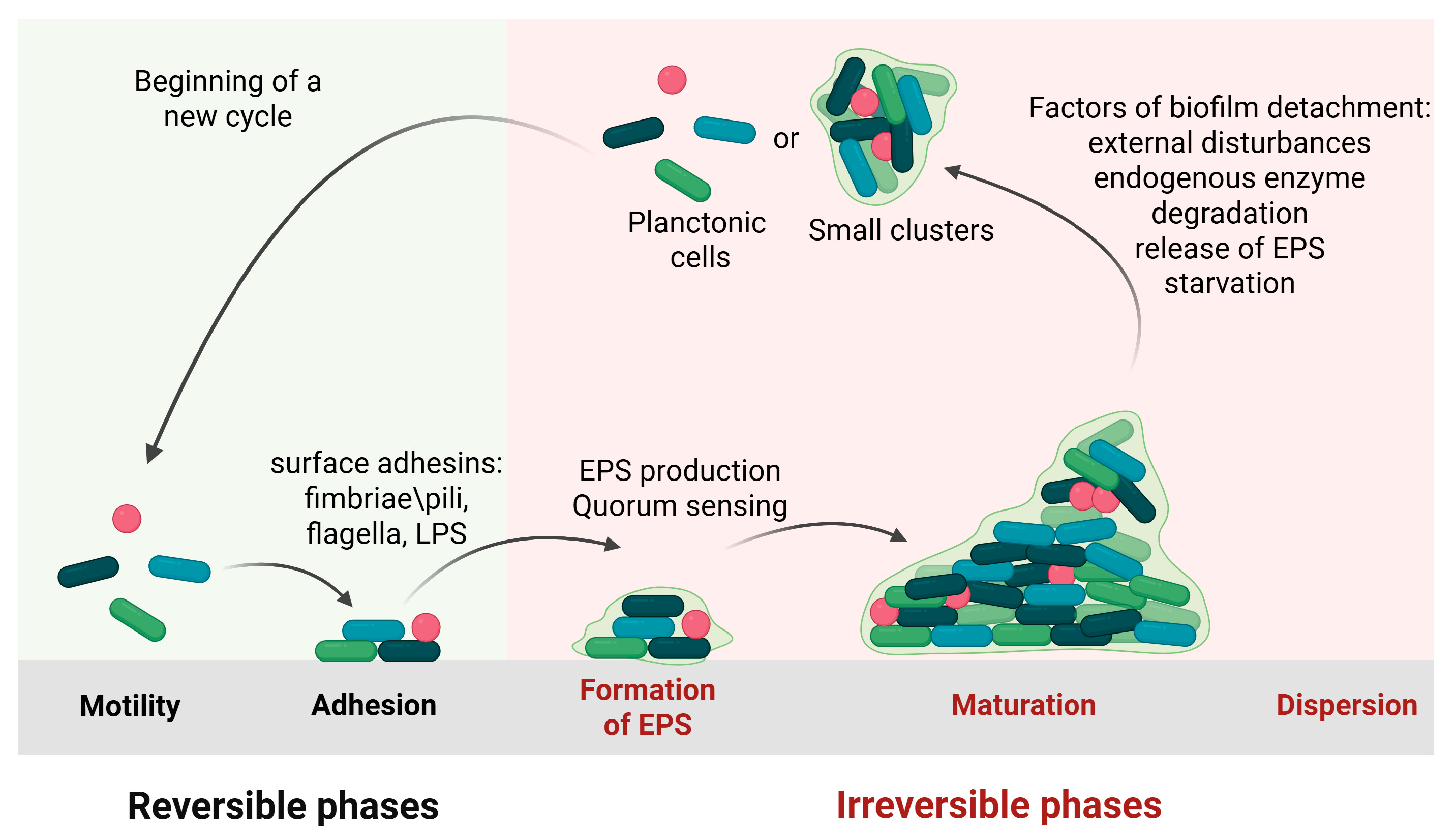
Figure 1. Schematic illustration of the key steps in biofilm formation on both biotic and abiotic surfaces (created in BioRender. Cochis, A. (2025) https://BioRender.com/j8kiffx, accessed on 1 August 2025).
Antibiotics 14 00802 g001
Figure 2. Dispersal biofilm: abrasion, erosion, and sloughing (created in BioRender. Cochis, A. (2025) https://BioRender.com/xyuiufv, accessed on 1 August 2025).
Antibiotics 14 00802 g002
Figure 3. Main components in the EPS matrix of biofilm (created in BioRender. Cochis, A. (2025) https://BioRender.com/g2n7f8z, accessed on 1 August 2025).
Antibiotics 14 00802 g003
Figure 4. Schematic explanation of the QS system in bacterial strains that regulates virulent behaviors (created in BioRender. Cochis, A. (2025) https://BioRender.com/g4wg72q, accessed on 1 August 2025).
Antibiotics 14 00802 g004
Figure 5. Gallium-doped zirconia reduces bacterial adhesion and proliferation. (a,b) Energy-dispersive X-ray spectroscopy (EDS) of zirconia (Z) and gallium-doped zirconia (ZGa); (c) low-magnification SEM micrographs showing a large bacterial biomass on Z and an almost clean surface on ZGa (scale bar = 200 μm); (d) high-magnification SEM micrographs highlighting the distinct bacterial populations on the two materials (scale bar = 1 μm). Reprinted/adapted with permission from [185], 2025, Alessandro Calogero Scalia.
Antibiotics 14 00802 g005
Table 1. Summary of other ESP-degrading enzymes with biofilm-dispersing activity.
Enzyme ClassesEnzyme NameComponent TargetBacterial TargetRef.
Glycoside hydrolaseDextranaseα-1,6-glucan (dextran)Streptococcus mutans (oral biofilm), Lactobacillus spp., [120,121,122]
Mutanaseα-1,3-glucan (mutan)Streptococcus mutans[121]
Cellulaseβ-1,4-glucan (cellulose)Pseudomonas fluorescens, Salmonella enterica, E. coli, Komagataeibacter xylinus[123,124]
Amylaseα-1,4-glucan (amylose)General polysaccharide degradation (minor components)[125]
α- or β-Mannosidaseα- or β-mannose-containing polysaccharidesKlebsiella sp., Pseudomonas sp. (minor components)[126,127]
ProteasesSubtilisin A (serine protease)Biofilm structural proteinsP. aeruginosa, S. aureus[128]
Papain (cysteine protease)Surface adhesin, extracellular proteinS. aureus, S. epidermidis[129,130]
Ficin (cysteine protease)Biofilm structural and adhesive proteinsS. aureus, S. epidermidis, S. mutans[131,132]
Bromelain (cysteine protease)Biofilm structural, adhesinsS. aureus, S. epidermidis, E. coli[129,133]
Metalloproteinases (zinc-dependent endopeptidase)Fibronectin-binding proteins (FnBPs), biofilm-associated protein (Bap), accumulation-associated protein (Aap)S. aureus, S. epidermidis, Enterococcus faecalis[133,134]
Alkaline protease (serine protease)FnBPs, Bap, AapS. aureus, S. epidermidis, E. coli[135]
DeoxyribonucleasesStreptodornase (DNase B)Extracellular DNA (eDNA)Gram-positive and Gram-negative bacteria[136,137]
NucBeDNAE. coli, Bacillus subtilis, Micrococcus luteus[76,138]
Micrococcal nucleaseCalcium-dependent endo-exonucleaseS. aureus, S. epidermidis, P. aeruginosa, B. subtilis[139]
Serratia nuclease (NucA)Single- and double-stranded eDNAE. coli, Bacillus subtilis[138]
Table 2. Antibacterial strategies: strengths, weaknesses, mitigation strategies, and toxicology profile.
StrengthsWeaknessesMitigation StrategiesToxicity Profile
Biofilm dispersing enzymesHigh specificity
Less likely to promote resistance development
Enhanced antimicrobial efficacy
Enzyme instability and poor retention
Limited efficacy against mature biofilms
Delivery challenges
High cost and limited scalability
Narrow spectrum of activity
Enzyme encapsulation in protective carriers can enhance stability, protect against degradation, and improve delivery to infection sites [217]
Enzyme engineering to improve stability and activity [218]
Combining different enzymes or co-administering with antibiotics can increase effectiveness [219]
Glycoside hydrolases (GHs) may be toxic to human cells in vitro [217]
DNases can cause DNA fragmentation in eukaryotic cells [220]
Proteases may damage eukaryotic cell membranes and induce apoptosis [221]
Chelating agentsReduced resistance development compared to other methods
Inhibition of bacterial growth
Biofilm disruption
Potential synergistic effects
High versatility
Low specificity (cannot distinguish between prokaryotic and eukaryotic cells)
Efficacy depends on bacterial species and their metal requirements
Delivery challenges
Combination with antibiotics to overcome resistance [222]
Development of agents targeting only prokaryotic cells [223]
Nanoparticle-based delivery to improve targeting [224]
May deplete essential metals in host cells, leading to dysfunction and toxicity [225]
Potential interference with host metal-dependent enzymes, causing oxidative stress [226]
Quorum sensing inhibitorsReduction of antibiotic resistance
Targeting of bacterial virulence factors
Availability of natural product-based options
Limited specificity
Susceptibility to washout
Challenges in clinical translation
Limited efficacy
Developing QSIs with high specificity can minimize off-target effects and improve efficacy [227]
Nanoparticle-based systems can enhance delivery and retention [228]
Further research into pathogen-specific QS pathways to design targeted inhibitors [229]
Potential interference with similar signaling pathways in human cells [230]
BiosurfactantsBiodegradable
Versatile
Stable under extreme conditions
Modifiable via genetic engineering
Low production yield
High production costs
Limited understanding and research
Lack of production and safety standards
Genetic engineering and nanotechnology approaches to improve production and properties [231]
Use of agricultural waste to reduce production costs [232]
Toxicity depends on type, concentration, exposure, and environmental conditions
Oxidizing agentsBroad-spectrum antimicrobial activity
Low likelihood of resistance development
Rapid disinfection
High versatility
Formation of harmful by-products
Short half-life of some agents
Environmental concerns
Combination with other antimicrobials to improve efficacy and reduce resistance risk [233]
Encapsulation in delivery systems for controlled release
Induction of oxidative stress
Mitochondrial dysfunction
Accumulation in tissues or organs [234]
Potential to trigger inflammation
Disclaimer/Publisher’s Note: The statements, opinions and data contained in all publications are solely those of the individual author(s) and contributor(s) and not of MDPI and/or the editor(s). MDPI and/or the editor(s) disclaim responsibility for any injury to people or property resulting from any ideas, methods, instructions or products referred to in the content.