Article

Antibacterial Effect of Eight Essential Oils against Bacteria Implicated in Bovine Mastitis and Characterization of Primary Action Mode of Thymus capitatus Essential Oil

1
Laboratory of Epidemiology and Veterinary Microbiology, Group of Bacteriology and Biotechnology, Pasteur Institute of Tunisia (IPT), University of Tunis El Manar (UTM), BP 74, 13 Place Pasteur, Belvédère, Tunis 1002, Tunisia
2
Higher Institute of Biotechnology of Beja, University of Jendouba, Beja 9000, Tunisia
3
Laboratory of Functional Physiology and Bio-Resources Valorization, Higher Institute of Biotechnology of Beja, University of Jendouba, Beja 9000, Tunisia
*
Author to whom correspondence should be addressed.
Antibiotics 2024, 13(3), 237; https://doi.org/10.3390/antibiotics13030237
Submission received: 30 October 2023 / Revised: 30 November 2023 / Accepted: 4 December 2023 / Published: 5 March 2024

Abstract

:
During the current investigation, eight essential oils (EOs) were tested for their antimicrobial activity against six species, belonging to the genus of staphylococcus, multi-resistant to antibiotics (S. epidermidis, S. cohni, S. wareneri, S. scuiri, S. chromogenes, S. pasteuri), three methicillin-resistant Staphylococcus aureus strains (MRSA) and two strains of Escherichia coli, producing extended-spectrum β-lactamase (ESBL) responsible for bovine mastitis. Our results indicated that the antimicrobial activities of eight EOs varied significantly among the types of EOs and bacterial species. Thymus capitatus and Trachyspermum ammi EOs display important antibacterial activity against all tested strains, with the inhibition zone diameters situated between 20 and 45 mm, while EOs of Artemisia absinthium, Eucalyptus globulus, Eucalyptus camaldulensis, Myrtus communis and Mentha pulegium exerted an intermediate activity. For Cymbopogon citratus, this effect depends on bacteria species. In fact, an important effect was observed against S. warneri, S. epidermidis, S. cohenii, S. pasteuri and MRSA (EC 39+) strains. In addition, the important lytic effect was observed against MRSA strains, showing that Gram-positive bacteria were more sensitive to T. capitatus EO than Gram-negative ones. Concerning the characterization of the mode action of T. capitatus, experiments of kill-time, bacteriolytic, loss of salt tolerance and loss of cytoplasmic material showed that the used EO was able to destroy cell walls and membranes followed by the loss of vital intracellular materials. In addition, it inhibits the normal synthesis of DNA, causing the bacterial death of E. coli and MRSA strains. This study shows the potential of using of EOs, particularly T. capitaus, to inhibit the growth of Gram-positive and Gram-negative bacteria multi-resistant to antibiotics causing bovine mastitis.

1. Introduction

Bovine mastitis is caused by several bacteria and, rarely, by yeasts, fungi, parasites or viruses [1]. This infection is often linked to trauma or attacks on the skin of the teat or at the level of the udder and sometimes to stenosis of the teat canal (lesions linked to the milking machine). The microorganisms causing mastitis are classified into three main categories, according to their sources, reservoir and their modes of transmission [1,2]. Staphylococcus aureus has been reported as the main causative agent of mastitis in cattle in several studies [3,4,5,6]. Furthermore, the emergence and spread of methicillin-resistant Staphylococcus aureus (MRSA) and methicillin-sensible Staphylococcus aureus (MSSA) are becoming a major concern for veterinary medicine as well as public health. MRSA is rarely detected as a causative agent of bovine mastitis in some countries, like Finland (1.5%), Germany (4.4%), Australia (4%) and Tunisia [6,7,8,9]. However, other studies reported the presence of MSSA: in Brazil (23%), Turkey (17.5%) and China (47.6%) [10,11,12]. This variability can be explained by the use of several categories of antibiotics belonging to the β-lactam family.
In addition to these contagious pathogens, Enterobacteriaceae are also the main environmental bacteria responsible for bovine mastitis. This family includes many genera, among which, we find Escherichia coli, which is frequently involved in mammary pathology [6,13,14]. In the same way, Klibi et al. [6] highlighted that the molecular characterization of Gram-negative bacteria and Staphylococci species showed a diversity of species with the respective dominance of E. coli (ten strains are extended-spectrum β-lactamase (ESBL) producers).
Generally, antibiotics are intended for the treatment of bovine mastitis. The presence of drug residues in milk constitutes a technological obstacle to the processing of milk and represents a danger to human health that is aggravated by the presence of multi-resistant bacteria. Since 2011, despite the World Health Organization (WHO) request to develop new drugs, a few molecules have been advanced. Plants can be considered as a source of natural bioactive substances that can be used in the manufacture of new antimicrobial drugs [15].
Recently, several findings have highlighted the different biological activities of essential oils (EOs), in particular their antibacterial power. In fact, Tariq et al. [16] signaled that EOs are more effective than conventional antibiotics against drug-resistant microbial strains. In a previous study, we investigated the antibacterial effect of eight EOs against Escherichia coli isolated from meat and able to produce extended-spectrum β-lactamase (ESBL) [17].
Although the chemical compositions and biological activities of EOs obtained from diverse plants were investigated, their antibacterial activities against Gram-negative and Gram-positive bacteria responsible for bovine mastitis have not been previously studied. In addition, until now, there are no published reports concerning the action mechanism of T. capitatus EO on the growth of Staphylococcus species multi-resistant to antibiotics. The aim of the current study is to evaluate the antibacterial effect of eight EOs (Eucalyptus globulus, Eucalyptus camaldulensis, Artemisia absinthium, Myrtus communis, Mentha pulegium, Trachyspermum ammi, Cymbopogon citratus and Thymus capitatus) against six species of Staphylococcus (S. wareneri, S. epidermidis, S. scuiri, S. chromogenes, S. cohni, S. pasteuri), methicillin-resistant Staphylococcus aureus (MRSA) and ESBLs producing E. coli strains. In addition, the characterization of the mechanism of antibacterial action of T. capitatus EO was investigated using four tests: kill-time analysis, bacteriolytic, loss of cytoplasmic material and loss of salt tolerance assays.

2. Materials and Methods

2.1. Plant Material

In the current report, eight essential oils (Eucalyptus globulus, Eucalyptus camaldulensis, Artemisia absinthium, Myrtus communis, Mentha pulegium, Trachyspermum ammi, Cymbopogon citratus, Thymus capitatus), purchased from the company “Carthago Essences Sousse, Tunisia”, were used to study their antibacterial effect against different strains multi-resistant to antibiotics causing clinical bovine mastitis in Tunisia.

2.2. Selected Bacteria and Growth Conditions

In order to study the antimicrobial activity of eight EOs, six species of Staphylococcus (S. wareneri, S. epidermidis, S. scuiri, S. chromogenes, S. cohni, S. Pasteursi), three methicillin-resistant Staphylococcus aureus (MRSA) and two strains of E. coli (Ec CTX and Ec Mac), isolated and identified by Klibi et al. [6], were used. The determination of antibiotic susceptibility of different strains was carried out by Klibi et al. [18] according to the recommendations of the CLSI “Clinical Laboratory Standards Institute 2015” and the French Society of Microbiology. Table 1 summarizes the different characteristics of used strains.
These bacteria were maintained by sub-culturing on brain heart infusion agar (BHI agar), which was favorable to their growth for 24 h at 37 °C.

2.3. Screening of Antimicrobial Activity of EOs

The sensibility of different bacterial species to eight EOs was evaluated using the agar well diffusion method, as described by Aouadhi et al. [17]. The qualitative antimicrobial activity results of tested EOs are expressed as the diameter of the growth-inhibition zone (including the disc diameter of 6 mm).

2.4. Evaluation of the Quantitative Antibacterial Power of EOs

The determination of minimum inhibitory concentrations (MICs) and the minimum bactericidal concentrations (MBCs) was utilized to quantify the antibacterial effect of eights EO. In fact, broth dilution, described by Aouadhi et al. [19], was used. The working culture, obtained after incubation at 37 °C for 18 h, was performed in Mueller–Hinton broth from three to four colonies. Each tested EO was diluted from 0.07 to 50% (v/v) in dimethyl sulfoxide (DMSO).
The positive control corresponded to the bacteria without EO, and the negative control corresponded to the appropriate medium and test oil. After incubation for 24 h at 37 °C, the MICs and MBCs were determined. To confirm MICs and MBC, 100 µL of each tube was inoculated on BHI agar. After incubation at 37 °C, the count of survivor’s bacteria was determined. Each experiment was repeated at least three times.

2.5. Primary Mode of Action of T. capitaus EO

2.5.1. Time-Kill Studies

This method makes it possible to characterize the antibacterial activity of T. capitatus EO over time against E. coli and MRSA strains. It evaluates the decrease in bacteria count in the presence of EO (at a concentration equal to its MIC) over several hours [20]. In fact, three to five bacterial colonies, obtained from an 18 h culture, are removed using a loop and deposited in 2 mL of peptone water. The obtained suspension was diluted to obtain a working culture (105 CFU/mL). EO was used at a concentration equal to its MIC. The growth of bacterial suspensions in presence or in absence (growth control) of T. capituatus EO was monitored for 24 h at 37 °C under stirring. At different times (0, 2, 4, 6, 8 and 24 h), an aliquot of 100 µL was taken to estimate its rate [21].

2.5.2. Bacteriolysis

This method allows for the determination of a possible bacteriolytic action of T. capitatus EO by measuring the absorbance of tested bacteria at 620 nm over time [21,22]. Indeed, two bacterial colonies, obtained from an 18 h culture, were used to inoculate 9 mL of nutrient medium. The culture was incubated at 37 °C for 18 h with shaking. The bacteria were then separated from the culture medium by centrifugation at 10,000 rpm for 12 min at 4 °C. The bacterial pellet was washed twice with sodium phosphate buffer (PBS), then resuspended in PBS-Tween 80 (0.01%, v/v). The obtained bacterial suspension was standardized at 1010 UFC/mL and placed in a sterile tube in the absence (negative control for bacteriolysis) or in the presence of EO at a concentration equal to the MIC. The obtained suspensions were subjected to stirring. After 120 min, they were homogenized, diluted to 1/100 and their absorbance was measured directly at 620 nm.
The results were expressed as a ratio (in percent) of the OD620 at each time point versus the OD620 at 0 min.

2.5.3. Loss of Cytoplasmic Material

The release of 280 nm absorbing materials of two strains (E. coli and MRSA) in presence of T. capitatus EO was used to evaluate the loss of cytoplasmic material, according to previous method described by Carson et al. [22].

2.5.4. Loss of Salt Tolerance

The loss of salt tolerance of E. coli and MRSA strains in the presence of T. capitatus EO was evaluated according to that previously described by Carson et al. [22]. In fact, untreated and treated suspensions of E. coli and MRSA strains, for 30 min with T. capitatus (the used concentration correspond to the MIC), were plated on nutrient agar containing different concentrations of NaCl (0 to 100 g/L). After incubation at 37 °C for 24 h, the colonies were counted. The numbers of UFC per milliliter on each nutrient agar plate were compared to those on the nutrient agar plate without NaCl.

2.6. Statistical Analysis

All experiments are expressed as the mean ± standard deviation (SD) of three replications. The obtained data were processed using Microsoft Excel 2007. Quantitative differences were assessed using the ANOVA procedure (SPSS 14.0 for Windows powershell 7.2) (p < 0.05) followed by Duncan’s multiple range test.

3. Results and Discussion

3.1. Antibacterial Effect of Essential Oils

During this study, the antimicrobial power of eight EOs against two strains of E. coli that is multi-resistant to antibiotics, producing extended-spectrum beta-lactamases (ESBL), six species belonging to the genus of Staphylococcus that are multi-resistant to antibiotics (S. epidermidis, S. cohni, S. wareneri, S. scuiri, S. chromogenes, S. pasteuri) and three MRSA strains causing bovine mastitis, was investigated.
The screening of qualitative and quantitative effects of different EOs was realized using three parameters: diameters of inhibition zones (IZs), MIC and MBC. The obtained data summarized in Table 2 and Table 3 clearly show that the antimicrobial activities of eight EOs varied significantly among the used EOs and bacteria species. According to Rossi et al. [23], the EOs are considered active if they produce microbial growth inhibition diameters equal to or greater than 20 mm. Considering this definition, we noted that T. capitatus and T. ammi EOs show important antibacterial activity against all tested strains, with IZ diameters situated between 20 and 45 mm, while the EOs of A. absinthium, Eucalyptus, M. communis and M. pulegium exerted intermediate activity. For C. citratus, this effect depends on bacteria species. In fact, an important effect was observed against S. warneri, S. epidermidis, S. cohenii, S. pasteuri, and MRSA EC 39+ strains. The lowest MIC (0.048% (v/v) was registered in the presence of T. capitatus and T. ammi EOs, and the highest MIC (12.5% (v/v) was observed in the presence of M. pulegium and Eucalyptus EOs. In addition, it can be signaled that the tested EOs were more active against Staphylococcus and MRSA strains than against E. coli strains. The diameters of the inhibition zone and the MIC varied between 14 and 45 mm and 0.048 and 12.5% for Staphylococcus, while, in the presence of E. coli, the diameters of the inhibition zone varied between 11 and 25 mm and the MIC situated between 0.39 and 6.25% for E. coli.
This difference in antimicrobial activity may be due to the difference in the chemical composition of the tested EOs. Generally, EOs are complex mixtures of natural compounds of about 20–60 constituents in varying quantities. The antimicrobial activity could be related to the major compounds of the EOs or to a synergistic effect between the major and minor compounds [24]. Nevertheless, some researchers reported that there is a relationship between the chemical composition and antimicrobial activity [25,26]. The highest antibacterial activity was observed in the presence of EO rich in aldehydes or phenols, such as carvacrol, eugenol or thymol, followed by EO containing terpene alcohol. EOs are inactive if the principal constituents were terpene hydrocarbons. However, weak activity was observed when the EOs containing ketone or esters such as β-myrcene, α-thujone or gerenyl acetate [27,28].
Thus, previous findings showed that the T. capitaus EO has very significant antibacterial activity compared to the other tested EOs, which might be due to its major constituents: thymol and carvacrol [17,29,30,31]. In previous studies, we demonstrated that thymol (81.49%), α-cubebene (3.44%), α-terpinene (3.83%) and β-ocimene (3.16%) were identified as the major components of T. capitatus [30]. In fact, Althunibat et al. [32] demonstrated that thymol and carvacrol exhibited important antimicrobial activity, with the most sensitive being the P. aeruginosa strain followed by S. aureus, E. aerogenes and E. coli. Moreover, EL Jalel et al. [26] signaled that T. capitatus from Abu-Draa has the highest antimicrobial activity against all tested strains compared to that from Sidi-Alhamery. Carvcrol was the major volatile component present in both essential oils of Abu-Draa and Sidi Al-Hamrey regions (58.56% and 24.28%, respectively) [26].
The antimicrobial activities of EO of T. ammi against E. coli and Staphylococcus species and MRSA are comparable to those obtained against E. coli strains isolated from meat. In fact, Aouadhi et al. [17] showed that EO extracted from T. ammi possesses important antibacterial activity, with the IZ situated between 20 and 22 mm, and the MIC ranged from 0.39 to 0.78% (v/v), respectively.
As shown in Table 2 and Table 3, it can be signaled that the EO of M. pulegium can be used as antimicrobial agent against pathogenic bacteria. These data are in agreement with those obtained by Aouadhi et al. [17], Ghazghazi et al. [33] and Ladjel et al. [34]. In addition, Amalich et al. [35] highlighted that the M. pulegium EO is more effective than amoxicillin against E. coli, Pseudomonas aeruginosa and Klebsiella pneumoniae. Concerning the chemical composition, Ghazghazi et al. [33] demonstrated that the main components of M. pulegium EO were menthone (41.7%), cis-isopulegone (31.71%) and isomenthone (15.03%).
Concerning A. absinthium, the antimicrobial activity of its EO depends on bacterial species. In fact, S. warneri and S. epidermidis were the most sensitive strains, with an MIC of 0.78% (v/v), while MRS strains were more resistant (MIC = 12.5%). In the same way, Belay et al. [36] demonstrated that EO obtained from A. absinthium is able to alter the biosynthesis of proteins, RNA, DNA and polysaccharide in the cells of S. aureus, causing the inhibition of its growth (MIC = 0.62 μL/mL). In addition, Riahi et al. [37] showed that EOs of A. absinthium harvested from four localities in Tunisia displayed antimicrobial activity (MIC varied between 12.5 and 25% (v/v)) against fungal strains, Gram-negative and Gram-positive bacteria. In addition, we demonstrated that the camphor, (Z)-sabinene hydrate and 1-terpinen-4-ol are the major compounds [37].
The antimicrobial activity of EO of M. communis observed in the current study may be due to alcoholic compounds such as borneol, having important antibacterial activity. In addition, other compounds belonging to ethers and hydrocarbon groups such as 1,8-cineole and α-pinene had moderate antimicrobial activity [38].
By analyzing the obtained data, we can conclude that, despite the antimicrobial effect of different EOs, it remains less important than that observed with the reference strains. This can be explained by the fact that the used bacteria have acquired different mechanisms of resistance to antibiotics, as shown in Table 1, and, therefore, have an impact on its resistance to different biological substances. In addition, the antimicrobial activity of used EOs does not depend on the structure of the bacterial cell wall and the arrangement of its outer membrane.

3.2. Characterization of Primary Action Mode of Thymus capitatus Essential Oil

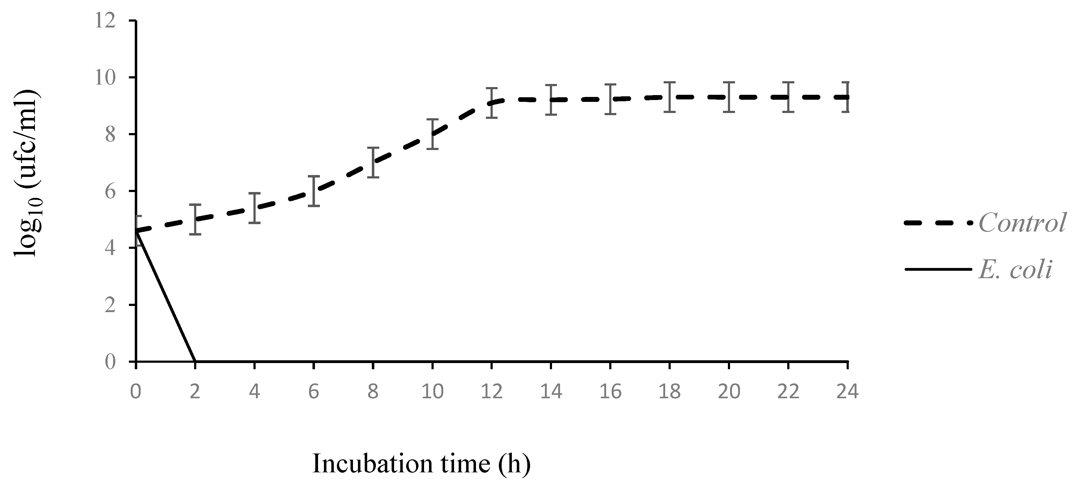
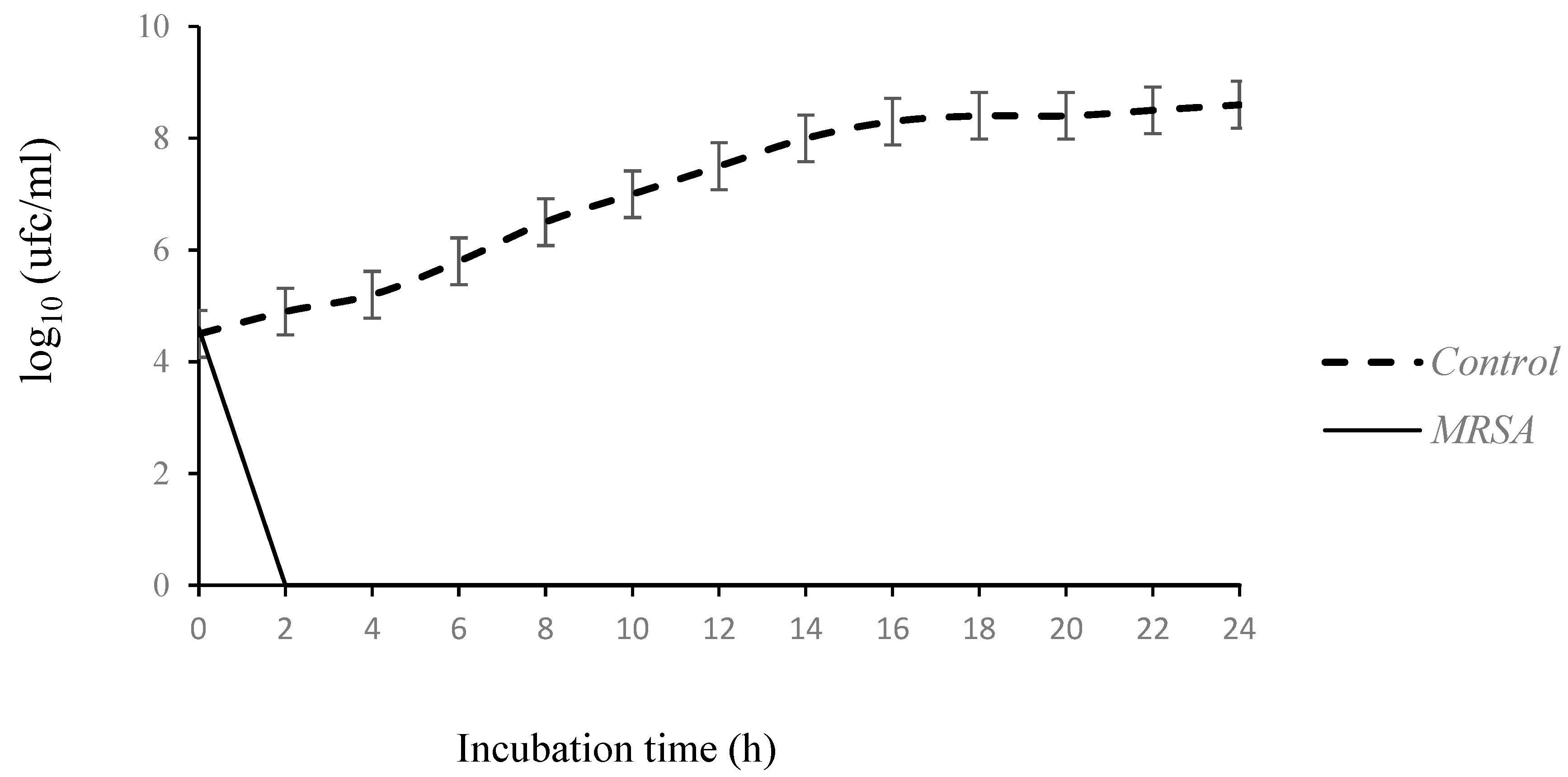
3.2.1. Dynamics of Action of EO by Measuring Bacterial Growth: Time-Kill Assay

The bacterial growth of MRSA 44 and of E. coli strains, without or with (concentrations of essential oil equal to MIC) T. capitatus EO, was measured over a period of 24 h. MRSA and E. coli were selected as model microorganisms for further study based on their distinct sensitivities to T. capitaus EO. The choice of T. capitatus EO, for the study of the mode of action, was oriented to select the most active EOs against MRSA and E. coli based on the disk diffusion method and the determination of MIC and MBC values. As shown in Figure 1, the control case revealed a classic growth curve with three phases (growth, stationary and decline). In fact, the E. coli and MRSA 44 counts increased from 4.6 log10 to 9.3 log10 cfu/mL and 8.4 lg10 cfu/mL, respectively, after 24 h. In the presence of EO, the growth of two bacterial strains was inhibited after one hour, showing the important bactericidal effect of T. capitaus EO, supporting the results of MIC and MBC. In addition, the evaluated EO caused a decrease in the growth of E. coli within the first ten minutes. Our data corroborate a previous study about the rapid antibacterial effect of T. capitatus EO against E. coli–BLSE producers [17,29], probably because it contained a high concentration of carvacrol and thymol. Xu et al. [39] showed that carvacrol is able to embrittle and depolarize the cytoplasmic membrane. Moreover, after 30 min, the hydroxyl group of carvacrol and its isomers, thymol, causes an increase in their hydrophilic capacity, altering microbial cell formation [39].

3.2.2. Bacteriolytic Effect

The screening for the bacteriolytic effect of T. capitatus EO against two species, MRSA and E. coli, is based on a measurement of the absorbance at 620 nm of bacterial suspensions in the absence or presence of EO, at a concentration equal to its MIC. The loss of absorbance is evaluated according to the initial absorbance. The obtained data clearly showed the absence of cell lysis of two used species in the absence of EO (the control case) since the absorbance was 100%. So, in the presence of EO, a decrease in the absorbance of two strains was observed. In fact, the percentage of OD decreased for E. coli to approximately 47% and for MRSA to 29%.
Based on this result, it can be concluded that T. capitatus EO exerts a bacteriolytic effect against E. coli, and MRSA species suggested that cell wall destruction is one of mechanism of action. The obtained data are comparable to those obtained by Aouadhi et al. [17], who demonstrated that T. capitatus exerts a bacteriolytic effect (OD in the presence of EO was 40%) against E. coli, producing extended-spectrum β-lactamases.
In addition, the important lytic effect was observed against MRSA compared to E. coli, showing that Gram-positive bacteria were more sensitive than Gram-negative ones towards the studied EO. This suggestion is consistent with previous studies carried out with several plant species [40,41] and is contrary to the investigation of Aouadhi et al. [30], who demonstrated that the EOs of T. capitatus harvested in two regions in Tunisia had powerful antimicrobial activity against all tested species (B. cereus, L. monocytogens, S. aureus, E. coli, P. aeruginosa, A. hydrophila, S. typhimurium).
Previous studies showed that the different antibacterial effects of some EOs against Gram-positive and Gram-negative bacteria can be attributed to the bacterial membrane. Indeed, the outer membrane of Gram-negative bacteria is rich in lipopolysaccharides. These molecules create a hydrophilic surface able to block the penetration of hydrophobic constituents of essential oils into the target cell membrane [40,41,42]. Consequently, E. coli producing extended-spectrum β-lactamases is relatively resistant to hydrophobic antibiotics. In contrast, for MRSA (Gram-positive bacteria), T. capitatus EO can block the enzyme system and progressivity of ion permeability thanks to the rupture of the cell membrane. Conner [43] highlighted that the thyme essential oil showed strong antibacterial activity against Staphylococcus aureus, Salmonella typhi and Pseudomonas aeruginosa. In addition, Horne et al. [44] showed that the EO of thyme generates a lytic effect.

3.2.3. Loss of Salt Tolerance

The growth of E. coli and MRSA strains on nutrient agar containing different concentrations of NaCl, with or without T. capiataus EO, was used to study their salt tolerance. The obtained data summarized in Table 4 corroborated that the addition of NaCl in the medium, after exposure to T. capitatus, reduced the ability of bacteria to form colonies. In fact, in the absence of EO and the presence of different concentrations of NaCl (0 to 10%), the growth percentage of the two used strains was 100%, thus showing that the E. coli and MRSA tolerate a high concentration of NaCl, while in the presence of T. capitatus EO at a concentration equal to its MIC, the total absence of MRSA colonies at different concentrations and a very low percentage growth (1% at a concentration of 5% and 0.3% at a concentration of 10%) for E. coli strains were observed. This observation was in concordance with those obtained by Aouadhi et al. [17] when they studied the effect of two EOs (T. capitatus and T. ammi) against the salt tolerance of extended-spectrum β-lactamase (ESBL)-producing E. coli isolated from meat. The two used EOs significantly reduced the ability of bacteria to grow on media containing NaCl [17].
The obtained data highlighted clearly that the second mechanism of action of T. capiatus EO was the loss of osmoregulation of bacterial cells to salt, showing that the tested EO affected the envelope of the cell and cytoplasm. Similarly, Gilbert et al. [45] showed that the permeability and osmoregulation of the cell membrane can be affected by EO since the latter can lead to sublethal damage to the cell membrane of bacteria. Several authors explained that the EO can increase bacterial cell membrane permeability, causing a leak of cellular components and degradation of ions [22,46,47]. Also, it is able to reduce membrane potential and to disrupt proton pumps and the liquidation of ATP [48]. For example, Rudramurthy et al. [49] showed that carvacrol affects the membrane fluidity, and permeability damages the cell membrane by altering the composition of fatty acids.

3.2.4. Loss of Cytoplasmic Material

Generally, the important macromolecules for cells are proteins and nucleic acids, which have an important role in cellular structure and genetic information [50]. The determination of absorbance at 260 nm (OD260) was used recently to understand the mechanism of action of EOs. This measurement is able to give us an idea of membrane integrity [51]. Table 5 summarizes the effect of T. capitatus EO on the integrity of the membrane of E. coli and MRSA. The OD260 of both filtrates after treatment increased significantly after 30 min, whereas in the control experiment, it remained practically stable (Table 5). Based on these data, it can be concluded that the EO of T. capitatus showed a release of absorbent constituents at 260 nm, so this oil affected the integrity and permeability of the membrane of E. coli and MRSA, leading to the leakage of nucleic acids through a damaged cytoplasmic membrane, inducing cell death. Bakkali et al. [52] showed that the EO damages mitochondrial membranes after it penetrates through the cytoplasmic membrane. Afterwards, free radicals, which oxidize and damage lipids, and proteins were produced by DNA in the mitochondria.

4. Conclusions

This is the first study to investigate the effect of different EOs against E. coli producing extended-spectrum β-lactamase (ESBL), six species belonging to the genus of Staphylococcus multi-resistant to antibiotics and three MRSA strains responsible for bovine mastitis. The obtained data highlighted the presence of a significant difference in the antibacterial activity between the eight EOs and according to the bacterial species. T. capitatus and T. ammi EOs showed important antibacterial activity against all tested strains, while the EOs of A. absinthium, Eucalyptus, M. communis and M. pulegium exerted intermediate activity. For C. citratus, this effect depends on bacteria species. Based on IZ, MIC and MBC values, it can be concluded that the used EOs possess antibacterial activity against Gram-positive and Gram-negative bacteria. But, using the cell lysis assay, Gram-positive bacteria are more sensitive to the action of essential oil than Gram-negative bacteria. T. capitaus EO (at a concentration equal to its MIC) affects the permeability and integrity of the cell membrane of E. coli and MRSA strains. In addition, it is able to cytoplasm material (DNA and proteins), causing cell death.

Author Contributions

C.A. conceived and designed the experiments; performed the experiments; analyzed and interpreted the data; and wrote the paper. A.J. and K.M. designed and performed the experiments. A.M. conceived and designed the experiments; contributed reagents, materials, analysis tools or data; and wrote the paper. All authors have read and agreed to the published version of the manuscript.

Funding

This research was funded by Internal collaborative project of Pasteur Institute of Tunis and the Tunisian Ministry of Higher Education and Scientific Research (LR11IPT03).

Institutional Review Board Statement

Not applicable.

Informed Consent Statement

Not applicable.

Data Availability Statement

Data included in article/referenced in article.

Conflicts of Interest

The authors declare no conflict of interest.

References

  1. Contreras, G.A.; Rodríguez, J.M. Mastitis: Comparative Etiology and Epidemiology. J. Mammary Gland Biol. Neoplasia 2011, 16, 339–356. [Google Scholar] [CrossRef]
  2. Motaung, T.E.; Petrovski, K.R.; Petzer, I.M.; Thekisoe, O.; Tsiloa, T.J. Importance of bovine mastitis in Africa. Anim. Health Res. Rev. 2017, 18, 58–69. [Google Scholar] [CrossRef]
  3. Jagielski, T.; Puacz, E.; Lisowski, A.; Siedlecki, P.; Dudziak, W.; Międzobrodzki, J.; Krukowski, H. Short communication: Antimicrobial susceptibility profiling and genotyping of Staphylococcus aureus isolates from bovine mastitis in Poland. J. Dairy Sci. 2014, 97, 6122–6128. [Google Scholar] [CrossRef]
  4. Jamali, H.; Radmehr, S.; Ismail, B. Short communication: Prevalence and antibiotic resistance of Staphylococcus aureus isolated from bovine clinical mastitis. J. Dairy Sci. 2014, 97, 2226–2230. [Google Scholar] [CrossRef]
  5. Wang, D.; Wang, Z.; Yan, Z.; Wu, J.; Ali, T.; Li, J.; Lv, Y.; Han, B. Bovine mastitis Staphylococcus aureus: Antibiotic susceptibility profile, resistance genes and molecular typing of methicillin-resistant and methicillin-sensitive strains in China. Infect. Genet. Evol. 2015, 31, 9–16. [Google Scholar] [CrossRef]
  6. Klibi, A.; Jouini, A.; El Andolsi, R.B.; Kmiha, S.; Ben Hamda, C.; Ghedira, K.; Hamrouni, S.; Ghram, A.; Maaroufi, A. Epidemiology of β-Lactamase-Producing Staphylococci and Gram Negative Bacteria as Cause of Clinical Bovine Mastitis in Tunisia. BioMed Res. Int. 2019, 2019, 2165316. [Google Scholar] [CrossRef]
  7. Brody, T.; Yavatkar, A.S.; Lin, Y.; Ross, J.; Kuzin, A.; Kundu, M.; Fann, Y.; Odenwald, W.F. Horizontal Gene Transfers Link a Human MRSA Pathogen to Contagious Bovine Mastitis Bacteria. PLoS ONE 2008, 3, 8. [Google Scholar] [CrossRef]
  8. Gindonis, V.; Taponen, S.; Myllyniemi, A.-L.; Pyörälä, S.; Nykäsenoja, S.; Salmenlinna, S.; Lindholm, L.; Rantala, M. Occurrence and characterization of methicillin resistant staphylococci from bovine mastitis milk samples in Finland. Acta Vet. Scan. 2013, 55, 61. [Google Scholar] [CrossRef]
  9. Worthing, K.A.; Abraham, S.; Pang, S.; Coombs, G.W.; Saputra, S.; Jordan, D.; Wong, H.S.; Abraham, R.J.; Trott, D.J.; Norris, J.M. Molecular Characterization of Methicillin-Resistant Staphylococcus aureus Isolated from Australian Animals and Veterinarians. Microb. Drug Resist. 2017, 24, 203–212. [Google Scholar] [CrossRef]
  10. Bardiau, M.; Yamazaki, K.; Duprez, J.N.; Taminiau, B.; Mainil, J.G.; Ote, I. Genotypic and phenotypic characterization of methicillin-resistant Staphylococcus aureus (MRSA) isolated from milk of bovine mastitis. Lett. Appl. Microbiol. 2013, 57, 181–186. [Google Scholar] [CrossRef]
  11. Pu, W.; Su, Y.; Li, J.; Li, C.; Yang, Z.; Deng, H.; Ni, C. High incidence of oxacillin-susceptible mecA-positive Staphylococcus aureus (OS-MRSA) associated with bovine mastitis in China. PLoS ONE 2014, 11, e88134. [Google Scholar] [CrossRef]
  12. Guimarães, F.F.; Manzi, M.P.; Joaquim, S.F.; Richini-Pereira, V.B.; Langoni, H. Short communication: Outbreak of methicillin-resistant Staphylococcus aureus (MRSA)-associated mastitis in a closed dairy herd. J. Dairy Sci. 2017, 100, 726–730. [Google Scholar] [CrossRef]
  13. Oliveira, L.; Hulland, C.; Ruegg, P.L. Characterization of clinical mastitis occurring in cows on 50 large dairy herds in Wisconsin. J. Dairy Sci. 2013, 96, 7538–7549. [Google Scholar] [CrossRef] [PubMed]
  14. Hogan, J.; Smith, K.L. Coliform mastitis. Vet. Res. 2003, 34, 507–519. [Google Scholar] [CrossRef] [PubMed]
  15. Abreu, A.C.; McBain, A.J.; Simoes, M. Plants as sources of new antimicrobials and resistance-modifying agents. Nat. Prod. Rep. 2012, 29, 1007–1021. [Google Scholar] [CrossRef] [PubMed]
  16. Tariq, S.; Wani, S.; Rasool, W.; Bhat, M.A.; Prabhakar, A.; Shalla, A.H.; Rather, M.A. A comprehensive review of the antibacterial, antifungal and antiviral potential of essential oils and their chemical constituents against drug-resistant microbial pathogens. Microb. Pathog. 2019, 134, 103580. [Google Scholar] [CrossRef]
  17. Aouadhi, C.; Jouini, J.; Mechichi, D.; Boulares, M.; Maaroufi, A. Characterization of primary action mode of two essential oils and evaluation of their antibacterial effect against extended- spectrum β-lactamase (ESBL)-producing Escherichia coli inoculated in turkey meat. Molecules 2022, 27, 2588. [Google Scholar] [CrossRef]
  18. Klibi, A.; Maaroufi, A.; Torres, C.; Jouini, A. Detection and characterization of methicillin resistant and susceptible coagulase-negative Staphylococci in milk from cows with clinical mastitis in Tunisia. Int. J. Antim. Agents 2018, 6, 930–935. [Google Scholar] [CrossRef]
  19. Aouadhi, C.; Ghazghazi, H.; Hasnaoui, B.; Maaroufi, A. Total phenolic content, antioxidant and antibacterial activities of Marrubium vulgare methanolic extract. Tunisian J. Med. Plants Nat. Prod. 2014, 11, 37–79. [Google Scholar]
  20. Viljoen, A.; van Vuuren, S.; Ernst, E.; Klepser, M.; Demirci, B.; Başer, H.; van Wyk, B.-E. Osmitopsis asteriscoides (Asteraceae)—The Antimicrobial Activity and Essential Oil Composition of a Cape-Dutch Remedy. J. Ethnopharm. 2003, 88, 137–143. [Google Scholar] [CrossRef]
  21. Guinoiseau, E.; Luciani, A.; de Rocca Serra, D.; Quilichini, Y.; Berti, L.; Lorenzi, V. Primary Mode of Action of Cistus ladaniferus L. Essential Oil Active Fractions on Staphyloccocus aureus strain. Adv. Microbiol. 2015, 5, 881–890. [Google Scholar] [CrossRef]
  22. Carson, C.F.; Mee, B.J.; Riley, T.V. Mechanism of Action of Melaleuca alternifolia (Tea Tree) Oil on Staphylococcus aureus Determined Par Time-Kill, Lysis, Leakage and Salt Tolerance Assays and Electron Microscopy. Ant. Agents. Chem. 2002, 46, 1914–1920. [Google Scholar] [CrossRef]
  23. Rossi, P.-G.; Berti, L.; Panighi, J.; Luciani, A.; Maury, J.; Muselli, A.; Serra, D.d.R.; Gonny, M.; Bolla, J.-M. Antibacterial action of essential oils from Corsica. J. Essent. Oil Res. 2007, 9, 176–182. [Google Scholar] [CrossRef]
  24. Carović-Stanko, K.; Orlić, S.; Politeo, O.; Strikić, F.; Kolak, I.; Milos, M.; Satovic, Z. Composition and antibacterial activities of essential oils of seven Ocimum taxa. Food Chem. 2010, 119, 196–201. [Google Scholar] [CrossRef]
  25. Djenane, D.; Yangüela, J.; Amrouche, T.; Boubrit, S.; Boussad, N.; Roncalés, P. Chemical composition and antimicrobial effects of essential oils of Eucalyptus globulus, Myrtus communis and Satureja hortensis against Escherichia coli O157:H7 and Staphylococcus aureus in minced beef. Food Sci. Technol. Int. 2011, 17, 505–515. [Google Scholar] [CrossRef] [PubMed]
  26. El-Jalel, L.F.; Elkady, W.M.; Gonaid, M.H.; El-Gareeb, K.A. Difference in chemical composition and antimicrobial activity of Thymus capitatus L. essential oil at different altitudes. Future J. Pharm. Sci. 2018, 4, 156–160. [Google Scholar] [CrossRef]
  27. Dorman, H.J.; Deans, S.G. Antimicrobial agents from plants: Antibacterial activity of plant volatile oils. J. Appl. Microbiol. 2000, 88, 308–316. [Google Scholar] [CrossRef] [PubMed]
  28. Inouye, S.; Yamaguchi, H.; Takizawa, T. Screening of the antibacterial effects of a variety of essential oils on respiratory tract pathogenns, using a modified dilution assay method. J. Infect. Chemother. 2001, 7, 251–254. [Google Scholar] [CrossRef]
  29. Jayari, A.; Jouini, A.; Boukhris, H.; Hamrouni, S.; Damergi, C.; Ahmed, S.B.H.; Maaroufi, A. Essential Oils from Thymus capitatus and Thymus algeriensis as Antimicrobial Agents to Control Pathogenic and Spoilage Bacteria in Ground Meat. J. Food Saf. 2021, 38, 5599374. [Google Scholar] [CrossRef]
  30. Aouadhi, C.; Ghazghazi, H.; Dallali, S.; Sebei, H.; Maaroufi, A.; Hasnaoui, B. Comparison of chemical composition, antioxidant and antimicrobial activities of Thymus capitatus L. essential oils from two Tunisian localities (Sousse and Bizerte). Int. J. Agr. Plant Prod. 2013, 4, 1772–1781. [Google Scholar]
  31. El Abed, N.; Kaabi, B.; Smaali, M.I.; Chabbouh, M.; Habibi, K.; Mejri, M.; Marzouki, M.N.; Ahmed, S.B.H. Chemical Composition, Antioxidant and Antimicrobial Activities of Thymus capitata Essential Oil with Its Preservative Effect against Listeria monocytogenes Inoculated in Minced Beef Meat. Evid. Based Compl. Alter. Med. 2014, 2014, 152487. [Google Scholar] [CrossRef]
  32. Althunibat, Y.O.; Qaralleh, H.; Sati, Y.; Al-Dalin, A.; Abboud, M.; Khleifat, K.; Majali, I.S.; Aldal, H.K.H.; Rayyan, W.A.; Jaafraa, A. Effect of Thymol and Carvacrol, the Major Components of Thymus capitatus on the Growth of Pseudomonas aeruginosa. J. Pure Appl. Microbiol. 2016, 10, 367–374. [Google Scholar]
  33. Ghazghazi, H.; Aouadhi, C.; Weslati, M.; Trakhn, F.; Houssine, S.; Maaroufi, A.; Hasnaoui, B. Chemical composition and the biological activities of Mentha pulegium leaves extracts against foodborne pathogens. J. Food Saf. 2013, 33, 239–246. [Google Scholar] [CrossRef]
  34. Ladjel, S.; Gherraf, N.; Hamada, D. Antimicrobial effect of essential oils from the Algerian medicinal plant Mentha rotundifolia L. J. Appl. Sci. Res. 2011, 7, 1665–1667. [Google Scholar]
  35. Amalich, S.; Zerkani, H.; Cherrat, A.; Soro, N.K.; Bourakhouadar, M.; Fadli, M.; Chevalier, J.; Saad, A.; Mezrioui, N.E.; Hassani, L. Essential oils from Moroccan plants as potential chemo-sensitizers restoring antibiotic activity in resistant Gram-negative bacteria. Int. J. Antimicrob. Agents 2016, 38, 325–330. [Google Scholar] [CrossRef]
  36. Belay, G.; Tariku, Y.; Kebede, T.; Hymete, A.; Mekonnen, Y. Ethnopharmacological investigations of essential oils isolated from five Ethiopian medicinal plants against eleven pathogenic bacterial strains. Phytopharmacology 2011, 1, 133–143. [Google Scholar]
  37. Riahi, L.; Ghazghazi, H.; Ayari, B.; Aouadhi, C.; Klay, I.; Chograni, H.; Ameur, C.; Zoghlami, N. Effect of environmental conditions on chemical polymorphism and biological activities among Artemisia absinthium L. essential oil provenances grown in Tunisia. Ind. Crops Prod. 2015, 66, 96–102. [Google Scholar] [CrossRef]
  38. Fadil, M.; Fikri-Benbrahim, K.; Rachiq, S.; Ihssane, B.; Lebrazi, S.; Chraibi, M.; Haloui, T.; Farah, A. Combined treatment of Thymus vulgaris L., Rosmarinus officinalis L. and Myrtus communis L. essential oils against Salmonella typhimurium: Optimization of antibacterial activity by mixture design methodology. Euro. J. Pharm. Biopharm. 2018, 126, 211–220. [Google Scholar] [CrossRef]
  39. Zhou, J.X.F.; Ji, B.-P.; Pei, R.-S.; Xu, N. The antibacterial mechanism of carvacrol and thymol against Escherichia coli. Lett. Appl. Microbiol. 2008, 47, 174–179. [Google Scholar] [CrossRef]
  40. Li, Z.-H.; Cai, M.; Liu, Y.-S.; Sun, P.L.; Luo, S.-L. Antibacterial Activity and Mechanisms of Essential Oil from Citrus medica L. var. sarcodactylis. Molecules 2019, 24, 1577. [Google Scholar] [CrossRef]
  41. Shakeri, A.; Khakdan, F.; Soheili, V.; Sahebkar, A.; Rassam, G.; Asili, J. Chemical composition, antibacterial activity, and cytotoxicity of essential oil from Nepeta ucrainica L. spp. kopetdaghensis. Ind. Crops Prod. 2014, 58, 315–321. [Google Scholar] [CrossRef]
  42. Saei-Dehkordi, S.S.; Tajik, H.; Moradi, M.; Khalighi-Sigaroodi, F. Chemical composition of essential oils in Zataria multiflora Boiss. from different parts of Iran and their radical scavenging and antimicrobial activity. Food Chem. Toxicol. 2010, 48, 1562. [Google Scholar] [CrossRef] [PubMed]
  43. Conner, N. Naturally occurring compounds. In Antimicrobials in Foods; Davidison, P., Branen, A.L., Eds.; Marcel Dekker: New York, NY, USA, 1993; pp. 441–468. [Google Scholar]
  44. Horne, D.; Holm, M.; Oberg, C.; Chao, S.; Young, D.G. Antimicrobial effects of essential oils on Streptococcus pneumoniae. J. Essent. Oil Res. 2001, 13, 387–392. [Google Scholar] [CrossRef]
  45. Gilbert, P. The revival of micro-organisms sublethally injured by chemical inhibitors. Soc. Appl. Bacteriol. Symp. 1984, 12, 175–197. [Google Scholar]
  46. Oussalah, M.; Caillet, S.; Lacroix, M. Mechanism of action of Spanish Oregano, Chinese Cinnamon, and savory essential oils against cell membranes and walls of Escherichia coli O157:H7 and Listeria monocytogenes. J. Food Prot. 2006, 69, 1046–1055. [Google Scholar] [CrossRef]
  47. Saad, N.Y.; Muller, C.D.; Lobstein, A. Major bioactivities and mechanism of action of essentials oils and their components. Flavour Fragr. J. 2013, 28, 269–279. [Google Scholar] [CrossRef]
  48. Turina, A.D.V.; Nolan, M.V.; Zygadio, J.A.; Perillo, M.A. Natural terpenes; self-assembly and membrane partitioning. Biophys. Chem. 2006, 122, 101–113. [Google Scholar] [CrossRef]
  49. Rudramurthy, G.R.; Swamy, M.K.; Sinniah, U.R.; Ghasemzadeh, A. Nanoparticles: Alternatives against Drug-Resistant Pathogenic Microbes. Molecules 2016, 21, 836. [Google Scholar] [CrossRef]
  50. Kohanski, M.A.; Dwyer, D.J.; Collins, J.J. How antibiotics kill bacteria: From targets to networks. Nat. Rev. Microbiol. 2010, 8, 423–435. [Google Scholar] [CrossRef]
  51. Bajpai, V.K.; Sharma, A.; Baek, K.H. Antibacterial mode of action of Cudrania tricuspidata fruit essential oil, affecting membrane permeability and surface characteristics of food-borne pathogens. Food Control 2013, 32, 582–590. [Google Scholar] [CrossRef]
  52. Bakkali, F.; Averbeck, S.; Averbeck, D.; Idaomar, M. Biological effects of essential oils–A review. Food Chem. Toxicol. 2008, 46, 446–475. [Google Scholar] [CrossRef] [PubMed]
Figure 1. Time-kill curves E. coli and methicillin resistant Staphylococcus aureus cultures untreated and treated with the essential oil of T. capitatus at concentration corresponding to the minimum inhibitory concentrations. Values given are means (error bars represent standard deviations) of three independent experiments.
Antibiotics 13 00237 g001aAntibiotics 13 00237 g001b
Table 1. Characteristics of used bacterial strains [6,18].
StrainsSpeciesPhenotype of ResistanceResistance Genes Detected
EC70BP−S. scuiriPEN, OXA, ERY, CLINblaZ, mecA, erm(B)
EC186BP+S. warneriPEN, OXA, FOX, CLIN, CIPblaZ, mecA
EC196BP+S. epidermidisPEN, OXA, FOX, TET, SXTblaZ, mecA, Dfr(A)
EC53+OS. pasteuriPEN, OXA, FOX, STRblaZ, mecA
EC39BP−S. chromogenesOXAmecA
EC60+OS. cohniiPEN, OXA, FOX, CLINblaZ, mecA
EC44BP+Methicillin resistant Staphylococcus aureus (MRSA)PEN, OXA, STR, ERYblaZ, MecA, msrA
EC159BP+PEN, STR, OXAblaZ, MecA
EC39+OPEN, OXAblaZ, MecA
172MACE. coliTET, AMP, SXT, TIC, SUL, SMNTEM-1b, tetB
78CTXTET, AMP, SXT, TIC, SUL, SMN, CHLTEM-1b, tetA, catA, strB
PEN: Penicillin, OXA: Oxacillin, ERY: Erythromycin, CLIN: Clindamycin, CIP: Ciprofloxacin, FOX: Céfoxitine, AMP: ampicillin; TIC: ticarcillin; TET: tetracyclin; SXT: sulfamethoxazole-trimethoprim; SUL: sulfonamide; SMN: streptomycin; CHL: chloramphenicol.
Table 2. Zone of inhibition expressed in diameter (mm) of eight essentials oils against extended-spectrum β-lactamase (ESBL)-producing Escherichia coli, six staphylococcus species and three MRSA strains.
StrainsDiameters of Inhibition Zone (mm)
E. globulusE. camaldulensisA. absinthiumM. communisM. pulegiumT. ammiC. citratusT. capitatus
S. scuiri13 ± 215 ± 212 ± 216 ± 213 ± 1.632 ± 112 ± 135 ± 1
S. warneri14 ± 0.520 ± 116 ± 119 ± 117 ± 133 ± 540 ± 0.6640 ± 2
S. epidermidis28 ± 0.518 ± 0.519 ± 118 ± 214 ± 230 ± 240 ± 0.3328 ± 0.5
S. pasteuri12 ± 0.516 ± 120 ± 0.523 ± 113 ± 136 ± 226 ± 1.230 ± 1
S. chromogenes16 ± 0.511 ± 0.514 ± 215 ± 215 ± 233 ± 215 ± 1.333 ± 3
S. cohnii18 ± 115 ± 216 ± 113 ± 112±0.528 ± 130 ± 224±1
MRSA EC44BP+14 ± 0.916 ± 214±121 ± 115 ± 0.540 ± 1.532 ± 245 ± 2
MRSA EC159BP+15 ± 0.616 ± 117 ± 316 ± 0.515 ± 125 ± 0.518 ± 1.526 ± 2
MRSA EC39+O14 ± 0.519 ± 0.515 ± 216 ± 214 ± 0.534 ± 244 ± 0.534±0.4
E. coli 172MAC12 ± 1.513 ± 111 ± 111 ± 114 ± 0.522 ± 113 ± 125 ± 0.5
E. coli 79CTX13 ± 0.518 ± 112 ± 120 ± 211 ± 120 ± 1.511 ± 0.525 ± 1.5
Table 3. Minimum inhibitory concentrations and minimum bactericidal concentrations (% v/v) of the tested essentials oils.
StrainsMinimum Inhibitory Concentrations (% v/v)
E. globulusE. camaldulensisA. absinthiumM. communisM. pulegiumT. capitatusT. ammiC. citratus
S. scuiri12.51.5612.53.1212.50.390.396.25
S. warneri1.561.560.783.1256.250.0970.0973.125
S. epidermidis12.51.560.786.2512.50.1950.1956.25
S. pasteuri12.51.566.250.7812.50.0970.0976.25
S. chromogenes3.1250.783.125253.120.0480.0486.25
S. cohnii3.1251.566.256.2512.50.0480.0480.78
MRSA EC44BP+6.2512.512.53.1256.250.0480.0483.125
MRSA EC159BP+6.2512.53.1253.12512.50.1950.1953.125
MRSA EC39+O6.253.12512.53.12512.50.0480.0483.125
E. coli 172MAC1.560.783.126.253.120.390.393.12
E. coli 79CTX6.256.253.121.563.120.390.391.56
StrainsMinimum Bactericidal Concentrations (% v/v)
E. globulusE. camaldulensisA. absinthiumM. communisM. pulegiumT. capitatusT. ammiC. citratus
S. scuiri253.125256.25250.781.5612.5
S. warneri3.1253.1251.563.12512.50.1970.396.25
S. epidermidis253.1251.566.25250.391.5612.5
S. pasteuri253.12512.56.25250.1950.3912.5
S. chromogenes6.251.566.256.256.260.0970.7812.5
S. cohnii6.253.12512.50.78250.0970.391.56
MRSA EC44BP+12.525253.12512.50.0970.786.25
MRSA EC159BP+12.5256.253.125250.390.0976.25
MRSA EC39+O12.56.25253.125250.0970.786.25
E. coli 172MAC3.121.566.2512.56.250.780.786.25
E. coli 79CTX12.512.56.253.126.250.780.783.12
Table 4. Effect of various concentrations of NaCl on the growth of E. coli and MRSA in the presence of T. capitatus EO at a concentration equal to its minimum inhibitory concentrations.
StrainsPercentage of Strain Growth (%)
ControlT. capitatus
2.5%5%10%2.5%5%10%
MRSA100100100000
E. coli100100100010.3
Table 5. Effect of T. capitatus EO on the absorbing at OD620 material of E. coli and MRSA strains.

Strains
Percentage of Initial OD620
ControlT. capitatus
030 min60 min030 min60 min
MRSA11.061.1212.872.99
E. coli11.021.1312.32.5
Disclaimer/Publisher’s Note: The statements, opinions and data contained in all publications are solely those of the individual author(s) and contributor(s) and not of MDPI and/or the editor(s). MDPI and/or the editor(s) disclaim responsibility for any injury to people or property resulting from any ideas, methods, instructions or products referred to in the content.

