Article

Widespread Multidrug Resistance of Arcobacter butzleri Isolated from Clinical and Food Sources in Central Italy

by
Claudia Gabucci
1,†,
Giulia Baldelli
2,†,
Giulia Amagliani
2,*,
Giuditta Fiorella Schiavano
3,
David Savelli
1,
Ilaria Russo
2,
Stefania Di Lullo
1,
Giuliana Blasi
1,
Maira Napoleoni
1,
Francesca Leoni
1,
Sara Primavilla
1,
Francesca Romana Massacci
1,
Giuliano Garofolo
4 and
Annalisa Petruzzelli
1
1
Istituto Zooprofilattico Sperimentale dell’Umbria e delle Marche “Togo Rosati”, 06126 Perugia, Italy
2
Department of Biomolecular Sciences, University of Urbino Carlo Bo, 61029 Urbino, Italy
3
Department of Humanities, University of Urbino Carlo Bo, 61029 Urbino, Italy
4
Istituto Zooprofilattico Sperimentale dell’Abruzzo e del Molise “G. Caporale”, 64100 Teramo, Italy
*
Author to whom correspondence should be addressed.
These authors contributed equally to this work.
Antibiotics 2023, 12(8), 1292; https://doi.org/10.3390/antibiotics12081292
Submission received: 14 July 2023 / Revised: 28 July 2023 / Accepted: 3 August 2023 / Published: 5 August 2023

Abstract

:
The Arcobacter genus comprises a group of bacteria widely distributed in different habitats that can be spread throughout the food chain. Fluoroquinolones and aminoglycosides represent the most common antimicrobial agents used for the treatment of Arcobacter infections. However, the increasing trend of the antimicrobial resistance of this pathogen leads to treatment failures. Moreover, the test implementation and interpretation are hindered by the lack of reference protocols and standard interpretive criteria. The purpose of our study was to assess the antibiotic resistance pattern of 17 A. butzleri strains isolated in Central Italy from fresh vegetables, sushi, chicken breast, and clinical human samples to provide new and updated information about the antimicrobial resistance epidemiology of this species. Antimicrobial susceptibility testing was carried out by the European Committee on Antimicrobial Susceptibility Testing (EUCAST)’s disc diffusion method. All the strains were multidrug resistant, with 100% resistance to tetracyclines and cefotaxime (third generation cephalosporins). Some differences were noticed among the strains, according to the isolation source (clinical isolates, food of animal origin, or fresh vegetables), with a higher sensitivity to streptomycin detected only in the strains isolated from fresh vegetables. Our data, together with other epidemiological information at the national or European Union (EU) level, may contribute to developing homogeneous breakpoints. However, the high prevalence of resistance to a wide range of antimicrobial classes makes this microorganism a threat to human health and suggests that its monitoring should be considered by authorities designated for food safety.

1. Introduction

The Arcobacter genus comprises a group of bacteria widely distributed in different habitats that can be spread throughout the food chain. Twenty-nine species have been recognized as belonging to the Arcobacter genus [1], but this number is dramatically increasing, with different potentially novel species described and detected in recent years in urban sewage, animals, and coastal waters [2,3,4,5,6].
Among the different species of Arcobacter genus, A. butzleri, together with A. cryaerophilus, A. skirrowii, and A. thereius, are considered human pathogens, and recent progress and outcomes on their pathogenicity, such as the in silico analysis by Martins et al. [7], has provided a better understanding of the resistance and virulence mechanisms. Currently, there is actually neither an Italian nor an EU regulatory requirement regarding the Arcobacter contamination of foods, but the designation for A. butzleri and A. cryaerophilus by the International Commission on Microbiological Specifications for Foods (ICMSF) as moderate risk for human health suggests their importance as potential foodborne pathogens [8].
Several different niches have been recognized as inhabited by A. butzleri, like water and raw and ready-to-eat (RTE) foods, including food products of animal origin, such as milk, cheese, seafood, and fish [9], thus highlighting that different food matrices could be the route for the spreading of this bacterium. A small number of studies about Arcobacter contamination in vegetables are available, but recent data revealed the presence of Arcobacter spp. in 20% of lettuce and 9% of rocket salad samples in Southern Italy [10], 4.4% of leafy green vegetables in Korea [11], 13.73% of vegetable samples collected from retail shops or local vendors in India [12], and 17% of fresh vegetables from Spain [13]. Several outbreaks of human gastroenteritis associated with Arcobacter have been linked to the consumption of contaminated fresh vegetables, which could act as an important source of infection [14,15]. Moreover, the contamination of products of animal origin has been reported more frequently in the last two decades, especially in poultry meat and meat products.
Additionally, various species of Arcobacter have been isolated from fish and shellfish, considered to be natural reservoir for this pathogen [16,17,18].
Similarly, the infectious dose has not yet been well established, and the available epidemiological data about arcobacteriosis seem to suggest a lower incidence than other foodborne diseases. This could also probably be due to the lack of routinely performed monitoring of this bacteria in clinical samples and to the absence of a standardized accurate analytical method for its detection and characterization [19], leading sometimes to misidentification with Campylobacter spp. Indeed, a study in South Africa showed A. butzleri to be the third most prevalent species in human feces, while in Belgium and France it was reported to be the fourth most prevalent species [20,21,22].
Arcobacter spp. has been associated with enteritis, bacteremia, endocarditis, gastroenteritis, and peritonitis [23]; it has been detected in the blood samples of patients with clinical conditions like liver cirrhosis and appendicitis [24,25], as well as from healthy humans [26]. The enteritis caused by Arcobacter is an acute diarrhea lasting for 3–15 days, sometimes becoming persistent or recurrent for more than two weeks or even as long as two months [27]. The condition is often accompanied by abdominal pain and nausea, and some patients also experience fever, chills, vomiting, and weakness. With a prevalence of 8%, A. butzleri was found to be the etiological agent of traveler’s diarrhea acquired by U.S. and European travelers to Guatemala, Mexico, and India [28,29].
Immunocompromised patients are of particular concern [30], since they may undergo recurrent infection [31]. Lastly, the possible involvement of some strains of A. butzleri in extra-intestinal illnesses has been shown, and experiments on in vivo models confirmed this hypothesis [32].
Besides the vast arsenal of potential virulence factors identified by Isidro et al. [33] through Whole-Genome Sequencing, members of the Arcobacter genus display characteristics that, in particular conditions, make them persistent and potentially hazardous for some categories of individuals, such as their biofilm formation capacity [34,35,36], their survival ability on different surfaces [37,38] even after sanitization and disinfection [39,40,41], and their biocide tolerance [42]. For these reasons, this genus is of clinical and veterinary importance and must be regarded as an emerging risk for human health.
Even though the symptoms of arcobacteriosis are usually self-limiting, their severity and time protraction may require antibiotic therapy. Fluoroquinolones and aminoglycosides represent the most common antimicrobial agents for the treatment of Arcobacter infections because they were found to be more effective compared to other antibiotics [43]. However, the available reports suggest that there is an increasing trend in antimicrobial resistance (AMR) of this emerging foodborne pathogen, leading to treatment failures with commonly used antimicrobials [44]. Indeed, different studies have confirmed the antibiotic resistance capacity of several strains of A. butzleri detected in clinical, water, and food specimens [33,45,46,47,48,49,50].
Until now, limited data about the antibiotic resistance of these strains have been available. For these reasons, this work aims to assess the antibiotic resistance pattern of 17 strains isolated in Central Italy from food matrices, such as fresh vegetables (endive, escarole, and radicchio), RTE fish products (sushi), chicken breast, and clinical samples (human feces), to provide new and updated information about the AMR features of this foodborne pathogenic species.

2. Results

2.1. Molecular Identification of Arcobacter Species

Multiplex PCR identification of the Arcobacter spp. strains of food and clinical origin provided amplification products referable to A. butzleri (372 bp) for all the samples (Table 1).

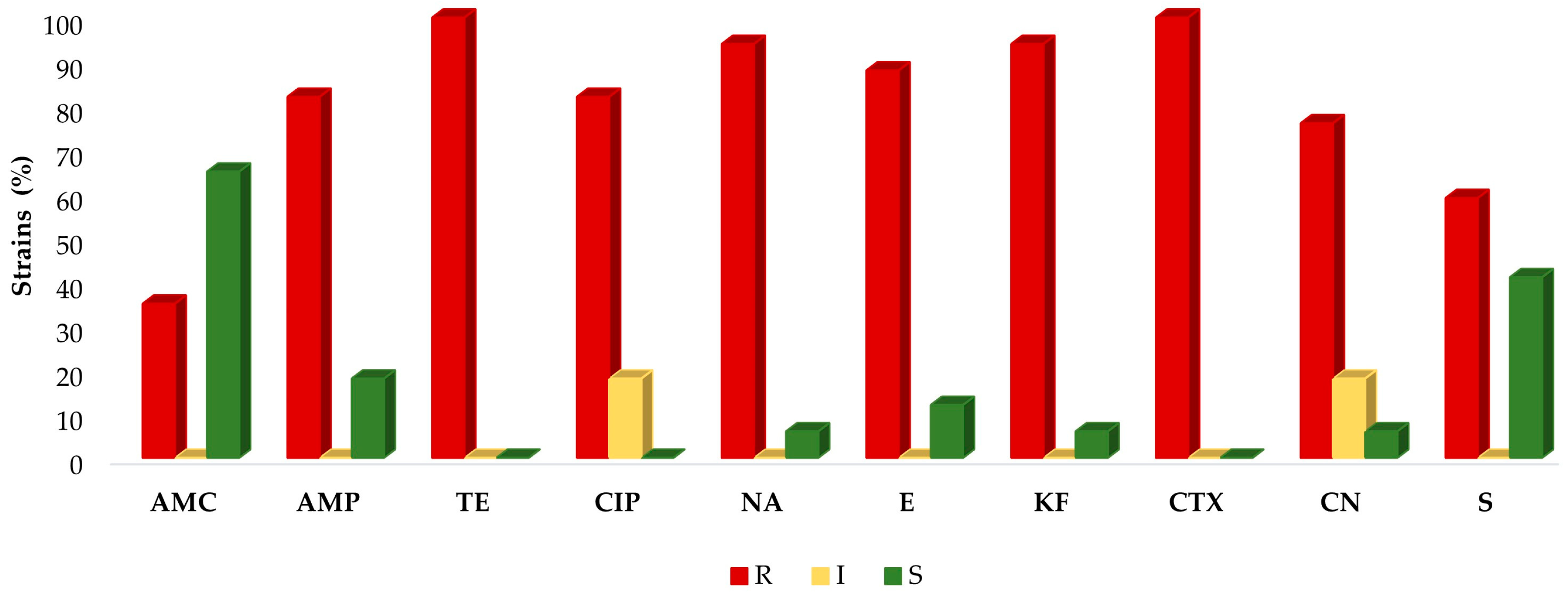
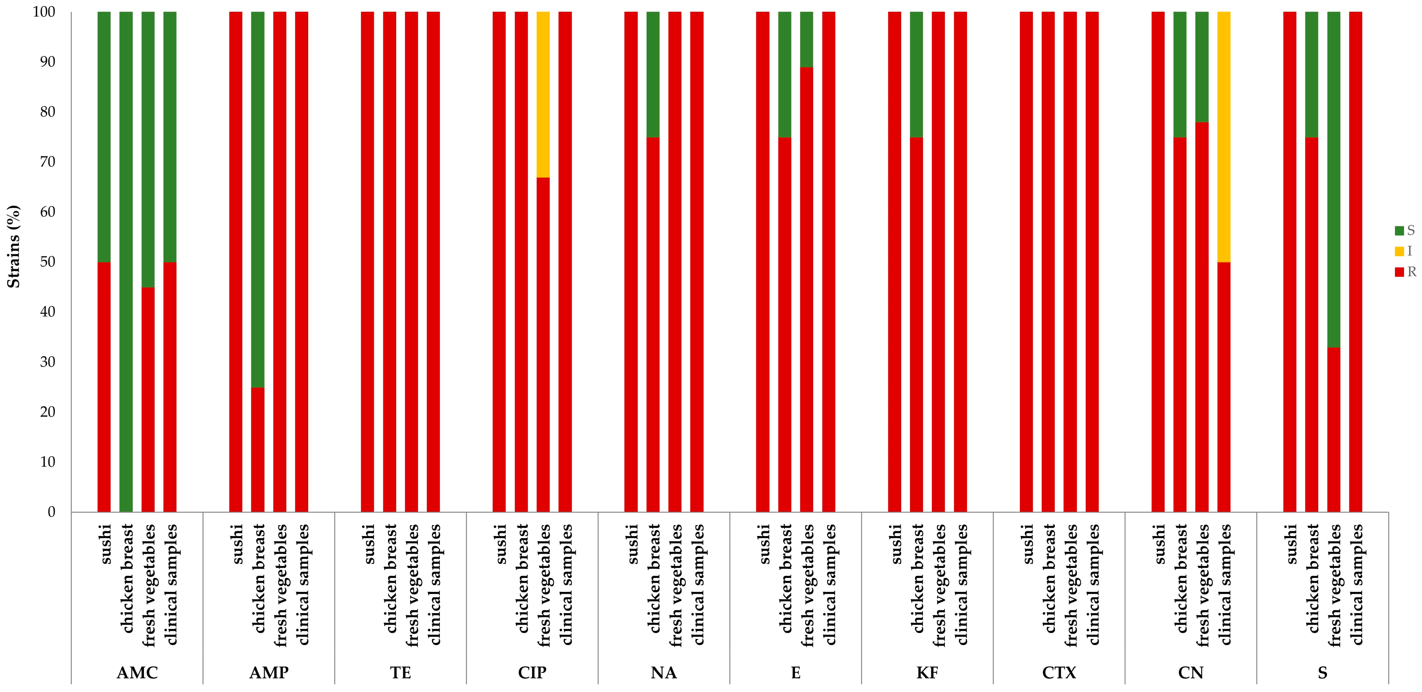
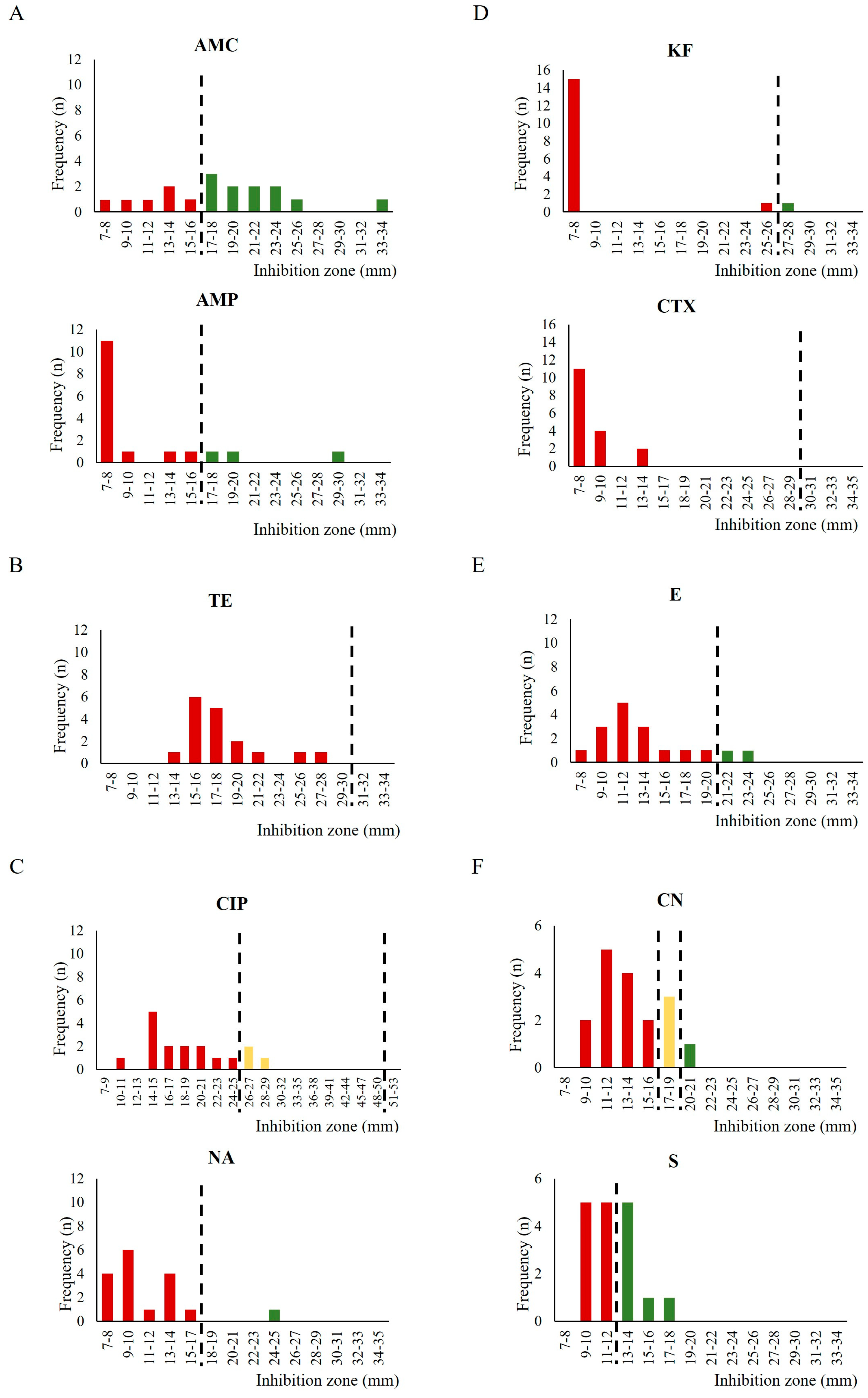
2.2. Antimicrobial Susceptibility Testing

The AMR was widely diffused among the tested strains (Table 2).
All the samples were resistant to tetracyclines and almost all to cephalosporins, except for a strain isolated from chicken breast (37809/1), which was sensitive to cefalotin. In the fluoroquinolones class, three strains (17.6%) showed intermediate resistance to ciprofloxacin, whereas the remaining were resistant; all strains but one (37809/1) (94%) were resistant to nalidixic acid. In contrast to the abovementioned results, 59% of strains were resistant to streptomycin and only 35% to amoxicillin–clavulanic acid (Figure 1). The prevalence of the resistant strains was significantly different (p < 0.001) among the various antibiotics tested.
Resistance to all antibiotics was found in one sushi-derived strain, while the other was sensitive to amoxicillin–clavulanic acid. In contrast, the strains isolated from chicken breast were all sensitive to the penicillins, except for one strain resistant to ampicillin (36981/2). Moreover, one strain (37809/1) was also sensitive to all aminoglycosides, macrolides, nalidixic acid, and cefalotin. As for the strains obtained from fresh vegetables, heterogeneous patterns were recorded: in the penicillin class, 45% of strains were resistant to amoxicillin–clavulanic acid and 100% to ampicillin. In the aminoglycoside group, 33% were resistant to streptomycin and 78% to gentamicin, with the remaining 22% showing intermediate resistance. In the fluoroquinolone class, 100% resistance to nalidixic acid and 33% intermediate and 67% full resistance to ciprofloxacin were observed. Moreover, 89% of the isolates were resistant to erythromycin, while 100% were resistant to tetracyclines and cephalosporins. One of the clinical strains (9291368) was resistant to all the antibiotic classes used in this study, while the other (8722325) was susceptible to amoxicillin–clavulanic acid and intermediate resistant to gentamicin (Figure 2). The prevalence of the resistant strains was significantly different (p < 0.001) according to the origin of isolation.
According to the European Centre for Disease Prevention and Control (ECDC)’s definition provided by Magiorakos et al. [51], all the A. butzleri strains tested were multidrug resistant (MDR).
The inhibition zone diameter (in mm) determined by the different antibiotics used in this study at their respective concentrations were investigated more closely (Figure 3). The dimensions of the inhibition zones were widely distributed for amoxicillin–clavulanic acid (7–34 mm); in other cases, such as for streptomycin, the values were closer to the cutoff (9–18 mm), whereas for the other antimicrobials, very similar values among the strains were observed, as recorded for cefalotin (7–8 mm).

3. Discussion

Arcobacter spp., and particularly A. butzleri, have gained increasing clinical significance in recent years. Due to their capacity to colonize several ecological niches, association with water reservoirs [18], and their presence in the intestinal tract and feces of healthy as well as diseased animals [54], contamination of food matrices of different categories by Arcobacter species may occur with a certain probability, and indeed it has been described worldwide [9,44]. Human infection occurs mainly through the ingestion of contaminated products, and fecal contamination of foods during various stages of production has been considered as the main route of contamination [55]. New consumers’ habits and trends, moving in the direction of raw or undercooked products (i.e., sushi), along with the large distribution in the food chain in industrialized countries are additional risk factors. Moreover, persistence in form of biofilm in the environments of the food production chain [34,35,36] and the resistance to disinfectants and sanitizing agents [41] have been described. All these features make A. butzleri a recognized human pathogen posing a potential threat to consumers’ health, with particular concern to immunocompromised people. Although the illness can be self-limited, clinical management of arcobacteriosis requires antimicrobial therapy with β-lactams, fluoroquinolones, and macrolides [56].
However, evidence of the AMR and MDR Arcobacter strains, often with high prevalence, has been reported [9]. The resistance mechanisms of Arcobacter to antibiotics were mainly chromosomal in nature [56]. Also, Isidro et al. elucidated the genetic bases of the AMR of A. butzleri and reported that this species harbors a large repertoire of efflux-pump-related genes and other antibiotic resistance determinants [33].
Concerning epidemiological data, the most recent studies indicated that Arcobacter strains frequently display high resistance rates for various antibiotic classes, including those agents currently in use for arcobacteriosis treatment. MDR rates, ranging from 20 to 93.8%, have been reported in the isolates from animals, humans, food products, and the environment [9,33,57,58,59,60,61,62].
Sciortino et al. found 100% and 89.2% A. butzleri resistance to the penicillin class of antimicrobials, ampicillin (AMP, 10 μg) and amoxicillin/clavulanic acid (AMC, 30 μg), respectively [46]. Similar results were shown by other authors, concerning the prevalence of resistance against ampicillin [49,63]. Our results are completely in line with those obtained by Silha and colleagues [61], who found 86.2% and 33.7% of the 80 strains isolated from poultry meat, water, and clinical sources to be resistant to ampicillin and amoxicillin/clavulanic acid, respectively.
Furthermore, Vicente-Martins, Oleastro, Domingues, and Ferreira [62] found 95.4% A. butzleri resistance to tetracycline (TE, 30 μg) among 65 strains isolated from retail food in Portugal; additionally, the resistance to tetracycline among all the tested strains was found in two studies [46,49], and our results confirmed these data. Also, for the fluoroquinolones, quite high percentages of resistant A. butzleri have been reported. From 27.7% to 41% of isolated strains in different studies were resistant to ciprofloxacin (CIP, 5 μg) [33,62], and from 93.8 to 100% were not susceptible to nalidixic acid [33,46,49,61,62,63]. These results agree with those obtained in our study for nalidixic acid, for which a 94% of resistance was assessed but not for ciprofloxacin; for this antimicrobial, in fact, we obtained 100% resistance or intermediate resistance.
Furthermore, literature data are available for the macrolide class of antimicrobials, and only two studies assessed a high resistance prevalence in A. butzleri strains [49,63]. Our data confirmed these results, with 88% of strains not susceptible to erythromycin (E, 15 μg). In contrast, other authors reported lower percentages of resistance against this antibiotic [46,62]. In addition, very alarming resistance rates were also described for cephalosporins, with 78.7-100% [61,64] and 98.5-100% [46,49,62] of A. butzleri strains shown as not sensitive to cefalotin (KF, 30μg) and cefotaxime (CTX, 30 μg), respectively. Also in this case, our data agreed with the available results, showing 94% and 100% of resistance prevalence in the tested strains.
Lastly, we obtained discordant results for aminoglycosides antibiotics, finding a higher percentage of resistance than those reported in the literature; particularly, we measured a 94% and 59% of resistance for gentamicin (CN, 10 μg) and streptomycin (S, 10 μg), respectively, which are in contrast to the sensitivity of almost all the A. butzleri strains reported by other authors [46,49,61,64]. In the present study, three strains (17.6% of the totality) were panresistant (i.e., resistant to all antibiotic classes used in this study).
To date, no reference protocols and standard interpretive criteria are available for the antimicrobial susceptibility testing (AST) of A. butzleri, hampering a univocal and comparable evaluation of the antimicrobial susceptibility of this bacterium [56]. The assessment of the AMR of A. butzleri isolates faces the difficulty that no clinical breakpoints are specified for this pathogen, yet. However, interpretative criteria for C. jejuni susceptibility testing recommended by the EUCAST (CIP5, E15, TE30) and BSAC zone diameter breakpoints for Enterobacteriaceae (AMC30, AMP10, NA30, KF30, CTX30, CN10, S10) were used to analyze the results [53,65,66], as previously reported by other authors [9,49,61,62,63,64].
Considering the lack of Arcobacter specific cutoff values, the graphical representation of the distribution of the inhibition zone diameters used in this work (Figure 3) could be more useful to compare our data with other epidemiological information at the national or EU level, in order to contribute to realize an epidemiological cutoff (ECOFF) [67]. Interestingly, arbitrarily analyzing the obtained results with the epidemiological cutoffs suggested by Zautner et al. [50], a much higher percentage of susceptible strains to ciprofloxacin, tetracycline, and erythromycin could be observed. For this reason, strains that exhibit smaller inhibition zones diameter, which were also considered to be resistant according to the EUCAST or BSAC guidelines, will be interesting candidates for investigating the genetic basis of the antibiotic resistance by Whole-Genome Sequencing. Most importantly, to standardize the information on the resistance pattern of this pathogen, it will be necessary for homogeneous breakpoints to be developed, at least at the European level.
Lastly, prevention and control measures to limit the pathogen spread and the related infections should also include food acid treatment (citric and lactic acid at 1–2%), good cooking practices, effective treatment of water resources and monitoring for their contamination, maintenance of slaughter hygiene and carcass control, HACCP, and good manufacturing practice application [44].
Certainly, the increasing and alarming drug resistance scenario demands the attention of researchers to find novel and alternative therapeutic options for preventing and controlling the spread of Arcobacter spp. in an effective way; this would be beneficial with regard to both public health and food safety viewpoints. A. butzleri AMR represents, therefore, a theme of concern; in this perspective, the investigation about the possible use of polyphenols as resistance modulators in A. butzleri has been proposed, showing that some stilbenes can increase its sensitivity, probably acting as efflux pump inhibitors [68].

4. Materials and Methods

4.1. Bacterial Strains

Arcobacter spp. food strains (n. 15), collected from June to October 2022 were included in this study. Bacterial strains were isolated from sushi (12%), fresh vegetables (53%), and chicken breast (23%), according to the isolation protocol proposed by Collado et al. [69].
The food matrices investigated were chosen based on the epidemiological data about Arcobacter prevalence in such matrices [9], including food items that are commonly eaten raw without any preparation phase.
Briefly, isolation was obtained after a first enrichment step of the food sample in Arcobacter broth supplemented with 8 mg/L cefoperazone, 10 mg/L amphotericin B, and 4 mg/L teicoplanin (Thermo Scientific, Waltham, MA, USA); then, the enriched broth was inoculated by passive filtration on Tryptic Soy Agar + 5% sheep blood, and incubated at 30 °C in a microaerophilic atmosphere generated using a jar gassing system (CampyGen 2.5L, Thermo Scientific). The suspected colonies were identified based on morphological criteria (small and round, with a translucent to beige color), Gram stain (Gram-negative), and oxidase test (oxidase-positive). Moreover, the study also included the analysis of two clinical strains (12%) isolated in 2018 and in 2022 from human feces of patients with acute gastroenteritis. Both clinical strains were isolated from patients undergoing acute infection with intestinal symptoms, who sought hospital care. The patients were a male and a female, aged 59 and 69, respectively, at the time of Arcobacter isolation. No further information about the patients nor the clinical course of infection was available.
The Arcobacter strains were routinely grown at 30 °C on Mueller Hinton (MH) agar plates (Thermo Scientific) supplemented with 5% defibrinated horse blood (Liofilchem, Roseto degli Abruzzi, Italy). All incubations were at 30 °C under microaerophilic conditions.

4.2. Multiplex PCR-Based Species Identification

All the strains were identified at the species level by multiplex PCR, with primers Cpn60-F (GCA CAT TCT ATT TTC AAA GAA GGG) and Cpn60-R (GAA TGG GTT ATT AAA CTC TGC) for A. butzleri; GyrAcry-F (AGT TCT GAA GCA ATA GAT TTA ATG G) and GyrAcry-R (CTG CAA TTC CTT CGA TTT GC) for A. cryaerophilus; Skirr-F (GGC GAT TTA CTG GAA CAC A) and Skirr-R (CGT ATT CAC CGT AGC ATA GC) for A. skirrowii. A. butzleri DSM 8739, A. cryaerophilus DSM 7289, and A. skirrowii DSM 7302 were used as positive controls [70].

4.3. Antibiotic Susceptibility Tests

The AST of the isolated strains was performed against ten antibiotics through the European Committee on Antimicrobial Susceptibility Testing (EUCAST)’s disc diffusion method [71]. The strains were tested against: amoxicillin–clavulanic acid (AMC, 30 μg; AMC30), ampicillin (AMP, 10 μg; AMP10), cefalotin (KF 30 μg; KF30), cefotaxime (CTX, 30 μg; CTX30), ciprofloxacin (CIP, 5 μg; CIP5), erythromycin (E, 15 μg; E15), gentamicin (CN, 10 μg; CN10), nalidixic acid (NA 30 μg; NA30), streptomycin (S, 10 μg; S10), and tetracycline (TE, 30 μg; TE30) (Oxoid, UK). The isolates were subcultured on MH agar supplemented with 5% defibrinated horse blood (Thermo Scientific) with incubation at 30 °C for 48 h under microaerophilic conditions. A 0.5 McFarland bacterial suspension was prepared in BHI broth, spread on MHF, and the antibiotic test discs were positioned. After incubation for 48 h at 30 °C in a microaerophilic atmosphere, the diameters of the inhibition zones were measured. The bacterial isolates with insufficient growth after 48 h were re-incubated, and the inhibition zone was measured to the closest millimeter after a total of 72 h. The experiments were performed in triplicate. The quality control strains used for resistance testing by agar diffusion were Campylobacter jejuni ATCC 33560 for the antibiotic test discs CIP5, E15, and TE30, and Enterococcus faecalis ATCC 29212 for AMP10 and AMC30.
According to other authors [1,45,46,62], since no breakpoints have been defined for Arcobacter spp. either by the EUCAST or the Clinical & Laboratory Standards Institute (CLSI), the strains were classified as resistant (R), susceptible (S), or intermediate (I) on the basis of EUCAST breakpoint tables for interpretation of the zone diameters [52] given for C. jejuni (CIP5, E15 and TE30) [52]. Moreover, the strains’ sensitivity to antibiotics other than those listed for C. jejuni was evaluated according to the interpretation guidelines of the British Society for Antimicrobial Chemotherapy (BSAC) Resistance Surveillance Programme [53,66] for Enterobacterales.
The strains resistant to at least three classes of antibiotics were classified as MDR [51].

4.4. Statistical Analysis

The chi-square test was used to assess the differences between the prevalence of the resistant strains to the antibiotics tested. Separate analyses were conducted on all the isolates or grouped according to their origin. p values lower than 0.05 were considered statistically significant.

5. Conclusions

The high prevalence of AMR to a wide range of antimicrobial classes found in this study for A. butzleri, including a percentage of resistance to aminoglycoside antibiotics higher than that reported in previous research, should be considered an important finding, especially since aminoglycosides are the main therapeutic option for human arcobacteriosis.
The scarce or unknown incidence of Arcobacter spp. infections, along with the lack of standardized methods for the detection of the pathogen and the assessment of its AMR may determine an underestimation of the potential threat to human health. Therefore, our data provide a contribution and suggest that Arcobacter spp. monitoring and the epidemiological surveillance of its AMR features should be considered by the authorities designated for food safety.

Author Contributions

Conceptualization, C.G., G.B. (Giulia Baldelli), G.A., G.F.S., D.S. and A.P.; methodology, formal analysis and data curation, C.G., G.B., G.A., G.F.S. and D.S., investigation, C.G., G.B. (Giulia Baldelli), D.S., I.R., S.D.L., M.N., S.P. and F.R.M. resources, C.G., D.S., G.B. (Giuliana Blasi), F.L., G.G. and A.P., writing—original draft preparation C.G., G.B. (Giulia Baldelli), G.A. and G.F.S., writing—review and editing, D.S. and A.P., supervision, G.A., G.F.S. and A.P., project administration, A.P., funding acquisition, A.P. All authors have read and agreed to the published version of the manuscript.

Funding

Italian Ministry of Health. Ricerca Corrente RC 13/2020 “Rischio correlato allo sviluppo di patogeni emergenti: studio sulla diffusione di Aliarcobacter spp. in alimenti”.

Institutional Review Board Statement

Not applicable.

Informed Consent Statement

Not applicable.

Data Availability Statement

The datasets generated and/or analyzed during the current study are not publicly available but are available from the principal investigator (PI) on reasonable request.

Conflicts of Interest

The authors declare no conflict of interest.

References

  1. Ferreira, S.; Oleastro, M.; Domingues, F. Current insights on Arcobacter butzleri in food chain. Curr. Opin. Food Sci. 2019, 26, 9–17. [Google Scholar] [CrossRef]
  2. Figueras, M.J.; Pérez-Cataluña, A.; Salas-Massó, N.; Levican, A.; Collado, L. ‘Arcobacter porcinus’ sp. nov., a novel Arcobacter species uncovered by Arcobacter thereius. New Microbes New Infect. 2017, 15, 104–106. [Google Scholar] [CrossRef] [PubMed]
  3. Pérez-Cataluña, A.; Salas-Massó, N.; Figueras, M.J. Arcobacter canalis sp. nov., isolated from a water canal contaminated with urban sewage. Int. J. Syst. Evol. Microbiol. 2018, 68, 1258–1264. [Google Scholar] [CrossRef]
  4. Callbeck, C.M.; Pelzer, C.; Lavik, G.; Ferdelman, T.G.; Graf, J.S.; Vekeman, B.; Schunck, H.; Littmann, S.; Fuchs, B.M.; Hach, P.F.; et al. Arcobacter peruensis sp. nov., a Chemolithoheterotroph Isolated from Sulfide- and Organic-Rich Coastal Waters off Peru. Appl. Environ. Microbiol. 2019, 85, e01344-19. [Google Scholar] [CrossRef]
  5. Kerkhof, P.-J.; On, S.L.W.; Houf, K. Arcobacter vandammei sp. nov., isolated from the rectal mucus of a healthy pig. Int. J. Syst. Evol. Microbiol. 2021, 71, 005113. [Google Scholar] [CrossRef] [PubMed]
  6. Park, S.; Jung, Y.-T.; Kim, S.; Yoon, J.-H. Arcobacter acticola sp. nov., isolated from seawater on the East Sea in South Korea. J. Microbiol. 2016, 54, 655–659. [Google Scholar] [CrossRef]
  7. Martins, I.; Mateus, C.; Domingues, F.; Oleastro, M.; Ferreira, S. Putative Role of an ABC Efflux System in Aliarcobacter butzleri Resistance and Virulence. Antibiotics 2023, 12, 339. [Google Scholar] [CrossRef]
  8. Buchanan, R.L.; Anderson, W.; Anelich, L.; Cordier, J.L.; Dewanti-Hariyadi, R.; Ross, T.; Zwietering, M.H. Microorganisms in Foods 7. Microbiological Testing in Food Safety Management; Springer: Cham, Switzerland, 2018; Volume 7. [Google Scholar]
  9. Chieffi, D.; Fanelli, F.; Fusco, V. Arcobacter butzleri: Up-to-date taxonomy, ecology, and pathogenicity of an emerging pathogen. Compr. Rev. Food Sci. Food Saf. 2020, 19, 2071–2109. [Google Scholar] [CrossRef]
  10. Mottola, A.; Ciccarese, G.; Sinisi, C.; Savarino, A.E.; Marchetti, P.; Terio, V.; Tantillo, G.; Barrasso, R.; Di Pinto, A. Occurrence and characterization of Arcobacter spp. from ready-to-eat vegetables produced in Southern Italy. Ital. J. Food Saf. 2021, 10, 8585. [Google Scholar] [CrossRef]
  11. Kim, N.H.; Park, S.M.; Kim, H.W.; Cho, T.J.; Kim, S.H.; Choi, C.; Rhee, M.S. Prevalence of pathogenic Arcobacter species in South Korea: Comparison of two protocols for isolating the bacteria from foods and examination of nine putative virulence genes. Food Microbiol. 2019, 78, 18–24. [Google Scholar] [CrossRef]
  12. Ramees, T.p.; Rathore, R.; Kumar, A.; Remesh, A.; Kumar, R.; Karthik, K.; Malik, Y.; Dhama, K.; Singh, R. Phylogenetic analysis of Arcobacter butzleri and Arcobacter skirrowii isolates and their detection from contaminated vegetables by multiplex PCR. J. Exp. Biol. Agric. Sci. 2018, 6, 307–314. [Google Scholar] [CrossRef]
  13. González, A.; Bayas Morejón, I.F.; Ferrús, M.A. Isolation, molecular identification and quinolone-susceptibility testing of Arcobacter spp. isolated from fresh vegetables in Spain. Food Microbiol. 2017, 65, 279–283. [Google Scholar] [CrossRef] [PubMed]
  14. González, A.; Ferrús, M.A. Study of Arcobacter spp. contamination in fresh lettuces detected by different cultural and molecular methods. Int. J. Food Microbiol. 2011, 145, 311–314. [Google Scholar] [CrossRef] [PubMed]
  15. Lee, M.H.; Choi, C. Survival of Arcobacter butzleri in Apple and Pear Purees. J. Food Saf. 2013, 33, 333–339. [Google Scholar] [CrossRef]
  16. Laishram, M.; Rathlavath, S.; Lekshmi, M.; Kumar, S.; Nayak, B.B. Isolation and characterization of Arcobacter spp. from fresh seafood and the aquatic environment. Int. J. Food Microbiol. 2016, 232, 87–89. [Google Scholar] [CrossRef]
  17. Leoni, F.; Chierichetti, S.; Santarelli, S.; Talevi, G.; Masini, L.; Bartolini, C.; Rocchegiani, E.; Naceur Haouet, M.; Ottaviani, D. Occurrence of Arcobacter spp. and correlation with the bacterial indicator of faecal contamination Escherichia coli in bivalve molluscs from the Central Adriatic, Italy. Int. J. Food Microbiol. 2017, 245, 6–12. [Google Scholar] [CrossRef]
  18. Barel, M.; Yildirim, Y. Arcobacter species isolated from various seafood and water sources; virulence genes, antibiotic resistance genes and molecular. World J. Microbiol. Biotechnol. 2023, 183, 12. [Google Scholar] [CrossRef]
  19. Figueras, M.J.; Levican, A.; Pujol, I.; Ballester, F.; Rabada Quilez, M.J.; Gomez-Bertomeu, F. A severe case of persistent diarrhoea associated with Arcobacter cryaerophilus but attributed to Campylobacter sp. and a review of the clinical incidence of Arcobacter spp. New Microbes New Infect. 2014, 2, 31–37. [Google Scholar] [CrossRef] [Green Version]
  20. Van den Abeele, A.M.; Vogelaers, D.; Van Hende, J.; Houf, K. Prevalence of Arcobacter species among humans, Belgium, 2008–2013. Emerg. Infect. Dis. 2014, 20, 1731–1734. [Google Scholar] [CrossRef] [Green Version]
  21. Prouzet-Mauléon, V.; Labadi, L.; Bouges, N.; Ménard, A.; Mégraud, F. Arcobacter butzleri: Underestimated enteropathogen. Emerg. Infect. Dis. 2006, 12, 307–309. [Google Scholar] [CrossRef] [Green Version]
  22. Samie, A.; Obi, C.L.; Barrett, L.J.; Powell, S.M.; Guerrant, R.L. Prevalence of Campylobacter species, Helicobacter pylori and Arcobacter species in stool samples from the Venda region, Limpopo, South Africa: Studies using molecular diagnostic methods. J. Infect. 2007, 54, 558–566. [Google Scholar] [CrossRef] [PubMed]
  23. Švarcová, K.; Pejchalová, M.; Šilha, D. The Effect of Antibiotics on Planktonic Cells and Biofilm Formation Ability of Collected Arcobacter-like Strains and Strains Isolated within the Czech Republic. Antibiotics 2022, 11, 87. [Google Scholar] [CrossRef]
  24. Yan, J.J.; Ko, W.C.; Huang, A.H.; Chen, H.M.; Jin, Y.T.; Wu, J.J. Arcobacter butzleri bacteremia in a patient with liver cirrhosis. J. Formos. Med. Assoc. 2000, 99, 166–169. [Google Scholar] [PubMed]
  25. Lau, S.K.; Woo, P.C.; Teng, J.L.; Leung, K.W.; Yuen, K.Y. Identification by 16S ribosomal RNA gene sequencing of Arcobacter butzleri bacteraemia in a patient with acute gangrenous appendicitis. Mol. Pathol. 2002, 55, 182–185. [Google Scholar] [CrossRef] [Green Version]
  26. Collado, L.; Figueras, M.J. Taxonomy, epidemiology, and clinical relevance of the genus Arcobacter. Clin. Microbiol. Rev. 2011, 24, 174–192. [Google Scholar] [CrossRef] [Green Version]
  27. Vandenberg, O.; Dediste, A.; Houf, K.; Ibekwem, S.; Souayah, H.; Cadranel, S.; Douat, N.; Zissis, G.; Butzler, J.P.; Vandamme, P. Arcobacter species in humans. Emerg. Infect. Dis. 2004, 10, 1863–1867. [Google Scholar] [CrossRef] [PubMed]
  28. Shah, A.H.; Saleha, A.; Zakaria, Z.; Marimuthu, M. Arcobacter—An emerging threat to animals and animal origin food products? Trends Food Sci. Technol. 2011, 22, 225–236. [Google Scholar] [CrossRef] [Green Version]
  29. McGregor, A.C.; Wright, S.G. Gastrointestinal symptoms in travellers. Clin. Med. 2015, 15, 93–95. [Google Scholar] [CrossRef] [Green Version]
  30. Arguello, E.; Otto, C.C.; Mead, P.; Babady, N.E. Bacteremia caused by Arcobacter butzleri in an immunocompromised host. J. Clin. Microbiol. 2015, 53, 1448–1451. [Google Scholar] [CrossRef] [Green Version]
  31. Soelberg, K.K.; Danielsen, T.K.L.; Martin-Iguacel, R.; Justesen, U.S. Arcobacter butzleri is an opportunistic pathogen: Recurrent bacteraemia in an immunocompromised patient without diarrhoea. Access Microbiol. 2020, 2, acmi000145. [Google Scholar] [CrossRef]
  32. Gölz, G.; Karadas, G.; Alutis, M.; Fischer, A.; Kühl, A.; Breithaupt, A.; Göbel, U.; Alter, T.; Bereswill, S.; Heimesaat, M. Arcobacter butzleri Induce Colonic, Extra-Intestinal and Systemic Inflammatory Responses in Gnotobiotic IL-10 Deficient Mice in a Strain-Dependent Manner. PLoS ONE 2015, 10, e0139402. [Google Scholar] [CrossRef] [PubMed]
  33. Isidro, J.; Ferreira, S.; Pinto, M.; Domingues, F.; Oleastro, M.; Gomes, J.P.; Borges, V. Virulence and antibiotic resistance plasticity of Arcobacter butzleri: Insights on the genomic diversity of an emerging human pathogen. Infect. Genet. Evol. 2020, 80, 104213. [Google Scholar] [CrossRef] [PubMed]
  34. Salazar-Sánchez, A.; Baztarrika, I.; Alonso, R.; Fernández-Astorga, A.; Martínez-Ballesteros, I.; Martinez-Malaxetxebarria, I. Arcobacter butzleri Biofilms: Insights into the Genes Beneath Their Formation. Microorganisms 2022, 10, 1280. [Google Scholar] [CrossRef]
  35. Martinez-Malaxetxebarria, I.; Girbau, C.; Salazar-Sánchez, A.; Baztarrika, I.; Martínez-Ballesteros, I.; Laorden, L.; Alonso, R.; Fernández-Astorga, A. Genetic characterization and biofilm formation of potentially pathogenic foodborne Arcobacter isolates. Int. J. Food Microbiol. 2022, 373, 109712. [Google Scholar] [CrossRef] [PubMed]
  36. Girbau, C.; Martinez-Malaxetxebarria, I.; Muruaga, G.; Carmona, S.; Alonso, R.; Fernandez-Astorga, A. Study of Biofilm Formation Ability of Foodborne Arcobacter butzleri under Different Conditions. J. Food Prot. 2017, 80, 758–762. [Google Scholar] [CrossRef] [PubMed]
  37. Mudadu, A.G.; Melillo, R.; Salza, S.; Mara, L.; Marongiu, L.; Piras, G.; Spanu, C.; Tedde, T.; Fadda, A.; Virgilio, S.; et al. Detection of Arcobacter spp. in environmental and food samples collected in industrial and artisanal sheep’s milk cheese-making plants. Food Control 2021, 126, 108100. [Google Scholar] [CrossRef]
  38. Šilha, D.; Hrušková, L.; Brožková, I.; Moťková, P.; Vytřasová, J. Survival of selected bacteria from the genus Arcobacter on various metallic surfaces. J. Food Nutr. Res. 2014, 53, 217–223. [Google Scholar]
  39. Falahchai, S.; Bahador, N.; Ghane, M. Isolation and Characterization of Arcobacter butzleri from Environmental Samples and Determination of their Pathogenic Gene Expression under Different Physicochemical Conditions. Pol. J. Environ. Stud. 2021, 30, 3963–3974. [Google Scholar] [CrossRef]
  40. Serraino, A.; Giacometti, F. Short communication: Occurrence of Arcobacter species in industrial dairy plants. J. Dairy Sci. 2014, 97, 2061–2065. [Google Scholar] [CrossRef] [Green Version]
  41. Šilha, D.; Šilhová, L.; Vytřasová, J.; Brožková, I.; Pejchalova, M. Survival of selected bacteria of Arcobacter genus in disinfectants and possibility of acquired secondary resistance to disinfectants. J. Microbiol. Biotechnol. Food Sci. 2016, 5, 326–329. [Google Scholar] [CrossRef] [Green Version]
  42. Rasmussen, L.H.; Kjeldgaard, J.; Christensen, J.P.; Ingmer, H. Multilocus sequence typing and biocide tolerance of Arcobacter butzleri from Danish broiler carcasses. BMC Res. Notes 2013, 6, 322. [Google Scholar] [CrossRef] [Green Version]
  43. Uljanovas, D.; Gölz, G.; Brückner, V.; Grineviciene, A.; Tamuleviciene, E.; Alter, T.; Malakauskas, M. Prevalence, antimicrobial susceptibility and virulence gene profiles of Arcobacter species isolated from human stool samples, foods of animal origin, ready-to-eat salad mixes and environmental water. Gut Pathog. 2021, 13, 76. [Google Scholar] [CrossRef] [PubMed]
  44. Ramees, T.P.; Dhama, K.; Karthik, K.; Rathore, R.S.; Kumar, A.; Saminathan, M.; Tiwari, R.; Malik, Y.S.; Singh, R.K. Arcobacter: An emerging food-borne zoonotic pathogen, its public health concerns and advances in diagnosis and control—A comprehensive review. Vet. Q. 2017, 37, 136–161. [Google Scholar] [CrossRef] [PubMed] [Green Version]
  45. Brückner, V.; Fiebiger, U.; Ignatius, R.; Friesen, J.; Eisenblätter, M.; Höck, M.; Alter, T.; Bereswill, S.; Gölz, G.; Heimesaat, M.M. Prevalence and antimicrobial susceptibility of Arcobacter species in human stool samples derived from out- and inpatients: The prospective German Arcobacter prevalence study Arcopath. Gut Pathog. 2020, 12, 21. [Google Scholar] [CrossRef] [PubMed] [Green Version]
  46. Sciortino, S.; Arculeo, P.; Alio, V.; Cardamone, C.; Nicastro, L.; Arculeo, M.; Alduina, R.; Costa, A. Occurrence and Antimicrobial Resistance of Arcobacter spp. Recovered from Aquatic Environments. Antibiotics 2021, 10, 288. [Google Scholar] [CrossRef]
  47. Khodamoradi, S.; Abiri, R. The incidence and antimicrobial resistance of Arcobacter species in animal and poultry meat samples at slaughterhouses in Iran. Iran. J. Microbiol. 2020, 12, 531–536. [Google Scholar] [CrossRef]
  48. Jribi, H.; Sellami, H.; Amor, S.B.; Ducournau, A.; Sifré, E.; Benejat, L.; Mégraud, F.; Gdoura, R. Occurrence and Antibiotic Resistance of Arcobacter Species Isolates from Poultry in Tunisia. J. Food Prot. 2020, 83, 2080–2086. [Google Scholar] [CrossRef]
  49. Fanelli, F.; Chieffi, D.; Di Pinto, A.; Mottola, A.; Baruzzi, F.; Fusco, V. Phenotype and genomic background of Arcobacter butzleri strains and taxogenomic assessment of the species. Food Microbiol. 2020, 89, 103416. [Google Scholar] [CrossRef]
  50. Zautner, A.E.; Riedel, T.; Bunk, B.; Spröer, C.; Boahen, K.G.; Akenten, C.W.; Dreyer, A.; Färber, J.; Kaasch, A.J.; Overmann, J.; et al. Molecular characterization of Arcobacter butzleri isolates from poultry in rural Ghana. Front. Cell. Infect. Microbiol. 2023, 13, 1094067. [Google Scholar] [CrossRef]
  51. Magiorakos, A.P.; Srinivasan, A.; Carey, R.B.; Carmeli, Y.; Falagas, M.E.; Giske, C.G.; Harbarth, S.; Hindler, J.F.; Kahlmeter, G.; Olsson-Liljequist, B.; et al. Multidrug-resistant, extensively drug-resistant and pandrug-resistant bacteria: An international expert proposal for interim standard definitions for acquired resistance. Clin. Microbiol. Infect. 2012, 18, 268–281. [Google Scholar] [CrossRef] [Green Version]
  52. European Committee on Antimicrobial Susceptibility Testing. In Breakpoint Tables for Interpretation of MICs and Zone Diameters; Version 13.0; European Committee on Antimicrobial Susceptibility Testing: Växjö, Sweden, 2023.
  53. BSAC. BSAC Methods for Antimicrobial Susceptibility Testing. Version 14.0, 05-01-2015. Available online: https://www.bsac.org.uk/wp-content/uploads/2012/02/BSAC-Susceptibility-testing-version-14.pdf (accessed on 10 June 2023).
  54. Çelik, C.; Pınar, O.; Sipahi, N. The Prevalence of Aliarcobacter Species in the Fecal Microbiota of Farm Animals and Potential Effective Agents for Their Treatment: A Review of the Past Decade. Microorganisms 2022, 10, 2430. [Google Scholar] [CrossRef]
  55. Collado, L.; Guarro, J.; Figueras, M.J. Prevalence of Arcobacter in meat and shellfish. J. Food Prot. 2009, 72, 1102–1106. [Google Scholar] [CrossRef]
  56. Ferreira, S.; Queiroz, J.A.; Oleastro, M.; Domingues, F.C. Insights in the pathogenesis and resistance of Arcobacter: A review. Crit. Rev. Microbiol. 2016, 42, 364–383. [Google Scholar] [CrossRef] [PubMed]
  57. Dekker, D.; Eibach, D.; Boahen, K.G.; Akenten, C.W.; Pfeifer, Y.; Zautner, A.E.; Mertens, E.; Krumkamp, R.; Jaeger, A.; Flieger, A.; et al. Fluoroquinolone-Resistant Salmonella enterica, Campylobacter spp., and Arcobacter butzleri from Local and Imported Poultry Meat in Kumasi, Ghana. Foodborne Pathog. Dis. 2019, 16, 352–358. [Google Scholar] [CrossRef] [PubMed] [Green Version]
  58. Kabeya, H.; Maruyama, S.; Morita, Y.; Ohsuga, T.; Ozawa, S.; Kobayashi, Y.; Abe, M.; Katsube, Y.; Mikami, T. Prevalence of Arcobacter species in retail meats and antimicrobial susceptibility of the isolates in Japan. Int. J. Food Microbiol. 2004, 90, 303–308. [Google Scholar] [CrossRef]
  59. Rathlavath, S.; Kohli, V.; Singh, A.S.; Lekshmi, M.; Tripathi, G.; Kumar, S.; Nayak, B.B. Virulence genotypes and antimicrobial susceptibility patterns of Arcobacter butzleri isolated from seafood and its environment. Int. J. Food Microbiol. 2017, 263, 32–37. [Google Scholar] [CrossRef]
  60. Shah, A.H.; Saleha, A.A.; Zunita, Z.; Murugaiyah, M.; Aliyu, A.B.; Jafri, N. Prevalence, distribution and antibiotic resistance of emergent Arcobacter spp. from clinically healthy cattle and goats. Transbound. Emerg. Dis. 2013, 60, 9–16. [Google Scholar] [CrossRef] [Green Version]
  61. Šilha, D.; Pejchalová, M.; Šilhová, L. Susceptibility to 18 drugs and multidrug resistance of Arcobacter isolates from different sources within the Czech Republic. J. Glob. Antimicrob. Resist. 2017, 9, 74–77. [Google Scholar] [CrossRef]
  62. Vicente-Martins, S.; Oleastro, M.; Domingues, F.C.; Ferreira, S. Arcobacter spp. at retail food from Portugal: Prevalence, genotyping and antibiotics resistance. Food Control 2018, 85, 107–112. [Google Scholar] [CrossRef]
  63. Elmali, M.; Yeşim Can, H. Occurence and antimicrobial resistance of Arcobacter species in food and slaughterhouse samples. Food Sci. Technol. 2017, 37, 280–285. [Google Scholar] [CrossRef] [Green Version]
  64. Rahimi, E. Prevalence and antimicrobial resistance of Arcobacter species isolated from poultry meat in Iran. Br. Poult. Sci. 2014, 55, 174–180. [Google Scholar] [CrossRef]
  65. Debelo, M.; Mohammed, N.; Tiruneh, A.; Tolosa, T. Isolation, identification and antibiotic resistance profile of thermophilic Campylobacter species from Bovine, Knives and personnel at Jimma Town Abattoir, Ethiopia. PLoS ONE 2022, 17, e0276625. [Google Scholar] [CrossRef]
  66. Brown, D.F.; Wootton, M.; Howe, R.A. Antimicrobial susceptibility testing breakpoints and methods from BSAC to EUCAST. J. Antimicrob. Chemother. 2016, 71, 3–5. [Google Scholar] [CrossRef] [Green Version]
  67. EUCAST. MIC and Zone Diameter Distributions and ECOFFs. Available online: https://www.eucast.org/mic_and_zone_distributions_and_ecoffs (accessed on 10 June 2023).
  68. Sousa, V.; Luís, Â.; Oleastro, M.; Domingues, F.; Ferreira, S. Polyphenols as resistance modulators in Arcobacter butzleri. Folia Microbiol. 2019, 64, 547–554. [Google Scholar] [CrossRef]
  69. Collado, L.; Inza, I.; Guarro, J.; Figueras, M.J. Presence of Arcobacter spp. in environmental waters correlates with high levels of fecal pollution. Environ. Microbiol. 2008, 10, 1635–1640. [Google Scholar] [CrossRef] [PubMed]
  70. Khan, I.U.H.; Cloutier, M.; Libby, M.; Lapen, D.R.; Wilkes, G.; Topp, E. Enhanced Single-tube Multiplex PCR Assay for Detection and Identification of Six Arcobacter Species. J. Appl. Microbiol. 2017, 123, 1522–1532. [Google Scholar] [CrossRef] [PubMed]
  71. Matuschek, E.; Brown, D.F.J.; Kahlmeter, G. Development of the EUCAST disk diffusion antimicrobial susceptibility testing method and its implementation in routine microbiology laboratories. Clin. Microbiol. Infect. 2014, 20, O255–O266. [Google Scholar] [CrossRef] [PubMed] [Green Version]
Figure 1. Prevalence of resistance to antimicrobials of all A. butzleri strains (n = 17). Resistant (red), intermediate resistant (yellow), or sensitive (green) strains to the antimicrobials tested. AMC: amoxicillin–clavulanic acid (30 μg); AMP: ampicillin (10 μg); TE: tetracycline (30 μg); CIP: ciprofloxacin (5 μg); E: erythromycin (15 μg); KF: cefalotin (30 μg); CTX: cefotaxime (30 μg); CN: gentamicin (10 μg); NA: nalidixic acid (30 μg); S: streptomycin (10 μg).
Antibiotics 12 01292 g001
Figure 2. Resistance patterns of the tested strains (n=17) according to their origin. Resistant (red), intermediate resistant (yellow), or sensitive (green) strains to the antimicrobials tested. AMC: amoxicillin–clavulanic acid (30 μg); AMP: ampicillin (10 μg); TE: tetracycline (30 μg); CIP: ciprofloxacin (5 μg); E: erythromycin (15) μg; KF: cefalotin (30μg); CTX: cefotaxime (30 μg); CN: gentamicin (10 μg); NA: nalidixic acid (30 μg); S: streptomycin (10 μg).
Antibiotics 12 01292 g002
Figure 3. Distribution of the inhibition zone diameter in the different classes of antimicrobials obtained through the AST on the A. butzleri strains investigated (n = 17). The dotted lines represent breakpoints according to EUCAST [52] or BSAC [53]. Only for CIP and CN, two dotted lines are reported to indicate the intermediate resistance and the resistance cutoff values. Resistant (red), intermediate resistant (yellow), or sensitive (green) strains to the antimicrobials tested. (A) Penicillins: AMC: amoxicillin–clavulanic acid (30 μg) and AMP: ampicillin (10 μg); (B) Tetracyclines: TE: tetracycline (30 μg); (C) Floroquinolones: CIP: ciprofloxacin (5 μg) and NA: nalidixic acid (30 μg); (D) Cephalosporins: KF: cefalotin (30 μg) and CTX: cefotaxime (30 μg); (E) Macrolides: E: erythromycin (15 μg); (F) Aminoglycosides: CN: gentamicin (10 μg) and S: streptomycin (10 μg).
Antibiotics 12 01292 g003
Table 1. Arcobacter strains included in this study, with isolation sources.
Strain IDSpeciesIsolation SourceIsolation Date
31164/3A. butzleriSushiJuly 2022
35709/3A. butzleriSushiAugust 2022
35683/1A. butzleriChicken breastAugust 2022
36981/2A. butzleriChicken breastAugust 2022
37809/1A. butzleriChicken breastAugust 2022
39884/1A. butzleriChicken breastAugust 2022
25176/2A. butzleriFresh vegetables, curly endiveJune 2022
29991/1A. butzleriFresh vegetables, escaroleJune 2022
32455/2A. butzleriFresh vegetables, curly endiveJuly 2022
35638/2A. butzleriFresh vegetables, curly endiveAugust 2022
40619/1A. butzleriFresh vegetables, escaroleSeptember 2022
43130/1A. butzleriFresh vegetables, escaroleSeptember 2022
43130/2A. butzleriFresh vegetables, curly endiveSeptember 2022
43130/3A. butzleriFresh vegetables, radicchioSeptember 2022
45224/3A. butzleriFresh vegetables, chicoryOctober 2022
8722325A. butzleriClinical isolateSeptember 2018
9291368A. butzleriClinical isolateAugust 2022
Table 2. The results of the antibiotic susceptibility tests of the 17 Arcobacter strains to 10 different antimicrobials with the disc diffusion method (EUCAST, 2023 and BSAC, 2015).
PenicillinsTetracyclinesFluoroquinolonesMacrolidesCephalosporinsAminoglycosides
Strain IDAMCAMPTECIPNAEKFCTXCNS
Food matricesSushi31164/3RRRRRRRRRR
35709/3SRRRRRRRRR
Chicken breast35683/1SSRRRRRRRR
36981/2SRRRRRRRRR
37809/1SSRRSSSRSS
39884/1SSRRRRRRRR
Fresh vegetables25176/2SRRRRRRRIS
29991/1RRRIRSRRRS
32455/2SRRIRRRRRS
35638/2RRRRRRRRRR
40619/1SRRRRRRRIS
43130/1RRRIRRRRRR
43130/2SRRRRRRRRS
43130/3RRRRRRRRRS
45224/3SRRRRRRRRR
Clinical 8722325SRRRRRRRIR
9291368RRRRRRRRRR
Background colors are for resistant (red), intermediate (yellow), or sensitive (green) strains. AMC: amoxicillin–clavulanic acid (30 μg); AMP: ampicillin (10 μg); TE: tetracycline (30 μg); CIP: ciprofloxacin (5 μg); E: erythromycin (15 μg); KF: cefalotin (30 μg); CTX: cefotaxime (30 μg); CN: gentamicin (10 μg); NA: nalidixic acid (30 μg); S: streptomycin (10 μg).
Disclaimer/Publisher’s Note: The statements, opinions and data contained in all publications are solely those of the individual author(s) and contributor(s) and not of MDPI and/or the editor(s). MDPI and/or the editor(s) disclaim responsibility for any injury to people or property resulting from any ideas, methods, instructions or products referred to in the content.

