Article

Screening of Antibiotic and Virulence Genes from Whole Genome Sequenced Cronobacter sakazakii Isolated from Food and Milk-Producing Environments

by
Ondrej Holý
1,
Julio Parra-Flores
2,*,
Jaroslav Bzdil
3,
Adriana Cabal-Rosel
4,
Beatriz Daza-Prieto
4,
Ariadnna Cruz-Córdova
5,*,
Juan Xicohtencatl-Cortes
5,
Ricardo Rodríguez-Martínez
5,
Sergio Acuña
6,
Stephen Forsythe
7 and
Werner Ruppitsch
4
1
Science and Research Center, Faculty of Health Sciences, Palacký University Olomouc, 77515 Olomouc, Czech Republic
2
Department of Nutrition and Public Health, Universidad del Bío-Bío, Chillán 3800708, Chile
3
Ptacy s.r.o., Valasska Bystrice 194, 75627 Valasska Bystrice, Czech Republic
4
Austrian Agency for Health and Food Safety, Institute for Medical Microbiology and Hygiene, 1220 Vienna, Austria
5
Intestinal Bacteriology Research Laboratory, Hospital Infantil de México Federico Gómez, Mexico City 06720, Mexico
6
Department of Food Engineering, Universidad del Bío-Bío, Chillán 3800708, Chile
7
FoodMicrobe.com Ltd., Adams Hill, Keyworth, Nottinghamshire NG12 5GY, UK
*
Authors to whom correspondence should be addressed.
Antibiotics 2023, 12(5), 851; https://doi.org/10.3390/antibiotics12050851
Submission received: 17 March 2023 / Revised: 27 April 2023 / Accepted: 3 May 2023 / Published: 5 May 2023
(This article belongs to the Special Issue Foodborne Pathogens: Features of Virulence and Antibiotic Resistance)

Abstract

:
The objective of this study was to use whole-genome sequencing (WGS) to screen for genes encoding for antibiotic resistance, fitness and virulence in Cronobacter sakazakii strains that had been isolated from food and powdered-milk-producing environments. Virulence (VGs) and antibiotic-resistance genes (ARGs) were detected with the Comprehensive Antibiotic Resistance Database (CARD) platform, ResFinder and PlasmidFinder tools. Susceptibility testing was performed using disk diffusion. Fifteen presumptive strains of Cronobacter spp. were identified by MALDI-TOF MS and ribosomal-MLST. Nine C. sakazakii strains were found in the meningitic pathovar ST4: two were ST83 and one was ST1. The C. sakazakii ST4 strains were further distinguished using core genome MLST based on 3678 loci. Almost all (93%) strains were resistant to cephalotin and 33% were resistant to ampicillin. In addition, 20 ARGs, mainly involved in regulatory and efflux antibiotics, were detected. Ninety-nine VGs were detected that encoded for OmpA, siderophores and genes involved in metabolism and stress. The IncFIB (pCTU3) plasmid was detected, and the prevalent mobile genetic elements (MGEs) were ISEsa1, ISEc52 and ISEhe3. The C. sakazakii isolates analyzed in this study harbored ARGs and VGs, which could have contributed to their persistence in powdered-milk-producing environments, and increase the risk of infection in susceptible population groups.

1. Introduction

Cronobacter is a genus of enteropathogenic bacteria consisting of seven species: Cronobacter sakazakii, C. malonaticus, C. universalis, C. turicensis, C. muytjensii, C. dublinensis and C. condimenti [1,2,3]. C. sakazakii and C. malonaticus are the species with the greatest clinical significance as they have been detected in both individual cases as well as in outbreaks [4].
C. sakazakii mainly affects immunocompromised groups ranging from premature newborns infants to older adults [5,6,7]. In infants, the serious symptoms include life-threatening meningitis, septicemia and necrotizing enterocolitis (EN), while in adults, urinary tract infections are more common. The mortality rate associated with the infection of this pathogen is between 15 and 80%, whereas, for cases with neonatal meningitis and septicemia, rates of 15 and 25%, respectively, are seen [8]. Neonatal meningitic cases have been associated with C. sakazakii sequence type 4 (ST4) infections [5]. Infant infections are mainly associated with the consumption of contaminated rehydrated powdered infant formula (PIF) for a target age of less than 6 months, although infection through expressed milk can also occur.
Cronobacter spp. can be isolated from other foods, such as infant cereals, milk replacers, water, and vegetables, as well as on food preparation surfaces, dairy plant equipment, and the hospital environment [9,10,11,12]. The organism has been isolated from infant formula manufacturing plants around the world and its prevalence ranges from 3 to 30% [4,13,14].
The Cronobacter genus underwent wide diversification during its evolution, with C. sakazakii and C. malonaticus being more pathogenic, while others have a less frequently reported impact on human health [4]. Information on the diversity, pathogenicity and virulence of C. sakazakii isolated from various sources is still scarce and under study. Our current understanding is that C. sakazakii infection is due to various virulence factors such as adherence and invasiveness in cell lines [7], gene encoding iron uptake systems, fimbriae, flagella, invasion, outer membranes, serum resistance and spreading, sialic acid utilization, capsule and endotoxin production [15,16,17,18]. In addition, C. sakazakii has shown resistance to various antibiotics [19,20], as well as the presence of antibiotic resistance genes, plasmids and mobile genetic elements [21,22].
Studies with whole-genome sequencing (WGS) have shown a high discrimination of the content of conserved and variable genetic information that can discriminate between different species in a more precise way. WGS is used as a tool for identification, typing (multilocus sequence typing (MLST)) core genome multilocus sequence typing ((cgMLST), CRISPR-Cas, serogroup, SNPs), and the detection of genes that confer resistance to antibiotics (ARGs) and/or virulence (VGs), which allows for more precise molecular epidemiological studies [23]. Therefore, the study of complete genomes and their comparison can reveal the role of ARGs and VGs in the pathogenicity of the organism [24].
Therefore, the objective of this study was to use WGS to screen for genes encoding resistance to ARGs, fitness or VGs in C. sakazakii isolated from food and the powdered-milk-producing plant environments.

2. Results

2.1. Sampling and Identification of Isolates

The isolates (n = 15) included in this study (Table 1) were presumptively identified as Cronobacter spp. by Matrix Assisted Laser Desorption Ionization—Time of Flight Mass Spectrometry (MALDI-TOF MS). The isolates were then speciated using ribosomal MLST (rMLST; 53 genes). Fourteen strains were identified and C. sakazakii and the remainders were identified as C. dublinensis (Table 2).
Most C. sakazakii were of the neonatal meningitic pathovar sequence type ST4 (60%, 9/15) [5]. The remainder were C. sakazakii ST83 (13.3%, 2/15); and single strains of ST1, ST93, ST822, and ST260. The prevalent C. sakazakii serotype was O:2 and only the ST1 isolate was serotype O:1. There was no correlation between source of isolation, or serotype, or STs for the C. sakazakii ST4 strains (Table 2).
Analysis of the 14 C. sakazakii isolates using cgMLST comprising 3678 core genes supported the conventional 7-loci MLST clustering of strains, but also showed that the ST4 strains could be further distinguished (Figure 1).

2.2. Antibiotic Resistance Profile

The strains analyzed in this study showed resistance to cephalothin (93%, 14/15), ampicillin (33.3%, 5/15), tetracycline (20%, 3/15), ceftazidime (13.33%, 2/15) and amoxicillin-clavulanic (6.66%, 1/15) (Table 2). Interestingly, only three strains were multidrug-resistant (MDR): 510176-19, 510177-19 and 510182-19. The strains were definied as MDR according to the four families of antibiotics to which these strains were resistant: ampicillin (penicillins), amoxicillin-clavulanic acid (B-lactam combination agents), ceftazidime, cephalothin (cephems) and tetracycline (tetracyclines) (Table 3).

2.3. Antibiotic Genes Detected in C. sakazakii Genomes

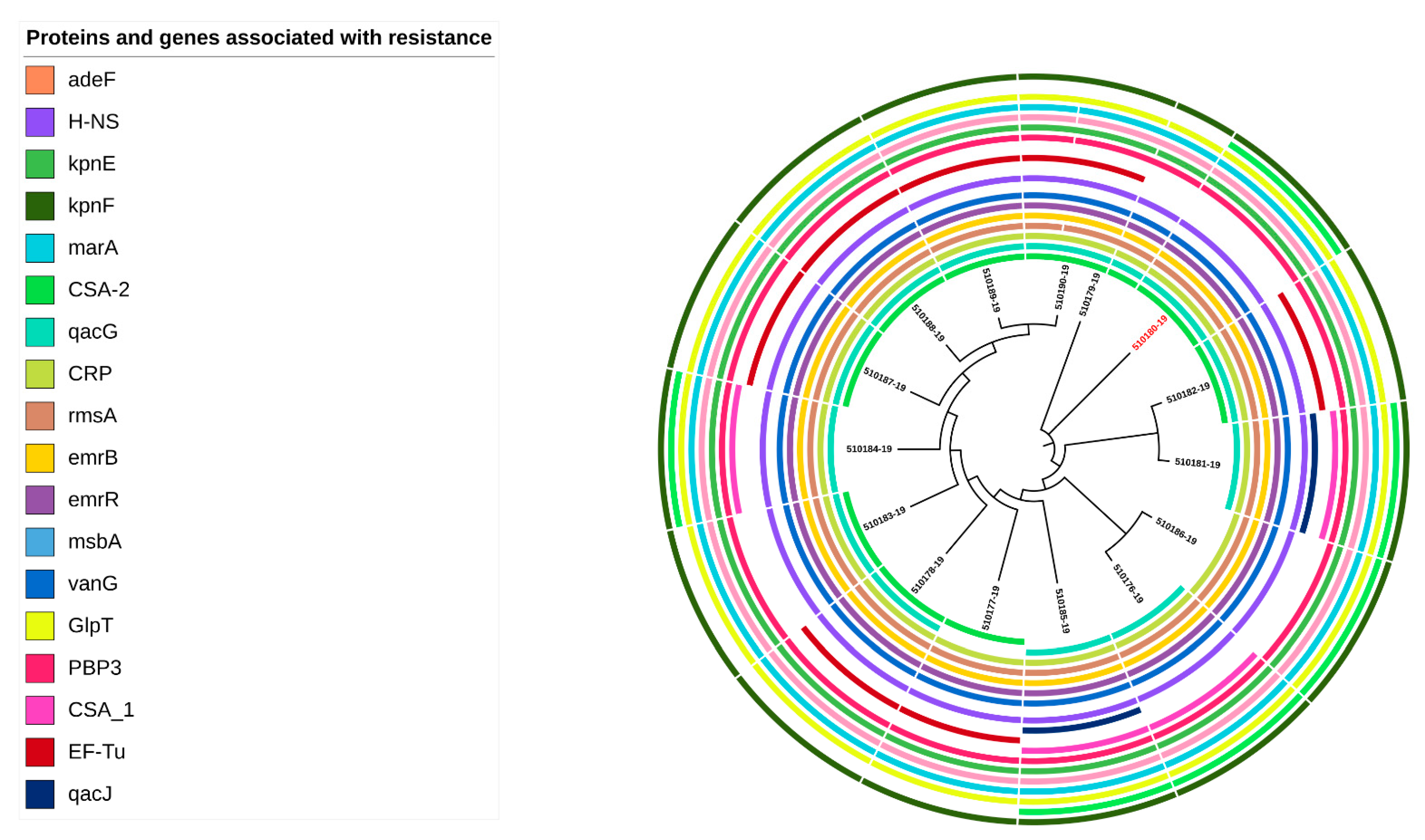
Twenty genes associated with antibiotic resistance were identified in the 14 C. sakazakii genomes. Some of these genes were classified as global regulators, such as HNS and CRP, which were associated with resistance-nodulation-cell division (RND) antibiotic efflux pump AMR families and rsmA a global translational regulatory protein RsmA. Other genes were related to antibiotic efflux, reduced antibiotic permeability, target modification or antibiotic inactivation (Supplementary Table S1). C. sakazakii strains carried the same genes (Figure 2) that were involved in general bacterial porin with reduced permeability to beta-lactams, small multidrug resistance (SMR) antibiotic efflux pump, ATP-binding cassette (ABC) antibiotic efflux pump, resistance-nodulation-cell division (RND), glycopeptide resistance gene cluster, Van ligase, antibiotic-resistant GlpT, and Penicillin-binding protein mutations conferring resistance to beta-lactam antibiotics related to phenotypic profiles (Supplementary Table S1).
Three genes (EF-tu, CSA-1 and qacJ) were identified in seven, four and two genomes, respectively (Figure 2). The EF-tu gene with a SNP R234F was detected in 46.7% (7/15) of the genomes. This gene belongs to the AMR gene family elfamycin and their mechanism of resistance is antibiotic target alteration. The gene CSA-1 was found in 26.7% (4/15) of the genomes. This gene belongs to AMR gene family CSA beta-lactamases, conferring resistance to cephalosporin by antibiotic inactivation. Lastly, the qacJ gene was identified in 13.3% (2/15) of the genomes of the associated gene family small multidrug-resistance (SMR) antibiotic efflux pump disinfecting agents and antiseptics (Supplementary Table S1).
Although most of the genes were identified in all the strains, three genes were absent in the analyzed genomes (Figure 2). The gene kpnF was not detected in 60% (9/15) of the genomes. This gene belongs to a major facilitator superfamily (MFS) antibiotic efflux pump. The gene CSA-2 was not found in 40% (6/15) of analyzed genomes. This gene encodes for a CSA beta-lactamase inactivating cephalosporins. The gene qacG was not identified in 13.3% (2/15) of genomes. This gene encodes for a small multidrug-resistance (SMR) antibiotic efflux pump associated with resistance to disinfecting agents and antiseptics; it uses the antibiotic efflux as a mechanism of resistance (Supplementary Table S1).
The genes kpnF, CSA-1, and qacJ were not identified in the C. dublinensis genome (510180-19); however, these genes were detected in most C. sakazakii genomes.

2.4. Virulence Genes in C. sakazakii Genomes

A total of 99 virulence and fitness genes were identified in the C. sakazakii and C. dublinensis genomes. The most common genes associated with virulence encode for ompA, siderophores and genes involved in metabolism or fitness (Figure 3, Supplementary Table S2).
Some genes could be involved in bacterial metabolism, such as: algG: bifunctional mannose-1-phosphate guanylyltransferase/mannose-6-phosphate isomerase; ctpC: manganese-exporting P-type ATPase CtpC; galF: UTP--glucose-1-phosphate uridylyltransferase GalF; gndA: NADP+-dependent 6-P-gluconate dehydrogenase; icl: isocitrate lyase, msrA/BpilB: methionine sulfoxide reductase; nar: nitrate reductase; pchD: transcriptional regulator.
The virulence genes detected in this study were related to: icmF1/tssM1, hsiC1/vipB/tssC, imp: type VI secretion system protein; hcp: type VI secretion system receptor/chaperone Hcp; iucA: aerobactin synthase, iucA, iutA: ferric aerobactin receptor IutA, entB: enterobactin synthase component B; fep: ferric enterobactin outer membrane transporter; fes: ferric enterobactin esterase; iroN: siderophore salmochelin receptor IroN, ompA: outer membrane protein; hec: B family hemolysin secretion/activation protein; kpsD: capsule polysaccharide ABC transporter substrate-binding protein; clvp: ATP-dependent protease ClpV; cpsG: colanic acid biosynthesis phosphomannomutase CpsG; gacS: RhoGAP domain-containing protein; ehaB: autotransporter adhesin EhaB; and cdiA: contact-dependent inhibition effector toxin. The flagella synthesis operon was also identified. Furthermore, plasminogen activator (cpa) and utilization of sialic acid (nanA,K,T) genes were detected only in the C. sakazakii strains and not in C. dublinensis. The small heat shock protein sHSP20 was detected in C. sakazakii ST4 and ST83, while the locus of heat resistance: yfdX1GI, yfdX2, hdeDGI, orf11, trxGI, kefB, and psiEGI was only found in the genomes of C. sakazakii ST4.
The genes: bvrS, ehaB, fepB, fliC, fliF, fliJ, hcpC, cpa, and nanA,K,T were not identified in the C. dublinensis genome (510180-19); however, these genes were detected in most C. sakazakii genomes.

2.5. Plasmids and Mobile Genetic Elements in C. sakazakii Genomes

The IncFIB(pCTU3) plasmid was detected in most (8/9) C. sakazakii isolates and the ST260 isolate (Table 3). IncFIB(pCTU2), pESA2, rep7a and Col4401 were found in strains 510186-19 and 510180-19, respectively. Regarding the mobile genetic elements (MGEs), the most prevalent were ISEsa1, ISEc52 and ISEhe3 (Table 4).

3. Discussion

C. sakazakii ST1, ST4 and, to a lesser extent, ST83, are the pathovars that have been most frequently found in PIF marketed in different countries, in PIF production plants and in invasive clinical cases such as fatal meningitis and septicemia [5,25,26,27,28,29]. The other strains, ST93, ST822 and ST260, have not been associated with cases of disease and are of less clinical importance. In this study, we identified nine C. sakazakii isolates of ST4 (CC4) serotype Csak O:2. These were mostly from environmental sources (5/9) (Table 2), whereas C. sakazakii 1 ST1 (CC1) serotype Csak O:1 was isolated from food. A previous study carried out on Cronobacter in the Americas showed that most of the isolates came from clinical, environmental and infant formula samples from North America (57.4%, n = 465) and Brazil (42.6%, n = 465). In addition, the study reported a total of 75 sequence types, with the most frequent being C. sakazakii ST4 (CC4) and ST1 (CC1) [30].
Analysis using a 3678 loci cgMLST scheme revealed a group of eight indistinguisable C. sakazakii ST4 isolates from food and the production environment. A previously reported multicenter study assessing the incidence of C. sakazakii throughout Europe found that 76.6% (59/77) of the human clinical strains of Cronobacter spp. corresponded to C. sakazakii. Twelve C. sakazakii isolates were ST4, and eight were ST1 [31]. There was only one allele difference between isolates associated with epidemiological outbreaks in Austria in 2016 and 2009, respectively.
C. dublinensis has primarily been reported in environmental samples and food ingredients. This is primarily considered an environmental commensal and has rarely been reported in clinical samples [4,32].
In the present study, the C. sakazakii strains were resistant to cephalothin, ampicillin tetracycline, ceftazidime and amoxicillin–clavulanic acid. In addition, two strains showed an MDR profile (ampicillin, cephalothin and tetracycline); one was ST 260 (510176-19), and the other was ST4 (510177-19). These two strains were isolated from different foods. Compared to other studies, the resistance observed in this study is low. For example, in Teheran city (Iran), of the 25 C. sakazakii strains recovered from PIF, 96% were MDR; these were mainly resistant to amoxicillin–clavulanic acid, amoxicillin, ampicillin, cefoxitin, cefepime, erythromycin, and ceftriaxone, and totally susceptible to trimethoprim/sulfamethoxazole and levofloxacin antibiotics [33]. In China, C. sakazakii from PIF and processing environments showed isolates resistant to amoxicillin–clavulanic acid, ampicillin, and cefazolin [34]. MDR isolates have been reported from domestic kitchens in middle Tennessee, (USA); the highest resistance was to penicillin (76.1%), tetracycline (66.6%), ciprofloxacin (57.1%), and nalidixic acid (47.6%) [35]. C. sakazakii antibiotic resistance is diverse and depends on the source and geographic location of the strain [33,34,35]. The genomic analysis has enabled the detection of genes involved in antibiotic resistance. The genes are related to cell permeability to beta-lactams and antibiotic efflux pumps. In other studies, blaCTX genes are commonly detected in strains resistant to cephalosporins, whereas, in this study, these genes were not detected [36,37]. Another detected gene was EF-Tu, which is included in the AMR gene family elfamycin and their mechanism of resistance is antibiotic target alteration. This gene has evolved to be a multifunctional protein in a wide variety of pathogenic bacteria [38]. Functions related to EF-Tu vary among microbial species; however, there is a common role of adherence and immune regulation [38]. The AmpC beta-lactamases designated CSA-1 and CSA-2 (from C. sakazakii) were found in the 14 C. sakazakii strains and have been reported previously [39].
The genomic analysis of the Cronobacter strains revealed a number of virulence-related genes. Several putative genes of the Type VI Secretion System cluster were found. The Type VI Secretion System (T6SS) is a protein secretion machinery that transports protein toxins into eukaryotic cells [40,41] and bacteriolytic effectors to target cells [42]. Some genes involved in the iron uptake category (fur, iroN and PSEEN_RS11615), responsible for the production of metal binding proteins, were identified in genomic sequences, and are associated with bacterial virulence [43]. The flagella operon was found in all genomes. Flagella has been involved in adherence and proinflammatory response in different pathogens [16]. The most frequently occuring gene, the outer membrane protein A (OmpA), was identified in the genomes. OmpA plays a critical role in host cell invasion [44].
In our research, all 14 C. sakazakii strains encoded the cpa gene. The cpa gene is possibly involved in serum resistance, as well as in the systemic spread of C. sakazakii. The isolates harbored the nanAKT cassette coding for the use of exogenous sialic acid. Only the C. sakazakii species and a few C. turicensis strains can catabolize this [45]. Sialic acid is found in gangliosides of the brain, and occurs naturally in breast milk [46]. Powdered infant formula is supplemented with this monosaccharide due to its association with brain development [4]. Sialic acid also regulates the expression of enzymes, such as sialidase and adhesins, or inhibits the transcription factors of the fimB gene involved in the adhesion and invasion of epithelial cells [47].
Another important observation was the presence of rpoS, ibpA, ibpB and clpk genes in the C. sakazakii genomes. These genes are involved in protection against environmental stress [48]. For example, the RpoS regulon, which acts as a transcriptional factor in response to general stress, develops cross-protection against other environmental disturbances, such as the response to oxidative stress and the response to heat stress. In E. coli, for example, it represents 10% of genes [49]. On the other hand, the genes encoding for heat shock proteins Hsp15, Hsp20 and HspQ, were found in thermotolerant C. sakazakii [50,51]. Therefore, when these environmental C. sakazakii are subjected to desiccation or drying processes, as in the preparation of PIF or other foods, they may become more resistant to these stresses, enabling them to persist either in the environments of these production plants or in the final products.
Sixty percent of the C. sakazakii strains in our study carried the IncFIB(pCTU3) plasmid, which has been previously reported in environmental sources and food [52,53], as shown in Table 3. The majority of strains carrying the plasmid were C. sakazakii ST4. This plasmid is associated with encoding for efflux pumps associated with heavy metals, which could allow the microorganism to adapt to the changing environmental conditions of osmotic stress [54].
In this study, C. sakazakii ST4 and ST1 were isolated from environmental strains (Table 2), and harbored genes that encode for antibiotic resistance proteins and virulence factors (Figure 2 and Figure 3). Consequently, there is the potential risk that these isolates could persist in the environment and become a source of food contamination, therefore presenting a risk to the health of consumers.

4. Materials and Methods

4.1. Sampling

A total of 15 presumptive strains of Cronobacter spp. were isolated from 2000 samples from food (n = 997), milk-producing environments (n = 855), and feces from live poultry (n = 148) on the territory of the Czech Republic in 2015. Sampling was performed by instructed veterinarians. Solid, loose and slurry materials were collected in 60–200 mL sterile plastic containers (Dispolab Ltd., Brno, Czech Republic) or in original packaging. Swabs were taken using Transbak system containing Amies broth with active carbon (Dispolab Ltd., Brno, Czech Republic). Environmental swabs, feces and meat samples were stored and transported to the laboratory at +4 °C. Other materials were transported at +21 °C. These strains were analyzed at the bacteriology department, Palacký University Olomouc, Czech Republic (Supplementary Table S3).

4.2. Isolation and Identification Methods of Cronobacter spp.

Cronobacter spp. strains were isolated according to the method described by Iversen et al. [55]. Food and environment samples were pre-enriched in buffered peptone water (BPW), followed by Enterobacteriaceae enrichment broth (BD Difco, Sparks, MD, USA), then on Brilliance CM 1035 chromogenic agar (Oxoid Thermo-Fisher, UK) and purified on trypticase soy agar (BD Difco, Sparks, MD, USA). Prior to sequencing, the strains were presumptively identified using Matrix Assisted Laser Desorption Ionization—Time of Flight Mass Spectrometry (MALDI-TOF MS) (Bruker, Billerica, MA, USA) and with the MALDI Biotyper Compass IVD 4.1.60 software (Bruker, Billerica, MA, USA) described by Lepuschitz et al. [56]. The identification of the Cronobacter spp. strains was confirmed with the Ribosomal Multilocus Sequence Typing (rMLST) software available at https://pubmlst.org/species-id (accessed on 2 February 2023) [57].

4.3. Whole-Genome Sequencing (WGS)

Cronobacter spp. isolates were cultured in Columbia blood agar plates (bioMérieux, Marcy-l’Étoile, France) at 37 °C for 24 h. For WGS, DNA was isolated from bacterial cultures with the MagAttract HMW DNA Kit (Qiagen, Hilden, Germany) according to the manufacturer’s instructions. The quantification of input DNA was performed with a Qubit 2.0 fluorometer (Thermo Fisher Scientific, Waltham, MA, USA) and the double-stranded DNA (dsDNA) BR assay kit (Thermo Fisher Scientific). Nextera XT chemistry (Illumina Inc., San Diego, CA, USA) was used to prepare sequencing libraries for a 2 × 300 bp paired-end sequencing run on an Illumina MiSeq sequencer. Samples were sequenced to achieve a minimum of 80-fold coverage using standard Illumina protocols. The resulting FASTQ files were quality-trimmed and de novo assembled with the SPAdes v3.11.1 software. Contigs were filtered for a minimum of 5-fold coverage and 200  bp minimum length with the Ridom SeqSphere+ software v8.3 (Ridom, Münster, Germany) [31]. Raw reads were quality-controlled using FastQC v0.11.9. Trimmomatic v0.36 [58] was used to remove adapter sequences and to trim the last 10 bp of each sequence and sequences with quality scores  < 20. Reads were assembled using SPAdes v3.11.1 [59]. Contigs were filtered for a minimum coverage of 5× and a minimum length of 200 bp using SeqSphere+ software v8.3.0 (Ridom GmbH, Würzburg, Germany) [58] (Supplementary Table S3).

4.4. Sequence Type (ST) and Core Genome Multilocus Sequence Typing (cgMLST) of Cronobacter sakazakii

A total of 3678 targets were used for core genome multilocus sequence typing (cgMLST) scheme of Cronobacter spp., with strain ATCC BAA-894 as a reference using a target gene loci task template of the Ridom SeqSphere+ software v8.3.0 (Ridom, Münster, Germany) [31,60]. According to this cgMLST scheme, isolates were visualized with a minimum spanning tree (MST) to establish their genotypic relationships and define those isolates with maximum differences of 10 alleles as clusters [31]. In addition, the sequences of the seven housekeeping genes of the conventional MLST for C. sakazakii (atpD, fusA, glnS, gltB, gyrB, infB and ppsA) were extracted and cross-checked against the Cronobacter MLST database https://pubmlst.org/organisms/cronobacter-spp/ (accessed on 2 February 2023) [61].

4.5. Determination of Serotypes

The profiles of gnd and galF genes specific to the Cronobacter spp. serotype O region were determined by WGS sequence analysis with the BIGSdb tool available in the PubMLST database (http://pubmlst.org/cronobacter/) (accessed on 2 February 2023) [18].

4.6. Antibiotic Susceptibility

To assess antibiotic susceptibility, the disk diffusion method was used, in accordance with the recommendations of the Clinical and Laboratory Standards Institute [62]. The commercial disks that were used consist of ampicillin (10 μg), amoxicillin–clavulanic acid (20/10 µg), ceftazidime (30 µg), ciprofloxacin (5 μg), chloramphenicol (30 μg), cefotaxime (30 μg), gentamicin (10 μg), cephalothin (30 μg), tetracycline (30 µg) and nalidixic acid (30 µg). The characterization of the resistance/susceptibility profiles was determined according to the manufacturer’s instructions. The Escherichia coli ATCC 25922 and Pseudomonas aeruginosa ATCC 27853 strains were used as internal control.

4.7. Detection of Antibiotic Resistance and Virulence Genes

The existence of virulence genes was confirmed by ResFinder tool from the Center of Genomic Epidemiology (CGE) (http://www.genomicepidemiology.org) (accessed on 5 March 2023) [63]. Thresholds for the target scanning procedure were set with a required identity of ≥90% to the reference sequence and an aligned reference sequence ≥99%. The Comprehensive Antibiotic Resistance Database (CARD) with the “perfect” and “strict” default settings for sequence analysis [64], the Task Template AMRFinderPlus 3.2.3 available in Ridom SeqSphere+ v7.8.0 software using the EXACT method at 100%, and BLAST alignment for protein identification available in the AMRFinderPlus database were used for antimicrobial resistance genes. For the search for genes and proteins associated with virulence and fitness, the Virulence Factor Database (VFDB), available at http://www.mgc.ac.cn/VFs/main.htm (accessed on 5 March 2023), was downloaded and compared with each of the 15 genomes using the blast tool ncbi-blast-2.13.0+-x64-arm-linux.tar.gz, available at https://ftp.ncbi.nlm.nih.gov/blast/executables/blast+/LAST/ (accessed on 5 March 2023) on command line.

4.8. Detection of Plasmids and Mobile Genetic Elements (MGEs)

The PlasmidFinder v2.1 and MobileElementFinder v1.0 tools were used to detect plasmids and mobile genetic elements (MGEs), respectively. The selected minimum identity was 95% and 90%, respectively (http://www.genomicepidemiology.org/) (accessed on 5 March 2023) [65,66].

5. Conclusions

The C. sakazakii isolates analyzed in this study harbored ARGs and VGs, which contributed to their persistence in powdered-milk-producing environments, and increased the risk of infection in susceptible population groups due to the presence of multi-resistant strains to antibiotics, reducing the possibility of treating of the infection [67,68].

Supplementary Materials

The following supporting information can be downloaded at: https://www.mdpi.com/article/10.3390/antibiotics12050851/s1. Supplementary Tables S1–S3 Virulence and antibiotic resistance genes, and sequencing parameters.

Author Contributions

Conceptualization, J.P.-F., O.H. and W.R.; methodology, J.P.-F., B.D.-P., A.C.-R., J.X.-C., R.R.-M. and W.R.; software, J.P.-F., B.D.-P. and R.R.-M.; validation, J.P.-F., B.D.-P. and W.R.; formal analysis, W.R. and A.C.-R.; investigation, J.B., O.H. and J.P.-F.; resources, J.P.-F. and S.A.; data curation, J.P.-F., S.F. and W.R.; writing—original draft preparation, J.P.-F., A.C.-C., S.F. and W.R.; writing—review and editing, J.P.-F., W.R., S.F. and S.A.; visualization, J.P.-F. and B.D.-P.; supervision, J.P.-F. and A.C.-C.; project administration, J.P.-F.; funding acquisition, J.P.-F., J.X.-C. and W.R. All authors have read and agreed to the published version of the manuscript.

Funding

This research was funded the Research Directorate of the Universidad del Bío-Bío, Project GI 195420/EF.

Institutional Review Board Statement

Not applicable.

Informed Consent Statement

Not applicable.

Data Availability Statement

The Cronobacter sakazakii and Cronobacter dublinensis isolates were submitted to https://pubmlst.org/organisms/cronobacter-spp with ID 3695-3697 and 3823-3834.

Acknowledgments

We thank the Institute of Medical Microbiology and Hygiene AGES-Austrian Agency for Health and Food Safety for their support.

Conflicts of Interest

The authors declare no conflict of interest.

References

  1. Iversen, C.; Mullane, N.; Mc Cardell, B.; Tall, B.; Lehner, A.; Fanning, S.; Stephan, R.; Joosten, H. Cronobacter gen. nov., a new genus to accommodate the biogroups of Enterobacter sakazakii, and proposal of Cronobacter sakazakii gen. nov.comb. nov., C. malonaticus sp. nov., C. turicensis sp. nov., C. muytjensii sp. nov., C. dublinensis sp. nov., Cronobacter genomospecies 1, and of three subspecies, C. dublinensis sp. nov.subsp. dublinensis subsp. nov., C. dublinensis sp. nov.subsp. lausannensis subsp. nov., and C. dublinensis sp. nov.subsp. lactaridi subsp. nov. Int. J. Syst. Evol. Microbiol. 2008, 58, 1442–1447. [Google Scholar]
  2. Joseph, S.; Cetinkaya, E.; Drahovska, H.; Levican, A.; Figueras, M.; Forsythe, S. Cronobacter condimenti sp. Nov., isolated from spiced meat, and Cronobacter universalis sp. nov., a species designation for Cronobacter sp. genomoespecies 1, recovered from a leg infection, water and food ingredients. Int. J. Syst. Evol. Microbiol. 2012, 62, 1277–1283. [Google Scholar] [CrossRef] [PubMed]
  3. Stephan, R.; Grim, C.; Gopinath, G.; Mammel, M.; Sathyamoorthy, V.; Trach, L.; Chase, H.; Fanning, S.; Tall, B. Re-examination of the taxonomic status of Enterobacter helveticus, Enterobacter pulveris and Enterobacter turicensis as members of the genus Cronobacter and their reclassification in the genera Franconibacter gen. nov. and Siccibacter gen. nov. as Franconibacter helveticus comb. nov., Franconibacter pulveris comb. nov. and Siccibacter turicensis comb. nov., respectively. Int. J. Syst. Evol. Microbiol. 2014, 64, 3402–3410. [Google Scholar] [PubMed]
  4. Forsythe, S.J. Updates on the Cronobacter Genus. Annu. Rev. Food Sci. Technol. 2018, 25, 23–44. [Google Scholar] [CrossRef] [PubMed]
  5. Hariri, S.; Joseph, S.; Forsythe, S.J. Cronobacter sakazakii ST4 strains and neonatal meningitis, United States. Emerg. Infect. Dis. 2013, 19, 175–177. [Google Scholar] [CrossRef]
  6. Patrick, M.; Mahon, B.; Greene, S.; Rounds, J.; Conquist, A.; Wymore, K.; Boothe, E.; Lathrop, S.; Palmer, A.; Bowen, A. Incidence of Cronobacter spp. infections, United States, 2003–2009. Emerg. Inf. Dis. 2014, 20, 1520–1523. [Google Scholar] [CrossRef]
  7. Holý, O.; Cruz-Cordova, A.; Xicohtencatl-Cortés, J.; Hochel, I.; Parra-Flores, J.; Petrzelova, J.; Fačevicová, K.; Forsythe, S.; Alsonosi, A. Occurrence of virulence factors in Cronobacter sakazakii and Cronobacter malonaticus originated from clinical samples. Microb. Pathog. 2019, 127, 250–256. [Google Scholar] [CrossRef]
  8. Holý, O.; Forsythe, S. Cronobacter spp. as emerging causes of healthcare-associated infection. J. Hosp. Infect. 2014, 86, 169–177. [Google Scholar] [CrossRef]
  9. Baumgartner, A.; Grand, M.; Liniger, M.; Iversen, C. Detection and frequency of Cronobacter spp. (Enterobacter sakazakii) in different categories of ready-to-eat foods other than infant formula. Int. J. Food Microbiol. 2009, 136, 189–192. [Google Scholar] [CrossRef]
  10. Vojkovska, H.; Karpiskova, R.; Orieskova, M.; Drahovska, H. Characterization of Cronobacter spp. isolated from food of plant origin and environmental samples collected from farms and from supermarkets in the Czech Republic. Int. J. Food Microbiol. 2016, 217, 130–136. [Google Scholar] [CrossRef]
  11. Parra-Flores, J.; Maury-Sintjago, E.; Rodriguez-Fernández, A.; Acuña, S.; Cerda, F.; Aguirre, J.; Holý, O. Microbiological quality of powdered infant formula in Latin America. J. Food. Prot. 2020, 83, 534–541. [Google Scholar] [CrossRef] [PubMed]
  12. Lindsay, D.; Laing, S.; Fouhy, K.I.; Souhoka, L.; Beaven, A.; Soboleva, T.; Malakar, P. Quantifying the uncertainty of transfer of Cronobacter spp. between fomites and floors and touch points in dairy processing plants. Food Microbiol. 2019, 84, 103256. [Google Scholar] [CrossRef] [PubMed]
  13. El-Sharoud, W.; O’Brien, S.; Negredo, C.; Iversen, C.; Fanning, S.; Healy, B. Characterization of Cronobacter recovered from dried milk and related products. BMC Microbiol. 2009, 9, 24. [Google Scholar] [CrossRef]
  14. Craven, H.H.; McAuley, C.M.; Duffy, L.L.; Fegan, N. Distribution, prevalence and persistence of Cronobacter (Enterobacter sakazakii) in the non-processing and processing environments of five milk powder factories. J. Appl. Microbiol. 2010, 109, 1044–1052. [Google Scholar] [CrossRef] [PubMed]
  15. Franco, A.; Kothary, M.; Gopinath, G.; Jarvis, K.; Grim, C.; Hu, L.; Datta, A.; McCardell, B.A.; Tall, B.D. Cpa, the outer membrane protease of Cronobacter sakazakii, activates plasminogen and mediates resistance to serum bactericidal activity. Infect. Immun. 2011, 79, 1578–1587. [Google Scholar] [CrossRef] [PubMed]
  16. Cruz-Córdova, A.; Rocha-Ramírez, L.; Ochoa, S.; Gónzalez-Pedrajo, B.; Espinosa, N.; Eslava, C.; Hernández-Chiñas, U.; Mendoza-Hernández, G.; Rodríguez-Leviz, A.; Valencia-Mayoral, P.; et al. Flagella from five Cronobacter species induce pro-inflammatory cytokines in macrophage derivatives from human monocytes. PLoS ONE 2012, 7, e52091. [Google Scholar] [CrossRef]
  17. Aldubyan, M.; Almami, I.; Benslimane, F.; Alsonosi, A.; Forsythe, S. Comparative outer membrane protein analysis of high and low-invasive strains of Cronobacter malonaticus. Front. Microbiol. 2017, 8, 2268. [Google Scholar] [CrossRef]
  18. Ogrodzki, P.; Forsythe, S. Capsular profiling of the Cronobacter genus and the association of specific Cronobacter sakazakii and C. malonaticus capsule types with neonatal meningitis and necrotizing enterocolitis. BMC Genom. 2015, 16, 758. [Google Scholar] [CrossRef]
  19. Lee, Y.-D.; Park, J.; Chang, H. Detection, antibiotic susceptibility and biofilm formation of Cronobacter spp. from various foods in Korea. Food Control 2012, 24, 225–230. [Google Scholar] [CrossRef]
  20. Parra-Flores, J.; Holý, O.; Riffo, F.; Lepuschitz, S.; Maury-Sintjago, E.; Rodríguez-Fernández, A.; Cruz-Córdova, A.; Xicohtencatl-Cortes, J.; Mancilla-Rojano, J.; Troncoso, M.; et al. Profiling the virulence and antibiotic resistance genes of Cronobacter sakazakii strains isolated from powdered and dairy formulas by whole-genome sequencing. Front. Microbiol. 2021, 12, 694922. [Google Scholar] [CrossRef]
  21. Shi, L.; Liang, Q.; Zhan, Z.; Feng, J.; Zhao, Y.; Chen, Y.; Huang, M.; Tong, Y.; Wu, W.; Chen, W.; et al. Co-occurrence of 3 different resistance plasmids in a multi-drug resistant Cronobacter sakazakii isolate causing neonatal infections. Virulence 2018, 9, 110–112. [Google Scholar] [CrossRef] [PubMed]
  22. Aly, M.A.; Domig, K.J.; Kneifel, W.; Reimhult, E. Whole genome sequencing-based comparison of food isolates of Cronobacter sakazakii. Front. Microbiol. 2019, 10, 1464. [Google Scholar] [CrossRef]
  23. Leopold, S.; Goering, R.; Witten, A.; Harmsen, D.; Mellmann, A. Bacterial whole-genome sequencing revisited: Portable, scalable, and standardized analysis for typing and detection of virulence and antibiotic resistance genes. J. Clin. Microbiol. 2014, 52, 2365–2370. [Google Scholar] [CrossRef] [PubMed]
  24. Lehner, A.; Tall, B.D.; Fanning, S.; Srikumar, S. Cronobacter spp.—Opportunistic foodborne pathogens: An update on evolution, osmotic adaptation and pathogenesis. Curr. Clin. Micro. Rpt. 2018, 5, 97–105. [Google Scholar] [CrossRef]
  25. Joseph, S.; Forsythe, S. Insights into the emergent bacterial pathogen Cronobacter spp., generated by multilocus sequence typing and analysis. Front. Food Microbiol. 2012, 3, 397. [Google Scholar] [CrossRef] [PubMed]
  26. Fei, P.; Jiang, Y.; Jiang, Y.; Yuan, X.; Yang, T.; Chen, J.; Wang, Z.; Kang, H.; Forsythe, S.J. Prevalence, molecular characterization, and antibiotic susceptibility of Cronobacter sakazakii isolates from powdered infant formula collected from Chinese retail markets. Front. Microbiol. 2017, 8, 2026. [Google Scholar] [CrossRef]
  27. Sonbol, H.; Joseph, S.; McAuley, C.; Craven, H.; Forsythe, S. Multilocus sequence typing of Cronobacter spp. from powdered infant formula and milk powder production factories. Int. Dairy J. 2013, 30, 1–7. [Google Scholar] [CrossRef]
  28. Csorba, C.; Pajić, M.; Blagojević, B.; Forsythe, S.; Radinović, M.; Velebit, B. Prevalence, characterization, and antibiotic susceptibility of Cronobacter spp. in a milk powder processing environment: The first reported case in Serbia. Food Sc. Nutr. 2022, 10, 554–563. [Google Scholar] [CrossRef]
  29. Grim, C.J.; Gopinath, G.R.; Jarvis, K.G.; Sathyamoorthy, V.; Trach, L.H.; Chase, H.R.; Tall, B.D. Genome sequence of Cronobacter sakazakii serogroup O:4, sequence type 4 strain CDC 2009-03746, isolated from a fatal case of infantile meningitis. Genome Announc. 2015, 3, e00492-15. [Google Scholar] [CrossRef]
  30. Costa, P.V.; Vasconcellos, L.; Forsythe, S.J.; Brandão, M.L. Diversity of Cronobacter genus isolated between 1970 and 2019 on the American continent and genotyped using multi-locus sequence typing. FEMS Microbiol. Lett. 2021, 368, 1–9. [Google Scholar] [CrossRef]
  31. Lepuschitz, S.; Ruppitsch, W.; Pekard-Amenitsch, S.; Forsythe, S.J.; Cormican, M.; Mach, R.L.; Piérard, D.; Allerberger, F.; The EUCRONI Study Group. Multicenter study of Cronobacter sakazakii infections in humans, Europe, 2017. Emerg. Infect. Dis. 2019, 25, 515–522. [Google Scholar] [CrossRef]
  32. Hu, J.; Li, X.; Du, X.; Cui, Z.; Cui, J. Identification and characterization of Cronobacter strains isolated from environmental samples. Curr. Microbiol. 2019, 76, 1467–1476. [Google Scholar] [CrossRef] [PubMed]
  33. Pakbin, B.; Brück, W.M.; Allahyari, S.; Rossen, J.W.A.; Mahmoudi, R. Antibiotic resistance and molecular characterization of Cronobacter sakazakii strains isolated from powdered infant formula milk. Foods 2022, 11, 1093. [Google Scholar] [CrossRef]
  34. Fei, P.; Jiang, Y.; Feng, J.; Forsythe, S.J.; Li, R.; Zhou, Y.; Man, C. Antibiotic and desiccation resistance of Cronobacter sakazakii and C. malonaticus isolates from powdered infant formula and processing environments. Front. Microbiol. 2017, 8, 316. [Google Scholar] [CrossRef]
  35. Kilonzo-Nthenge, A.; Rotich, E.; Godwin, S.; Nahashon, S.; Chen, F. Prevalence and antimicrobial resistance of Cronobacter sakazakii isolated from domestic kitchens in middle Tennessee, United States. J. Food Prot. 2012, 75, 1512–1517. [Google Scholar] [CrossRef]
  36. Zurfluh, K.; Nuësch-Inderbinen, M.; Morach, M.; Zihler Berner, A.; Hächler, H.; Stephan, R. Extended-spectrum-β-lactamaseproducing Enterobacteriaceae isolated from vegetables imported from the Dominican Republic, India, Thailand, and Vietnam. Appl. Environ. Microbiol. 2015, 81, 3115–3120. [Google Scholar] [CrossRef] [PubMed]
  37. Holý, O.; Alsonosi, A.; Hochel, I.; Röderová, M.; Zatloukalova, S.; Mlynárčik, P.; Kolář, M.; Petrželová, J.; Alazraq, A.; Chmelař, D. Antibiotic susceptibility of Cronobacter spp. isolated from clinical samples. Pol. J. Microbiol. 2019, 68, 5–14. [Google Scholar] [CrossRef]
  38. Harvey, K.L.; Jarocki, V.M.; Charles, I.G.; Djordjevic, S.P. The diverse functional roles of elongation Factor Tu (EF-Tu) in microbial pathogenesis. Front Microbiol. 2019, 10, 2351. [Google Scholar] [CrossRef]
  39. Müller, A.; Hächler, H.; Stephan, R.; Lehner, A. Presence of AmpC beta-lactamases, CSA-1, CSA-2, CMA-1, and CMA-2 conferring an unusual resistance phenotype in Cronobacter sakazakii and Cronobacter malonaticus. Microb. Drug Resist. 2014, 20, 275–280. [Google Scholar] [CrossRef] [PubMed]
  40. Durand, E.; Cambillau, C.; Cascales, E.; Journet, L. VgrG, Tae, Tle, and beyond: The versatile arsenal of type VI secretion effectors. Trends Microbiol. 2014, 22, 498–507. [Google Scholar] [CrossRef] [PubMed]
  41. Jang, H.; Gopinath, G.R.; Eshwar, A.; Srikumar, S.; Nguyen, S.; Gangiredla, J.; Patel, I.R.; Finkelstein, S.B.; Negrete, F.; Woo, J.; et al. The Secretion of toxins and other exoproteins of Cronobacter: Role in virulence, adaption, and persistence. Microorganisms 2020, 8, 229. [Google Scholar] [CrossRef]
  42. Coulthurst, S. The Type VI secretion system: A versatile bacterial weapon. Microbiology (Reading) 2019, 165, 503–515. [Google Scholar] [CrossRef]
  43. Singh, N.; Goel, G.; Raghav, M. Insights into virulence factors determining the pathogenicity of Cronobacter sakazakii. Virulence 2015, 6, 433–440. [Google Scholar] [CrossRef]
  44. Mohan Nair, M.K.; Venkitanarayanan, K. Role of bacterial OmpA and host cytoskeleton in the invasion of human intestinal epithelial cells by Enterobacter sakazakii. Pediatr. Res. 2007, 62, 664–669. [Google Scholar] [CrossRef] [PubMed]
  45. Joseph, S.; Hariri, S.; Masood, N.; Forsythe, S. Sialic acid utilization by Cronobacter sakazakii. Microb. Inform. Exp. 2013, 3, 3. [Google Scholar] [CrossRef] [PubMed]
  46. Wang, B.; McVeagh, P.; Petocz, P.; Brand-Miller, J. Brain ganglioside and glycoprotein sialic acid in breastfed compared with formula-fed infants. Am. J. Clin. Nutr. 2003, 78, 1024–1029. [Google Scholar] [CrossRef] [PubMed]
  47. Severi, E.; Hood, D.W.; Thomas, G.H. Sialic acid utilization by bacterial pathogens. Microbiology (Reading) 2007, 153, 2817–2822. [Google Scholar] [CrossRef]
  48. Lee, C.; Wigren, E.; Lünsdorf, H.; Römling, U. Protein homeostasis-more than resisting a hot bath. Curr. Opin. Microbiol. 2016, 30, 147–154. [Google Scholar] [CrossRef]
  49. Schellhorn, H.E. Function, evolution, and composition of the RpoS regulon in Escherichia coli. Front. Microbiol. 2020, 11, 560099. [Google Scholar] [CrossRef]
  50. Bojer, M.S.; Struve, C.; Ingmer, H.; Hansen, D.S.; Krogfelt, K.A. Heat resistance mediated by a new plasmid encoded Clp ATPase, ClpK, as a possible novel mechanism for nosocomial persistence of Klebsiella pneumoniae. PLoS ONE 2010, 5, e15467. [Google Scholar] [CrossRef]
  51. Boll, E.J.; Frimodt-Møller, J.; Olesen, B.; Krogfelt, K.A.; Struve, C. Heat resistance in extended-spectrum beta-lactamase-producing Escherichia coli may favor environmental survival in a hospital setting. Res. Microbiol. 2016, 167, 345–349. [Google Scholar] [CrossRef]
  52. Niu, H.; Mingzhe, Y.; Qi, Y.; Liu, Y.; Wang, X.; Dong, Q. Heat shock in Cronobacter sakazakii induces direct protection and cross-protection against simulated gastric fluid stress. Food Microbiol. 2022, 103, 10394. [Google Scholar] [CrossRef]
  53. Brooks, K.; Eze, J.; Onalenna, O.; Rahube, T.O. Analysis of antibiotic resistance from a rural community and wastewater contaminated environment linked to human and animal activities. J. Hazard Mater. Adv. 2023, 9, 100232. [Google Scholar] [CrossRef]
  54. Jang, H.; Eshwar, A.; Lehner, A.; Gangiredla, J.; Patel, I.R.; Beaubrun, J.J.-G.; Chase, H.R.; Negrete, F.; Finkelstein, S.; Weinstein, L.M.; et al. Characterization of Cronobacter sakazakii strains originating from plant-origin foods using comparative genomic analyses and zebrafish infectivity studies. Microorganisms 2022, 10, 1396. [Google Scholar] [CrossRef]
  55. Iversen, C.; Forsythe, S.J. Isolation of Enterobacter sakazakii and other Enterobacteriaceae from powdered infant formula milk and related products. Food Microbiol. 2004, 21, 771–776. [Google Scholar] [CrossRef]
  56. Lepuschitz, S.; Sorschag, S.; Springer, B.; Allerberger, F.; Ruppitsch, W. Draft genome sequence of carbapenemase-producing Serratia marcescens isolated from a patient with chronic obstructive pulmonary disease. Genome Announc. 2017, 5, e01288-17. [Google Scholar] [CrossRef] [PubMed]
  57. Jolley, K.A.; Bliss, C.M.; Bennett, J.S.; Bratcher, H.B.; Brehony, C.; Colles, F.M.; Wimalarathna, H.; Harrison, O.B.; Sheppard, S.K.; Cody, A.J.; et al. Ribosomal multilocus sequence typing: Universal characterization of bacteria from domain to strain. Microbiology (Reading) 2012, 158, 1005–1015. [Google Scholar] [CrossRef] [PubMed]
  58. Bolger, A.; Lohse, M.; Usadel, B. Trimmomatic: A flexible trimmer for Illumina sequence data. Bioinformatics 2014, 30, 2114–2120. [Google Scholar] [CrossRef]
  59. Bankevich, A.; Nurk, S.; Antipov, D.; Gurevich, A.; Dvorkin, M.; Kulikov, A.S.; Lesin, V.M.; Nikolenko, S.I.; Pham, S.; Prjibelski, A.D.; et al. SPAdes: A new genome assembly algorithm and its applications to single-cell sequencing. J. Comput. Biol. 2012, 19, 455–477. [Google Scholar] [CrossRef]
  60. Jünemann, S.; Sedlazeck, F.J.; Prior, K.; Albersmeier, A.; John, U.; Kalinowski, J.; Mellmann, A.; Goesmann, A.; von Haeseler, A.; Stoye, J.; et al. Updating benchtop sequencing performance comparison. Nat. Biotechnol. 2013, 31, 294–296. [Google Scholar] [CrossRef]
  61. Baldwin, A.; Loughlin, M.; Caubilla-Barron, J.; Kucerova, E.; Manning, G.; Dowson, C.; Forsythe, S. Multilocus sequence typing of Cronobacter sakazakii and Cronobacter malonaticus reveals stable clonal structures with clinical significance, which do not correlate with biotypes. BMC Microbiol. 2009, 9, 223. [Google Scholar] [CrossRef]
  62. Clinical and Laboratory Standards Institute (CLSI). Performance Standards for Antimicrobial Susceptibility Testing, 30th ed.; Clinical and Laboratory Standars Institute Publisher Supplement M100: Wayne, PA, USA, 2020; pp. 1–263. [Google Scholar]
  63. Bortolaia, V.; Kaas, R.F.; Ruppe, E.; Roberts, M.C.; Schwarz, S.; Cattoir, V.; Philippon, A.; Allesoe, R.L.; Rebelo, A.R.; Florensa, A.R.; et al. ResFinder 4.0 for predictions of phenotypes from genotypes. J. Antimicrob. Chemother. 2020, 75, 3491–3500. [Google Scholar] [CrossRef] [PubMed]
  64. Jia, B.; Raphenya, A.R.; Alcock, B.; Waglechner, N.; Guo, P.; Tsang, K.; Lago, B.; Dave, B.; Pereira, S.; Sharma, A.; et al. CARD 2017: Expansion and model-centric curation of the comprehensive antibiotic resistance database. Nucleic Acids Res. 2017, 45, D566–D573. [Google Scholar] [CrossRef] [PubMed]
  65. Carattoli, A.; Zankari, E.; García-Fernández, A.; Voldby-Larsen, M.; Lund, O.; Villa, L.; Møller-Aarestrup, F.; Hasman, H. In silico detection and typing of plasmids using PlasmidFinder and plasmid multilocus sequence typing. Antimicrob. Agents Chemother. 2014, 58, 3895–3903. [Google Scholar] [CrossRef]
  66. Johansson, M.; Bortolaia, V.; Tansirichaiya, S.; Aarestrup, F.M.; Roberts, A.P.; Petersen, T.N. Detection of mobile genetic elements associated with antibiotic resistance in Salmonella enterica using a newly developed web tool: MobileElementFinder. J. Antimicrob. Chemother. 2021, 76, 101–109. [Google Scholar] [CrossRef]
  67. Cechin, C.D.F.; Carvalho, G.G.; Bastos, C.P.; Kabuki, D.Y. Cronobacter spp. in foods of plant origin: Occurrence, contamination routes, and pathogenic potential. Crit. Rev. Food Sci. Nutr. 2022, 22, 1–15. [Google Scholar] [CrossRef]
  68. Gan, X.; Li, M.; Xu, J.; Yan, S.; Wang, W.; Li, F. Emerging of multidrug-resistant Cronobacter sakazakii isolated from infant supplementary food in China. Microbiol. Spectr. 2022, 10, 0119722. [Google Scholar] [CrossRef]
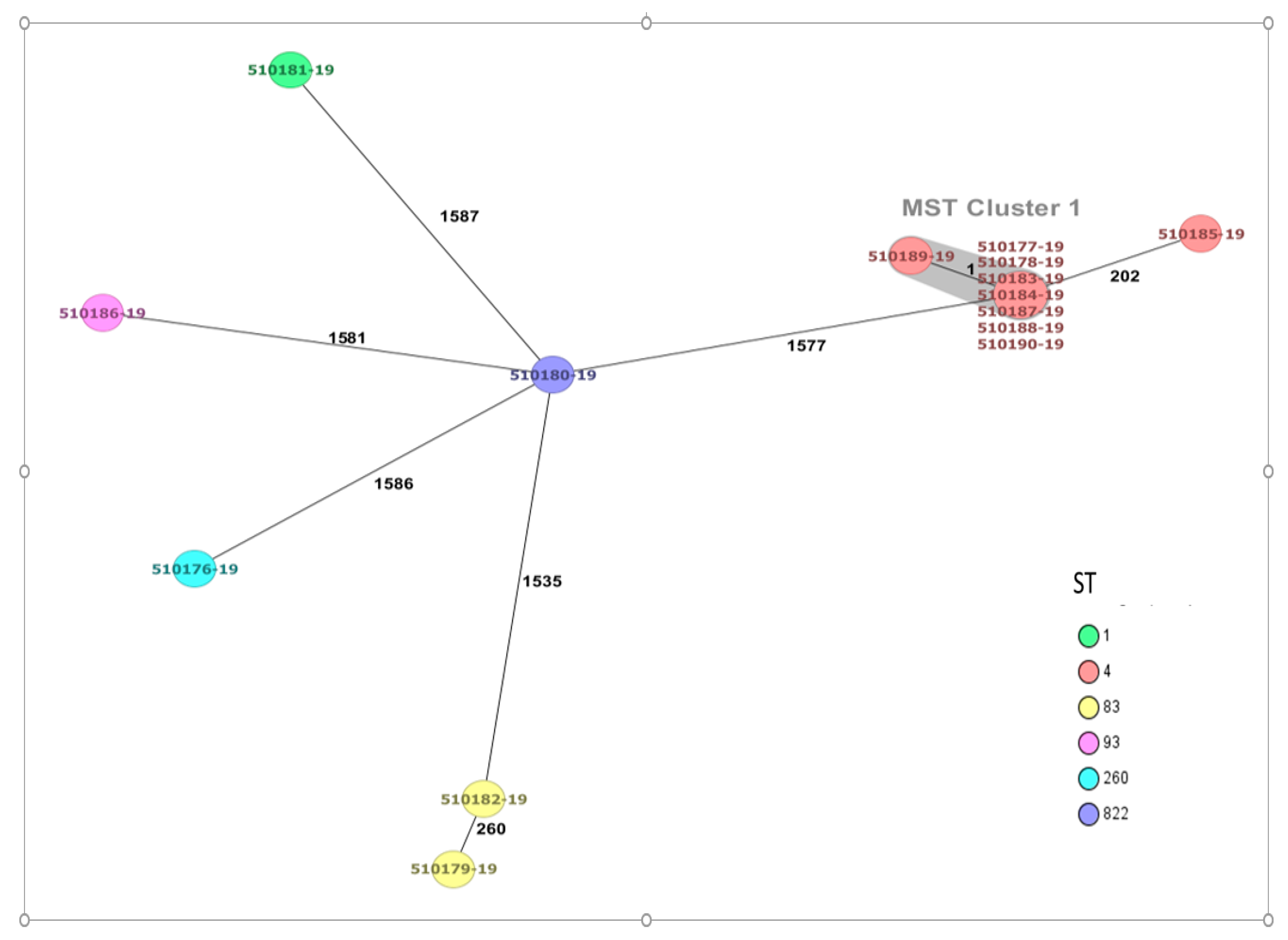
Figure 1. Minimum spanning tree (MST) of fourteen C. sakazakii strains. The isolates are represented as colored circles according to their sequence type (ST) as defined using the 7-loci MLST scheme (STs). Black numbers on the connection lines indicate the number of allelic differences between isolates from the cgMLST scheme comprising 3678 target genes for C. sakazakii. Isolates falling under the cluster threshold of 10 alleles are marked in grey as clusters.
Antibiotics 12 00851 g001
Figure 2. Antibiotic-resistant genes of fifteen Cronobacter spp. strains identified by Comprehensive Antibiotic Resistance Database (CARD). C. dublinensis genome is shown in red color. The visualization was performed with the iTol program.
Antibiotics 12 00851 g002
Figure 3. Virulence genes identified in C. sakazakii and C. dublinensis genomes (red color font). The visualization was performed with the iTol program.
Antibiotics 12 00851 g003
Table 1. Numbers and types of examined samples in the study.
Group of SamplesSpecific CommodityNumber of Examined Samples
Swabs from the food production environmentSwabs855
PoultryFeaces148
FoodChockolate72
Caramel55
Spice (dill, pepper)260
Poultry meat77
Dried cow milk244
Wheat flour91
Seeds (soy, pistachio, barley, mustard)198
Total 2000
Table 2. Identification of Cronobacter spp. strains isolated from different sources by MALDI-TOF MS and whole-genome sequencing rMLST.
Sample ID* PubMLST IDSourceWGS rMLST ResultSTCCSerotype
(gnd-galF Alleles)
Collection Date
510177-193823FoodCronobacter sakazakii44Csak O:22015
510178-193824FoodCronobacter sakazakii44Csak O:22015
510183-193828Production environmentCronobacter sakazakii44Csak O:22015
510184-193829Production environmentCronobacter sakazakii44Csak O:22015
510185-193830FoodCronobacter sakazakii44Csak O:22015
510187-193831FoodCronobacter sakazakii44Csak O:22015
510188-193832Production environmentCronobacter sakazakii44Csak O:22015
510189-193833Production environmentCronobacter sakazakii44Csak O:22015
510190-193834Production environmentCronobacter sakazakii44Csak O:22015
510181-193826FoodCronobacter sakazakii11Csak O:12015
510179-193825Production environmentCronobacter sakazakii8383Csak O:72015
510182-193827FoodCronobacter sakazakii8383Csak O:72015
510176-193695FoodCronobacter sakazakii260-Csak O:12015
510186-193697HenCronobacter sakazakii93-Csak O:72015
510180-193696FoodCronobacter dublinensis822-ND2015
* MLST database ID; WGS rMLST: Whole-Genome Sequencing ribosomal MLST; ST: sequence type; CC: clonal complex; -: no associated clonal complex; ND: not determined.
Table 3. Antibiotic resistance profile of Cronobacter spp. strains.
STStrains aAM
(10 µg)
AMC
(20/10 µg)
CAZ
(30 µg)
CIP
(5 µg)
CL
(30 µg)
CTX
(30 µg)
GE
(10 µg)
KF
(30 µg)
TE
(30 µg)
W
(30 µg)
1510181-19SSSSSSSRSS
4510177-19RSSSSSSRRS
4510178-19SSSSSSSRSS
4510183-19SRSSSSSRSS
4510184-19SSSSSSSRRS
4510185-19SSSSSSSRSS
4510187-19SSSSSSSRSS
4510188-19RSSSSSSRSS
4510189-19SSSSSSSRSS
4510190-19SSSSSSSRSS
83510179-19RSSSSSSRSS
83510182-19RSRSSSSRSS
260510176-19RSSSSSSRRS
822510180-19 *SSSSSSSSSS
a All strains were C. sakazakii except for 510180-19; * which was C. dublinensis. AM: ampicillin; AMC: amoxicillin-clavulanic acid; CAZ: Ceftazidime; CIP: ciprofloxacin; CL: chloramphenicol; CTX: cefotaxime; GE: gentamicin; KF: cephalothin; TE: tetracycline; W: nalidixic acid; R: Resistance; S: Susceptibility.
Table 4. Plasmids and mobile genetic elements of Cronobacter spp. strains.
ID Strain aSTPlasmidPlasmid Accession NumberMobile Genetic Elements
510177-194IncFIB(pCTU3)FN543096ISEhe3, ISEsa1, ISEc52
510178-194IncFIB(pCTU3)FN543096ISEhe3, ISEsa1, ISEc52
510183-194IncFIB(pCTU3)FN543096ISEc52
510184-194IncFIB(pCTU3)FN543096ISEhe3, ISEsa1
510185-194----------ISEhe3
510187-194IncFIB(pCTU3)FN543096ISEhe3, ISEsa1, IS26, ISEc52
510188-194IncFIB(pCTU3)FN543096ISEsa1,IS26, ISEc52
510189-194IncFIB(pCTU3)FN543096ISEhe3, ISEsa1
510190-194IncFIB(pCTU3)FN543096ISEhe3, ISEsa1, ISEc52
510179-1983rep7aSAU83488-----
510180-19822IncFII(pCTU2)
pESA2
FN543095
CP000784
ISEch12
510186-1993Col440lCp023920.1ISEsa1, ISKpn34
510176-19260IncFIB(pCTU3)FN543096ISEsa1, IS26, cn_6897_IS26
a All strains were C. sakazakii except for 510180-19 which was C. dublinensis.
Disclaimer/Publisher’s Note: The statements, opinions and data contained in all publications are solely those of the individual author(s) and contributor(s) and not of MDPI and/or the editor(s). MDPI and/or the editor(s) disclaim responsibility for any injury to people or property resulting from any ideas, methods, instructions or products referred to in the content.