Article

National Surveillance of Tetracycline, Erythromycin, and Clindamycin Resistance in Invasive Streptococcus pyogenes: A Retrospective Study of the Situation in Spain, 2007–2020

Laboratorio de Referencia e Investigación en Taxonomía, Centro Nacional de Microbiología, Instituto de Salud Carlos III, Majadahonda, 28220 Madrid, Spain
*
Author to whom correspondence should be addressed.
Antibiotics 2023, 12(1), 99; https://doi.org/10.3390/antibiotics12010099
Submission received: 16 December 2022 / Revised: 3 January 2023 / Accepted: 4 January 2023 / Published: 6 January 2023
(This article belongs to the Section Mechanism and Evolution of Antibiotic Resistance)

Abstract

:
Background: This work reports on antimicrobial resistance data for invasive Streptococcus pyogenes in Spain, collected by the ‘Surveillance Program for Invasive Group A Streptococcus’, in 2007–2020. Methods: emm typing was determined by sequencing. Susceptibility to penicillin, tetracycline, erythromycin, and clindamycin was determined via the E-test. tetM, tetO, msrD, mefA, ermB, ermTR, and ermT were sought by PCR. Macrolide-resistant phenotypes (M, cMLSB, and iMLSB) were detected using the erythromycin–clindamycin double-disk test. Resistant clones were identified via their emm type, multilocus sequence type (ST), resistance genotype, and macrolide resistance phenotype. Results: Penicillin susceptibility was universal. Tetracycline resistance was recorded for 237/1983 isolates (12.0%) (152 carried only tetM, 48 carried only tetO, and 33 carried both). Erythromycin resistance was detected in 172/1983 isolates (8.7%); ermB was present in 83, mefA in 58, msrD in 51, ermTR in 46, and ermT in 36. Clindamycin resistance (methylase-mediated) was present in 78/1983 isolates (3.9%). Eight main resistant clones were identified: two that were tetracycline-resistant only (emm22/ST46/tetM and emm77/ST63/tetO), three that were erythromycin-resistant only (emm4/ST39/mefA-msrD/M, emm12/ST36/mefA-msrD/M, and emm28/ST52/ermB/cMLSB), and three that were tetracycline–erythromycin co-resistant (emm11/ST403/tetM-ermB/cMLSB, emm77/ST63/tetO-ermTR/iMLSB, and emm77/ST63/tetM-tetO-ermTR/iMLSB). Conclusions: Tetracycline, erythromycin, and clindamycin resistance rates declined between 2007 and 2020. Temporal variations in the proportion of resistant clones determined the change in resistance rates.

1. Introduction

Streptococcus pyogenes, also called group A Streptococcus, causes illnesses of different severities. Invasive infections are less common than superficial infections but are associated with higher mortality [1,2]. A large number of virulence factors contribute to the pathogenicity of S. pyogenes, including the presence of the surface M protein and a set of exotoxins, the members of which act as superantigens [2,3]. The microbiological typing of strains is based on the emm gene (which codes for the M protein), which determines the emm type (formerly called the serotype) [3]. Superantigens can trigger an uncontrolled immune response known as a cytokine storm, which plays an essential role in the onset of STSS and necrotizing fasciitis, some of the most severe infections [2,3].
Penicillins/beta-lactams are the first-line treatments for S. pyogenes invasive infections; clindamycin in combination with penicillin is recommended for the most severe infections due to clindamycin’s antitoxic activity. Macrolides, vancomycin, and linezolid can replace beta-lactams in intolerant patients. Macrolides are also commonly used in superficial infections, such as pharyngitis [4,5]. Although tetracyclines are not used to treat S. pyogenes infections, resistance knowledge is epidemiologically important. Macrolide and tetracycline resistance genes are basically acquired by horizontal transfer [6,7,8]. Temporal variations in antimicrobial resistance genes (ARGs) are related to temporal variations in emm types carrying these genes [9,10,11,12,13]. The study of acquired ARGs is, therefore, essential in defining the circulating clones.
S. pyogenes remains susceptible to penicillin [2]. The macrolide resistance mechanisms most commonly encountered involve the MefA–MsrD active efflux pump and Erm methylases [2,14,15]. mefA and msrD are part of the same operon and are involved in the expulsion of 14- and 15-membered ring macrolides; they are responsible for the macrolide-resistant M phenotype [15]. The erm genes code for proteins that methylate 23S rRNA, modifying the binding site of macrolides, lincosamides, and streptogramin B (MLSB). This results in cross-resistance among these antibiotics and the expression of the macrolide-resistant MLSB phenotype, which can be constitutively expressed (cMLSB) or inducibly expressed in the presence of erythromycin (iMLSB) [14,15]. The methylase genes possessed by S. pyogenes include the chromosomal ermB [14,15] and ermTR [15,16], in addition to the plasmidic ermT [15,17]. Tetracycline resistance is effectively due to ribosomal protection proteins encoded by tetM and tetO [2,15]. Tetracycline and macrolide co-resistance is usual in S. pyogenes, a consequence of the presence of genetic elements—i.e., phages, transposons, plasmids, conjugative integrative elements, etc.—that carry resistance determinants for both groups of antibiotics [6,7,8,9].
The surveillance of S. pyogenes is important; only then can the characteristics of the circulating strains be known, changes in trends detected, and early control as well as preventive measures put in place. The ‘Surveillance Program for Invasive Group A Streptococcus’ (SPIGAS) has been implemented by the National Centre for Microbiology (NCM) [18] since 1994, and its main objective is to obtain microbiological information on invasive strains circulating in Spain [19] through phenotypic, genotypic, and/or genomic research. All Spanish hospitals and public health laboratories can voluntarily take part in this free of charge program.
In Spain, in the period of 2007–2019, the most prevalent invasive emm types—emm1, emm89, and emm3—were basically susceptible to antimicrobials [19]. Here, we analyze the less frequently resistant emm types, the ARGs, resistance rates, and trends; we also compare our results with those of previous studies.
Using the genotype and phenotype information for the resistant emm types detected by SPIGAS, the aim of the present work is to provide better knowledge on the tetracycline, erythromycin, and clindamycin resistance shown by invasive S. pyogenes in Spain in the period of 2007–2020.

2. Results

2.1. Clinical and Epidemiological Data for Resistant S. pyogenes Isolates

In the period 2007–2020, (n = 1983) S. pyogenes isolates that caused invasive illness were analyzed by SPIGAS. A total of 315 (15.9%) were resistant to tetracycline, erythromycin, and/or clindamycin. These resistant isolates were detected in 13/17 of Spain’s autonomous regions (involving 29/50 of the country’s provinces). The affected age range was 0–99 years, with a median of 47 years; patients ≥ 75 years of age (n = 61, 19.4%) made up the most affected age group. A total of 168 (53.3%) patients were male, and 139 were (44.1%) female; for 8 patients (2.5%) no gender information was available. The most commonly examined samples were blood (136 samples, 43.2%) and wound exudate (106, 33.7%). Clinical manifestations included sepsis (84, 26.7%), cellulitis (70, 22.2%), wound infections (33, 10.5%), ulcers (21, 6.7%), arthritis (20, 6.3%), scarlet fever (18, 5.7%), pneumonia (16, 5.1%), necrotizing fasciitis (11, 3.5%), abscesses (11, 3.5%), and others (31, 9.8%).

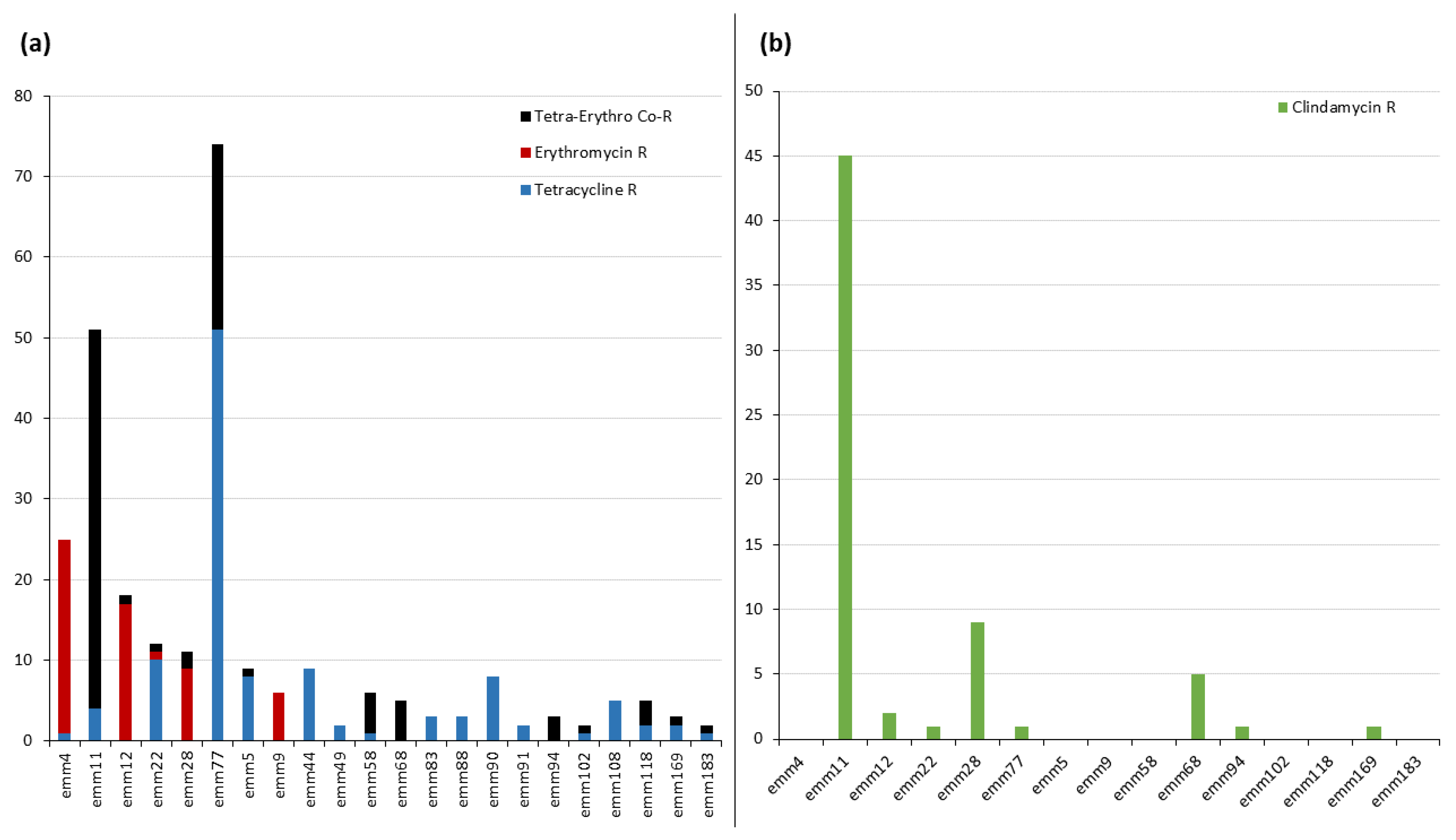
2.2. emm Types Resistant to Tetracycline, Erythromycin, and Clindamycin

All isolates were susceptible to penicillin; MIC50 and MIC90 values of 0.016 and 0.023 mg/L were recorded, respectively. Table 1 shows the data for susceptibility to tetracycline, erythromycin, and clindamycin for the six majority emm types (>10 resistant isolates) significantly associated (p ≤ 0.05) with some kind of resistance. Tetracycline resistance was observed in 237 isolates (12.0%). emm11, emm22, and emm77 were the majority emm types associated with resistance to this antibiotic; significantly associated minority emm types (≤ 10 resistant isolates) included emm5, emm44, emm49, emm58, emm68, emm83, emm88, emm90, emm91, emm94, emm102, emm108, emm118, emm169, and emm183. Erythromycin resistance was detected in 172 isolates (8.7%), mostly associated with emm4, emm11, emm12, and emm77 (majority types), but also with emm9, emm58, emm68, emm94, and emm118 (minority types). Clindamycin resistance was recorded for 78 isolates (3.9%); this was mostly associated with emm11 and emm28 (majority), but also with emm68 (minority). Finally, co-resistance to tetracycline and erythromycin (4.9% of isolates) was associated mostly with emm11 and emm77 (majority), but also with emm58 and emm68 (minority). Figure 1 shows the emm type distribution of isolates resistant to tetracycline, erythromycin, and clindamycin.

2.3. Tetracycline and Erythromycin Resistance: Genotype Analysis

Among the tetracycline-resistant population, the tetM gene was detected in 185 (78.1%) isolates, while tetO was detected in 81 (34.2%). Among the erythromycin-resistant population, the ermB gene was detected in 83 isolates (48.3%), mefA in 58 (33.7%), msrD in 51 (29.7%), ermTR in 46 (26.7%), and ermT in 36 (20.9%). An association was seen between the mefA and msrD genes (p < 0.00001; relative risk, RR = 17.3), between ermB and tetM (p = 0.0002; RR = 2.2), and between ermTR and tetO (p = 0.0002; RR = 2.9). More than 20 different resistant genotypes were detected among the total 315 examined isolates. Table 1 shows the emm types and the most common genotypes associated with tetracycline, erythromycin, and clindamycin resistance. A total of 126 erythromycin-resistant isolates (73.2%) carried the genes for just one macrolide resistance mechanism (Table 1), while the rest (26.8%) carried the genes for two or more. Among the latter, ermT was the most common gene detected (possessed by 76% of isolates).

2.4. Macrolide and Lincosamide Resistance: Phenotype Analysis

The cMLSB phenotype was detected in 76 (44.1%) isolates resistant to erythromycin, of which 96.1% carried the ermB gene. The M phenotype was identified in 49 (28.5%) isolates; 95.9% of these carried mefA–msrD. The iMLSB phenotype was present in 46 (26.7%) isolates, most of which (82.6%) possessed ermTR. No erythromycin-susceptible but clindamycin-resistant isolates were detected. Figure 2 shows the distribution of macrolide-resistant phenotypes over the study period.

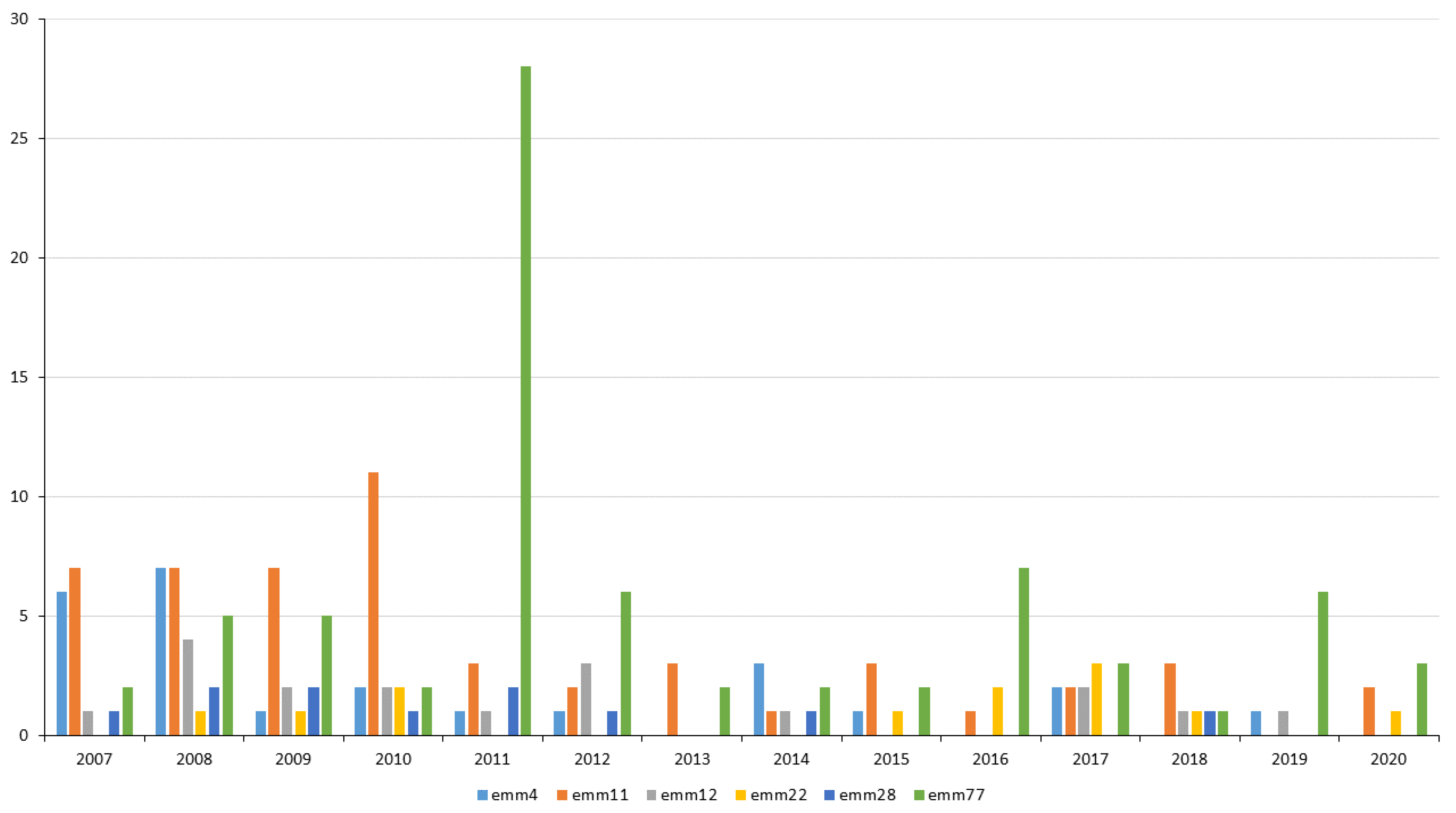
2.5. Changes in Resistance in 2007–2020

Figure 3 shows the changes in antimicrobial resistance from 2007–2020 by year and by 4–5-year periods. In 2007–2010, the resistance rates to tetracycline, erythromycin, and clindamycin were 15.9%, 14.4%, and 7.1%, respectively. In the same order, these rates were 11.9%, 6.3%, and 3.3% in 2011–2015, and 9.4%, 6.9%, and 2.4% in 2016–2020. Figure 4 shows the distribution of resistant emm types over time. emm4 was more prevalent in 2007–2008, emm11 in 2007–2010, emm12 in 2008, and emm28 in 2007–2012. emm22 was the least prevalent majority emm type; indeed, it was not detected in 2007, 2011–2014, and 2019. emm77 was persistently detected, although it showed annual fluctuations in prevalence, with a remarkably large number of isolates detected in 2011.

2.6. Resistant Clones

Table 2 shows the characteristics of the eight most resistant clones. The tetracycline-resistant-only clones were emm22/ST46/tetM and emm77/ST63/tetO. Erythromycin-only resistance was recorded for the emm4/ST39/mefA-msrD/M, emm12/ST36/mefA-msrD/M, and emm28/ST52/ermB/cMLSB clones. The main tetracycline–erythromycin co-resistant clones were emm11/ST403/tetM-ermB/cMLSB, emm77/ST63/tetO-ermTR/iMLSB, and emm77/ST63/tetM-tetO-ermTR/iMLSB.

3. Discussion

Antimicrobial resistance is a major public health concern and, therefore, is a focus of SPIGAS [18]. Actions must be taken to deal with this problem, such as international and national policies for the better management of antimicrobials, which are being implemented in many countries [21,22]. However, deep knowledge about resistance, its causes, and trends is also necessary. This work tackles these aspects.
The only noteworthy epidemiological difference between the resistant S. pyogenes isolates examined in the present work and the susceptible/resistant isolates examined in a previous study [19] was patient age. Infections caused by the present resistant isolates were more common in people ≥ 75 years of age, whereas those caused by the susceptible/resistant isolates of the latter work were most common in 0–4 year olds. This difference may be explained by the fact that the two most representative resistant emm types detected in the present work were emm11 and emm77 (Table 1 and Figure 1), both of which were associated (p ≤ 0.05) with infections in elderly people in the 2021 study [19].
Penicillin susceptibility was universal. However, complacency is not recommendable since a few cases of diminished penicillin susceptibility have been described by other authors [23].
Macrolides are the treatment of choice for severe infections in beta-lactam-intolerant patients. Previous studies have shown that erythromycin resistance in S. pyogenes has declined in Spain since the 1990s, and that the M phenotype has gradually been replaced by the MLSB phenotype [10,11,12,13,24]. The replacement of phenotypes is mainly explained by changes in the prevalence of emm4, emm11, and emm77—which are associated with the M, cMLSB, and iMLSB phenotypes, respectively (Table 2) [10,11,12,13,24]. The present work searched for the erythromycin resistance genes that have been described for S. pyogenes [2,14,15]. Among these, the most prevalent was ermB, followed by mefA (linked to msrD in 51/58 isolates), ermTR, and finally ermT. The phenotyping and genotyping results agreed (see Table 1) [14,25]. The ermB genotype was associated with the strongest macrolide resistance (MICs > 256 mg/L) and nearly always with the cMLSB phenotype, which shows constitutive resistance to erythromycin and clindamycin. The mefA–msrD genotype showed the least resistance (MICs 16–32 mg/L) and was associated with the M phenotype (i.e., erythromycin-resistant and clindamycin-susceptible), as was also the case for the seven isolates that only carried mefA. The ermTR genotype basically corresponded to the iMLSB phenotype, which exhibits strong resistance to erythromycin (although less strong than the ermB genotype), as well as inducible resistance to clindamycin. In agreement with that which has been previously reported [17], the ermT genotype was only detected in one isolate (which expressed the iMLSB phenotype [Table 1]). However, the ermT gene was mainly detected in combination with other macrolide resistance genes, especially with the mefA–msrD–ermT, ermB–ermT, and ermTR–ermT genotypes, in which its presence did not alter the expression of the M, cMLSB, and iMLSB phenotypes, respectively. How erythromycin resistance is influenced by the presence of ermT cannot be concluded from the results of this study, although ermT was much more prevalent than expected according to previous work [17]; it is recommended that its presence always be investigated.
Clindamycin in combination with penicillin is of prime importance in the treatment of the most severe S. pyogenes infections. Methylase-mediated clindamycin resistance was associated only with the MLSB phenotype [14,15]. Other less common resistance mechanisms involving, e.g., lincosamide nucleotidyltransferases [14,15] were not sought out given the absence of phenotypically compatible isolates.
Tetracycline resistance is of great concern since the tet genes are carried on mobile genetic elements (MGEs) that promote their horizontal transmission [6,7,8]. The present results indicate a declining trend in tetracycline resistance over the years, but other recent studies have reported tetracycline resistance rates ranging from 6.8% [12] to 60.6% [9]. Tetracycline resistance (12% of total isolates) was more extended than erythromycin resistance (8.7%) among the present isolates. In total, eighteen emm types were associated with tetracycline resistance (p ≤ 0.05), and nine were with erythromycin resistance [13]. As has been commonly described [12,13], tetM was more prevalent than tetO (185 vs. 81 isolates). No significant differences were seen among the MICs of the isolates with the tetM, tetO, or tetM-tetO genotypes (Table 1), suggesting that tetM and tetO provide similar resistance but have no additive or synergistic effects. Tetracycline and erythromycin co-resistance can be explained by the presence of MGEs that carry the tetM–ermB and tetO–ermTR genetic associations [6,7,8,9,14] (represented by emm11 and emm77, respectively).
The main resistant clones detected in this work (Table 2) have been described as being globally distributed [26]. Their temporal fluctuation in terms of prevalence conditioned the resistance rates recorded over the 14-year study period [11,13,24] (Figure 3 and Figure 4). Over this period, the highest tetracycline, erythromycin, and clindamycin resistance rates were, for the most part, owed to the presence of the emm11/ST403/tetM-ermB/MLSBc clone [10], the most representative co-resistant clone in numerical terms. The strong presence of the emm4/ST39/mefA-msrD/M, emm12/ST36/mefA-msrD/M, and emm28/ST52/ermB/MLSBc clones explains the high rates of erythromycin resistance seen during the first 4-year period. emm4 and emm12 conditioned the predominance of the M phenotype in 2007–2008, while emm11 and emm28 conditioned that of cMLSB in 2009–2010 (Figure 2, Figure 3 and Figure 4). In general, resistance rates declined in 2011–2015 and 2016–2020, although some exceptions were registered. In 2011, the highest tetracycline resistance rate was detected (21.8%, Figure 3) owing to the over-representation of the emm77/ST63/tetO clone (Figure 4), which was involved in two geographically distant surgical outbreaks. This majority clone (55% of emm77 isolates, Table 2), which was only resistant to tetracycline, was detected in 2007–2013. However, since 2013, emm77/ST63/tetO has been replaced by the emm77/ST63/tetO-ermTR/iMLSB and emm77/ST63/tetM-tetO-ermTR/iMLSB co-resistant clones, which have acquired the ermTR gene. This explains the slight increase in erythromycin resistance (iMLSB phenotype) detected during 2016–2020. Finally, the small, constant number of isolates of the emm22/ST46/tetM clone seemed to have little effect on tetracycline resistance in the period of 2007–2020.
The present study suffers from the limitation that MLST was only performed on a limited number of isolates; it is likely that the typing of all resistant isolates would have revealed wider genetic diversity. Furthermore, while this work provides a general view of antimicrobial resistance and its associated genes in Spain, the results shed no light on the MGEs involved in the transmission of these genes, which remain unknown [6,7,8,9]. Whole-genome sequencing is needed if we are to better understand the genetic environment of the resistance genes. This is the first nationwide study on antimicrobial resistance in invasive S. pyogenes in Spain, and comparing it with similar studies has been difficult because some variables differed in important ways, i.e., geographical origin, invasive vs. non-invasive isolates, and study period. Additionally, the lack of knowledge on the treatment that patients received does not permit us to know its influence on susceptibility patterns.

4. Materials and Methods

4.1. Basic Microbiological Typing

Three hundred and fifteen resistant bacterial isolates (one isolate per patient and clinical episode) were analyzed by SPIGAS between 2007 and 2020 [18]. The M protein gene (emm type) was acquired by sequencing the 180 hypervariable nucleotides of the gene’s 5′ end by using the Center for Disease Control and Prevention’s protocol and database [27]. The exotoxin genes, speA, speC, speG, speH, speJ, smeZ, and ssa, were detected by PCR [19,28]. A basic antibiogram was selected according to clinical and epidemiological criteria. Penicillin G, tetracycline, erythromycin, and clindamycin susceptibilities were tested using E-test strips (bioMérieux, Marcy-l’Etoile, France) following the recommendations and interpretative criteria of the European Committee on Antimicrobial Susceptibility Testing (EUCAST) [20]. Penicillin G, erythromycin, and clindamycin were mainly selected according to clinical—i.e., treatment—criteria, and tetracycline was selected according to epidemiological criteria.

4.2. Tetracycline, Erythromycin, and Clindamycin Resistance: Phenotype and Genotype Analyses

Isolates resistant to tetracycline (MIC > 1 mg/L) were checked by PCR for the presence of tetM [29] and tetO [30]. Isolates resistant to erythromycin (MIC > 0.5 mg/L) were analyzed to determine their phenotypes with respect to resistance to macrolides, lincosamides, and B streptogramins (M, cMLSB, and iMLSB phenotypes) by using the erythromycin–clindamycin double-disk test [25]. They were also examined by PCR for the presence of msrD [31], mefA [30], ermB [32], ermTR [16], and ermT [32].

4.3. Multilocus Sequence Typing and Description of Resistant Clones

Multilocus sequence typing (MLST) [33] included the amplification and partial sequencing of the seven housekeeping genes gki, gtr, murI, mutS, recP, xpt, and yqiL [34]; each allelic combination corresponded to a specific sequence type (ST). An MLST analysis was performed on a number of isolates of the majority resistant emm types, i.e., nine emm4-, seven emm11-, five emm12-, four emm22-, four emm28-, and seven emm77-type isolates. The resistant clones were identified by the combination of their emm type, ST, tetracycline- as well as macrolide-resistant genotype, and macrolide-resistant phenotype. It was assumed that isolates that shared their emm type, genotype, and phenotype would have identical STs.

4.4. Statistical Analysis

Categorical variables (emm type, age, gender, clinical manifestation, clinical sample, MICs, ARGs, and macrolide-resistance phenotype) were compared using the Fischer exact test. Age was grouped into six ranges: 0–4, 5–14, 15–39, 40–64, 65–74, and ≥ 75 years old. Significance was set up at p ≤ 0.05. All calculations were carried out by using Stata v.17 software.

4.5. Ethical Approval

Bacterial strains were collected as part of standard patient care and were sent to a public national reference laboratory (Centro Nacional de Microbiología, Majadahonda, Spain) for microbiological typing. This study focused on bacteria, and no identifiable human data were used; ethical approval was, therefore, not required.

5. Conclusions

Over the study period, erythromycin resistance in invasive S. pyogenes in Spain was found to be clustered in the clones with the emm4, emm11, emm12, and emm77 genotypes; the genes most commonly involved (in descending order) were ermB, mefA, and ermTR. Macrolide-resistant cMLSB was the most frequently detected phenotype. Clindamycin resistance was always mediated by methylases. emm11, emm22, and emm77 were associated with tetracycline resistance, with the tetM gene more extended than tetO. Tetracycline–erythromycin resistance was common and grouped in the clones with the emm11 and emm77 genotypes. Tetracycline, erythromycin, and clindamycin resistance rates declined from 2007 to 2020, with temporal variations in resistant clones corresponding to changes in resistance rates.

Author Contributions

Conceptualization, P.V. and S.V.; methodology, P.V., M.B., M.J.M.-P., N.G., S.P.-R. and G.C.; validation, P.V. and M.B.; formal analysis, P.V., M.B., M.J.M.-P., N.G., S.P.-R. and G.C.; investigation, P.V., M.J.M.-P. and S.V.; resources, P.V., M.B., M.J.M.-P., N.G., S.P.-R. and S.V.; data curation, P.V.; writing—original draft preparation, P.V.; writing—review and editing, M.B., M.J.M.-P., N.G., S.P.-R., G.C. and S.V.; supervision, P.V. and S.V.; project administration, P.V. and S.V.; funding acquisition, P.V. and S.V. All authors have read and agreed to the published version of the manuscript.

Funding

This research was partially funded by the Instituto de Salud Carlos III, grant number MPY377/18.

Institutional Review Board Statement

Not applicable.

Informed Consent Statement

Not applicable.

Data Availability Statement

Not applicable.

Acknowledgments

We thank clinicians and microbiologists from across Spain for their interest and participation in SPIGAS; this work would not be possible without their collaboration. We also thank Adrian Burton for editing and language assistance (Physical Evidence Scientific Translations, http://physicalevidence.es, accessed on 15 December 2022).

Conflicts of Interest

The authors declare no conflict of interest. The funders had no role in the design of the study; in the collection, analyses, or interpretation of the data; in the writing of the manuscript; or in the decision to publish the results.

References

  1. Carapetis, J.R.; Steer, A.C.; Mulholland, E.K.; Weber, M. The global burden of group A streptococcal diseases. Lancet Infect. Dis. 2005, 5, 685–694. [Google Scholar] [CrossRef]
  2. Walker, M.J.; Barnett, T.C.; McArthur, J.D.; Cole, J.N.; Gillen, C.M.; Henningham, A.; Sriprakash, K.S.; Sanderson-Smith, M.L.; Nizet, V. Disease manifestations and pathogenic mechanisms of group A Streptococcus. Clin. Microbiol. Rev. 2014, 27, 264–301. [Google Scholar] [CrossRef] [Green Version]
  3. Bisno, A.L.; Brito, M.O.; Collins, C.M. Molecular basis of group A streptococcal virulence. Lancet Infect. Dis. 2003, 3, 191–200. [Google Scholar] [CrossRef]
  4. Center for Disease Control and Prevention (CDC). Group A Streptococcal (GAS) Disease. Available online: https://www.cdc.gov/groupastrep/diseases-hcp/index.html (accessed on 28 December 2022).
  5. Johnson, A.F.; LaRock, C.N. Antibiotic treatment, mechanisms for failure, and adjunctive therapies for infections by group A Streptococcus. Front. Microbiol. 2021, 12, 760255. [Google Scholar] [CrossRef]
  6. Varaldo, P.E.; Montanari, M.P.; Giovanetti, E. Genetic elements responsible for erythromycin resistance in streptococci. Antimicrob. Agents Chemother. 2009, 53, 343–353. [Google Scholar] [CrossRef] [Green Version]
  7. Brenciani, A.; Bacciaglia, A.; Vecchi, M.; Vitali, L.A.; Varaldo, P.E.; Giovanetti, E. Genetic elements carrying erm(B) in Streptococcus pyogenes and association with tet(M) tetracycline resistance gene. Antimicrob. Agent. Chemother. 2007, 51, 1209–1216. [Google Scholar] [CrossRef] [Green Version]
  8. Brenciani, A.; Tiberi, E.; Bacciaglia, A.; Petrelli, D.; Varaldo, P.E.; Giovanetti, E. Two distinct genetic elements are responsible for erm(TR)-mediated erythromycin resistance in tetracycline-susceptible and tetracycline-resistant strains of Streptococcus pyogenes. Antimicrob. Agent. Chemother. 2011, 55, 2106–2112. [Google Scholar] [CrossRef] [Green Version]
  9. Berbel, D.; Càmara, J.; González-Díaz, A.; Cubero, M.; López de Egea, G.; Martí, S.; Tubau, F.; Domínguez, A.; Ardanuy, C. Deciphering mobile genetic elements disseminating macrolide resistance in Streptococcus pyogenes over a 21 year period in Barcelona, Spain. J. Antimicrob. Chemother. 2021, 76, 1991–2003. [Google Scholar] [CrossRef]
  10. Pérez-Trallero, E.; Montes, M.; Orden, B.; Tamayo, E.; García-Arenzana, J.M.; Marimón, J.M. Phenotypic and genotypic characterization of Streptococcus pyogenes isolates displaying the MLSB phenotype of macrolide resistance in Spain, 1999 to 2005. Antimicrob. Agent. Chemother. 2007, 51, 1228–1233. [Google Scholar] [CrossRef] [Green Version]
  11. Ardanuy, C.; Domenech, A.; Rolo, D.; Calatayud, L.; Tubau, F.; Ayats, J.; Martín, R.; Liñares, J. Molecular characterization of macrolide- and multidrug-resistant Streptococcus pyogenes isolated from adult patients in Barcelona, Spain (1993–2008). J. Antimicrob. Chemother. 2010, 65, 634–643. [Google Scholar] [CrossRef]
  12. Rubio-López, V.; Valdezate, S.; Álvarez, D.; Villalón, P.; Medina, M.J.; Salcedo, C.; Sáez-Nieto, J.-A. Molecular epidemiology, antimicrobial susceptibilities and resistance mechanisms of Streptococcus pyogenes isolates resistant to erythromycin and tetracycline in Spain (1994-2006). BMC Microbiol. 2012, 12, 215. [Google Scholar] [CrossRef] [Green Version]
  13. Montes, M.; Ardanuy, C.; Tamayo, E.; Domènech, A.; Liñares, J.; Pérez-Trallero, E. Epidemiological and molecular analysis of Streptococcus pyogenes isolates causing invasive disease in Spain (1998-2009): Comparison with non invasive isolates. Eur. J. Clin. Microbiol. Infect. Dis. 2011, 30, 1295–1302. [Google Scholar] [CrossRef]
  14. Leclerq, R. Mechanisms of resistance to macrolides and lincosamides: Nature of the resistance elements and their clinical implications. Clin. Infect. Dis. 2002, 34, 482–492. [Google Scholar] [CrossRef] [Green Version]
  15. Roberts, M.C. Tetracycline and MLS Nomenclature. 2022. Available online: http://faculty.washington.edu/marilynr/ (accessed on 8 November 2022).
  16. Seppälä, H.; Skurnik, M.; Soini, H.; 5, M.C.; Huovinen, P. A novel erythromycin resistance methylase gene (ermTR) in Streptococcus pyogenes. Antimicrob. Agents Chemother. 1998, 42, 257–262. [Google Scholar] [CrossRef] [Green Version]
  17. Woodbury, R.L.; Klammer, K.A.; Xiong, Y.; Bailiff, T.; Glennen, A.; Bartkus, J.M.; Lynfield, R.; Van Beneden, C.; Beall, B.W. Plasmid-borne erm(T) from invasive, macrolide-resistant Streptococcus pyogenes strains. Antimicrob. Agents Chemother. 2008, 52, 1140–1143. [Google Scholar] [CrossRef] [Green Version]
  18. Instituto de Salud Carlos III. Centro Nacional de Microbiología. Área de Orientación Diagnóstica. Programas de Vigilancia de Bacterias. Vigilancia de la Infección Estreptocócica. Available online: https://www.12.es/QueHacemos/Servicios/DiagnosticoMicrobiol%C3%B3gicoyProgramasVigilancia/Paginas/default.aspx (accessed on 15 December 2022).
  19. Villalón, P.; Sáez-Nieto, J.A.; Rubio-López, V.; Medina-Pascual, M.J.; Garrido, N.; Carrasco, G.; Pino-Rosa, S.; Valdezate, S. Invasive Streptococcus pyogenes disease in Spain: A microbiological and epidemiological study covering the period 2007–2019. Eur. J. Clin. Microbiol. Infect. Dis. 2021, 40, 2295–2303. [Google Scholar] [CrossRef]
  20. The European Committee of Antimicrobial Susceptibility Testing. Clinical Breakpoints–Bacteria (V12.0). 2022. Available online: https://www.eucast.org/fileadmin/src/media/PDFs/EUCAST_files/Breakpoint_tables/v_12.0_Breakpoint_Tables.pdf (accessed on 8 November 2022).
  21. European Centre for Disease Prevention and Control (ECDC). Available online: https://www.ecdc.europa.eu/en/publications-data/directory-online-resources-prevention-and-control-antimicrobial-resistance-amr (accessed on 28 December 2022).
  22. Plan Nacional de Resistencia a Antibióticos (PRAN). Available online: https://www.resistenciaantibioticos.es/es (accessed on 28 December 2022).
  23. Vannice, K.S.; Ricaldi, J.; Nanduri, S.; Fang, F.C.; Lynch, J.B.; Bryson-Cahn, C.; Wright, T.; Duchin, J.; Kay, M.; Chochua, S.; et al. Streptococcus pyogenes pbp2x mutation confers reduced susceptibilit to β–lactam antibiotics. Clin. Infect. Dis. 2019, 71, 201–204. [Google Scholar] [CrossRef] [Green Version]
  24. Montes, M.; Tamayo, E.; Mojica, C.; García-Arenzana, J.M.; Esnal, O.; Pérez-Trallero, E. What causes erythromycin resistance in Streptococcus pyogenes? Dynamics of four clones in a southern European región from 2005 to 2012. J. Antimicrob. Chemother. 2014, 69, 1474–1482. [Google Scholar] [CrossRef] [Green Version]
  25. Giovanetti, E.; Montanari, M.P.; Mingoia, M.; Varaldo, P.E. Phenotypes and genotypes of erythromycin-resistant Streptococcus pyogenes strains in Italy and heterogeneity of inducibly resistant strains. Antimicrob. Agents Chemother. 1999, 43, 1935–1940. [Google Scholar] [CrossRef] [Green Version]
  26. Willems, R.J.L.; Hanage, W.P.; Bessen, D.E.; Feil, E.J. Population biology of Gram-positive Pathogens: High-risk clones for dissemination of antibiotic resistance. FEMS Microbiol. Rev. 2011, 35, 872–900. [Google Scholar] [CrossRef]
  27. Center for Disease Control and Prevention. Streptococcus Laboratory. 2022. Available online: https://www.cdc.gov/streplab/groupa-strep/index.html (accessed on 8 November 2022).
  28. Schmitz, F.J.; Beyer, A.; Charpentier, E.; Henriques Normark, B.; Schade, M.; Fluit, A.C.; Hafner, D.; Novak, R. Toxin-gene profile heterogeneity among endemic invasive European group A streptococcal isolates. J. Infect. Dis. 2003, 188, 1578–1586. [Google Scholar] [CrossRef] [Green Version]
  29. Poyart, C.; Jardy, L.; Quesne, G.; Berche, P.; Trieu-Cuot, P. Genetic basis of antibiotic resistance in Streptococcus agalactiae strains isolated in a French hospital. Antimicrob. Agents Chemother. 2003, 47, 794–797. [Google Scholar] [CrossRef] [Green Version]
  30. Malhotra-Kumar, S.; Lammens, C.; Piessens, J.; Goossens, H. Multiplex PCR for simultaneous detection of macrolide and tetracycline resistance determinants in streptococci. Antimicrob. Agents Chemother. 2005, 49, 4798–4800. [Google Scholar] [CrossRef] [Green Version]
  31. Luthje, P.; Schwarz, S. Molecular basis of resistance to macrolides and lincosamides among staphylococci and streptococci from various animal sources collected in the resistance monitoring program BfT-GermVet. Int. J. Antimicrob. Agents 2007, 29, 528–535. [Google Scholar] [CrossRef]
  32. Sutcliffe, J.; Grebe, T.; Tait-Kamradt, A.; Wondrack, L. Detection of erythromycin-resistant determinants by PCR. Antimicrob. Agents Chemother. 1996, 40, 2562–2566. [Google Scholar] [CrossRef] [Green Version]
  33. Jolley, K.A.; Brain, J.E.; Maiden, M.C.J. Open-access bacterial population genomics: BIGSdb software, the PubMLST.org website. Wellcome Open Res. 2018, 3, 124. [Google Scholar] [CrossRef]
  34. Enright, M.C.; Spratt, B.G.; Kalia, A.; Cross, J.H.; Bessen, D. Multilocus sequence typing of Streptococcus pyogenes and the relationships betweem emm type and clone. Infect. Immun. 2001, 69, 2416–2427. [Google Scholar] [CrossRef]
Figure 1. Antimicrobial-resistant Streptococcus pyogenes emm types. (a) emm types resistant to tetracycline and erythromycin. (b) Clindamycin resistance in erythromycin-resistant emm types. Vertical axis = number of isolates. R, resistance.
Antibiotics 12 00099 g001
Figure 2. Streptococcus pyogenes macrolide-resistant phenotypes. Distribution by year of isolates expressing the M, cMLSB, and iMLSB phenotypes. Vertical axis = number of isolates.
Antibiotics 12 00099 g002
Figure 3. Change over time in antimicrobial resistance in Streptococcus pyogenes. Rates of tetracycline, erythromycin, and clindamycin resistance distributed (a) yearly and (b) by 4–5-year periods. R, resistance. Vertical axis = number of isolates. Total R represents the sum of the resistant isolates.
Antibiotics 12 00099 g003
Figure 4. Annual distribution of majority resistant Streptococcus pyogenes emm types. Data are for the emm4, emm11, emm12, emm22, emm28, and emm77 resistant isolates. Vertical axis = number of isolates.
Antibiotics 12 00099 g004
Table 1. emm types and genotypes of invasive Streptococcus pyogenes associated with tetracycline, erythromycin, and clindamycin resistance in Spain, 2007–2020.
Type aN bTetracycline cErythromycin cClindamycin c
n%MIC50MIC90n%MIC50MIC90n%MIC50MIC90
emm
Total198323712.00.25121728.70.1250.25783.90.1250.25
emm416110.60.250.52414.90.1251600.00.1250.25
emm11635181.016324774.6>256>2564571.4>256>256
emm1211510.860.250.51815.70.191621.70.1250.25
emm22361130.60.251625.60.1250.2512.80.1250.25
emm289722.10.250.51111.30.125>25699.20.1250.5
emm77817491.432322328.40.191611.20.1250.25
Genotype
tetM-152-1648--------
tetO-48-3248--------
tetM-tetO-33-2432--------
mefA-msrD-----40-1632000.1250.25
ermB-----61->256>25657->256>256
ermTR-----24-12>2561-0.1250.25
ermT-----1->256>256000.1250.125
MIC, minimum inhibitory concentration; S, susceptible; R, resistant; and -, not analyzed. a: Data are for the majority emm types significantly associated (p ≤ 0.05) with tetracycline, erythromycin, and/or clindamycin resistance, as well as for the most representative genotypes. b: Total number of isolates. c: The number (n) and percentage (%) of resistant isolates are indicated for each antibiotic. MICs are expressed in mg/L. Antimicrobial susceptibility is interpreted according to EUCAST criteria [20]. Tetracycline: ≤1, S; >2, R. Erythromycin: ≤0.25, S; >0.5, R. Clindamycin: ≤0.5, S; >0.5, R.
Table 2. Data of the main resistant clones of invasive Streptococcus pyogenes in Spain, 2007–2020.
emm TypeNo. Isolates aMLST bGenotype cPhenotype d
STntetMtetOmsrDmefAermBermTRermT
Only tetracycline resistance
emm2210/12462+-
emm7741/74633-+
Only erythromycin resistance
emm419/25395 ++---M
emm1212/18364 ++---M
emm286/11522 --+--cMLSB
Tetracycline–erythromycin co-resistance
emm1133/514035+---+--cMLSB
emm778/74632-+---+-iMLSB
emm778/74632++---+-iMLSB
a: Assumed number of clonal isolates with respect to the total number of resistant isolates. b: MLST, multilocus sequence typing; ST, sequence type; and n, number of isolates analyzed by MLST for each clone. c: +, present; -, absent. d: Macrolide-resistant phenotype. M, M phenotype; cMLSB, constitutive MLSB phenotype; and iMLSB, inducible MLSB phenotype.
Disclaimer/Publisher’s Note: The statements, opinions and data contained in all publications are solely those of the individual author(s) and contributor(s) and not of MDPI and/or the editor(s). MDPI and/or the editor(s) disclaim responsibility for any injury to people or property resulting from any ideas, methods, instructions or products referred to in the content.

