Review

Recent Developments in Methicillin-Resistant Staphylococcus aureus (MRSA) Treatment: A Review

by
Palanichamy Nandhini
1,
Pradeep Kumar
2,
Suresh Mickymaray
3,
Abdulaziz S. Alothaim
3,
Jayaprakash Somasundaram
4 and
Mariappan Rajan
1,*
1
Biomaterials in Medicinal Chemistry Laboratory, Department of Natural Products Chemistry, School of Chemistry, Madurai Kamaraj University, Madurai 625021, India
2
Department of Pharmacy and Pharmacology, School of Therapeutic Sciences, Faculty of Health Sciences, University of the Witwatersrand, Johannesburg 2193, South Africa
3
Department of Biology, College of Science, Al-Zulfi, Majmaah University, Majmaah 11952, Saudi Arabia
4
Department of Pharmaceutics, College of Pharmacy, University of Hafr Al Batin, Hafar Al Batin 39524, Saudi Arabia
*
Author to whom correspondence should be addressed.
Antibiotics 2022, 11(5), 606; https://doi.org/10.3390/antibiotics11050606
Submission received: 7 February 2022 / Revised: 28 February 2022 / Accepted: 23 March 2022 / Published: 29 April 2022
(This article belongs to the Special Issue Antimicrobial Resistance and Healthcare Associated Infections)

Abstract

:
Staphylococcus aureus (S. aureus) is a Gram-positive bacterium that may cause life-threatening diseases and some minor infections in living organisms. However, it shows notorious effects when it becomes resistant to antibiotics. Strain variants of bacteria, viruses, fungi, and parasites that have become resistant to existing multiple antimicrobials are termed as superbugs. Methicillin is a semisynthetic antibiotic drug that was used to inhibit staphylococci pathogens. The S. aureus resistant to methicillin is known as methicillin-resistant Staphylococcus aureus (MRSA), which became a superbug due to its defiant activity against the antibiotics and medications most commonly used to treat major and minor infections. Successful MRSA infection management involves rapid identification of the infected site, culture and susceptibility tests, evidence-based treatment, and appropriate preventive protocols. This review describes the clinical management of MRSA pathogenesis, recent developments in rapid diagnosis, and antimicrobial treatment choices for MRSA.

1. Introduction

1.1. Evolution of MRSA Treatment

Staphylococcus aureus (S. aureus) is a Gram-positive, clustered, spherical-shaped bacterium; it is primarily a human and animal pathogen. It belongs to the family of Staphylococcaceae and the genus known as Staphylococcus. A Scottish surgeon, Sir Alexander Ogston (1881), while performing a procedure, discovered that Staphylococcus could cause wound infections in living organisms [1]. He introduced the term Staphylococcus for the genus in 1882, and Rosenbach (1884) detached the genus into S. aureus and S. albus [2,3]. In 1939, Cowan distinguished S. epidermidis as a separate species based on coagulase testing [4]. Normally, S. aureus can also be found in healthy individuals [5]. It doesn’t cause any infection on healthy skin; however, upon entering the internal tissues or bloodstream, it may cause serious diseases [6]. S. aureus can cause minor skin infections such as impetigo, scalded skin, pimples, boils, abscesses, etc. It also causes life-threatening diseases such as meningitis, pneumonia, endocarditis, bacteremia, and sepsis [7,8,9,10].
According to studies, 20% of individuals are nasal carriers of S. aureus, and 30% are intermittent carriers [11]. As a result, S. aureus is one of the major reasons for the spread of hospital and community-acquired infections, which brings about serious consequences and life-threatening diseases. The discovery of antibiotics helped to treat the infectious diseases caused by S. aureus. The antibiotic penicillin was discovered in 1928 by Sir Alexander Fleming. The purified form of penicillin came in 1941 and saved the lives of many war victims during World War II. Within 2 years of introducing penicillin, S. aureus resistance had emerged [12]. The first penicillin-resistant S. aureus strain was identified in 1942 [13]. The semisynthetic antibiotic methicillin was designed in 1950s, and methicillin-resistant S. aureus (MRSA) was clinically detected in 1960s [14]. The first MRSA strains were found in the United Kingdom, and this epidemic was primarily constrained to Europe. Soon after, MRSA was identified in the United States, Japan, and Australia.
MRSA strains have the ability to produce a penicillin-binding protein (PBP) related with a diminished affinity for most semisynthetic penicillins. The PBP can be encoded by an obtained gene, mecA [15,16,17,18]. The methicillin-resistant genetic component (mecA) is carried on a mobile genetic element (MGE) selected staphylococcal cassette chromosome mec (SCCmec) [19,20]. The emergence of methicillin-resistant strains of staphylococci is because of the acquirement and inclusion of the mobile genetic elements into the chromosomes of the vulnerable strains.

1.2. Seriousness of MRSA

S. aureus, commonly known as “Staph” bacteria, is generally found on the skin or nose of about one-third of the inhabitants. These bacteria usually cause minor skin infections in healthy individuals. However, when S. aureus bacteria become resistant to any antibiotic, it can cause serious opportunistic infections or diseases. According to the survey taken by the US Centers for Disease Control and Prevention (CDC), around 5% of the populace carries the MRSA strain.
MRSA was first found in 1961. It is resistant to many antibiotics such as methicillin, penicillin, oxacillin, cloxacillin, cefazolin, cefoxitin, and other common antibiotics [21]. MRSA can spread via close contact with infected people. It can transmit from an object which contains MRSA to a human, or from a human carrier to another human. In general, any infections from hospitals or other healthcare centers are known as nosocomial infections. MRSA infection that occurs in hospital-associated/healthcare settings is termed as HA-MRSA. MRSA infection that occurs in community-associated settings is known as CA-MRSA. It can easily transmit between healthy people via skin-to-skin contact. CA-MRSA can easily affect elderly or debilitated people living in crowded conditions.
MRSA infections can resist the effects of numerous common antibiotics; hence, it is more intricate to treat. MRSA infections can influence the bloodstream, heart, bones, lungs, and joints of MRSA casualties. The world health organization (WHO) has listed MRSA as a “priority pathogen” due to its successful clones and ability to spread life-threatening diseases. Figure 1 represents the targets of antibiotics and the bacterial resistance mechanism against antibiotics.
In the 1980s and 1990s, the epidemic of MRSA spread across developed countries due to the rise of new clones. A study taken by Hetem and coworkers recommended that HA-MRSA clones tend to transfer at higher rates between hospitalized patients [22]. In 2008, over 380,000 HA-MRSA infections were acquired in European Union (EU) member states, Iceland, and Norway. The Brazilian clone (ST239-lll) is the major clone related to HA-MRSA infections and has been reported in various parts of Africa [23].
CC398 is the most widely recognized livestock-associated MRSA (LA-MRSA) strain worldwide. The origin of LA-MRSA seems to be related to pigs. CC398 shows little host specificity than other S. aureus isolates and moves simply between the hosts. CC398 is mainly associated with the food animal species such as cats, dogs, cows, poultry, and rabbits. It can also cause skeletal infections in broiler chickens. People in direct contact with livestock infected by MRSA, such as slaughterhouse workers, veterinarians and farmers, are at high risk of colonization with LA-MRSA.
Every year, 35,000 deaths occur in the United States because of antibiotic-resistant infections. The Public Health Agency of Canada (PHAC) has regarded MRSA as one of the priority antimicrobial-resistant pathogens. It can persistently evolve with a constant emergence of new genetically variant strains. Studies taken by the WHO reveal that people with MRSA infections are 64% more likely to die than people with other infections.

1.3. Current Threat

The spread of MRSA has been detected in every province of the planet. MRSA is currently endemic in most hospitals around the world. According to CDC assessments, MRSA has been reported as a serious threat after considering diverse factors, such as trouble on healthcare infrastructure and the community, prevalence, and escalating trends of resistance, treatability, mortality, preventability, and transmissibility. On the economic side, the annual healthcare costs for treating MRSA infections are estimated at $3 billion per year. CA-MRSA has been the principal MRSA infection in the past 10–20 years due to its successful clones [24].
The Asian Network for Surveillance of Resistant Pathogens (ANSORP) study shows MRSA infection rates of 38.1% for the Philippines, 57% for Thailand, and 74.1% for Vietnam in 2004–2006. Another report reveals that 18,000 patients lost their lives in the US during 2005, and the proportion of the mortality rate is higher than HIV/AIDS-associated fatalities.
The data collected from the Risk Stratification-based Surveillance (RSS) program in 2011 unveiled the truth that the MRSA ratio among clinical S. aureus isolates ranged from 28% in Indonesia and 59% in the Philippines. Studies taken by two multicenters in India showed MRSA infection proportions of 41% and 45%, respectively, in 2008–2009. A 2020 article revealed that MRSA commonly affects the skin and soft tissues. Bacteremia owing to S. aureus has been associated with 15% to 60% mortality rates. MRSA can also affect children to adults with no age restriction. MRSA bacteremia may lead to sudden death when there is no rapid diagnosis and treatment. According to some surveys taken in the US, up to 30% of children have MRSA on their skin or noses. Most children and adults didn’t know that they were MRSA carriers; hence, the bacteria live on their skin or in their noses cause serious complications upon relevant opportunistic health issues.

2. Current Therapeutic Strategies and Existing Drugs

2.1. Natural Extracts for MRSA Intervention

Recent studies concerning medicinal plants unveiled the truth that herbals can be a prolific source of antibacterial activities [25]. The herbals G. mangostana and Q. infectoria, in combination, has considerable activity against MRSA. Bassett et al., reported that tea-tree oil (TTO) shows favorable results in treating skin inflammation [26].
The phytochemicals in medicinal plants have various actions against resistant bacteria [27]. The multiple actions are:
The increased penetrability of the cell wall membrane.
Prohibition of the efflux pump systems.
Modification of the active site.
Enzymatic ruin and alteration of bacterial enzymes.
According to a study taken by Nassan et al., the aqueous extract of clove and cinnamon shows significant antibacterial activity against E. coli and S. aureus [28]. Badei et al., suggested that the combination of essential oils (cardamom, cinnamon, and clove) and phenol has antibacterial activity against 13 bacterial strains, 7 molds, and 2 yeasts [29]. Nature gives the necessary substrates to potentially fight against the pathogens that cause serious defects to humans.

2.2. Natural Drugs

Several natural products show potent antibacterial activity against S. aureus. Few natural compounds show potential bactericidal effects against drug-resistant S. aureus. The application of phytomedicines to treat drug-resistant bacterial infections has considerably increased worldwide in recent years.
Curcumin, Garlic, Ginger, Thai longan honey, Juncus and Luzula species, Greek oregano, Baru plant, and Lichen are natural products that show great activity against drug-resistant S. aureus. Apple cider vinegar has acidic as well as antimicrobial properties. Acetic acid is the main substance in vinegar. It can kill harmful bacteria or prevent them from multiplying. It is frequently used to treat eczema, dry skin, and skin infections.
Manuka honey has been suggested to be effective against MRSA because of its bactericidal activity. Recent studies unveiled that Manuka honey can inhibit more than twenty MRSA isolates at 4.4% (v/v) [30]. Another study shows that the growth of five MRSA strains was restrained by Manuka honey at 12.5% v/v dilution [31].
Malaysian tualang honey also has therapeutic potential, including wound recuperating properties and antimicrobial activity. Tualang honey can inhibit the growth of MRSA, P. aeruginosa, S. aureus, and E. coli with minimum inhibitory concentrations (MICs) comparable with those of Manuka honey [32].
Maureen U. Okwu et al., reported the anti-MRSA activities of some potential medicinal plants [33]. In this study, ethanol and methanol extracts of the medicinal plants were tested against the MRSA strains owing to the higher antimicrobial activity of alcoholic extracts than aqueous extracts (Table 1).
The phytochemical constituents present in the plants have shown good antibacterial activity against MRSA. For example, the flavonoids present in the plants form a complex with the bacterial cell wall, soluble protein, and extracellular substrates, and tannins inactivate envelop proteins and enzymes to lyse the bacteria.

2.3. Existing Synthetic Chemical Entities

Penicillin was initially used for the treatment of S. aureus infections. Primarily, methicillin is used to treat bacterial infections caused by S. aureus. Methicillin should be taken as an intravenous or intramuscular injection because it will be inactivated by gastric acid in the stomach when taken orally.
Vancomycin has been used for over 50 years to treat MRSA bacteremia. Vancomycin and its derivatives hamper the peptidoglycan (PG) biosynthesis of the Gram-positive bacteria by forming a strong hydrogen bond interaction with the D-Ala-A-Ala moiety of PG biosynthetic precursors [53].
Daptomycin is a cyclic lipopeptide, permitted in 2003 to be utilized for soft-tissue infections. It has a distinctive mode of action to kill bacteria which is concentration-dependent and works by binding to Gram-positive bacterial membranes. In vitro studies have proved that daptomycin has bacterial activity equivalent to or greater than vancomycin and linezolid [54].
Mupirocin is used to cure bacterial skin infections. It interferes with the isoleucyl t-RNA synthase in Gram-positive bacteria, inhibiting bacterial protein synthesis [55]. The recently launched drug linezolid helps to treat MRSA infections and other drug-resistant bacterial infections. Linezolid has a wide range of activity against MRSA, methicillin-resistant S. epidermidis (MRSE), vancomycin-resistant S. aureus (VRSA), and other Gram-positive pathogens; for example, penicillin-resistant streptococci and Bacillus fragilis [56]. Tigecycline, oritavancin, dalbavancin, iclaprim, cethromycin, and delafloxacin are semisynthetic drugs and show better activity against MRSA by inhibiting the peptidoglycan biosynthesis or protein synthesis.

2.4. Combining Synthetic and Natural Drugs

Synergistic or combination therapy is a new approach against resistant microorganisms such as MRSA, VRSA, etc. The use of plant extracts in combination with conventional antibiotics shows promising results in treating MRSA infections. The microdilution method or checkerboard method helps to determine the antimicrobial interaction between natural drugs and synthetic drugs. The alkaloids squalamine and lysergol, polyphenols, vitamins, and flavonoids have good synergistic effects against Gram-negative bacteria. Chiu-Fai Kuok et al., discussed the synergistic effect of natural products such as M. officinalis, M. charantia, V. officinalis, and D. genkwa with potential antibiotics [57]. This study unveiled that the tiliroside’s mechanism can inhibit MRSA’s penicillin-binding protein 2a (PBP2a) and penicillin-binding protein 4 (PBP4).
According to the study taken by Jency Blesson et al., the synergistic mixture of gentamicin and C. esculenta aqueous extract showed the maximum antibacterial activity against MRSA [58]. This study reveals that the bioactive compound present in the plant extract binds to the cell wall of the MRSA and increases the permeability of the cell wall. It enhances the flow of antibiotics into MRSA. The steroidal alkaloid tomatidine, produced by solanaceous plants, has potent antibacterial activity against S. aureus alone or with aminoglycosides [59]. Piperine is a bioactive compound present in pepper that shows good antibacterial activity against MRSA infections with the combination of gentamicin [60]. Synergism or a combination of drugs is a new concept in developing drug molecules and treating drug-resistant bacteria. The combination of drugs shows better results than the activity of individual medications. Hence, unique and effective drugs against resistant bacteria can be discovered via this method.

2.5. Multi-Drug Strategies

Combining two or more drugs shows more significant effects, and combination therapy has gained prevalent recognition, particularly in infectious diseases. As per the Safety and Innovation Act approved by the United States Congress in 2012, numerous antibiotics, for example, dalbavancin, tedizolid, and oritavancin were endorsed by the Food and Drug Administration (FDA). This act gives new impetus to the researchers via fast-tracked antibiotic endorsements and an additional 5 years of patent protection [61]. The Spanish Society of Clinical Microbiology and Infectious Diseases has already recommended combination therapy in certain conditions [62]. Researchers have also been examining the function of vancomycin or daptomycin in combination with β-lactam antibiotics. The combination therapy of antibiotics is widely used in clinical practice and it is being investigated as a novel approach in the fight against MRSA [63].
Linezolid, tigecycline, and daptomycin are the three novel antibacterial drugs for treating MRSA infections. Linezolid is a complete synthetic oxazolidinone that resists the initiation of protein synthesis in MRSA [64]. A study related to carbapenem with linezolid shows an excellent synergistic effect against MRSA infection. The combination of daptomycin and fosfomycin exhibits the highest synergistic effect against MRSA strains.
The daptomycin and oxacillin combination also shows a synergistic effect against MRSA strains [65]. A survey taken by Debbia et al., shows that daptomycin and gentamicin had a potential synergistic effect against S. aureus in a time-killing analyses [66]. Rifampicin and daptomycin combination has increased the MRSA biofilm clearance and showed a successful clinical rate in animal models [67]. Identifying the antimicrobial peptides (AMPs) to pair with conventional antibiotics shows promising results in treating S. aureus infections. The combination of Polysporin and Neosporin is used to treat skin infections caused by S. aureus [68]. Stan Deresinski discussed the treatment of serious MRSA infections using vancomycin in combination with other antibiotics [69]. In this study, the combination of vancomycin with a second antibiotic, rifampin or gentamicin, was suggested to treat MRSA infections.
Dilworth et al., illustrated a higher probability of destruction of MRSA from the bloodstream in patients treated with a blend of vancomycin and β-lactam antibiotic (n = 50) compared to those who obtained vancomycin alone (n = 30). Forty-eight patients (96%) in the combination therapy group and twenty-four patients (80%) in the vancomycin monotherapy group achieved abolition. In the subgroup of patients with endocarditis (n = 11 in each group), all 11 (100%) in the combination group and 9 (81.8%) in the vancomycin monotherapy group achieved microbiological abolition [70].
Antibiotic-loaded nanoparticles (NPs) have also been investigated by many researchers against resistant bacteria (Table 2). NPs have attractive antibacterial activity towards the resistant bacteria; hence more study is essential regarding the combinational methods, targeted and controlled release aspects of attaining resistance-free drugs [71].

2.6. Bacteriophage Therapy

Bacteriophage therapy, or phage therapy, is a cost-effective treatment method that uses viruses to treat bacterial infections. Phages can affect bacteria but are harmless to the populace, animals, and plants. Phage therapy has been proved as an effective treatment method for staphylococcal lung infections, P. aeruginosa infections, eye infections, and surgical wound infections. Phages are widely used in Russia and Georgia to treat bacterial infections that don’t respond to conventional antibiotics. Natalia et al., investigated the lytic activity of kayviruses against multidrug-resistant S. aureus [87]. They have studied morphology, genetics, biological properties, host range, latent time, phage burst size, adsorption rate, and lysis profiles of three phages. The morphological studies of the phages (vB_SauM-A, vB_SauM-C, and vB_SauM-D) showed a myovirion morphology, and the phage vB_SauM-A showed rapid adsorption, short latent period, and large burst size. Genomic studies revealed that the phages possess large genomes with low G+C content and similarity, like phage K.

2.7. Bacteriophage-Antibiotic Therapy

Phages are effective against treatable and resistant bacteria. They can prevent the bacteria from sharing antibiotic-resistant genes. Phages can be used alone or with antibiotics to treat multi-drug resistant bacterial infections. During the treatment, phages can multiply themselves and destroy the bacteria’s cell wall and cell membrane to kill the bacteria. Bartłomiej et al., discussed the bacteriophage-antibiotic therapy to treat Acinetobacter baumannii biofilm in a human urine model [88]. They used a cocktail composed of the Multi-Drug Resistant (MDR) Acinetobacter baumannii infecting bacteriophages in combination with antibiotics to demolish the bacterial biofilm in human urine. This study revealed that bacteriophage-antibiotic therapy could reduce biofilm biomass in a human urine model. The antibiotics used in the treatment showed an excellent synergistic effect with phage cocktails.

2.8. Vaccines for MRSA Treatment

The high antibiotic resistance profile of MRSA indicates the need for new interventions such as vaccines and new antibiotics. Translational science studies helps to assess vaccine candidates’ efficacy using animal models and in vitro and ex vivo models. In addition to bacteriophages, vaccines, monoclonal antibodies, and centyrins are being developed. Some of these strategies have been tested in humans with encouraging results. Clegg et al., reviewed the importance of identifying novel vaccine formulations that elicit potent humoral and cellular immune responses [89]. They have defined that the entering of vaccines into clinical trials offers the best success in treating MRSA infections, and a better understanding of the synergism of immunotherapies, antibiotics, and vaccines can help to design future clinical trials.

3. Current Treatment Approaches and Advances

3.1. Chemotherapeutics for MRSA

Chemotherapy is a drug treatment generally used to kill fast-growing cells in the human body using powerful chemicals. Chemotherapy drugs can be used alone or combined with other medications to treat various diseases or infections. Patients with malignancy, especially hematological malignancy, are at high risk of MRSA infection. Vancomycin is the primary antibiotic for the treatment of MRSA infections.
Teicoplanin, telavancin, ceftaroline, daptomycin, oxazolidinones, and vancomycin are the prominent antibiotics widely used to treat MRSA infections. To treat MRSA infections, a combination of chemotherapy with vancomycin or arbekacin plus a β-lactam antibiotic is recommended, and a granulocyte colony-stimulating factor is clinically helpful when the granulocytopenia was induced by chemotherapy. Hiramatsu et al., found that nybomycin can strongly obstruct the function of the mutated DNA gyrase of quinolone-susceptible S. aureus [90]. Recent studies unveiled the truth that pleuromutilins can be used to treat impetigo caused by methicillin-susceptible S. aureus (MSSA) and Streptococcus pyogenes.
Jacqueline C et al., reported the most active bactericidal drug of ceftaroline fosamil, in the bunny model of MRSA endocarditis [91]. For each MRSA strain, bunnies were randomized to no treatment (controls), a ceftaroline fosamil dose comparable to 10 mg/kg/12 h in human beings (600 mg twice daily), daptomycin at a dose equivalent to 6 mg/kg/24 h in human beings, or a tigecycline dose equal to 100 mg/24 h in humans plus 50 mg/12 h. Ceftaroline and daptomycin showed high bactericidal efficiency based on MRSA growth diminution rates (>5 log10 CFU/g). In contrast, tigecycline didn’t show bactericidal efficiency, and MRSA growth diminution rates were <2 log10 CFC/g compared to controls. However, ceftaroline’s MRSA sterilization rate was 100% compared to the 57% attained by daptomycin, with resistant mutants only seen in the daptomycin treatment group.
Linezolid has also been reported as an effective drug against MRSA bacteremia [92]. In a prospective open randomized trial, clinical triumph at the cure analysis was achieved in 19 of 24 (79.2%) linezolid recipients and 16 of 21 (76.2%) vancomycin recipients. Tsai et al., investigated the efficacy of teicoplanin against MRSA bacteremia [93]. A total of 146 adult patients with MRSA bacteremia were treated with teicoplanin. In that case, a considerable number of patients in the high-dose regimen group (6 mg/kg/12 h) showed favorable outcomes compared to patients treated with a standard-dose (6 mg/kg/24 h) (84.1% vs. 41.2%; p < 0.01). This study proposed the high-dose teicoplanin treatment for MRSA bacteremia rather than the standard dose of teicoplanin treatment.
Stryjewski et al., discussed the treatment of complicated skin and soft-tissue infections caused by Gram-positive bacteria using telavancin versus standard therapy [94]. In this case, 167 patients were selected and given at least one dose of study medication. Of the patients with S. aureus infection at baseline (n = 102), 80% of the patients (telavancin group) were cured, and 77% of the patients in the standard therapy group were cured. Cure rates of 82% and 69% were shown in the telavancin and traditional therapy groups, respectively, for patients with MRSA infection at baseline (n = 48).

3.2. Nanomedicinal Platforms

The WHO states that antibiotic-resistant bacteria are one of the most extraordinary obstacles to global health and progress. Nanomaterial approaches to fight against MRSA are the emerging paradigm that shows the challenges faced by humans due to infections caused by resistant bacteria [95]. Specific NPs can fight with resistant bacteria via several mechanisms.
In April 2011, IBM and Institute of Bioengineering and Nanotechnology (IBN) researchers discovered a new type of polymer that can detect and destroy resistant bacteria and infectious diseases such as MRSA. These biodegradable nanostructures can be infused directly into the human body or applied topically to the skin.
Mamun et al., reported the functions and properties of nanoantibiotics to combat antibiotic resistance [96]. Due to having a high surface-to-volume ratio and specific surface region, NPs possess an extremely high contact area than molecular materials of a similar mass. Organic NPs (ONPs) synthesized from organic compounds such as proteins, lipids, carbohydrates, nucleic acids, etc., can directly interact with the bioactive components of bacteria. The metallic NPs and ONPs can concurrently exhibit three distinct antimicrobial mechanisms when functionalized with antibiotics.
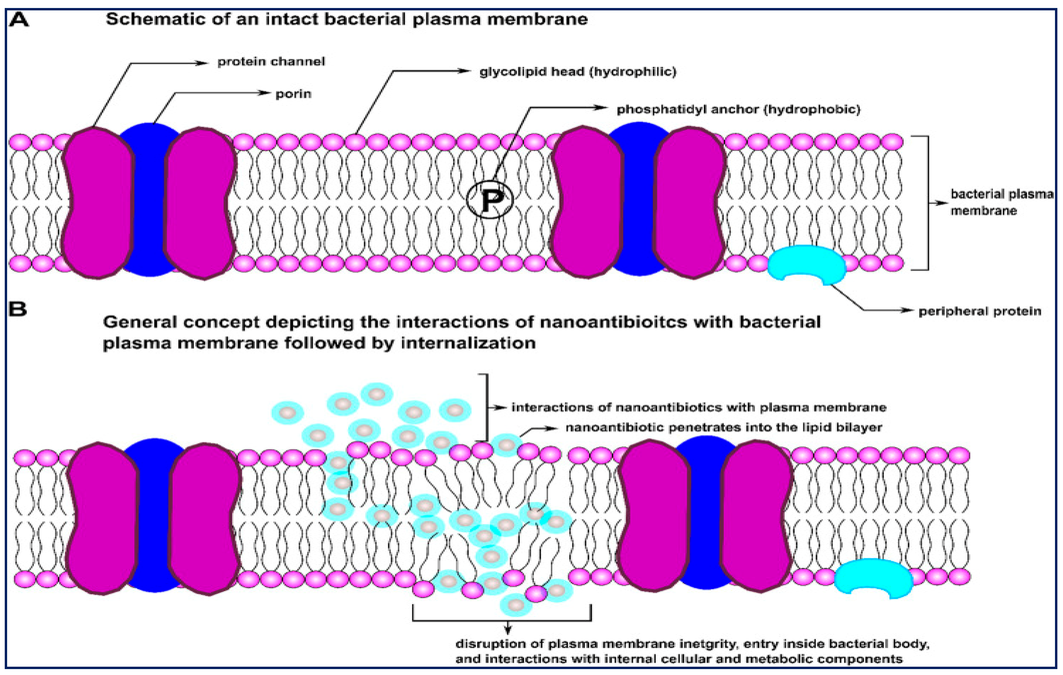
The oxidative mechanism includes: stress induction through reactive oxygen species (ROS) and free-radical generation, electron transport chain restraint, plasmid damage, disruption of the cell membrane, DNA damage, interruption of enzyme activity, metal ion release including the crumbling of metal ions from nanoconjugates into dissolve form, interruption of the electron transport chain, and non-oxidative attributes such as surface energy, size, shape, surface roughness, cytoplasm release, atomically thin structures, zeta potential (surface charge), stability, increased specific surface region/volume ratio, high surface reactivity, conductivity, the bioavailability of NPs, drug release systems at target sites, physical interactions between particles, and cell wall/membranes and particle-antibiotic interface, etc. These features of NPs permit appropriate contact with bacterial cells and improved penetration capacities crossing the cell membrane while meddling with cellular elements and metabolic machinery. Figure 2 represents how nanoscale particles function as carriers of drug molecules across bacterial membrane barriers.
Daniel Hasan et al., reported the formulation of quatsomes for enhanced delivery of vancomycin against MRSA [97]. The responsive quatsomes were prepared from pH-responsive quaternary bicephalic surfactants (StBAclm) and cholesterol (Chol). Drug-loaded quatsomes were developed by adding the bicephalic surfactant, vancomycin, and Chol at different concentrations. This was followed by adding 40 mL of distilled water, and the mixture was further sonicated for 10, 15, 20, and 30 min using a probe-sonicator in an ice bath. The dispersions were further stirred at 500 rpm using a magnetic stirrer for 24 h at room temperature. After 24 h of equilibration, the potential aggregates were detached from the blank (StBAclm-Qt) and the drug-loaded vancomycin-StBAclm-Qt quatsomes using a filter/syringe. These all showed an enhanced antimicrobial effect of the drug-loaded quatsomes at an eight-fold lower concentration when compared to bare vancomycin. The drug-loaded quatsomes also demonstrated the capability to abolish MRSA biofilm.
Giersing et al., discussed nano-patterning technologies and surface alteration to deter MRSA biofilm formation [98]. The drug-loaded NPs could overcome the limitations of conventional antibiotic treatments. Metal containing NPs have the potential to inhibit the growth of MRSA infections. When the positively charged ions of the NPs combine with the negatively charged parts of the bacterial cell membrane, the bacterial cell wall or membrane disruption occurs. This creates pores in the membrane and leads to cell death by dissipating the H+ gradient across the membrane [99]. ROS formation results in DNA damage and cell fatality when the NP’s internalization occurs in the bacterial cytosol [100]. The development of nanotechnology gives a promising solution for treating resistant bacterial infections.

3.3. Antimicrobial Activity of Surfactant Molecules

Generally, the antimicrobial activity of surfactants depends on the alkyl chain length, and surfactants show increased antibacterial activity while having decreased chain length. Cationic surfactants such as quaternary ammonium compounds show good antibacterial activity because they have high cationic charge and hydrophobicity. The positive control of cationic surfactants leads to a more efficient interaction with the negatively charged bacterial membrane. Robert et al., discussed the effectiveness of humimycins, N-acylated linear heptapeptides, against MRSA [101]. They have altered the structure of the lipopeptides, including hydrophobicity and enantiomeric amino acid substitutions, to explore their potential against MRSA. This study revealed that the dihydroxy analogue, synthesized with myristic anhydride, shows four times better activity against MRSA in minimum inhibitory concentration assays.

3.4. Radiation Therapy

Radiation therapy is generally used for cancer treatment that uses intense beam energy or photons, or other types of energy, to kill cancer cells. During brachytherapy, radiation will be placed inside the human body. Pulse laser therapy effectively kills MRSA and shortens the therapy time. It doesn’t give any heat or pain sensation; hence, it is ideal for clinical applications. Diabetic patients with open wounds are highly susceptible to MRSA when the infection takes hold and creates a biofilm or a slimy buildup of bacteria that become more difficult to treat.
Boston University College of Engineering researchers invented a new technique using radiation therapy that can kill 99.9% of MRSA [102]. The golden pigment is the universal signature of S. aureus. MRSA is exposed to blue light that causes a traumatic photobleaching effect and destroys the pigment molecule responsible for the cell’s golden color. Vulnerable spots will appear on the cell membrane when the golden pigment molecules are decomposed under blue light. The blue light can destabilize MRSA cells and enough to kill about 90% of the bacterial culture. The powerful oxidizing agent H2O2 can kill the remaining, and critical, 10%. The research in mice using blue light and H2O2 to treat MRSA infections revealed that this method could speed up the healing of skin wounds infected with MRSA.

4. In Vitro and In Vivo Studies

MRSA isolates extracellular matrix production is the critical virulence factor and survival mechanism for this superbug [103]. An in vitro study refers to work performed in a controlled environment such as a test tube or petri dish. In vitro studies can be achieved only outside of a living organism. In vivo research can be done with or within an entire living organism. Studies in animal models or human clinical trials are in vivo studies.
There is no coercion to submit animal protocols to Institutional Animal Care and Use Committee (IACUCs) for doing in vitro studies [104]. This method helps to reduce the requirement for experienced laboratory personnel in animal handling. The loss of glycosylation of the antibody might make the antibody product unsuitable for in vivo experiments.
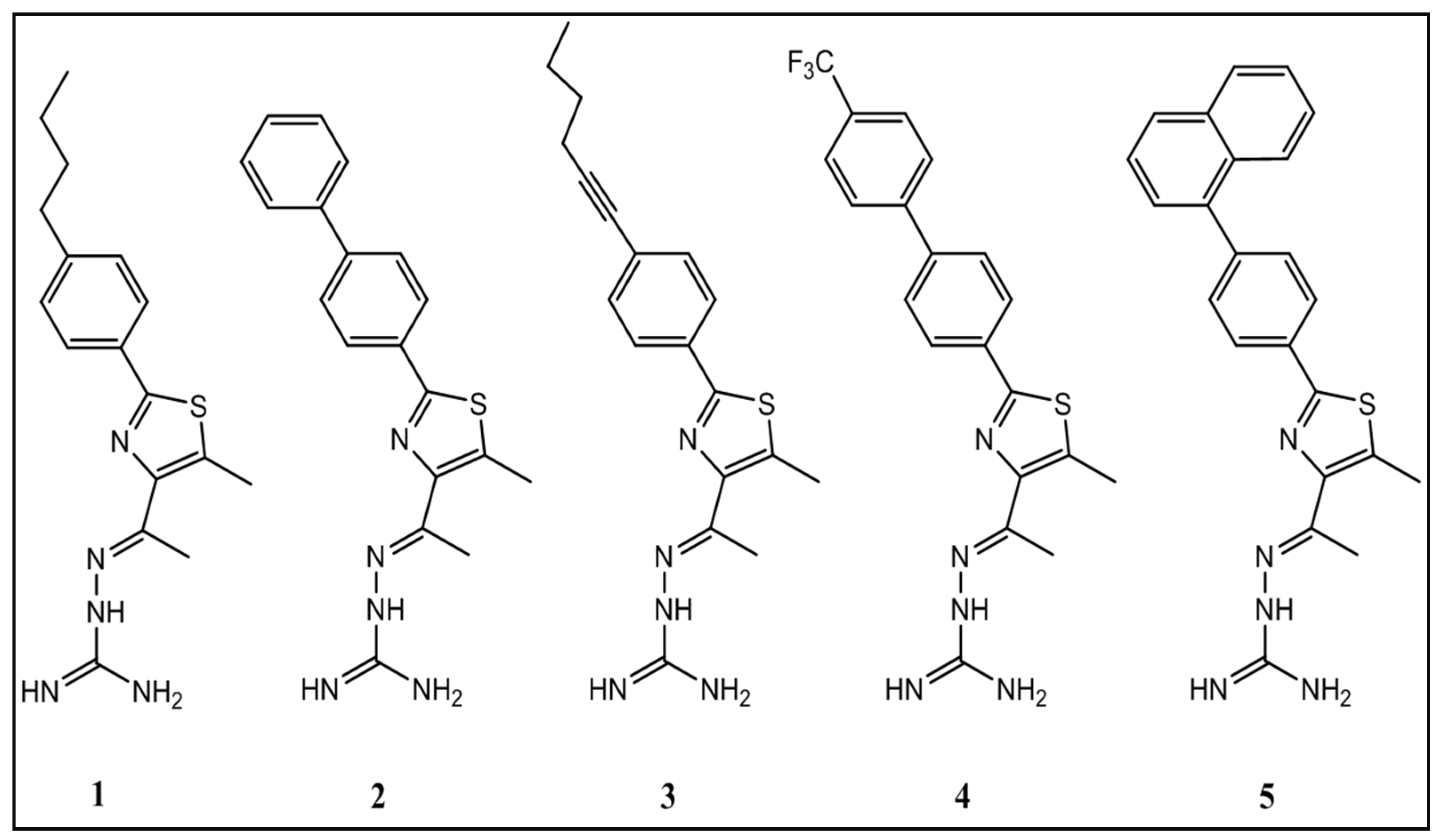
Mohammad et al., examined the potential of thiazole compounds against MRSA [105]. They have reported that phenylthiazole compounds can exhibit effective antimicrobial activity in in vitro studies against various arrays of clinically meaningful MRSA strains. Derivatives of the compound were synthesized, and the structure-activity relationship has been elucidated. Figure 3 represents the Chemical structures of thiazole compounds.
These derivatives unveiled that the aminoguanidine moiety at thiazole-C5 is essential for the antibacterial activity. A nonpolar, hydrophobic group is preferential at thiazole-C2. Analogs to the lipophilic alkyl tail of phenylthiazole compounds were subsequently developed to improve the antimicrobial activity of the thiazole compounds, toxicity profile, and their physicochemical properties. These phenylthiazole compounds have numerous admirable characteristics in vitro, including quick bactericidal activity against MRSA, a low frequency of bacterial resistance developing, and they have exposed the possibility of being used in a blend with other antibiotics, such as vancomycin, against MRSA. The compounds (15) were tested (at concentrations of 5 µg/mL, 10 µg/mL, 20 µg/mL, and 40 µg/mL) against a human keratinocyte (HaCaT) cell line to determine the potential toxic effect to mammalian skin cells using in vitro analysis.
The cells were cultured in Dulbecco’s modified Eagle medium (DMEM) supplemented with 10% fetal bovine serum (FBS) at 37 °C with CO2 (5%). Control cells obtained Dimethyl sulfoxide (DMSO) alone at a concentration equivalent to that in drug-treated cell samples. The cells were incubated with the compounds (in triplicate) in a 96-well plate at 37 °C with CO2 (5%) for two hours before the addition of the assay reagent MTS 3-(4,5-dimethylthiazole-2-yl)-5-(3-carboxymethoxyphenyl)-2-(4-sulfophenyl)-2H-tetrazolium). Absorbance readings (at OD490) were calculated using a kinetic microplate reader. After treatment with each compound, the extent number of viable cells was expressed as a percentage of the viability of DMSO-treated control cells. The toxicity data were examined through a one-way Analysis of variance (ANOVA), with post hoc Dunnett’s multiple comparisons tests (p < 0.05), utilizing GraphPad Prism 6.0. In vitro studies revealed that the thiazole compounds are nontoxic to human keratinocytes and negatively affect mammalian tissues. Furthermore, the antimicrobial activities of the thiazole compounds were confirmed by performing an in vivo analysis.
The in vivo process evades the necessity to teach the antibody producer tissue-culture methods. When performing the in vivo method (i.e., mouse ascites method), a very high mAb concentration will be produced for this reason, there is no need for additional concentration procedures, which can denature the antibody and reduce efficiency. In vivo strategies can create significant pain or trouble in mice. The mAb produced via in vivo methods can contain diverse contaminations that might need purification.
The in vivo studies were performed with eight groups (n = 5) of eight-week-old female Balb/c mice. The mice were disinfected with ethanol (70%) and shaved on the back center (roughly a one-inch by one-inch square region around the injection site) one day before infection. The bacterial inoculum preparation, an aliquot of an overnight culture of MRSA USA300, was transferred to a clean tryptic soy broth (TSB) and shaken at 37 °C until an OD600 value of ~1.0 was accomplished.
The cells were centrifuged, washed once with phosphate buffer solution (PBS), re-centrifuged, and then resuspended in PBS. The mice were given an intradermal injection (20 µL) containing ~2.76 × 10⁸ CFU/mL MRSA USA300. An open injury was created at the injection site, 48 h post-infection. Topical treatment was initiated consequently with each group of mice getting thiazole compounds (2%, using petroleum jelly as the vehicle), mupirocin (2%, using petroleum jelly as the vehicle), thiazole compound-1 (2%, using Lipoderm as an alternative vehicle) and a control group obtaining the control vehicle (20 mg, petroleum jelly) alone. Each group of mice receiving a particular treatment procedure was housed individually in a ventilated cage with proper bedding, food, and water. The mice were tested twice a day during infection and treatment to ensure there were no unfavorable reactions. The mice were humanely euthanized by CO2 asphyxiation 24 h after administering the last dose. The region around the skin injury was delicately swabbed with ethanol (70%) and expelled. Then, the tissue was homogenized in 1 mL of TSB. The homogenized tissue was consecutively diluted in PBS before plating onto mannitol salt agar plates. The plates were incubated for 20–22 h at 37 °C before feasible CFCs were counted, and MRSA diminution in the skin injury post-treatment was determined for each group. Using a one-way ANOVA, data were analyzed with post hoc Holm-Sidak’s multiple comparison tests (p < 0.05), utilizing GraphPad Prism 6.0.
Thiazole-1 showed effective activity against all drug-resistant staphylococcal strains with the MIC value of 1.3 µg/mL. The biphenyl and butyne analogs (2 and 3, respectively) have MIC values ranging from 2.8 to 5.6 µg/µL. All five thiazole compounds have potent antimicrobial activity against MRSA strains that exhibit resistance to antibiotics, including β-lactams, fluoroquinolones (USA800), tetracycline (USA300), and erythromycin (USA300 and USA1000).
These compounds also exhibited intense antimicrobial activity against S. aureus NRS107 (MIC values range from 1.3 to 13.3 µg/mL), a strain showing high resistance to mupirocin (MIC of 1024.0 µg/mL). Moreover, compounds 1 and 2 (MIC of 1.3 and 2.8 µg/mL, respectively) are more active than mupirocin (MIC of 4.0 µg/mL) against three additional MRSA strains (USA800, USA1000, and USA1100). When tested against four MRSA strains, Clindamycin was determined to have a 0.1 µg/mL MIC. The results showed that the thiazole compounds have intense activity against CA-MRSA strains and MRSA isolates liable for infected wounds in patients (Table 3).
In this study, the antimicrobial activity of phenylthiazole compounds has been illustrated using in vitro and in vivo analysis against clinically pertinent MRSA strains liable for skin and wound infections. The thiazole compounds were reported as nontoxic to human keratinocytes and exhibited potent antimicrobial activity against MRSA.

5. Clinical Analysis

Clinical analysis is performed via blood, plasma, serum, urine, fluids, tissues, and occasionally on other matrices. Generally, analytes are measured in serum due to their easy access, and it is used to reflect target concentrations in tissue or receptors. Intensive care patients have a higher risk compared with medical patients. They have a higher rate of developing MRSA infection within the first 4 days of admission.
Patients with surgical wounds, ulcers, and intravenous catheterization also have risk factors, with hazard ratios of 2.9, 3.0, and 4.7 [106]. Protection of patients with ulcers, intravenous catheters, and surgical wounds from MRSA outbreaks helps to reduce the spread and seriousness of MRSA infections. Using antibiotics, herbals, radiation therapy, or other treatment methods may help patients to recover from MRSA infections. Proper knowledge about MRSA, earlier treatment, and reducing the risk factors can also help to decrease the mortality rate of MRSA infections.
Jang et al., assessed the patients who had been suffering from S. aureus bacteremia [107]. Among the 377 patients with S. aureus bacteremia, 41 cases (11%) were persistent even with proper antibiotics. Of these, 35 of the 41 persistent bacteremia patients had persistence due to MRSA. Salvage treatment comprised of either adding an aminoglycoside or rifampicin to vancomycin or a vancomycin substitution with linezolid alone or blending with a carbapenem. This study revealed that linezolid substitution was considerably more efficient, with MRSA clearance in 75% of cases compared with 17% with vancomycin-based treatment continuation.
Lai et al., estimated the use of high-dose daptomycin in patients with severe Gram-positive infections, with 63 out of 67 (94%) patients getting high-dose daptomycin as a salvage therapy. MRSA was segregated in 56.7% of patients, and 80.6% of patients were bacteremic. The clinical and microbiological triumph rates were 77.6% and 82.1%, respectively [108].
Caelli et al., compared the use of a combination of 4% TTO nasal ointment and 5% TTO body wash (intervention) with a standard 2% mupirocin nasal ointment and triclosan body wash (routine) for the annihilation of MRSA [109]. A total of 30 in-patients were selected and erratically assigned to be treated with TTO or standard routine care for 3 days. The patients also obtained intravenous vancomycin, and all the patients were evaluated for MRSA carriage after 48 and 96 h of the discontinuance of topical treatment. More patients in the intervention than in the control group were cleared of infection (5/8 versus 2/10). Two patients in the intervention group were treated for 34 days and removed the infection, while the other remained chronically colonized.

6. Computational Analysis

Computer-aided design is a preliminary stand for screening novel inhibitors, and the statistics help to find new applications in drug development. Computational analysis helps to understand mainly by studying mathematical models implemented on computers. Computation is any type of calculation that includes arithmetical and non-arithmetical processes and pursues a well-defined model. Computational analysis helps researchers to carry out thousands of simulated experiments.
Finite Element Method (FEM) is a computational analysis that is widely used in orthopedic biomechanics [110]. Various computational methods are available to identify the best compound for the treatment of MRSA infections, including docking studies, ADMET analysis, and the characterization of the respective mechanism. The target protein molecules can be designed via homology modeling or collected from the Protein Data Bank (PDB) database and the potential drug molecules suggested from the PubChem database [111]. Computational models are used to track contagious diseases in populaces that help to identify the most efficient interventions and adjust interventions to diminish the spread of diseases. Scientists use computational modeling to design the safest drugs for patients with minor side effects.
Skariyachan et al., has reported the computer-aided screening and assessment of herbal therapeutics against MRSA infections [112]. In this study, absorption, distribution, metabolism, excretion, toxicity (ADMET) and docking studies were used to screen the drug-likeness and effectiveness of diverse herbal compounds. The structures of the proteins PBP2A and Panton-Valentine Leukocidin (PVL) were used as the drug targets. The 3D structures of all plant-based ligands were collected from the drug database, and the structures were reclaimed from Kyoto Encyclopedia of Genes and Genomes (KEGG), PubChem, and Chemspider.
Computational biology tools can predict the molecules’ absorption, distribution, metabolism, excretion, and toxicity (ADMET). The 74 ligands identified for this study have been analyzed for their drug-likeliness, ADME properties, and toxicity investigation by Pre-ADMET. The ADME includes the extent and rate of absorption, distribution, metabolism, and excretion. Pre-ADMET uses Caco2-cell (heterogeneous human epithelial colorectal adenocarcinoma cell lines) and MDCK (Madlin-Darby Canine Kidney) cell models for oral drug absorption prediction, and skin penetrability and human gastrointestinal absorption models for oral and transdermal drug absorption prediction. Pre-ADMET helps to predict toxicity based on the mutagenicity of AMES parameters and rodent carcinogenicity assays of rats and mice. The crystal structures of PBP2A (1VQQ) and PVL toxin (1T5R) were reclaimed from the Protein Data Bank, and the harmonized files were used for the study. The chosen herbal compounds were docked with the target proteins by AutoDock. The program uses a Monte Carlo replicated annealing for configurational investigation using grid-based molecular affinity potentials and confers bioactive conformation by energy minimization.
The docking studies unveiled a truth that the bioactive constituent of Aloe vera, β-sitosterol (3S,8S,9S,10R,13R,14S,17R) 17 [(2R,5R)-5-ethyl-6-methylheptan-2-yl]-10, 13-dimethyl 2,3,4,7,8,9,11,12,14,15,16,17-dodecahydro-1H-cyclopenta [a] phenanthren-3-ol) exhibited excellent binding energies of −7.40 kcal/mol and −6.34 kcal/mol for PBP2A and PVL toxin, respectively. In the same way, Meliantriol (1S-1-[(2R,3R, 5R)-5-hydorxy-3-[(3S,5R,9R,10R,13S,14S,17S)-3-hydroxy 4,4,10,13,14-pentamethyl-2,3,5, 6,9,11,12,15,16,17-decahydro-1H-cyclopenta [a] phenanthren-17-yl] oxolan-2-yl]-2-methylpropane-1,2 diol), bioactive compound in Azadirachta indica (Neem) exhibited the binding energies of −6.02 kcal/mol for PBP2A and −8.94 for PVL toxin. The in-silico and in vitro studies revealed that the herbal extracts of Aloevera, Guava, Neem, Pomegranate, and Tea can be used as therapeutics against MRSA infections.

Molecular Dynamics

Traditional docking methods have significant limitations related to the semi-flexible treatment of ligands (small molecules) and targets (proteins) [113]. Atomistic molecular dynamics (MDs) have emerged as an alternative for simulating macromolecular complexes. MDs helps to explore drug-target recognition and binding from mechanical and energetic perspectives and determine the binding and unbinding kinetic constants. Many computational works have been developed based on combined docking MD strategies. Solvent mapping or co-solvent MD simulation techniques have been widely used to identify binding sites and hot spots on protein surfaces at a dynamic level.
MD simulation helps to reveal unstable binding modes according to the structural perspective. This technique helps to negate physically unreliable docking solutions or helps to find new ones [114]. Post-processing via MD helps to refine the energy assessed by scoring functions (re-scoring). A wide range of theoretical methods are needed for re-scoring approaches, and the energies are computed as ensemble averages [115]. Re-scoring techniques range from partially empirical methods such as Linear Interaction Energy through an authentic free-energy process. The probable solution may lie between the Molecular Mechanics-Poisson-Boltzmann Surface Area and the Molecular Mechanics-Generalized Born Surface Area re-scoring approach, which helps to balance reasonable accuracy and computational costs [116].
Another method has been used to generate an ensemble of protein conformations by performing MD before docking. In this method, the targeting flexibility in docking will be taken into account indirectly and Virtual Screening (VS) through pre-generated discrete conformations. McCammon and co-workers implemented the “relaxed complex scheme”, which helps to perform extensive MD simulation of the protein in the apo form [117,118]. Then, snapshots were taken at regular time intervals, and each protein targeted in the subsequent docking exercise. The problem with this method is choosing the appropriate protein structures to be used to build the ensemble. A large number of protein conformations may worsen the VS performances. Cluster analysis techniques can help to reduce redundancy in the conformation set and ensure that only dissimilar structures are included. Enhanced sampling techniques can be used in this method to further increase the protein conformations’ variability [119].
Dynamic docking methods are different from the other methods due to the possibility of characterizing the protein-ligand binding process. Dynamic docking exploits the advances in sampling strategies. There are two conceptual layers of complexity in the static, dynamic method. The first layer is related to engendering the binding modes, and the second layer is about evaluating the reliability of the identified “poses.” This can be achieved by considering the binding energy. Dynamic docking methods are helpful in the drug discovery program, and feasible advances may happen in the future by improving hardware architecture and machine-learning techniques.

7. Future Directions for MRSA Treatment

MRSA infections are continuously growing burden for human society due to the poor prognosis and high costs associated with this infection [120]. Most of the antibiotics available in the market become less active against resistant bacteria, particularly against MRSA; hence, new strategies are needed to treat MRSA infections.
The future treatment methods for MRSA infection may have the following aspects:
  • Nanocarriers with good surface area for controlled release of antibiotic with minimum inhibitory concentrations.
  • The design and development of antibody-based biologic agent treatments for the management of serious infections of MRSA.
  • Multi-drug strategies for treating drug-resistant bacterial infections such as synthetic drugs, synthetic and natural drugs, and natural medicines.
  • Breakage of biofilms formed by MRSA using a suitable targeting carrier system with the biotic drugs.

8. Conclusions

Many lives have been saved from numerous contagious diseases by the discovery of antibiotics, but MRSA infections are formidable, versatile, and unpredictable. Vancomycin is mainly used to treat MRSA infections, either alone or in combination. However, intermediate resistance levels, vigilant therapeutic drug monitoring, and MIC creep are the path blockers for vancomycin against MRSA infections. Improving efficient anti-MRSA treatments and the execution of infection control methodologies are of expeditious need. Advanced research in this field may reduce the clinical impact of this pathogen. The combination of antibiotics with NPs can be a precedent shift to distribute the ideal concentration of a drug in the target place by bypassing resistance mechanisms of bacteria. The combination of antibiotics with NPs can accomplish all the requirements of an effective antibacterial agent and save millions of lives. The efficiency and biosafety of the designed and synthesized drugs can be studied using in vivo and ex vivo analyses. These studies may reduce the difficulties in human trials.

Author Contributions

Composed the manuscript and prepared the figures, P.N. and P.K. Analyzed, edited, and commented on the manuscript, S.M., A.S.A. and J.S. Conceptualization, conceived the manuscript design, analyzed and approved the manuscript, M.R. All authors have read and agreed to the published version of the manuscript.

Funding

This research is financially supported by the plan of the Science and Engineering Research Board (Ref: EEQ/2020/000201) New Delhi, India and the Deanship of Scientific Research, Majmaah University, Kingdom of Saudi Arabia for research support under project number: *R-2022-109*.

Acknowledgments

M. Rajan acknowledges significant financial support from the plan of the Science and Engineering Research Board (Ref: EEQ/2020/000201) New Delhi, India. The authors would like to thank the Deanship of Scientific Research, Majmaah University, Kingdom of Saudi Arabia for research support under project number: *R-2022-109*.

Conflicts of Interest

The authors declare no conflict of interest.

References

  1. Ogston, A. Report upon microorganisms in surgical diseases. Br. Med. J. 1881, 1, 369–375. [Google Scholar] [CrossRef] [PubMed] [Green Version]
  2. Ogston, A. Micrococcus poisoning. J. Anat. Physiol. 1882, 16, 526–567. [Google Scholar] [PubMed]
  3. Cowan, S.T.; Shaw, C.; Williams, R.E. Type strain for Staphylococcus aureus Rosenbach. J. Gen. Microbiol. 1954, 10, 174–176. [Google Scholar] [CrossRef] [PubMed] [Green Version]
  4. Cowan, S.T. Classification of staphylococci by slide agglutination. J. Pathol. Bacteriol. 1939, 48, 169–173. [Google Scholar] [CrossRef]
  5. Taylor, T.A.; Unakal, C.G. Staphylococcus aureus. StatPearls; 2020. Available online: https://www.ncbi.nlm.nih.gov/books/NBK441868/ (accessed on 27 May 2021). [PubMed]
  6. Turner, N.A.; Sharma-Kuinkel, B.K.; Maskarinec, S.A.; Eichenberger, E.M.; Shah, P.P.; Carugati, M.; Holland, T.L.; Fowler, V.G., Jr. Methicillin-resistant Staphylococcus: An overview of basic and clinical research. Nat. Rev. Microbiol. 2019, 17, 203–218. [Google Scholar] [CrossRef]
  7. Diekema, D.J.; Pfaller, M.A.; Schmitz, F.J.; Smayevsky, J.; Bell, J.; Jones, R.N.; Beach, M.; SENTRY Participants Group. Survey of infections due to Staphylococcus species: Frequency of occurrence and antimicrobial susceptibility of isolates collected in the United States, Canada, Latin America, Europe, and the Western Pacific region for the SENTRY Antimicrobial Surveillance Program, 1997–1999. Clin. Infect. Dis. 2001, 32 (Suppl. 2), S114–S132. [Google Scholar]
  8. Schito, G.C. The importance of the development of antibiotic resistance in Staphylococcus aureus. Clin. Microbiol. Infect. 2006, 12 (Suppl. 1), S3–S8. [Google Scholar] [CrossRef] [Green Version]
  9. Lindsay, J.A.; Holden, M.T. Staphylococcus aureus: Superbug, super genome? Trends Microbiol. 2004, 12, 378–385. [Google Scholar] [CrossRef]
  10. Lowy, F.D. Staphylococcus aureus infections. N. Engl. J. Med. 1998, 339, 520–532. [Google Scholar] [CrossRef]
  11. Wertheim, H.F.; Melles, D.C.; Vos, M.C.; van Leeuwen, W.; van Belkum, A.; Verbrugh, H.A.; Nouwen, J.L. The role of nasal carriage in Staphylococcus aureus infections. Lancet Infect. Dis. 2005, 5, 751–762. [Google Scholar] [CrossRef]
  12. Kirby, W.M. Extraction of a highly potent penicillin inactivator from penicillin resistant staphylococci. Science 1994, 99, 452–453. [Google Scholar] [CrossRef] [PubMed]
  13. Rammelkamp, C.H.; Maxon, T. Resistance of Staphylococcus aureus to the action of penicillin. Proc. Soc. Exp. Biol. Med. 1942, 51, 386–389. [Google Scholar] [CrossRef]
  14. Jevons, M.P. Celbenin-resistant staphylococci. Br. Med. J. 1961, 1, 124–125. [Google Scholar] [CrossRef]
  15. Hartman, B.J.; Tomasz, A. Low-affinity penicillin-binding protein associated with beta-lactam resistance in Staphylococcus aureus. J. Bacteriol. 1984, 158, 513–516. [Google Scholar] [CrossRef] [PubMed] [Green Version]
  16. Reynolds, P.E.; Brown, D.F. Penicillin-binding proteins of beta-lactam-resistant strains of Staphylococcus aureus. Effect of growth conditions. FEBS Lett. 1985, 192, 28–32. [Google Scholar] [CrossRef] [Green Version]
  17. Utsui, Y.; Yokota, T. Role of an altered penicillin-binding protein in methicillin and cephem-resistant Staphylococcus aureus. Antimicrob. Agents Chemother. 1985, 28, 397–403. [Google Scholar] [CrossRef] [PubMed] [Green Version]
  18. Matcuhashi, M.; Song, M.D.; Ishino, F.; Wachi, M.; Doi, M.; Inoue, M.; Ubukata, K.; Yamashita, N.; Konno, M. Molecular cloning of the gene of a penicillin-binding protein supposed to cause high resistance to beta-lactam antibiotics in Staphylococcus aureus. J. Bacteriol. 1986, 167, 975–980. [Google Scholar] [CrossRef] [Green Version]
  19. Katayama, Y.; Ito, T.; Hiramatsu, K. A new class of genetic element, staphylococcus cassette chromosome mec, encodes methicillin resistance in Staphylococcus aureus. Antimicrob. Agents Chemother. 2000, 44, 1549–1555. [Google Scholar] [CrossRef] [Green Version]
  20. Hiramatsu, K.; Asada, K.; Suzuki, E.; Okonogi, K.; Yokota, T. Molecular cloning and nucleotide sequence determination of the regulator region of mecA gene in methicillin-resistant Staphylococcus aureus (MRSA). FEBS Lett. 1992, 298, 133–136. [Google Scholar] [CrossRef] [Green Version]
  21. Mickymaray, S.; Alturaiki, W.; Al-Aboody, M.S.; Mariappan, P.; Rajenderan, V.; Alsagaby, S.A.; Kalyanasundram, U.; Alarfajj, A.A. Antibacterial efficacy of bacteriocin produced by marine Bacillus subtilis against clinically important extended spectrum beta-lactamase strains and methicillin-resistant Staphylococcus aureus. IJMRHS 2018, 7, 75–83. [Google Scholar]
  22. Hetem, D.J.; Westh, H.; Boye, K.; Jarlov, J.O.; Bonten, M.J.; Bootsma, M.C. Nosocomail transmission of community-associated methicillin-resistant Staphylococcus aureus in Danish hospitals. J. Antimicrob. Chemother. 2012, 67, 1775–1780. [Google Scholar] [CrossRef] [PubMed] [Green Version]
  23. Moodley, A.; Oosthuysen, W.F.; Duse, A.G.; Marais, E.; South African MSG. Molecular characterization of clinical methicillin-resistant Staphylococcus aureus isolates in South Africa. J. Clin. Microbiol. 2010, 48, 4608–4611. [Google Scholar] [CrossRef] [PubMed] [Green Version]
  24. King, M.D.; Humphrey, B.J.; Wang, Y.F.; Kourbatova, E.V.; Ray, S.M.; Blumberg, H.M. Emergence of community-acquired methicillin-resistant Staphylococcus aureus USA 300 clone as the predominant cause of skin and soft-tissue infections. Ann. Intern. Med. 2006, 144, 309–317. [Google Scholar] [CrossRef] [Green Version]
  25. Semple, S.J.; Reynolds, G.D.; O’Leary, M.C.; Flower, R.L. Screening of Australian medicinal plants for antiviral activity. J. Ethnopharmacol. 1998, 60, 163–172. [Google Scholar] [CrossRef]
  26. Bassett, I.B.; Pannowitz, D.L.; Barnetson, R.S. A comparative study of tea-tree oil versus benzylperoxide in the treatment of acne. Med. J. Aust. 1990, 153, 455–458. [Google Scholar] [CrossRef] [PubMed]
  27. Hemaiswarya, S.; Kruthiventi, A.K.; Doble, M. Synergism between natural products and antibiotics against infectious diseases. Phytomedicine 2008, 15, 639–652. [Google Scholar] [CrossRef]
  28. Nassan, M.A.; Mohamed, E.H.; Abdelhafez, S.; Ismail, T.A. Effect of clove and cinnamon extracts on experimental model of acute hematogenous pyelonephritis in albino rats: Immunopathological and antimicrobial study. Int. J. Immunopathol. Pharmacol. 2015, 28, 60–68. [Google Scholar] [CrossRef] [Green Version]
  29. Badei, A.; Faheld, S.; El-Akel, A.; Mahnoud, B. Application of some species in flavoring and preservation of cookies: 2-Antimicrobial and sensory properties of cardamom, cinnamon and clove. Dtsch. Lebensm. Rundsch. 2002, 98, 261–265. [Google Scholar]
  30. Hussain, M.B.; Hannan, A.; Absar, M.; Butt, N.S. In vitro susceptibility of methicillin-resistant Staphylococcus aureus to honey. Complement. Ther. Clin. Pract. 2017, 27, 57–60. [Google Scholar] [CrossRef]
  31. Sherlock, O.; Dolan, A.; Athman, R.; Power, A.; Gethin, G.; Cowman, S.; Humphreys, H. Comparison of the antimicrobial activity of Ulmo honey from Chile and Manuka honey against methicillin-resistant Staphylococcus aureus, Escherichia coli and Pseudomonas aeruginosa. BMC Complement. Altern. Med. 2010, 10, 47. [Google Scholar] [CrossRef] [Green Version]
  32. Tan, H.T.; Rahman, R.A.; Gan, S.H.; Halim, A.S.; Hassan, S.A.; Sulaiman, S.A.; Kaur, B.K. The antibacterial properties of Malaysian tualang honey against wound and enteric microorganisms in comparison to manuka honey. BMC Complement. Altern. Med. 2009, 9, 34. [Google Scholar] [CrossRef] [PubMed] [Green Version]
  33. Okwu, M.U.; Olley, M.; Akpoka, A.O.; Izevbuwa, O.E. Methicillin-resistant Staphylococcus aureus (MRSA) and anti-MRSA activities of extracts of some medicinal plants: A brief review. AIMS Microbiol. 2019, 5, 117–137. [Google Scholar] [CrossRef] [PubMed]
  34. Sahu, M.C.; Padhy, R.N. In vitro antibacterial potency of Butea monosperma Lam. against twelve clinically isolated multidrug resistant bacteria. Asian. Pac. J. Trop. Dis. 2013, 3, 217–226. [Google Scholar] [CrossRef]
  35. Voravuthikunchai, S.P.; Kitpipit, L. Activity of medicinal plant extracts against hospital isolates of Methicillin-resistant Staphylococcus aureus (MRSA). Clin. Microb. Infect. 2005, 11, 493–512. [Google Scholar] [CrossRef] [PubMed] [Green Version]
  36. Gomber, C.; Saxena, S. Anti-staphylococcal potential of Callistemon rigidus. Cent. Eur. J. Med. 2006, 2, 79–88. [Google Scholar] [CrossRef] [Green Version]
  37. Aliyu, A.B.; Musa, A.M.; Abdullahi, M.S. Activity of plant extracts used in Northern Nigerian traditional medicine against Methicillin-resistant Staphylococcus aureus (MRSA). Niger. J. Pharmaceu. Sci. 2008, 7, 1–8. [Google Scholar]
  38. Akinjogunla, O.J.; Yah, C.S.; Eghafona, N.O. Antibacterial activity of the leave extracts of Nymphaea lotus (Nymphaeceae) on Methicillin-resistant Staphylococcus aureus and Vancomycin-resistant S. aureus isolated from clinical samples. Annals. Biol. Res. 2010, 1, 174–184. [Google Scholar]
  39. Invasive Species Compendium-CABI. Available online: https://www.cabi.org/isc/datasheet/28765 (accessed on 27 May 2021).
  40. Invasive Species Compendium-CABI. Available online: https://www.cabi.org/isc/datasheet/24882 (accessed on 27 May 2021).
  41. Invasive Species Compendium-CABI. Available online: https://www.cabi.org/isc/datasheet/39510 (accessed on 27 May 2021).
  42. Invasive Species Compendium-CABI. Available online: https://www.cabi.org/isc/datasheet/45141 (accessed on 27 May 2021).
  43. National Resources Conservation Service, United States Department of Agriculture (USDA). Available online: https://plants.usda.gov/core/profile?symbol=PUGR2 (accessed on 27 May 2021).
  44. Globinmed. Available online: https://www.globinmed.com/index.php?option (accessed on 27 May 2021).
  45. Arefin, K.; Rahman, M.; Uddin, M.Z.; Hassan, A. Angiosperm flora of Satchari Natural Park, Habiganj, Bangladesh. Bangl. J. Plant. Taxon. 2011, 18, 117–140. [Google Scholar] [CrossRef] [Green Version]
  46. Al-Alusi, N.T.; Kadir, F.A.; Ismail, S.; Abdullah, M. In vitro interaction of combined plants: Tinospora crispa and Swietenia mahagoni against Methicillin-resistant Staphylococcus aureus (MRSA). Afr. J. Microbiol. Res. 2010, 4, 2309–2312. [Google Scholar]
  47. Wikaningtyas, P.; Sukandar, E.Y. The antibacterial activity of selected plants towards resistant bacteria isolated from clinical specimens. Asian. Pac. J. Trop. Biomed. 2016, 6, 16–19. [Google Scholar] [CrossRef] [Green Version]
  48. Zuo, G.Y.; Zhang, X.J.; Yang, C.X.; Han, J.; Wang, G.C.; Bian, Z.Q. Evaluation of traditional Chinese medicinal plants for anti-MRSA activity with reference to the treatment record of infectious diseases. Molecules 2012, 17, 2955–2967. [Google Scholar] [CrossRef] [PubMed] [Green Version]
  49. Heyman, H.M.; Hussein, A.A.; Meyer, J.J.M.; Lall, N. Antibacterial activity of South African medicinal plants against Methicillin-resistant Staphylococcus aureus (MRSA). Pharm. Biol. 2009, 47, 67–71. [Google Scholar] [CrossRef] [Green Version]
  50. Uddin, Q.; Samiulla, L.; Singh, V.K.; Jamil, S.S. Phytochemical and Pharmacological Profile of Withania somnifera Dunal: A review. J. Appl. Pharm. Sci. 2012, 2, 170–175. [Google Scholar]
  51. Sucilathangam, G.; Gomatheswari, S.N.; Velvizhi, G.; Vincent, C.P.; Palaniappan, N. Detection of antibacterial activity of Medicinal plant Quercus infectoria against methicillin-resistant Staphylococcus aureus (MRSA) isolates in clinical samples. J. Pharm. Biomed. Sci. 2012, 14, 1–5. [Google Scholar]
  52. Armas, J.R.; Quiroz, J.R.; Roman, R.A.; Sanchez, J.O.; Pacheco, M.P.; Valdivia, L.H.; Rivera, E.C.; Asmat, R.C.; Anampa-Guzmán, A. Antibacterial activities of essential oils from three medicinal plants in combination with EDTA against MRSA. Br. Microbiol. Res. J. 2016, 17, 1–10. [Google Scholar] [CrossRef]
  53. Belley, A.; McKay, G.A.; Arhin, F.F.; Sarmiento, I.; Beaulieu, S.; Fadhil, I.; Parr, T.R., Jr.; Moeck, G. Oritavancin disrupts membrane integrity of Staphylococcus aureus and vancomycin-resistant enterococci to effect rapid bacterial killing. Antimicrob. Agents Chemother. 2010, 54, 5369–5371. [Google Scholar] [CrossRef] [Green Version]
  54. Carpenter, C.F.; Chambers, H.F. Daptomycin: Another novel agent for treating infections due to drug-resistant Gram-positive pathogens. Clin. Infect. Dis. 2004, 38, 994–1000. [Google Scholar] [CrossRef] [Green Version]
  55. Hughes, J.; Mellows, G. Inhibition of isoleucyl-transfer ribonucleic acid synthetase in Echerichia coli by pseudomonic acid. Biochem. J. 1978, 176, 305–318. [Google Scholar] [CrossRef] [Green Version]
  56. French, G.L. What’s new and not so new on the antimicrobial horizon? Clin. Microbiol. Infect. 2008, 14 (Suppl. 6), 19–29. [Google Scholar] [CrossRef] [Green Version]
  57. Kuok, C.F.; Hoi, S.O.; Hoi, C.F.; Chan, C.H.; Fong, I.H.; Ngok, C.K.; Meng, L.R.; Fong, P. Synergistic antibacterial effects of herbal extracts and antibiotics on methicillin-resistant Staphylococcus aureus: A computational and experimental study. Exp. Biol. Med. 2017, 242, 731–743. [Google Scholar] [CrossRef] [Green Version]
  58. Blesson, J.; Saji, C.V.; Nivya, R.M.; Kumar, R. Synergistic Antibacterial Activity of Natural Plant Extracts and Antibiotics against Methicillin Resistant Staphylococcus aureus (MRSA). WJPPS 2015, 4, 741–763. [Google Scholar]
  59. Jiang, Q.W.; Chen, M.W.; Cheng, K.J.; Yu, P.Z.; Wei, X.; Shi, Z. Therapeutic potential of steroidal alkaloids in cancer and other diseases. Med. Res. Rev. 2016, 36, 119–143. [Google Scholar] [CrossRef]
  60. Khameneh, B.; Iranshahy, M.; Ghandadi, M.; Atashbeyk, D.G.; Bazzaz, B.S.F.; Iranshahi, M. Investigation of the antibacterial activity and efflux pump inhibitory effect of co-loaded piperine and gentamicin nanoliposomes in methicillin-resistant Staphylococcus aureus. Drug. Dev. Ind. Pharm. 2015, 41, 989–994. [Google Scholar] [CrossRef] [PubMed]
  61. Tillotson, J.; Tillotson, G.S. The Regulatory Pathway for Antifungal Drugs: A US Perspective. Clin. Infect. Dis. 2015, 61, S678–S683. [Google Scholar] [CrossRef] [PubMed] [Green Version]
  62. Gudiol, F.; Aguado, J.M.; Almirante, B.; Bouza, E.; Cercenado, E.; Dominguez, M.A.; Gasch, O.; Lora-Tamayo, J.; Miró, J.M.; Palomar, M.; et al. Executive summary of the diagnosis and treatment of bacteremia and endocarditis due to Staphylococcus aureus. A clinical guideline from the Spanish Society of Clinical Microbiology and Infectious Diseases (SEIMC). Enferm. Infecc. Microbiol. Clínica 2015, 33, 626–632. [Google Scholar] [CrossRef]
  63. Bal, A.M.; David, M.Z.; Garau, J.; Gottlieb, T.; Mazzei, T.; Scaglione, F.; Tattevin, P.; Gould, I.M. Future trends in the treatment of Methicillin-Resistant Staphylococcus aureus (MRSA) infection: An in-depth review of newer antibiotics active against an enduring pathogen. J. Glob. Antimicrob. Resist. 2017, 10, 295–303. [Google Scholar] [CrossRef]
  64. Ippolito, J.A.; Kanyo, Z.F.; Wang, D.; Franceschi, F.J.; Moore, P.B.; Steitz, T.A.; Duffy, E.M. Crystal structure of the oxazolidinone antibiotic linezolid bound to the 50S ribosomal subunit. J. Med. Chem. 2008, 51, 3353–3356. [Google Scholar] [CrossRef]
  65. Lee, Y.C.; Chen, P.Y.; Wang, J.T.; Chang, S.C. A study on combination of daptomycin with selected antimicrobial agents: In vitro synergistic effect of MIC value of 1 mg/L against MRSA strains. BMC Pharmacol. Toxicol. 2019, 20, 25. [Google Scholar] [CrossRef] [Green Version]
  66. Debbia, E.; Pesce, A.; Schito, G.C. In vitro activity of LY146032 alone and in combination with other antibiotics against gram-positive bacteria. Antimicrob. Agents Chemother. 1988, 32, 279–281. [Google Scholar] [CrossRef] [Green Version]
  67. Saleh-Mghir, A.; Muller-Serieys, C.; Dinh, A.; Massias, L.; Cremieux, A.C. Adjunctive rifampin is crucial to optimizing daptomycin efficacy against rabbit prosthetic joint infection due to methicillin-resistant Staphylococcus aureus. Antimicrob. Agents Chemother. 2011, 55, 4589–4593. [Google Scholar] [CrossRef] [Green Version]
  68. Trookman, N.S.; Rizer, R.L.; Weber, T. Treatment of minor wounds from dermatologic procedures: A comparison of three topical wound care ointments using a laser wound model. J. Am. Acad. Dermatol. 2011, 64, S8–S15. [Google Scholar] [CrossRef] [PubMed]
  69. Deresinski, S. Vancomycin in Combination with other Antibiotics for the Treatment of Serious Methicillin-Resistant Staphylococcus aureus Infections. Clin. Infect. Dis. 2009, 49, 1072–1079. [Google Scholar] [CrossRef] [PubMed] [Green Version]
  70. Dilworth, T.J.; Ibrahim, O.; Hall, P.; Sliwinski, J.; Walraven, C.; Mercier, R.C. β-Lactams enhance vancomycin activity against Methicillin-Resistant Staphylococcus aureus bacteremia compared to vancomycin alone. Antimicrob. Agents Chemother. 2014, 58, 102–109. [Google Scholar] [CrossRef] [PubMed] [Green Version]
  71. Ruddaraju, L.K.; Pammi, S.V.N.; Guntuku, G.S.; Padavala, V.S.; Kolapalli, V.R.M. A review on anti-bacterials to combat resistance: From ancient era of plants and metals to present and future perspectives of green nano technological combinations. Asian J. Pharm. Sci. 2020, 15, 42–59. [Google Scholar] [CrossRef] [PubMed]
  72. Gurunathan, S. Biologically synthesized silver nanoparticles enhances antibiotic activity against gram-negative bacteria. J. Ind. Eng. Chem. 2015, 29, 217–226. [Google Scholar] [CrossRef]
  73. Ghosh, S.; Patil, S.; Ahire, M.; Kitture, R.; Kale, S.; Pardesi, K.; Cameotra, S.S.; Bellare, J.; Dhavale, D.D.; Jabgunde, A.; et al. Synthesis of silver nanoparticles using Dioscorea bulbifera tuber extract and evaluation of its synergistic potential in combination with antimicrobial agents. Int. J. Nanomed. 2012, 7, 483–496. [Google Scholar]
  74. Patra, J.K.; Baek, K.H. Biosynthesis of silver nanoparticles using aqueous extract of silky hairs of corn and investigation of its antibacterial and anticandidal synergistic activity and antioxidant potential. IET Nanobiotechnol. 2016, 10, 326–333. [Google Scholar] [CrossRef]
  75. Basker, S. Synergistic efficacy of antibiotics and silver nanoparticles synthesized from Eichhornia crassipes. Res. Plant. Biol. 2016, 6, 1–5. [Google Scholar] [CrossRef] [Green Version]
  76. Patra, J.K.; Baek, K.H. Novel green synthesis of gold nanoparticles using Citrullus lanatus rind and investigation of proteasome inhibitory activity, antibacterial, and antioxidant potential. Int. J. Nanomed. 2015, 10, 7253–7264. [Google Scholar]
  77. Patra, J.K.; Baek, K.H. Antibacterial activity and synergistic antibacterial potential of biosynthesized silver nanoparticles against foodborne pathogenic bacteria along with its anticandidal and antioxidant effects. Front. Microbiol. 2017, 8, 167. [Google Scholar] [CrossRef] [Green Version]
  78. Padalia, H.; Moteriya, P.; Chanda, S. Green synthesis of silver nanoparticles from marigold flower and its synergistic antimicrobial potential. Arab. J. Chem. 2015, 8, 732–741. [Google Scholar] [CrossRef] [Green Version]
  79. Rastogi, L.; Kora, A.J.; Sashidhar, R.B. Antibacterial effects of gum kondagogu reduced/stabilized silver nanoparticles in combination with various antibiotics: A mechanistic approach. Appl. Nanosci. 2015, 5, 535–543. [Google Scholar] [CrossRef] [Green Version]
  80. Singh, R.; Wagh, P.; Wadhwani, S.; Gaidhani, S.; Kumbhar, A.; Bellare, J.; Chopade, B.A. Synthesis, optimization, and characterization of silver nanoparticles from Acinetobacter calcoaceticus and their enhanced antibacterial activity when combined with antibiotics. Int. J. Nanomed. 2013, 8, 4277–4290. [Google Scholar]
  81. Gandhi, H.; Khan, S. Biological synthesis of silver nanoparticles and its antibacterial activity. J. Nanomed. Nanotechnol. 2016, 7, 366. [Google Scholar] [CrossRef] [Green Version]
  82. Barapatre, A.; Ram Aadil, K.; Jha, H. Synergistic antibacterial and antibiofilm activity of silver nanoparticles biosynthesized by lignin degrading fungus. Bioresour. Bioprocess. 2016, 3, 8. [Google Scholar] [CrossRef] [Green Version]
  83. Naqvi, S.Z.H.; Kiran, U.; Ali, M.I.; Jamal, A.; Hameed, A.; Ahmed, S.; Ali, N. Combined efficacy of biologically synthesized silver nanoparticles and different antibiotics against multidrug-resistant bacteria. Int. J. Nanomed. 2013, 8, 3187–3195. [Google Scholar] [CrossRef] [Green Version]
  84. Kumar, N.; Das, S.; Jyoti, A.; Kaushik, S. Synergistic effect of silver nanoparticles with doxycycline against klebsiella pneumonia. Int. J. Pharm. Sci. 2016, 8, 183–186. [Google Scholar]
  85. Wypij, M.; Czarnecka, J.; Swiecimska, M.; Dahm, H.; Rai, M.; Golinska, P. Synthesis, characterization and evaluation of antimicrobial and cytotoxic activities of biogenic silver nanoparticles synthesized from Streptomyces xinghaiensis OF1 strain. World J. Microbiol. Biotechnol. 2018, 34, 23. [Google Scholar] [CrossRef] [Green Version]
  86. Fayaz, A.M.; Balaji, K.; Girilal, M.; Yadav, R.; Kalaichelvan, P.T.; Venketesan, R. Biogenic synthesis of silver nanoparticles and their synergistic effect with antibiotics: A study against gram-positive and gram-negative bacteria. Nanomedicine 2010, 6, 103–109. [Google Scholar] [CrossRef]
  87. Łubowska, N.; Grygorcewicz, B.; Kosznik-Kwaśnicka, K.; Zauszkiewicz-Pawlak, A.; Węgrzyn, A.; Dołęgowska, B.; Piechowicz, L. Characterization of the Three New Kayviruses and Their Lytic Activity against Multidrug-Resistant Staphylococcus aureus. Microorganisms 2019, 7, 471. [Google Scholar] [CrossRef] [Green Version]
  88. Grygorcewicz, B.; Wojciuk, B.; Roszak, M.; Łubowska, N.; Błażejczak, P.; Jursa-Kulesza, J.; Rakoczy, R.; Masiuk, H.; Dołęgowska, B. Environmental Phage-Based Cocktail and Antibiotic Combination Effects on Acinetobacter baumannii Biofilm in a Human Urine Model. Microb. Drug Resist. 2021, 27, 25–35. [Google Scholar] [CrossRef] [PubMed]
  89. Clegg, J.; Soldaini, E.; McLoughlin, R.M.; Rittenhouse, S.; Bagnoli, F.; Phogat, S. Staphylococcus aureus Vaccine Research and Development: The Past, Present and Future, Including Novel Therapeutic Strategies. Front. Immunol. 2021, 12, 705360. [Google Scholar] [CrossRef] [PubMed]
  90. Hiramatsu, K.; Katayama, Y.; Matsuo, M.; Sasaki, T.; Morimoto, Y.; Sekiguchi, A.; Baba, T. Multi-drug resistant Staphylococcus aureus and future chemotherapy. J. Infect. Chemother. 2014, 20, 593–601. [Google Scholar] [CrossRef] [PubMed] [Green Version]
  91. Jacqueline, C.; Amador, G.; Batard, E.; Mabecque, V.L.; Miegeville, A.F.; Biek, D.; Caillon, J.; Potel, G. Comparison of ceftaroline fosamil, daptomycin and tigecycline in an experimental rabbit endocarditis model caused by methicillin-susceptible, methicillin-resistant and glycopeptide-intermediate Staphylococcus aureus. J. Antimicrob. Chemother. 2011, 66, 863–866. [Google Scholar] [CrossRef] [Green Version]
  92. Wilcox, M.H.; Tack, K.J.; Bouza, E.; Herr, D.L.; Ruf, B.R.; Ijzerman, M.M.; Croos-Dabrera, R.V.; Kunkel, M.; Knirsch, C. Complicated skin and skin-structure infections and catheter-related bloodstream infections: Noninfereiority of linezolid in a phase 3 study. Clin. Infect. Dis. 2009, 48, 203–212. [Google Scholar] [CrossRef]
  93. Tsai, C.Y.; Lee, C.H.; Chien, C.C.; Chen, I.L. Impact of teicoplanin maintenance dose and MIC values on the clinical outcomes of patients treated for methicillin-resistant Staphylococcus aureus bacteremia. Infect. Drug Resist. 2018, 11, 1205–1217. [Google Scholar] [CrossRef] [Green Version]
  94. Stryjewski, M.E.; O’Riordan, W.D.; Lau, W.K.; Pien, F.D.; Dunbar, L.M.; Vallee, M.; Fowler, V.G., Jr.; Chu, V.H.; Spencer, E.; Barriere, S.L.; et al. FAST Investigator Group. Telavancin versus Standard Therapy for Treatment of Complicated Skin and Soft-Tissue Infections Due to Gram-Positive Bacteria. Clin. Infect. Dis. 2005, 40, 1601–1607. [Google Scholar] [CrossRef] [Green Version]
  95. Huh, A.J.; Kwon, Y.J. Nanoantibiotics: A new paradigm for treating infectious diseases using nanomaterials in the antibiotics resistant era. J. Control. Release 2011, 156, 128–145. [Google Scholar] [CrossRef]
  96. Mamun, M.M.; Sorinolu, A.J.; Munir, M.; Vejerano, E.P. Nanoantibiotics: Functions and Properties at the Nanoscale to Combat Antibiotic Resistance. Front. Chem. 2021, 9, 687660. [Google Scholar] [CrossRef]
  97. Hassan, D.; Omolo, C.A.; Fasiku, V.O.; Elrashedy, A.A.; Mocktar, C.; Nkambule, B.; Soliman, M.E.S.; Govender, T. Formulation of pH-Responsive Quatsomes from Quaternary Bicephalic Surfactants and Cholesterol for Enhanced Delivery of Vancomycin against Methicillin Resistant Staphylococcus aureus. Pharmaceutics 2020, 12, 1093. [Google Scholar] [CrossRef]
  98. Giersing, B.K.; Dastgheyb, S.S.; Modjarrad, K.; Moorthy, V. Status of vaccine research and development of vaccines for Staphylococcus aureus. Vaccine 2016, 34, 2962–2966. [Google Scholar] [CrossRef] [PubMed] [Green Version]
  99. Knetsch, M.L.W.; Koole, L.H. New Strategies in the Development of Antimicrobial Coatings: The Example of Increasing Usage of Silver and Silver Nanoparticles. Polymers 2011, 3, 340–366. [Google Scholar] [CrossRef]
  100. Vimbela, G.V.; Ngo, S.M.; Fraze, C.; Yang, L.; Stout, D.A. Antibacterial properties and toxicity from metallic nanomaterials. Int. J. Nanomed. 2017, 12, 3941–3965. [Google Scholar] [CrossRef] [PubMed] [Green Version]
  101. Tancer, R.J.; Baynes, K.; Wiedman, G.R. Synergy among humimycins against methicillin-resistant Staphylococcus aureus. Pept. Sci. 2020, 113, e24197. [Google Scholar] [CrossRef]
  102. Dong, P.T.; Mohammad, H.; Hui, J.; Leanse, L.G.; Li, J.; Liang, L.; Dai, T.; Seleem, M.N.; Cheng, J.X. Photolysis of Staphyloxanthin in Methicillin-Resistant Staphylococcus aureus Potentiates Killing by Reactive Oxygen Species. Adv. Sci. 2019, 6, 1900030. [Google Scholar] [CrossRef] [PubMed]
  103. Eftekhar, F.; Dadaei, T. Biofilm formation and detection of IcaAB genes in clinical isolates of methicillin resistant Staphylococcus aureus. Iran. J. Basic Med. Sci. 2011, 14, 132–136. [Google Scholar]
  104. National Research Council (US) Committee on Methods of Producing Monoclonal Antibodies; National Academies Press (US): Washington, DC, USA. Available online: https://www.ncbi.nlm.nih.gov/books/NBK100199 (accessed on 27 May 2021).
  105. Mohammad, H.; Cushman, M.; Seleem, M.N. Antibacterial Evaluation of Synthetic Thiazole Compounds In vitro and In vivo in a Methicillin-Resistant Staphylococcus aureus (MRSA) Skin Infection Mouse Model. PLoS ONE 2015, 10, e0142321. [Google Scholar] [CrossRef] [Green Version]
  106. Coello, R.; Glynn, J.R.; Gaspar, C.; Picazo, J.J.; Fereres, J. Risk factors for developing clinical infection with methicillin-resistant Staphylococcus aureus (MRSA) amongst hospital patients initially only colonized with MRSA. J. Hosp. Infect. 1997, 37, 39–46. [Google Scholar] [CrossRef] [Green Version]
  107. Jang, H.C.; Kim, S.H.; Kim, K.H.; Kim, C.J.; Lee, S.; Song, K.H.; Jeon, J.H.; Park, W.B.; Kim, H.B.; Park, S.W.; et al. Salvage treatment for persistent methicillin-resistant Staphylococcus aureus bacteremia: Efficacy of linezolid with or without carbapenem. Clin. Infect. Dis. 2009, 49, 395–401. [Google Scholar] [CrossRef] [Green Version]
  108. Lai, C.C.; Sheng, W.H.; Wang, J.T.; Cheng, A.; Chuang, Y.C.; Chen, Y.C.; Chang, S.C. Safety and efficacy of high-dose daptomycin as salvage therapy for severe gram-positive bacterial sepsis in hospitalized adult patients. BMC Infect. Dis. 2013, 13, 66. [Google Scholar] [CrossRef] [Green Version]
  109. Caelli, M.; Porteous, J.; Carson, C.F.; Heller, R.; Riley, T.V. Tea tree oil as an alternative topical decolonization agent for methicillin-resistant Staphylococcus aureus. J. Hosp. Infect. 2000, 46, 236–237. [Google Scholar] [CrossRef]
  110. Huiskes, R.; Chao, E.Y.S. A survey of finite element analysis in orthopedic biomechanics: The first decade. J. Biomech. 1983, 16, 385–409. [Google Scholar] [CrossRef]
  111. Sangeetha, M.; Saranya, T.S.; Sathianarayanan, S.; Vyshnavi, A.M.H.; Namboori, P.K.K. Design and Development of Potential Flavonoid Moiety for Pbp2a Inhibition for MRSA Therapy—A Computational Technique. Biomed. Pharma. J. 2020, 13, 687–692. [Google Scholar] [CrossRef]
  112. Skariyachan, S.; Krishnan, R.S.; Siddapa, S.B.; Salian, C.; Bora, P.; Sebastian, D. Computer aided screening and evaluation of herbal therapeutics against MRSA infections. Bioinformation 2011, 7, 222–233. [Google Scholar] [CrossRef] [PubMed] [Green Version]
  113. Gioia, D.; Bertazzo, M.; Recanatini, M.; Masetti, M.; Cavalli, A. Dynamic Docking: A Paradigm Shift in Computational Drug Discovery. Molecules 2017, 22, 2029. [Google Scholar] [CrossRef] [PubMed] [Green Version]
  114. Cavalli, A.; Bottegoni, G.; Raco, C.; De Vivo, M.; Recanatini, M. A computational study of the binding of propidium to the peripheral anionic site of human acetylcholinesterase. J. Med. Chem. 2004, 47, 3991–3999. [Google Scholar] [CrossRef]
  115. Alonso, H.; Bliznyuk, A.A.; Gready, J.E. Combining docking and molecular dynamic simulations in drug design. Med. Res. Rev. 2006, 26, 531–568. [Google Scholar] [CrossRef]
  116. Decherchi, S.; Masetti, M.; Vyalov, I.; Rocchia, W. Implicit solvent methods for free energy estimation. Eur. J. Med. Chem. 2015, 91, 27–42. [Google Scholar] [CrossRef] [Green Version]
  117. Amaro, R.E.; Baron, R.; McCammon, J.A. An improved relaxed complex scheme for receptor flexibility in computer-aided drug design. J. Comput. Aided Mol. Des. 2008, 22, 693–705. [Google Scholar] [CrossRef] [Green Version]
  118. Lin, J.H.; Perryman, A.L.; Schames, J.R.; McCammon, J.A. Computational drug design accommodating receptor flexibility: The relaxed complex scheme. J. Am. Chem. Soc. 2002, 124, 5632–5633. [Google Scholar] [CrossRef]
  119. Buonfiglio, R.; Ferraro, M.; Falchi, F.; Cavalli, A.; Masetti, M.; Recanatini, M. Collecting and Assessing Human Lactate Dehydrogenase-A Conformations for Structure-Based Virtual Screening. J. Chem. Inf. Model. 2013, 53, 2792–2797. [Google Scholar] [CrossRef] [PubMed]
  120. Rasmussen, R.V.; Fowler Jr, V.G.; Skov, R.; Bruun, N.E. Future challenges and treatment of Staphylococcus aureus bacteremia with emphasis on MRSA. Future Microbiol. 2011, 6, 43–56. [Google Scholar] [CrossRef] [PubMed] [Green Version]
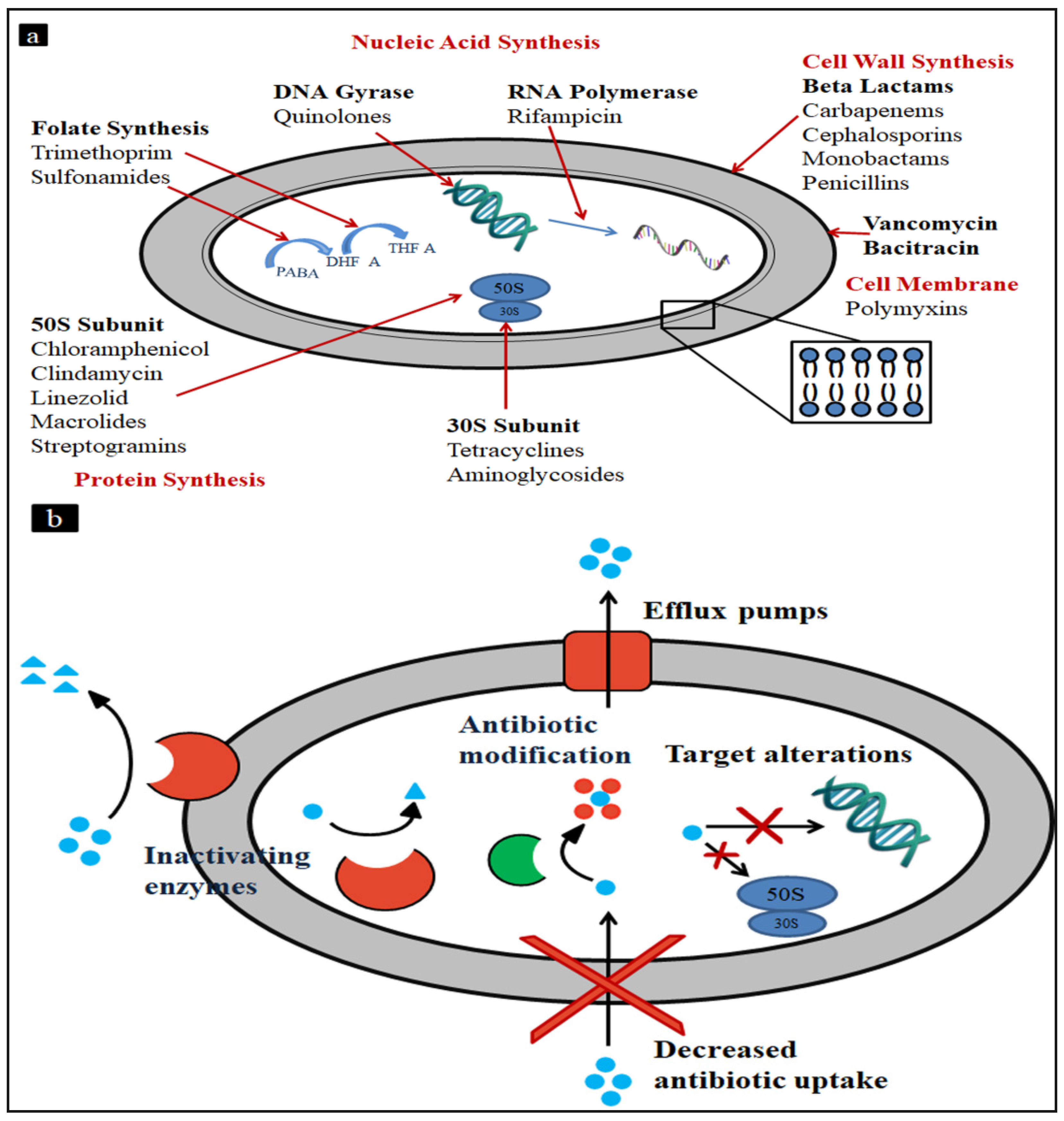
Figure 1. (a) Antibiotic targets and (b) bacterial resistance mechanism.
Antibiotics 11 00606 g001
Figure 2. Schematic representation of (A) an intact bacterial cell membrane and (B) the effect of nanoantibiotics on the reliability of a bacterial cell membrane (Adapted from Ref. [96]).
Antibiotics 11 00606 g002
Figure 3. Chemical structures of thiazole compounds 1–5 (Adapted from Ref. [105]).
Antibiotics 11 00606 g003
Table 1. Anti-MRSA activities of medicinal plants.
S. NoBotanical NameCommon NamePlant Part UsedExtracting SolventMIC/MBC (mg/mL) MRSARef.
1.Butea monosperma Lam.Flame-of-the-forestLeafEthanol5.91/13.30[34]
2.Acacia catechu (L. f.) WilldCutch tree, black catechuWoodEthanol1.6–3.2/25[35]
3.Callistemon rigidus R.Br.Stiff bottlebrushLeafMethanol0.00125–0.08[36]
4.Acacia albida Del.GawoStem barkMethanol3.0/4.0[37]
5.Nymphaea lotus Linn.White lotusLeafEthanol5.0–10.0/10.0–30.0[38]
6.Impatiens balsaminaGarden balsamLeafEthanol6.3/25[39]
7.Garcinia mangostana L.MangosteenFruit shellEthanol0.05–0.4/0.1–0.4[40]
8.Peltophorum pterocarpum (DC.)Yellow flame treeBarkEthanol0.1–0.8/6.3[41]
9.Psidium guajava L.GuavaLeafEthanol0.2–1.6/6.3[42]
10.Punica granatum L.PomegranateFruit shellEthanol0.2–0.4/1.6–3.2[43]
11.Uncaria gambir (Hunter) Roxb.Gambier, White cutchLeaf, stemEthanol0.4–0.8/3.2[44]
12.Walsura robustaBonlichuWoodEthanol1.6–3.2/25[45]
13.Swietenia mahagoniMahagoniSeedEthanol0.2–0.78/0.78–1.56[46]
14.Senna alataCandle bushLeafEthanol0.5/ND[47]
15.Garcinia Morella Desr.GambogeWhole plantEthanol0.016–0.064/0.064–0.26[48]
16.Melianthus major L.Giant honey flowerLeavesEthanol0.78/3.12[49]
17.Melianthus comosus VahlHoney flowerLeavesEthanol0.39/1.56[49]
18.Withania somnifera L.AshwagandhaRoots & leavesEthanol1.56/>6.25[50]
19.Quercus infectoria OlivierOak gallsNutgallsEthanol0.4–3.2/3.2–6.3[51]
20.Thymus vulgaris L.ThymeLeavesEssential oil0.057/ND[52]
Table 2. The synergy of plant-based or microbial-based NPs and antibiotics for antibacterial activity.
Nano ParticlesSource of Reducing AgentCombination of AntibioticTargeted BacteriaRef.
Ag NPLeaf extract of Typha angustifoliaGentamicin, cefotaxime, meropenemE. coli, K. pneumonia[72]
Ag NPDioscorea bulbifera tuber extractβ-lactam (piperacillin) and macrolide (erythromycin)A. baumannii[73]
Ag NPSilky hairs of aqueous corn extractKanamycin and rifampicinE. coli, S. aureus[74]
Ag NPEichhornia crassipesVancomycin, penicillin, streptomycin, and tetracyclineS. aureus, E. coli, K. pneumonia, Enterococcus[75]
Au NPCitrullus lanatus rind extractKanamycin, rifampicinBacillus cereus, E. coli, Listeria monocytogenes, S. aureus, S. typhi[76]
Ag NPCorn leaf waste of Zea mays extractKanamycin and rifampicinE. coli ATCC 42890, Bacillus cereus ATCC 13061 19115, and S. aureus ATCC 49444[77]
Ag NPFlower broth of Tagetes erectaCommercial antibioticsGram-positive (S. aureus and B. cereus), Gram-negative (E. coli and P. aeruginosa) bacteria[78]
Ag NPGum kondagoguCiprofloxacin, streptomycin, and gentamycinGram-positive (S. aureus 25923, S. aureus 49834) and Gram-negative (E. coli 25922, P. aeruginosa 27853)[79]
Ag NPAcinetobacter calcoaceticusAminoglycosides, β-lactams, cephalosporins, tetracyclinesA. baumannii, P. aeruginosa, S. aureus, S. typhi[80]
Ag NPE. coliBacitracin, ampicillin, kanamycinCorynebacterium diphtheria, E. coli, K. pneumonia[81]
Ag NPAspergillus flavus and Emericella nidulansAmikacin, kanamycin and streptomycinE. coli, S. aureus and P. aeruginosa[82]
Ag NPAspergillus flavusCiprofloxacin, vancomycin, gentamycinGram-positive and Gram-negative bacteria[83]
Ag NPBacteria from petroleum soilDoxycyclineKlebsiella pneumonia[84]
Ag NPStreptomyces xinghaiensis OF1 strainAmpicillin, kanamycin, and tetracyclineE. coli, P. aeruginosa, S. aureus and Klebsiella pneumonia[85]
Ag NPTrichoderma virideAmpicillin, kanamycin, and erythromycinGram-positive and Gram-negative bacteria[86]
Table 3. Minimum inhibitory concentration (MIC in µg/mL) of thiazole compounds 15, clindamycin and mupirocin (tested in triplicate) against MRSA and mupirocin-resistant S. aureus (NRS107) strain isolated from skin wounds [105].
CompoundS. aureus Strain Number
NRS107USA300USA400USA800USA1000USA1100
11.31.31.31.31.31.3
22.82.82.82.82.82.8
32.85.65.65.62.85.6
413.313.313.313.313.313.3
56.46.412.86.412.86.4
Mupirocin1024.01.01.04.04.04.0
Clindamycin0.11.80.10.10.10.1
Publisher’s Note: MDPI stays neutral with regard to jurisdictional claims in published maps and institutional affiliations.