Article

Thymus zygis Essential Oil: Phytochemical Characterization, Bioactivity Evaluation and Synergistic Effect with Antibiotics against Staphylococcus aureus

1
CICS-UBI—Health Sciences Research Centre, University of Beira Interior, Av. Infante D. Henrique, 6200-506 Covilhã, Portugal
2
Center of Potential and Innovation of Natural Resources, Polytechnic Institute of Guarda, 6300-559 Guarda, Portugal
3
FibEnTech—Department of Chemistry, Fibrous Materials and Environmental Technologies, University of Beira Interior, Rua Marquês d’Ávila e Bolama, 6201-001 Covilhã, Portugal
*
Author to whom correspondence should be addressed.
Antibiotics 2022, 11(2), 146; https://doi.org/10.3390/antibiotics11020146
Submission received: 22 December 2021 / Revised: 16 January 2022 / Accepted: 20 January 2022 / Published: 24 January 2022

Abstract

:
Staphylococcus aureus is a nosocomial bacterium causing different infectious diseases, ranging from skin and soft-tissue infections to more serious and life-threatening infections such as sepsis, meningitis and endocarditis, which may be exacerbated by antibiotic resistance. Plant products may be seen as an alternative as antibacterial agents, namely, against S. aureus. Thus, the aim of this work was to characterize the chemical composition and evaluate the bioactive properties of the T. zygis essential oil (EO), with a focus on antimicrobial activity against S. aureus. Gas chromatography coupled with mass spectrometry was used to assess the chemical composition of the T. zygis EO, and the antioxidant activity was evaluated using the DPPH method and β-carotene-bleaching assay. The antimicrobial activity against S. aureus strains, the interaction with different antibiotics and the attenuation of this bacterium’s virulence were evaluated. The T. zygis EO showed antioxidant activity acting through two different mechanisms and antibacterial activity against S. aureus, with antibiofilm and antihaemolytic properties. This EO also demonstrated synergistic or additive interactions in combination with ampicillin, ciprofloxacin or vancomycin against S. aureus strains and, in some cases, changed the antibiotic-resistance phenotype from resistant to susceptible. Therefore, the present work demonstrates the good bioactive properties of the EO of T. zygis, mainly the antimicrobial activity against S. aureus, revealing its potential to be used as an antibacterial agent.

1. Introduction

Antibiotics are used as the primary weapon against infections; while, at first, antibiotics were highly effective, their inappropriate use and high selective pressure have led to the emergence and spread of antibiotic-resistant bacteria [1]. In fact, antibiotic resistance has increased dramatically in recent decades and is now considered one of the greatest global health threats [2,3].
Staphylococcus aureus is a Gram-positive facultative anaerobic human pathogen of both nosocomial and community-acquired infections worldwide [4,5]. S. aureus is a commensal bacterium located on the skin and mucous membranes, but also a virulent bacterial pathogen associated with high morbidity and mortality [6,7,8,9]. This opportunistic pathogen can cause numerous acute and chronic infections [5,10], such as moderately severe skin infections, fatal pneumonia, sepsis, meningitis, endocarditis, or toxic-shock syndrome [7,11,12]. The higher rates of colonization, augmented use of surgical implants, immunosuppressive conditions, and escalation of antibiotic resistance have increased the prevalence of these infections [10]. Due to frequently occurring antibiotic resistance in S. aureus isolates, S. aureus infections are particularly problematic, and methicillin-resistant S. aureus (MRSA) is of utmost importance clinically. The World Health Organization states that people with MRSA infections are 64% more likely to die than people with drug-sensitive infections, and so it is on the list of microorganisms for which further investigation is critical [13].
When bacteria become resistant to first-line medicines, alternative therapies may be used [3]. The development of novel antibiotics remains a dominant approach for the treatment of bacterial-associated infections; however, this discovery is challenging [1]. Thus, it is important to explore alternative strategies and molecules to fight antibiotic-resistant S. aureus [1]. One possible solution is to combine antibiotics with other nonantibiotic drugs or to combine antibiotics with adjuvants or antimicrobials selected from the reservoir of bioactive compounds in nature [3,14].
Plant products have been used in folk medicine throughout human history and are the primary source of healthcare for much of the world’s population [2]. Hereupon, the need for novel antibacterial therapies has led to an increase in research into natural products as antibacterial agents [2]. Plants naturally produce a wide diversity of secondary metabolites, such as essential oils (EOs), that serve as defence compounds protecting against pathogens; therefore, they are important sources for the discovery of natural bioactive products [1].
Essential oils are complex blends of secondary metabolites, mainly terpenes and terpenoids [15], extracted by steam distillation, hydrodistillation or solvent extraction [16,17], which are usually stored in resin ducts, oil ducts, glands or trichomes of the plants [16]. EOs are natural products obtained from aromatic plant materials, with a broad spectrum of valuable biological properties and recognized uses in various areas (pharmaceutical, food, cosmetic and textile industries) [17,18]. They have been known to present antibacterial activity for centuries and so have been investigated for this purpose [2].
Thymus zygis, also known as red thyme, is predominantly found in the Mediterranean region, Asia, Southern Europe and North Africa and has been used for a long time as a spice or drug [19,20]. Its EO is known for its bioactive properties, such as antibacterial [21], antifungal [22,23] antiviral [24], antigiardial [25], insecticidal [26,27] and other properties.
Thus, considering the relevance of S. aureus resistance to antibiotics and the bioactive effects of T. zygis, this work aimed to evaluate the chemical composition of the T. zygis EO and to provide a better understanding of its antioxidant properties and antimicrobial activity against S. aureus strains, as well as cytotoxicity. The effect of the T. zygis EO on the virulence attenuation of S. aureus and also its interaction with antibiotics were evaluated.

2. Results

2.1. T. zygis EO Chemical Composition

The analysis of the chemical composition of the T. zygis EO through gas chromatography coupled with mass spectrometry (GC-MS) showed eighteen compounds, accounting for 94% of the total composition of the EO. The main components were identified as thymol (43.17%), carvacrol (13.00%) and p-cymene (10.58%) (Table 1).

2.2. T. zygis EO Antioxidant Activity

A very strong antioxidant activity was exhibited by the T. zygis EO according to the DPPH method and based on Scherer and Godoy classification [28], with IC50 values of 2.00 ± 0.15% (Table 2). The antioxidant activity of the T. zygis EO proved to have an identical effect to the Trolox standard with similar AAI values. Regarding the antioxidant activity according to a β-carotene-bleaching assay, the T. zygis EO showed antioxidant activity through the inhibition of lipid peroxidation (Table 2). For this reason, it can be said that the T. zygis EO has antioxidant activity through at least two different mechanisms, the inhibition of lipid peroxidation and sequestration of free radicals.

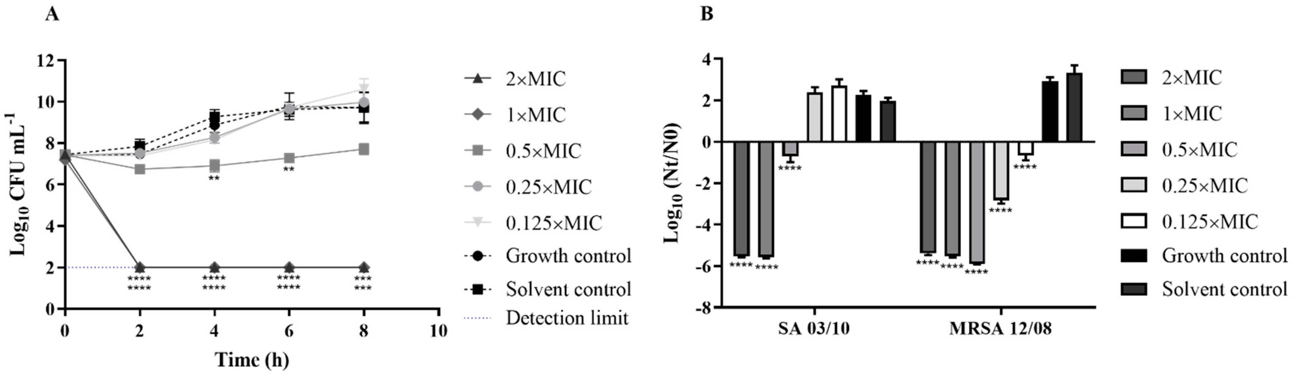
2.3. T. zygis EO Antibacterial Activity

The antimicrobial activity of the T. zygis EO was evaluated by using different methodologies and considering different parameters. Thus, first, it was screened through the disc-diffusion methodology, considering the EO and its volatile compounds (Table 3). According to the obtained results, higher antimicrobial activity was found against S. aureus ATCC 25923, with an inhibition halo of 35.10 ± 4.57 mm. The S. aureus SA 03/10 strain was the most resistant to the T. zygis EO, with an inhibition halo of 20.67 ± 1.59 mm. Concerning the evaluation of the volatile compounds of the T. zygis EO (Table 3), the T. zygis EO’s compounds released from the disc during incubation demonstrated inhibitory activity against all three S. aureus strains, showing inhibition halos between 16.26 ± 5.15 and 27.54 ± 4.10 mm. Regarding the study of the antimicrobial activity through the MIC determination (Table 3), the essential oil of T. zygis presented the same MIC value of 0.05% for the strains S. aureus ATCC 25922 and SA 03/10 and 0.1% against MRSA 12/08.
The antimicrobial activity can also be observed by looking at the time–kill curves showing that the T. zygis EO had a bactericidal effect at 1× and 2×MIC for all the strains of S. aureus (Figure 1). Furthermore, after 4 h of incubation, significant reductions in the logarithmic bacterial counts were observed for MRSA 12/08 with 0.5× MIC, 0.25× MIC and 0.125× MIC of T. zygis EO (p < 0.0001), while only the subinhibitory concentration of 0.5× MIC led to a significant reduction in S. aureus SA 03/10 (p < 0.0001).
The combined use of non-antibiotic compounds (known as antibiotic adjuvants) and antibiotics can be a strategy to enhance the activity of antibiotics and thus increase the susceptibility of resistant strains of bacteria [2,15]. According to the results presented in Figure 2, the T. zygis EO showed interactions with the antibiotics ampicillin, ciprofloxacin and vancomycin, demonstrating a synergistic (FICI ≤ 0.5) or additive (0.5 < FICI ≤ 1) effect, according to the classification of [29]. With this association, a synergic interaction occurred for the MRSA 12/08 strain with ampicillin or ciprofloxacin and the T. zygis EO, while the other combinations presented an additive interaction. Furthermore, the T. zygis EO resensitised S. aureus SA 03/10 to the antibiotics ampicillin, ciprofloxacin and vancomycin, and S. aureus MRSA 12/08, to ampicillin or ciprofloxacin.

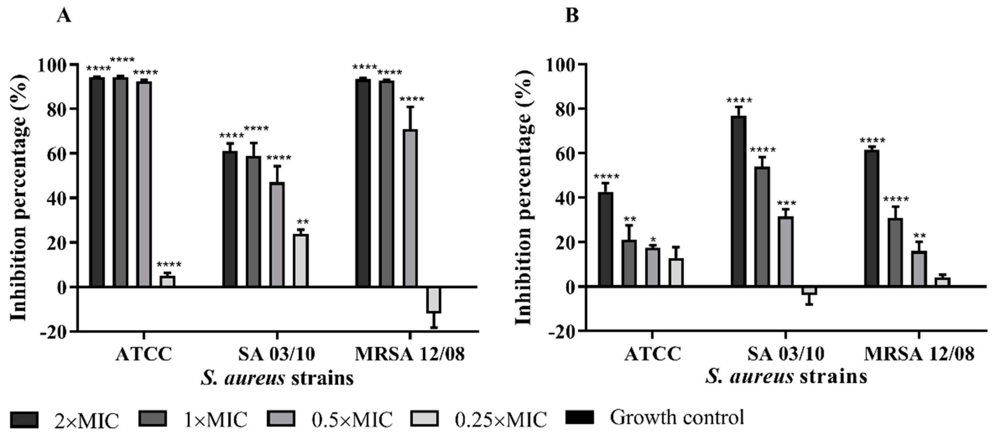
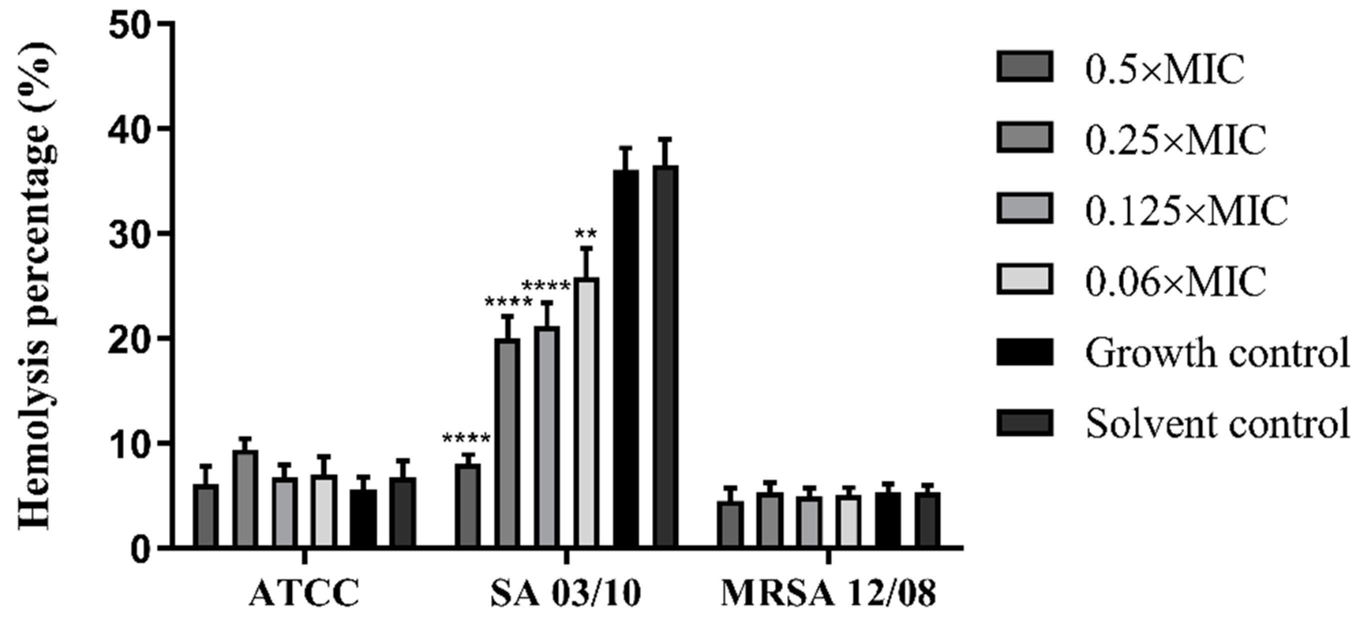
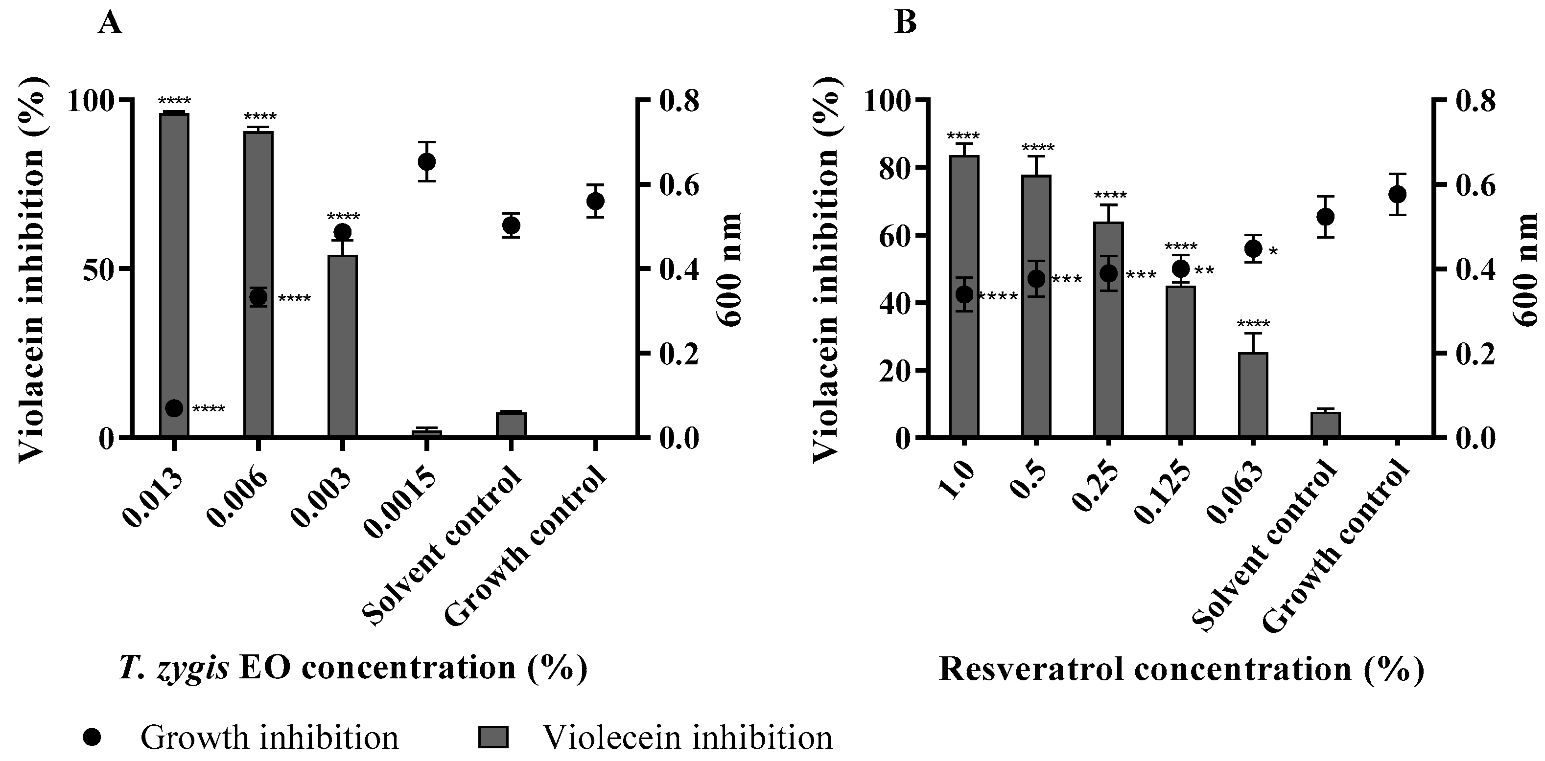
2.4. T. zygis EO Anti-Virulence Activity

The essential oil of T. zygis was shown to affect virulence factors of S. aureus, such as by the inhibition of biofilm formation, the elimination of biofilms formed or affecting their haemolytic ability. The T. zygis EO was shown to be able to inhibit the formation of biofilms by the strains of S. aureus and also to partially eliminate preformed biofilms even at subinhibitory concentrations (Figure 3 and Figure 4). The effect of the essential oil of T. zygis in inhibiting biofilm formation was more pronounced than that in eliminating preformed biofilms, with the exception of the S. aureus SA 03/10 strain.
The interference of the T. zygis EO with the haemolytic ability of the strains was also evaluated. Of the three strains of S. aureus under study, only the SA 03/10 strain demonstrated haemolytic capacity. The pre-exposure of S. aureus to subinhibitory T. zygis EO concentrations was shown to significantly reduce the haemolytic activity of S. aureus SA 03/10 compared to the respective controls (Figure 5) in a dose-dependent way.
As quorum sensing is a mechanism that allows bacteria to control the regulation and the secretion of virulence factors, we further tested its potential inhibition by the EO [17,30]. Using C. violaceum as a biosensor strain to evaluate the potential of the T. zygis EO as a quorum-sensing inhibitor, it was observed that the concentration of 0.006% led to a significant reduction in the violet pigment production (p < 0.0001) without affecting the growth of C. violaceum (Figure 6). The concentration of 0.006% inhibited almost 100% of the violacein production, and the concentration of 0.003% inhibited approximately 50% of the pigment. It can also be observed that the T. zygis EO worked in a dose-dependent manner, and when the concentration of the T. zygis EO was decreased, the violacein inhibition also decreased. Resveratrol was used as a positive control, and there was a statistically significant inhibition of violacein production at all the concentrations under study.
To understand the biocompatibility of the T. zygis EO for human cells, the effect of this essential oil was studied using a normal human dermal-fibroblast cell line for the evaluation of cytotoxicity (Figure 7). The incubation with the T. zygis EO reduced the viability of these cells in a dose-dependent manner. The results obtained in an MTT assay showed that the viability of NHDF was more than 70% when they were seeded in contact with the T. zygis EO at concentrations between 0.0125 and 0.0030%, for 24 h. Therefore, considering the ISO 10993:5–2009, it is possible to consider that such concentrations are non-cytotoxic when compared to the negative-control group (untreated cells).

3. Discussion

EOs have been used for centuries in perfumery, cosmetics and medicine and as part of spices and herbs in foods, and they are associated with a broad range of bioactive properties such as antibacterial and antioxidant activities [31]. The potential use of essential oils for developing promising antimicrobial agents with potential against S. aureus has been widely studied [32,33]. In the literature, it is described that T. zygis EO has antibacterial and antifungal activity against several microorganisms [34,35,36] and several other bioactive properties [15,21,37]; however, its interaction with antibiotics and several forms of antimicrobial activity required more in-depth studies.
The bioactive properties of EOs are correlated with their compositions. Therefore, this determination is important and may allow relating the composition with the biological activities [18,38]. The composition of the T. zygis EO used in this work is similar to that presented in the literature, where thymol is presented as the major compound [35,36,39,40] and the cymene and carvacrol are found with considerable percentages in relation to other compounds [25,35,40,41].
Antioxidants are important because they can compete with free radicals and avoid the propagation of oxidation reactions [18]. An increase in free-radical production and decline in the activities of antioxidant-enzyme systems can damage membranes, lipids and lipoproteins and can induce DNA mutations [42,43], and the implications of lipid peroxidation can lead to a diverse number of pathological disorders [42,43,44]. The results in this work show the ability of T. zygis EOs to scavenge free radicals, as well as the results for the inhibition of lipid peroxidation [42], and are corroborated by other authors [19,45]. Different samples of T. zygis EOs were evaluated by Carrasco et al. [43], and it was shown that T. zygis EO with a high proportion of thymol led to better antioxidant activity, using different methodologies. As can be seen in the review of Escobar et al. [46], there are several studies showing the antioxidant activity of the isolated compound thymol. Considering that thymol is the major compound of the T. zygis EO under study, the high antioxidant activity may be mainly due to this compound.
The broad spectrum of antibacterial activity of many EOs suggests a wide range of applications as antibacterial agents [2]. The T. zygis EO and its volatile compounds showed good antimicrobial activity against S. aureus, as previously described [47]. The authors related the high amount of monoterpenes in the vapour of the EO with the presented activity, as it is easier for these compounds to attack the bacterium compared to the liquid phase [47]. In accordance with our results, it was described that T. zygis EO also shows good inhibition of the growth of MRSA isolates [48]. The amount of thymol was also correlated with a better antimicrobial activity of T. zygis EO against S. aureus, pointing to its role in the activity of the EO [19], which may correlate with the activity observed in this work. The antimicrobial activity of the T. zygis EO was further validated by time–kill curves, demonstrating its bactericidal action even at subinhibitory concentrations, similarly to what has been described for other essential oils [49,50,51] and for A. baumannii and K. pneumoniae strains with T. zygis EO [15].
Drug discovery has looked to natural products for the purpose of combating infections caused by multiresistant bacteria [2]. The combination of multitarget antivirulence compounds, such as EOs, and antibiotics can help to restore the effectiveness of antibiotics, as can be seen in the review of Owen and Laird [2], and is a promising approach for combating antibiotic-resistant S. aureus [1,2,15]. In fact, T. zygis EO shows promising results in this area. The majority of the combinations of the T. zygis EO and antibiotics investigated mainly showed additive interactions; however, several of these additive combinations restored antibiotic sensitivity according to Clinical and Laboratory Standards Institute breakpoints [52]. The combination of the T. zygis EO and ampicillin, ciprofloxacin or vancomycin changed the resistance phenotype from resistant to sensitive in SA 03/10 strain (Figure 2). Regarding the two synergistic effects obtained, namely, that between the T. zygis EO and the antibiotic ampicillin or ciprofloxacin against the MRSA 12/08 strain, the presence of the EO also changed the resistance phenotype from resistant to sensitive in this strain. Moreover, the isobolograms show additive or synergistic effects between the combinations of T. zygis EO and ampicillin, ciprofloxacin or vancomycin (Figure 2). These results correlate with the synergistic interaction between thymol and ampicillin previously described [53]. In fact, as reviewed by Langeveld et al. [3], several studies showed interactions between thymol and different classes of antibiotics among different microorganisms. This interaction may be associated with the mechanism of action of thymol. Wang et al. [54] showed that thymol disrupted S. aureus cell membrane integrity, which may decrease cell viability and also increase the ability of other drugs to permeate the membrane [54].
The data obtained here showed that the combinations of the T. zygis EO and the antibiotics ampicillin, ciprofloxacin and vancomycin were able to decrease the MICs of antibiotics substantially and restore sensitivity to them, showing the EO’s potential in combating antibiotic-resistant S. aureus strains. As far as we know, there are no studies about the interaction of T. zygis EOs with antibiotics.
Antibiotic-resistant S. aureus poses a severe threat to human health, and antivirulence therapy is a potential antibacterial strategy for combating S. aureus-associated infections [1]. In fact, the T. zygis EO presents activity against some of the virulence factors of S. aureus. Among these factors, biofilms are associated with indwelling-medical-device-associated infections, endocarditis, osteomyelitis, conjunctivitis and other diseases [12]. Furthermore, biofilms can be a form of resistance to antimicrobials, host defence systems and external stresses [5,30,32]. The T. zygis EO decreased S. aureus virulence through the inhibition of biofilm formation and even the elimination of previously formed biofilms, even at subinhibitory concentrations. The antibiofilm-formation effect was further validated by SEM analysis. This antibiofilm efficacy of T. zygis EO was already described in the literature [40,48].
S. aureus is a major human pathogen that produces diverse virulence factors, such as α-haemolysin (Hla; also known as α-toxin) [4,7,9], one of the main cytotoxic agents secreted by S. aureus, which has been implicated in the pathogeneses of sepsis, pneumonia and severe skin infections [4,9]. The EO from T. zygis in the present work was shown to reduce the haemolytic capacity of S. aureus, comparable to other EOs that presented a similar effect [55,56]. To the best of our knowledge, this is the first report showing the efficacy of T. zygis EO in reducing the haemolytic capacity of S. aureus.
One of the antivirulence strategies aims to interfere with cell–cell communication or quorum sensing. Thus, the discovery of quorum-sensing inhibitor candidates has been presented as a step in the path toward the integration of the antivirulence strategy into the management and treatment of S. aureus infections [10]. The essential oil of T. zygis demonstrated the ability to inhibit the formation of violacein in C. violaceum, indicating its potential as an inhibitor of quorum sensing. Thus, the results show that, in addition to T. zygis having antimicrobial activity against planktonic cells, it also reduces virulence factors such as quorum sensing and biofilm formation.
The chemical composition and the biological activities of EOs are important to know, but determining their utilization limits, including their safety, is also important [18].
Similarly to our work, where a strong decrease in MTT reduction was observed for 0.25 µL/mL, the cytotoxicity of the EO of T. zygis against different cell lines was previously shown. In a fetal mouse-skin dendritic cell line (FSDC), no cytotoxic effect was observed at concentrations ranging from 0.08 to 0.16 µL/mL, and only for higher concentrations of the T. zygis EO (0.32 µL/mL) was a strong decrease in MTT reduction for the FSDC cell line noted [20]. Moreover, the EO of T. zygis did not cause a significant alteration in the viability of macrophages (RAW 264.7) and bovine aortic endothelial cells when compared to controls [25]. Nonetheless, when the T. zygis EO was tested on normal fetal lung fibroblasts (MRC-5 cell line), a significant decrease in cell viability was observed, comparable to the that for the positive control Triple antibiotic paste (metronidazole, ciprofloxacin and minocycline, at the ratio 1:1:1) [40]. In these studies, the essential oils of T. zygis had differences in their composition compared to the one studied in the present work, which must be considered when the biomedical application of these essential oils is envisioned.
Nonetheless, at the concentrations of T. zygis EO for which the cytotoxicity of the EO is low, interesting biological activities were also obtained. In the range of concentrations showing a low level of cytotoxicity, the T. zygis EO was shown (i) to significantly reduce the logarithmic bacterial counts of S. aureus, (ii) to potentiate the effects of the studied antibiotics, (iii) to inhibit the haemolytic capacity in the SA 03/10 strain or inhibit biofilm formation, and (iv) to potentiate the elimination of preformed biofilms in the S. aureus strains, as well as inhibiting quorum sensing in C. violaceum.

4. Materials and Methods

4.1. Essential Oil and Bacterial Strains

The commercial Thymus zygis essential oil was acquired from the company Pharmaplant (Algarve, Portugal) and was obtained by steam distillation from the aerial parts. The essential oil was protected from light and stored at 4 °C until further use.
The reference strain S. aureus ATCC 25923 and two clinical isolates S. aureus SA 03/10 and S. aureus MRSA 12/08 were used as test microorganisms.

4.2. GC-MS Analysis of the T. zygis Essential Oil

The T. zygis EO was analysed on an Agilent Technologies 7890A GC-System apparatus equipped with a fused silica DB-5 capillary column (Agilent J&W Column, part number: 122–5032) with a 30 m × 0.25 mm inner diameter and 0.25 µm film thickness coupled with a mass spectrometer (MS) (Agilent Technologies 5975C, Inert XL MSD) Triple-Axis detector. The operating conditions for the mass spectrometer were set as follows: ion source temperature, 250 °C; ionization voltage, 70 eV; interface temperature, 280 °C. As the carrier gas, helium was used at a flow rate of 1 mL/min. The initial oven temperature was 40 °C, with a hold time of 5 min, and it was increased to 250 °C at a rate of 5 °C/min. A 1 µL volume of T. zygis EO at a concentration of 10% (v/v) in dichloromethane was injected. The NIST Mass Spectral Software and Agilent GC/MSD ChemStation Software were used to calculate the relative concentration and perform identification.

4.3. Antioxidant Activity

The antioxidant activity of the T. zygis EO was evaluated using two methodologies, the 2,2-diphenyl-1-picrylhydrazyl radical (DPPH) and the β-carotene-bleaching assays, according to Coimbra et al. [57] with some adaptations. In the first one, 5 µL of each concentration of T. zygis EO methanolic solution was separately mixed with 195 µL of DPPH methanolic solution in 96-well microtiter plates. Methanol was used as the negative control, and Trolox (Acros Organics, Geel, Belgium) and gallic acid (Acros Organics, Geel, Belgium), as standards. The absorbances were measured at 515 nm, and the antioxidant activity was expressed through the antioxidant activity index (AAI), calculated according to:
AAI = (final concentration of DPPH in the control sample/IC50.
In the second methodology, 56.6 μL of a T. zygis EO methanolic dilution was added to 943.4 μL of the emulsion. The emulsion was prepared with 500 µL of β-carotene solution at 20 mg/mL in chloroform, 40 µL of linoleic acid, 400 µL of Tween 40 and 1 mL of chloroform. Then, the chloroform was evaporated under vacuum at 45 °C, and 100 mL of oxygenated distilled water was added to form an emulsion. Butylated hydroxytoluene (BHT, purity 99%, Acros Organics, Geel, Belgium) was used as a standard, and methanol, as a negative control. The absorbances were read at 470 nm, against a blank containing an emulsion without β-carotene, and the percentage of inhibition of β-carotene oxidation was calculated using the equation:
% Inhibition = ((Abs samplet = 1h − Abs controlt = 1 h)/(Abs controlt = 0 h − Abs controlt = 1 h))×100.
All tests were performed in triplicate.

4.4. Antimicrobial Activity

4.4.1. Disc-Diffusion Method and Vapour-Phase Antimicrobial Activity Determination

The disc-diffusion method was performed to evaluate the susceptibility of the S. aureus strains to the T. zygis EO as described by Luís et al. [58]. Tetracycline at 20 µg/disc was used as a positive control. The susceptibility of the S. aureus strains to the volatile compounds of the T. zygis EO was evaluated as described by Duarte et al. [59]. These methodologies were performed with tryptic soy agar (TSA) medium. The inhibition halos were measured in millimetres, and the results are presented as means ± standard deviations. At least three independent assays were performed.

4.4.2. Determination of the Minimum Inhibitory Concentration (MIC)

The susceptibility of the S. aureus strains to the T. zygis EO was evaluated through the broth microdilution method according to Coimbra et al. [57] with modifications. Briefly, in 96-well plates, the essential oil was serially diluted with tryptic soy broth (TSB, RPD microbiology, Barcelona, Spain). Dimethyl sulfoxide (DMSO) was used as the solvent for the improvement of the solubility, with a maximum concentration of 2% (v/v) (no growth inhibition). The inoculum, with a concentration of 0.5 McFarland, was diluted in medium, and 50 µL was added to the wells to obtain a final volume of 100 µL and a concentration of 5 × 10⁵ colony-forming units (CFU)/mL per well. The MIC was determined as the lowest concentration of T. zygis EO without visible growth. At least three independent determinations with duplicates were performed, and the results are presented as modal values.

4.4.3. Time–Kill Curves

The time–kill curve assay was performed based on Ferreira and Domingues [60] with minor modifications. Briefly, S. aureus strains grown overnight were used to prepare a cellular suspension to give a final cell concentration of 106 CFU/mL, and it was exposed to several concentrations of T. zygis EO (from 0.125× to 2× MIC). A solvent control with DMSO (1% (v/v)) and growth control were also performed. The viable counts were determined by the drop-plate method at 0, 2, 4, 6 and 8 h of incubation from the tubes incubated at 37 °C. The independent experiments were performed at least thrice.

4.4.4. Antibiofilm Activity of T. zygis EO

Biofilm Formation

The inhibition of biofilm formation was based on the previously described method of Stepanović et al. [61] with modifications. Briefly, S. aureus strains were grown overnight at 37 ˚C, at 250 rpm, in TSB. Afterwards, the turbidity of the suspension was adjusted to an OD600 nm ~1.5 and diluted to achieve a final concentration in the wells of 1 × 10⁷ CFU/mL. Serial two-fold dilutions of T. zygis EO (0.25 to 2× MIC) were prepared in TSB, supplemented with 0.5% glucose, in 96-well flat-bottom polystyrene microtiter plates, and 100 μL of each bacterial suspension was added to the wells to obtain a final volume of 200 µL. The plates were incubated at 37 °C for 24 h. For the positive control, the bacterial suspension with medium was used, whereas for the negative control, only the culture medium was used. A solvent control in the presence of DMSO (0.125 to 1%) was also performed. After incubation, the contents of the plates were poured off, and each well was washed twice with 200 µL of distilled water to remove the loosely attached cells. The remaining attached bacteria were fixed with methanol (200 µL) for 20 min; after methanol removal, the plates were air dried. Staining was achieved with 0.1% (w/v) crystal violet (200 µL) for 10 min, the dye was removed, and the wells were washed thrice with 400 µL of distilled water. The crystal violet bound was dissolved with 33% (v/v) glacial acetic acid per well (200 µL), and the absorbance at 570 nm was determined using a microplate reader. At least five replicates of three independent experiments were conducted.

Biofilm Dispersion

The effect of the T. zygis EO on preformed biofilms was evaluated based on Duarte et al. [62] with some adaptations. Briefly, biofilms were prepared as mentioned above by inoculating 100 μL of the bacterial suspension into the wells of 96-well flat-bottom polystyrene microtiter plates containing 100 μL of TSB supplemented with 0.5% glucose. Following incubation at 37 °C for 24 h, the medium was removed and 100 μL of each T. zygis EO or DMSO concentration was added to the biofilm in the wells. The plates were further incubated at 37 °C for 24 h. For the positive control, 100 μL of culture medium was added, whereas for the negative control, only the culture medium was used. After incubation, the biofilm biomass was evaluated by the crystal-violet staining method as described above. At least five replicates of three independent experiments were conducted.

4.4.5. Inhibition of Quorum Sensing

The anti-quorum-sensing activity of the T. zygis EO was assessed with the biosensor strain Chromobacterium violaceum ATCC 12472 and performed based on Asensio et al. [63] with some modifications. A bacterial suspension of C. violaceum ATCC 12472 was obtained from an overnight culture at 30 °C and 250 rpm in Luria–Bertani (LB) broth and then diluted in fresh LB broth to achieve DO600 nm 0.02. T. zygis EO and resveratrol (positive control) were serially 2-fold diluted with LB (final concentrations of 0.0015 to 0.013% and 0.063 to 0.10%, respectively), and 500 µL of each solution was applied to 48-well flat-bottom polystyrene microtiter plates. DMSO with a final concentration of 0.125% was used as the solvent control. Then, 500 µL of the bacterial suspension was added to the wells, and the plate was incubated at 30 °C without shaking for 48 h. After incubation, 750 µL from each well was transferred to a centrifuge tube and centrifuged at 5000× g for 3 min. The supernatants were discarded, and the pellets were vigorously vortexed with 750 µL of DMSO to dissolve the violacein. The samples were centrifuged again at 8000× g for 5 min to remove the C. violaceum cells and to evaluate the violacein production. A 200 µL volume of violacein-containing supernatant was added into a 96-well microplate in triplicate, and the optical density at 585 nm was measured using a plate reader. The growth inhibition of C. violaceum was evaluated by suspending the removed cells in 750 µL of distilled water, 200 µL of the suspension was applied into a 96-well microplate in triplicate, and the absorbance was measured at 600 nm. The violacein inhibition (%) was calculated using the equation 100 − ((ODsample/ODgrowth control) × 100).

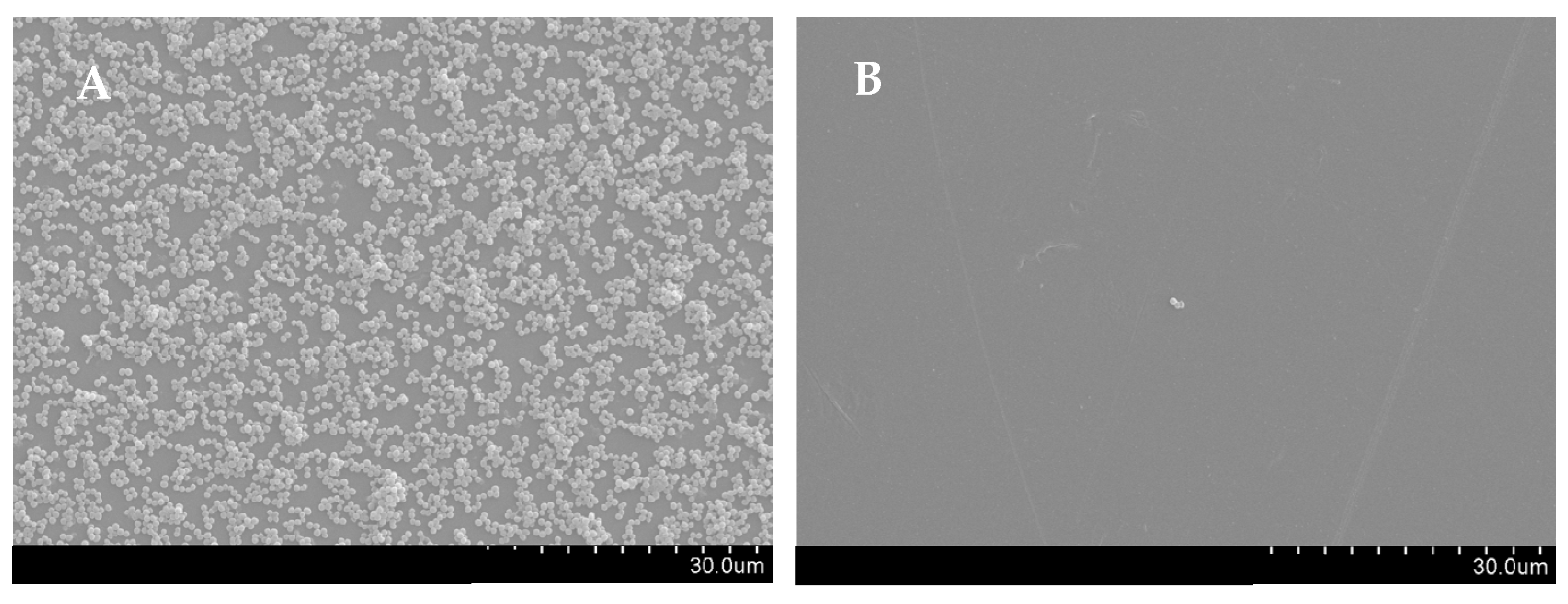
4.4.6. Scanning Electron Microscopy (SEM)

The effect of T. zygis EO on biofilm formation by the strain S. aureus MRSA 12/08 was observed through SEM according to Luís et al. [64] with slight modifications. Biofilm formation was performed as described above but, in this case, in 24-well plates containing a polystyrene coupon with dimensions of 1 cm × 1 cm. Initially, the coupons were washed and submerged in a 70% ethanol solution overnight, followed by exposure to UV radiation for 30 min on both sides. A 500 µL volume of T. zygis EO (1× MIC), DMSO (0.5% (v/v), solvent control) or TSB supplemented with 0.5% glucose (growth control) was added to the plate, and 500 µL of bacterial suspension was added. After 24 h of incubation at 37 °C, the wells were washed twice with an isotonic saline solution (0.85% (w/v) NaCl) and fixed with 500 µL of 2% glutaraldehyde and 4% formaldehyde solution in PBS for 3 h at room temperature. The coupons were then carefully washed with PBS, dehydrated in a graded ethanol series (25, 50, 70, 90 and twice with 100%) and dried in a desiccator overnight. Lastly, the coupons were mounted on a stub, sputter-coated with gold and examined with a scanning electron microscope (Hitachi S-3400 N).

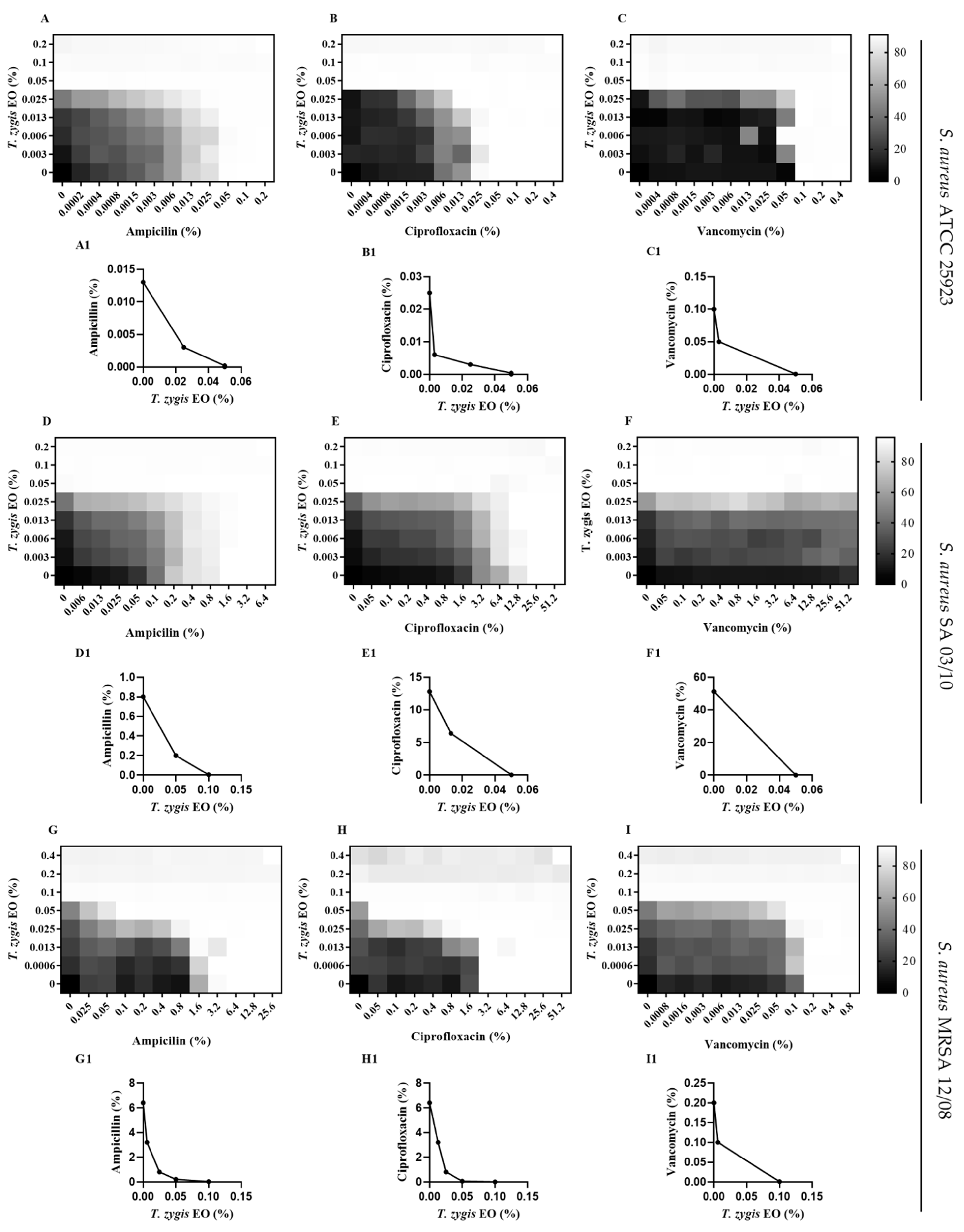
4.4.7. Checkerboard Assay

The checkerboard method was used to test the combined effect of the T. zygis EO and antibiotics, according to Silva et al. [65] with some adaptations. The inoculum was prepared as described in Section 4.4.2 and the suspension was then diluted 1:67 in TSB to ensure a final cell concentration of 5 × 10⁵ CFU/mL in each well. Two microplates were prepared, one where T. zygis EO was successively diluted with TSB, vertically, with a final volume of 50 µL, and another plate, where successive dilutions of the antibiotics (ampicillin, ciprofloxacin or vancomycin) were carried out with TSB, in the horizontal direction. Subsequently, with a multichannel pipette, 50 µL from the plate with the antibiotic was transferred to the plate with the T. zygis EO, with the addition of 50 µL of inoculum to obtain a final volume of 150 µL per well. The concentrations of T. zygis EO and the antibiotics were selected based on the MIC values previously determined. The plate was incubated at 37 °C for 24 h.
The results for the combined effects of the T. zygis EO and antibiotics were calculated and are expressed in terms of the fractional inhibitory concentration index (FICI), equal to the sum of the fractional inhibitory concentration (FIC) of the T. zygis EO and FICs of the antibiotics. The FIC was defined as the MIC of the EO and antibiotic in combination divided by the MIC of the EO and antibiotic used alone. If FICI ≤ 0.5, it was considered to have a synergistic effect; for 0.5 < FICI ≤ 1, there was an additive effect; 1 < FICI < 4 showed an indifferent effect; and with FICI ≥ 4, the effect was antagonistic [29].

4.4.8. Effect of the T. zygis EO on the Haemolytic Capacity of S. aureus

The haemolytic activity of the S. aureus strains was evaluated as described by Lee et al. [5] with adaptations. Briefly, the S. aureus strains were grown overnight at 37 °C, at 250 rpm, for 16 h, and used to prepare a cellular suspension at a final concentration of 106 CFU/mL. Tubes were prepared by adding T. zygis EO (0.06 to 0.5× MIC) in TSB and the cellular suspension in a final volume of 3 mL. A solvent control with DMSO (0.125% (v/v)) and a growth control were performed, and all the tubes were incubated at 37 °C for 20 h. After the incubation, 100 µL from each tube was transferred to a U-bottom 96-well plate, and 100 µL of 2% (v/v) human erythrocytes were added. A negative control (PBS without bacteria) and positive control for total haemolysis (1% (v/v) Triton-X 100) were also included. The erythrocytes were collected from one healthy volunteer into a blood collection tube with ethylenediamine tetraacetic acid (EDTA) and washed thrice with PBS, and a stock solution was prepared in the same buffer. The plate was incubated at 37 °C for 1 h and, after the incubation, was centrifuged at 1000× g for 5 min. A 100 µL volume of each supernatant was transferred to a 96-well flat-bottom microtiter plate, and the absorbance at 492 nm was measured. At least four replicates of three independent experiments were conducted.

4.5. Evaluation of T. zygis EO Biocompatibility

The cytotoxicity of the T. zygis EO was evaluated using normal human dermal fibroblasts (NHDF cells isolated from the dermis of adult skin and acquired from PromoCell GmbH (Heidelberg, Germany)) that were initially seeded in 96-well flat-bottom culture plates with 2×10⁴ cells/well and containing DMEM-F12 supplemented with 10% FBS. Adherent cells were grown in an incubator with a humidified atmosphere containing 5% CO2 at 37 °C for a day. Then, the culture medium was removed, and the cells were incubated with several concentrations of T. zygis EO (0.0030 and 0.4%) for 24 h. Cells cultured with EtOH (96%) were used as a positive control, and those without materials were used as a negative control. The 3-(4,5-dimethylthiazol-2-yl)-2,5-diphenyltetrazolium bromide (MTT) assay was used to monitor the cell metabolic activity. For that, the medium was removed, and a PBS solution of 5 mg/mL of MTT (50 µL in each well) was added to each sample (n = 5). The plate was incubated in a 5% CO2 atmosphere for 4 h at 37 °C. To dissolve the pigmented formazan formed, 200 µL of DMSO (0.04 N) was added to the cells for 30 min. Afterwards, a microplate reader (Biorad xMark microplate spectrophotometer, Waltham, MA, USA) was used to read the absorbance at 570 nm.

5. Conclusions

To summarize, this work shows that T. zygis EO presents good antioxidant and antimicrobial properties. T. zygis EO presents activity against resistant S. aureus strains with bactericidal activity, while showing antibiofilm and antihaemolytic activities against S. aureus. The T. zygis EO’s prospects for improving the effect of antimicrobial agents was highlighted, since the combination of the T. zygis EO with the antibiotics ampicillin, ciprofloxacin and vancomycin potentiated the effects of these antibiotics against the S. aureus strains. These results show the possible use of T. zygis EO as an alternative antibacterial agent for the control of S. aureus.

Author Contributions

Conceptualization, S.F.; methodology, A.C., S.M., M.R., P.C. and L.S.; formal analysis, A.C., S.M. and S.F.; writing—original draft preparation, A.C.; writing—review and editing, A.C., S.M., M.R., P.C., L.S, A.P.D. and S.F.; supervision, A.P.D. and S.F.; funding acquisition, A.P.D. and S.F. All authors have read and agreed to the published version of the manuscript.

Funding

This work was developed within the scope of the CICS-UBI projects UIDB/00709/2020 and UIDP/00709/2020, financed by national funds through the Portuguese Foundation for Science and Technology/MCTES. The authors are very grateful for the support given by research unit Fiber Materials and Environmental Technologies (FibEnTech-UBI), on the extent of the project reference UIDB/00195/2020, funded by the Fundação para a Ciência e a Tecnologia, IP/MCTES through national funds (PIDDAC).

Institutional Review Board Statement

Not applicable.

Informed Consent Statement

Not applicable.

Data Availability Statement

Data are contained within the text.

Acknowledgments

Susana Ferreira acknowledges UBI and FCT by the contract of Scientific Employment according to DL57/2016.

Conflicts of Interest

The authors declare no conflict of interest.

References

  1. Wu, S.C.; Liu, F.; Zhu, K.; Shen, J.Z. Natural products that target virulence factors in antibiotic-resistant Staphylococcus aureus. J. Agric. Food Chem. 2019, 67, 13195–13211. [Google Scholar] [CrossRef] [PubMed]
  2. Owen, L.; Laird, K. Synchronous application of antibiotics and essential oils: Dual mechanisms of action as a potential solution to antibiotic resistance. Crit. Rev. Microbiol. 2018, 44, 414–435. [Google Scholar] [CrossRef] [PubMed]
  3. Langeveld, W.T.; Veldhuizen, E.J.A.; Burt, S.A. Synergy between essential oil components and antibiotics: A review. Crit. Rev. Microbiol. 2014, 40, 76–94. [Google Scholar] [CrossRef] [PubMed]
  4. Lee, K.; Lee, J.H.; Kim, S.I.; Cho, M.H.; Lee, J. Anti-biofilm, anti-hemolysis, and anti-virulence activities of black pepper, cananga, myrrh oils, and nerolidol against Staphylococcus aureus. Appl. Microbiol. Biotechnol. 2014, 98, 9447–9457. [Google Scholar] [CrossRef] [PubMed]
  5. Lee, J.H.; Kim, Y.G.; Park, J.G.; Lee, J. Supercritical fluid extracts of Moringa oleifera and their unsaturated fatty acid components inhibit biofilm formation by Staphylococcus aureus. Food Control 2017, 80, 74–82. [Google Scholar] [CrossRef]
  6. Zhang, Y.; Wang, J.; Dong, J.; Wei, J.; Wang, Y.; Dai, X.; Wang, X.; Luo, M.; Tan, W.; Deng, X.; et al. Inhibition of α-toxin production by subinhibitory concentrations of naringenin controls Staphylococcus aureus pneumonia. Fitoterapia 2013, 86, 92–99. [Google Scholar] [CrossRef]
  7. Otto, M. Staphylococcus aureus toxins. Curr. Opin. Microbiol. 2014, 17, 32–37. [Google Scholar] [CrossRef] [Green Version]
  8. Vandenesch, F.; Lina, G.; Henry, T. Staphylococcus aureus hemolysins, bi-component leukocidins, and cytolytic peptides: A redundant arsenal of membrane-damaging virulence factors? Front. Cell. Infect. Microbiol. 2012, 2, 12. [Google Scholar] [CrossRef] [Green Version]
  9. Singh, V.; Phukan, U.J. Interaction of host and Staphylococcus aureus protease-system regulates virulence and pathogenicity. Med. Microbiol. Immunol. 2019, 208, 585–607. [Google Scholar] [CrossRef]
  10. Quave, C.L.; Horswill, A.R. Flipping the switch: Tools for detecting small molecule inhibitors of staphylococcal virulence. Front. Microbiol. 2014, 5, 706. [Google Scholar] [CrossRef] [Green Version]
  11. Korem, M.; Gov, Y.; Shirron, N.; Shuster, A.; Rosenberg, M. Alcohol increases hemolysis by staphylococci. FEMS Microbiol. Lett. 2007, 269, 153–159. [Google Scholar] [CrossRef] [PubMed] [Green Version]
  12. Cheung, G.Y.C.; Bae, J.S.; Otto, M. Pathogenicity and virulence of Staphylococcus aureus. Virulence 2021, 12, 547–569. [Google Scholar] [CrossRef] [PubMed]
  13. World Health Organization. WHO’s First global Report on Antibiotic Resistance Reveals Serious, Worldwide Threat to Public Health. Available online: https://www.who.int/news/item/30-04-2014-who-s-first-global-report-on-antibiotic-resistance-reveals-serious-worldwide-threat-to-public-health. (accessed on 9 December 2021).
  14. Ju, J.; Xie, Y.; Yu, H.; Guo, Y.; Cheng, Y.; Qian, H.; Yao, W. Synergistic interactions of plant essential oils with antimicrobial agents: A new antimicrobial therapy. Crit. Rev. Food Sci. Nutr. 2020, 1–12. [Google Scholar] [CrossRef] [PubMed]
  15. Vázquez-Ucha, J.C.; Martínez-Guitián, M.; Lasarte-Monterrubio, C.; Conde-Pérez, K.; Arca-Suárez, J.; Álvarez-Fraga, L.; Pérez, A.; Crecente-Campo, J.; Alonso, M.J.; Bou, G.; et al. Syzygium aromaticum (clove) and Thymus zygis (thyme) essential oils increase susceptibility to colistin in the nosocomial pathogens Acinetobacter baumannii and Klebsiella pneumoniae. Biomed. Pharmacother. 2020, 130, 110606. [Google Scholar] [CrossRef] [PubMed]
  16. Raut, J.S.; Karuppayil, S.M. A status review on the medicinal properties of essential oils. Ind. Crops Prod. 2014, 62, 250–264. [Google Scholar] [CrossRef]
  17. Trifan, A.; Luca, S.V.; Greige-Gerges, H.; Miron, A.; Gille, E.; Aprotosoaie, A.C. Recent advances in tackling microbial multidrug resistance with essential oils: Combinatorial and nano-based strategies. Crit. Rev. Microbiol. 2020, 46, 338–357. [Google Scholar] [CrossRef]
  18. Ribeiro-Santos, R.; Andrade, M.; Sanches-Silva, A.; de Melo, N.R. Essential oils for food application: Natural substances with established biological activities. Food Bioprocess. Technol. 2018, 11, 43–71. [Google Scholar] [CrossRef]
  19. Cutillas, A.B.; Carrasco, A.; Martinez-Gutierrez, R.; Tomas, V.; Tudela, J. Thyme essential oils from Spain: Aromatic profile ascertained by GC–MS, and their antioxidant, anti-lipoxygenase and antimicrobial activities. J. Food Drug Anal. 2018, 26, 529–544. [Google Scholar] [CrossRef] [Green Version]
  20. Gonçalves, M.J.; Cruz, M.T.; Cavaleiro, C.; Lopes, M.C.; Salgueiro, L. Chemical, antifungal and cytotoxic evaluation of the essential oil of Thymus zygis subsp. sylvestris. Ind. Crops Prod. 2010, 32, 70–75. [Google Scholar] [CrossRef]
  21. Lagha, R.; Abdallah, F.B.; AL-Sarhan, B.O.; Al-Sodany, Y. Antibacterial and biofilm inhibitory activity of medicinal plant essential oils against Escherichia coli isolated from UTI patients. Molecules 2019, 24, 1161. [Google Scholar] [CrossRef] [Green Version]
  22. Debonne, E.; Vermeulen, A.; Van Bockstaele, F.; Soljic, I.; Eeckhout, M.; Devlieghere, F. Growth/no-growth models of in-vitro growth of Penicillium paneum as a function of thyme essential oil, pH, aw, temperature. Food Microbiol. 2019, 83, 9–17. [Google Scholar] [CrossRef] [PubMed]
  23. Yang, V.W.; Clausen, C.A. Antifungal effect of essential oils on southern yellow pine. Int. Biodeterior. Biodegrad. 2007, 59, 302–306. [Google Scholar] [CrossRef] [Green Version]
  24. Santoyo, S.; Jaime, L.; García-Risco, M.R.; Lopez-Hazas, M.; Reglero, G. Supercritical fluid extraction as an alternative process to obtain antiviral agents from thyme species. Ind. Crops Prod. 2014, 52, 475–480. [Google Scholar] [CrossRef]
  25. Machado, M.; Dinis, A.M.; Salgueiro, L.; Cavaleiro, C.; Custódio, J.B.A.; Do Céu Sousa, M. Anti-Giardia activity of phenolic-rich essential oils: Effects of Thymbra capitata, Origanum virens, Thymus zygis subsp. sylvestris, and Lippia graveolens on trophozoites growth, viability, adherence, and ultrastructure. Parasitol. Res. 2010, 106, 1205–1215. [Google Scholar]
  26. Sangha, J.S.; Astatkie, T.; Cutler, G.C. Ovicidal, larvicidal, and behavioural effects of some plant essential oils on diamondback moth (Lepidoptera: Plutellidae). Can. Entomol. 2017, 149, 639–648. [Google Scholar] [CrossRef]
  27. Park, C.G.; Jang, M.; Yoon, K.A.; Kim, J. Insecticidal and acetylcholinesterase inhibitory activities of Lamiaceae plant essential oils and their major components against Drosophila suzukii (Diptera: Drosophilidae). Ind. Crops Prod. 2016, 89, 507–513. [Google Scholar] [CrossRef]
  28. Scherer, R.; Godoy, H.T. Antioxidant activity index (AAI) by the 2,2-diphenyl-1-picrylhydrazyl method. Food Chem. 2009, 112, 654–658. [Google Scholar] [CrossRef]
  29. Roudashti, S.; Zeighami, H.; Mirshahabi, H.; Bahari, S.; Soltani, A.; Haghi, F. Synergistic activity of sub-inhibitory concentrations of curcumin with ceftazidime and ciprofloxacin against Pseudomonas aeruginosa quorum sensing related genes and virulence traits. World J. Microbiol. Biotechnol. 2017, 33, 50. [Google Scholar] [CrossRef] [PubMed]
  30. Algburi, A.; Comito, N.; Kashtanov, D.; Dicks, L.M.T.; Chikindas, M.L. Control of biofilm formation: Antibiotics and beyond. Appl. Environ. Microbiol. 2017, 83, e02508-16. [Google Scholar] [CrossRef] [PubMed] [Green Version]
  31. Perricone, M.; Arace, E.; Corbo, M.R.; Sinigaglia, M.; Bevilacqua, A. Bioactivity of essential oils: A review on their interaction with food components. Front. Microbiol. 2015, 6, 1–7. [Google Scholar] [CrossRef] [Green Version]
  32. Idrees, M.; Sawant, S.; Karodia, N.; Rahman, A. Staphylococcus aureus biofilm: Morphology, genetics, pathogenesis and treatment strategies Int. J. Environ. Res. Public Health 2021, 18, 7602. [Google Scholar] [CrossRef] [PubMed]
  33. Vieira, M.; Bessa, L.J.; Martins, M.R.; Arantes, S.; Teixeira, A.P.S.; Mendes, Â.; Da Costa, P.M.; Belo, A.D.F. Chemical composition, antibacterial, antibiofilm and synergistic properties of essential oils from Eucalyptus globulus LABILL. and seven Mediterranean aromatic plants. Chem. Biodivers. 2017, 14, e1700006. [Google Scholar] [CrossRef] [PubMed] [Green Version]
  34. Dorman, H.J.D.; Deans, S.G. Chemical composition, antimicrobial and in vitro antioxidant properties of Monarda citriodora var. citriodora, Myristica fragrans, Origanum vulgare ssp. hirtum, Pelargonium sp. and Thymus zygis oils. J. Essent. Oil Res. 2004, 16, 145–150. [Google Scholar] [CrossRef]
  35. Pina-Vaz, C.; Rodrigues, A.G.; Pinto, E.; Costa-de-Oliveira, S.; Tavares, C.; Salgueiro, L.; Cavaleiro, C.; Gonçalves, M.; Martinez-de-Oliveira, J. Antifungal activity of Thymus oils and their major compounds. J. Eur. Acad. Dermatol. Venereol. 2004, 18, 73–78. [Google Scholar] [CrossRef] [PubMed]
  36. Ballester-Costa, C.; Sendra, E.; Fernández-López, J.; Pérez-Álvarez, J.A.; Viuda-Martos, M. Chemical composition and in vitro antibacterial properties of essential oils of four Thymus species from organic growth. Ind. Crops Prod. 2013, 50, 304–311. [Google Scholar] [CrossRef]
  37. Sánchez-Hidalgo, M.; Montalbán-López, M.; Cebrián, R.; Valdivia, E.; Martínez-Bueno, M.; Maqueda, M. AS-48 bacteriocin: Close to perfection. Cell. Mol. Life Sci. 2011, 68, 2845–2857. [Google Scholar] [CrossRef]
  38. Rota, C.; Herrera, A.; Martınez, R.M.; Sotomayor, J.A.; Jordán, M.J. Antimicrobial activity and chemical composition of Thymus vulgaris, Thymus zygis and Thymus hyemalis essential oils. Food Control 2008, 19, 681–687. [Google Scholar] [CrossRef]
  39. Andrés, M.F.; González-coloma, A.; Muñoz, R.; De la Peña, F.; Julio, L.F.; Burillo, J. Nematicidal potential of hydrolates from the semi industrial vapor-pressure extraction of Spanish aromatic plants. Environ. Sci. Pollut. Res. 2018, 25, 29834–29840. [Google Scholar] [CrossRef]
  40. Marinković, J.; Ćulafić, D.M.; Nikolić, B.; Đukanović, S.; Marković, T.; Tasić, G.; Ćirić, A.; Marković, D. Antimicrobial potential of irrigants based on essential oils of Cymbopogon martinii and Thymus zygis towards in vitro multispecies biofilm cultured in ex vivo root canals. Arch. Oral Biol. 2020, 117, 104842. [Google Scholar] [CrossRef]
  41. Solarte, A.L.; Astorga, R.J.; De Aguiar, F.C.; De Frutos, C.; Barrero-Domínguez, B.; Huerta, B. Susceptibility Ddistribution to essential oils of Salmonella enterica strains involved in animal and public health and comparison of the Typhimurium and Enteritidis Serotypes. J. Med. Food 2018, 21, 946–950. [Google Scholar] [CrossRef]
  42. Youdim, K.A.; Deans, S.G.; Finlayson, H.J. The antioxidant properties of thyme (Thymus zygis L.) essential oil: An inhibitor of lipid peroxidation and a free radical scavenger. J. Essent. Oil Res. 2002, 14, 210–215. [Google Scholar] [CrossRef]
  43. Carrasco, A.; Tomas, V.; Tudela, J.; Miguel, M.G. Comparative study of GC-MS characterization, antioxidant activity and hyaluronidase inhibition of different species of Lavandula and Thymus essential oils. Flavour Fragr. J. 2015, 31, 57–69. [Google Scholar] [CrossRef] [Green Version]
  44. Jordán, M.J.; Martínez, R.M.; Martínez, C.; Moñino, I.; Sotomayor, J.A. Polyphenolic extract and essential oil quality of Thymus zygis ssp. gracilis shrubs cultivated under different watering levels. Ind. Crops Prod. 2009, 29, 145–153. [Google Scholar] [CrossRef]
  45. Ballester-Costa, C.; Sendra, E.; Fernández-López, J.; Pérez-Álvarez, J.A.; Viuda-Martos, M. Assessment of antioxidant and antibacterial properties on meat homogenates of essential oils obtained from four Thymus species achieved from organic growth. Foods 2017, 6, 59. [Google Scholar] [CrossRef] [PubMed] [Green Version]
  46. Escobar, A.; Pérez, M.; Romanelli, G.; Blustein, G. Thymol bioactivity: A review focusing on practical applications. Arab. J. Chem. 2020, 13, 9243–9269. [Google Scholar] [CrossRef]
  47. Ghabraie, M.; Vu, K.D.; Tata, L.; Salmieri, S.; Lacroix, M. Antimicrobial effect of essential oils in combinations against five bacteria and their effect on sensorial quality of ground meat. LWT Food Sci. Technol. 2016, 66, 332–339. [Google Scholar] [CrossRef] [Green Version]
  48. Abdallah, F.B.; Lagha, R.; Gaber, A. Biofilm inhibition and eradication properties of medicinal plant essential oils against methicillin-resistant Staphylococcus aureus clinical isolates. Pharmaceuticals 2020, 13, 369. [Google Scholar] [CrossRef]
  49. Brochot, A.; Guilbot, A.; Haddioui, L.; Roques, C. Antibacterial, antifungal, and antiviral effects of three essential oil blends. Microbiol. Open 2017, 6, e00459. [Google Scholar] [CrossRef]
  50. Wang, X.; Shen, Y.; Thakur, K.; Han, J.; Zhang, J.G.; Hu, F.; Wei, Z.J. Antibacterial activity and mechanism of ginger essential oil against Escherichia coli and Staphylococcus aureus. Molecules 2020, 25, 3955. [Google Scholar] [CrossRef]
  51. Bilia, A.R.; Guccione, C.; Isacchi, B.; Righeschi, C.; Firenzuoli, F.; Bergonzi, M.C. Essential oils loaded in nanosystems: A developing strategy for a successful therapeutic approach. Evid. Complement. Altern. Med. 2014, 2014, 651593. [Google Scholar] [CrossRef] [Green Version]
  52. Clinical and Laboratory Standards Institute (CLSI). Performance Standards for Antimicrobial Susceptibility Testing, Approvated standard, CLSI Document M100-S31, 31st ed.; CLSI: Wayne, PA, USA, 2021; Volume 31. [Google Scholar]
  53. Palaniappan, K.; Holley, R.A. Use of natural antimicrobials to increase antibiotic susceptibility of drug resistant bacteria. Int. J. Food Microbiol. 2010, 140, 164–168. [Google Scholar] [CrossRef] [PubMed]
  54. Wang, L.H.; Zhang, Z.H.; Zeng, X.A.; Gong, D.M.; Wang, M.S. Combination of microbiological, spectroscopic and molecular docking techniques to study the antibacterial mechanism of thymol against Staphylococcus aureus: Membrane damage and genomic DNA binding. Anal. Bioanal. Chem. 2017, 409, 1615–1625. [Google Scholar] [CrossRef] [PubMed]
  55. Shi, C.; Zhao, X.; Yan, H.; Meng, R.; Zhang, Y.; Li, W.; Liu, Z.; Guo, N. Effect of tea tree oil on Staphylococcus aureus growth and enterotoxin production. Food Control 2016, 62, 257–263. [Google Scholar] [CrossRef]
  56. Qiu, J.; Li, H.; Su, H.; Dong, J.; Luo, M.; Wang, J.; Leng, B.; Deng, Y.; Liu, J.; Deng, X. Chemical composition of fennel essential oil and its impact on Staphylococcus aureus exotoxin production. World J. Microbiol. Biotechnol. 2012, 28, 1399–1405. [Google Scholar] [CrossRef]
  57. Coimbra, A.T.; Luís, Â.F.S.; Batista, M.T.; Ferreira, S.M.P.; Duarte, A.P.C. Phytochemical characterization, bioactivities evaluation and synergistic effect of Arbutus unedo and Crataegus monogyna extracts with amphotericin B. Curr. Microbiol. 2020, 77, 2143–2154. [Google Scholar] [CrossRef]
  58. Luís, Â.; Duarte, A.P.; Pereira, L.; Domingues, F. Chemical profiling and evaluation of antioxidant and anti-microbial properties of selected commercial essential oils: A comparative study. Medicines 2017, 4, 36. [Google Scholar] [CrossRef] [Green Version]
  59. Duarte, A.; Luís, Â.; Oleastro, M.; Domingues, F.C. Antioxidant properties of coriander essential oil and linalool and their potential to control Campylobacter spp. Food Control 2016, 61, 115–122. [Google Scholar] [CrossRef]
  60. Ferreira, S.; Domingues, F. The antimicrobial action of resveratrol against Listeria monocytogenes in food-based models and its antibiofilm properties. J. Sci. Food Agric. 2016, 96, 4531–4535. [Google Scholar] [CrossRef]
  61. Stepanović, S.; Ćirković, I.; Ranin, L.; Švabić-Vlahović, M. Biofilm formation by Salmonella spp. and Listeria monocytogenes on plastic surface. Lett. Appl. Microbiol. 2004, 38, 428–432. [Google Scholar] [CrossRef]
  62. Duarte, A.; Alves, A.C.; Ferreira, S.; Silva, F.; Domingues, F.C. Resveratrol inclusion complexes: Antibacterial and anti-biofilm activity against Campylobacter spp. and Arcobacter butzleri. Food Res. Int. 2015, 77, 244–250. [Google Scholar] [CrossRef]
  63. Asensio, C.M.; Quiroga, P.R.; Al-Gburi, A.; Huang, Q.; Grosso, N.R. Rheological behavior, antimicrobial and Qquorum sensig inhibition study of an argentinean oregano essential oil nanoemulsion. Front. Nutr. 2020, 7, 1–12. [Google Scholar] [CrossRef] [PubMed]
  64. Luís, Â.; Silva, F.; Sousa, S.; Duarte, A.P.; Domingues, F. Antistaphylococcal and biofilm inhibitory activities of gallic, caffeic, and chlorogenic acids. Biofouling 2014, 30, 69–79. [Google Scholar] [CrossRef] [PubMed]
  65. Silva, F.; Ferreira, S.; Duarte, A.; Mendona, D.I.; Domingues, F.C. Antifungal activity of Coriandrum sativum essential oil, its mode of action against Candida species and potential synergism with amphotericin B. Phytomedicine 2011, 19, 42–47. [Google Scholar] [CrossRef] [PubMed]
Figure 1. Time–kill curves for Staphylococcus aureus ATCC 25923 (A) and Log10 (Nt/N0) of Staphylococcus aureus SA 03/10 and MRSA 12/08 strains at 4 h (B) incubated with T. zygis EO from 0.125× MIC to 2× MIC at 37 °C. Pointed line corresponds to the detection limit of the method. ** (p < 0.01); *** (p < 0.001); **** (p < 0.0001).
Antibiotics 11 00146 g001
Figure 2. Checkerboards of T. zygis EO and (A,D,G) ampicillin (FICI = 0.74–1; 0.54–1; 0.56–1), (B,E,H) ciprofloxacin (FICI = 0.75–1; 0.75–1; 1) and (C,F,I) vancomycin (FICI = 0.31; 0.27–0.38; 0.56–1) for growth inhibition of S. aureus ATCC 25923, S. aureus SA 03/10 and S. aureus MRSA 12/08. The graphs (A1I1) are the corresponding isobolograms. In the checkerboard graphics, white indicates 0% growth and black indicates 100% growth in relative terms. Points on isobolograms represent combinations of T. zygis EO and antibiotics (relative to their MICs alone) that exhibited > 90% growth inhibition.
Antibiotics 11 00146 g002
Figure 3. Effects of different concentrations of T. zygis EO on the formation of biofilms (A) and on elimination of pre-established biofilms (B) of S. aureus strains. Biofilm formation was estimated by the crystal-violet assay, and results are expressed as % of biofilm biomass inhibition regarding the correspondent solvent control (DMSO). * (p < 0.05); ** (p < 0.01); *** (p < 0.001); **** (p < 0.0001).
Antibiotics 11 00146 g003
Figure 4. SEM micrographs showing the effect of the T. zygis EO on the biofilm formation: (A) untreated S. aureus MRSA 12/08; (B) S. aureus biofilm formed in the presence of T. zygis EO at 1× MIC. Micrographs are presented at 1500× magnification.
Antibiotics 11 00146 g004
Figure 5. Effects of different concentrations of T. zygis EO on haemolytic capacity of S. aureus strains. ** (p < 0.01); **** (p < 0.0001).
Antibiotics 11 00146 g005
Figure 6. Quorum-sensing inhibition by T. zygis EO (A) and resveratrol (B) against Chromobacterium violaceum. Percentage of violacein inhibition (%) by different concentrations of EO or resveratrol and evaluation of microbial viability (OD 600 nm) after 48 h of incubation. * (p < 0.05); ** (p < 0.01); *** (p < 0.001); **** (p < 0.0001).
Antibiotics 11 00146 g006
Figure 7. T. zygis EO biocompatibility for normal human dermal-fibroblast cell line measured by MTT assay after 24 h of treatment. The negative control was performed using untreated cells, and cells cultured with EtOH (96%) were used as a positive control. Results are expressed as means ± standard deviations of at least three independent experiments. * (p < 0.05); **** (p < 0.0001).
Antibiotics 11 00146 g007
Table 1. Chemical composition of T. zygis essential oil according to GC-MS.
CompoundsRetention TimeKovats Index%
α-Thujene10.759290.73
α-Pinene10.989361.01
Camphene11.509501.19
β-Myrcene13.149891.29
α-Terpinene13.9810171.38
p-Cymene14.31102410.58
Limonene14.4210300.56
γ-Terpinene15.5010608.04
Trans-Sabinene hydrate15.7110981.14
β-Linalool16.8510993.77
Camphor18.1611431.10
Trans-pinocarveol18.3111400.89
Borneol18.9011663.79
4-Terpineol19.2111770.46
Thymol22.78129043.17
Carvacrol22.99130013.00
β-Caryophyllene26.0314201.43
Caryophyllene oxide30.0115810.59
The value in bold represents the major component of T. zygis EO.
Table 2. Results for antioxidant activity of T. zygis EO and standards measured using the DPPH method (mean ± standard deviation) and β-carotene-bleaching assay (results expressed as medians).
DPPH Methodβ-Carotene-Bleaching Assay
SamplesIC50 (%)AAIAntioxidant Activity ClassificationIC50 (%)
T. zygis2.00 ± 0.1512.87 ± 3.65Very strong0.27
Gallic acid2.14 ± 0.3922.16 ± 3.53Very strong-
Trolox3.26 ± 1.2115.02 ± 0.64Very strong-
BHT---0.10
T. zygis EO IC50s are presented as % (v/v) and standards as % (w/v); AAI—Antioxidant activity index; BHT—Butylated hydroxytoluene.
Table 3. Diameters of inhibition zones for disc-diffusion method and volatile compounds of T. zygis EO.
Inhibition Zone (mm)MIC (%)
SpeciesT. zygis
(10 µL/Disc)
Tetracycline
(20 µg/Disc)
Volatile Compounds (10 µL/Disc)T. zygisTetracycline
S. aureus ATCC 2592335.10 ± 4.5731.17 ± 2.7327.54 ± 4.100.050.013
S. aureus SA 03/1020.67 ± 1.598.24 ± 0.4916.26 ± 5.150.056.4
S. aureus MRSA 12/0830.93 ± 4.648.42 ± 0.7516.45 ± 3.630.16.4
MIC—minimum inhibitory concentration of T. zygis EO (%, v/v) and tetracycline (%, w/v). Values for inhibition zone are presented as means ± standard deviations, and MIC values of T. zygis EO and tetracycline are presented as modal values.
Publisher’s Note: MDPI stays neutral with regard to jurisdictional claims in published maps and institutional affiliations.

