Article

Syzygium aromaticum Extracts as a Potential Antibacterial Inhibitors against Clinical Isolates of Acinetobacter baumannii: An In-Silico-Supported In-Vitro Study

1
Department of Botany and Microbiology, Faculty of Science, Al-Azhar University, Assuit 71524, Egypt
2
Department of Botany and Microbiology, Faculty of Science, South Valley University, Qena 83523, Egypt
3
Department of Pathology, College of Medicine, Taif University, P.O. Box 11099, Taif 21944, Saudi Arabia
*
Authors to whom correspondence should be addressed.
These authors contributed equally to this work.
Antibiotics 2021, 10(9), 1062; https://doi.org/10.3390/antibiotics10091062
Submission received: 17 July 2021 / Revised: 20 August 2021 / Accepted: 28 August 2021 / Published: 1 September 2021
(This article belongs to the Topic Compounds with Medicinal Value)

Abstract

:
Imipenem is the most efficient antibiotic against Acinetobacter baumannii infection, but new research has shown that the organism has also developed resistance to this agent. A. baumannii isolates from a total of 110 clinical samples were identified by multiplex PCR. The antibacterial activity of Syzygium aromaticum multiple extracts was assessed following the GC-Mass spectra analysis. The molecular docking study was performed to investigate the binding mode of interactions of guanosine (Ethanolic extract compound) against Penicillin- binding proteins 1 and 3 of A. baumannii. Ten isolates of A. baumannii were confirmed to carry recA and iutA genes. Isolates were multidrug-resistant containing blaTEM and BlaSHV. The concentrations (0.04 to 0.125 mg mL−1) of S. aromaticum ethanolic extract were very promising against A. baumannii isolates. Even though imipenem (0.02 mg mL−1) individually showed a great bactericidal efficacy against all isolates, the in-silico study of guanosine, apioline, eugenol, and elemicin showed acceptable fitting to the binding site of the A. baumannii PBP1 and/or PBP3 with highest binding energy for guanosine between −7.1 and −8.1 kcal/mol respectively. Moreover, it formed π-stacked interactions with the residue ARG76 at 4.14 and 5.6, Å respectively. These findings might support the in vitro study and show a substantial increase in binding affinity and enhanced physicochemical characteristics compared to imipenem.

1. Introduction

One of the principal global health hazards is recognized by the Multi-Drug Resistant (MDR) gram-negative bacterial diseases which contribute worldwide to nosocomial infections [1]. In Egypt, antimicrobial resistance monitoring could be used as a prerequisite to avoid nosocomial infections [2]. Acinetobacter baumannii is a gram-negative aerobic coccobacillus, common in hospital settings, particularly in intensive care (ICU). Septicemia, pneumonia, endocarditis, meningitis, and dermatitis are among the bacterial nosocomial infections they cause [3]. A. baumannii is generally antibiotic-resistant because of decreased permeability, efflux pumping systems, inactivation of enzymes, and the formation of biofilm [4,5]. Thus, they are often β-lactams, aminoglycosides, and quinolones resistant [6]. Several virulence factors such as colicin V production (cvaC), curli fibers (csg), siderophores such as aerobactin (iutA), and cytotoxic necrotizing factor (cnf) are responsible for the pathogenicity of A. baumannii [7]. β-lactam antibiotics such as cephalosporins, carbapenems, and penicillins represent approximately 60% of the used antibiotics [8]. A. baumannii resistance is mainly due to Extended-Spectrum Beta-Lactamases (ESBLs); which could disrupt various β-lactam antimicrobial agents as penicillins and their derivatives [9]. ESBLs are encoded by specific genes such as; blaTEM (encoded for penicillins-resistance), blaSHV (sulfhydryl variable), and blaCTX (encoded for cephalosporins-resistance) [10,11,12]. Many researchers have identified the causes of this resistance to pathogens. One of the mechanisms relating to beta-lactam bacterial resistance is the reduction of binding affinity between penicillin-binding proteins (PBP) and beta-lactam antibiotics [13,14]. The earliest in vitro experimentations have demonstrated that imipenem and/or sulbactam binds to penicillin-binding proteins (PBPs) on Acinetobacter spp. accordingly, it has been proposed that bacteria are killed by this mechanism. [15,16]. Detection of latent virulence genes and/or antibiotic resistance genes in clinical isolates of A. baumannii has several epidemiologically important findings that allow the research to track the spread of this bacterium’s infectious diseases [17]. To combat multi-drug resistant gram-negative bacteria and their biofilm, natural phytocompounds that mimic enzymes are necessary. [18,19]. The discovery of new products used for the treatment of severe diseases was reported in herbal medicines as a valuable source [20]. Many plant species like Syzygium aromaticum have different pharmacological and antibacterial actions because of their constituents, such as glycosides, hormones, tannins, alkaloids, and saponins [21,22]. Cloves (Syzygium aromaticum, syn. Eugenia aromaticum, or Eugenia caryophyllata) are the aromatic desiccated buds [23]. It belongs to the genus Eugenia (family Myrtaceae). Clove is known universally. It is used mostly in food, medicinal products, perfume, and cosmetics. [24]. There is also a compelling requirement to identify novel pharmacological targets and to understand the function of possible therapies in the treatment of MDR A. baumannii infections. In the present study, the prevalence, antibiogram, PCR detection of virulence, and antibiotics resistant genes of A. baumannii isolated from different clinical samples were investigated. Subsequently, the phytochemical investigations of Syzygium aromaticum different extracts were carried out using gas chromatography-mass spectrometer technique (GC-MS) (Thermo scientific Technologies Australia, Trace 1310 Series). The antibacterial activity of the extracts or imipenem was tested in vitro against A. baumannii isolates. Docking studies were conducted to determine and compare the interactions of both Syzygium aromaticum compounds and the antibiotic imipenem inside the bacterial outer membrane enzymes’ active sites.

2. Results

2.1. Patients and Clinical Characteristics

A total of 110 patients with clinical evidence of nosocomial infection (respiratory diseases, elevated liver and/or kidney functions, etc.) were enrolled in the study (Table 1). Of them, 63 (56.88) were females and 47 (43.12%) were males. The majority (50.9%—38 female; 18 male) of participants were found in the age group between 30 and 60 years. Thirty patients (7 female; 23 male) with a percentage of approximately (27.3%) of the study participants were above 60 years old, while 21.8% (18 female; 6 male) were below 30 years old. Table 1 depicts the clinical characteristics of the study participants.

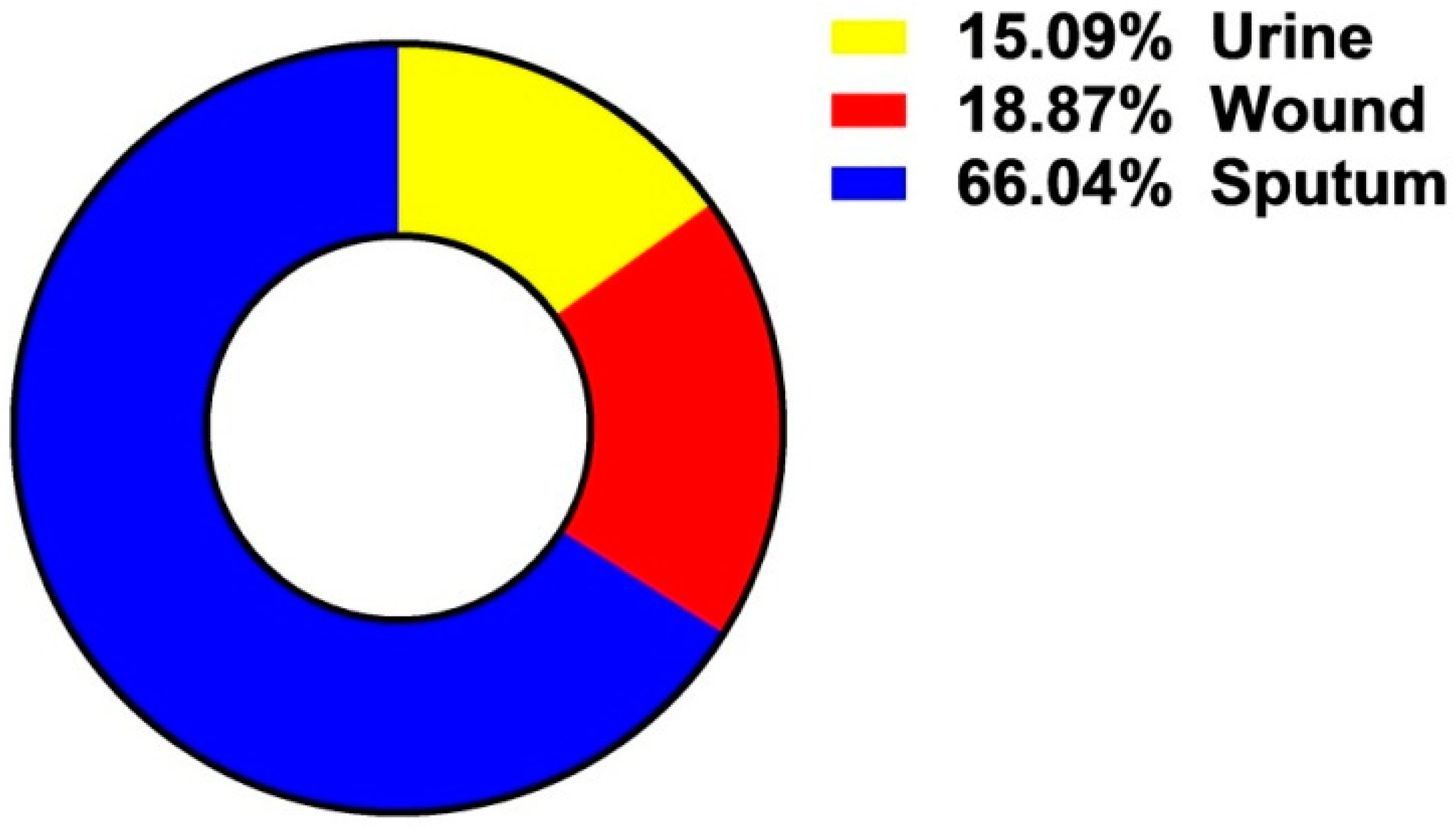
The Incidence of A. baumannii Isolates among Examined Clinical Samples

The prevalence of the isolated bacteria in the clinical samples is illustrated in Figure 1. Among the 110 clinical specimens, the highest percentage of A. baumannii was 66.04% followed by 18.87% and 15.09% was recorded for sputum, wound, and urine samples respectively (Figure 1). The total bacterial count in urine culture showed that 70% (21/30) of female patients had a range of 104 to 105 CFU/mL compared to 65% (13/20) of male patients. While 5% of female and 2% of male patients had CFU values ≥ 106.

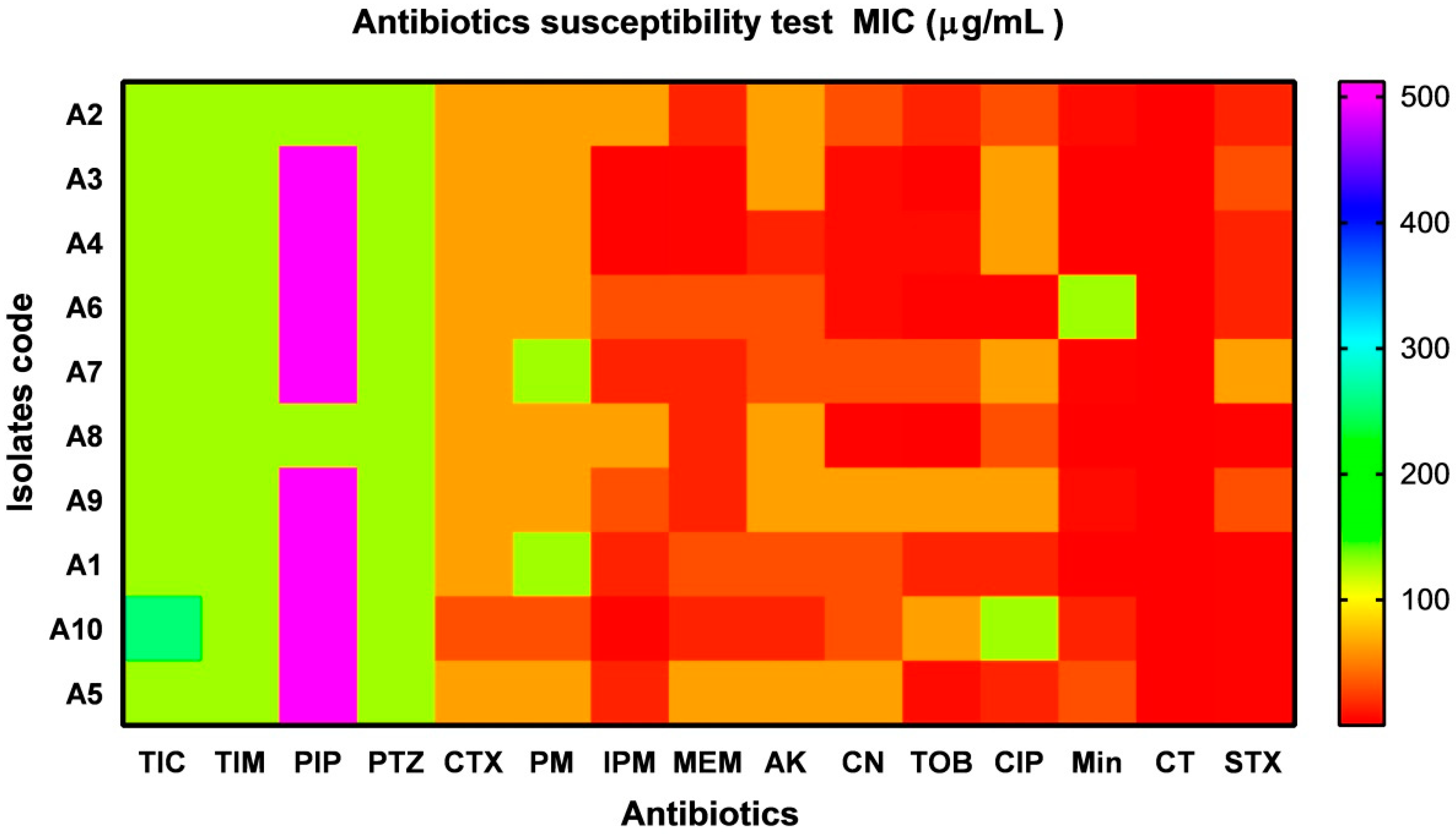
2.2. Antimicrobial Susceptibility Testing

A total of 10 (9.09%) of A. baumannii isolates were recovered from sputum samples (n = 7; 70%), urine (n = 2; 20%) and pus (n = 1; 10%). The rate of resistant isolates to a panel of antibiotics with different potency was illustrated as MIC values (µg mL−1) in a heat- map as shown in Figure 2. All isolates (n = 10; 100%) displayed a high resistance pattern to Ticarcillin, Ticarcillin/Clavulanic Acid, Piperacillin, Piperacillin/Tazobactam, Cefotaxime, Cefepime, Ciprofloxacin, and Amikacin. Around (80%) are resistant to Gentamicin and Tobramycin; and (70%) are resistant to imipenem and meropenem. An intermediate resistant levels with (60 and 40%) to Trimethoprim/Sulfamethoxazole and Minocycline respectively. Interestingly, sensitivity levels within Colistin and Minocycline were observed. According to the Vitek-2 compact system test, three out of 10 A. baumannii isolates (codes A4, A7, and A9) were found to share the same phenotype and antibiotic pattern profile. Hence, these isolates were skipped. The rest of the isolates that showed different phenotypes and antibiotic patterns such as (A1, A2, A3, A5, A6, and A8) were considered for further experiments.

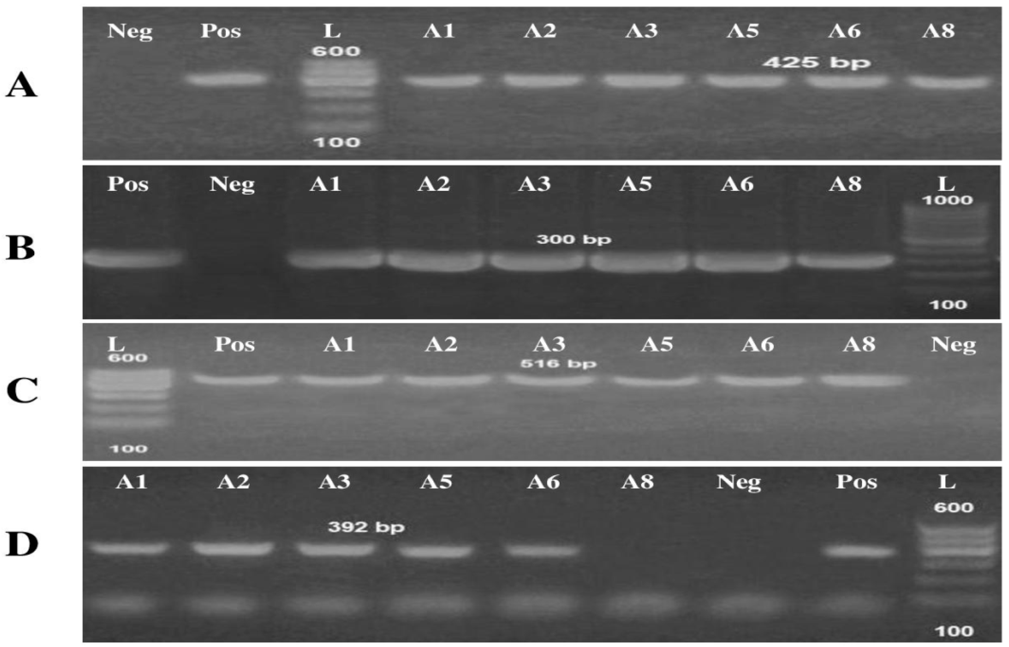
Detection of Virulence and Antibiotic-Resistant Genes

The multiplex PCR screening for virulence and antibiotic-resistant genes showed that 100% of A. baumannii were carrying the recA and iutA virulence genes (Table 2; Figure 3). On the other hand, the antibiotic-resistant genes, blaTEM was present in all A. baumannii isolates. BlaSHV was present with a percentage of approximately 83% since the isolate (A8) missed that gene (Table 2; Figure 3).

2.3. Gas Chromatography-Mass Spectrometry (GC/MS) Analysis

Plant content analysis and extraction play an important role in the progress, restoration, and quality management of herbal formulations. As a result, one of the primary goals of this analysis was to identify the bioactive compounds found in the S. aromaticum extracts to assess their role in improving the antibacterial activity against A. baumannii isolates. Eighteen signal peaks related to separate components were obtained by gas chromatography-mass spectrometry (GC/MS) in the aqueous extract of S. aromaticum (Table 3). The main constituents were α-pinene (18.82%); Beta-caryophyllene (15.12%); oleic acid (14.52%); camphor (11.75%); globuolol (11.35%); Loganetin (8.51%); Apioline (5.45%) and Hexadecanoic acid (4.59%). The ethanol extract possesses 12 signal peaks for compounds such as oleic acid (27.22%); Guanosine (8.91%); indole (6.83%) and 1-Eicosene (6.3%). Finally, the highest percentage content in the ethyl acetate extract gives Linoleic acid (36.16%); Citral (13.48%), and Hexadecanoic acid (11.95%). Other active compounds with their peak number, concentration (peak area %), and retention time (RT) are presented in Table 3.

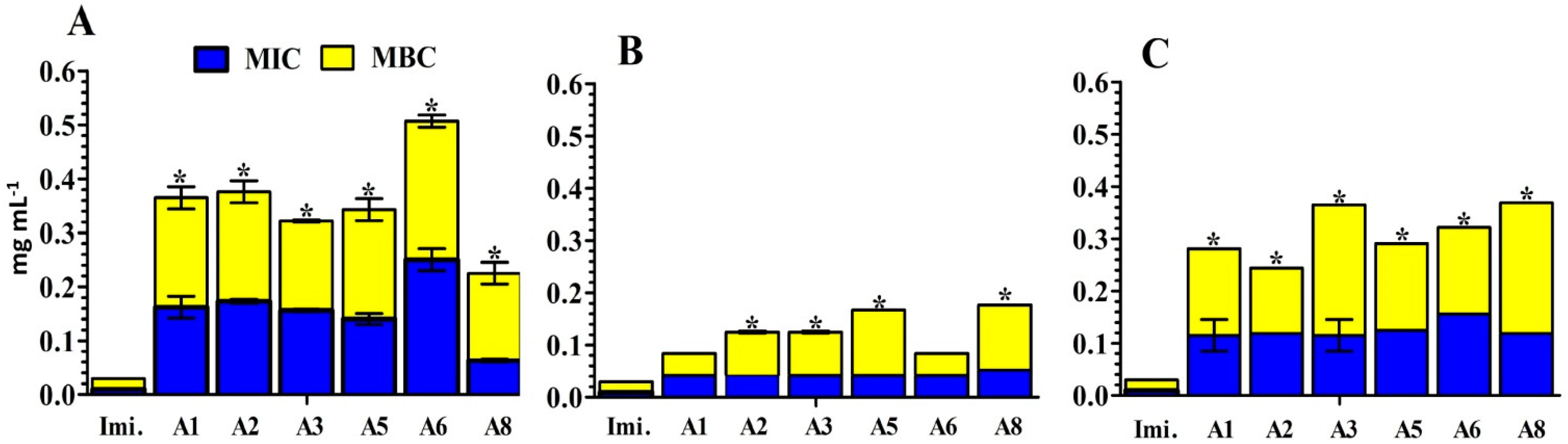
2.4. In Vitro Assay for the Antibacterial Activity of S. aromaticum Extracts

Antibacterial activity of S. aromaticum different extracts and Imipenem against A. baumannii isolates was analyzed by minimal inhibitory concentrations (MIC) by determining the bacterial viability using a colorimetric INT-formazan assay. As a result, we determined the minimal bactericidal concentrations (MBC) which confirmed the killing of A. baumannii isolates over time (24 h). The individual use of S. aromaticum aqueous and/or ethyl acetate extracts against A. baumannii isolates (A1, A2, A3, A5, A6, and A8) exhibited MBC values varying from 0.17 to 0.25 mg mL−1, respectively (Figure 4A,C). Within isolates A1 and A6, ethanol extracts revealed MBC with significant values (0.04 to 0.125 mg mL−1) respectively when compared to Imipenem. (Figure 4B). It is worth mentioning that Imipenem showed a great bactericidal efficacy against all isolates with a concentration of 0.02 mg mL−1 are adequate to kill all the tested A. baumannii isolates (Figure 4).

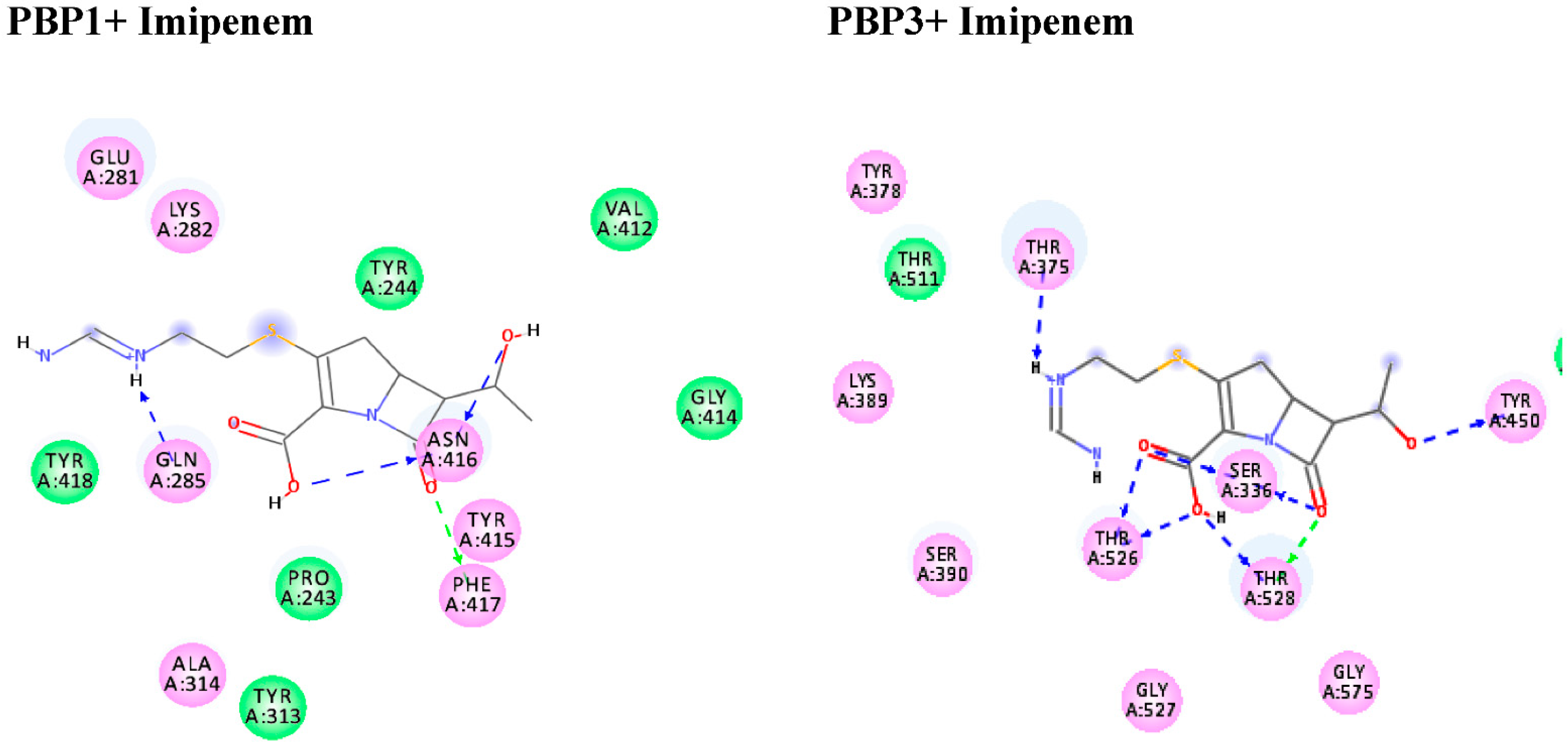
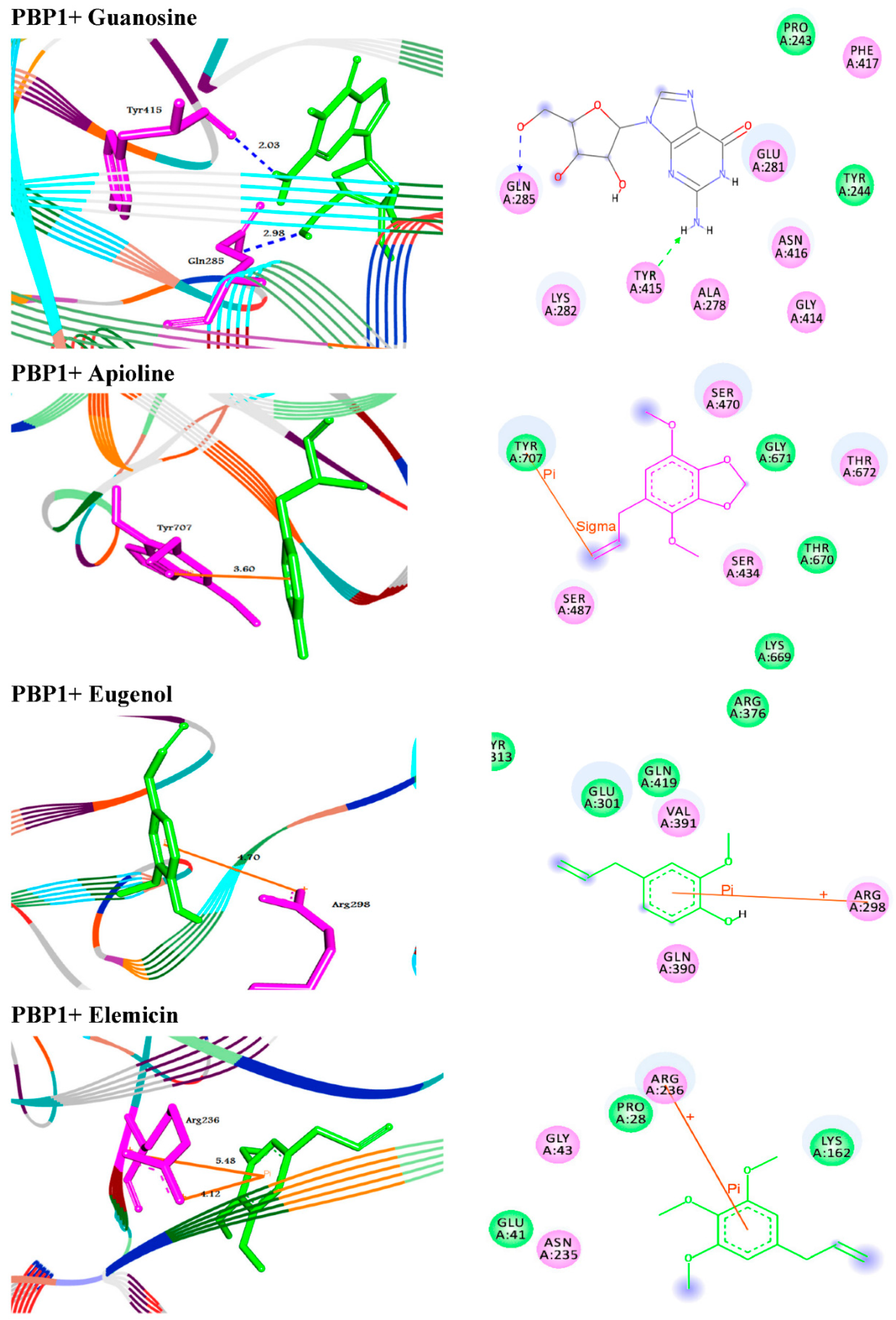
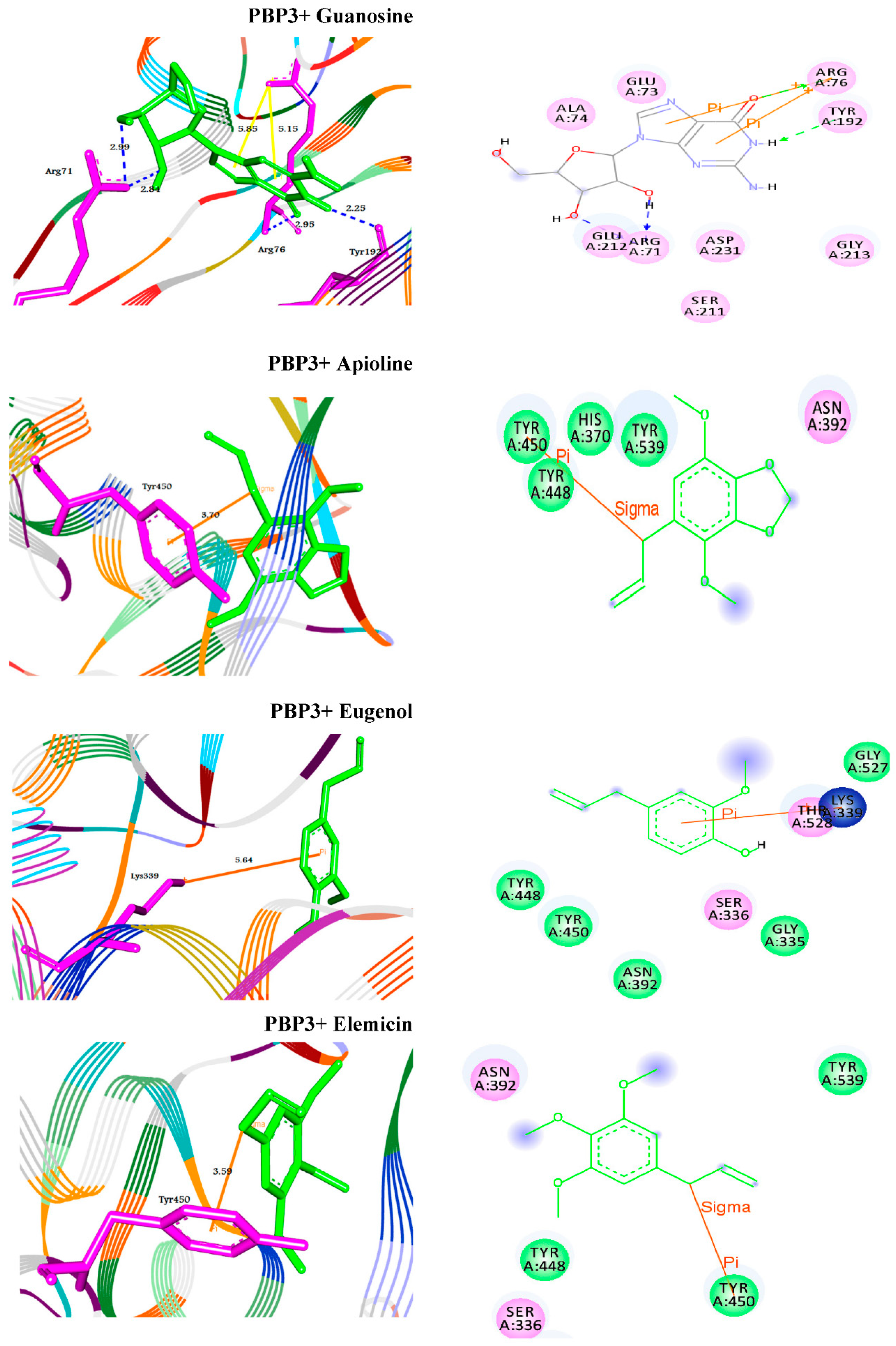
2.5. Molecular Docking Studies of Standard Antibiotic and Herbal Ligands

From the obtained data in Table 4, the antibiotic (Imipenem) suited the binding sites of (PBP1 and PBP3) well, with binding energies ranging from −6.8 to −6.5 kcal/mol respectively (Figure 5). Moreover, the molecular docking simulations were performed for Guanosine, Apioline, Eugenol, and Elemicin against the target proteins (PBP1 and 3) to support the in vitro study via their mode of interactions (Figure 6 and Figure 7). The screened compound (guanosine) displayed respectable fitting to the same binding sites of the targets and having binding energies of −7.1 to −8.1 kcal/mol, respectively. The compound docked to the target protein PBP1 through HB interactions with the residues GLN285 and TYR415 at 2.98 and 2.03 Å, respectively. In addition, it formed two types of interactions such as HB and π- stacking with the target protein PBP3. The guanosine exhibited HBs with the residues ARG71, ARG76 and TYR192 at the distances of 2.97, 2.84, 2.93, and 2.25 Å, respectively. Moreover, it formed π-stacked interactions with the residue ARG76 at 4.14 and 5.6 Å, respectively (Figure 5).
The extracted phyto-compounds (Apioline, Eugenol, and Elemicin) were successfully docked to PBP1 with the binding energies −5.6, −5.4 and −5.3 Å, respectively (Table 4, Figure 6). Apioline docked with the target through π-sigma interaction with the residue TYR707 at 3.60 Å. While, Eugenol docked through π-cation interaction with ARG298 at 4.70 Å. Finally, Elemicin formed two π-cation interactions with the residue ARG236 at 5.48 and 4.12 Å, respectively. For the second enzyme PBP3, the molecules exhibited binding affinities to the active site pockets with docking scores −6.0, −5.8, and −5.2 Å, respectively. They showed π-stacking, similar to π-sigma and π-cation interactions with the residues TYR450 and Lys339 (Table 4, Figure 7).

3. Discussion

The risk of antibiotic-resistant nosocomial infections becomes life-threatening in the intensive care unit and other areas of hospital care [25]. Multi-drug resistant bacteria such as A. baumannii and P. aeruginosa cause many of these infections [6]. The current study showed the proportion of nosocomial infections due to the gram-negative bacilli A. baumannii isolated from clinical samples as urine (15.09%); wound (18.87%); and sputum (66.04%) among the patients (Figure 1). These bacteria are a common cause of urinary tract infections (UTIs) that upset the kidney, leading to pyelonephritis, as well as the bladder, resulting in cystitis [26]. UTI symptoms included elevated kidney function and high levels of A. baumannii in the patients’ sputum (66.04%) (Table 1). This result is conceivable since UTI symptoms are not a reliable indication of illness. The presence of bacteria must be confirmed by urine culture in order to diagnose UTI [27]. The urine culture in our results indicated the presence of bacteriuria in 70% of female patients and 65% of male patients. The percentage of A. baumannii in our study was in agreement with the results obtained by Al-Agamy et al. [10] the highest prevalence of A. baumannii isolates was in respiratory samples followed by wound samples and urine (Figure 1). Our findings are in agreement with the results obtained by Abdulzahra et al. [28]. The antibiotic susceptibility test for A. baumannii isolates indicated that all isolates (n = 10; 100%) were multi-drug resistant to Ticarcillin, Ticarcillin/Clavulanic Acid, Piperacillin, Piperacillin/Tazobactam, Cefotaxime, Cefepime, Ciprofloxacin, and Amikacin, while 70–80% are extensively drug-resistant. Resistance decreased to 40% with Minocycline and all isolates were sensitive to Colistin (Figure 2). Our findings are in covenant with earlier results concerning the MDR isolates [29]. Some of the most significant virulence genes of A. baumannii are colicin V production, curi fibers (csg), siderophores, such as aerobactin (iutA), and cytotoxic necrotizing factors (cnf) [30,31]. Virulence genes such as recA and iutA were present in all isolates of A. baumannii (Table 2; Figure 3). The same findings were confirmed earlier for 18.75% of A. baumannii isolates from the hospital environment were carrying iutA genes [32]. Antibiotic-resistant genes blaTEM and blaSHV were the most common in A. baumannii as (100%) and (83%) respectively (Table 2; Figure 3). This finding is consistent with the results reported by Beriş et al. [11,33], where the prevalence of blaTEM and blaSHV was 55.7% and 7.7%, respectively. The highest percentage of blaTEM (100%) and absence of blaCTX in all isolates are in concert with the findings reported by Al-Agamy et al. [10]. The current study confirmed the promising efficacy of all Syzygium aromaticum extracts against all A. baumannii isolates with the lowest MBC values varied from 0.04 to 0.125 mg mL−1 that recorded for the ethanolic one (Figure 4). This could be due to the presence of some active phytochemicals such as guanosine, α- Pinene, Beta-caryophyllene; oleic acid; camphor, globulol, and loganetin that were detected in the GC-MS analysis (Table 3). α-pinenes were detected earlier as the main component in S. aromaticum aqueous extract [34]. The bactericidal activities of α-pinenes against both gram-negative and positive bacteria were reported earlier by Mercier et al. [35]. α-pinenes kill bacteria by damaging membrane structure and function [36]. A membrane’s expansion and fluidity were enhanced as a result of its lipophilic nature. Beta-caryophyllene is the most common antibacterial constituent in essential oils of the Syzygium genus, it is detected in S. aromaticum, S. cumini, S. polyanthum and S. samarangense [37]. The mechanism of action was discussed recently as Beta-caryophyllene alters the bacterial membrane permeability and causes non-selective pore formation that kills the bacterial cells [38]. Oleic acid in Syzgium aromaticum could inhibit bacterial growth by inhibiting the bacterial fatty acid synthesis [39]. The antibacterial activity of camphor present in lavender essential oil was reported by De Azeredo et al. [40]. Another important phyto-component such as globulol was discussed for their antifungal and antibacterial activity [41]. Limonene has previously been reported in an aqueous extract of Syzygium aromaticum by Jimoh et al. [42]. Finally, guanosine has been reported as a potent antibacterial activity both in vitro and in vivo [43]. Moreover, in silico molecular docking and modeling techniques [44,45] were performed in the current study for the ethanolic extracted compound (guanosine) for better understanding their mode of action through the interaction with the active site pockets of PBP1 and PBP3, and to identify new inhibitors as antibacterial agents. The result showed that the target compounds exhibited binding energy higher than standard drug imipenem (−7.1 and −8.1 Å, respectively) against the target proteins (Table 4; Figure 5, Figure 6 and Figure 7). The SAR (structure activity relationship) analysis showed that the antibacterial activity of guanosine can be modulated by the presence of pyrimidine and indole moieties, and hydroxyl and amino groups (Figure 5). Therefore, guanosine may be considered a good inhibitor for PBP1 and PBP3 proteins. In recent times, computational modeling and molecular-dynamic simulations have shown that phytoligands, such as Apioline, Eugenol, and Elemicin, have good binding potential with minimum binding energy and that they are stabilized by an interaction between antibacterial imipenem (carbapenam) of the present three generations, with their common objectives in A. baumannii [14]. Virtual screening findings and drug-like tests have shown that phyto-compounds are a hit molecule due to a much greater binding affinity (−9.4 kcal/mol) compared with imipenem in their target PBP1 of A. baumannii [13]. Displayed guanosine as a relevant screened molecule that has binding energies of −7.1 to −8.1 kcal/mol at the binding site for PBP1 and PBP3 proteins, respectively. Form the tabulated data in Table 4; we can conclude that guanosine has the best docking score and intermolecular interactions as compared with the other selected compounds (Apioline, Eugenol, and Elemicin). As a result, it might be a promising pharmacological option for combating A. baumannii. Given the increasing importance of pyrimidine compounds, particularly in recent years, which have several applications in various fields, most notably in medicine as antibacterial agents [46]. In addition, pyrimidines are nitrogen heterocyclic aromatic compounds with great interest as they constitute an important class of natural products. The substituted pyrimidines such as Brodiprim, Iclaprim, Trimethoprim, and Pyrimethamine are biologically important compounds and act as effective antibacterial drugs [47]. Therefore, Guanosine with pyrimidine ring and other phyto-compounds such as Apioline, Eugenol, and Elemicin was selected as drug candidate against PBP1 and PBP3 proteins.

4. Materials and Methods

4.1. Sampling Collection, Isolation, and Processing

One hundred and ten specimens; urine (n = 50), sputum (n = 40), and wound swabs (n = 20) were randomly collected under complete aseptic conditions from different hospitals and clinics in Qena province, Egypt (from January to December 2018). In total, 5 mL of clean-catch urine from patients suspected of urinary tract infection (UTI) is obtained in a sterile container. Urine samples were inoculated on MacConkey agar and/or Blood agar (Merck, Germany) at 37 °C for 24 h and observed for bacterial growth. Blood agar colonies were counted using a colony counter and checked for significant bacteriuria. The culture that grew ≥105 CFU/mL, was measured as significant bacteriuria. For heterogeneous colonies, sub-culturing of individual distinct colonies was performed to ensure pure cultures. The sterile cotton swabs dipped into normal saline with the Levine method were obtained aseptically from two wound samples of each participant [48]. Sputum samples were taken from patients over a self-induced cough into sterile cups and sent for culture. Samples were primarily identified using typical laboratory approaches including growth on MacConkey agar and/or Blood agar (Merck, Germany) and Gram staining. Plates were incubated at 37 °C and examined for detectable bacterial growth after 48 h of incubation. The isolated bacteria were further identified using the automated system Vitek-2 (bioMérieux, France).

4.2. Phenotypic Identification of the Isolates by Vitek-2 Systems

The bacterial isolates were identified with the Automated Identification Biomerieux Vitek-2 System via morphological, classical biochemical studies. In total, the 41 tests included 18 sugar assimilation and fermentation; 2 decarboxylase tests, and 3 different tests on the 64 ID-GNB plastic well (for urease, utilization of malonate, and tryptophane deaminase). With a vacuum card, the organism’s 0.50 McFarland suspension is inoculated, made from a blood agar plate of 18–20 h (BioMe’rieux), and is automatically screened and inserted manually inside the Vitek-2 reader. The fluorescence of the inoculator is measured every 15 min [49]. All isolates identified as Acinetobacter baumannii were used for further characterization.

4.3. Antimicrobial Susceptibility Testing

For all recovered isolates, antibiograms were calculated according to the disc diffusion method mentioned earlier and compared with the standard chart (CLSI, 2017) [50]. MIC was detected by Vitek-2 compact system (bioMérieux, France), the susceptibility of Acinetobacter baumannii isolates was tested for 15 antibiotics (Bioanalyse ®). The used antibiotics were Amikacin, Tobramycin, Gentamicin, Ticarcillin, Ticarcillin/Clavulanic Acid, Piperacillin, Piperacillin/Tazobactam, Cefotaxime, Cefepime, Imipenem, Meropenem, Ciprofloxacin, Levofloxacin, Trimethoprim/Sulfamethoxazole and Minocycline. Multidrug resistance was defined as resistance to three or more antibiotics of the different classes [51]. A. baumannii isolates with the same phenotype and antibiotic pattern profile were excluded, only different isolates were considered for further experiments.

4.4. Recognition of Virulence and Antibiotic-Resistant Genes of A. baumannii Isolates

Molecular representation of the recovered A. baumannii was approved by multiplex PCR. The detection of eight encoding genes of virulence and antibiotic resistance was performed by using 16 primers. Set extraction of DNA was carried according to QIAamp DNA mini kit instructions. Genes encoding different virulence factors recA and iutA. For antibiotic resistance genes blaTEM, and blaSHV were analyzed by multiplex PCR.

4.5. DNA Amplification for the Selected Virulence and Antibiotic Resistance Genes of Isolates

As previously stated, DNA extraction was carried out under QIAamp DNA mini-kit instructions (QIAGEN, Germany, GmbH) [52]. Concisely, 200 μL of the sample suspension was inoculated with 10 μL of proteinase K and 200 μL of lysis buffer at 56 °C for 10 min. The lysate was added to 200 μL of 100% ethanol following incubation. The sample was washed and centrifuged according to the recommendations of the manufacturer. Nucleic acid was eluted with 100 μL of elution buffer provided in the kit. PCR amplification was performed using oligonucleotide primer (METABION, Germany) that were utilized in a 25 μL reaction containing 12.5 μL of EMERALDAMP Max PCR Master Mix (TAKARA, Japan), 1 μL of each primer of 20 pmol concentration, 5.5 μL of dist. water and 6 μL of DNA template. Applied thermal cyclers have been used to react in the biosystem. Table 5 summarizes all amplicon sizes and cycling conditions. The products of PCR were separated by electrophoresis on 1.5% agarose gel (APPLICHEM, Germany, GmbH) in 1xTBE buffer at room temperature using gradients of 5 V/cm. For gel analysis, 15 μL of the products were loaded in each gel slot. Gelpilot 100 bp and 100 bp plus ladders (QIAGEN, Germany, GmbH) and GeneRuler 100 bp ladder (FERMENTAS, THERMO) was used as a marker for electrophoresis to determine the fragment sizes. The gel was photographed by a gel documentation system (ALPHA INNOTECH, BIOMETRA) and the data were analyzed through computer software (AUTOMATIC IMAGE CAPTURE, USA).

4.6. Plant Material and Extraction

Syzygium aromaticum (clove) was purchased from a local market in Qena city, Egypt. Dry seeds were washed with sterile water and further dried, ground into a fine powder using a tissue grinder (IKA®® A10, Germany). Plant species have been visually imaged for documentation under the Department of Botany and Microbiology, Science Faculty, South Valley University, Qena, Egypt, for further taxonomic identification purposes. Three solvents were used for the extract of bioactive components from Syzygium aromaticum, as follows: 10 g of plant powder was soaked separately in 100 mLof hot distilled water, ethanol, and/or ethyl acetate for 7 days with continuous stirring (150 rpm) at room temperature by using a bigger bill shaker, USA. The obtained extracts were filtered through a Buchner funnel with Whatman No.1 filter paper and evaporated by a rotary evaporator (BUCHI R-114, Switzerland) under reduced pressure to dryness at 45 °C. All extract residue were dissolved in dimethyl sulfoxide (DMSO) except the aqueous extract, which dissolved in sterile distilled water at a concentration of 100 mg/mL [56]. All extracts were sterilized using a syringe filter equipped with a 45 μm membrane filter, then kept at −4 °C.

4.7. Determination of the Minimum Inhibitory Concentration (MIC) and Minimum Bactericidal Concentration (MBC) by INT Reduction Assay

The determination of MIC and MBC were assayed as described earlier [57]. The freshly prepared culture of A. baumannii isolates was adjusted to OD595 of 0.01. 100 μL of each isolate culture was put into sterilized 96-well plates. Then, 20 μL of the original S. aromaticum extracts (100 mg mL−1) was added (serial dilutions of 10−1–10−10 were used, eight replicates were made for each dilution into complete raw of the 96-well plate). Imipenem (10 mg mL−1) and un-inoculated media were tested as the positive and negative control, respectively. After 24 h incubation at 37 °C, MIC was determined by the addition of 40μL of p-iodonitrotetrazolium violet chloride (INT) (0.2 mg/mL, Sigma-Aldrich) to the plates and re-incubated at 37 °C for 30 min. The lowest concentration which banned color change is the MIC [58,59]. The MBC was determined by transferring 50 mL from each well of overnight MIC plates (and/or higher) to sterile (TSA) fresh plates. Viable colonies were counted after 24 h at 37 °C. The limit of detection for this assay was 101 CFU/mL.

4.8. Gas Chromatography-Mass Spectrometry (GC-MS) Analysis

S. aromaticum different extracts were subjected to gas chromatography-mass spectrometer technique (GC-MS) (Thermo scientific technologies, Trace 1310) with capillary column TG-5 (30 m × 250 μm × 0.25 μm) system were used. The mass detector used in split mode and helium gas with a flow rate of 1.5 mL/min was used as a carrier. The injector was operated at 230 °C and the oven temperature for the initial setup was 60 °C for 2 min ramp 10/min to 300 °C for 8 min. Mass spectra were taken at 70 eV, total GC running time was 35 min.

4.9. Molecular Docking Studies of Standard Antibiotic and Herbal Ligands

The objective of this work is to create a new, sensitive, and possible imipenem ligand derivative against the target A. baumannii penicillin-binding protein 1 and/or 3 (PBP1–PBP3). It begins by studying the crystal structure of imipenem treated PBP1 and PBP3, finding the active site of the protein (pocket) and examining interactivities with imipenem. A further step is to create novel ligand molecules from S. aromaticum derived bioactive compounds, including guanosine based on the imipenem structure complex with PBP1/3; the calculations will then be completed to calculate the free binding energy and physical-chemical characteristics of every molecule. To identify the causes of shifting powers, a binding interaction between the chosen analogs and the receiver may also be analyzed. The 3D structures of A. baumannii PBP1 and PBP3 were downloaded from the protein data bank [60]. The PDB files were energy minimized and optimized by the removal of water molecules and atomic clashes to get a stable confirmation. A receptor grid was then generated at the centroid of the active site cavity to perform the screening approach [61,62]. The ligand molecule was sketched using Chem Draw Ultra 0.7, then converted to SDF format using Open Babel software [63]. The docking study was carried out using the PyRx-virtual screening tool [64].

4.10. Statistical Data Analysis

Data were analyzed using the Mann–Whitney U test or a Kruskal–Wallis test followed by post hoc Dunn’s multiple comparisons. Differences were considered significant at p values of ≤0.05. For all statistical analyses, GraphPad Prism version 8 was used.

5. Conclusions

A. baumannii represents vital problems because of the high percentage of antibiotics resistance, their encoding gene for virulence, and antibiotics resistance. Although ethanol extract of Syzygium aromaticum was promising for the in vitro study against A. baumannii, improving the potency of beta-lactam antibiotics can be an overwhelming pre-condition for antibiotic resistance; it is thus vital that new analogues of A. baumannii PBP1 and/or PBP3 imipenem are developed, with a substantially higher relative binding energy free. The results show how imipenem analogues may be designed using a phyto-compound such as guanosine, apioline, eugenol, and elemicin against the target proteins (A. baumannii PBP1 and/or PBP3) in the in-silico drug design. This stimulating analogue exhibits increased physico-chemical characteristics, as well as greater binding affinity. Therefore, guanosine and other biologically active compounds extracted from S. aromaticum are eco-friendly and might be employed as an alternative antimicrobial agent against A. baumannii isolates.

Author Contributions

Conceptualization, W.S.; Data curation, A.M. and W.S.; Funding acquisition, B.H.E.; Investigation, W.S. and B.H.E.; Methodology, A.M. and F.E.S.; Resources, B.H.E.; Supervision, M.M.A., F.E.S. and W.S.; Visualization, M.M.A.; Writing—original draft, A.M.; Writing—review and editing, F.E.S. and W.S. All authors have read and agreed to the published version of the manuscript.

Funding

This research was funded by Taif University researchers supporting project number: TURSP- 2020/127.

Institutional Review Board Statement

The study protocol was approved by the local Medical Ethics Committees of the Medical University of Assiut, Egypt, which has been approved by the Egyptian Ministry of Higher Education and Scientific Research on 11/2009. All participants received an ethically approved clinical sampling and informed consent from General Hospital and Private medical research laboratories for Qena province in Egypt during the study work. The procedures were conducted according to applicable guidelines and regulations and written consent was given by the subjects.

Informed Consent Statement

Informed consent was obtained from all subjects involved in the study.

Data Availability Statement

The data presented in this study are available on request from the corresponding author. The data are not publicly available due to privacy restrictions.

Acknowledgments

The authors acknowledged Taif University, Taif, Saudia Arabia, for supporting the study (Taif University researchers supporting project number: TURSP-2020/127). The authors would like to extend their sincere appreciation to Aboubakr Haredi Abdelmonsef, Chemistry Department, Faculty of Science, South Valley University, Qena, Egypt for performing the docking analysis.

Conflicts of Interest

The authors declare no conflict of interest.

References

  1. Algammal, A.M.; Hetta, H.F.; Batiha, G.E.; Hozzein, W.N.; El Kazzaz, W.M.; Hashem, H.R.; Tawfik, A.M.; El-Tarabili, R.M. Virulence-determinants and antibiotic-resistance genes of MDR- E. coli isolated from secondary infections following FMD-outbreak in cattle. Sci. Rep. 2020, 10, 19779. [Google Scholar] [CrossRef] [PubMed]
  2. Abouelfetouh, A.; Torky, A.S.; Aboulmagd, E. Phenotypic and genotypic characterization of carbapenem-resistant Acinetobacter baumannii isolates from Egypt. Antimicrob. Resist. Infect. Control 2019, 8, 185. [Google Scholar] [CrossRef] [Green Version]
  3. Pritsch, M.; Zeynudin, A.; Messerer, M.; Baumer, S.; Liegl, G.; Schubert, S.; Löscher, T.; Hoelscher, M.; Belachew, T.; Rachow, A.; et al. First report on bla NDM-1-producing Acinetobacter baumannii in three clinical isolates from Ethiopia. BMC Infect. Dis. 2017, 17, 180. [Google Scholar] [CrossRef] [PubMed] [Green Version]
  4. Santajit, S.; Indrawattana, N. Mechanisms of antimicrobial resistance in ESKAPE pathogens. BioMed Res. Int. 2016, 2016, 2475067. [Google Scholar] [CrossRef] [PubMed] [Green Version]
  5. Chakravarty, B. Genetic mechanisms of antibiotic resistance and virulence in Acinetobacter baumannii: Background, challenges and future prospects. Mol. Biol. Rep. 2020, 47, 4037–4046. [Google Scholar] [CrossRef] [PubMed]
  6. Motbainor, H.; Bereded, F.; Mulu, W. Multi-drug resistance of blood stream, urinary tract and surgical site nosocomial infections of Acinetobacter baumannii and Pseudomonas aeruginosa among patients hospitalized at Felegehiwot referral hospital, Northwest Ethiopia: A cross-sectional study. BMC Infect. Dis. 2020, 20, 92. [Google Scholar] [CrossRef] [Green Version]
  7. Cerezales, M.; Xanthopoulou, K.; Wille, J.; Krut, O.; Seifert, H.; Gallego, L.; Higgins, P.G. Mobile genetic elements harboring antibiotic resistance determinants in Acinetobacter baumannii isolates from Bolivia. Front. Microbiol. 2020, 11, 919. [Google Scholar] [CrossRef]
  8. Magiorakos, A.P.; Srinivasan, A.; Carey, R.T.; Carmeli, Y.; Falagas, M.T.; Giske, C.T.; Harbarth, S.; Hindler, J.T.; Kahlmeter, G.; Olsson-Liljequist, B.; et al. Multidrug-resistant, extensively drug-resistant and pandrug-resistant bacteria: An international expert proposal for interim standard definitions for acquired resistance. Clin. Microbiol. Infect. 2012, 18, 268–281. [Google Scholar] [CrossRef] [Green Version]
  9. Ghaima, K.K. Distribution of extended spectrum beta-lactamase (ESBL) genes among Acinetobacter baumannii isolated from burn infections. MOJ Cell Sci. Rep. 2018, 5, 42–46. [Google Scholar] [CrossRef] [Green Version]
  10. Al-Agamy, M.H.; Khalaf, N.G.; Tawfick, M.M.; Shibl, A.M.; El Kholy, A. Molecular characterization of carbapenem-insensitive Acinetobacter baumannii in Egypt. Int. J. Infect. Dis. 2014, 22, 49–54. [Google Scholar] [CrossRef] [Green Version]
  11. Beriş, F.Ş.; Budak, E.E.; Gülek, D.; Uzun, A.; Cizmeci, Z.; Mengeloğlu, F.Z.; Direkel, Ş.; Cetinkol, Y.; Altıntop, A.; Iraz, M.; et al. Investigation of the frequency and distribution of beta-lactamase genes in the clinical isolates of Acinetobacter baumannii collected from different regions of Turkey: A multicenter study. Mikrobiyoloji Bul. 2016, 50, 511–521. [Google Scholar] [CrossRef] [Green Version]
  12. Smiline, A.S.G.; Vijayashree, J.P.; Paramasivam, A. Molecular characterization of plasmid-encoded blaTEM, blaSHV and blaCTX-M among extended spectrum β-lactamases [ESBLs] producing Acinetobacter baumannii. Br. J. Biomed. Sci. 2018, 75, 200–202. [Google Scholar] [CrossRef] [PubMed]
  13. Salih, T.; Salih, H.A. In Silico Design and Molecular Docking Studies of Carbapenem Analogues Targeting Acinetobacter baumannii PBP1A Receptor. Al Mustansiriyah J. Pharm. Sci. 2020, 20, 35–50. [Google Scholar]
  14. Skariyachan, S.; Manjunath, M.; Bachappanavar, N. Screening of potential lead molecules against prioritised targets of multi-drug-resistant-Acinetobacter baumannii–insights from molecular docking, molecular dynamic simulations and in vitro assays. J. Biomol. Struct. Dyn. 2019, 37, 1146–1169. [Google Scholar] [CrossRef]
  15. Penwell, W.F.; Shapiro, A.B.; Giacobbe, R.A.; Gu, R.F.; Gao, N.; Thresher, J.; McLaughlin, R.E.; Huband, M.D.; DeJonge, B.L.; Ehmann, D.E.; et al. Molecular mechanisms of sulbactam antibacterial activity and resistance determinants in Acinetobacter baumannii. Antimicrob. Agents Chemother. 2015, 59, 1680–1689. [Google Scholar] [CrossRef] [Green Version]
  16. Cayô, R.; Rodríguez, M.C.; Espinal, P.; Fernández-Cuenca, F.; Ocampo-Sosa, A.A.; Pascual, Á.; Ayala, J.A.; Vila, J.; Martínez-Martínez, L. Analysis of genes encoding penicillin-binding proteins in clinical isolates of Acinetobacter baumannii. Antimicrob. Agents Chemother. 2011, 55, 5907–5913. [Google Scholar] [CrossRef] [PubMed] [Green Version]
  17. Leungtongkam, U.; Thummeepak, R.; Kitti, T.; Tasanapak, K.; Wongwigkarn, J.; Styles, K.M.; Wellington, E.M.; Millard, A.D.; Sagona, A.P.; Sitthisak, S. Genomic analysis reveals high virulence and antibiotic resistance amongst phage susceptible Acinetobacter baumannii. Sci. Rep. 2020, 10, 16154. [Google Scholar] [CrossRef] [PubMed]
  18. Elamary, R.; Salem, W.M. Optimizing and purifying extracellular amylase from soil bacteria to inhibit clinical biofilm-forming bacteria. PeerJ 2020, 8, e10288. [Google Scholar] [CrossRef] [PubMed]
  19. Yassien, A.S.; Hassan, M.M.; Elamary, R.B. Prevalence of lipase producer Aspergillus niger in nuts and anti-biofilm efficacy of its crude lipase against some human pathogenic bacteria. Sci. Rep. 2021, 11, 7981. [Google Scholar] [CrossRef]
  20. Ekor, M. The growing use of herbal medicines: Issues relating to adverse reactions and challenges in monitoring safety. Front. Pharmacol. 2014, 4, 177. [Google Scholar] [CrossRef] [Green Version]
  21. Batiha, G.E.S.; Alkazmi, L.M.; Wasef, L.G.; Beshbishy, A.M.; Nadwa, E.H.; Rashwan, E.K. Syzygium aromaticum L. (Myrtaceae): Traditional uses, bioactive chemical constituents, pharmacological and toxicological activities. Biomolecules 2020, 10, 2. [Google Scholar]
  22. Selles, S.M.A.; Kouidri, M.; Belhamiti, B.T.; Ait Amrane, A. Chemical composition, in-vitro antibacterial and antioxidant activities of Syzygium aromaticum essential oil. J. Food Meas. Charact. 2020, 14, 2352–2358. [Google Scholar] [CrossRef]
  23. Singh, R.; Lawrence, R.; Lawrence, K.; Agarwal, B.; Gupta, R.K.; Dar, S. Antioxidant and antibacterial activity of Syzigium aromaticum. Chem. Sci. Trans. 2015, 4, 239–245. [Google Scholar]
  24. Singh, J.; Baghotia, A.; Goel, S.P. Eugenia caryophyllata Thunberg (Family Myrtaceae): A review. Int. J. Res. Pharm. Biomed. Sci. 2012, 3, 1469–1475. [Google Scholar]
  25. Exner, M.; Bhattacharya, S.; Christiansen, B.; Gebel, J.; Goroncy-Bermes, P.; Hartemann, P.; Heeg, P.; Ilschner, C.; Kramer, A.; Larson, E.; et al. Antibiotic resistance: What is so special about multidrug-resistant Gram-negative bacteria? GMS Hyg. Infect. Control 2017, 12, Doc05. [Google Scholar] [CrossRef]
  26. Thomson, C.; Armitage, A. Urinary tract infection. In Oxford Textbook of Medicine; Warrell, D.A., Cox, T.M., Firth, J.D., Eds.; Oxford Uni Press: Oxford, UK, 2010; pp. 4103–4122. [Google Scholar]
  27. Elamary, R.B.; Albarakaty, F.M.; Salem, W.M. Efficacy of Acacia nilotica aqueous extract in treating biofilm-forming and multidrug resistant uropathogens isolated from patients with UTI syndrome. Sci. Rep. 2020, 10, 11125. [Google Scholar] [CrossRef]
  28. Abdulzahra, A.T.; Khalil, M.A.; Elkhatib, W.F. First report of colistin resistance among carbapenem-resistant Acinetobacter baumannii isolates recovered from hospitalized patients in Egypt. New Microbes New Infect. 2018, 26, 53–58. [Google Scholar] [CrossRef]
  29. Younis, R.I.; Nasef, S.A.; Salem, W.M. Detection of Multi-Drug Resistant Food-borne Bacteria in Ready-to-Eat Meat Products in Luxor City, Egypt. SVU-Int. J. Vet. Sci. 2019, 2, 20–35. [Google Scholar] [CrossRef] [Green Version]
  30. Eijkelkamp, B.A.; Stroeher, U.H.; Hassan, K.A.; Paulsen, I.T.; Brown, M.H. Comparative analysis of surface-exposed virulence factors of Acinetobacter baumannii. BMC Genom. 2014, 15, 1020. [Google Scholar] [CrossRef] [Green Version]
  31. Eraç, B.; Yılmaz, F.F.; Hoşgör Limoncu, M.; Oztürk, I.; Aydemir, S. Investigation of the virulence factors of multidrug-resistant Acinetobacter baumannii isolates. Mikrobiyoloji Bul. 2014, 48, 70–81. [Google Scholar] [CrossRef]
  32. Al-Kadmy, I.M.S.; Ali, A.N.M.; Salman, I.M.A.; Khazaal, S.S. Molecular characterization of Acinetobacter baumannii isolated from Iraqi hospital environment. New Microbes New Infect. 2018, 21, 51–57. [Google Scholar] [CrossRef]
  33. Moradi, J.; Hashemi, F.B.; Bahador, A. Antibiotic resistance of Acinetobacter baumannii in Iran: A systemic review of the published literature. Osong Public Health Res. Perspect. 2015, 6, 79–86. [Google Scholar] [CrossRef] [Green Version]
  34. Tahir, H.U.; Sarfraz, R.A.; Ashraf, A.; Adil, S. Chemical composition and antidiabetic activity of essential oils obtained from two spices (Syzygium aromaticum and Cuminum cyminum). Int. J. Food Prop. 2016, 19, 2156–2164. [Google Scholar] [CrossRef]
  35. Mercier, B.; Prost, J.; Prost, M. The essential oil of turpentine and its major volatile fraction (α-and β-pinenes): A review. Int. J. Occup. Med. Environ. Health 2009, 22, 331–342. [Google Scholar] [CrossRef] [PubMed] [Green Version]
  36. Andrews, R.E.; Parks, L.W.; Spence, K.D. Some effects of Douglas fir terpenes on certain microorganisms. Appl. Environ. Microbiol. 1980, 40, 301–304. [Google Scholar] [CrossRef] [Green Version]
  37. Hamad, A.; Mahardika, M.G.P.; Yuliani, I.; Hartanti, D. Chemical constituents and antimicrobial activities of essential oils of Syzygium polyanthum and Syzygium aromaticum. Rasayan J. Chem. 2017, 10, 564–569. [Google Scholar]
  38. Moo, C.L.; Yang, S.K.; Osman, M.A.; Yuswan, M.H.; Loh, J.Y.; Lim, W.M.; Lim, S.H.E.; Lai, K.S. Antibacterial Activity and Mode of Action of β-caryophyllene on Bacillus cereus. Pol. J. Microbiol. 2020, 69, 49. [Google Scholar] [CrossRef] [Green Version]
  39. Zheng, C.J.; Yoo, J.S.; Lee, T.G.; Cho, H.Y.; Kim, Y.H.; Kim, W.G. Fatty acid synthesis is a target for antibacterial activity of unsaturated fatty acids. FEBS Lett. 2005, 579, 5157–5162. [Google Scholar] [CrossRef] [PubMed] [Green Version]
  40. De Azeredo, G.A.; Stamford, T.L.M.; Nunes, P.C.; Neto, N.J.G.; De Oliveira, M.E.G.; De Souza, E.L. Combined application of essential oils from Origanum vulgare L. and Rosmarinus officinalis L. to inhibit bacteria and autochthonous microflora associated with minimally processed vegetables. Food Res. Int. 2011, 44, 1541–1548. [Google Scholar] [CrossRef] [Green Version]
  41. Tan, M.; Zhou, L.; Huang, Y.; Wang, Y.; Hao, X.; Wang, J. Antimicrobial activity of globulol isolated from the fruits of Eucalyptus globulus Labill. Nat. Prod. Res. 2008, 22, 569–575. [Google Scholar] [CrossRef]
  42. Jimoh, S.O.; Arowolo, L.A.; Alabi, K.A. Phytochemical screening and antimicrobial evaluation of Syzygium aromaticum extract and essential oil. Int. J. Curr. Microbiol. Appl. Sci. 2017, 6, 4557–4567. [Google Scholar] [CrossRef]
  43. Hu, J.; Hu, Q.; He, X.; Liu, C.; Kong, Y.; Cheng, Y.; Zhang, Y. Stimuli--responsive hydrogels with antibacterial activity assembled from guanosine, aminoglycoside, and a bifunctional anchor. Adv. Healthc. Mater. 2020, 9, 1901329. [Google Scholar] [CrossRef] [PubMed]
  44. El-Saghier, A.M.; El-Naggar, M.; Hussein, A.H.M.; El-Adasy, A.-B.A.; Olish, M.; Abdelmonsef, A.H. Eco-Friendly Synthesis, Biological Evaluation, and In Silico Molecular Docking Approach of Some New Quinoline Derivatives as Potential Antioxidant and Antibacterial Agents. Front. Chem. 2021, 9, 679967. [Google Scholar] [CrossRef] [PubMed]
  45. Gomha, S.M.; Abdelhady, H.A.; Hassain, D.Z.; Abdelmonsef, A.H.; El-Naggar, M.; Elaasser, M.M.; Mahmoud, H.K. Thiazole-Based Thiosemicarbazones: Synthesis, Cytotoxicity Evaluation and Molecular Docking Study. Drug Des. Dev. Ther. 2021, 15, 659–677. [Google Scholar] [CrossRef]
  46. Horchani, M.; Hajlaoui, A.; Harrath, A.H.; Mansour, L.; Jannet, H.B.; Romdhane, A. New pyrazolo-triazolo-pyrimidine derivatives as antibacterial agents: Design and synthesis, molecular docking and DFT studies. J. Mol. Struct. 2020, 1199, 127007. [Google Scholar] [CrossRef]
  47. Sharma, V.; Chitranshi, N.; Agarwal, A.K. Significance and biological importance of pyrimidine in the microbial world. Int. J. Med. Chem. 2014, 2014, 202784. [Google Scholar] [CrossRef] [PubMed] [Green Version]
  48. Levine, N.S.; Lindberg, R.B.; Mason, A.D., Jr.; Pruitt, B.A., Jr. The quantitative swab culture and smear: A quick, simple method for determining the number of viable aerobic bacteria on open wounds. J. Trauma 1976, 16, 89–94. [Google Scholar] [CrossRef]
  49. Joyanes, P.; del Carmen Conejo, M.; Martínez-Martínez, L.; Perea, E.J. Evaluation of the VITEK 2 system for the identification and susceptibility testing of three species of non-fermenting gram-negative rods frequently isolated from clinical samples. J. Clin. Microbiol. 2001, 39, 3247–3253. [Google Scholar] [CrossRef] [Green Version]
  50. Clinical and Laboratory Standard Institute (CLSI). Performance Standards for Antimicrobial Susceptibility Testing, 27th ed.; CLSI supplement M100; Clinical and Laboratory Standard Institute: Wayne, PA, USA, 2017. [Google Scholar]
  51. Reller, L.B.; Weinstein, M.; Jorgensen, J.H.; Ferraro, M.J. Antimicrobial susceptibility testing: A review of general principles and contemporary practices. Clin. Infect. Dis. 2009, 49, 1749–1755. [Google Scholar]
  52. Prakash, D.; Saxena, R.S. Antimicrobial susceptibility pattern of human pathogenic bacteria related to Enterobacteriaceae family causing urinary tract infection. Adv. Appl. Sci. Res. 2013, 4, 98–104. [Google Scholar]
  53. Chiang, M.C.; Kuo, S.C.; Chen, Y.C.; Lee, Y.T.; Chen, T.L.; Fung, C.P. Polymerase chain reaction assay for the detection of Acinetobacter baumannii in endotracheal aspirates from patients in the intensive care unit. J. Microbiol. Immunol. Infect. 2011, 44, 106–110. [Google Scholar] [CrossRef] [PubMed] [Green Version]
  54. Yaguchi, K.; Ogitani, T.; Osawa, R.; Kawano, M.; Kokumai, N.; Kaneshige, T.; Noro, T.; Masubuchi, K.; Shimizu, Y. Virulence factors of avian pathogenic Escherichia coli strains isolated from chickens with coli septicemia in Japan. Avian Dis. 2007, 51, 656–662. [Google Scholar] [CrossRef]
  55. Colom, K.; Pérez, J.; Alonso, R.; Fernández-Aranguiz, A.; Lariño, E.; Cisterna, R. Simple and reliable multiplex PCR assay for detection of blaTEM, blaSHV and blaOXA–1 genes in Enterobacteriaceae. FEMS Microbiol. Lett. 2003, 223, 147–151. [Google Scholar] [CrossRef] [Green Version]
  56. Ahmed, O.; Mohamed, H.; Salem, W.; Afifi, M.; Song, Y. Efficacy of Ethanolic Extract of Syzygium aromaticum in the Treatment of Multidrug-Resistant Pseudomonas aeruginosa Clinical Isolates Associated with Urinary Tract Infections. Evid.-Based Complement. Altern. Med. 2021, 2021, 6612058. [Google Scholar] [CrossRef]
  57. Salem, W.; Shibat El-hamed, D.; Sayed, W.; Elamary, R. Alterations in virulence and antibiotic-resistant genes of multidrug-resistant Salmonella serovars isolated from poultry: The bactericidal efficacy of Allium sativum. Microb. Pathog. 2017, 108, 91–100. [Google Scholar] [CrossRef]
  58. Eloff, J.N.A. Sensitive and quick microplate method to determine the minimal inhibitory concentration of plant extracts for bacteria. Planta Med. 1998, 64, 711–713. [Google Scholar] [CrossRef] [PubMed] [Green Version]
  59. Lall, N.; Henley-Smith, C.J.; De Canha, M.N.; Oosthuizen, C.B.; Berrington, D. Viability reagent, PrestoBlue, in comparison with other available reagents, utilized in cytotoxicity and antimicrobial assays. Int. J. Microbiol. 2013, 2013, 420601. [Google Scholar] [CrossRef] [PubMed] [Green Version]
  60. Berman, H.M.; Battistuz, T.; Bhat, T.N.; Bluhm, W.F.; Bourne, P.E.; Burkhardt, K.; Feng, Z.; Gilliland, G.L.; Iype, L.; Jain, S.; et al. The protein data bank. Acta Crystallogr. Sect. D Biol. Crystallogr. 2002, 58, 899–907. [Google Scholar] [CrossRef]
  61. Haredi Abdelmonsef, A.; Eldeeb Mohamed, M.; El-Naggar, M.; Temairk, H.; Mohamed Mosallam, A. Novel Quinazolin-2,4-Dione Hybrid Molecules as Possible Inhibitors Against Malaria: Synthesis and in silico Molecular Docking Studies. Front. Mol. Biosci. 2020, 7, 105. [Google Scholar] [CrossRef]
  62. Rashdan, H.; Shehadi, I.; Abdelmonsef, A.H. Synthesis, Anticancer Evaluation, Computer-Aided Docking Studies, and ADMET Prediction of 1,2,3-Triazolyl-Pyridine Hybrids as Human Aurora B Kinase Inhibitors. ACS Omega 2021, 6, 1445–1455. [Google Scholar] [CrossRef]
  63. O’Boyle, N.M.; Banck, M.; James, C.A.; Morley, C.; Vandermeersch, T.; Hutchison, G.R. Open Babel: An Open chemical toolbox. J. Cheminformatics 2011, 3, 33. [Google Scholar] [CrossRef] [PubMed] [Green Version]
  64. Dallakyan, S.; Olson, A.J. Small-Molecule Library Screening by Docking with PyRx. In Chemical Biology; Springer: Berlin/Heidelberg, Germany, 2015; Volume 1263, pp. 243–250. ISBN 9780123944474. [Google Scholar]
Figure 1. The incidence of Acinetobacter baumannii isolates among examined clinical samples.
Antibiotics 10 01062 g001
Figure 2. The heat-map illustrates the antibiograms for all the recovered A. baumannii isolates (A1: A10) by ViteK-2 systems. The intensity of colors indicates the numerical value of the MIC (µg/mL). TIC; Ticarcillin: TIM; Ticarcillin/Clavulanic Acid: PIP; Piperacillin; PTZ; Piperacillin/Tazobactam: CTX; Cefotaxime: PM; Cefepime; IPM; Imipenem: MEM; Meropenam: AK; Amikacin: CN; Gentamicin; TOB; Tobramycin: CIP; Ciprofloxacin: Min; Minocycline: CT; Colistin: SXT; Trimethoprim/Sulfamethxazole. Interpretations breakpoint of antibiotic susceptibility is based on CLSI criteria.
Antibiotics 10 01062 g002
Figure 3. 1.5% agarose gel electrophoresis of multiplex PCR of virulence and antibiotic resistant genes for the A. baumannii isolates. (A): recA (425 bp) gene for identification A. baumannii. Lane L: Gel pilot 100 bp plus ladder (cat.no. 239045) supplied from QIAGEN (USA) as molecular size DNA marker Lane Pos: Positive control for recA gene confirmed by reference laboratory for quality control. Lane Neg: Negative control. A1 for urine isolates; A5 for wound isolates, A2, A3, A6, A8 for respiratory isolates. (B): iutA (300 bp) virulence gene. (C,D): blaTEM (516 bp) and blaSHV (392 bp) antibiotic resistance genes, respectively.
Antibiotics 10 01062 g003
Figure 4. The differences of MIC and MBC values of Imipenem and S. aromaticum different extracts against A. baumannii isolates. Shown are the medians from at least three independent measurements for MIC (blue) and MBC (yellow). S. aromaticum different extracts (100 mg mL−1) (A): aqueous extract; (B): ethanolic extract; and (C): ethyl acetate extract. Imi: Imipenem (10 mg mL−1). The error bars indicate the interquartile range. Significant differences between the data sets are marked by asterisks (p < 0.05; Kruskal–Wallis test and post hoc Dunn’s multiple comparisons). A1: A8 are the A. baumannii isolates codes. A1: isolates from urine sample; A5: from wound samples; A2, A3, A6, and A8 for respiratory sample.
Antibiotics 10 01062 g004
Figure 5. Molecular interactions of Imipenem with penicillin-binding protein 1 and 3 (PBP1 and PBP3) in Acinetobacter baumannii. Shown are the 2D binding modes upon docking. HBs are represented in blue and green dotted line colors.
Antibiotics 10 01062 g005
Figure 6. Molecular interactions of Guanosine, Apioline, Eugenol, and Elemicin with penicillin-binding protein 1 (PBP1) in Acinetobacter baumannii. Shown are the 3D (left) and 2D (right) binding modes upon docking. HBs are represented in blue and green dotted line colors while π- interactions are shown in yellow.
Antibiotics 10 01062 g006
Figure 7. Molecular interactions of Guanosine, Apioline, Eugenol, and Elemicin with penicillin-binding protein 3 (PBP3) in Acinetobacter baumannii. Shown are the 3D (left) and 2D (right) binding modes upon docking. HBs are represented in blue and green dotted line colors while π- interactions are shown in yellow and orange line colors.
Antibiotics 10 01062 g007
Table 1. Data and patient characters for the examined clinical samples.
FemaleMalePatient Character
56.88%No = 6343.12%No = 47
28.51812.776below (30)Age (year)
60.33838.3018between (30–60)
11.2748.9323above (60)
Prevalence of underlying disease
20.6396.383Respiratory disease.
16.1710.635Diabetes mellitus.
27.01721.310Diabetic and hypertension
15.871010.635Diabetic and respiratory disease
11.11717.028Diabetic (respiratory disease and hypertension)
9.51610.635Patients with elevated liver function test.
11.1719.169Patients with elevated kidney function test.
0.004.262Catheter presence
Table 2. Virulence and antibiotic resistant genes for the Acinetobacter baumannii isolates.
Types of Samples
(Isolate No.)
Virulence GenesAntibiotic Resistance Genes
recAiutAblaTEMBlaSHV
Urine (A1)1111
Wound (A5)1111
Sputum (A2,A3,A6, A8 *)4443
Total6665
A1: isolates from urine sample; A5: from wound samples; A2, A3, A6 and A8 for respiratory sample. * A8 isolate missed BlaSHV.
Table 3. GC-MS for bioactive chemical components of different extracts from S. aromaticum *.
Aqueous Extracts
No.RT (min)Compound NameM. FormulaM.wtArea (%)
17.38LimoneneC10H161361.56
28.61α-PineneC10H1613618.82
311.26(2E)-3,7-dimethylocta-2,6-dienalC10H16O1520.86
413.18CamphorC10H16O15211.75
514.33CyclododeceneC12H221660.76
614.802,4 DecadienalC10H16O1521.05
718.06α -ChamigreneC15H242040.52
818.21à-GuaieneC15H242040.59
918.57Beta-caryophylleneC15H2420415.12
1019.32Ethyl benzoylacetateC11H12O31921.05
1119.52GlobulolC15H26O22211.35
1222.76ApiolineC12H14O42225.45
1329.05Hexadecanoic acidC16H32O22564.59
1429.78LoganetinC11H16O52288.51
1530.75IsobergaptenC12H8O42161.09
1632.66Oleic acidC18H34O228214.52
1732.86Isochiapin BC20H26N2O23260.62
1842.01LuceninC27H30O166100.56
Ethanolic extract
128.33PentenenitrileC5H7N814.37
229.66Ethyl oleateC20H38O23102.54
332.17cis-10-Nonadecenoic acidC19H36O22965.37
435.12IndoleC8H7N1176.83
537.21GuanosineC10H13N5O52838.91
639.55Oleic acidC18H34O228227.22
739.75ChlorozotocinC9H16ClN3O73130.52
840.621-EicoseneC20H402806.30
946.77NonadeceneC19H382661.20
1048.853-HexacosanolC26H54O3822.21
1151.96NonacosaneC29H604080.36
1253.70Dodecanoic acidC12H24O22002.34
Ethyl acetate extract
113.23LinaloolC10H18O1541.24
213.94CarveolC10H16O1528.08
314.46CitralC10H16O15213.48
415.07EugenolC10H12O21641.91
515.231-HexadeceneC16H322240.16
618.30Phenol, 2,4-bis(1,1-dimethylethyl)-C14H22O2060.13
720.62ElemicinC12H16O32083.32
821.73Farnesyl acetateC17H28O22641.44
922.42HeptadecaneC17H362400.28
1023.94Tetradecanoic acidC14H28O22280.73
1127.11Hexadecanoic acid, methyl esterC17H34O22700.12
1227.71AlantolactoneC15H20O22326.36
1328.16Hexadecanoic acidC16H32O225611.95
1428.56EremanthinC15H18O22302.76
1530.32Linoleic acid methyl esterC19H34O22940.73
1630.43Oleic acid methyl esterC19H36O22960.37
1731.60Linoleic acidC18H32O228036.16
1831.71Oleic AcidC18H34O22826.39
1931.95Octadecanoic acidC18H36O22843.23
2043.71Ethyl iso-allocholateC26H44O54360.22
*: RT: Retention time per minute; active compounds detected by GC mass; area (%): percentage of compound; M. formula: molecular formula; M.wt: molecular weight of the compound (g/mol).
Table 4. The binding affinity (kcal/mol) of some phyto-compounds and Imipenem with PBP1 and PBP3 after molecular docking.
PBP1 (3udx)PBP3 (3ue3)
Binding Energy
kcal/mol
Docked Complex
(Amino Acid–Ligand)
Distance (Å)Binding Energy
kcal/mol
Docked Complex
(Amino Acid–Ligand)
Distance (Å)
Imipenem−6.8H–bond −6.5H–bond
GLN285:NE2–Imipenem2.15 SER336–Imipenem1.94
ASN416–Imipenem2.55 SER336–Imipenem2.17
PHE417–Imipenem2.96 TYR450–Imipenem2.23
THR526–Imipenem2.18
THR526–Imipenem1.99
THR528–Imipenem2.53
THR528–Imipenem2.97
Guanosine−7.1H–bond −8.1H–bond
GLN285:NE2–Guanosine2.98 ARG71:NH1–Guanosine2.97
TYR415:O–Guanosine ARG71:NH1–Guanosine2.84
2.03 ARG76:N–Guanosine2.93
TYR192:O–Guanosine2.25
π–π interaction
ARG76:N–Guanosine4.14
ARG76:N–Guanosine5.06
Apioline−5.6π–sigma interaction −6.0π–sigma interaction
TYR707–Apioline3.60 TYR450–Apioline3.70
Eugenol−5.4π–cation interaction −5.8π–cation interaction
ARG298:NH2–Eugenol4.70 LYS339:NZ–Eugenol5.64
Elemicin−5.3π–cation interaction −5.2π–sigma interaction
ARG236:NH1–Elemicin5.48 TYR450–Elemicin3.59
ARG236:NH2–Elemicin4.12
The 3D structures of PBP1 and PBP3 were downloaded from the protein data bank (with pdb IDs: 3udx and 3ue3, respectively).
Table 5. Primers sequences, target genes, amplicon sizes, and cycling conditions.
Target GeneSequenceAmplified Segment (bp)Primary DenaturationAmplification (35 Cycles)References
Secondary DenaturationAnnealingExtensionFinal Extension
Virulence genes used for Acinetobacter baumannii isolates *
recACCTGAATCTTCTGGTAAAAC
GTTTCTGGGCTGCCAAACATTAC
42594 °C/5 min94 °C/30 s50 °C/45 s72 °C/30 s72 °C/10 min[53]
iutAGGCTGGACATGGGAACTGG
CGTCGGGAACGGGTAGAATCG
30094 °C/5 min94 °C/30 s63 °C/30 s72 °C/45 s72 °C/7 min[54]
Antibiotics resistance genes
blaTEMATCAGCAATAAACCAGC
CCCCGAAGAACGTTTTC
51694 °C/5 min94 °C/30 s54 °C/40 s72 °C/45 s72 °C/10 min[55]
blaSHVAGGATTGACTGCCTTTTTG
ATTTGCTGATTTCGCTCG
39294 °C/5 min94 °C/30 s54 °C/40 s72 °C/45 s72 °C/10 min[55]
*: The specific sequences that were amplified for each of the used primers (Metabion, Germany).
Publisher’s Note: MDPI stays neutral with regard to jurisdictional claims in published maps and institutional affiliations.

