Article

Effects of Different β-Lactam Antibiotics on Indirect Tomato (Solanum lycopersicum L.) Shoot Organogenesis and Agrobacterium tumefaciens Growth Inhibition In Vitro

by
Nataliya V. Varlamova
1,
Yuliya I. Dolgikh
1,
Andrey O. Blinkov
1,
Ekaterina N. Baranova
2,3 and
Marat R. Khaliluev
1,4,*
1
Laboratory of Plant Cell Engineering, All-Russia Research Institute of Agricultural Biotechnology, Timiryazevskaya 42, 127550 Moscow, Russia
2
Laboratory of Cell Biology, All-Russia Research Institute of Agricultural Biotechnology, Timiryazevskaya 42, 127550 Moscow, Russia
3
Laboratory of Plant Protection, N.V. Tsitsin Main Botanical Garden of Russian Academy of Sciences, Botanicheskaya 4, 127276 Moscow, Russia
4
Department of Biotechnology, Institute of Agrobiotechnology, Russian State Agrarian University—Moscow Timiryazev Agricultural Academy, Timiryazevskaya 49, 127550 Moscow, Russia
*
Author to whom correspondence should be addressed.
Antibiotics 2021, 10(6), 660; https://doi.org/10.3390/antibiotics10060660
Submission received: 25 April 2021 / Revised: 15 May 2021 / Accepted: 26 May 2021 / Published: 1 June 2021

Abstract

:
A β-lactams that act by inhibiting the bacterial cell wall biosynthesis are one of the most common classes of antibiotics applied to suppress the growth of latent bacterial infection associated with the plant tissue culture, as well as in the Agrobacterium-mediated transformation techniques. Plant sensitivity to antibiotics usually is species-, genotype-, or even tissue-specific and mainly depends on concentrations, growth conditions, and culture system. In the presented article, we estimated a comparative effect of four β-lactam antibiotics (Claforan®, timentin, amoxicillin, and Amoxiclav®) at different concentrations in an agar-solidified Murashige and Skoog (MS) culture medium supplemented with 5 mg L−1 6-benzylaminopurine (6-BA) and 0.1 mg L−1 indole-3-acetic acid (IAA) on in vitro callus induction and shoot organogenesis from hypocotyl and cotyledon explants of two tomato cultivars (Rekordsmen, Moryana). The role of clavulanic acid in combination with amoxicillin (Amoxiclav®) in the shoot organogenesis frequency and number of shoots per explant has been demonstrated. Additionally, the growth inhibition of Agrobacterium tumefaciens AGL0 strain according to agar disk-diffusion assay was studied. As a result, both stimulatory (timentin, amoxicillin, and Amoxiclav®) and inhibitory (Claforan®) effects of β-lactam antibiotics on in vitro morphogenetic responses of tomato were noted. It was found that clavulanic acid, which is part of the commercial antibiotic Amoxiclav®, significantly increased the shoot regeneration frequency from cotyledon and hypocotyl explants of Rekordsmen tomato cultivar. Possible reasons for the stimulating effect of clavulanic acid on the induction of shoot organogenesis are discussed. According to agar disk-diffusion assay, the maximum diameter of growth inhibition zones (43.9 mm) was identified using 200 mg L−1 timentin. The in vitro antibacterial activity of tested β-lactam antibiotics was arranged in the following order: timentin > Claforan® > amoxicillin ≥ Amoxiclav®. Thus, to suppress the growth of internal and latent bacterial infection of tomato plant tissue culture, as well as for transformation of Moryana and Rekordsmen cultivars by A. tumefaciens strain AGL0, we recommend adding of 100–200 mg L−1 timentin or 400–800 mg L−1 Amoxiclav® to the shoot induction medium.

1. Introduction

Agrobacterium-mediated transformation is the commonly used method for stable integration of foreign genes into the genome and consequent generations of tomato (Solanum lycopersicum L.) and many other dicotyledonous transgenic plants [1,2]. Direct methods to introduce of foreign DNA into the tomato genome, such as bioballistic transformation [3,4], microinjection-based transformation [5], electroporation-mediated transformation [6], and PEG-mediated protoplast transformation [7,8], are used much less frequently. This is due to the fact that Agrobacterium-mediated transformation has a number of significant advantages compared with direct methods of introducing exogenous DNA. These include the relative simplicity of transformation procedure and lower cost of equipment, high competence of tomato somatic cells for Agrobacterium infection, the ability to transfer a large DNA fragments with low-copy number transgene integration into the plant genome, and stable Mendelian inheritance [2,9].
Regardless of the genetic transformation techniques, the regeneration of full-fledged and fertile shoots from various tomato explants in vitro is a prerequisite. Most frequently, shoot regeneration is achieved from tomato somatic cells via indirect organogenesis through the callus phase [1,10,11,12]. Other approaches for shoot regeneration in vitro in tomato through direct organogenesis [13,14] as well as direct or indirect somatic embryogenesis [12,15,16,17] are much less common.
Plant morphogenesis is a complicated and integrated process, a regulation which is carried out at the cellular, tissue, organ, and organismal level. The in vitro morphogenetic capacity of various somatic tomato tissues is genetically controlled [18,19,20,21]. The morphogenetic responses of tomato somatic cells during Agrobacterium-mediated transformation highly depends on the genotype, physiological age and explant type, culture medium as well as quantitative and qualitative component of plant growth regulators (PGRs), Agrobacterium strain, type of plasmid vector, and different physical and cultural conditions, as well as other factors [1,22,23].
A. tumefaciens is a gram-negative soil bacterium that is pathogenic to plant tissue cultures [2]. Therefore, Agrobacterium-mediated transformation can undoubtedly be considered a combination of biotic (contamination of pathogenic bacteria) and abiotic (explant wounding, prolonged cultivation on culture medium supplemented with PGRs and antibiotics at high concentrations for callus induction, shoot regeneration, as well as selection of transgenic plants) stress factors that has a complex multiple effect [24]. Stressful conditions initiate primary stress-induced response, displayed in reactive oxygen species generation and activation of signaling cascades, which ultimately significantly reduces the regeneration and transformation capacity [25]. It was previously demonstrated that the nanomolar concentrations of antioxidant SkQ1, which penetrates the cell membrane and accumulates in mitochondria, stimulated indirect shoot organogenesis of Zea mays L., Triticum aestivum L., Saccharum officinarum L., and Medicago sp. [26].
Co-cultivation of tomato explants with Agrobacterium is carried out mainly at a low temperature (15–18 °C) in darkness for 48–72 h. It is believed that during this time period, the entire cycle of Agrobacterium-mediated transformation occurs, from T-DNA excision to insertion into the plant genome. An increase in the co-cultivation period (96 h or more) and temperature leads to intensive multiplication of bacterial cells, premature contamination, and death of explants [27,28]. Tissue browning and necrotization of explants inoculated with Agrobacterium in the early stages of cultivation are due to plant cells’ death caused by the plant–pathogen interaction [29]. In this regard, subculturing explants after the co-cultivation stage on a culture medium supplemented with antibiotics is essential for suppressing Agrobacterium growth.
β-lactams (penicillins, cephalosporins, carbapenems, monobactams, and β-lactamase inhibitors) that act by inhibiting the bacterial cell wall biosynthesis [30,31] are one of the most common classes of antibiotics applied to suppress the growth of internal and latent bacterial infection associated with the plant tissue culture, as well as in the Agrobacterium-mediated transformation techniques. Clavulanic acid derived from Streptomyces clavuligerus is a β-lactam molecule that has little intrinsic antibacterial activity that functions as a mechanism-based β-lactamase inhibitor. Potassium clavulanate (clavulanic acid as a salt of potassium) combined with β-lactam antibiotics such as amoxycillin (trade names Augmentin®, Amoxiclav®, Clavamox®, and others) or ticarcillin (trade name Timentin®) to fight antibiotics resistance by preventing their degradation by β-lactamase enzymes, broadening their spectrum of susceptible to bacterial infections [32]. The choice of antibiotic is based on the following requirements: (1) affordable price, (2) highly bacteriostatic and bactericidal effects, (3) no negative effect on the growth and morphogenesis of plant tissue cultures, (4) stability in liquid or solidified culture medium; (5) stability in stock solutions after storage at −20 or −80 °C and periodic thaw–freeze cycles [33].
The presented article is devoted to the comparative effect of various β-lactam antibiotics, such as cefotaxime, amoxycillin, and ticarcillin, on in vitro callus induction and shoot organogenesis of two tomato cultivars (Rekordsmen, Moryana), as well as growth inhibition of the Agrobacterium tumefaciens AGL0 strain. In addition, the role of clavulanic acid in the form of a potassium salt on tomato morphogenesis was evaluated.

2. Results

2.1. The Effects of Different β-Lactam Antibiotics on In Vitro Tomato Morphogenesis

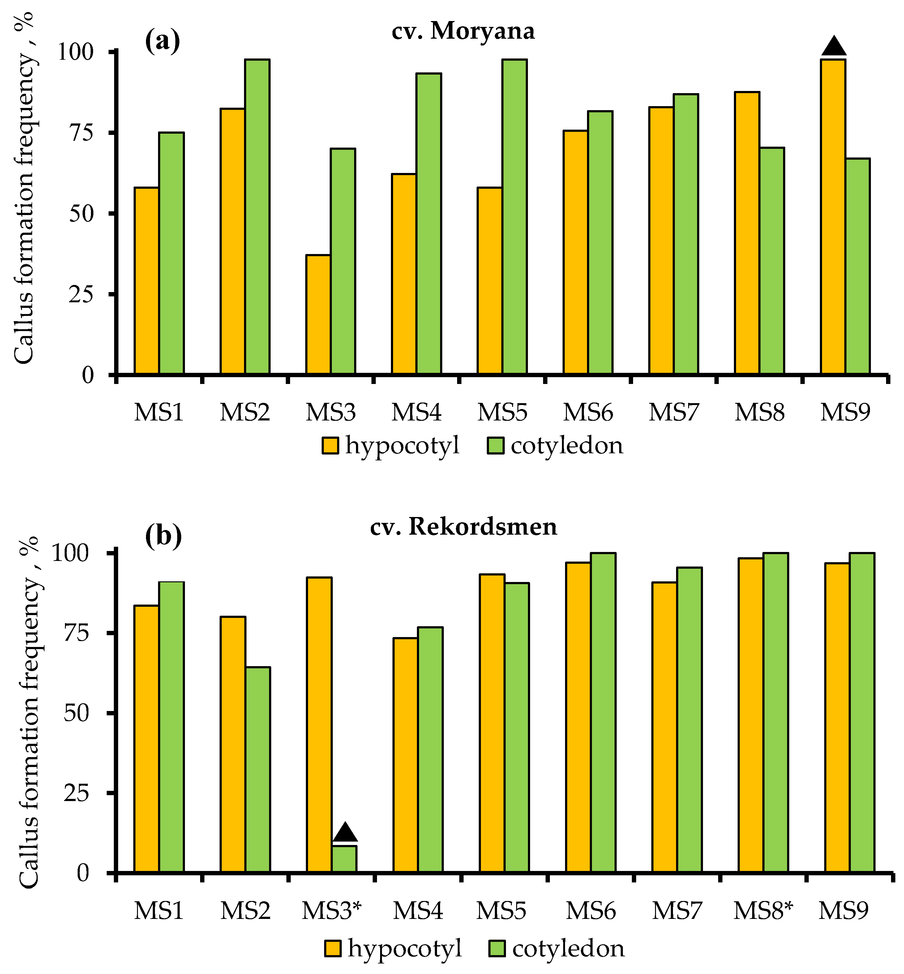
2.1.1. Callus Induction and Frequency of Its Formation

Cotyledon and hypocotyl explants of both tomato cultivars increased in size at the beginning of passaging when cultivated on Murashige and Skoog medium (MS) with PGRs (control MS1) and experimental culture media supplemented with a β-lactam antibiotic (MS2–MS9) (Table 1). Thickening of the overgrown tissues on explants could be observed already on the fifth day of cultivation, from which the callus was subsequently formed. Dense and light green calli were formed on the 10–12 days after planting on the abaxial part of cotyledons, as well as on the edge of cotyledon petioles. The formation of callus tissue on tomato hypocotyl occurred on the entire explant surface. The histological analysis of callus tissue revealed formation of a meristematic zone among hypertrophied cortical parenchyma cells. The meristematic zones consist of meristematic cell masses as well as de novo formed tracheal elements located in their central part (Figure 1).
The results of the three-way ANOVA test showed statistical differences at 5% significance level in callus formation frequency between different tomato explants and the studied culture media. In addition, the differences were significant for the interaction of factors “culture medium × genotype”, “culture medium × explant”, “genotype × explant”, and “culture medium × explant × genotype” (Table S1). The frequency of callus formation from hypocotyl and cotyledon explants of Moryana and Rekordsmen cultivars on control MS1 culture medium was 58.0% and 75.0%, as well as 83.6% and 91.3%, respectively (Figure 2).
Significant differences in the frequency of callus formation between hypocotyl (97.6%) and cotyledon (67.0%) explants in Moryana cultivar were revealed on the culture medium (MS9) supplemented with 800 mg L−1 of Amoxiclav®. In contrast, a dramatic inhibition of callus formation (8.5%) from cotyledons of Rekordsmen tomato cultivar was observed during the cultivation on a MS3 medium supplemented with a high concentration of cefotaxime. No significant differences were found between the culture media on the average callus formation frequency for two explant types of Moryana tomato cultivar. Similar data were noted for the Rekordsmen cultivar, except for MS3 and MS8 culture media, on which the values of callus formation frequency were, respectively, significantly lower and higher in comparison with the control.

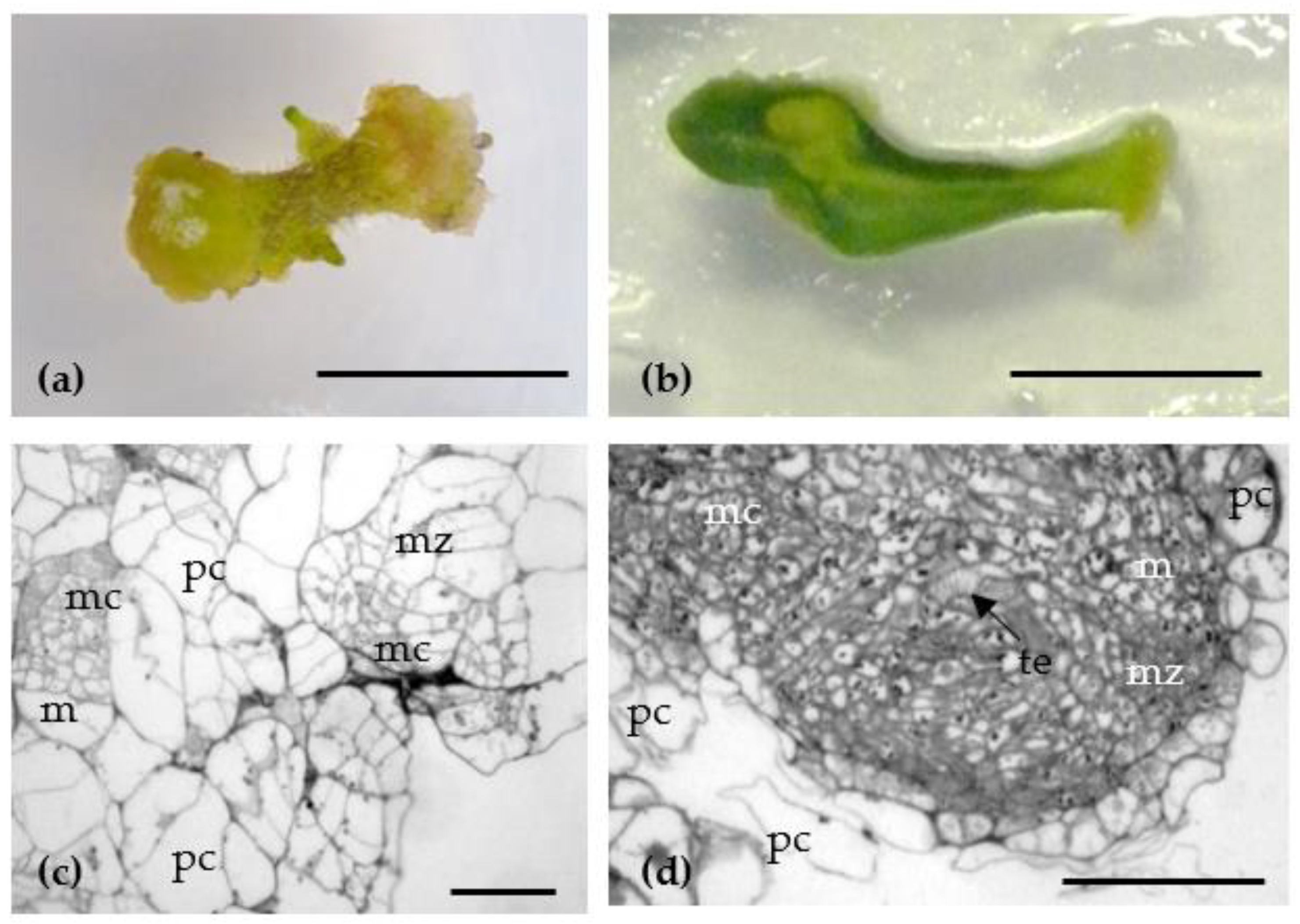
2.1.2. Efficiency of Tomato Shoot Organogenesis

The efficiency of tomato shoot organogenesis was assessed by the frequency of somatic organogenesis (Table 2), as well as the average number of regenerated shoots per explant (Table 3). The frequency of tomato shoot organogenesis from hypocotyl and cotyledon explants of Moryana and Rekordsmen cultivars on control MS1 culture medium supplemented with PGRs was 4.4% and 1.5%, as well as 67.1% and 42.7%, respectively (Table 2). When explants of the Moryana cultivar were cultured on most media with the addition of antibiotics, no significant differences were found in the shoot organogenesis frequency both in comparison with control and among themselves according to Duncan’s multiple range test. The exception was MS4 and MS6 culture media supplemented with 100 mg L−1 timentin and 200 mg L−1 amoxicillin, respectively, which stimulated the shoots organogenesis from hypocotyl explants, increasing their frequency by 8.5 and 6.8 times. In contrast to the MS7 culture medium containing 400 mg L−1 amoxicillin, the MS8 medium, characterized by the presence of clavulanic acid in the commercial antibiotic Amoxiclav®, significantly increased the shoot regeneration frequency from cotyledon and hypocotyl explants of Rekordsmen tomato cultivar.
A more pronounced effect of β-lactam antibiotics on the induction of somatic shoot organogenesis from different tomato explants was demonstrated for Rekordsmen cultivar. Adding timentin (MS4, MS5), Amoxiclav® (MS8, MS9), and amoxicillin at a concentration of 200 mg L−1 (MS6) to the culture medium leads to a significant increase in the shoot organogenesis frequency of from hypocotyl explants (17.6–77.8%). A similar response was observed for the cotyledon explants of Rekordsmen cultivar in the MS5, MS8 and MS9 culture medium. At the same time, the frequency of somatic shoot organogenesis from cotyledon explants on a culture medium containing Amoxiclav® (MS8, MS9) was 100% compared with control treatment (42.7%), and one significantly exceeded other experimental variants. Cultivation of cotyledon explants on MS2 medium supplemented with 400 mg L−1 Claforan® dramatically reduced rates of shoot organogenesis (1.2%), while a twofold increase in its concentration completely inhibited the regenerative process.
Both stimulatory and inhibitory effects of β-lactam antibiotics were noted when evaluating the average number of regenerated shoots per explant (Table 3). Compared to the control, a significant increase in the number of regenerated shoots per hypocotyl explant (2.89–3.24) was found on MS5, MS6, and MS8 culture media only for the Rekodsmen cultivar. For cotyledon explants of both tomato cultivars, the stimulatory effect was established for most variants of culture media. At the same time, the average number of shoots produced per cotyledon explant for Moryana and Rekodsmen cultivars varied from 3.2 and 5.6 to 5.6 and 9.2, respectively.
Thus, on average for both explant types and tomato genotypes, the addition of timentin, amoxicillin, and Amoxiclav® to t MS1 culture medium increased the shoot organogenesis frequency, as well as the number of shoots produced per explant. It was found that cotyledons of both tomato genotypes were characterized by a higher frequency of shoot organogenesis (74.2% and 56.4% for Moryana and Rekodsmen cultivars, respectively) and average shoot number produced per explant (4.1 and 3.5 for Moryana and Rekodsmen cultivars, respectively) than hypocotyl segments. Thus, it is not found significant differences between tomato genotypes on regenerative capacity for both explant types and variants of culture media.

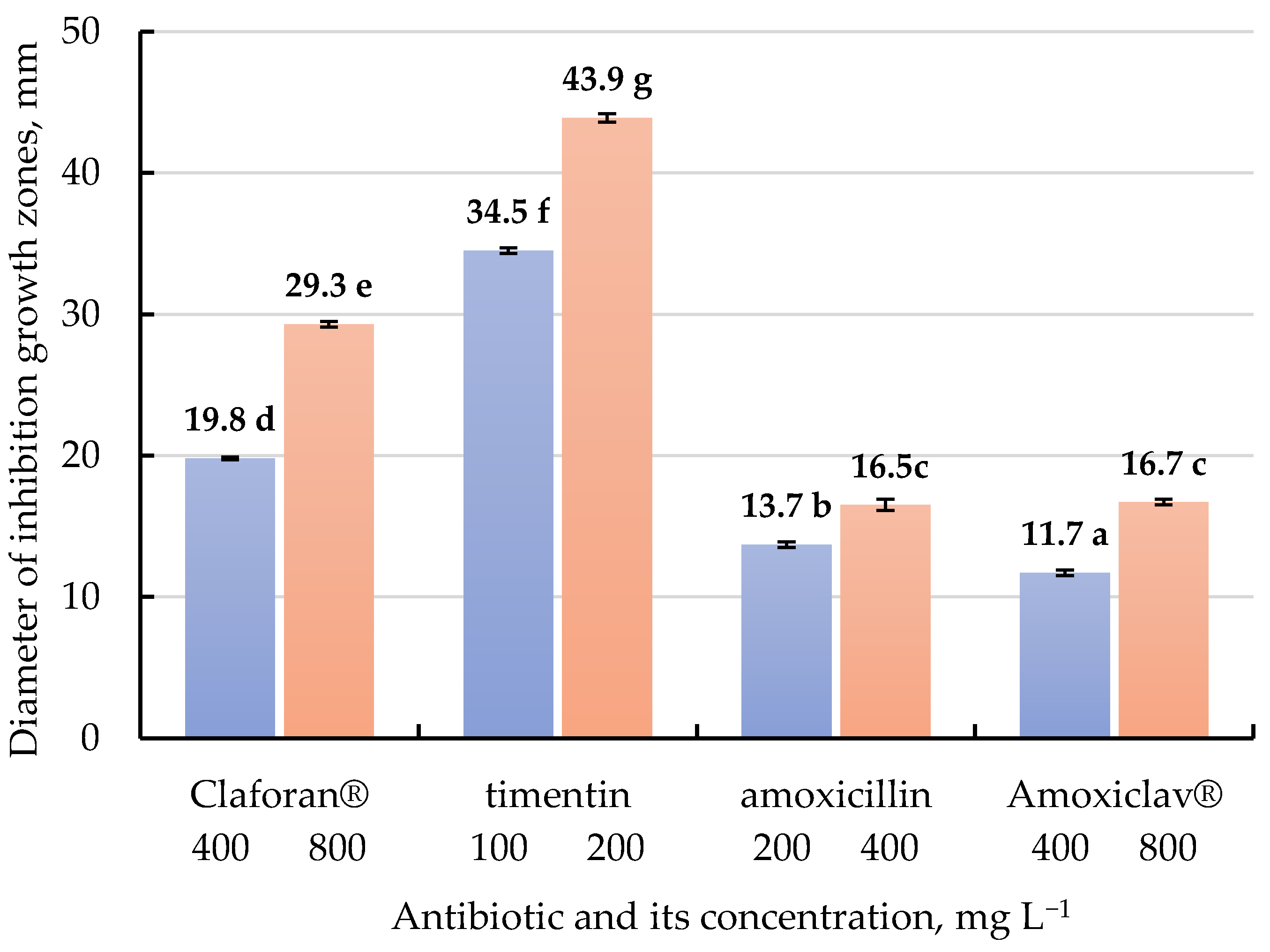
2.2. The Effects of Different β-Lactam Antibiotics on A. tumefaciens Growth Inhibition In Vitro

The results shown in Figure 3 and Figure S1 represent the antibacterial activity of different β-lactam antibiotics in solidified Luria-Bertani (LB) medium against growth inhibition of A. tumefaciens strain AGL0 using agar disk-diffusion assay. Depending on the concentration and type of antibiotic, the diameter of growth inhibition zones varied from 11.7 to 43.9 mm. The higher antibiotic concentration in LB medium increased diameter of the growth inhibition zones. Thus, the antibacterial activity of tested β-lactam antibiotics was arranged in the following order: timentin > Claforan® > amoxicillin ≥ Amoxiclav®.

3. Discussion

Optimization of an effective protocol for tomato regeneration system in vitro as well as Agrobacterium-mediated transformation is still routine, since morphogenesis efficiency is largely dependent on genetic, physiological, and physical factors [1,5,10,11,12,13,14,15,16,17,21,22,23]. To suppress bacterial infection during the introduction of plant material into an in vitro culture, clonal micropropagation and Agrobacterium-mediated transformation plant material is long-term subcultured on a medium supplemented with antibiotics. This is because the cultivation of explants on nutritionally-rich media stimulates an increase of microorganism titer in the cytoplasm, intercellular spaces, and vascular tissues, which ultimately leads to the visible manifestation after n-number of passages, necrosis, and death of plant tissue. Agrobacterium contamination of plant tissues does not allow the detection of the true transgenic status of transformants by molecular genetic techniques. In addition, endophytic bacterial microorganisms associated with the plant tissue culture (latent bacterial contamination) determine the regeneration and transformation capacity [34,35].
The comparative effect of various types and concentrations of β-lactam antibiotics to inhibit the Agrobacterium growth in vitro [36,37] and in planta [38,39,40,41,42], as well as on tomato morphogenesis responses [43,44,45], has been previously shown. In pioneering works devoted to the introduction of foreign DNA into the tomato genome by Agrobacterium-mediated transformation, the authors used classical carbenicillin [46,47]. With the advent of next generation semi-synthetic β-lactams, such as cefotaxime (Claforan®) [22,48,49], cefoxitin (mefoxin) [39], meropenem and imipenem [38,39], moxalactam [39], amoxicillin (Augmentin®, Amoxiclav®, Clavamox®) [42,50], and ticarcillin (Timentin®) [10,41,51], singly or coupled with β-lactamase inhibitor (clavulanic acid), they have been utilized at different concentrations to eliminate bacteria during tomato transformation. A number of studies have demonstrated the effective combined use of two overgrowth-control antibiotics in tomato regeneration medium, e.g., 250 mg L−1 cefotaxime and 500 mg L−1 carbenicillin [23], as well as 250 mg L−1 cefotaxime and ticarcillin [40]. In addition to antimicrobial activity, antibiotics have a pronounced positive effect on the regeneration capacity in tomato tissue culture [40,41,42,43,44,45] and many other crops [52,53,54,55,56]. However, their role in stimulating the morphogenesis in vitro is not fully understood, but it has been assumed that the antibiotics or degradation products mimic PGRs, since some of them possess an auxin-like structure [52,56,57].
In our earlier experiments, we evaluated different PGRs on callus induction and shoot organogenesis in vitro from hypocotyl and cotyledon explants of Rekordsmen cultivar and other commercial tomato genotypes [10,11]. In order to reveal the possible stimulating and inhibiting effects of various β-lactam antibiotics on tomato morphogenesis, in this study, we used the MS culture medium supplemented with 5 mg L−1 6-BA and 0.1 mg L−1 IAA (MS1), which does not provide high efficiency of shoot regeneration. As a result, culture media were identified that negatively (MS3 medium supplemented with 800 mg L−1 of cefotaxime) or positively (MS8 supplemented with 400 mg L−1 of Amoxiclav®) influenced the callus formation of Rekordsmen cultivar. Several studies have demonstrated no obvious inhibition effect of cefotaxime on callus induction and growth in tomato [41], but it significantly reduced shoot regeneration [41,42,45,49,50]. Kazemi et al. [49] suggested that reducing the negative effects of cefotaxime on tomato regeneration frequency and shoot number per cotyledonary explant was detected when replacing FeEDTA (iron chelate) with FeEDDHA (iron(3+)[ethylenediamine-N,N′-bis(hydroxyphenylacetic acid)]) in MS culture medium. In contrast, a stimulatory effect of cefotaxime in maize callus cultures [53] and carrot protoplast cultures [56] has been previously demonstrated. In our case, the addition of Claforan® at different concentrations in the MS1 culture medium dramatically reduced shoot organogenesis or completely inhibited the regenerative process.
Furthermore, our study identified different concentrations of β-lactams, such as timentin, amoxicillin, and Amoxiclav®, that significantly increased the shoot organogenesis frequency, as well as the number of shoots produced per explant from tomato tissue culture. It was found that clavulanic acid, which is part of the commercial antibiotic Amoxiclav®, significantly increased the shoot regeneration frequency from cotyledon and hypocotyl explants of Rekordsmen tomato cultivar. A stimulatory effect of ticarcillin and amoxicillin in tomato tissue cultures has also been previously shown [41,42,43,44,45,50]. Since ticarcillin is metabolized to phenylacetic acid, a naturally occurring weak auxin [57], increasing timentin concentration might improve tomato regeneration potential. Clavulanic acid is rapidly hydrolyzed at a 25 °C via the reactive amino ketone (1-amino-2-oxo-butan-4-ol) to pyrazine end-products (2,5-bis(2-hydroxyethyl)pyrazine, 3-carboxyethyl-2,5-bis(2-hydroxyethyl)pyrazine, and 3-ethyl-2,5-bis(2-hydroxyethyl)pyrazine) as well as carbon dioxide and acetaldehyde [58,59]. The stimulating effect of clavulanic acid on morphogenetic responses of tomato is most likely associated with direct action or products of its degradation as catalysts alone or combined with other β-lactam antibiotics or PGRs. Enhanced regenerative capacity may be due to stimulation of these antibiotics on the number of meristematic zones formed in callus tissue, from which subsequent shoot organogenesis occurred. Thus, the obtained results agree with the previously presented data that plant sensitivity to antibiotics usually is species-, genotype-, or even tissue-specific and mainly depends on concentrations, growth conditions and culture system.
Agar disk-diffusion assay is the official routine in vitro method used in many clinical trials and laboratory testing for antimicrobial susceptibility evaluation of antibiotics and other protective compounds [60,61,62,63]. With this assessment method, we evaluated the effectiveness of four β-lactam antibiotics on the growth elimination of A. tumefaciens strain AGL0 at concentrations that are most often applied in tomato transformation. As a result, the maximum diameter of growth inhibition zones was identified using 200 mg L−1 timentin (43.9 mm). The in vitro antibacterial activity of tested β-lactam antibiotics was arranged in the following order: timentin > Claforan® > amoxicillin ≥ Amoxiclav®. On the basis of agar disk-diffusion assay, the high efficiency of timentin at minimal concentrations (50 mg L−1) in inhibiting the growth of A. tumefaciens strain EHA105 compared with carbenicillin and cefotaxime was previously demonstrated [37]. Determination of the minimum inhibitory concentration (MIC) and minimum bactericidal concentration (MBC) revealed a high in vitro antibacterial activity of meroperem (carbapenem antibiotic) against A. tumefaciens strains AGL0, LBA4404, C58, and EHA101 [36,38]. Ogawa and Mii [36] found that the antibacterial activities of 12 β-lactams tested against strain LBA4404 were equal to or higher than those tested against strain EHA101. The mentioned above indicates the need for screening the type and concentration of antibiotic depending on the Agrobacterium strain, taking into account its negatively effects on the morphogenesis of plant tissue cultures.

4. Materials and Methods

4.1. Plant Material

Two commercial tomato (S. lycopersicum L.) cultivars (Rekordsmen and Moryana) were used in the experiment for obtaining donor seedlings. Tomato seeds were kindly provided by the All-Russian Research Institute of Irrigated Vegetable, Melon, and Ground Growing (Astrakhan oblast, Kamyziyak, Russia).

4.2. Explant Source, Antibiotics, and Culture Conditions

The seeds were surface sterilized in 96% ethanol for 30 s and in 20% solution (v/v) of the commercial bleach ‘Ace’ with a few drops of Tween-20 for 7 min, then rinsed with sterilized distilled water five times for 1 min each. After surface sterilization, the seeds were cultured on MS basal medium [64] without PGRs supplemented with 3% (w/v) sucrose (PanReac AppliChem, Madrid, Spain) and 0.7% (w/v) agar (PhytoTechnology Laboratories, Lenexa, KS, USA). The pH was adjusted with 1M KOH solution to 5.7–5.8 before autoclaving. The culture medium was sterilized by autoclaving at 121 °C and 1.1 atm for 20 min.
Whole cotyledons with 2–3 mm petioles as well as hypocotyl segments (approximately 10–15 mm in length) excised from the middle part of 10–12-day-old aseptic seedlings were used as explants. For callus induction and shoot regeneration, the explants were cultivated on agar-solidified MS medium supplemented with 5 mg L−1 6-benzylaminopurine (6-BA) (Sigma-Aldrich, St. Louis, MI, USA) and 0.1 mg L−1 indole-3-acetic acid (IAA) (Sigma-Aldrich, St. Louis, MI, USA) (MS1) as well as on MS1 culture medium containing four types of β-lactam antibiotics at different concentrations (MS2–MS9) (Table 1).
Antibiotics and PGRs were dissolved in distilled water, filter-sterilized (0.22 μm Millipore, Burlington, MA, USA), and stored until use at −20 °C. They were added after autoclaving to a culture medium cooled to 45 °C. Hypocotyl segments and cotyledons were placed in the Petri dishes horizontally and with the abaxial surface in contact with the culture medium, respectively. The explants were subcultured to fresh medium every 15 days. Donor seedlings and explants were kept in a climatic test chamber (Sanyo WLR-351H, Osaka, Japan) at 25 ± 2 °C under a 16-h photoperiod and light intensity of 65 μmol m−2 s−1.

4.3. Histological Analysis of Callus Tissue

Fragments of callus tissue formed from hypocotyl and cotyledon explants on MS1 culture media supplemented with 5 mg L−1 6-BA and 0.1 mg L−1 IAA were fixed for 24 h in 2.5% glutaraldehyde (Merck, Darmstadt, Germany) dissolved in 0.1 M Sorensen’s phosphate buffer (pH 7.2) with 1.5% sucrose. Then, the samples were washed, post-fixed in 1% OsO4 (Sigma-Aldrich, USA), and dehydrated in ethanol of increasing concentrations (30%, 50%, 70%, 96%, and 100%) and in propylene oxide (Fluka, Hamburg, Germany). The samples were embedded in mixture of Epon-812 and Araldite (Merck, Germany) according to the standard procedure. For light microscopy, semi-thin sections (1–2 μm) were prepared using glass knives and ultramicrotome LKB-V (LKB, Sweden), placed on glass slides, and embedded in epoxide resin. Samples were photographed using Olympus BX51 microscope (Olympus, Tokyo, Japan) with Color View II camera (Soft Imaging System, Münster, Germany).

4.4. Agar Disk-Diffusion Method

The agar disk-diffusion method was used to determine the Agrobacterium growth inhibition in vitro by various antibiotics at different concentrations. A. tumefaciens strain AGL0 was grown in a 250-mL capacity conical glass flask overnight at 28 °C in 50 mL of LB liquid medium [65] containing 50 mg L−1 rifampicin in a rotary shaker (150 rpm). An overnight culture of AGL0 strain was resuspended to OD600 = 0.6 and subsequently diluted 1000 times with LB liquid medium. Then, a diluted Agrobacterium suspension was inoculated on the surface of glass Petri dishes (100 × 15 mm) containing 25 mL agar-solidified LB medium supplemented with 50 mg/L rifampicin. To the sterile filter paper discs (10 mm in diameter), 50 μL solutions of different antibiotic concentrations such as Claforan®, timentin, amoxicillin, and Amoxiclav® (Table 1) were added. A sterile disc without antibiotic was used as negative control. Subsequently, filter paper discs were placed on the surface of agar-solidified LB medium. The Petri dishes were incubated at 37 °C for 48 h, then diameters of inhibition growth zones were measured.

4.5. Accounting Data and Statistical Analysis

To assess the effect of antibiotics on tomato morphogenesis, the frequency of callus formation and shoot organogenesis as well as the average number of shoots per explant were determined using following formulas:
Callus formation frequency (%) = [number of explants successfully forming callus/total number of explants] × 100;
Shoot organogenesis frequency (%) = [number of regenerating explants/number of explants forming callus] × 100;
Average number of shoots per explant = [shoot number/number of regenerating explants] × 100.
Evaluation of the morphogenetic potential was carried out on the 45th day of cultivation. Each variant of treatment was performed in three replications. For one replication, 10 cotyledons and 15 hypocotyl segments per each variant of culture medium were used. Experimental data were assessed at 5% significance level using the analysis of variance (ANOVA) and Duncan’s multiple range tests with AGROS software (version 2.11, Russia). Data of callus formation and shoot organogenesis (%) were arcsin X transformed prior to statistical analysis. The average number of shoots per explant were X + 1 transformed prior ANOVA [66].
To assess the effect of varying antibiotic concentrations for eliminating Agrobacterium, the diameters of inhibition growth zones were measured. The agar disk-diffusion test was performed three times in three biological replicates (three Petri dishes with three filter paper discs for each variant).

5. Conclusions

In the presented article, we estimated a comparative effect of various β-lactam antibiotics, such as cefotaxime, amoxycillin, and ticarcillin at different concentrations, on in vitro callus induction and shoot organogenesis from hypocotyl and cotyledon explants of two tomato cultivars (Rekordsmen, Moryana). Additionally, the growth inhibition of A. tumefaciens AGL0 strain according to agar disk-diffusion assay was implemented. As a result, both stimulatory (timentin, amoxicillin, and Amoxiclav®) and inhibitory (Claforan®) effects of β-lactam antibiotics on in vitro morphogenetic responses of tomato were noted. It was found that clavulanic acid, which is part of the commercial antibiotic Amoxiclav®, significantly increased the shoot regeneration frequency from cotyledon and hypocotyl explants of Rekordsmen tomato cultivar. The stimulating effect of clavulanic acid on morphogenetic responses of tomato is most likely associated with direct action or products of its degradation as catalysts alone or combined with other β-lactam antibiotics or PGRs. Thus, the obtained results agree with the previously presented data that plant sensitivity to antibiotics usually is genotype- and tissue-specific and mainly depends on its concentrations. According to agar disk-diffusion assay, the maximum diameter of growth inhibition zones (43.9 mm) was identified using 200 mg L−1 timentin. The in vitro antibacterial activity of tested β-lactam antibiotics was arranged in the following order: timentin > Claforan® > amoxicillin ≥ Amoxiclav®. Thus, to suppress the growth of internal and latent bacterial infection associated with tomato plant tissue culture, as well as for tomato transformation of Moryana and Rekordsmen cultivars by A. tumefaciens strain AGL0, we recommend adding 100–200 mg L−1 timentin or 400–800 mg L−1 Amoxiclav® to the culture medium.

Supplementary Materials

The following are available online at https://www.mdpi.com/article/10.3390/antibiotics10060660/s1, Table S1: The results of three-way ANOVA test to evaluate the significance of culture medium components, tomato genotype and explant type on the callus formation frequency; Figure S1: Antibacterial activity of different β-lactam antibiotics on growth inhibition of A. tumefaciens strain AGL0 using agar disk-diffusion assay.

Author Contributions

N.V.V. and Y.I.D. conducted the experiments on tomato culture in vitro. A.O.B. and N.V.V. conducted the agar disk-diffusion assay. E.N.B. conducted the histological estimation of tomato callus tissue. M.R.K. participated in manuscript conceptualization and statistical analysis of experimental data and writing, reviewing, and editing of manuscript; approved the final manuscript for publication, and agreed to be accountable for all aspects of the manuscript. All authors have read and agreed to the published version of the manuscript.

Funding

The reported study was supported by assignments 0574-2019-002 (All-Russia Research Institute of Agricultural Biotechnology) and 18-118021490111-5 (Tsitsin Main Botanical Garden of Russian Academy of Sciences) of the Ministry of Science and Higher Education of the Russian Federation.

Institutional Review Board Statement

Not applicable.

Informed Consent Statement

Not applicable.

Data Availability Statement

Data sharing is not applicable to this article.

Acknowledgments

The authors are grateful to the All-Russian Research Institute of Irrigated Vegetable, Melon and Ground Growing for providing tomato seeds of the Moryana and Rekodsmen cultivars for experiments.

Conflicts of Interest

The authors declare no conflict of interest.

References

  1. Gerszberg, A.; Hnatuszko-Konka, K.; Kowalczyk, T.; Kononowicz, A.K. Tomato (Solanum lycopersicum L.) in the service of biotechnology. Plant Cell Tiss. Organ Cult. 2015, 120, 881–902. [Google Scholar] [CrossRef] [Green Version]
  2. Hwang, H.H.; Yu, M.; Lai, E.M. Agrobacterium-mediated plant transformation: Biology and applications. Arab. Book 2017, 2017, 1–31. [Google Scholar] [CrossRef] [PubMed] [Green Version]
  3. Ruma, D.; Dhaliwal, M.S.; Kaur, A.; Gosal, S.S. Transformation of tomato using biolistic gun for transient expression of the β-glucuronidase gene. Indian J. Biotechnol. 2009, 8, 363–369. [Google Scholar]
  4. Abu-El-Heba, G.A.; Hussein, G.M.; Abdalla, N.A. A rapid and efficient tomato regeneration and transformation system. Landbauforsch. Volkenrode 2008, 58, 103. [Google Scholar]
  5. Vinoth, S.; Gurusaravanan, P.; Jayabalan, N. Optimization of factors influencing microinjection method for Agrobacterium tumefaciens-mediated transformation of tomato. Appl. Biochem. Biotechnol. 2013, 169, 1173–1187. [Google Scholar] [CrossRef] [PubMed]
  6. Nakata, K.; Tanaka, H.; Yano, K.; Takagi, M. An effective transformation system for Lycopersicon peruvianum by electroporation. Jpn. J. Breed. 1992, 42, 487–495. [Google Scholar] [CrossRef] [Green Version]
  7. Ray, S.; Lahiri, S.; Halder, M.; Mondal, M.; Choudhuri, T.R.; Kundu, S. An efficient method of isolation and transformation of protoplasts from tomato leaf mesophyll tissue using the binary vector pCambia 1302. Int. Adv. Res. J. Sci. Eng. Technol. 2015, 2, 146–150. [Google Scholar]
  8. Nicolia, A.; Andersson, M.; Hofvander, P.; Festa, G.; Cardi, T. Tomato protoplasts as cell target for ribonucleoprotein (RNP)-mediated multiplexed genome editing. Plant Cell Tiss. Organ. Cult. 2021, 144, 463–467. [Google Scholar] [CrossRef]
  9. Rao, A.Q.; Bakhsh, A.; Kiani, S.; Shahzad, K.; Shahid, A.A.; Husnain, T.; Riazuddin, S. The myth of plant transformation. Biotechnol. Adv. 2009, 27, 753–763. [Google Scholar] [CrossRef]
  10. Khaliluev, M.R.; Kharchenko, P.N.; Dolgov, S.V. Development of regeneration system and study of the transformation potential of a commercial tomato variety. Russ. Agric. Sci. 2010, 36, 175–179. [Google Scholar] [CrossRef]
  11. Khaliluev, M.R.; Bogoutdinova, L.R.; Baranova, G.B.; Baranova, E.N.; Kharchenko, P.N.; Dolgov, S.V. Influence of genotype, explant type, and component of culture medium on in vitro callus induction and shoot organogenesis of tomato (Solanum lycopersicum L.). Biol. Bull. 2014, 41, 512–521. [Google Scholar] [CrossRef]
  12. Kumar, S.; Jindal, S.K.; Sarao, N.K.; Dhaliwal, M.S. Development of an efficient In vitro regeneration protocol in tomato (Solanum lycopersicum L.). Agric. Res. J. 2017, 54, 475–479. [Google Scholar] [CrossRef]
  13. Rashid, R.; Bal, S.S. Effect of hormones on direct shoot regeneration in hypocotyl explants of tomato. Not. Sci. Biol. 2010, 2, 70–73. [Google Scholar] [CrossRef] [Green Version]
  14. Devi, R.; Dhaliwal, M.S.; Gosal, S.S. In vitro direct plant regeneration protocol for tomato genotypes. Indian J. Horticult. 2013, 70, 369–372. [Google Scholar]
  15. Saeed, W.; Naseem, S.; Gohar, D.; Ali, Z. Efficient and reproducible somatic embryogenesis and micropropagation in tomato via novel structures-Rhizoid Tubers. PLoS ONE 2019, 14, e0215929. [Google Scholar] [CrossRef] [PubMed] [Green Version]
  16. Godishala, V.; Mangamoori, L.; Nanna, R. Plant regeneration via somatic embryogenesis in cultivated tomato (Solanum lycopersicum L.). J. Cell Tiss. Res. 2011, 11, 2521. [Google Scholar]
  17. Ashakiran, K.; Chidambareswaran, M.; Govindasamy, V.; Sivankalyani, V.; Girija, S. Somatic embryogenesis for Agrobacterium mediated transformation of tomato-Solanum lycopersicum. L. Int. J. Biotechnol. Appl. 2011, 3, 72–79. [Google Scholar]
  18. Trujillo-Moya, C.; Gisbert, C.; Vilanova, S.; Nuez, F. Localization of QTLs for in vitro plant regeneration in tomato. BMC Plant Biol. 2011, 11, 1–13. [Google Scholar] [CrossRef] [Green Version]
  19. Basté, E.M.; Pratta, G.R.; Zorzoli, R. Genetic analysis of the in vitro culture response in tomato. Plant Cell Tiss. Organ Cult. 2007, 88, 233–239. [Google Scholar] [CrossRef]
  20. Sánchez-López, J.; Atarés, A.; Jáquez-Gutiérrez, M.; Ortiz-Atienza, A.; Capel, C.; Pineda, B.; García-Sogo, B.; Yuste-Lisbona, F.J.; Lozano, R.; Moreno, V. Approaching the genetic dissection of indirect adventitious organogenesis process in tomato explants. Plant Sci. 2021, 302, 110721. [Google Scholar] [CrossRef]
  21. Lee, M.H.; Lee, J.; Jie, E.Y.; Choi, S.H.; Jiang, L.; Ahn, W.S.; Kim, C.Y.; Kim, S.W. Temporal and spatial expression analysis of shoot-regeneration regulatory genes during the adventitious shoot formation in hypocotyl and cotyledon explants of tomato (cv. Micro-Tom). Int. J. Mol. Sci. 2020, 21, 5309. [Google Scholar] [CrossRef]
  22. Stavridou, E.; Τzioutziou, N.A.; Madesis, P.; Labrou, N.E.; Nianiou-Obeidat, I. Effect of different factors on regeneration and transformation efficiency of tomato (Lycopersicum esculentum) hybrids. Czech J. Gen. Plant Breed. 2019, 55, 120–127. [Google Scholar] [CrossRef]
  23. Chetty, V.J.; Ceballos, N.; Garcia, D.; Narváez-Vásquez, J.; Lopez, W.; Orozco-Cárdenas, M.L. Evaluation of four Agrobacterium tumefaciens strains for the genetic transformation of tomato (Solanum lycopersicum L.) cultivar Micro-Tom. Plant Cell Rep. 2013, 32, 239–247. [Google Scholar] [CrossRef] [PubMed]
  24. Enikeev, A.G.; Kopytina, T.V.; Semenova, L.A.; Natyaganova, A.V.; Gamanetz, L.V.; Volkova, O.D. Agrobacterial transformation as complex biotical stressing factor. J. Stress Physiol. Biochem. 2008, 4, 11–19. [Google Scholar]
  25. Şen, A. Oxidative stress studies in plant tissue culture. In Antioxidant Enzyme; El-Missiry, M.A., Ed.; InTech: Rijeka, Croatia, 2012; pp. 59–88. [Google Scholar]
  26. Dolgikh, Y.I.; Stepanova, A.Y.; Trusova, S.V.; Chichkova, N.V.; Vartapetian, A.B. Mitochondria-targeted antioxidant provides for enhanced morphogenetic potential in plant tissue cultures. Russ. J. Plant Physiol. 2013, 60, 706–712. [Google Scholar] [CrossRef]
  27. Ahsan, N.; Lee, S.-H.; Lee, D.-G.; Anisuzzaman, M.; Alam, M.F.; Yoon, H.-S.; Choi, M.S.; Yang, J.-K.; Lee, B.H. The effects of wounding type, preculture, infection method and cocultivation temperature on the Agrobacterium-mediated gene transfer in tomatoes. Ann. Appl. Biol. 2007, 151, 363–372. [Google Scholar] [CrossRef]
  28. Guo, M.; Zhang, Y.L.; Meng, Z.J.; Jiang, J. Optimization of factors affecting Agrobacterium-mediated transformation of Micro-Tom tomatoes. Genet. Mol. Res. 2012, 11, 661–671. [Google Scholar] [CrossRef]
  29. Dan, Y.; Zhang, S.; Zhong, H.; Yi, H.; Sainz, M.B. Novel compounds that enhance Agrobacterium-mediated plant transformation by mitigating oxidative stress. Plant. Cell Rep. 2015, 34, 291–309. [Google Scholar] [CrossRef]
  30. Pandey, N.; Cascella, M. Beta lactam antibiotics. In StatPearls; StatPearls Publishing: Treasure Island, FL, USA, 2020. [Google Scholar]
  31. Zango, U.U.; Ibrahim, M.; Shawai, S.A.A. A review on β-lactam antibiotic drug resistance. MOJ Drug. Des. Dev. Ther. 2019, 3, 52–58. [Google Scholar]
  32. Saudagar, P.S.; Survase, S.A.; Singhal, R.S. Clavulanic acid: A review. Biotechnol. Adv. 2008, 26, 335–351. [Google Scholar] [CrossRef]
  33. Cheng, Z.M.; Schnurr, J.A.; Kapaun, J.A. Timentin as an alternative antibiotic for suppression of Agrobacterium tumefaciens in genetic transformation. Plant Cell Rep. 1998, 17, 646–649. [Google Scholar] [CrossRef]
  34. Dunaeva, S.E.; Osledkin, Y.S. Bacterial microorganisms associated with the plant tissue culture: Identification and possible role. Sel’skokhozyaistvennaya Biol. 2015, 50, 3–15. [Google Scholar] [CrossRef] [Green Version]
  35. Cheong, E.J.; Na, M.; Jeong, U. The effect of endophytic bacteria on in vitro shoot growth of Prunus yedoensis and its identification and elimination. Vitro Cell. Dev. Biol. Plant. 2019, 56, 200–206. [Google Scholar] [CrossRef]
  36. Ogawa, Y.; Mii, M. Screening for highly active β-lactam antibiotics against Agrobacterium tumefaciens. Arch. Microbiol. 2004, 181, 331–336. [Google Scholar] [CrossRef] [PubMed]
  37. Priya, A.M.; Pandian, S.K.; Manikandan, R. The effect of different antibiotics on the elimination of Agrobacterium and high frequency Agrobacterium-mediated transformation of indica rice (Oryza sativa L.). Czech J. Genet. Plant Breed. 2012, 48, 120–130. [Google Scholar] [CrossRef] [Green Version]
  38. Farzaneh, A.; Adel, Y.; Ali, N.; Younes, G. Determine effective concentrations of β-lactam antibiotics against three strains of Agrobacterium tumefaciens and phytotoxicity on Tomato and Tobacco. Int. J. Agron. Plant. Prod. 2013, 11, 2919–2925. [Google Scholar]
  39. Ogawa, Y.; Mii, M. Meropenem and moxalactam: Novel β-lactam antibiotics for efficient Agrobacterium-mediated transformation. Plant Sci. 2007, 172, 564–572. [Google Scholar] [CrossRef]
  40. Hu, W.; Phillips, G.C. A combination of overgrowth-control antibiotics improves Agrobacterium tumefaciens-mediated transformation efficiency for cultivated tomato (L. esculentum). Vitro Cell. Dev. Biol. Plant 2001, 37, 12–18. [Google Scholar] [CrossRef]
  41. Ling, H.Q.; Kriseleit, D.; Ganal, M.W. Effect of ticarcillin/potassium clavulanate on callus growth and shoot regeneration in Agrobacterium-mediated transformation of tomato (Lycopersicon esculentum Mill.). Plant Cell Rep. 1998, 17, 843–847. [Google Scholar] [CrossRef] [PubMed]
  42. Ieamkhang, S.; Chatchawankanphanich, O. Augmentin® as an alternative antibiotic for growth suppression of Agrobacterium for tomato (Lycopersicon esculentum) transformation. Plant Cell Tiss. Organ Cult. 2005, 82, 213–220. [Google Scholar] [CrossRef]
  43. Mamidala, P.; Nanna, R.S. Influence of antibiotics on regeneration efficiency in tomato. Plant Omics J. 2009, 2, 135–140. [Google Scholar]
  44. Costa, M.G.C.; Nogueira, F.T.S.; Figueira, M.L.; Otoni, W.C.; Brommonschenkel, S.H.; Cecon, P.R. Influence of the antibiotic timentin on plant regeneration of tomato (Lycopersicon esculentum Mill.) cultivars. Plant Cell Rep. 2000, 19, 327–332. [Google Scholar] [CrossRef] [PubMed]
  45. Gerszberg, A.; Grzegorczyk-Karolak, I. Influence of selected antibiotics on the tomato regeneration in in vitro cultures. Not. Bot. Hort. Agrobot. 2019, 47, 558–564. [Google Scholar] [CrossRef] [Green Version]
  46. Horsch, R.B.; Frye, J.E.; Hoffman, N.F.; Eichoholtz, D.; Rogers, S.G.; Fraley, R.T. A simple and general method for transferring genes into plants. Science 1985, 227, 1229–1231. [Google Scholar]
  47. McCormick, S.; Niedermeyer, J.; Fry, J.; Barnason, A.; Horsch, R.; Fraley, R. Leaf disc transformation of cultivated tomato (L. esculentum) using Agrobacterium tumefaciens. Plant Cell Rep. 1986, 5, 81–84. [Google Scholar] [CrossRef] [PubMed]
  48. Perveen, S.; Khan, T.A.; Shaheen, H.; Naz, R.; Hyder, M.Z.; Ijaz, B.; Saqlan Naqvi, S.M.; Yasmin, T. Morpho-physiological investigations in transgenic tomato (Solanum lycopersicum L.) over expressing OsRGLP1 gene. Vitro Cell. Dev. Biol. Plant 2021, 1–16. [Google Scholar] [CrossRef]
  49. Kazemi, E.M.; Jonoubi, P.; Majd, A.; Pazhouhandeh, M. Reduction of negative effects of cefotaxime in tomato transformation by using FeEDDHA. Int. J. Farm. Alli. Sci. 2014, 3, 538–542. [Google Scholar]
  50. Gulevich, A.A.; Kurenina, L.V.; Baranova, E.N. Application of a system for targeting Fe-dependent superoxide dismutase and choline oxidase enzymes to chloroplast as a strategy for effective plant resistance to abiotic stresses. Russ. Agric. Sci. 2018, 44, 118–123. [Google Scholar] [CrossRef]
  51. Shpakovski, G.V.; Spivak, S.G.; Berdichevets, I.N.; Babak, O.G.; Kubrak, S.V.; Kilchevsky, A.V.; Aralov, A.V.; Slovokhotov, I.Y.; Shpakovski, D.G.; Baranova, E.N.; et al. A key enzyme of animal steroidogenesis can function in plants enhancing their immunity and accelerating the processes of growth and development. BMC Plant Biol. 2017, 17, 189. [Google Scholar] [CrossRef] [Green Version]
  52. Mineykina, A.; Shumilina, D.; Bondareva, L.; Soldatenko, A.; Domblides, E. Effect of beta-lactam antibiotics on microspore embryogenesis in Brassica species. Plants 2020, 9, 489. [Google Scholar] [CrossRef]
  53. Danilova, S.A.; Dolgikh, Y.I. The stimulatory effect of the antibiotic cefotaxime on plant regeneration in maize tissue culture. Russ. J. Plant Physiol. 2004, 51, 559–562. [Google Scholar] [CrossRef]
  54. Gambhir, G.; Kumar, P.; Srivastava, D.K. Effect of antibiotic sensitivity on different cultured tissues and its significance in genetic transformation of cabbage Brassica oleracea. Biosci. Biotechnol. Res. Comm. 2017, 10, 652–661. [Google Scholar] [CrossRef]
  55. Stanišić, M.; Ninković, S.; Savić, J.; Ćosić, T.; Banjac, N. The effects of β-lactam antibiotics and hygromycin B on de novo shoot organogenesis in apple cv. Golden Delicious. Arch. Biol. Sci. 2018, 70, 179–190. [Google Scholar] [CrossRef] [Green Version]
  56. Grzebelus, E.; Skop, L. Effect of β-lactam antibiotics on plant regeneration in carrot protoplast cultures. Vitro Cell. Dev. Biol. Plant 2014, 50, 568–575. [Google Scholar] [CrossRef] [Green Version]
  57. Holford, P.; Newbury, H.J. The effects of antibiotics and their breakdown products on the in vitro growth of Antirrhinum majus. Plant Cell Rep. 1992, 11, 93–96. [Google Scholar] [CrossRef]
  58. Brethauer, S.; Held, M.; Panke, S. Clavulanic acid decomposition is catalyzed by the compound itself and by its decomposition products. J. Pharm. Sci. 2008, 97, 3451–3455. [Google Scholar] [CrossRef]
  59. Gómez-Ríos, D.; Ramírez-Malule, H.; Neubauer, P.; Junne, S.; Ríos-Estepa, R. Degradation kinetics of clavulanic acid in fermentation broths at low temperatures. Antibiotics 2019, 8, 6. [Google Scholar] [CrossRef] [Green Version]
  60. Balouiri, M.; Sadiki, M.; Ibnsouda, S.K. Methods for in vitro evaluating antimicrobial activity: A review. J. Pharm. Anal. 2016, 6, 71–79. [Google Scholar] [CrossRef] [Green Version]
  61. Schumacher, A.; Vranken, T.; Malhotra, A.; Arts, J.J.C.; Habibovic, P. In vitro antimicrobial susceptibility testing methods: Agar dilution to 3D tissue-engineered models. Eur. J. Clin. Microbiol. Infect Dis. 2018, 37, 187–208. [Google Scholar] [CrossRef] [Green Version]
  62. Chiu, C.-T.; Lai, C.-H.; Huang, Y.-H.; Yang, C.-H.; Lin, J.-N. Comparative analysis of gradient diffusion and disk diffusion with agar dilution for susceptibility testing of Elizabethkingia anophelis. Antibiotics 2021, 10, 450. [Google Scholar] [CrossRef]
  63. Böhm, M.E.; Razavi, M.; Flach, C.F.; Larsson, D.G. A Novel, Integron-regulated, class C β-lactamase. Antibiotics 2020, 9, 123. [Google Scholar] [CrossRef] [PubMed] [Green Version]
  64. Murashige, T.; Skoog, F.A. Revised medium for rapid growth and bioassays with tobacco tissue culture. Physiol. Plant. 1962, 15, 473–497. [Google Scholar] [CrossRef]
  65. Bertani, G. Studies on lysogenesis I. The mode of phage liberation by lysogenic Escherichia coli. J. Bacterial. 1951, 62, 293–300. [Google Scholar] [CrossRef] [PubMed] [Green Version]
  66. Dospekhov, B.A. Methods of Field Experience; Agropromizdat: Moscow, Russia, 1985; p. 350. [Google Scholar]
Figure 1. Callus formation from hypocotyl (a) and cotyledon (b) explants on MS1 culture media supplemented with 5 mg L−1 6-benzylaminopurine (6-BA) and 0.1 mg L−1 indole-3-acetic acid (IAA) and its histological analysis (c,d). Abbreviations: mz—meristematic zone; mc—meristematic cell; pc—parenchyma cell; te—tracheal element. Bars—1 cm (a,b) and 50 μm (c,d).
Antibiotics 10 00660 g001
Figure 2. Influence of MS culture media supplemented with different types and concentrations of β-lactam antibiotics (MS2–MS9) on the callus formation frequency from hypocotyl and cotyledon explants of tomato cv. Moryana (a) and Rekordsmen (b). Abbreviations: ▲—variants of culture media that have significant differences (α = 0.05) between cotyledon and hypocotyl explants; *—variants of culture media significantly different from the control treatment (MS1).
Antibiotics 10 00660 g002
Figure 3. Influence of different β-lactam antibiotics on A. tumefaciens growth inhibition (strain AGL0) using agar disk-diffusion assay. The figure shows mean values ± standard errors. Means followed by the same letter are not significantly different at α = 0.05 according to the Duncan’s multiple range test.
Antibiotics 10 00660 g003
Table 1. Types and concentrations of β-lactam antibiotics used in MS culture media for in vitro callus induction and shoot organogenesis of tomato.
Culture
Medium
MS2MS3MS4MS5MS6MS7MS8MS9
Antibioticbrand
name
Claforan®,
Sanofi-Aventis
(France)
Timentin, PhytoTechnology Laboratories
(USA)
Amoxicillin, PhytoTechnology Laboratories (USA)Amoxiclav®,
Lek d.d. (Slovenia)
generic nameCefotaxime sodiumTicarcillin
disodium + clavulanate potassium (15:1)
AmoxicillinAmoxicillin + clavulanate potassium
Concentration, mg L−1400800100200200400400800
Table 2. The effects of genotype, explant type, and culture medium components on the frequency of tomato shoot organogenesis in vitro.
Culture MediumAntibiotic and Its Concentration,
mg L−1
Frequency of Shoot Organogenesis, %
cv. Moryanacv. RekordsmenMean (Culture Medium) 3
HypocotylsCotyledonsMean 2HypocotylsCotyledonsMean
MS14.4 1 ab67.1 fghijk35.8 cdef1.5 ab42.7 fghij22.1 c29.0 b
MS2Claforan®4004.3 ab71.6 ijk38.0 cdef11.9 bcde1.2 ab6.6 b22.3 b
MS3Claforan®8000 a56.8 efghij28.4 c0.8 ab0 a0.4 a14.4 a
MS4Timentin10037.4 de77.5 jk57.5 g17.6 cdef46.6 ghij32.1 cde44.8 d
MS5Timentin20016.8 bcd89.2 k53.0 efg26.5 defgh80.6 l53.6 fg53.3 de
MS6Amoxicillin20030.2 cde81.6 jk56.0 fg30.6 efgh68.4 jkl49.5 efg52.8 de
MS7Amoxicillin40010.9 bc87.0 k54.4 defg6.7 abcd68.4 ijkl37.6 cdef46.0 cd
MS8Amoxiclav®40010.9 bc70.3 hijk46.1 cdefg77.8 kl100 mn88.9 i67.5 f
MS9Amoxiclav®8006.7 ab67.1 ghijk36.9 cdef55.7 hijkl100 n77.9 hi57.4 ef
Mean (explant) 413.5 a74.2 b25.5 a56.4 b
Mean (genotype) 543.9 a40.9 a
Means followed by the same letter are not significantly different at α = 0.05 according to the Duncan’s multiple range test. Data were arcsin X transformed prior to statistical analysis. 1 Influence of culture medium component and explant type on the shoot organogenesis frequency. 2 Influence of culture medium component and genotype on the shoot organogenesis frequency. 3 Influence of culture medium component on the shoot organogenesis frequency. 4 Influence of explant type on the shoot organogenesis frequency. 5 Influence of genotype on the shoot organogenesis frequency.
Table 3. The effects of genotype, explant type, and culture medium components on the average number of tomato shoots produced per explant in vitro.
Culture MediumAntibiotic and Its Concentration,
mg L−1
Average Number of Shoots per Explant
cv. Moryanacv. RekordsmenMean (Culture Medium) 3
HypocotylsCotyledonsMean 2HypocotylsCotyledonsMean
MS11.9 1 bc2.0 bcde2.0 bcd1.3 ab2.3 bcde1.8 abc1.9 ab
MS2Claforan®4001.6 ab3.7 ghi2.7 bcdefg2.6 bcde1.5 abcd2.0 bcde2.4 bc
MS3Claforan®8001.0 a2.3 bcdef1.7 ab1.3 ab1.0 a1.1 a1.4 a
MS4Timentin1003.0 cdefghi5.6 j4.3 jkl2.0 abcde2.3 bcde2.2 bcdef3.2 cd
MS5Timentin2002.9 cdefghi4.3 ij3.6 gh6ijk2.9 cde5.3 fgh4.1 ijk3.8 d
MS6Amoxicillin2002.6 bcdefg3.5 fghi3.0 defghi3.0 de3.5 efg3.3 fghij3.1 cd
MS7Amoxicillin4002.2 bcde3.2 efghi2.7 cdefg2.0 abcde9.2 i5.6 l4.2 d
MS8Amoxiclav®4002.0 bcd3.1 defghi2.6 bcdefg3.2 e6.3 h4.8 kl3.7 d
MS9Amoxiclav®8002.3bcdef4.0 hi3.1 efghij2.7 bcde5.3 gh4.0 hijk3.6 d
Mean (explant) 42.0 a3.5 b2.0 a4.0 b
Mean (genotype) 52.8 a3.0 a
Means followed by the same letter are not significantly different at α = 0.05 according to the Duncan’s multiple range test. The average number of shoots per explant were X + 1 transformed prior ANOVA. 1 Influence of culture medium component and explant type on the number of shoots per explant. 2 Influence of culture medium component and genotype on the number of shoots per explant. 3 Influence of culture medium component on the number of shoots per explant. 4 Influence of explant type on the number of shoots per explant. 5 Influence of genotype on the number of shoots per explant.
Publisher’s Note: MDPI stays neutral with regard to jurisdictional claims in published maps and institutional affiliations.

