Article

The Application of a Nanomaterial Optical Fiber Biosensor Assay for Identification of Brucella Nomenspecies

1
Department of Physics, College of Science, Virginia Tech, Blacksburg, VA 24061, USA
2
Department of Biomedical Sciences and Pathobiology, Virginia-Maryland College of Veterinary Medicine, Virginia Tech, Blacksburg, VA 24061, USA
3
Long Island University, Brookville, NY 11548, USA
*
Author to whom correspondence should be addressed.
Biosensors 2019, 9(2), 64; https://doi.org/10.3390/bios9020064
Submission received: 22 March 2019 / Revised: 10 May 2019 / Accepted: 14 May 2019 / Published: 21 May 2019
(This article belongs to the Special Issue Spectroscopy-Based Biosensors)

Abstract

:
Bacteria in the genus Brucella are the cause of brucellosis in humans and many domestic and wild animals. A rapid and culture-free detection assay to detect Brucella in clinical samples would be highly valuable. Nanomaterial optical fiber biosensors (NOFS) are capable of recognizing DNA hybridization events or other analyte interactions with high specificity and sensitivity. Therefore, a NOFS assay was developed to detect Brucella DNA from cultures and in tissue samples from infected mice. An ionic self-assembled multilayer (ISAM) film was coupled to a long-period grating optical fiber, and a nucleotide probe complementary to the Brucella IS711 region and modified with biotin was bound to the ISAM by covalent conjugation. When the ISAM/probe duplex was exposed to lysate containing ≥100 killed cells of Brucella, or liver or spleen tissue extracts from Brucella-infected mice, substantial attenuation of light transmission occurred, whereas exposure of the complexed fiber to non-Brucella gram-negative bacteria or control tissue samples resulted in negligible attenuation of light transmission. Oligonucleotide probes specific for B. abortus, B. melitensis, and B. suis could also be used to detect and differentiate these three nomenspecies. In summary, the NOFS biosensor assay detected three nomenspecies of Brucella without the use of polymerase chain reaction within 30 min and could specifically detect low numbers of this bacterium in clinical samples.

Graphical Abstract

1. Introduction

Brucellae are bacterial pathogens responsible for brucellosis of domestic and wild animals and are zoonotic pathogens for humans. Brucella spp. are small gram-negative, nonmotile, aerobic, and slow-growing coccobacilli. Despite the recognition of brucellae as a single genospecies based on DNA-DNA hybridization studies, they are systematically classified based on host specificity. The main terrestrial nomenspecies are B. abortus (cattle), B. melitensis (goats and sheep), B. suis (pigs), B. canis (dogs), B. ovis (sheep), and B. neotomae (woodrats) [1,2]. In addition, Brucella spp. can also be isolated from marine mammals [3,4]. Human infections are acquired by consuming unpasteurized milk and dairy products or by direct exposure to animals and their carcasses. Human brucellosis resulting from direct exposure is primarily a disease of farmers, shepherds, veterinarians, microbiologists, butchers, and slaughterhouse workers [5,6].
Wild animals play an important role in the epidemiology of Brucella infections. Brucella spp. remain enzootic in wild elk and bison in the Greater Yellowstone region that includes areas of Montana, Idaho, and Wyoming. As a result, these animals are a reservoir for B. abortus in the United States [7]. Transmission of Brucella spp. to susceptible cattle normally occurs by ingestion or oral contact with infected fetuses that have been aborted, fetal fluids and membranes, or uterine discharges [8]. Elk that congregate on feeding grounds from November through April overlap with the peak time period when Brucella is transmitted to other animals (February through June) [9]. Maichak et al. reported that as many as 12% of the elk attending feeding grounds come into contact with non-infectious elk fetuses placed on these sites [10]. Bison normally congregate in large numbers, which increases the likelihood they will come into contact with Brucella-infected fetuses and discharges. Such congregation increases the possibility that infected bison could transmit Brucella to cattle herds in the area [7]. As a result, farmers may unnecessarily kill elk or bison that wander out of the park and onto private farmlands.
Development of reliable and cost-effective diagnostic tests for use in elk and other wildlife is a high research priority. Reliable and portable diagnostic assays that can be carried out in the field by non-specialist personnel are urgently needed to minimize the spread of the disease among wildlife and its transmission to domestic animals and humans.
Biosensors combine biological molecules with a physicochemical transducer. Biological components incorporated into biosensors may include nucleic acids, enzymes, antibodies, etc., and the transducer may be optical, electrochemical, thermometric, or piezoelectric. Regardless, the detection of the target biological material results in a measurable signal. The advantages of optical fibers (light, inexpensive, and low interference) have established them as essential instruments of sensor technology [11]. Biosensors that consist of optical fibers transmit light based on total internal reflection through their transduction elements. The sensor produces a signal that can be analyzed and is in proportion to the concentration of the molecule that binds to the biological element on the sensor. Grating devices in the optical fiber induce a periodic variation in the refractive index of the optical fiber’s core. As a result, there is a significant drop in the amount of light transmitted through the fiber at a specific wavelength. The specific wavelength can be changed to account for temperature, pressure, or the type of binding event [12].
Layer-by-layer films, also known as ionic self-assembled multilayer (ISAM) films, are a novel type of materials that enable the user to modify the structure and thickness of the thin film at nanometer levels. The assembly of such films is simple and inexpensive [13,14]. As a result, optical fibers containing these nanoscale overlays substantially enhance, through direct light transmission, the detection of antigen binding to antibody or DNA hybridizing to complementary DNA. Furthermore, these sensors can be organized into a device that is rugged and portable [15,16]. For the detection of infectious agents, these fiber-optic biosensors can be used as rapid diagnostic or screening tests prior to culture, serology, or other means of diagnosis. A variety of fiber grating-based biosensor platforms have recently been developed [17,18,19,20]. For the work described here, a nanomaterial optical fiber biosensor (NOFS) assay was successfully used to detect Brucella DNA in culture lysates and in infected animal tissues.

2. Materials and Methods

2.1. Oligonucleotide Primers and Probes

The oligonucleotide probes and primers (Table 1) were designed manually based on the DNA sequences of the respective genes/regions in GenBank and were purchased from Integrated DNA Technologies, Collinsville, IL, USA. The IS711 DNA region is present in all known nomenspecies of Brucella, but not in other bacteria. Therefore, the primers IS711-For and IS711-Rev and probes IS711-BIO and IS711-DIG from the IS711 region (Table 1) were used for detection of all Brucella nomenspecies and to distinguish them from other bacterial species. In order to identify and differentiate the three major Brucella nomenspecies (Brucella abortus, B. melitensis, and B. suis), the oligonucleotide probes BruAb2_0168 (GenBank accession AE017224.1), Melitensis_0466 (GenBank accession AE008918.1), and Suis_TraJ (GenBank accession CP024421.1) (Table 1) were used. A search using the NCBI BLAST program confirmed the specificity of the DNA regions used for identifying and distinguishing the respective Brucella species.

2.2. Bacterial Strains and Culture Conditions Used

The Brucella nomenspecies and other bacterial species used as controls in this study are listed in Table 2. The bacteria were grown to mid-log phase in brain heart infusion (BHI) broth. Bacteria were harvested by centrifugation, washed, resuspended in phosphate buffered saline, pH 7.2 (PBS), and killed by boiling for 20 min (confirmed by viable plate count). Serial dilutions of killed cell suspensions were made in PBS, and genomic DNA was harvested using the DNAeasy Blood and Tissue kit (Qiagen, Valencia, CA, USA).

2.3. PCR

PCR, when used, was performed in 25 μL volumes and included 10 pmol each of the primers Primer-IS711-For and Primer-IS711-Rev (Table 1), 1 mM MgCl2, 200 μM dNTPS, 1X concentration of OneTaq Standard Reaction Buffer (New England Biolabs, Ipswich, MA, USA), 1.25 units of OneTaq DNA Polymerase (New England Biolabs), and template DNA. Template DNA included either 26 ng of genomic DNA or 1 µL of heat-killed, cell lysate from 1 × 10⁵ cells/mL to 3 × 10¹⁰ cells/mL. Reaction conditions were an initial denaturation temperature of 95 °C for 5 min, followed by 30 cycles of 95 °C/1 min, 57 °C/1 min, 72 °C/1 min, and a final extension at 72 °C/10 min.

2.4. Enzyme-Linked Immunosorbent Assay (ELISA)

An ELISA was designed using Magnalink Streptavidin Magnetic beads (Solulink Inc., San Diego, CA, USA). The protocol was as described by the manufacturer (Solulink, Inc.) with modification. Briefly, 60 pmol of the biotinylated probe (Probe-IS711-BIO) (Table 1) in 250 μL of nucleic acid binding and wash buffer (50 mM Tris-HCl, 150 mM NaCl, 0.05% Tween 20, pH 8.0) was incubated for 30 min at room temperature with the beads in 1.5 mL microcentrifuge tubes. The heat-denatured PCR products or genomic DNA were incubated with the beads coupled to the biotinylated probe in hybridization buffer (3× SSC, 0.05% Tween 20) for 2 h at 45 °C. A digoxigenin-labeled probe (Probe-IS711-DIG) (Table 1) in hybridization buffer was then incubated with the bead/probe/DNA triplex for 2 h at 45 °C. The DIG Detection Starter Kit from Roche (Sigma-Aldrich, St. Louis, MO, USA) was used to determine binding of the probe to the triplex complex. The ELISA was designed solely to confirm that the designed probe would hybridize with the Brucella genomic DNA, and not as a diagnostic assay itself. Therefore, quantitative data were not obtained.

2.5. Fabrication of the ISAM Film

ISAM films were fabricated using procedures described by the authors [21]. The ISAM method simply involves the alternate dipping of a charged substrate (optical fiber) into an aqueous solution of a polycation and an aqueous solution of a polyanion at room temperature. The optical fiber was immersed in an aqueous 10 mM poly-allylamine hydrochloride (PAH) (pH 7.0) solution for 2 min then rinsed three times in distilled water. The fiber was then immersed in a similar aqueous solution of 10 mM poly-1-[p-(3′-carboxy-4′-hydroxyphenylazo) benzenesulfonamido]-1,2-ethanediyl (PCBS) (pH 7.0) for 2 min and rinsed again. The final layer was always the negatively-charged PCBS. These two steps were repeated until the optimal number of bilayers was obtained, which, for this assay, was four layers (Figure 1).

2.6. Coupling the Probe to the ISAM Film

The ISAM film was incubated with 0.6 mL of 0.17 M freshly prepared N-(3-dimethylaminodipropyl)-N’-ethylcarbodiimide (EDC), 0.17 M N-hydroxysulfosuccinimide (NHS), and 60 pmol of biotinylated oligonucleotide probe in PBS, pH 7.0, at room temperature for 30 min.

2.7. Conjugation of Streptavidin to the ISAM Film
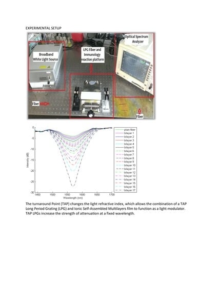
An alternative method to couple the probe onto the ISAM film involved using a streptavidin intermediate. Four bilayers were deposited onto the optical fiber, leaving PCBS with negatively-charged carboxyl groups exposed. Then, 40 μL of streptavidin (1 mg/mL in PBS, pH 7.0) was mixed with 0.6 mL of cross-linker solution (0.17 M EDC and 0.17 M NHSS in PBS, pH 7.0). The mixture was added to the fiber and incubated for 8 h, with mixing every 15 min. The fiber was then rinsed and the biotinylated probe was added for spontaneous coupling to streptavidin.

2.8. NOFS Assay

The NOFS assay consists of turnaround point long-period gratings (TAP-LPGs) with ISAM films adsorbed on fiber cladding. The TAP-LPGs are TrueWave RSTM (OFS) single-mode optical fibers with a grating period of 116 μm written by a 248 nm excimer laser through a chrome-plated mask. The grating couples to the LP0,14 cladding mode of the fiber. White light model SLD-11OESL003 (FiberLabs, Inc. Fugimino-Shi, Saitama, Japan) was coupled to the optical fiber, and the spectra were measured by an optical spectrum analyzer (ANDO AQ6317) following the deposition of materials onto the TAP-LPG.
Bacteria grown in broth medium were harvested and washed in PBS. Serials dilutions of cultures were inoculated to agar medium to determine the colony forming units (CFU)/mL. Brucella cultures were lysed by boiling for 30 min. Loss of viability was confirmed by viable plate count before removing the bacteria from the biosafety level-3 laboratory. Prior to beginning the assay, preparations of genomic DNA, PCR products, and lysates of bacterial cells were boiled for 5 min. The film/probe duplex was incubated with the heat-denatured sample (genomic DNA, DNA regions amplified by PCR, amplified DNA from killed cells, dilutions of lysed cells, or dilutions of extracts of tissues from infected animals) for 50 min to allow hybridization between the probe and sample DNA to occur [21]. The TAP-LPG was tuned beyond the turnaround point such that the two narrow-band peaks merged into a single broadband peak that changed attenuation strength as the coupling between the core and cladding mode was modified by the addition of material to the cladding surface. As light in the range of 1400–1700 nm was transmitted through the ISAM fiber, an optical analyzer was used to record the attenuation in light transmission at 1550 nm. This attenuation in light transmission occurred due to the increase in coupling of light out of the core of the optical fiber due to sample DNA hybridizing to the DNA probe. An example of the series of spectra, as material binds to the cladding surface, is shown in Figure 2. The thickness of the ISAM films used is determined in order to set the attenuation at approximately half of the maximum attenuation that occurs before peak split into two separate narrowband peaks.

2.9. Detection of Brucella DNA in Tissues of Infected Mice

Groups of two female BALB/c mice each were inoculated intraperitoneally with 6 × 10⁴ CFU/mouse of B. abortus strain 2308, B. melitensis strain 16 M, or B. suis strain 1330. Two mice were injected with PBS as controls. One week after inoculation the mice were euthanized, and 0.1 g of spleen and liver samples were collected. The tissues were ground with 1 mL of PBS. Half of the volume of the extracts of the ground tissues were used in viable plate count determination to determine the number of bacteria/g of tissue. The remaining half (500 µL of heat-denatured cell-free extract corresponding to 0.05 g of tissue) was used per each run of the NOFS assay, as previously described [21]. DNA in these samples were not amplified by PCR prior to NOFS testing. Serial dilutions of the extract were also cultured onto BHI agar, and bacterial colony counts were determined as CFU after 72 h of incubation at 37 °C with 5% CO2.

2.10. Statistical Analyses

The standard deviations of the means were calculated from assays repeated at least three times. The online calculator (http://www.danielsoper.com/statcalc3/calc.aspx?id=43) was used to determine the analysis of variance, which was used to compare the transmission attenuation between different samples. The online calculator (http://www.socscistatistics.com/tests/studentttest/Default2.aspx) was used to calculate p-values from the Student t-test and to compare the attenuation of light transmission recorded for infected versus control tissue extracts. Student t-tests were also used for analysis of the attenuation of light transmission after exposure of the probe to two different PCR products. Results with calculated p-values of less than 0.05 was considered significant. The cutoff value in percent attenuation of light transmission that was used to differentiate negative from positive samples was calculated by multiplying the standard deviation of the true negative isolates tested by 3. This cutoff value could change depending on the optical fiber used and varied from 0.6% light attenuation to 3.2% light attenuation. Larger cutoff values were due to larger standard deviations of the negative controls.

3. Results

3.1. Specificity of the DNA Probes

DNA amplification of Brucella and heterologous species using oligonucleotide primers to the IS711 region (Table 1) confirmed the specificity of the IS711 region for Brucella nomenspecies. An approximately 300 bp-size amplicon was obtained when 50 ng of genomic DNA or lysates containing at least 5 × 10³ killed cells of each Brucella nomenspecies was used in PCR reactions. However, visible amplicons were not seen in agarose gels when lysates representing 8 × 10² or fewer Brucella cells were used in PCR reactions. PCR amplicons were also not seen when lysates containing up to 3 × 10⁷ killed cells of Escherichia coli, Pseudomonas aeruginosa, or Salmonella Typhimurium (negative controls) were used (Figure 3).

3.2. Validation of Target DNA for Hybridization to the DNA Probe

An ELISA was used to validate that target DNA hybridized to probes of the IS711 region. After the DNA and initial bead-bound probe were allowed to hybridize, a second DIG-labeled oligonucleotide IS711 probe to a different region of the DNA was added. Only if the sample DNA bound to the first probe would the second DIG-labelled probe bind and specifically detect Brucella DNA. The use of genomic DNA or lysate of killed cells in the absence of PCR did not produce a colorimetric change, indicating there was inadequate complementary DNA sequence from the genomic DNA to be detected in this assay. However, following DNA amplification of the test sample (genomic DNA or lysate containing 8 × 10³ cells of Brucella), a positive reaction was obtained (Figure 4), but not if lysate representing 8 × 10² or fewer Brucella cells were tested (not shown). These results were consistent with results obtained by gel electrophoresis of PCR products and confirmed that the probe successfully bound to amplified DNA from the IS711 region and was valid for use in the NOFS assay.

3.3. Identification of Brucella Nomenspecies by NOFS Assay

Reaction of the ISAM/probe (IS711) duplex with the entire 25 µL of PCR amplicons from a lysate representing 104 cells of B. abortus strain 2308, B. melitensis strain 16 M, or B. suis strain 1330 in 500 µL of PBS resulted in 18.7%, 18.6% and 20.11% attenuation of light transmission, respectively, with positive results being above 0.6% light attenuation. When lysate from 102 cells of these same nomenspecies were tested, 8.8%, 14.2% and 13.6% attenuation of light transmission was obtained, respectively (Figure 5). These results indicated that the NOFS assay was capable of detecting PCR products with at least 102 cells of Brucella, which is much lower than the number of cells that could be detected by gel electrophoresis or ELISA. In contrast, when lysate from 104 cells of P. aeruginosa, E. coli, or S. Typhimurium were tested by the NOFS assay following PCR, less than 0.2% attenuation of transmission was obtained for any of the non-Brucella species tested (Figure 5).
Reaction of the ISAM/IS711 probe duplex containing streptavidin with lysate representing 4 × 10² or 4 × 10⁴ cells of heat-killed B. abortus without the use of PCR resulted in 4.3% and 14.5% transmission attenuation, respectively. Reaction of the same duplex lysate representing 5 × 10⁴ cells of heat-killed E. coli failed to produce a positive transmission attenuation (Figure 6). These results confirmed that the assay could specifically detect low numbers of Brucella without the use of PCR.
When 10 replicates of each of the Brucella nomenspecies above were tested with lysates containing 102 cells/reaction by NOFS with streptavidin and without PCR, all were positive for attenuation of light transmission and significantly greater in comparison to three different non-Brucella species tested in duplicate as negative controls (p ≤ 0.0004, pooled averages). The average light attenuation for B. abortus was 3.81% ± 0.92%, for B. suis was 3.50% ± 1.15%, and for B. melitensis was 5.15% ± 1.63% (all above the respective cutoff value for a positive result). Light attenuation results using lysates containing 104 cells/reaction of the negative control species E. coli, P. aeruginosa, and Salmonella were 0.41% ± 1.28%, 0.93% ± 1.68%, and 1.04% ± 0.89%. These results could be obtained in 30 min and confirmed that the NOFS assay was a highly sensitive, specific, and rapid assay for the detection of Brucella DNA.

3.4. NOFS Assay to Detect and Distinguish Different Brucella Nomenspecies and to Distingusih Brucella from Non-Brucella Bacterial Types

When the ISAM/probe BruAb2_0168 complex (specific for B. abortus but not for other Brucella nomenspecies) was reacted directly with lysate containing 105 heat-killed cells of B. abortus strain 2308 (without PCR), light transmission was attenuated by 5.4%. However, when the same ISAM/probe complex was reacted with a similar number of B. melitensis 16 M cells, transmission was attenuated only by 0.2%. In a separate assay, when the ISAM/probe Suis_TraJ complex (specific for B. suis) was reacted with lysate containing 105 cells of B. suis strain 1330, light transmission was attenuated by 3.8%. However, when the same ISAM/probe complex was reacted with lysate containing 105 cells of B. abortus, no positive attenuation of transmission was observed. When the ISAM/probe IS711 complex was reacted with lysate representing 105 cells of 15 non-Brucella bacterial samples (Table 3), less than 2.2% light transmission attenuation was observed (all below the respective positive cutoff value). Thus, the NOFS assay was specific for Brucella and could detect and distinguish different Brucella nomenspecies.

3.5. Identification of Brucella in Tissues from Infected Mice by NOFS Assay

When the ISAM/probe BruAb2_0168 complex (specific for B. abortus) was reacted with 2 spleen or 2 liver extracts from B. abortus-infected mice, light transmission was attenuated by 6.79% ± 0.34% and 3.38% ± 0.78%, respectively (positive values were above 3.2% light attenuation for these assays). The average bacterial loads in the spleen and liver extracts used in the assays were 3.8 × 10⁴ and 4 × 10³ cells, respectively. However, when the same ISAM/probe complex was reacted with 2 spleen or 2 liver extracts from control mice inoculated with PBS, there was no positive attenuation of transmission for any of the samples (mean attenuation was −1.47% and −1.78%, respectively). Therefore, the NOFS assay could detect B. abortus in infected mouse spleen and liver. When the ISAM/probe Melitensis_0466 (specific for B. melitensis) was reacted with 2 spleen or 2 liver extracts from B. melitensis-infected mice, light transmission was attenuated by 7.6% and 6.1%, respectively. However, when the same ISAM/probe complex was reacted with 2 spleen or 2 liver extracts from PBS-injected mice, positive attenuation of transmission was not seen. Similarly, when the ISAM/probe Suis_TraJ complex (specific for B. suis) was reacted with 2 spleen or 2 liver extracts from B. suis-infected mice, light transmission was attenuated by 6.9% and 5.1%, respectively, but light transmission was less than 1% when the probe complex was reacted with 2 spleen or 2 liver extracts from PBS-injected mice (Table 4).

4. Discussion

Biosensors are becoming established diagnostic modalities and, when combined with PCR, have been used for detection of DNA using impedance spectroscopy [22,23] and piezoelectric gold electrode [24]. However, such biosensors are very expensive (i.e., may cost over $10,000 apiece), require high maintenance by experienced personnel, and have the additional PCR step. Therefore, these assays may also not be practical for many laboratories. Optical transduction methods such as surface plasmon resonance (SPR) are rapid and sensitive devices that have been developed for detection of bacterial agents [25]. However, these assays require the use of LED and spectroscopy to generate excited light and receive a signal. SPR sensors are also expensive and require highly trained personnel. Unlike other published biosensors, the NOFS assay described here utilizes nanometer-thick layers that can include a variety of materials, such as DNA, antibodies, and antigens. TAP-LPGs that are coupled to a DNA probe specific to the bacterium and supplemented with additional layers of biotin-streptavidin further enhance the limit of detection of the assay. As a result, PCR was not required for adequate detection of DNA with this NOFS assay. When the target DNA binds to the complementary oligonucleotide probe, thus altering the thickness of the film, the refractive index is also changed. As a result, the transmission characteristics of the fiber are modified, resulting in attenuation of the percent light transmitted. Due to the high specificity of the compatible DNA probe and target, specific DNA can be detected.
DNA probe assays have previously been used to identify the common nomenspecies of Brucella [26]. Specific primers have been used with DNA amplification to successfully differentiate B. abortus biovars 1, 2, and 4, B. melitensis, B. suis biovar 1, and B. ovis. [27,28]. Several investigators have shown that by targeting highly conserved genes (i.e., 16S rRNA [29], 16S-23S intergenic spacer regions [30], bcsp31 or IS711 for all Brucella species [31,32], alkB for B. abortus, and BMEI1162 for B. melitensis [27,33]), probes and primers can be developed for direct detection of these agents. In this communication, DNA amplification was used to confirm that the IS711 DNA region was specific for all three nomenspecies of Brucella, and an ELISA was used to demonstrate that the oligonucleotide probe specifically binds to the IS711 region of Brucella. The NOFS assays, which included a biotin-streptavidin linker, were used to detect as few as 100 cells of Brucella with a high degree of sensitivity and specificity in the set of samples studied here, even without prior PCR amplification. The NOFS assay was also capable of detecting Brucella in the tissues of infected mice. The probes to BruAb2_0168, Melitensis_0466, and Suis_TraJ DNA regions were specific for B. abortus, B. melitensis, and B. suis, respectively, as determined by NOFS. Therefore, these designed oligonucleotide probes could be used to distinguish each of the respective Brucella nomenspecies from each other or heterologous bacteria. Major advantages of the NOFS assay were that it could be completed in less than 1 h, did not require particular expertise to perform, and did not require a large amount of bench space.
The detection of antibodies to the lipopolysaccharide (LPS) O-antigen by serological methods is the accepted diagnostic method for brucellosis in all hosts. However, specificity can be problematic due to the structural similarity of the O-antigen side chain of Brucella with that of other bacteria, particularly Yersinia enterocolitica O:9, Vibrio cholerae, and E. coli 0:157. At this time, no other antigens have been identified that can successfully replace the LPS O-antigen in diagnostic assays. Molecular diagnostic tests are now important methods in clinical microbiology, although they remain restricted to larger laboratories that have the funds, expertise, and equipment to utilize this technology. Real-time (q)PCR assays can detect the DNA of infectious disease agents with same day results [34,35,36]. However, qPCR technology is restrictive due to the large cost of equipment and expertise needed to carry out these assays. Therefore, qPCR is normally not available in medical settings that have or utilize small laboratories, particularly in rural communities where infections due to Brucella may be more prevalent. Furthermore, Brucella can be exceptionally difficult to detect in blood, and although isolation from animal tissues may be more productive, as we show here by detection of Brucella by NOFS from mouse tissues, such isolation is normally not practical with human tissues. Nonetheless, the NOFS assay can be modified to detect antibodies to Brucella by coupling the antigen to the fiber, rather than a DNA probe, or alternatively coupling antibodies to the fiber to detect a specific antigen. We have described such an assay using antibody-coupled sensors to detect methicillin-resistant Staphylococcus aureus [21] and Francisella tularensis [37].
The most prominent reservoir of B. abortus in the United States in in bison and elk in the Yellowstone National Park area [7]. Cattle farmers are particularly concerned that bison or elk that wander onto their farmlands may be infected with Brucella and transmit the agent to their cattle, resulting in their loss of Brucella-free status. The NOFS assay has the advantage that samples collected from anesthetized animals or aborted fetuses could be used in a small regional facility to rapidly detect the presence of B. abortus. In addition, B. suis is the most prevalent Brucella nomenspecies in the United States and is present in feral hogs in 14 U.S. states [38]. B. suis can be transmitted to humans through hunting (field dressing and butchering) or other close contact [39]. Although Brucella diagnosis in humans can be difficult due to non-specific flu-like symptoms, detection of the agent in the animal’s tissues can strongly support the diagnosis. Therefore, with the correct primers and probes, this NOFS assay can be adapted to detect any of the Brucella nomenspecies.
The NOFS assay described here is at the proof-of-principle stage. Further work will be required to develop a diagnostic test ready for regulatory approval. Such work will require a large number of Brucella strains of the different nomenspecies, as well as additional negative control strains, to adequately determine the sensitivity and specificity of the assay. Although the limit of detection of the assay has been determined (about 100 cells/mL), due to the low number of strains of Brucella available to us, sensitivity (defined as the true positive rate divided by false positives) was not able to be accurately calculated. Although the specificity of the assay appeared to be 100%, additional control strains encompassing a wide variety of species would need to be tested to confirm this. In addition, the NOFS assay can easily be modified to detect antigens, antibodies, or DNA from a wide variety of infectious agents, including viruses, fungi, and parasites, as well as other bacteria. In addition, the assay could be used to detect DNA encoding for antibiotic resistance genes to aid in screening patients that may be colonized with bacteria carrying specific antibiotic resistance genes. Such an assay would be highly beneficial in hospital infection control situations, particularly with an assay that can be completed in a short period of time for reasonable cost, and without the need for highly trained personnel.

Author Contributions

Project administration: T.J.I. and J.R.H.; investigation, A.B.B., Z.Z., and K.M.; formal analysis: A.B.B., Z.Z., and K.M.; writing—original draft preparation, A.B.B. and K.M.; writing—review and editing, T.J.I., J.R.H., A.B.B., and K.M.

Funding

This study was funded, in part, by USDA-NIFA award 2010-37610-30877.

Acknowledgments

We thank Ben Fox for excellent technical assistance.

Conflicts of Interest

The authors declare no conflict of interest.

References

  1. Corbel, M.J. International committee on systematic bacteriology subcommittee on the taxonomy of Brucella—Report of the Meeting, 5 September 1986, Manchester, England. Int. J. Syst. Bacteriol. 1988, 38, 450–452. [Google Scholar] [CrossRef]
  2. Mercier, E.; JumasBilak, E.; AllardetServent, A.; Ocallahan, D.; Ramuz, M. Polymorphism in Brucella strains detected by studying distribution of two short repetitive DNA elements. J. Clin. Microbiol. 1996, 34, 1299–1302. [Google Scholar]
  3. Jahans, K.L.; Foster, G.; Broughton, E.S. The characterisation of Brucella strains isolated from marine mammals. Vet. Microbiol. 1997, 57, 373–382. [Google Scholar] [CrossRef]
  4. Verger, J.M.; Grayon, M.; Cloeckaert, A.; Lefevre, M.; Ageron, E.; Grimont, F. Classification of Brucella strains isolated from marine mammals using DNA-DNA hybridization and ribotyping. Res. Microbiol. 2000, 151, 797–799. [Google Scholar] [CrossRef]
  5. Buchanan, T.M.; Hendrick, S.L.; Patton, C.M.; Feldman, R.A. Brucellosis in United States, 1960–1972—Abattoir associated disease. 3. Epidemiology and evidence for acquired immunity. Medicine 1974, 53, 427–439. [Google Scholar] [CrossRef] [PubMed]
  6. Young, E.J. Human Brucellosis. Rev. Infect. Dis. 1983, 5, 821–842. [Google Scholar] [CrossRef]
  7. Cheville, N.F.; McCullough, D.R.; Paulson, L.R. Brucellosis in the Greater Yellowstone area. National Research Council. In Brucellosis in the Greater Yellowstone Area; The National Acadimies Press: Washington, DC, USA, 1998. [Google Scholar]
  8. Thorne, E.T. Brucellosis. In Infectious Diseases of Wild Mammals, 3rd ed.; Williams, E.S., Barker, I.K., Eds.; Wiley Online Library: Hoboken, NJ, USA, 2001; pp. 72–395. [Google Scholar]
  9. Cross, P.C.; Edwards, W.H.; Scurlock, B.M.; Maichak, E.J.; Rogerson, J.D. Effects of management and climate on elk brucellosis in the Greater Yellowstone Ecosystem. Ecol. Appl. 2007, 17, 957–964. [Google Scholar] [CrossRef] [PubMed]
  10. Maichak, E.J.; Scurlock, B.M.; Rogerson, J.D.; Meadows, L.L.; Barbknecht, A.E.; Edwards, W.H.; Cross, P.C. Effects of management, behavior, and scavenging on risk of brucellosis transmission ifn Elk of Western Wyoming. J. Wildl. Dis. 2009, 45, 398–410. [Google Scholar] [CrossRef]
  11. Ramsden, J.J. Optical biosensors. J. Mol. Recognit. 1997, 10, 109–120. [Google Scholar] [CrossRef]
  12. Kersey, A.D.; Davis, M.A.; Patrick, H.J.; LeBlanc, M.; Koo, K.P.; Askins, C.G.; Putnam, M.A.; Friebele, E.J. Fiber Grating Sensors. J. Lightwave Technol. 1997, 15, 1442–1463. [Google Scholar] [CrossRef]
  13. Decher, G. Fuzzy nanoassemblies. Science 1997, 277, 1232–1237. [Google Scholar] [CrossRef]
  14. Decher, G.H.J.D.; Hong, J.D.; Schmitt, J. Buildup of ultrathin multilayer films by a self-assembly process: Consecutively alternating adsorption of anionic and cationic polyelectrolytes on charged surfaces. Thin Solid Film. 1992, 210/211, 831–835. [Google Scholar] [CrossRef]
  15. Wang, Z.; Heflin, J.R.; Van Cott, K.; Stolen, R.H.; Ramachandran, S.; Ghalmi, S. Biosensors employing ionic self-assembled multilayers adsorbed on long-period fiber gratings. Sens. Actuators B Chem. 2009, 139, 618–623. [Google Scholar] [CrossRef]
  16. Wang, Z.; Heflin, J.; Stolen, R.; Ramachandran, S. Analysis of optical response of long period fiber gratings to nm-thick thin-film coating. Opt. Express 2005, 13, 2808–2813. [Google Scholar] [CrossRef]
  17. Bandara, A.B.; Zuo, Z.; Ramachandran, S.; Ritter, A.; Heflin, J.R.; Inzana, T.J. Detection of methicillin-resistant staphylococci by biosensor assay consisting of nanoscale films on optical fiber long-period gratings. Biosens. Bioelectron. 2015, 70, 433–440. [Google Scholar] [CrossRef] [PubMed]
  18. Chiavaioli, F.; Baldini, F.; Tombelli, S.; Trono, C.; Giannetti, A. Biosensing with optical fiber gratings. Nanophotonics 2017, 6, 663–679. [Google Scholar] [CrossRef]
  19. Esposito, F.; Sansone, L.; Taddei, C.; Campopiano, S.; Giordano, M.; Iadicicco, A. Ultrasensitive biosensor based on long period grating coated with polycarbonate-graphene oxide multilayer. Sens. Actuators B Chem. 2018, 274, 517–526. [Google Scholar] [CrossRef]
  20. Piestrzyńska, M.; Dominik, M.; Kosiel, K.; Janczuk-Richter, M.; Szot-Karpińska, K.; Brzozowska, E.; Shao, L.; Niedziółka-Jonsson, J.; Bock, W.J.; Śmietana, M. Ultrasensitive tantalum oxide nano-coated long-period gratings for detection of various biological targets. Biosens. Bioelectron. 2019, 133, 8–15. [Google Scholar] [CrossRef]
  21. Bandara, A.B.; Zuo, Z.; McCutcheon, K.; Ramachandran, S.; Heflin, J.R.; Inzana, T.J. Identification of Histophilus somni by a nanomaterial optical fiber biosensor assay. J. Vet. Diagn. Investig. 2018, 30, 821–829. [Google Scholar] [CrossRef] [PubMed]
  22. Corrigan, D.K.; Schulze, H.; Henihan, G.; Ciani, I.; Giraud, G.; Terry, J.G.; Walton, A.J.; Pethig, R.; Ghazal, P.; Crain, J.; et al. Impedimetric detection of single-stranded PCR products derived from methicillin resistant Staphylococcus aureus (MRSA) isolates. Biosens. Bioelectron. 2012, 34, 178–184. [Google Scholar] [CrossRef]
  23. Wang, Z.; Zhang, J.; Chen, P.; Zhou, X.; Yang, Y.; Wu, S.; Niu, L.; Han, Y.; Wang, L.; Chen, P.; et al. Label-free, electrochemical detection of methicillin-resistant Staphylococcus aureus DNA with reduced graphene oxide-modified electrodes. Biosens. Bioelectron. 2011, 26, 3883–3886. [Google Scholar] [CrossRef]
  24. Tombelli, S.; Minunni, M.; Santucci, A.; Spiriti, M.M.; Mascini, M. A DNA-based piezoelectric biosensor: Strategies for coupling nucleic acids to piezoelectric devices. Talanta 2006, 68, 806–812. [Google Scholar] [CrossRef]
  25. Yang, K.L.A.; Wu, S.Y.; Kwok, H.C.; Ho, H.P.; Kong, S.K. Using loop-mediated isothermal DNA amplification (LAMP) and spectral surface plasmon resonance (SPR) to detect methicillin-resistance S. aureus (MRSA). In Proceedings of the 2012 International Conference on Biomedical Engineering and Biotechnology, Macao, China, 28–30 May 2012. [Google Scholar] [CrossRef]
  26. Gotuzzo, E.; Carrillo, C.; Guerra, J.; Llosa, L. An evaluation of diagnostic methods for brucellosis—The value of bone marrow culture. J. Infect. Dis. 1986, 153, 122–125. [Google Scholar] [CrossRef] [PubMed]
  27. Bricker, B.J.; Halling, S.M. Differentiation of Brucella abortus BV-1, Brucella abortus BV-2, and Brucella abortus BV-4, Brucella mellitensis, Brucella ovis, and Brucella suis BV-1 by PCR. J. Clin. Microbiol. 1994, 32, 2660–2666. [Google Scholar]
  28. Matar, G.M.; Khneisser, I.A.; Abdelnoor, A.M. Rapid laboratory confirmation of human brucellosis by PCR analysis of a target sequence on the 31-kilodalton Brucella antigen DNA. J. Clin. Microbiol. 1996, 34, 477–478. [Google Scholar] [PubMed]
  29. Wellinghausen, N.; Nockler, K.; Sigge, A.; Bartel, M.; Essig, A.; Poppert, S. Rapid detection of Brucella spp. in blood cultures by fluorescence in situ hybridization. J. Clin. Microbiol. 2006, 44, 1828–1830. [Google Scholar] [CrossRef]
  30. Rijpens, N.P.; Jannes, G.; VanAsbroeck, M.; Rossau, R.; Herman, L.M.F. Direct detection of Brucella spp. in raw milk by PCR and reverse hybridization with 16S-23S rRNA spacer probes. Appl. Environ. Microbiol. 1996, 62, 1683–1688. [Google Scholar]
  31. Bricker, B.J.; Ewalt, D.R.; Halling, S.M. Brucella ‘HOOF-Prints’: Strain typing by multi-locus analysis of variable number tandem repeats (VNTRs). BMC Microbiol. 2003, 3, 15. [Google Scholar] [CrossRef]
  32. Ewalt, D.R.; Bricker, B.J. Validation of the abbreviated Brucella AMOS PCR as a rapid screening method for differentiation of Brucella abortus field strain isolates and the vaccine strains, 19 and RB51. J. Clin. Microbiol. 2000, 38, 3085–3086. [Google Scholar]
  33. Probert, W.S.; Schrader, M.N.; Khuong, N.Y.; Bystrom, S.L.; Graves, M.H. Real-time multiplex PCR assay for detection of Brucella spp., B. abortus, and B. melitensis. J. Clin. Microbiol. 2004, 42, 1290–1293. [Google Scholar] [CrossRef] [PubMed]
  34. Schmalz, G.; Tsigaras, S.; Rinke, S.; Kottmann, T.; Haak, R.; Ziebolz, D. Detection of five potentially periodontal pathogenic bacteria in peri-implant disease: A comparison of PCR and real-time PCR. Diagn. Microbiol. Infect. Dis. 2016, 85, 289–294. [Google Scholar] [CrossRef] [PubMed]
  35. Thorn, M.; Rorsman, F.; Ronnblom, A.; Sangfelt, P.; Wanders, A.; Eriksson, B.M.; Bondeson, K. Active cytomegalovirus infection diagnosed by real-time PCR in patients with inflammatory bowel disease: A prospective, controlled observational study. Scand. J. Gastroenterol. 2016, 51, 1075–1080. [Google Scholar] [CrossRef] [PubMed]
  36. Fellahi, S.; Harrak, M.E.; Kuhn, J.H.; Sebbar, G.; Bouaiti, E.A.; Khataby, K.; Fihri, O.F.; Houadfi, M.E.; Ennaji, M.M. Comparison of SYBR green I real-time RT-PCR with conventional agarose gel-based RT-PCR for the diagnosis of infectious bronchitis virus infection in chickens in Morocco. BMC Res. Notes 2016, 9, 231. [Google Scholar] [CrossRef]
  37. Cooper, K.L.; Bandara, A.B.; Wang, Y.; Wang, A.; Inzana, T.J. Photonic biosensor assays to detect and distinguish subspecies of Francisella tularensis. Sensors 2011, 11, 3004–3019. [Google Scholar] [CrossRef]
  38. eXtension. Brucellosis in Feral Hogs. Cooperative Extension Service. 2012. Available online: https://articles.extension.org/pages/65499/brucellosis-in-feral-hogs (accessed on 18 March 2019).
  39. Giurgiutiu, D.; Banis, C.; Hunt, E.; Mincer, J.; Nicolardi, C.; Weltman, A.; Stanek, D.; Matthews, S.; Siegenthaler, C.; Blackmore, C.; Tiller, R.; et al. Centers for Disease Control. Brucella suis infection associated with feral swine hunting—Three States, 2007–2008. Morb. Mortal. Wkly. Rep. 2009, 58, 618–621. [Google Scholar]
Figure 1. Assembly of the ionic self-assembled multilayer (ISAM) film. Polycationic and polyanionic solutions were alternately deposited on the optical fiber to form the ISAM film.
Biosensors 09 00064 g001
Figure 2. Transmission spectra after different steps of the assay. Adding the probe to the ISAM-coated fiber caused a large increase in attenuation. The attenuation was further increased after exposing the sensor to the positive control. However, further exposure of the fiber to the negative control did not result in any further change in attenuation, as was expected. As a result, the negative control spectrum overlaps and is indistinguishable from that of the positive control.
Biosensors 09 00064 g002
Figure 3. PCR amplicons from Brucella and control strains. Lanes and lysates representing the number of cells from nomenspecies used for PCR: 1 and 21, molecular size standards; 5 and 11, blank wells; 2–4, 3 × 10⁷, 3 × 10⁵, and 3 × 10³ cells of Pseudomonas aeruginosa (the same negative results for the same number of cells are not shown for Escherichia coli and Salmonella Typhimurium); 6–10, 8 × 10⁶, 8 × 10⁵, 8 × 10⁴, 8 × 10³, and 8 × 10² cells of B. abortus; 12–16, 6 × 10⁶, 6 × 10⁵, 6 × 10⁴, 6 × 10³, and 6 × 10² cells of B. suis; 17–20, 5 × 10⁶, 5 × 10⁵, 5 × 10⁴, and 5 × 10³ cells of B. melitensis; 22, positive control (26 ng of B. abortus genomic DNA; 23, negative control.
Biosensors 09 00064 g003
Figure 4. Magnetic bead ELISA. All tubes contained the beads, biotinylated probe of IS711 gene, and digoxigenin-labelled probe to a distinct IS711 DNA region. Tube 1 contained the PCR amplicon from a lysate of 7 × 10⁶ cells of Brucella abortus. Tube 2 contained all the reaction components of tube 1, but the genomic DNA was not amplified by PCR.
Biosensors 09 00064 g004
Figure 5. Detection of Brucella DNA amplified by PCR from B. suis, B. abortus, and B. melitensis by nanomaterial optical fiber biosensors (NOFS) assay. Each experiment consisted of 3 sequential steps: The biosensor was first tested with sample amplified by PCR from a lysate containing 104 cells of a negative control strain (E. coli, Salmonella, or P. aeruginosa), followed by an amplified sample of lysate from 104 Brucella cells, then lysate from an amplified Brucella culture containing 102 cells.
Biosensors 09 00064 g005
Figure 6. Detection of B. abortus DNA from lysates of killed cells by NOFS assay without PCR amplification. This experiment consisted of 3 sequential steps: The biosensor was first tested with lysate representing 5 × 10⁴ cells of a negative control strain (E. coli), followed by lysate containing 4 × 10⁴ cells of B. abortus, followed by lysate containing 4 × 10² cells of B. abortus.
Biosensors 09 00064 g006
Table 1. Oligonucleotide probes and primers used for detecting major Brucella nomenspecies.
Probe/Primer NameSequence (5′ to 3′)Comments
Probe-IS711-BIOAAGCCAACACCCGGCCATTATGGT The probe was biotinylated at the 3′ end
Probe-IS711-DIGGGCCTACCGCTGCGAATAThe probe was labeled with digoxigenin at the 5′ end
Primer-IS711-ForTTGGCCTTGATCTGAGCCGT
Primer-IS711-RevATCGAAAGTCCACGCAGATG
Probe-BruAb2_0168TGGAACGACCTTTGCAGGCGAGATCBiotin label at the 3′ end; specific for B. abortus
Probe_Melitensis_0466CCAGCTTTTGGCCTTTTCCAGATTGBiotin label at the 3′ end; specific for B. melitensis
Probe_Suis-TraJCCATGAGCGCCCGCATGTCCTCTTGBiotin label at the 3′ end; specific for B. suis
Table 2. Bacterial species and strains used.
Bacterial SpeciesStrain NameSource
Brucella abortus2308 Virginia Tech Biosafety 3 laboratory
Brucella melitensis16 MVirginia Tech Biosafety 3 laboratory
Brucella suis1330Virginia Tech Biosafety 3 laboratory
Klebsiella pneumoniae2237Virginia Tech Veterinary Teaching Hospital
Klebsiella pneumoniae2237-13Virginia Tech Veterinary Teaching Hospital
Proteus mirabilis2172BVirginia Tech Veterinary Teaching Hospital
Proteus mirabilis13-2319Virginia Tech Veterinary Teaching Hospital
Proteus mirabilis13-2401Virginia Tech Veterinary Teaching Hospital
Staphylococcus aures29213Virginia Tech Veterinary Teaching Hospital
Enterococcus faecalis2174Virginia Tech Veterinary Teaching Hospital
Enterococcus faecalis13-2321-2Virginia Tech Veterinary Teaching Hospital
Escherichia coli2174Virginia Tech Veterinary Teaching Hospital
Escherichia coliTop10ThermoFisher Scientific
Escherichia coli13-2438Virginia Tech Veterinary Teaching Hospital
Salmonella arizonae13-2453Virginia Tech Veterinary Teaching Hospital
Enterobacter aerogenes13-2329-2Virginia Tech Veterinary Teaching Hospital
Enterococcus faecium13-2174-CVirginia Tech Veterinary Teaching Hospital
Enterococcus faecium13-2248-2Virginia Tech Veterinary Teaching Hospital
Table 3. Percent NOFS light attenuation from non-Brucella bacterial types.
Bacterial SpeciesStrain NameTransmission Attenuation (Mean ± Standard Deviation)
Klebsiella pneumoniae2237−1.67% ± 0.15%
Klebsiella pneumoniae2237-132.09% ± 2.63%
Proteus mirabilis2172B0.65% ± 0.31%
Proteus mirabilis13-23190.70% ± 1.64%
Proteus mirabilis13-24010.25% ± 0.95%
Staphylococcus aures292131.30% ± 0.70%
Enterococcus faecalis21741.57% ± 1.03%
Enterococcus faecalis13-2321-21.04% ± 0.85%
Escherichia coli21740.23% ± 0.06%
Escherichia coliTop10−0.62% ± 0.94%
Escherichia coli13-24381.54% ± 0.12%
Salmonella arizonae13-2453−0.59% ± 1.27%
Enterobacter aerogenes13-2329-21.64% ± 0.46%
Enterococcus faecium13-2174-C2.14% ± 0.10%
Enterococcus faecium13-2248-21.69% ± 0.38%
Table 4. Percent NOFS a light attenuation from spleen and liver extracts from mice infected with Brucella spp.
ChallengeOrganAverage % Attenuation ± Standard DeviationTest Result b
With probe BruAb2_0168 (specific for B. abortus) B. abortusSpleen (from mice 1 and 2)6.80 ± 0.32Positive
Liver (from mice 1 and 2)3.38 ± 0.78Positive
PBSSpleen (from mice 7 and 8)−0.74 ± 1.12Negative
Liver (from mice 7 and 8)−1.78 ± 0.29Negative
With probe Melitensis_0466 (specific for B. melitensis) B. melitensisSpleen (from mice 3 and 4)7.60 ± 1.86Positive
Liver (from mice 3 and 4)6.13 ± 2.81Positive
PBSSpleen (from mice 7 and 8)−1.78 ± 2.60Negative
Liver (from mice 7 and 8)−0.64 ± 0.41Negative
With probe Suis_TraJ (specific for B. suis) B. suisSpleen (from mice 5 and 6)6.94 ± 0.59Positive
Liver (from mice 5 and 6)5.10 ± 1.45Positive
PBSSpleen (from mice 7 and 8)0.72 ± 2.93Negative
Liver (from mice 7 and 8)0.34 ± 1.59Negative
a All samples included streptavidin as a linker in the NOFS assay. b The positive cutoff value for this assay was 3.2%.

