
Microfluidic Devices for Forensic DNA Analysis: A Review


Abstract
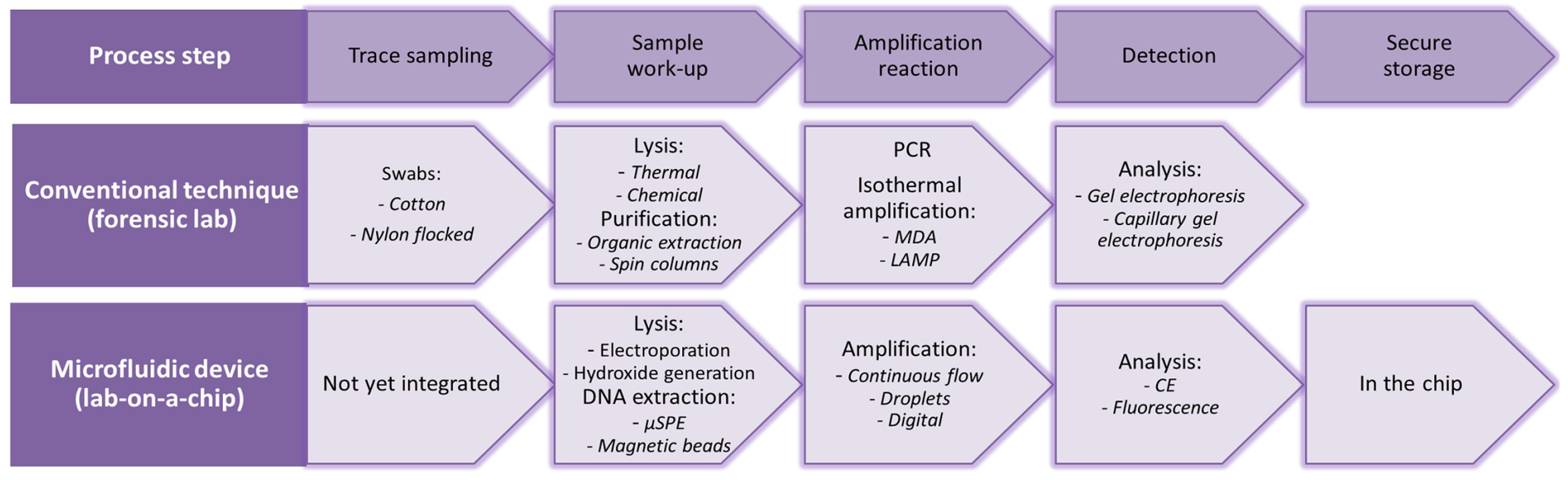
:1. Introduction
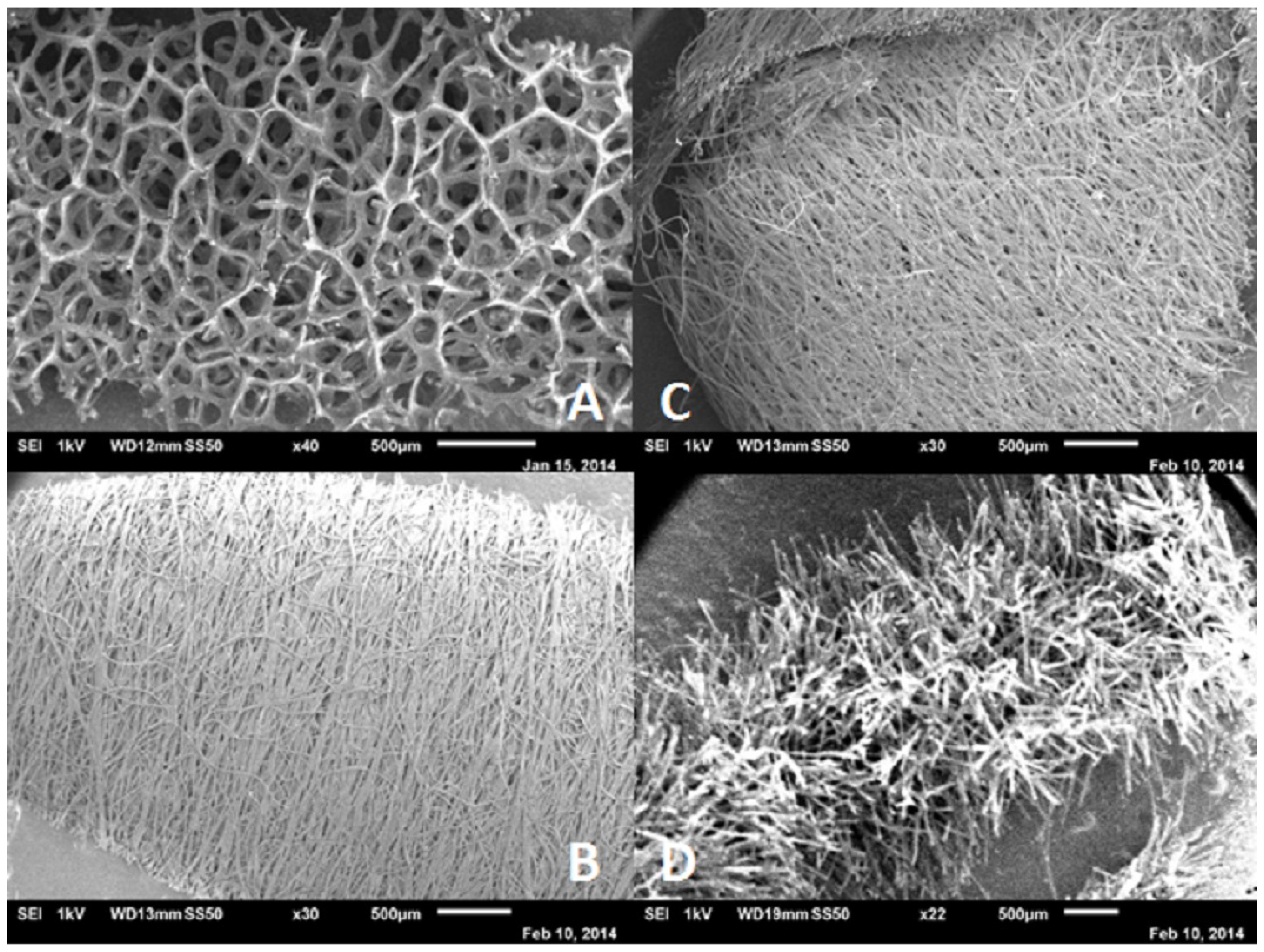
2. Trace Sampling
3. Sample Work-Up
3.1. Cell Lysis
3.2. DNA Extraction and Purification
3.2.1. (μ)SPE
3.2.2. Magnetic Beads
3.2.3. Differential Extraction
4. DNA Amplification
4.1. PCR
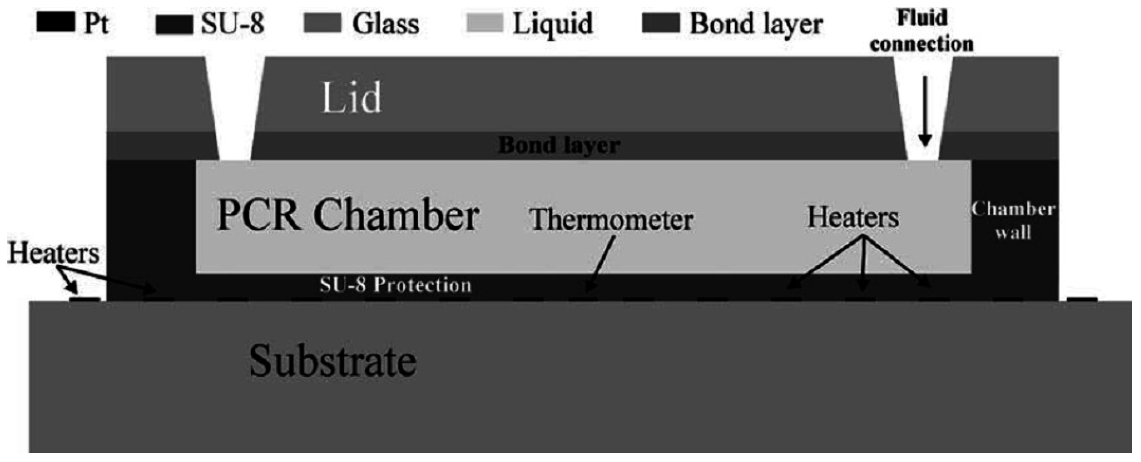
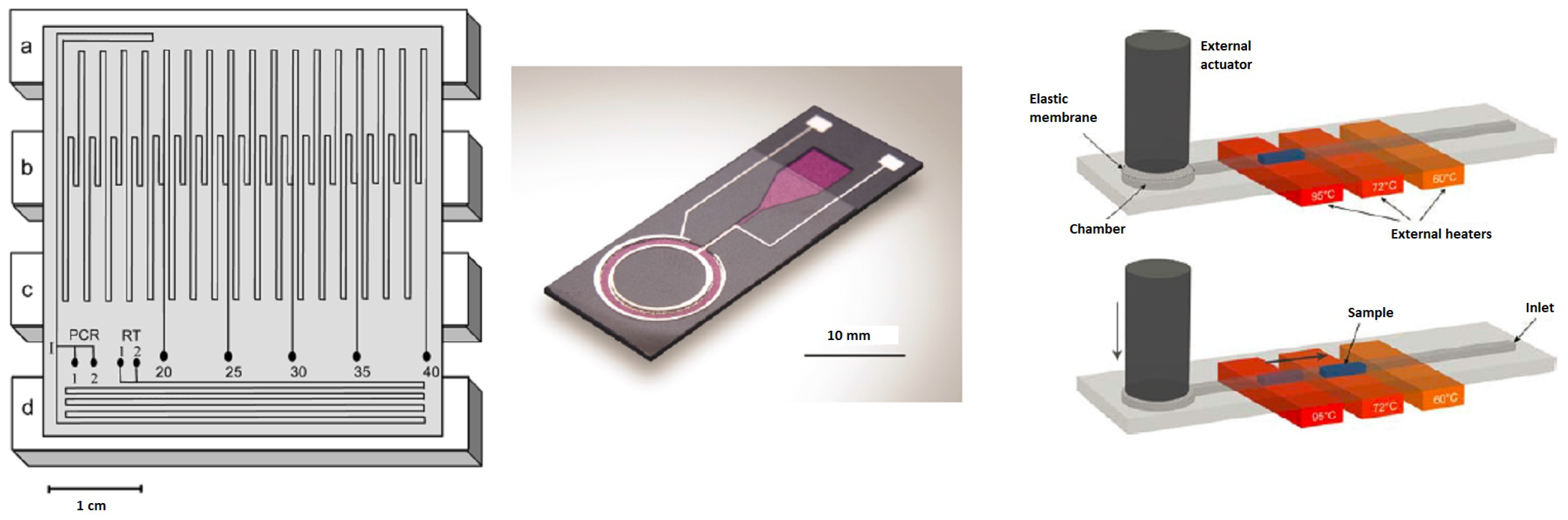
4.1.1. Well-Based Chips
4.1.2. Continuous-Flow Chips
Fixed-Loop Chips
Closed-Loop Chips
Oscillatory Chips
4.1.3. PCR Speed Records
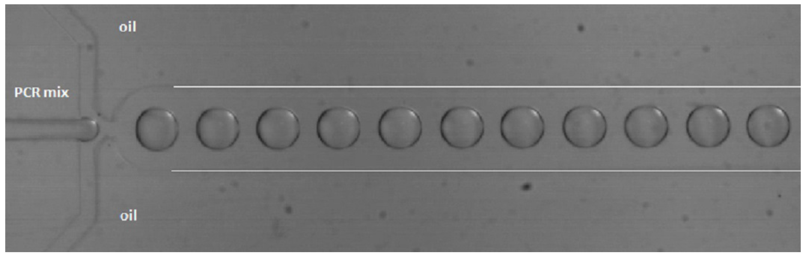
4.1.4. PCR in Droplets
4.1.5. dPCR
4.2. Isothermal Amplification
4.2.1. LAMP
4.2.2. MDA
5. Detection
5.1. Absorbance Detection
5.2. Fluorescence Detection
5.2.1. DNA Dyes
5.2.2. Fluorescent dNTPs
5.2.3. Fluorescent Primers
5.3. Capillary Electrophoresis
6. Secure Storage
7. Chip Materials for DNA Analysis
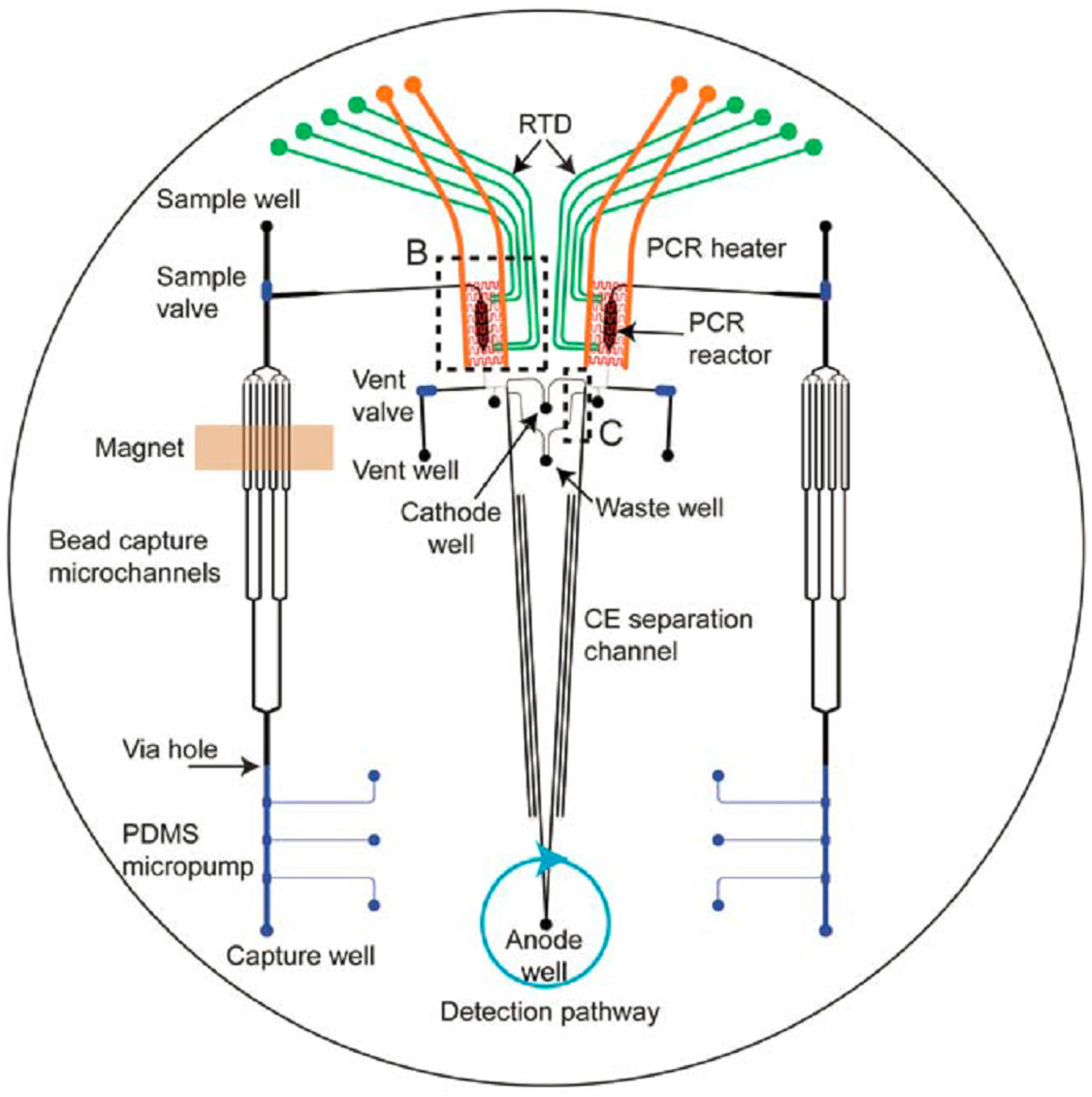
8. Chips with Integrated Functionality
8.1. Research Chips
8.2. Commercial Chip-Based Systems
9. Outlook
Acknowledgments
Conflicts of Interest
References
- Mapes, A.A.; Kloosterman, A.D.; Poot, C.J. DNA in the criminal justice system: The DNA success story in perspective. J. Forensic Sci. 2015, 60, 851–856. [Google Scholar] [CrossRef] [PubMed]
- Van Asten, A.C. On the added value of forensic science and grand innovation challenges for the forensic community. Sci. Justice 2014, 54, 170–179. [Google Scholar] [CrossRef] [PubMed]
- Kloosterman, A.; Mapes, A.; Geradts, Z.; van Eijk, E.; Koper, C.; van den Berg, J.; Verheij, S.; van der Steen, M.; van Asten, A. The interface between forensic science and technology: How technology could cause a paradigm shift in the role of forensic institutes in the criminal justice system. Philos. Trans. B 2015, 370. [Google Scholar] [CrossRef] [PubMed]
- Butler, J.M. Forensic DNA Typing; Elsevier Academic Press: Cambridge, MA, USA, 2005. [Google Scholar]
- Wu, J.; Kodzius, W.; Cao, R.; Wen, W. Extraction, amplification and detection of DNA in microfluidic chip-based assays. Microchim. Acta 2013, 181, 1611–1631. [Google Scholar] [CrossRef]
- Reinholt, S.J.; Baeumner, A.J. Microfluidic isolation of nucleic acids. Angew. Chem. Int. Ed. 2014, 53, 13988–14001. [Google Scholar] [CrossRef] [PubMed]
- Reedy, C.R.; Price, C.W.; Sniegowski, J.; Ferrance, J.P.; Begley, M.; Landers, J.P. Solid phase extraction of DNA from biological samples in a post-based, high surface area poly (methyl methacrylate)(PMMA) micro-device. Lab Chip 2011, 11, 1561–1700. [Google Scholar] [CrossRef] [PubMed]
- Kim, J.; Johnson, M.L.; Hill, P.; Gale, B.K. Microfluidic sample preparation: Cell lysis and nucleic acid purification. Integr. Biol. 2009, 1, 574–586. [Google Scholar] [CrossRef] [PubMed]
- Le Gac, S.; van den Berg, A. Cell Capture and Lysis on a Chip. In Unravelling Single Cell Genomics: Micro and Nanotools; Royal Society of Chemistry: Enschede, The Netherlands, 2010; Chapter 12. [Google Scholar]
- Waters, L.C.; Jacobson, S.C.; Kroutchinina, N.; Khandurina, J.; Foote, R.S.; Ramsey, J.M. Microchip device for cell lysis, multiplex PCR amplification, and electrophoretic sizing. Anal. Chem. 1998, 70, 158–162. [Google Scholar] [CrossRef] [PubMed]
- Lee, C.-Y.; Lee, G.-B.; Lin, J.-L.; Huang, F.-C.; Liao, C.-S. Integrated microfluidic systems for cell lysis, mixing/pumping and DNA amplification. J. Micromech. Microeng. 2005, 15, 1215–1223. [Google Scholar] [CrossRef]
- Wiederkehr, R.S.; Jones, B.; Peeters, S.; Stakenborg, T.; Ibrahim, O.; Fiorini, P.; Tanaka, H.; Yamashita, I.; Matsuno, T.; Lagae, L. On-chip multiplex for amplification directly from whole blood. In Proceedings of the 17th International Conference on Miniaturized Systems for Chemistry and Life Sciences—MicroTAS, Freiburg, Germany, 27–31 October 2013; pp. 1776–1778.
- Tsougeni, K.; Papadakis, G.; Gianneli, M.; Grammoustianou, A.; Constantoudis, V.; Dupuy, B.; Petrou, P.S.; Kakabakos, S.E.; Tserepi, A.; Gizeli, E.; et al. Plasma nanotextured polymeric lab-on-a-chip for highly efficient bacteria capture and lysis. Lab Chip 2016, 16, 120–131. [Google Scholar] [CrossRef] [PubMed]
- Lu, H.; Schmidt, M.A.; Jensen, K.F. A microfluidic electroporation device for cell lysis. Lab Chip 2005, 5, 23–29. [Google Scholar] [CrossRef] [PubMed]
- Jen, C.-P.; Hsiao, J.-H.; Maslov, N.A. Single-cell chemical lysis on microfluidic chips with arrays of micro wells. Sensors 2011, 12, 347–358. [Google Scholar] [CrossRef] [PubMed]
- Fox, M.B.; Esveld, D.C.; Valero, A.; Luttge, R.; Mastwijk, H.C.; Bartels, P.V.; van Den Berg, A.; Boom, R.M. Electroporation of cells in microfluidic devices: A review. Anal. Bioanal. Chem. 2006, 385, 474–485. [Google Scholar] [CrossRef] [PubMed]
- Jiang, F.; Chen, J.N.; Yu, J. Design and application of a microfluidic cell lysis microelectrode chip. Instrum. Sci. Technol. 2015, 44, 223–232. [Google Scholar] [CrossRef]
- Di Carlo, D.; Ionescu-Zanetti, C.; Zhang, Y.; Hung, P.; Lee, L.P. On-chip cell lysis by local hydroxide generation. Lab Chip 2004, 5, 171–178. [Google Scholar] [PubMed]
- Nevill, J.T.; Cooper, R.; Dueck, M.; Breslauer, D.N.; Lee, L.P. Integrated microfluidic cell culture and lysis on a chip. Lab Chip 2007, 7, 1689–1695. [Google Scholar] [CrossRef] [PubMed]
- Lee, H.J.; Kim, J.-H.; Lim, H.K.; Cho, E.C.; Huh, N.; Ko, C.; Park, J.C.; Choi, J.W.; Lee, S.S. Electrochemical cell lysis device for DNA extraction. Lab Chip 2010, 10, 626–633. [Google Scholar] [CrossRef] [PubMed]
- Di Carlo, D.; Jeong, K.H.; Lee, L.P. Reagentless mechanical cell lysis by nanoscale barbs in microchannels for sample preparation. Lab Chip 2003, 3, 287–291. [Google Scholar] [CrossRef] [PubMed]
- Lin, Y.-H.; Lee, G.-B. An optically induced cell lysis device using dielectrophoresis. Appl. Phys. Lett. 2009, 94, 033901. [Google Scholar] [CrossRef]
- Huang, S.-H.; Hung, L.-Y.; Lee, G.-B. Continuous nucleus extraction by optically-induced cell lysis on a batch-type microfluidic platform. Lab Chip 2016, 16, 1447–1456. [Google Scholar] [CrossRef] [PubMed]
- Shahini, M. Development of Cell Lysis Techniques in Lab on a Chip. Ph.D. Thesis, University of Waterloo, Waterloo, ON, Canada, 2013. [Google Scholar]
- Brown, R.B.; Audet, J. Current techniques for single-cell lysis. J. R. Soc. Interface 2008, 5 (Suppl. 2), S131–S138. [Google Scholar] [CrossRef] [PubMed]
- Valero, A. Single Cell Electroporation on Chip. Ph.D. Thesis, University of Twente, Enschede, The Netherlands, October 2006. [Google Scholar]
- Valero, A.; Post, J.N.; van Nieuwkasteele, J.W.; Ter Braak, P.M.; Kruijer, W.; van Den Berg, A. Gene transfer and protein dynamics in stem cells using single cell electroporation in a microfluidic device. Lab Chip 2008, 8, 62–67. [Google Scholar] [CrossRef] [PubMed]
- Shahini, M.; van Wijngaarden, F.; Yeow, J.T.W. Fabrication of electro-microfluidic channel for single cell electroporation. Biomed. Microdev. 2013, 15, 759–766. [Google Scholar] [CrossRef] [PubMed]
- Price, C.W.; Leslie, D.C.; Landers, J.P. Nucleic acid extraction techniques and application to the microchip. Lab Chip 2009, 9, 2484–2494. [Google Scholar] [CrossRef] [PubMed]
- Tian, H.; Hühmer, A.F.R.; Landers, J.P. Evaluation of silica resins for direct and efficient extraction of DNA from complex biological matrices in a miniaturized format. Anal. Biochem. 2000, 283, 175–191. [Google Scholar] [CrossRef] [PubMed]
- Hagan, K.A.; Bienvenue, J.M.; Moskaluk, C.A.; Landers, J.P. Microchip-based solid-phase purification of RNA from biological samples. Anal. Chem. 2008, 80, 8453–8460. [Google Scholar] [CrossRef] [PubMed]
- Wolfe, K.A.; Breadmore, M.C.; Ferrance, J.P.; Power, M.E.; Conroy, J.F.; Norris, P.M.; Landers, J.P. Toward a microchip-based solid-phase extraction method for isolation of nucleic acids. Electrophoresis 2002, 23, 727–733. [Google Scholar] [CrossRef]
- Breadmore, M.C.; Wolfe, K.A.; Arcibal, I.G.; Leung, W.K.; Dickson, D.; Giordano, B.C.; Mary, E.; Ferrance, J.P.; Feldman, S.H.; Norris, P.M.; et al. Microchip-based purification of DNA from biological samples. Anal. Chem. 2003, 75, 1880–1886. [Google Scholar] [CrossRef] [PubMed]
- Duarte, G.R.M.; Price, C.W.; Augustine, B.H.; Carrilho, E.; Landers, J. Dynamic solid phase DNA extraction and PCR amplification in polyester-toner (PeT) based microchip. Anal. Chem. 2011, 83, 5182–5189. [Google Scholar] [CrossRef] [PubMed]
- Zhang, X.; Wu, X.; Peng, R.; Li, D. Electromagnetically controlled microfluidic chip for DNA extraction. Measurement 2015, 75, 23–28. [Google Scholar] [CrossRef]
- Barbaro, A.; Cormaci, P. Validation of DNA typing from skeletal remains using the Invitrogen Charge Switch® Forensic DNA Purification Kit. Forensic Sci. Int. Genet. Suppl. Ser. 2008, 1, 398–400. [Google Scholar] [CrossRef]
- Invitrogen. ChargeSwitch® PCR Clean-Up Kit. 2006. Available online: http://tools.invitrogen.com/content/sfs/manuals/chargeswitch_PCR_man.pdf (accessed on 12 February 2016).
- Hopwood, A.J.; Hurth, C.; Yang, J.; Cai, Z.; Moran, N.; Lee-Edghill, J.G.; Nordquist, A.; Lenigk, R.; Estes, M.D.; Haley, J.P.; et al. Integrated microfluidic system for rapid forensic DNA analysis: Sample collection to DNA profile. Anal. Chem. 2010, 82, 6991–6999. [Google Scholar] [CrossRef] [PubMed]
- Lien, K.Y.; Lee, W.C.; Lei, H.Y.; Lee, G.B. Integrated reverse transcription polymerase chain reaction systems for virus detection. Biosens. Bioelectron. 2007, 22, 1739–1748. [Google Scholar] [CrossRef] [PubMed]
- Gu, S.Q.; Zhang, Y.X.; Zhu, Y.; Du, W.; Yao, B.; Fang, Q. Multifunctional picoliter droplet manipulation platform and its application in single cell analysis. Anal. Chem. 2011, 83, 7570–7576. [Google Scholar] [CrossRef] [PubMed]
- Yang, J.; Brooks, C.; Estes, M.D.; Hurth, C.M.; Zenhausern, F. An integratable microfluidic cartridge for forensic swab samples lysis. Forensic Sci. Int. Genet. 2014, 8, 147–158. [Google Scholar] [CrossRef] [PubMed]
- Bienvenue, J.M.; Duncalf, N.; Marchiarullo, D.; Ferrance, J.P.; Landers, J.P. Microchip-based cell lysis and DNA extraction from sperm cells for application to forensic analysis. J. Forensic Sci. 2006, 51, 266–273. [Google Scholar] [CrossRef] [PubMed]
- Voorhees Norris, J.; Evander, M.; Horsman-Hall, K.M.; Nilsson, J.; Laurell, T.; Landers, J.P. Acoustic differential extraction for forensic analysis of sexual assault evidence. Anal. Chem. 2009, 81, 6089–6095. [Google Scholar] [CrossRef] [PubMed]
- Horsman, K.M.; Bienvenue, J.M.; Blasier, K.R.; Landers, J.P. Forensic DNA analysis on microfluidic devices: A review. J. Forensic Sci. 2007, 52, 784–799. [Google Scholar] [CrossRef] [PubMed]
- Belgrader, P.I.; Yuan, B. Sonication to Selectively Lyse Different Cell Types. U.S. Patent 7,785,869, 31 August 2010. [Google Scholar]
- Horsman, K.M.; Barker, S.L.R.; Ferrance, J.P.; Forrest, K.A.; Koen, K.A.; Landers, J.P. Separation of sperm and epithelial cells in a microfabricated device: Potential application to forensic analysis of sexual assault evidence. Anal. Chem. 2005, 77, 742–749. [Google Scholar] [CrossRef] [PubMed]
- Bienvenue, J.M.; Legendre, L.A.; Ferrance, J.P.; Landers, J.P. An integrated microfluidic device for DNA purification and PCR amplification of STR fragments. Forensic Sci. Int. Genet. 2010, 4, 178–186. [Google Scholar] [CrossRef] [PubMed]
- Hagan, K.A.; Reedy, C.R.; Bienvenue, J.M.; Dewald, A.H.; Landers, J.P. A valveless microfluidic device for integrated solid phase extraction and polymerase chain reaction for short tandem repeat (STR) analysis. Analyst 2011, 136, 1928–1937. [Google Scholar] [CrossRef] [PubMed]
- Meulenbroek, A.J.; Kloosterman, A.D. DNA-onderzoek van minimale biologische sporen; gevoelige problematiek. Analyse 2009, 64, 108–120. [Google Scholar]
- Van Oorschot, R.A.; Ballantyne, K.N.; Mitchell, R.J. Forensic trace DNA: A review. Investig. Genet. 2010, 1. [Google Scholar] [CrossRef] [PubMed]
- Nicklas, J.A.; Buel, E. Quantification of DNA in forensic samples. Anal. Bioanal. Chem. 2003, 376, 1160–1167. [Google Scholar] [CrossRef] [PubMed]
- Lee, S.B.; McCord, B.; Buel, E. Advances in forensic DNA quantification: A review. Electrophoresis 2014, 35, 3044–3052. [Google Scholar] [CrossRef] [PubMed]
- Tan, E.; Turingan, R.S.; Hogan, C.; Vasantgadkar, S.; Palombo, L.; Schumm, J.W.; Selden, R.F. Fully integrated, fully automated generation of short tandem repeat profiles. Investig. Genet. 2013, 4. [Google Scholar] [CrossRef] [PubMed]
- Jovanovich, S.; Bogdan, G.; Belcinski, R.; Buscaino, J.; Burgi, D.; Butts, E.L.R.; Chear, K.; Ciopyk, B.; Eberhart, D.; El-Sissi, O.; et al. Developmental validation of a fully integrated sample-to-profile rapid human identification system for processing single-source reference buccal samples. Forensic Sci. Int. Genet. 2015, 16, 181–194. [Google Scholar] [CrossRef] [PubMed]
- Auroux, P.-A.; Koc, Y.; deMello, A.; Manz, A.; Day, P.J.R. Miniaturised nucleic acid analysis. Lab Chip 2004, 4, 534–546. [Google Scholar] [CrossRef] [PubMed]
- El-Ali, J.; Perch-Nielsen, I.R.; Poulsen, C.R.; Bang, D.D.; Telleman, P.; Wolff, A. Simulation and experimental validation of a SU-8 based PCR thermocycler chip with integrated heaters and temperature sensor. Sens. Actuators A Phys. 2004, 110, 3–10. [Google Scholar] [CrossRef]
- Zhang, Y.; Ozdemir, P. Microfluidic DNA amplification: A review. Anal. Chim. Acta 2009, 638, 115–125. [Google Scholar] [CrossRef] [PubMed]
- Obeid, P.J.; Christopoulos, T.K.; Crabtree, H.J.; Backhouse, C.J. Microfabricated device for DNA and RNA amplification by continuous-flow polymerase chain reaction and reverse transcription-polymerase chain reaction with cycle number selection. Anal. Chem. 2003, 75, 288–295. [Google Scholar] [CrossRef] [PubMed]
- West, J.; Karamata, B.; Lillis, B.; Gleeson, J.P.; Alderman, J.; Collins, J.K.; Lane, W.; Mathewson, A.; Berney, H. Application of magnetohydrodynamic actuation to continuous flow chemistry. Lab Chip 2002, 2, 224–230. [Google Scholar] [CrossRef] [PubMed]
- Frey, O.; Bonneick, S.; Hierlemann, A.; Lichtenberg, J. Autonomous microfluidic multi-channel chip for real-time PCR with integrated liquid handling. Biomed. Microdev. 2007, 9, 711–718. [Google Scholar] [CrossRef] [PubMed]
- Liu, H.B.; Ramalingam, N.; Jiang, Y.; Dai, C.C.; Hui, K.M.; Gong, H.Q. Rapid distribution of a liquid column into a matrix of nanoliter wells for parallel real-time quantitative PCR. Sens. Actuators B Chem. 2009, 135, 671–677. [Google Scholar] [CrossRef]
- Zhang, Y.; Zhu, Y.; Yao, B.; Fang, Q. Nanolitre droplet array for real time reverse transcription polymerase chain reaction. Lab Chip 2011, 11, 1545–1549. [Google Scholar] [CrossRef] [PubMed]
- Liu, Y.; Li, C.; Li, Z.; Chan, S.D.; Eto, D.; Wu, W.; Zhang, J.P.; Chien, R.-L.; Wada, H.G.; Greenstein, M.; et al. On-chip quantitative-PCR using integrated real-time detection by capillary electrophoresis. Electrophoresis 2016, 37, 545–552. [Google Scholar] [CrossRef] [PubMed]
- Kopp, M.U.; Mello, A.J.; Manz, A. Chemical amplification: Continuous-flow PCR on a chip. Science 1998, 280, 1046–1048. [Google Scholar] [CrossRef] [PubMed]
- Obeid, P.J.; Christopoulos, T.K. Continuous-flow DNA and RNA amplification chip combined with laser-induced fluorescence detection. Anal. Chim. Acta 2003, 494, 1–9. [Google Scholar] [CrossRef]
- Qi, H.; Wang, X.; Chen, T.; Ma, X.; Zuo, T. Fabrication and characterization of a polymethyl methacrylate continuous-flow PCR microfluidic chip using CO2 laser ablation. Microsyst. Technol. 2009, 15, 1027–1030. [Google Scholar] [CrossRef]
- Moschou, D.; Vourdas, N.; Kokkoris, G.; Papadakis, G.; Parthenios, J.; Chatzandroulis, S.; Tserepi, A. All-plastic, low-power, disposable, continuous-flow PCR chip with integrated microheaters for rapid DNA amplification. Sens. Actuators B Chem. 2014, 199, 470–478. [Google Scholar] [CrossRef]
- Hatch, A.C.; Ray, T.; Lintecum, K.; Youngbull, C. Continuous flow real-time PCR device using multi-channel fluorescence excitation and detection. Lab Chip 2014, 14, 562–568. [Google Scholar] [CrossRef] [PubMed]
- Sadler, D.J.; Changrani, R.; Roberts, P.; Chou, C.F.; Zenhausern, F. Thermal management of BioMEMS: Temperature control for ceramic-based PCR and DNA detection devices. IEEE Trans. Compon. Packag. Technol. 2003, 26, 309–316. [Google Scholar] [CrossRef]
- Chen, Z.; Qian, S.; Abrams, W.R.; Malamud, D.; Bau, H.H. Thermosiphon-based PCR reactor: Experiment and modeling. Anal. Chem. 2004, 76, 3707–3715. [Google Scholar] [CrossRef] [PubMed]
- Bu, M.; Melvin, T.; Ensell, G.; Wilkinson, J.S.; Evans, A.G.R. Design and theoretical evaluation of a novel microfluidic device to be used for PCR. J. Micromech. Microeng. 2003, 13, S125–S130. [Google Scholar] [CrossRef]
- Wang, W.; Li, Z.-X.; Luo, R.; Lü, S.-H.; Xu, A.-D.; Yang, Y.-J. Droplet-based micro oscillating-flow PCR chip. J. Micromech. Microeng. 2005, 15, 1369. [Google Scholar] [CrossRef]
- Nie, J.; Zhao, Y.; Peng, N. Multichannel oscillatory-flow PCR micro-fluidic chip with controllable temperature gradient. Microsyst. Technol. 2015, 21, 41–48. [Google Scholar] [CrossRef]
- Giordano, B.C.; Ferrance, J.; Swedberg, S.; Hühmer, A.F.R.; Landers, J.P. Polymerase chain reaction in polymeric microchips: DNA amplification in less than 240 seconds. Anal. Biochem. 2001, 291, 124–132. [Google Scholar] [CrossRef] [PubMed]
- Neuzil, P.; Zhang, C.; Pipper, J.; Oh, S.; Zhuo, L. Ultra fast miniaturized real-time PCR: 40 cycles in less than six minutes. Nucleic Acids Res. 2006, 34, e77. [Google Scholar] [CrossRef] [PubMed]
- Neuzil, P.; Pipper, J.; Hsieh, T.M. Disposable real-time microPCR device: Lab-on-a-chip at a low cost. Mol. BioSyst. 2006, 2, 292–298. [Google Scholar] [CrossRef] [PubMed]
- Fuchiwaki, Y.; Nagai, H.; Saito, M.; Tamiya, E. Ultra-rapid flow-through polymerase chain reaction microfluidics using vapor pressure. Biosens. Bioelectron. 2011, 27, 88–94. [Google Scholar] [CrossRef] [PubMed]
- Son, J.H.; Cho, B.; Hong, S.; Lee, S.H.; Hoxha, O.; Haack, A.J.; Lee, L.P. Ultrafast photonic PCR. Light Sci. Appl. 2015, 4, e280. [Google Scholar] [CrossRef]
- Son, J.H.; Hong, S.; Haack, A.J.; Gustafson, L.; Song, M.; Hoxha, O.; Lee, L.P. Rapid optical cavity PCR. Adv. Healthc. Mater. 2016, 5, 167–174. [Google Scholar] [CrossRef] [PubMed]
- Gu, H.; Duits, M.H.G.; Mugele, F. Droplets formation and merging in two-phase flow microfluidics. Int. J. Mol. Sci. 2011, 12, 2572–2597. [Google Scholar] [CrossRef] [PubMed]
- Baroud, C.N.; Gallaire, F.; Dangla, R. Dynamics of microfluidic droplets. Lab Chip 2010, 10, 2032–2045. [Google Scholar] [CrossRef] [PubMed]
- Choi, J.W.; Kang, D.-K.; Park, H.; deMello, A.J.; Chang, S.-I. High-throughput analysis of protein-protein interactions in picoliter-volume droplets using fluorescence polarization. Anal. Chem. 2012, 84, 3849–3854. [Google Scholar] [CrossRef] [PubMed]
- Theberge, A.B.; Courtois, F.; Schaerli, Y.; Fischlechner, M.; Abell, C.; Hollfelder, F.; Huck, W.T.S. Microdroplets in microfluidics: An evolving platform for discoveries in chemistry and biology. Angew. Chem. Int. Ed. 2010, 49, 5846–5868. [Google Scholar] [CrossRef] [PubMed]
- Zhang, Y.; Jiang, H.-R. A review on continuous-flow microfluidic PCR in droplets: Advances, challenges and future. Anal. Chim. Acta 2016, 914, 7–16. [Google Scholar] [CrossRef] [PubMed]
- Beer, N.R.; Hindson, B.J.; Wheeler, E.K.; Sara, B.; Rose, K.A.; Kennedy, I.M.; Colston, B.W. On-chip, real-time, single-copy polymerase chain reaction in picoliter droplets. Anal. Chem. 2007, 79, 8471–8475. [Google Scholar] [CrossRef] [PubMed]
- Mohr, S.; Zhang, Y.H.; Macaskill, A.; Day, P.J.R.; Barber, R.W.; Goddard, N.J.; Emerson, D.R.; Fielden, P.R. Numerical and experimental study of a droplet-based PCR chip. Microfluid. Nanofluid. 2007, 3, 611–621. [Google Scholar] [CrossRef]
- Schaerli, Y.; Wootton, R.C.; Robinson, T.; Stein, V.; Dunsby, C.; Neil, M.A.A.; French, P.M.W.; deMello, A.J.; Abell, C.; Hollfelder, F. Continuous-flow polymerase chain reaction of single-copy DNA in microfluidic microdroplets. Anal. Chem. 2008, 81, 302–306. [Google Scholar] [CrossRef] [PubMed]
- Kiss, M.M.; Ortoleva-Donnelly, L.; Beer, N.R.; Warner, J.; Bailey, C.G.; Colston, B.W.; Rothberg, J.M.; Link, D.R.; Leamon, J.H. High-throughput quantitative PCR in picoliter droplets. Anal. Chem. 2008, 80, 8975–8981. [Google Scholar] [CrossRef] [PubMed]
- Leng, X.; Zhang, W.; Wang, C.; Cui, L.; Yang, C.J. Agarose droplet microfluidics for highly parallel and efficient single molecule emulsion PCR. Lab Chip 2010, 10, 2841–2843. [Google Scholar] [CrossRef] [PubMed]
- Hatch, A.C.; Fisher, J.S.; Tovar, A.R.; Hsieh, A.T.; Lin, R.; Pentoney, S.L.; Yang, D.L.; Lee, A.P. 1-Million droplet array with wide-field fluorescence imaging for digital PCR. Lab Chip 2011, 11, 3838–3845. [Google Scholar] [CrossRef] [PubMed]
- Zhang, W.; Zhang, W.; Liu, Z.; Li, C.; Zhu, Z.; Yang, C.J. Highly parallel single molecule amplification approach based on agarose droplet PCR for efficient and cost-effective aptamer selection. Anal. Chem. 2012, 84, 350–355. [Google Scholar] [CrossRef] [PubMed]
- Geng, T.; Novak, R.; Mathies, R.A. Single-cell forensic short tandem repeat typing within microfluidic droplets. Anal. Chem. 2014, 86, 703–712. [Google Scholar] [CrossRef] [PubMed]
- Beer, N.R.; Wheeler, E.K.; Lee-Houghton, L.; Watkins, N.; Nasarabadi, S.; Hebert, N.; Leung, P.; Arnold, D.W.; Bailey, C.G.; Colston, B.W. On-chip single-copy real-time reverse-transcription PCR in isolated picoliter droplets. Anal. Chem. 2008, 80, 1854–1858. [Google Scholar] [CrossRef] [PubMed]
- Zhang, H.; Jenkins, G.; Zou, Y.; Zhu, Z.; Yang, C.J. Massively parallel single-molecule and single-cell emulsion reverse transcription polymerase chain reaction using agarose droplet microfluidics. Anal. Chem. 2012, 84, 3599–3606. [Google Scholar] [CrossRef] [PubMed]
- Geng, T.; Mathies, R.A. Minimizing inhibition of PCR-STR typing using digital agarose droplet microfluidics. Forensic Sci. Int. Genet. 2015, 14, 203–209. [Google Scholar] [CrossRef] [PubMed]
- Basova, E.Y.; Foret, F. Droplet microfluidics in (bio)chemical analysis. Analyst 2015, 140, 22–38. [Google Scholar] [CrossRef] [PubMed]
- Sykes, P.J.; Neoh, S.H.; Brisco, M.J.; Hughes, E.; Condon, J.; Morley, A.A. Quantitation of targets for PCR by use of limiting dilution. Biotechniques 1992, 13, 444–449. [Google Scholar] [PubMed]
- White, R.A.; Blainey, P.C.; Fan, S.R.; Quake, H.C. Digital PCR provides sensitive and absolute calibration for high throughput sequencing. BMC Genom. 2009, 10. [Google Scholar] [CrossRef]
- Sanders, R.; Huggett, J.F.; Bushell, C.A.; Cowen, S.; Scott, D.J.; Foy, C.A. Evaluation of digital PCR for absolute DNA quantification. Anal. Chem. 2011, 83, 6474–6484. [Google Scholar] [CrossRef] [PubMed]
- Wang, P.; Jing, F.; Li, G.; Wu, Z.; Cheng, Z.; Zhang, J.; Zhang, H.; Jia, C.; Jin, Q.; Mao, H.; et al. Absolute quantification of lung cancer related microRNA by droplet digital PCR. Biosens. Bioelectron. 2015, 74, 836–842. [Google Scholar] [CrossRef] [PubMed]
- Asiello, P.J.; Baeumner, A.J. Miniaturized isothermal nucleic acid amplification, a review. Lab Chip 2011, 11, 1420–1430. [Google Scholar] [CrossRef] [PubMed]
- Craw, P.; Balachandran, W. Isothermal nucleic acid amplification technologies for point-of-care diagnostics: A critical review. Lab Chip 2012, 12, 2469–2486. [Google Scholar] [CrossRef] [PubMed]
- Johne, R.; Müller, H.; Rector, A.; van Ranst, M.; Stevens, H. Rolling-circle amplification of viral DNA genomes using phi29 polymerase. Trends Microbiol. 2009, 17, 205–211. [Google Scholar] [CrossRef] [PubMed]
- Gill, P.; Ghaemi, A. Nucleic acid isothermal amplification technologies—A review. Nucleosides Nucleotides Nucleic Acids 2008, 27, 224–243. [Google Scholar] [CrossRef] [PubMed]
- Safavieh, M.; Kanakasabapathy, M.K.; Tarlan, F.; Ahmed, M.U.; Zourob, M.; Asghar, W.; Shafiee, H. Emerging loop-mediated isothermal amplification-based microchip and micro-device technologies for nucleic acid detection. ACS Biomater. Sci. Eng. 2016, 2, 278–294. [Google Scholar] [CrossRef]
- Mori, Y.; Nagamine, K.; Tomita, N.; Notomi, T. Detection of loop-mediated isothermal amplification reaction by turbidity derived from magnesium pyrophosphate formation. Biochem. Biophys. Res. Commun. 2001, 289, 150–154. [Google Scholar] [CrossRef] [PubMed]
- Gadkar, V.; Rillig, M.C. Evaluation of loop-mediated isothermal amplification (LAMP) to rapidly detect arbuscular mycorrhizal fungi. Soil Biol. Biochem. 2008, 40, 540–543. [Google Scholar] [CrossRef]
- Dean, F.B.; Hosono, S.; Fang, L.; Wu, X.; Faruqi, A.F.; Bray-Ward, P.; Sun, Z.; Zong, Q.; Du, Y.; Du, J.; et al. Comprehensive human genome amplification using multiple displacement amplification. Proc. Natl. Acad. Sci. USA 2002, 99, 5261. [Google Scholar] [CrossRef] [PubMed]
- Silander, K.; Saarela, J. Whole genome amplification with Phi29 DNA polymerase to enable genetic or genomic analysis of samples of low DNA yield. Methods Mol. Biol. 2008, 439, 1–18. [Google Scholar] [PubMed]
- Vincent, M.; Xu, Y.; Kong, H. Helicase-dependent isothermal DNA amplification. EMBO Rep. 2004, 5, 795–800. [Google Scholar] [CrossRef] [PubMed]
- Motré, A.; Li, Y.; Kong, H. Enhancing helicase-dependent amplification by fusing the helicase with the DNA polymerase. Gene 2008, 420, 17–22. [Google Scholar] [CrossRef] [PubMed]
- Jeong, Y.J.; Park, K.; Kim, D.E. Isothermal DNA amplification in vitro: The helicase-dependent amplification system. Cell. Mol. Life Sci. 2009, 66, 3325–3336. [Google Scholar] [CrossRef] [PubMed]
- Lizardi, P.M.; Huang, X.; Zhu, Z.; Bray-Ward, P.; Thomas, D.C.; Ward, D.C. Mutation detection and single-molecule counting using isothermal rolling-circle amplification. Nat. Genet. 1998, 19, 225–232. [Google Scholar] [CrossRef] [PubMed]
- Liu, D.; Daubendiek, S.L.; Zillman, M.A.; Ryan, K.; Kool, E.T. Rolling circle DNA synthesis: Small circular oligonucleotides as efficient templates for DNA polymerases. J. Am. Chem. Soc. 1996, 118, 1587–1594. [Google Scholar] [CrossRef] [PubMed]
- Walker, G.T.; Little, M.C.; Nadeau, J.G.; Shank, D.D. Isothermal in vitro amplification of DNA by a restriction enzyme/DNA polymerase system. Proc. Natl. Acad. Sci. USA 1992, 89, 392–396. [Google Scholar] [CrossRef] [PubMed]
- Fang, X.; Chen, H.; Yu, S.; Jiang, X.; Kong, J. Predicting viruses accurately by a multiplex microfluidic loop-mediated isothermal amplification chip. Anal. Chem. 2010, 83, 690–695. [Google Scholar] [CrossRef] [PubMed]
- Gansen, A.; Herrick, A.; Dimov, I.K.; Lee, L.; Chiu, D.T. Digital LAMP in a sample self-digitization (SD) chip. Lab Chip 2012, 12, 2247–2254. [Google Scholar] [CrossRef] [PubMed]
- Duarte, C.; Salm, E.; Dorvel, B.; Reddy, B., Jr.; Bashir, R. On-chip parallel detection of foodborne pathogens using loop-mediated isothermal amplification. Biomed. Microdev. 2013, 15, 821–830. [Google Scholar] [CrossRef] [PubMed]
- Zhou, Q.-J.; Wang, L.; Chen, J.; Wang, R.-N.; Shi, Y.-H.; Li, C.-H.; Zhang, D.-M.; Yan, X.-J.; Zhang, Y.-J. Development and evaluation of a real-time fluorogenic loop-mediated isothermal amplification assay integrated on a microfluidic disc chip (on-chip LAMP) for rapid and simultaneous detection of ten pathogenic bacteria in aquatic animals. J. Microbiol. Methods 2014, 104, 26–35. [Google Scholar] [CrossRef] [PubMed]
- Sayad, A.A.; Ibrahim, F.; Uddin, S.M.; Pei, K.X.; Mohktar, M.S.; Madou, M.; Thong, K.L. A microfluidic lab-on-a-disc integrated loop mediated isothermal amplification for foodborne pathogen detection. Sens. Actuators B Chem. 2016, 227, 600–609. [Google Scholar] [CrossRef]
- Marcy, Y.; Ishoey, T.; Lasken, R.S.; Stockwell, T.B.; Walenz, B.P.; Halpern, A.L.; Beeson, K.Y.; Goldberg, S.M.D.; Quake, S.R. Nanoliter reactors improve multiple displacement amplification of genomes from single cells. PLoS Genet. 2007, 3, 1703–1708. [Google Scholar] [CrossRef] [PubMed]
- Yang, Y.; Rho, H.S.; Stevens, M.; Tibbe, A.G.J.; Gardeniers, J.G.E.; Terstappen, L.W.M.M. Microfluidic device for DNA amplification of single cancer cells isolated from whole blood by self-seeding micro wells. Lab Chip 2015, 15, 4331–4337. [Google Scholar] [CrossRef] [PubMed]
- Kaprou, G.D.; Papadakis, G.; Papageorgiou, D.P.; Kokkoris, G.; Papadopoulos, V.; Kefala, I.; Gizeli, E.; Tserepi, A. Miniaturized devices for isothermal DNA amplification addressing DNA diagnostics. Microsyst. Technol. 2016, 22, 1529–1534. [Google Scholar] [CrossRef]
- Shi, D.; Huang, J.; Chuai, Z.; Chen, D.; Zhu, X.; Wang, H.; Peng, J.; Wu, H.; Huang, Q.; Fu, W. Isothermal and rapid detection of pathogenic microorganisms using a nano rolling circle amplification-surface plasmon resonance biosensor. Biosens. Bioelectron. 2014, 62, 280–287. [Google Scholar] [CrossRef] [PubMed]
- Ma, X.; Xu, W.; Chen, C.; Lu, Z.; Li, J. A microfabrication-free nanoliter droplet array for nucleic acid detection combined with isothermal amplification. Analyst 2015, 140, 4370–4373. [Google Scholar] [PubMed]
- Kalsi, S.; Valiadi, M.; Tsaloglou, M.-N.; Parry-Jones, L.; Jacobs, A.; Watson, R.; Turner, C.; Amos, R.; Hadwen, B.; Buse, J.; et al. Rapid and sensitive detection of antibiotic resistance on a programmable digital microfluidic platform. Lab Chip 2015, 15, 3065–3075. [Google Scholar] [CrossRef] [PubMed]
- Santiago-Felipe, S.; Tortajada-Genaro, L.A.; Puchades, R.; Maquieira, Á. Parallel solid-phase isothermal amplification and detection of multiple DNA targets in microliter-sized wells of a digital versatile disc. Microchim. Acta 2016, 183, 1195–1202. [Google Scholar] [CrossRef]
- Kunze, A.; Dilcher, M.; Abd El Wahed, A.; Hufert, F.; Niessner, R.; Seidel, M. On-chip isothermal nucleic acid amplification on flow-based chemiluminescence microarray analysis platform for the detection of viruses and bacteria. Anal. Chem. 2016, 88, 898–905. [Google Scholar] [CrossRef] [PubMed]
- Nagamine, K.; Hase, T.; Notomi, T. Accelerated reaction by loop-mediated isothermal amplification using loop primers. Mol. Cell. Probes 2002, 16, 223–229. [Google Scholar] [CrossRef] [PubMed]
- Notomi, T.; Okayama, H.; Masubuchi, H.; Yonekawa, T.; Watanabe, K.; Amino, N.; Hase, T. Loop-mediated isothermal amplification of DNA. Nucleic Acids Res. 2000, 28, e63. [Google Scholar] [CrossRef] [PubMed]
- Mori, Y.; Kitao, M.; Tomita, N.; Notomi, T. Real-time turbidimetry of LAMP reaction for quantifying template DNA. J. Biochem. Biophys. Methods 2004, 59, 145–157. [Google Scholar] [CrossRef] [PubMed]
- Cai, T.; Lou, G.Q.; Yang, J.; Xu, D.; Meng, Z.H. Development and evaluation of real-time loop-mediated isothermal amplification for hepatitis B virus DNA quantification: A new tool for HBV management. J. Clin. Virol. 2008, 41, 270–276. [Google Scholar] [CrossRef] [PubMed]
- Deguo, W.; Guicheng, H.; Fugui, W.; Yonggang, L.; Daxi, R. Drawback of loop-mediated isothermal amplification. Afr. J. Food Sci. 2008, 2, 83–86. [Google Scholar]
- Luo, J.; Fang, X.; Ye, D.; Li, H.; Chen, H.; Zhang, S.; Kong, J. A real-time microfluidic multiplex electrochemical loop-mediated isothermal amplification chip for differentiating bacteria. Biosens. Bioelectron. 2014, 60, 84–91. [Google Scholar] [CrossRef] [PubMed]
- Watthanapanpituck, K.; Kiatpathomchai, W.; Chu, E.; Panvisavas, N. Identification of human DNA in forensic evidence by loop-mediated isothermal amplification combined with a colorimetric gold nanoparticle hybridization probe. Int. J. Legal Med. 2014, 128, 923–931. [Google Scholar] [CrossRef] [PubMed]
- illustra GenomiPhi V2 DNA Amplification Kit. 2006. Available online: https://www.gelifesciences.com/gehcls_images/GELS/Related%20Content/Files/1314774443672/litdocGPHI_V2_25660030_revB_20110831102610.pdf (accessed on 1 August 2016).
- Kumar, G.; Garnova, E.; Reagin, M.; Vidali, A. Improved multiple displacement amplification with Phi 29 DNA polymerase for genotyping of single human cells. BioTechniques 2008, 44, 879–890. [Google Scholar] [CrossRef] [PubMed]
- Salas, M.; Blanco, L.; Lázaro, J.M.; de Vega, M. The bacteriophage φ29 DNA polymerase. IUBMB Life 2008, 60, 82–85. [Google Scholar] [CrossRef] [PubMed]
- Ballantyne, K.N.; van Oorschot, R.A.H.; John Mitchell, R.; Koukoulas, I. Molecular crowding increases the amplification success of multiple displacement amplification and short tandem repeat genotyping. Anal. Biochem. 2006, 355, 298–303. [Google Scholar] [CrossRef] [PubMed]
- Ballantyne, K.N.; van Oorschot, R.A.H.; Mitchell, R.J. Comparison of two whole genome amplification methods for STR genotyping of LCN and degraded DNA samples. Forensic Sci. Int. 2007, 166, 35–41. [Google Scholar] [CrossRef] [PubMed]
- Singer, V.L.; Jones, L.J.; Yue, S.T.; Haugland, R.P. Characterization of PicoGreen reagent and development of a fluorescence-based solution assay for double-stranded DNA quantitation. Anal. Biochem. 1997, 249, 228–238. [Google Scholar] [CrossRef] [PubMed]
- Tanner, N.A.; Zhang, Y.; Evans, T.C., Jr. Visual detection of isothermal nucleic acid amplification using pH-sensitive dyes. BioTechniques 2015, 59, 59–68. [Google Scholar] [CrossRef] [PubMed]
- Rodriguez-Manzano, J.; Karymov, M.A.; Begolo, S.; Selck, D.A.; Zhukov, D.V.; Jue, E.; Ismagilov, R.F. Reading out single-molecule digital RNA and DNA isothermal amplification in nanoliter volumes with unmodified camera phones. ACS Nano 2016, 10, 3102–3113. [Google Scholar] [CrossRef] [PubMed]
- Molecular Probes®. The Molecular Probes® Handbook. 2012. Available online: http://www.invitrogen.com/site/us/en/home/References/Molecular-Probes-The-Handbook.html (accessed on 3 May 2016).
- Sang, F.; Ren, J. Capillary electrophoresis of double-stranded DNA fragments using a new fluorescence intercalating dye EvaGreen. J. Sep. Sci. 2006, 29, 1275–1280. [Google Scholar] [CrossRef] [PubMed]
- Ross, J.S.; Cronin, M. Whole cancer genome sequencing by next-generation methods. Am. J. Clin. Pathol. 2011, 136, 527–539. [Google Scholar] [CrossRef] [PubMed]
- Liu, L.; Li, Y.; Li, S.; Hu, N.; He, Y.; Pong, R.; Lin, D.; Lu, M.; Law, L. Comparison of next-generation sequencing systems. BioMed Res. Int. 2012, 2012, 1–11. [Google Scholar] [CrossRef] [PubMed]
- Liu, P.; Scherer, J.R.; Greenspoon, S.A.; Chiesl, T.N.; Mathies, R.A. Integrated sample cleanup and capillary array electrophoresis microchip for forensic short tandem repeat analysis. Forensic Sci. Int. Genet. 2011, 5, 484–492. [Google Scholar] [CrossRef] [PubMed]
- Chen, D.; Mauk, M.; Qiu, X.; Liu, C.; Kim, J.; Ramprasad, S.; Ongagna, S.; Abrams, W.R.; Malamud, D.; Corstjens, P.L.; et al. An integrated, self-contained microfluidic cassette for isolation, amplification, and detection of nucleic acids. Biomed. Microdev. 2010, 12, 705–719. [Google Scholar] [CrossRef] [PubMed]
- Le Roux, D.; Root, B.E.; Reedy, C.R.; Hickey, J.A.; Scott, O.N.; Bienvenue, J.M.; Landers, J.P.; Chassagne, L.; de Mazancourt, P. DNA analysis using an integrated microchip for multiplex PCR amplification and electrophoresis for reference samples. Anal. Chem. 2014, 86, 8192–8199. [Google Scholar] [CrossRef] [PubMed]
- Pascali, J.P.; Bortolotti, F.; Tagliaro, F. Recent advances in the application of CE to forensic sciences, an update over years 2009–2011. Electrophoresis 2012, 33, 117–126. [Google Scholar] [CrossRef] [PubMed]
- Mitnik, L.; Carey, L.; Burger, R.; Desmarais, S.; Koutny, L.; Wernet, O.; Matsudaira, P.; Ehrlich, D. High-speed analysis of multiplexed short tandem repeats with an electrophoretic micro-device. Electrophoresis 2002, 23, 719–726. [Google Scholar] [CrossRef]
- Chen, Y.; Young Choi, J.; Jin Choi, S.; Seo, T.S. Sample stacking capillary electrophoretic micro-device for highly sensitive mini Y short tandem repeat genotyping. Electrophoresis 2010, 31, 2974–2980. [Google Scholar] [CrossRef] [PubMed]
- Date-Chong, M.; Hudlow, W.R.; Buoncristiani, M.R. Evaluation of the RapidHIT 200 and RapidHIT GlobalFilerExpress kit for fully automated STR genotyping. Forensic Sci. Int. Genet. 2016, 23, 1–8. [Google Scholar] [CrossRef] [PubMed]
- Aboud, M.J.; Gassmann, M.; McCord, B. Ultrafast STR separations on short-channel microfluidic systems for forensic screening and genotyping. J. Forensic Sci. 2015, 60, 1164–1170. [Google Scholar] [CrossRef] [PubMed]
- Frippiat, C.; Zorbo, S.; Leonard, D.; Marcotte, A.; Chaput, M.; Aelbrecht, C.; Noel, F. Evaluation of novel forensic DNA storage methodologies. Forensic Sci. Int. Genet. 2011, 5, 386–392. [Google Scholar] [CrossRef] [PubMed]
- Hamilton Company. Netherlands Forensic Institute Orders BiOS Automated Biobanking System. 2012. Available online: http://www.hamiltoncompany.com/about-us/news-and-events/press-releases/2012-press-releases/netherlands-forensic-institute-orders-bios-automated-biobanking-system (accessed on 13 May 2016).
- Lee, S.B.; Clabaugh, K.C.; Silva, B.; Odigie, K.O.; Coble, M.D.; Loreille, O.; Scheible, M.; Fourney, R.M.; Stevens, J.; Carmody, G.R.; et al. Assessing a novel room temperature DNA storage medium for forensic biological samples. Forensic Sci. Int. Genet. 2012, 6, 31–40. [Google Scholar] [CrossRef] [PubMed]
- Ahn, C.H.; Choi, J.W.; Beaucage, G.; Nevin, J.; Lee, J.B.; Puntambekar, A.; Lee, R.J.Y. Disposable smart lab on a chip for point-of-care clinical diagnostics. Proc. IEEE 2004, 92, 154–173. [Google Scholar] [CrossRef]
- Abgrall, P.; Gue, A.M. Lab-on-chip technologies: Making a microfluidic network and coupling it into a complete microsystem: A review. J. Micromech. Microeng. 2007, 17, R15. [Google Scholar] [CrossRef]
- Cho, Y.K.; Kim, J.; Lee, Y.; Kim, Y.A.; Namkoong, K.; Lim, H.; Oh, K.W.; Kim, S.; Han, J.; Park, C.; et al. Clinical evaluation of micro-scale chip-based PCR system for rapid detection of hepatitis B virus. Biosens. Bioelectron. 2006, 21, 2161–2169. [Google Scholar] [CrossRef] [PubMed]
- Aboud, M.J.; Gassmann, M.; McCord, B.R. The development of mini pentameric STR loci for rapid analysis of forensic DNA samples on a microfluidic system. Electrophoresis 2010, 31, 2672–2679. [Google Scholar] [CrossRef] [PubMed]
- Geissler, M.; Beauregard, J.A.; Charlebois, I.; Isabel, S.; Normandin, F.; Voisin, B.; Boissinot, M.; Bergeron, M.G.; Veres, T. Extraction of nucleic acids from bacterial spores using bead-based mechanical lysis on a plastic chip. Eng. Life Sci. 2011, 11, 174–181. [Google Scholar] [CrossRef]
- Ogura, M.; Agata, Y.; Watanabe, K.; McCormick, R.M.; Hamaguchi, Y.; Aso, Y.; Mitsuhashi, M. RNA chip: Quality assessment of RNA by microchannel linear gel electrophoresis in injection-molded plastic chips. Clin. Chem. 1998, 44, 2249. [Google Scholar] [PubMed]
- Mueller, O.; Hahnenberger, K.; Dittmann, M.; Yee, H.; Dubrow, R.; Nagle, R.; Ilsley, D. A microfluidic system for high-speed reproducible DNA sizing and quantitation. Electrophoresis 2000, 21, 128–134. [Google Scholar] [CrossRef]
- Pekin, D.; Skhiri, Y.; Baret, J.C.; le Corre, D.; Mazutis, L.; Salem, C.B.; Millot, F.; El Harrak, A.; Hutchison, J.B.; Larson, J.W.; et al. Quantitative and sensitive detection of rare mutations using droplet-based microfluidics. Lab Chip 2011, 11, 2156–2166. [Google Scholar] [CrossRef] [PubMed]
- Fang, W.F.; Ting, S.C.; Hsu, C.W.; Chen, Y.T.; Yang, J.T. Locally enhanced concentration and detection of oligonucleotides in a plug-based microfluidic device. Lab Chip 2012, 12, 923–931. [Google Scholar] [CrossRef] [PubMed]
- Marcus, J.S.; Anderson, W.F.; Stephen, R. Parallel picoliter RT-PCR assays using microfluidics. Anal. Chem. 2006, 78, 956–958. [Google Scholar] [CrossRef] [PubMed]
- Deal, K.S.; Easley, C.J. A self-regulated, droplet-based sample chopper for microfluidic absorbance detection. Anal. Chem. 2011, 84, 1510–1516. [Google Scholar] [CrossRef] [PubMed]
- Hatch, A.C.; Fisher, J.S.; Pentoney, S.L.; Yang, D.L.; Lee, A.P. Tunable 3D droplet self-assembly for ultra-high-density digital micro-reactor arrays. Lab Chip 2011, 11, 2509–2517. [Google Scholar] [CrossRef] [PubMed]
- Yang, P.; Ji, J.; Guo, L.; Zhao, Y.; Ji, C.; Liu, B. Interfacial organic synthesis in a simple droplet-based microfluidic system. Lab Chip 2012, 12, 1373–1377. [Google Scholar]
- Morganti, E.; Collini, C.; Potrich, C.; Ress, C.; Adami, A.; Lorenzelli, L.; Pederzolli, C. A micro polymerase chain reaction module for integrated and portable DNA analysis systems. J. Sens. 2011, 2011, 1–7. [Google Scholar] [CrossRef]
- Crabtree, H.J.; Lauzon, J.; Morrissey, Y.C.; Taylor, B.J.; Liang, T.; Johnstone, R.W.; Stickel, A.J.; Manage, D.P.; Atrazhev, A.; Backhouse, C.J.; et al. Inhibition of on-chip PCR using PDMS-glass hybrid microfluidic chips. Microfluid. Nanofluid. 2012, 13, 383–398. [Google Scholar] [CrossRef]
- Heyries, K.A.; Tropini, C.; van Insberghe, M.; Doolin, C.; Petriv, O.I.; Singhal, A.; Leung, K.; Hughesman, C.B.; Hansen, C.L. Megapixel digital PCR. Nat. Methods 2011, 8, 649–651. [Google Scholar] [CrossRef] [PubMed]
- Zhu, Q.; Qiu, L.; Yu, B.; Xu, Y.; Gao, Y.; Pan, T.; Tian, Q.; Song, Q.; Jin, W.; Jin, Q.; et al. Digital PCR on an integrated self-priming compartmentalization chip. Lab Chip 2014, 14, 1176–1185. [Google Scholar] [CrossRef] [PubMed]
- Nakano, M.; Komatsu, J.; Matsuura, S.; Takashima, K.; Katsura, S.; Mizuno, A. Single-molecule PCR using water-in-oil emulsion. J. Biotechnol. 2003, 102, 117–124. [Google Scholar] [CrossRef]
- Lagally, E.T.; Simpson, P.C.; Mathies, R.A. Monolithic integrated microfluidic DNA amplification and capillary electrophoresis analysis system. Sens. Actuators B Chem. 2000, 63, 138–146. [Google Scholar] [CrossRef]
- Chen, X.; Cui, D.; Liu, H.; Li, C.; Chen, J. Continuous flow microfluidic device for cell separation, cell lysis and DNA purification. Anal. Chim. Acta 2007, 584, 237–243. [Google Scholar] [CrossRef] [PubMed]
- Liu, P.; Li, X.; Greenspoon, S.A.; Scherer, J.R.; Mathies, R.A. Integrated DNA purification, PCR, sample cleanup, and capillary electrophoresis microchip for forensic human identification. Lab Chip 2011, 11, 1041–1048. [Google Scholar] [CrossRef] [PubMed]
- Jha, S.K.; Chand, R.; Han, D.; Jang, Y.-C.; Ra, G.-S.; Kim, J.S.; Nahm, B.-H.; Kim, Y.-S. An integrated PCR microfluidic chip incorporating aseptic electrochemical cell lysis and capillary electrophoresis amperometric DNA detection for rapid and quantitative genetic analysis. Lab Chip 2012, 12, 4455–4464. [Google Scholar] [CrossRef] [PubMed]
- Lounsbury, J.A.; Karlsson, A.; Miranian, D.C.; Cronk, S.M.; Nelson, D.A.; Li, J.I.; Haverstick, D.M.; Kinnon, P.; Saul, D.J.; Landers, J.P. From sample to PCR product in under 45 minutes: A polymeric integrated micro-device for clinical and forensic DNA analysis. Lab Chip 2013, 13, 1384–1393. [Google Scholar] [CrossRef] [PubMed]
- Oblath, E.A.; Hampton Henley, W.; Alarie, J.P.; Ramsey, J.M. A microfluidic chip integrating DNA extraction and real-time PCR for the detection of bacteria in saliva. Lab Chip 2013, 13, 1325–1332. [Google Scholar] [CrossRef] [PubMed]
- Northrup, M.A.; Benett, B.; Hadley, D.; Landre, P.; Lehew, S.; Richards, J.; Stratton, P. A miniature analytical instrument for nucleic acids based on micromachined silicon reaction chambers. Anal. Chem. 1998, 70, 918–922. [Google Scholar] [CrossRef] [PubMed]
- Jung, W.; Yang, J.; Barrett, M.; Duane, B.; Brooks, C.; Hurth, C.; Nordquist, A.; Smith, S.; Zenhausern, F. Recent improvement in miniaturization and integration of a DNA analysis system for rapid forensic analysis (MiDAS). J. Forensic Investig. 2014, 2, 7. [Google Scholar]
- Liu, P.; Yeung, S.H.I.; Crenshaw, K.A.; Crouse, C.A.; Scherer, J.R.; Mathies, R.A. Real-time forensic DNA analysis at a crime scene using a portable microchip analyzer. Forensic Sci. Int. Genet. 2008, 2, 301–309. [Google Scholar] [CrossRef] [PubMed]
- Xu, J.; Lv, X.; Wei, Y.; Zhang, L.; Li, Y.; and Deng, R.; Xu, X. Air bubble resistant and disposable microPCR chip with a portable and programmable device for forensic test. Sens. Actuators B Chem 2015, 212, 472–480. [Google Scholar] [CrossRef]
- Romsos, E.L.; Vallone, P.M. Rapid PCR of STR markers: Applications to human identification. Forensic Sci. Int. Genet. 2015, 18, 90–99. [Google Scholar] [CrossRef] [PubMed]
- Kloosterman, A.D.; Mckeown, B.; Elliott, K.; Gardeniers, J.G.E.; Bruijns, B.B.; Mapes, A.A. Workshop: Mobile DNA-Technologies. 2012. Available online: http://www.forensic.to/abstract_book_eafs2012.pdf (accessed on 27 April 2016).
- LGC Forensics. ParaDNA. 2011. Available online: http://paradna.lgcforensics.com/ (accessed on 3 May 2016).
- Ball, G.; Dawnay, N.; Stafford-Allen, B.; Panasiuk, M.; Rendell, P.; Blackman, S.; Duxbury, N.; Wells, S. Concordance study between the ParaDNA Intelligence Test, a Rapid DNA profiling assay, and a conventional STR typing kit (AmpFlSTR SGM Plus. Forensic Sci. Int. Genet. 2015, 16, 48–51. [Google Scholar] [CrossRef] [PubMed]
- Blackman, S.; Dawnay, N.; Ball, G.; Stafford-Allen, B.; Tribble, N.; Rendell, P.; Neary, K.; Hanson, E.K.; Ballantyne, J.; Kallifatidis, B.; et al. Developmental validation of the ParaDNA® Intelligence System—A novel approach to DNA profiling. Forensic Sci. Int. Genet. 2015, 17, 137–148. [Google Scholar] [CrossRef] [PubMed]
- IntegenX. RapidHIT. 2014. Available online: http://integenx.com/ (accessed on 3 May 2016).
- Hennessy, L.K.; Franklin, H.; Li, Y.; Buscaino, J.; Chear, K.; Gass, J.; Mehendale, N.; Williams, S.; Jovanovich, S.; Harris, D.; et al. Developmental validation studies on the RapidHIT Human DNA Identification System. Forensic Sci. Int. Genet. Suppl. Ser. 2013, 4, e7–e8. [Google Scholar] [CrossRef]
- Gangano, S.; Elliott, K.; Anoruo, K.; Gass, J.; Buscaino, J.; Jovanovich, S.; Harris, D. DNA investigative lead development from blood and saliva samples in less than two hours using the RapidHIT Human DNA Identification System. Forensic Sci. Int. Genet. Suppl. Ser. 2013, 4, e43–e44. [Google Scholar] [CrossRef]
- Verheij, S.; Clarisse, L.; van den Berge, M.; Sijen, T. RapidHIT 200, a promising system for rapid DNA analysis. Forensic Sci. Int. Genet. Suppl. Ser. 2013, 4, e254–e255. [Google Scholar] [CrossRef]
- NEC. By Technologies: Portable DNA Analyzer. 2015. Available online: http://www.nec.com/en/global/solutions/security/technologies/dna_analyzer_technologies.html (accessed on 9 May 2016).
- NEC. By Products: Portable DNA Analyzer. 2015. Available online: http://www.nec.com/en/global/solutions/security/products/portable_dna_analyzer.html (accessed on 9 May 2016).


| Type | Material | Cycles * | Detection | Year and Ref. |
|---|---|---|---|---|
| Well-based | SU-8 | Melting curve experiment 35 (199 bp, 90 min) | SYBR Green (melting curve) Electropherogram (off-chip) | (2004) [56] |
| PDMS/Glass (droplet array) | 40 (several amplicons, 65 min) | EvaGreen (real-time + melting curve) | (2009) [61] | |
| Silicon (droplet array) | 45 (18–25 bp, 65 min + 66 min) micro RNA, RT-PCR | TaqMan probes (real-time) | (2011) [62] | |
| Polycarbonate | 32 (243 and 96 bp, 45 min) | CE (on-chip) | (2016) [63] | |
| Fixed-loop | Glass | 20 (176 bp, 1.5–19 min) | Gel + EtBr (off-chip) | (1998) [64] |
| Glass | 20, 25, 30, 35 and 40 (230 bp, 17 min for 40 cycles) | SYBR Green (off-chip) | (2003) [65] | |
| PMMA | 20 (990 bp, 57 min) | Gel + EtBr (off-chip) | (2009) [66] | |
| Pyralux | 30 (90 bp, 5 min) | Gel + EtBr (off-chip) | (2014) [67] | |
| FEPtubing | 40 (Plasmid clones and Escherichia coli 40 min) | TaqMan probes | (2014) [68] | |
| Closed-loop | Ceramic | 40 (209 bp, 27–70 min) | Electronic | (2003) [69] |
| Teflon | 35 (305 and 700 bp, 73 min) | Gel + EtBr (off-chip) | (2004) [70] | |
| Oscillatory | Silicon/Pyrex | 20–30 (only theoretical model) | Gel + EtBr (off-chip) | (2003) [71] |
| Silicon | 35 (Human papillomavirus, 15 min) | Gel + EtBr (off-chip) | (2005) [72] | |
| PDMS | 12(−20) (Plasmid DNA, 3–4 min) | SYBR Green | (2007) [60] | |
| PDMS/Glass | 30 (Hepatitis B virus, 23 min) | TaqMan probe | (2014) [73] |
| Type | Material | Cycles * | Droplet Size | Detection | Year and Ref. |
|---|---|---|---|---|---|
| T-junction | Silicon/Pyrex | 40 (unknown amplicon, 108 min) | 8–15 pL | FAM | (2007) [85] |
| Polycarbonate | 32 (60 bp, 14–19 min) | 100–155 m | Fluorescence | (2007) [86] | |
| SU-8/PMMA | 34 (85 bp, 17 min) | 131 pL | Gel + SYBR Green (off-chip) | (2009) [87] | |
| Flow focus | PDMS/Glass | 34 (245 bp, 35 min) | 65 pL | FAM and Alexa Fluor 594 | (2008) [88] |
| Glass | 25 (101 bp, 46 min) | 3 nL (agarose) | SYBR Green (off-chip) | (2010) [89] | |
| PDMS | 40–45 (150–300 bp, 30–90 min) | 50 pL | FAM | (2011) [90] | |
| Glass | 25 (several amplicons, 46 min) | pL (agarose) | SYBR Green (off-chip) | (2012) [91] | |
| PDMS/Glass | 32 (STR, 152 min) | nL (agarose) | CE (off-chip) | (2014) [92] |
| Method | Polymerase | Temperature | Primers | Speed/Yield | Remarks |
|---|---|---|---|---|---|
| LAMP [106,107] | Bst | 60–65 C | 2 or 3 sets | 10–20 g in 30–60 min 10 copies < 1 h 3-fold every half cycle | Highly specific Detection by turbidity Complex primer design |
| MDA [108,109] | φ29 | 30 C | Random hexamers | Exponential amplification | High processivity Direct amplification of lysate (no purification) |
| HDA [110,111,112] | Helicase | 37 C (mesophilic) 60–65 C (thermophilic) | 1 set | 10 ng from 10 copies Exponential amplification | UvrD helicase has limited speed and processivity Helimerase is more efficient |
| RCA[103,113,114] | φ29 Klenow | 37 C | 1 or 2 primers | 53 nucleotides/s 70 kbp in 20 min | Circular template needed Can be used with padlock probes |
| SDA [115] | exo Klenow | 37 C | 1 set | Exponential amplification 10-fold 10-fold after 5 h | Inefficient at long amplicons Denaturation needed Complex primer design |
| Method | Material | Amplicon | Volume | Detection | Year and Ref. |
|---|---|---|---|---|---|
| LAMP | Silicon | Virulence genes (various, 20 min) | 50 μL (10 chambers) | Turbidity and SYBR Green | (2011) [116] |
| PDMS | λDNA (48,502 bp input, 70 min) | 2 μL (for nL droplets) | Calcein | (2012) [117] | |
| PDMS/Glass | Virulence genes (various, 60 min) | 30 nL droplets | EvaGreen | (2013) [118] | |
| PMMA | Pathogenic bacteria (various, 30 min) | 48 μL (1.414 μL/reaction well) | Gel + SYBR Green (off-chip) | (2014) [119] | |
| PMMA | Salmonella (-, 70 min) | 25 μL | SYBR Green | (2016) [120] | |
| MDA | PDMS | E. coli (whole genome, 10–16 h) | 60 nL | SYBR Green (off-chip) | (2007) [121] |
| PDMS | MCF-7 cells (whole genome, 16 h) | 1.4 nL | qPCR (off-chip) | (2015) [122] | |
| HDA | Polymeric | BRCA1 gene (113 and 157 bp, 15 min) | 35 μL | Gel | (2015) [123] |
| RCA | Glass | 16 S rDNA (various, 110 min) off-chip amplification | 30 μL | SPR-Biosensor | (2014) [124] |
| Glass | OLR1 gene (-, 30 min) | 500 nL | EvaGreen | (2015) [125] | |
| RPA | Various | E. coli (bla gene, 15 min) | 270 nL | Cy5 labeled probes | (2015) [126] |
| DVD-R discs | Various (-, 2 h) | 3 μL (sample volume) | Optical density | (2016) [127] | |
| PMMA/Glass | 2 viruses and 1 bacterium (142, 144 and 181 bp, 48 min) | 54 μL | Luminol | (2016) [128] |
| Chip Material | Lysis | Extraction | Cycles * | Detection | Remarks | Year and Ref. |
|---|---|---|---|---|---|---|
| Glass | - | - | PCR 20 (136 bp, 10 min) | CE | 280 nL PCR chambers Valve design | (2000) [177] |
| PDMS/Glass | Thermal 2 min @ 95 C | - | PCR 30 (273 bp, 27 min) | Gel + EtBr Off-chip detection | EOFpumping | (2005) [11] |
| PMMA + silicon + PDMS/Glass | Chemical Guanidine lysis buffer | SPE Porous silicon | PCR 35 (293 bp, 50 min) | SYBR Green and Gel | Region for cell separation Blood sample | (2007) [178] |
| Polycarbonate | Chemical Lysis buffer | SPE Silica membrane | (RT-)PCR 25–35 (Bacterial/viral, 28–130 min) | Up-converting phosphor reporter particles Antibody-antigen | Paraffin valves Dried reagents in the chip | (2010) [149] |
| Borofloat glass | - | SPE Silica beads | PCR 28 (STR, 140 min) | - | STR fragments Off-chip detection | (2010) [47] |
| Borofloat glass | - | SPE Silica beads | PCR 32 (STR, 60 min) | - | STR fragments Off-chip detection | (2011) [48] |
| Glass + PDMS | - | Beads Magnetic | PCR 32 (STR, 40 min) | CE Biotin label | STR fragments Resistance temperature detector | (2011) [179] |
| Glass + PDMS | Electrochemical 0–10 V DC | - | PCR 25 (100–595 bp, -) | CE-AD and Gel + EtBr | Amperometric detection Combination of modules | (2012) [180] |
| PMMA + PDMS | DNA liberation Enzymatic | - | PCR 32 (STR, 26 min) | - | STR fragments IR heating | (2013) [181] |
| PDMS/Glass | - | Membrane Aluminum oxide | PCR 60 (Bacterial, 101 min) | FAM probes | 2-step PCR | (2013) [182] |
| Cyclic olefin polymer | - | - | PCR 27 (STR, 45 min) | Electrophoretic PowerPlex | STR fragments For reference samples | (2014) [150] |
© 2016 by the authors; licensee MDPI, Basel, Switzerland. This article is an open access article distributed under the terms and conditions of the Creative Commons Attribution (CC-BY) license (http://creativecommons.org/licenses/by/4.0/).
Share and Cite
Bruijns, B.; Van Asten, A.; Tiggelaar, R.; Gardeniers, H. Microfluidic Devices for Forensic DNA Analysis: A Review. Biosensors 2016, 6, 41. https://doi.org/10.3390/bios6030041
Bruijns B, Van Asten A, Tiggelaar R, Gardeniers H. Microfluidic Devices for Forensic DNA Analysis: A Review. Biosensors. 2016; 6(3):41. https://doi.org/10.3390/bios6030041
Chicago/Turabian StyleBruijns, Brigitte, Arian Van Asten, Roald Tiggelaar, and Han Gardeniers. 2016. "Microfluidic Devices for Forensic DNA Analysis: A Review" Biosensors 6, no. 3: 41. https://doi.org/10.3390/bios6030041
APA StyleBruijns, B., Van Asten, A., Tiggelaar, R., & Gardeniers, H. (2016). Microfluidic Devices for Forensic DNA Analysis: A Review. Biosensors, 6(3), 41. https://doi.org/10.3390/bios6030041