Review

Respiratory Monitoring in Motion: An Overview of Wearable Methods and Algorithmic Approaches for Reliable Assessment

by
Michal Pecik
*,
Erik Vavrinsky
*,
Diana Vitazkova
,
Helena Kosnacova
,
Juraj Nevrela
and
Erik Foltan
Institute of Electronics and Photonics, Faculty of Electrical Engineering and Information Technology, Slovak University of Technology, Ilkovicova 3, 81219 Bratislava, Slovakia
*
Authors to whom correspondence should be addressed.
Biosensors 2026, 16(6), 306; https://doi.org/10.3390/bios16060306
Submission received: 14 April 2026 / Revised: 18 May 2026 / Accepted: 21 May 2026 / Published: 23 May 2026
(This article belongs to the Special Issue Advances in Flexible and Wearable Biosensors)

Abstract

Advances in wearable device and sensor technologies progressively shift respiratory monitoring from the clinical setting to real-world conditions. This rapidly developing field allows for more accurate diagnostics. However, reliable monitoring during dynamic activities remains challenging due to artifacts caused by movement, postural changes, electrode drift, and variability in breathing patterns. Therefore, this review focuses on wearable methodologies capable of determining respiratory rate and potentially tidal volume during strenuous physical activities. Direct sensing approaches, including chest and abdominal belts, bioimpedance principles, and inertial sensing units, are complemented by indirect methods derived from ECG and PPG signals. Hybrid systems, which are also discussed, represent a very promising approach. Special attention is paid to signal processing, machine learning, and multimodal sensor fusion algorithms that improve robustness and reliability. By systematically analyzing hardware and software combinations, validation protocols, and current limitations, this article identifies emerging trends in adaptive respiratory monitoring. This review aims to guide the development of next-generation wearable systems.

1. Introduction

Respiratory rate (RR) and tidal volume (VT) are fundamental indicators of physiological status and cardiopulmonary function. Unlike many cardiovascular parameters, respiratory signals respond rapidly to changes in workload and exertion, providing early insights into physiological stress and adaptation. Changes in respiratory dynamics may even precede changes in heart rate or oxygen saturation. Previous studies have shown that respiratory activity is predominantly regulated by rapid neural inputs, including central command signals and afferent feedback from working muscles, while metabolic stimuli contribute to a delayed response [1,2,3]. This multitemporal regulation explains the strong association between respiratory signals and exercise intensity across a wide range of conditions. Emerging evidence suggests that respiratory parameters may even reflect physical exertion more consistently than traditional physiological markers, such as blood lactate, particularly in situations involving postexercise muscle damage, glycogen depletion, or metabolic disturbances [4,5,6]. In this context, the RR has been recognized as one of the most sensitive physiological markers, responding not only to physical effort and exercise-induced fatigue but also to emotional stress, cognitive load, thermal stress, and various pathological conditions. Nicolo et al. [7] further emphasized that respiratory monitoring has broad implications, extending from healthcare and clinical deterioration assessment to occupational safety and sports performance optimization, while also highlighting that respiratory parameters remain substantially underutilized despite the growing availability of wearable sensing technologies. Conventional reference techniques, such as spirometry and pneumotachography, allow the direct measurement of inspiratory and expiratory flow and therefore remain the highly accurate gold standard for determining both RR and VT. However, the need for face masks or nasal interfaces significantly limits subjects’ comfort and mobility, alters natural breathing patterns, and limits their applicability to short-term and laboratory experiments [8].
The current status quo in respiratory monitoring is dominated by a diverse range of technologies optimized for static conditions, such as sleep analysis or home-based clinical diagnostics [9,10,11]. This ecosystem includes traditional chest straps, modern bioamplifiers utilizing thoracic impedance, and sensors capturing micro-movements through seismocardiography and ballistocardiography. Remote, non-contact solutions—ranging from radar and integrated optical fibers to thermal and RGB camera systems—have also gained prominence for their ability to monitor breathing without physical attachments. Additionally, acoustic sensing and gas analysis via specialized masks offer deep physiological insights into lung function and breathing patterns. Massaroni et al. [12] provided a physically oriented framework for contact sensing that details the transduction mechanisms underlying strain, impedance, airflow, and acoustic systems. In their subsequent review [13], the authors categorized chest wall monitoring technologies into electrical, optical, and inertial modalities, complementing the hardware analysis with state-of-the-art signal processing strategies. Together, these works confirm that robust respiratory monitoring requires a joint design of sensor physics and algorithmic extraction rather than an isolated optimization of these areas. While these modalities provide high accuracy and comfort in stable environments, they often face significant limitations under dynamic movement due to susceptibility to motion artifacts and reliance on fixed environmental infrastructure [14]. Regarding signal reliability, Monaco et al. [15] demonstrated that while modalities such as inertial measurement units (IMUs), respiratory induction plethysmography (RIP), and bioimpedance provide acceptable accuracy under static conditions, performance during dynamic movement degrades significantly. A central limitation also remains the fact that subject-specific calibration is required for quantitative estimation of VT, as opposed to spirometry. This issue reflects a broader challenge: movement artifacts are not just noise but biomechanically coupled signals that overlap spectrally and morphologically. This article can be considered a continuation of our previous publication “Advances in Respiratory Monitoring: A Comprehensive Review of Wearable and Remote Technologies” [14], which focused primarily on wearable and remote monitoring in relatively controlled clinical contexts, especially sleep applications. That work emphasized acoustic sensing, breath analysis, and blood gas estimation, but did not prioritize motion-robust algorithms or high-intensity activity scenarios. The strong interest in this work and its positive reception in the research community motivated us to follow up with this review, which addresses respiratory monitoring in dynamic and real-world conditions, where motion influence, adaptive filtering, multimodal fusion, and algorithmic robustness become critical.
Recent advances in wearable respiratory sensor technologies are improving non-invasive monitoring. Devices are offered in a variety of forms, including advanced chest belts, adhesive patches, wristbands, and smart textiles, each offering different trade-offs between signal quality, comfort, and robustness [9]. Nevertheless, reliable estimation of RR and VT during complex physical activities remains a major unsolved challenge [16]. Motion artifacts, variability in electrode–skin contact, changes in posture, and non-stationary breathing patterns substantially degrade the quality of the obtained signal. These effects are particularly pronounced in high-intensity scenarios where respiratory signals are strongly modulated by both mechanical motion and neural control mechanisms. While several comprehensive reviews have addressed wearable and remote respiratory monitoring device technologies, fewer studies have critically examined their performance under dynamic real-life conditions with an explicit focus on reliability and robustness [12,17,18]. This gap exists even though the changes in ventilatory parameters during moderate, high-intensity, and prolonged exercise have already been well summarized in the recent literature [2,19].
This paper aims to introduce methodologies for respiratory monitoring by evaluating their hardware implementations and related algorithmic approaches. In addition to individual sensing modalities, this paper also explores the role of multisensor fusion, adaptive signal processing, and machine learning (ML) techniques in improving robustness. The focus is on systems designed for real-life situations. Stationary, bedside, or remotely sensed approaches, such as ballistocardiography mattresses, acoustic sensors, radar, and camera-based systems, are excluded. Preference is given to systems that report quantitative respiratory parameters, in particular RR and, where applicable, VT trends.
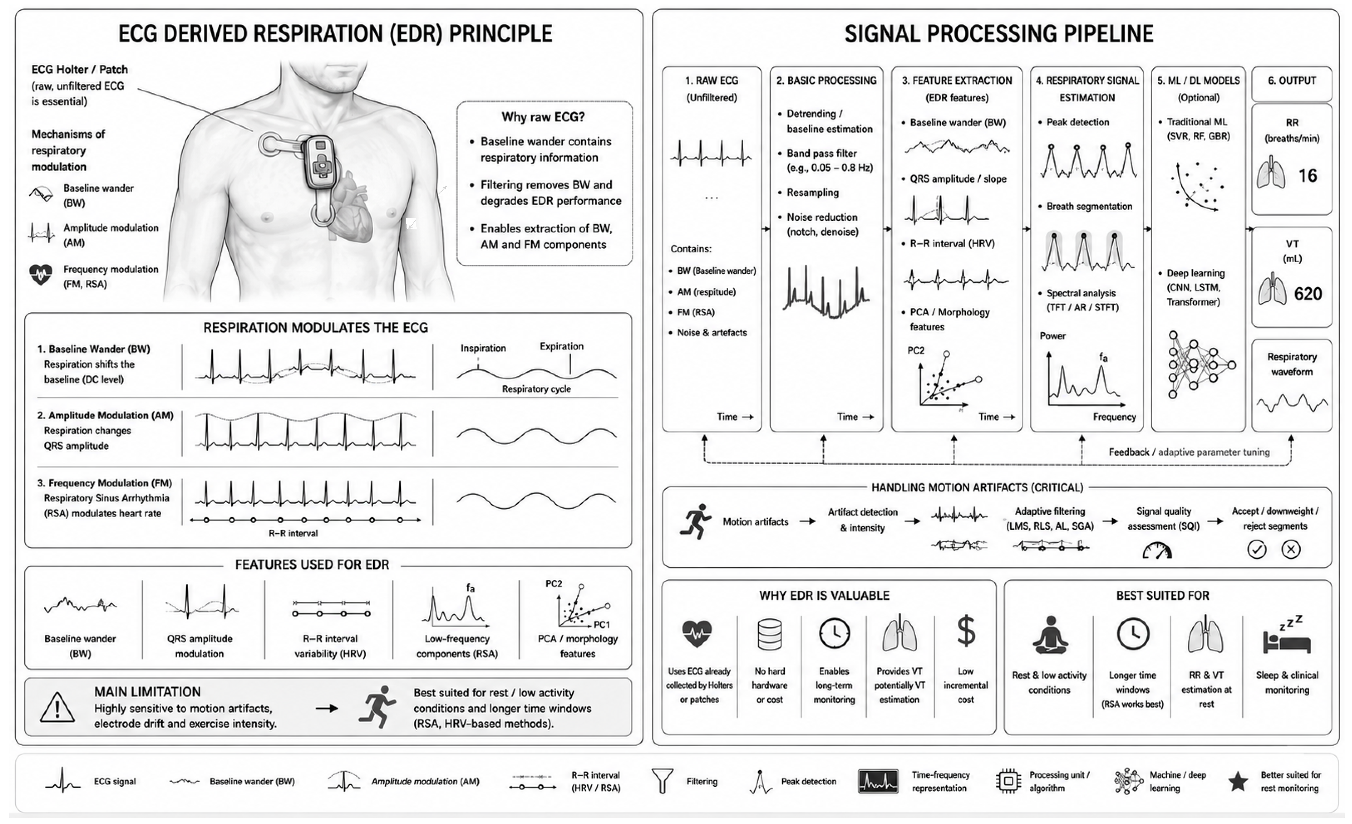
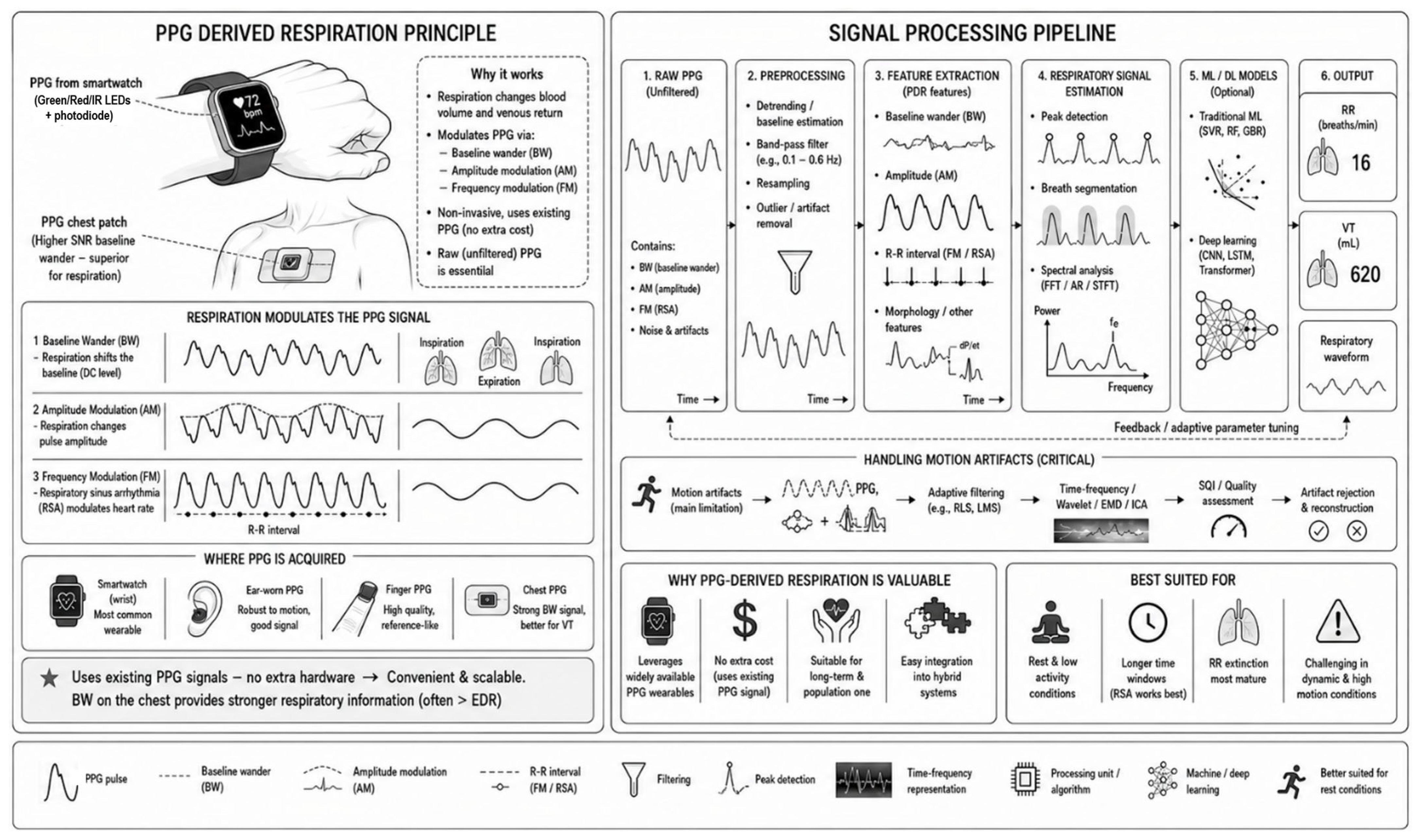
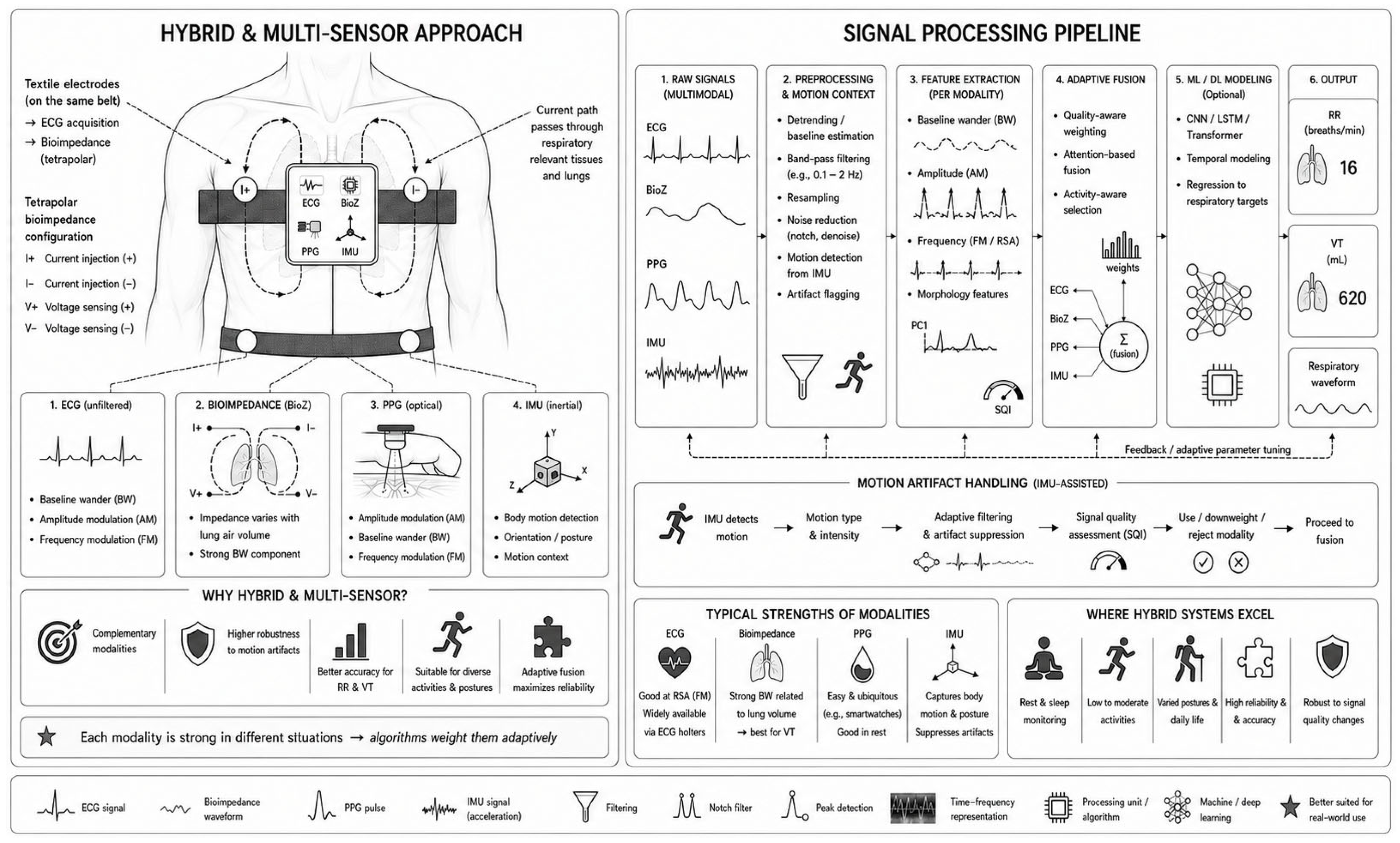
In terms of sensing, the approaches evaluated fall into two main categories [12]. The first includes direct respiratory measurement methods that capture physical manifestations, including chest and abdominal motion [20], sensed using resistive [21,22], capacitive [23,24], inductive [25,26], or optical strain sensors [27], bioimpedance measurements [28], and kinematic signals derived from inertial measurement units [29,30,31,32,33] that includes accelerometers, gyroscopes, and magnetometers [34]. The second category covers indirect methods that estimate respiration from modulation effects observed in cardiovascular signals, primarily electrocardiography (ECG) and photoplethysmography (PPG).
The term dynamic real-life conditions refers to ambulatory and physically demanding scenarios ranging from daily activities such as walking and posture transitions to vigorous exercise and competitive sports. Emphasis is placed on monitoring during conditions involving substantial body movement, including running, cycling, fitness activities, and sport-specific applications. This focus is motivated by our ongoing research on multimodal respiratory monitoring in highly dynamic environments, specifically motorsport racing and aerobatic aviation, where large motion artifacts, rapid body movements, vibration, and changing body orientation substantially complicate physiological sensing. Nevertheless, studies involving lower-intensity activities or static posture changes were also included when their findings were relevant for understanding wearable performance under progressively dynamic conditions. Interestingly, several studies have already reported considerable differences in respiratory estimation accuracy between sitting, standing, and walking conditions, suggesting that even relatively subtle biomechanical and physiological changes, such as altered diaphragm mechanics or thoracic stabilization, can significantly influence respiratory sensing performance.
Compared with static respiratory monitoring, dynamic monitoring introduces substantially greater technical and methodological challenges. The primary limitation is the presence of motion artifacts, sensor displacement, unstable skin contact, soft tissue deformation, and activity-dependent changes in respiratory mechanics, all of which can obscure physiologically relevant respiratory components. These problems are especially critical for wearable systems, where unobtrusive long-term use must be balanced against signal quality, sensor stability, energy consumption, and computational complexity. An additional challenge identified throughout the reviewed literature is the considerable heterogeneity of experimental protocols, reference modalities, evaluation metrics, and testing scenarios, which strongly limits direct comparison between studies and complicates objective assessment of technological progress. The need for standardized validation methodologies and benchmarking procedures, therefore, represents an important future direction and is further discussed later in this review.
To ensure methodological transparency, a brief description of the literature search strategy is provided. This review primarily focuses on studies published between 2015 and 2025, while selected earlier seminal works were included when historically or technically relevant. Relevant publications were identified using the ResearchGate, PubMed, Scopus, and Google Scholar databases. The search process combined keywords related to wearable and dynamic respiratory monitoring, including “wearable respiration monitoring”, “respiratory rate estimation”, “tidal volume estimation”, “motion artifacts”, “dynamic conditions”, “exercise monitoring”, “smart textiles”, “bioimpedance”, “seismocardiography”, “ECG-derived respiration”, “PPG-derived respiration”, and “multimodal respiratory sensing”. Emphasis was placed on studies evaluating respiratory monitoring under ambulatory, exercise, or free-living conditions, as well as on recent advances in multimodal sensing, artifact suppression, and machine-learning-based signal processing. To achieve scientific consistency, we define conceptual and statistical frameworks. Validation refers to the assessment of a system’s performance against a gold standard. Reliability denotes the stability of the measurement across different subjects or sessions, encompassing both repeatability (consistency under identical conditions over short intervals) and reproducibility (consistency across different observers, environments, or longer time periods). Robustness is defined as the ability to maintain operational integrity. From a data characterization perspective, breathing pattern recognition involves identifying specific rhythms and timing, often facilitated by respiratory waveform reconstruction. In dynamic conditions, the averaging window is a critical parameter used to stabilize features. Statistically, the strength of the relationship between the wearable and the reference is expressed via linear correlation, quantified by the correlation coefficient. To assess clinical bias and the range of errors, limits of agreement are established through Bland–Altman analysis, while uncertainty is accounted for to quantify the potential dispersion of the measured values. Finally, although metrics such as “accuracy”, “error”, and “agreement” represent distinct statistical entities, we retain the original terminology used in the cited studies to remain faithful to the specific methodologies employed, treating them as collective indicators of performance relative to a reference standard.
The main body of this review is organized according to the above-mentioned sensing paradigms. First, it addresses direct respiratory sensing, followed by derived or modulation-based approaches, and finally hybrid and multisensor systems. Each section has a consistent structure, starting with a brief theoretical background, continuing with representative devices and studies with particular emphasis on sensor design, placement, and signal processing strategies. To maintain readability, individual studies are described briefly, with only the most relevant information included. More detailed experimental results and specific technical parameters are summarized separately in tables provided at the end of each section. Each main section also concludes with a summary that highlights the main findings and limitations of the respective approach. In addition, a separate section is dedicated to relevant review articles to provide a broader context and encourage cross-comparison. This review concludes with a final discussion that synthesizes findings across all methodological groups and outlines the main challenges and priorities for future research.
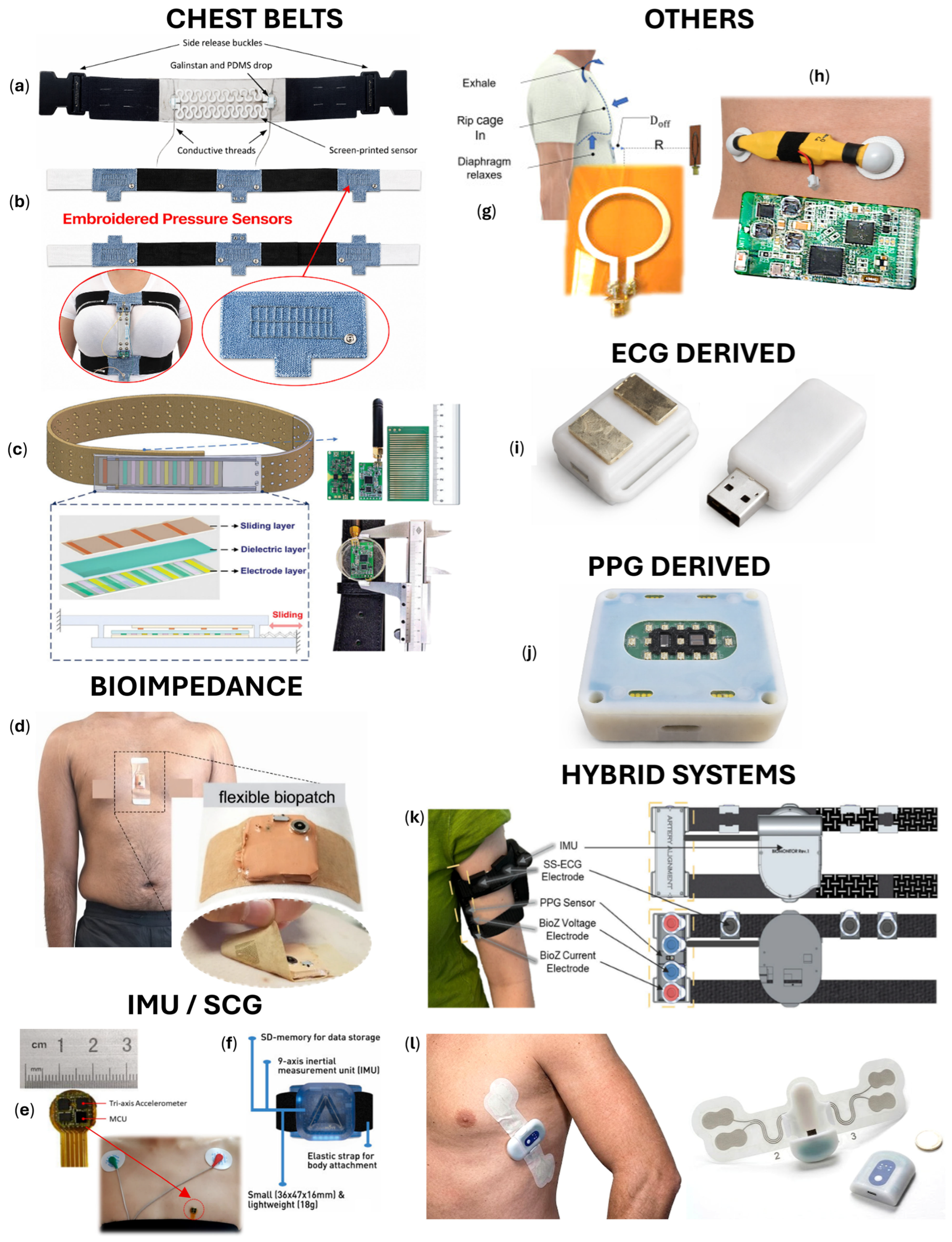
An overview of representative wearable respiratory sensors discussed in this work is presented in Figure 1, which serves as a visual reference for the sensing categories addressed in the following sections.

2. Direct Measurement Methods

2.1. Chest and Abdominal Bands

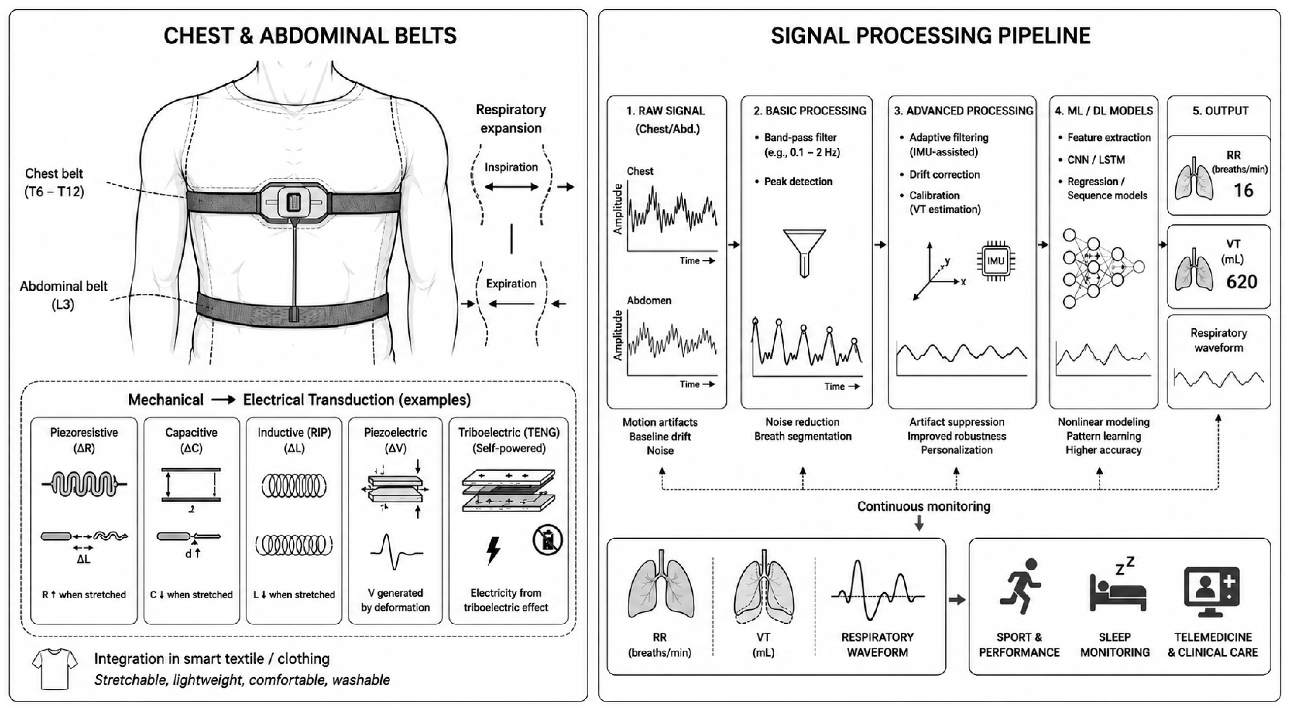
Devices based on chest and abdominal motions represent one of the most established and physiologically intuitive approaches (Figure 2) (Table 1). These sensors provide direct access to respiratory mechanics by capturing the cyclic expansion and contraction of the chest and abdomen and converting the resulting mechanical deformation into electrical or optical signals, making them well-suited for ambulatory and activity-related monitoring [13]. Common implementations include elastic strain sensors, inductive plethysmography belts, capacitive transducers, piezoelectric elements, and textile-integrated strain sensors, as well as optically interrogated strain sensors, such as Fiber Bragg gratings (FBGs) [45]. Textile breathing belts and smart clothing represent a significant development as they offer improved comfort, adaptability, and continual wearability while maintaining sensitivity to respiratory movements. Thanks to lightweight and deformable materials, such systems allow unrestricted movement as a necessary condition to use them in real life [19]. To keep the overview structured, the following subsections are organized according to the underlying sensing principle and signal transduction mechanism.

2.1.1. Piezoresistive Systems

Piezoresistive systems represent one of the most widely investigated approaches for wearable respiratory motion sensing due to their high strain sensitivity, simple electronics, flexibility, and compatibility with textile integration. These systems are increasingly incorporated into wearable patches, chest bands, and smart garments for continuous respiratory monitoring. These systems are increasingly integrated into wearable patches or bands due to their high strain sensitivity and compatibility with textile integration.
Chu et al. [46] introduced a crack-based piezoresistive strain sensor fabricated as a thin metal film on a silicone elastomer substrate. The reversible disconnection of microcracks resulted in a high gauge factor, enabling the detection of very small thoracic strains. The system allows simultaneous estimation of the RR and VT, and the results also demonstrated reliable reconstruction of respiratory waveforms. Vanegas et al. [47] designed a system based on a piezoresistive FlexiForce sensor attached to a chest strap system. A compact 3D-printed casing integrated a microcontroller, acquisition circuit, battery, and Bluetooth. Evaluation determined a 27 s analysis window as optimal, yielding low error rates of 4.02% for the time-based algorithm and 3.40% for the counting-based algorithm. In subsequent work, the same group analyzed signal drift induced by movement and body constitution [48]. Loranca Gómez et al. [49] proposed a low-cost piezoresistive textile band and compared its performance with an MPU6050 IMU. While respiratory patterns were reliably detected, the piezoresistive signal exhibited higher susceptibility to noise, suggesting that hybrid sensor fusion with inertial data may enhance robustness for dynamic system applications. Innovations in materials were further demonstrated by Lin et al. [50], who introduced a disposable graphene nanosheet-coated strain sensor, “Motion Tape”, designed for universal integration into elastic chest straps. The modular configuration with a snap fastener allows for interchangeability with existing straps. Validation against a commercial reference demonstrated accurate waveform reconstruction and identical RR measurements. High-intensity validation was described by Di Paco et al. [51], who evaluated a custom chest strap against a metabolic cart during a maximal cardiopulmonary exercise test (CPET) in elite soccer players. The device showed high absolute agreement, a strong linear correlation, and a root mean square error (RMSE) of 2.42 rpm, supporting its applicability even under maximal cardiopulmonary load. Solanki et al. [52] presented a smart-textile system “RespWear”. The system combined a textile pressure sensor belt for monitoring respiration-induced chest motion with a wireless microcontroller-based acquisition unit. Validation was performed on four participants during different breathing rates and body positions, including standing, sitting, bending, and reclining. An OptiTrack infrared camera system served as the reference method. The proposed system achieved a strong correlation coefficient of 0.836 for RR estimation. Later, this system was innovated and named “SolumWear”, with which Cay et al. [36] conducted an evaluation on 10 subjects, confirming the correlation. The system also demonstrated low computational and communication latency, confirming the feasibility of near-real-time monitoring using wearable devices. Screen-printed piezoresistive approaches were introduced by Al-Halhouli et al. [35]. The sensor features a unique silver “horseshoe” pattern electrode on a stretchable substrate to reduce stress concentration and preserve conductivity under high strain. The sensor achieved high RR accuracy across sitting, standing, and Fowler’s 45° position. Egwu et al. [53] proposed a TinyML-based framework for real-time respiratory monitoring using embroidered textile strain sensors. By implementing 8-bit quantized CNN and wavelet-based dense neural network (DNN) architectures on an STM32L4 microcontroller, the system enables fully embedded signal inference directly on the wearable device. Validation on public and custom TexHype datasets demonstrated that the CNN achieved superior accuracy (MAE 1.23 rpm), while the wavelet-based DNN offered lower computational overhead with an incremental power consumption of only 3.3 mW. This study highlights the efficacy of edge AI in balancing the trade-offs between estimation precision, latency, and energy efficiency in smart garment applications.
Sensor placement and multisensor configurations were examined by Laufer et al. [54], who applied regression analysis and bootstrapping techniques to identify optimal thoracic measurement positions for VT estimation. The analysis, using a camera capturing 102 markers, revealed that three circumferential and one distance changes carried the majority of VT information.
Some studies have also increasingly focused on fabrication strategies and textile integration techniques. A study by Tang et al. [55] proposed a smart clothing system based on strain-sensing yarn integrated into fabric using a novel stitching methodology. Using finite element analysis, the authors showed that self-locked yarn configurations and reduced needle pitch improved local stress concentration and enhanced sensing sensitivity. The resulting textile sensor demonstrated a low detection limit of 0.1%, a rapid response time of 280 ms, durability over 10,000 cycles, and strong resistance to washing, humidity, and perspiration. The study also optimized stitch trace lengths for respiration and heartbeat monitoring and integrated the sensor into a complete smart clothing platform. A related fabrication-oriented study by Arslan-Catak et al. [56] developed a sustainable water-based conductive ink composed of carbon nanotubes and cellulose nanocrystals for textile printing. Besides optimizing ink composition, the authors systematically investigated how different textile sensor geometries influence piezoresistive sensitivity during cyclic loading simulating chest movement. Their results demonstrated that sensor geometry significantly affects respiratory sensing performance, highlighting the importance of structural textile design in addition to the conductive material itself.
So far, we have mentioned single-band sensors; now, we will move on to those that measure respiration in multiple channels. Romano et al. [57] integrated two piezoresistive textile sensors, MedTex P130 (Statex Produktions und Vertriebs GmbH, Bremen, Germany), into a smart T-shirt and optimized their size and placement on the chest to improve RR extraction. The system was evaluated on two volunteers against the Zephyr Bioharness 3.0 during sitting, standing, walking, running, and stair climbing. An important finding is that the sensor placed on the side of the chest is more suitable for static activities, the sensor placed on the back for dynamic activities, and the combined output from the sensors outperforms the individual sensors by approximately two times. Di Tocco et al. [58] assessed the feasibility of a multisensor configuration that included four conductive textile strain sensors wired into a Wheatstone bridge to capture thoracic deformation, and combined them with an IMU to estimate HR via mechanical cardiac vibrations. In sitting, standing, and supine positions, and later also on a jockey during horse racing [59], the system achieved consistently low errors in both RR and HR estimation, demonstrating the advantage of distributed sensing for posture robustness.

2.1.2. Piezoelectric Systems

Regarding piezoelectric sensors, Yuan et al. [60] introduced a flexible polyvinylidene fluoride (PVDF) thin-film sensor with a biomimetic lateral line structure inspired by the fish geometry that improved sensitivity to weak thoracic deformations. The device generated stable voltage outputs proportional to VT under varying physiological conditions. Furthermore, Lei et al. [61] investigated a PVDF film encapsulated in polydimethylsiloxane (PDMS) for respiration monitoring during walking conditions. The patch demonstrated robustness against motion artifacts, with RR values showing no significant statistical difference compared to a commercial respiratory effort transducer and maintaining high correlation during ambulation. More application-oriented research was described by Ji et al. [62], who integrated a flexible piezoelectric belt into an aircraft seat system for simultaneous ECG, respiration, and motion monitoring. Extraction of these parameters, combined with a long short-term memory recurrent neural network (LSTM-RNN) classifier, enabled detection of sleep apnea–hypopnea syndrome, with 84–85% accuracy. Although this application was designed for sedentary conditions, the fusion of motion signals with recurrent neural networks illustrates a scalable framework potentially adaptable to dynamic respiration monitoring scenarios. We are leaving the article in the selection because our future research activities will include monitoring respiration during aerobatic flying.

2.1.3. Inductance Systems

Another method, respiratory induction plethysmography (RIP), measures changes in the self-inductance of coils placed around the chest and abdomen. Stretching during inspiration changes the loop inductance, modulating the oscillator frequency and producing a proportional voltage output [63,64]. Accurate estimation of VT requires appropriate weighting of the thoracic and abdominal signals, which is usually achieved by calibration, such as isovolumic or qualitative diagnostic [25]. Ratnagiri et al. [65] analyzed thoracoabdominal motion relationships using the RIP system pneuRIP [66]. An ML elastic-net regularized model identified thoracoabdominal asynchrony, achieving 61.3% accuracy via phase difference and 90.3% using inverse cumulative percentage metrics.
Beyond hardware design, several studies have focused on improving RIP signal processing to enhance respiratory parameter estimation. Holm et al. [67] introduced BreathFinder, an open-source algorithm for RR detection from RIP signals, demonstrating a robust framework for respiratory cycle identification that is also relevant for noise suppression in dynamic conditions. Complementarily, Finnsson et al. [68] proposed a correction and linearization approach for RIP signals against oronasal pneumotachography, substantially improving the accuracy of small respiration and depth estimation. Bias was reduced from 2–12% to 1–9%. Although their study was conducted in the context of sleep pathology, the presented signal correction strategy is highly relevant for wearable monitoring, as it addresses nonlinearities and deformation-related distortions that are likewise encountered during physical activity.

2.1.4. Capacitive Systems

Capacitive stretch sensors estimate respiration by detecting changes in inter-electrode distance during thoracic expansion. Early, conceptually simple implementations convert fabric deformation directly into capacitance changes. Kim et al. [69] validated an easy-wear capacitive belt against a BIOPAC reference, reporting RR errors below 2% across six postures. Enokibori et al. [70] introduced the “Spiro Vest”, an e-textile garment integrating two capacitive length sensors on the chest and abdomen to infer VT from fabric deformation. Vicente [71] evaluated two polyglycerol–sebacate sensor variants—porous and pyramidal microstructures—to estimate tidal volume from capacitance changes. Both variants correlated strongly with a commercial airflow transducer, yielding a mean absolute error (MAE) in the order of 100 mL.
Progressing from these basic textile systems, several studies addressed electrode geometry and materials to improve sensitivity and robustness. Ali et al. [72] proposed a textile sensor that operates without direct skin contact, implemented by screen-printing an electrode stack with an optimized 1:3:1 sensor:reflector:ground ratio on a polyester–cotton substrate. Park et al. [73] developed a flexible PDMS waist belt incorporating silver nanowires and carbon fibers. The design combined enhanced sensitivity and mechanical durability and applied a finite impulse response (FIR) filter to mitigate motion artifacts. Designs prioritizing wearability tackle the trade-off between comfort and measurement fidelity. Kobayashi et al. [74] presented a low-compression smart garment that exerted minimal torso pressure while maintaining strong agreement with spirometry and a mean RR difference below 0.1 rpm across postures.
More complex systems fuse complementary sensing modalities for robust performance in dynamic conditions. Bernhart et al. [75] combined a capacitive pressure interface (PSoC™62) with an elastic belt to detect respiration via pressure changes between the rib cage and the elastic belt, and used an MPU6050 IMU to detect stride and motion. In validation with endurance runners, the multimodal system achieved high F1 scores for step and respiration event detection. At the highest level of complexity are approaches that leverage advanced signal processing, machine learning, and engineered microstructures. Kim and Kim [76] applied a convolutional neural network (CNN) and ResNet architectures to breathing-pattern classification. The optimized ResNet substantially outperformed a conventional CNN (overall accuracy 96% vs. 87%), particularly for the most challenging classes.

2.1.5. Optical Systems

Fiber optic sensors provide a promising alternative to conventional electronic methods. These systems typically operate on the principle of detecting changes in light transmission, such as intensity modulation or macro-bending effects, induced by mechanical expansion of the thorax. A prominent subset of this technology, FBG sensors, is based on wavelength shifts and offers high sensitivity and multiplexing capability, though interrogation cost and temperature cross-sensitivity remain key limitations. For a detailed overview of these sensors and their integration into wearable systems, we recommend the comprehensive review by Krizan et al. [77]. Zha et al. [78] designed a stretchable elastomer optical fiber sensor incorporated into a belt, achieving ≤ 1 rpm RR error and ≤3 bpm HR error, with an MAPE 5.25% and an RMSE of 1.28 rpm during different postures. Huo et al. [79] reviewed the design of flexible optical fiber wearable sensors, highlighting biocompatibility, lightweight design, and integration with IoT and ML for real-time monitoring, while noting challenges in long-term stability and cost that must be addressed to transition these devices into clinical and sports training equipment.

2.1.6. TENG Systems

The growing interest in self-powered sensing has intensified research on triboelectric nanogenerators (TENGs). Li et al. [80] introduced a lightweight retractable sensor based on a rotating thin-film TENG, capable of enduring more than 1 million stretching cycles. When integrated with a Wi-Fi module, an STM32 microcontroller (STMicroelectronics, Geneva, Switzerland), and a charge amplifier, the system generated AC signals proportional to thoracic expansion, enabling wireless respiratory monitoring. Similarly, Shi et al. [37] proposed a compact TENG sensor employing triple-phase interpolation electrodes to quantify thoracic displacement with sub-millimeter resolution and operational durability exceeding 700,000 cycles. Expanding the functional scope of TENG-based monitoring, Xu et al. [81] demonstrated simultaneous measurement of RR and VT, achieving an MAE < 0.2 rpm and strong agreement with spirometry. The system also reconstructed volume–time curves with a relative MAE of 2.43%. Beyond triboelectric approaches, Sharma et al. [82] introduced a smart respiration belt utilizing giant magnetoresistance sensing to detect magnetic field variations induced by chest expansion. In a cohort of 12 subjects, the system achieved a maximum deviation of ±2 rpm compared with a BIOPAC reference.
While respiratory masks are often associated with stationary clinical settings, specialized implementations utilizing TENGs warrant inclusion due to their emerging role in high-intensity dynamic environments. This application is particularly relevant to aviation and aerobatics—domains that align with the objectives of our forthcoming research. Zhao et al. [83] demonstrated this potential by integrating an ML-enabled triboelectric textile sensor directly into an oxygen mask for real-time monitoring in extreme conditions. By utilizing plasma-modified surfaces and nanoscale engineering to enhance sensitivity, the system achieved a respiratory pattern recognition accuracy of 97.2%. To ensure reliability during active use, an ML-assisted classifier was employed to effectively distinguish authentic respiratory signals from motion and environmental artifacts.

2.1.7. Commercial Systems

Finally, commercially available wearable respiratory monitoring systems should also be considered, as they represent the practical translation of the previously discussed sensing principles into real-world applications. While most commercial solutions are inherently multimodal, the following section focuses primarily on their dominant respiratory sensing mechanism and its application in dynamic monitoring scenarios.
A representative example is the Airgo belt (MyAir, Inc., Boston, MA, USA) [84], a resistance-based thoracic circumference sensor that integrates stretchable silver-coated yarn and IMU for RR, VT, and motion detection. As a CE Class IIa certified medical device, it serves as a non-intrusive proxy for spirometry, respiratory pattern detection, and sleep disorder screening. Validation against a metabolic cart [21] and subsequent analysis using detrended fluctuation analysis [34] demonstrate its reliable performance during both nighttime rest and daytime activity. Notably, this study found that the limits of agreement (LoAs) increased by a factor of approximately three during dynamic activities compared to static conditions [21]. Another widely utilized device is the Zephyr Bioharness 3.0 (Medtronic, Minneapolis, MN, USA) [85]. Because it frequently serves as a reference standard in other studies, its rigorous independent validation is essential. Panni et al. [86] evaluated it against spirometry using Monte Carlo uncertainty propagation, demonstrating high RR accuracy with LoAs within −2 to 3 rpm and a very strong linear correlation. Similar findings were reported by Hailstone and Kilding [87], who confirmed the reliability during dynamic treadmill exercise, and by Kim et al. [88], under more demanding running conditions, including both maximal incremental exercise and prolonged moderate-intensity running in the heat. Their results showed that the Zephyr maintained acceptable agreement with the reference method, although accuracy decreased under more physiologically and environmentally challenging conditions. The device’s test–retest reliability has also been well documented [87,89]. Romano et al. [90] proposed a signal quality index (SQI) algorithm that evaluates the morphology of individual breaths to further enhance the accuracy of such systems. By comparing these waveforms against an average respiratory template, the algorithm successfully identified and excluded unreliable respiratory cycles without the need for an external reference signal. Validation of this SQI across rest, walking, running, and cycling conditions demonstrated consistent improvements in RR estimation accuracy between 2.8% and 30.7%, depending on the situation. Beyond algorithmic enhancements, optimal sensor placement and calibration strategies are critical factors for accurate respiratory monitoring, particularly when estimating VT. Investigating these parameters, studies evaluating a thoraco-abdominal sensor (SA9311M, Thought Technology Ltd., Montreal, QC, Canada) demonstrated that thoracic placement (at the T6 and T12 vertebrae) yields more accurate VT estimation than abdominal positioning (at the L3 vertebra) [91,92]. Furthermore, the authors found that implementing an individualized calibration formula significantly outperformed the universal calibration approach. However, it is important to note that during a longitudinal assessment spanning two separate visits, the maximum mean relative error for the VT increased significantly over time, whereas the mean relative error for the RR remained relatively stable.
A notable emerging trend in wearable respiratory monitoring is the miniaturization of traditional full-chest belts into smart patch formats. These patches offer distinct advantages, including reduced size, enhanced user comfort, and the seamless integration of supplementary sensors. Conversely, they typically require direct skin adhesion, and a potential trade-off is a slight reduction in measurement accuracy. This is particularly relevant for VT estimation, as patches cover a significantly smaller surface area of the chest compared to circumferential belts. Exemplifying this technological shift is, for example, the Resmetrix (Resmetrix Medical Ltd., Haifa, Israel) [93], a patch-based, chest-worn system designed for the continuous monitoring of breathing patterns, RR, and VT. To support clinical utility, the device transmits data to facilitate the automatic, AI-powered detection of exacerbation-related abnormalities.
Expanding beyond localized patches, another prominent approach involves integrating sensors directly into smart garments and textiles. These systems offer the distinct advantage of capturing data over a larger surface area of the tors, which is highly beneficial for accurate VT estimation. A highly successful commercial solution in this domain is the Hexoskin biometric shirt (Carré Technologies Inc., Montreal, QC, Canada), which combines ECG, respiratory inductance plethysmography (RIP), and 3D accelerometry for multimodal monitoring [94]. Widely adopted in scientific research, its validation against spirometry and 12-lead ECG demonstrated the highest agreement for VT and minute ventilation (VE) during submaximal exercise, though larger deviations were observed at rest and during maximal effort [95]. Heart rate and RR errors remained below 10%, and adjusting for sex and body weight further improved VE estimation. Similarly, Villar et al. [96] reported a minimal RR bias of 0.3 rpm and LoAs of ±2 rpm during submaximal incremental walking. The system also proves effective in natural environments, such as tracking respiration topography in tobacco users, utilizing individualized calibration procedures [97]. Further demonstrating the viability of smart commercial garments under highly dynamic conditions, Innocenti et al. [98] evaluated two vests, the ComfTech vest (Howdy Senior, ComfTech s.r.l.®, Monza, Italy) and the Tyme Wear vest (Tyme WearTM, Boston, MA, USA), and the BioHarness 3.0 strap during soccer-specific movements. When compared against a reference metabolic mask, the extracted respiratory frequency closely tracked the reference signal. Breath-by-breath analysis yielded mean absolute percentage errors (MAPEs) of 7.03% for ComfTech, 8.65% for Tyme Wear, and 14.60% for BioHarness. Notably, these errors decreased significantly to 1.85%, 3.27%, and 7.30%, respectively, when the data was averaged over 30 s windows, highlighting the importance of temporal smoothing in dynamic sports applications. What is also visible and can be considered an important conclusion: the vests outperformed the belt system in accuracy.
Table 1. Chest- and abdominal belt-based respiratory monitoring.
Sensor TypeApplicationSensing ElementKey ParametersRef.
Skin-
attached chest strain sensor
RR 1, VT 2,
respiratory waveform
Crack-based
piezoresistive thin metal film
Textile sensor (EeonTex LTT-SLPA-20K), silicone elastomer substrate, miniature, BL 3, linear response, MLR 4 algorithm for VT, validated vs. spirometry, during motion: RR R2 = 0.83, VT: concordance
correlation coefficient 0.75, bias −77 mL,
LoAs 5 −374–220 mL, SEE 6 = 131 mL (26%)
[46]
Chest beltRRPiezoresistive sensor
(FlexiForce A201)
BLE 7, 3D-printed casing integrating microcontroller and acquisition system, 21 subjects, optimal analysis window 27 s, time-based algorithm error 4.02%,
counting-based algorithm error 3.40%
[47,48]
Chest beltRR, respiratory waveformDisposable
graphene nanosheet-coated piezoresistive strain sensor
Snap fastener interface, 4-channel, 12-bit
ADC resolution, sampling rate max. 66 Hz, data storage 32 GB mSD 8 card, BL, 2000 mAh LiPo battery, 90 min working time, accurate respiratory waveform reconstruction
[50]
Chest belt RRStrain-based chest piezoresistive sensor integrated in elastic strapValidation during maximal CPET 9 vs. metabolic cart, BLE, 26 soccer players, high-intensity cardiopulmonary load, high absolute agreement ICC 10 = 0.97,
linear correlation 0.96, RMSE 11 = 2.42 rpm
[51]
Smart textile chest beltRR6× embroiled
piezoresistive
textile pressure sensor
16-bit, sampling rate 64 Hz, BPF 12 0.1–0.35 Hz, wireless data, posture-independent RR estimation, 10 subjects, validated vs. OptiTrack IR 13 camera, correlation coefficient 0.836, MAE 14: standing (deep 3.25%, normal 12.3%, fast 3.03%), sitting (22.91%, 11.61%, −0.58%), latency: 4.84 s (computational), 2.13 ms (communication)[36]
Chest beltRRScreen-printed
piezoresistive
sensor
Silver horseshoe-pattern electrode on stretchable substrate, validation vs. airflow, RR evaluated in sitting, standing and Fowler’s 45° position, minimal RR error 0.055 rpm across postures, LoAs −0.91–0.998[35]
Smart textileRREmbroidered
meander-pattern textile strain
sensor
STM32L4 microcontroller, CNN 15 + wavelet-based DNN 16, TinyML/embedded edge AI 17, public strain-sensor + TexHype dataset, MAE: 1.23 rpm (CNN), 2.21 rpm (DNN), inference latency: 5.8–6.2 s (CNN), 18–96 ms (DNN), power overhead 3.3 mW[53]
Smart shirt/chest beltVT3× strain gauges piezoresistiveOptimization of sensor distribution by 102 motion capture points, coefficient of determination 0.97,
average VT error 104.4 mL
[54]
Smart shirtRR2× piezoresistive textile sensors (MedTex P130)2 subjects, validated vs. Zephyr Bioharness 3.0, static activities: lateral chest sensor MAE 0.1–0.3 rpm, back sensors MAE 1.1–3.2 rpm, during walking: lateral chest sensor MAE 1.9, back sensor MAE ≈ 0.1 rpm, 9 subjects, during sitting, standing, walking, running, and stair climbing–results by sensor combination MAE to 0.32 rpm, individual sensors: MAE 0.53 rpm and 0.78 rpm[57]
Two chest beltsRR, HR4× conductive
piezoresistive
textile sensors sewn on elastic belts + IMU 18
Textile sensor (EeonTex LG-SLPA), IMU (LSM9DS1), µSD storage, 8 h battery life, sampling rate 100 Hz, validated vs. Zephyr BioHarness, 8 subjects, RR
average error ~0.17–0.35 rpm (sitting/standing), ~2.95 rpm (supine), RR percentage error ~1.21% (sitting), ~3.49% (standing), ~9.25% (supine)
[58]
Chest sensorRR, VTPiezoelectric PVDF 19 thin-film sensorBio-inspired lateral line geometry to enhance
sensitivity to low-amplitude thoracic deformation, passive self-powered sensing, stable voltage output proportional to VT, BL, low detection limit 0.5 mN,
sensitivity 0.24 V/N, response time 4 ms
[60]
Chest patchRRPiezoelectric PVDF film
encapsulated in PDMS 20
Improved mechanical stability, motion-robust
during dynamic walking, RR showed no statistically significant difference p > 0.05
[61]
Seat belt
integrated system
RR, ECG 21,
motion, OSA 22
Flexible
piezoelectric belt sensor
Signal fusion with LSTM-RNN 23 classifier, ML 24-based multimodal framework to respiration analysis with motion context, OSA accuracy 84–85%[62]
Chest and abdominal beltsRR2× RIP 25 beltML-based analysis, regularized model, 51 pediatric subjects, thoracoabdominal asynchrony accuracy: 61.3% (phase difference features),
90.3% (inverse cumulative percentage metric)
[65,66]
Chest beltRRRIPBreathFinder algorithm, 31 subjects, static conditions, dataset comprising 8782 (7.3 h) manually annotated breaths, RR detection accuracy 94%[67]
Chest beltRRCapacitive sensorFlexible electrodes/dielectric layer, validation vs. BIOPAC MP150, 6 postures, RR MAE < 2% (longer period)[69]
e-Textile
garment
RR, VT2× capacitive
textile length
sensors
Dual-sensor configuration capturing thoracic and
abdominal, BL, 3 subjects, walking, sampling rate 100 Hz, VT error reduction 60%
[70]
Chest
attachment
VTCapacitive
pressure sensors
Validated vs. airflow, 38 subjects, mean correlation > 0.91, porous substrate: sensitivity 0.09 kPa−1, MAE 122 mL, pyramidal substrate:
sensitivity 0.015 kPa−1, MAE 100 mL
[71]
Textile chest
belt
RRTextile capacitive sensor with screen-printed electrodes Electrodes on polyester cotton fabric, optimized
electrodes ratio 1:3:1 (sensor:reflector:ground),
validation vs. manual counting, frequency-based readout, sensitivity 6.2%, RR accuracy 98.68%,
[72]
Waist beltRRCapacitive
pressure sensor
PDMS dielectric with Ag nanowire and carbon fiber electrodes, optimized for belt, sensitivity 0.161 kPa−1, dynamic range 200 kPa, mechanical durability > 6000 cycles, FIR 26 filtering to suppress motion[73]
Smart
garment
RRDouble layer
capacitive
bending angle sensor
Minimizing mechanical constraint, compression pressure 0.77 ± 0.21 kPa, validation vs. spirometry, 20 subjects, strong correlation 0.97–0.99 across
postures, mean RR difference < 0.1 rpm
[74]
Chest beltRR, strideCapacitive
pressure sensor + IMU
Sensing interface (PSoCTM62), IMU (MPU6050) for stride and motion detection, validation vs. ergospirometry in endurance runners, F1 score 93.2% (step), 97.4% (exhalation), 97.2% (inhalation)[75]
Abdominal garmentRR, respiratory waveformTextile capacitive sensors with
embroidery
electrodes
100 × 50 mm electrodes, DL 27 models, respiratory
pattern estimation: accuracy: 0.87 (CNN), 0.96
(ResNet 28), precision under challenging breathing:
0.6 (CNN), 0.8 (ResNet)
[76]
Chest beltRR, HR 29Elastomer optical fiber sensor 10 subjects, different postures, validated vs. manual counting, RR error ≤ 1 rpm, HR error ≤ 3 bpm, MAPE 30 5.25%, RMSE 1.28 rpm[78]
Chest beltRRFlexible optical
fiber sensor
Sensor embedded in wearable substrates,
compatible with textile integration, enables IoT 31 connectivity, different respiratory rates experiment, ML-based real-time monitoring,
MAE 0.31 rpm (2.29%)
[79]
Chest beltRRRetractable
thin-film TENG 32
sensor
Self-powered, miniaturized, mechanical durability > 1,000,000 stretching cycles, resolution 0.13 mm,
integrated Wi-Fi 33 module and STM32 controller
[80]
Chest beltRRTENG sensor with triple-phase interpolation
electrodes
Self-powered, resolution > 1 mm, mechanical
durability > 700,000 stretching cycles, wireless data transmission, compact design suitable for
integration into wearable systems
[37]
Chest beltRR, VTTENG sensorSelf-powered, validation vs. spirometry, MAE < 0.2 rpm for RR, correlation 0.88,
VT reconstruction relative MAE 2.43%
[81]
Chest beltRRGiant magnetoresistance sensorNon-contact, integrated into elastic belt, validated vs. BIOPAC, 12 subjects, maximum RR error ± 2 rpm[82]
Pilot maskRR, respiratory waveformTribolometric
fibers in pilot
oxygen mask
ML-assisted respiratory pattern classification,
sensitivity 2.02 V·kPa−1, response time 96 ms, 420% output voltage enhancement, accuracy 97.2%
[83]
Chest belt
(commercial)
RR, VT, sleep, OSA, activityStretchable sensor + IMUBL, IP67, 6-weeks autonomy, ML and AI analysis, CE Medical Device Class IIa certified, 21 subjects,
RR LoA: ±5.9 rpm (standing), ±7.9 rpm (seated), ±10.6 (supine), ±25.8 (low intensity), ±19.5 (medium-high intensity), ±31.5 (maximal intensity), normalized minute ventilation relative median error > 5.9% (standing), 7% (seated), 3.4% (supine), 9.3% (low intensity), 34.7% (medium-high intensity), 40.6% (maximal intensity), α ≈ 0.74–0.75 (RR), α ≈ 0.88–0.97 (VT)
[21,34,84]
Chest belt
(commercial)
ECG, HR, RR, temp 34, activity ECG electrodes + capacity pressure pad + 3D
accelerometer + thermistor
BL, IP55, 12 h battery life, ECG (250 Hz), RR (25 Hz), temp (1 Hz), acceleration (100 Hz), FDA 510(k) CE (Class II), weight 71 g, smoothing and high pass filter, RR accuracy ± 1 rpm, LoAs −2–3 rpm (static and dynamic), ±5 rpm (maximal incremental running test), ±8.3 (running trial in the heat), linear correlation 0.95, typical error 4.4–8.7%, bias −0.6–0.2 rpm, test-retest reliability typical error 1.4–2.8 rpm (4.3–7.3%)[85,86,87,88]
Chest belt
(commercial)
ECG, HR, RR, temp, activityECG electrodes + capacity pressure pad + 3D
accelerometer + thermistor
SQI 35-based approaches, morphology exclusion of unreliable cycles, 33 subjects, MAPE reduction 18.5% (rest), 22.2% (walking), 2.8% (running), 14.1% (cycling), 30.7% (high intensity interval training)[85,90]
Chest belt
(commercial)
RR, VT,
respiratory waveform
Stretch-sensitive girth sensorRequires external DAQ/amplifier, non-calibrated VT, individualized calibration,
mean relative error 13–26% (RR), 19–35% (VT)
[91,92]
Chest patch
(commercial)
RR, VT, HR, temp, activityProprietary stretchable sensor BL, AI algorithm,
respiratory patterns and deterioration
[93]
Smart textile biometric shirt
(commercial)
RR, VT, ECG, HR, HRV 36, sleep, activity, VO2 37 max,RIP 16-based belts + ECG electrodes + 3D
accelerometer
RIP (128 Hz); ECG (256 Hz), accelerometer (64 Hz), BL, validation vs. spirometry and 12-lead ECG, 17 subjects, HR and RR errors < 10%, agreement for VT ≤ 5.3% (submaximal exercise), ≤15.3% (rest), ≤11.7% (maximal effort), VT estimation improved with sex and body-weight adjustment (r2 = 0.89)[94,95]
Strain-based systems
(commercial)
HRStrain-based systems15 soccer players, validated vs. metabolic mask, MAPE: 7.03% (ComfTech), 8.65% (Tyme Wear), 14.60% (BioHarness), LoA: ±12 rpm, ±15.7 rpm, ±24.4 rpm, MAPE (30s averaging window): 1.85%, 3.27%, 7.30%[98]
1 Respiration rate, 2 tidal volume, 3 Bluetooth, 4 multiple linear regression, 5 limits of agreement, 6 standard error of estimate, 7 Bluetooth low energy, 8 MicroSD card, 9 cardiopulmonary exercise testing, 10 intraclass correlation coefficient, 11 root mean square error, 12 bandpass filter, 13 infrared, 14 mean absolute error, 15 convolutional neural network, 16 dense neural network, 17 artificial intelligence, 18 inertial measurement unit, 19 polyvinylidene fluoride, 20 polydimethylsiloxane, 21 electrocardiography, 22 obstructive sleep apnea, 23 long short-term memory recurrent neural network, 24 machine learning, 25 respiratory induction plethysmography, 26 finite impulse response, 27 deep learning, 28 residual network, 29 heart rate, 30 mean absolute percentage error, 31 Internet of Things, 32 triboelectric nanogenerator, 33 wireless fidelity, 34 temperature, 35 signal quality index, 36 heart rate variability, 37 volume of oxygen.

2.2. Bioimpedance Methods

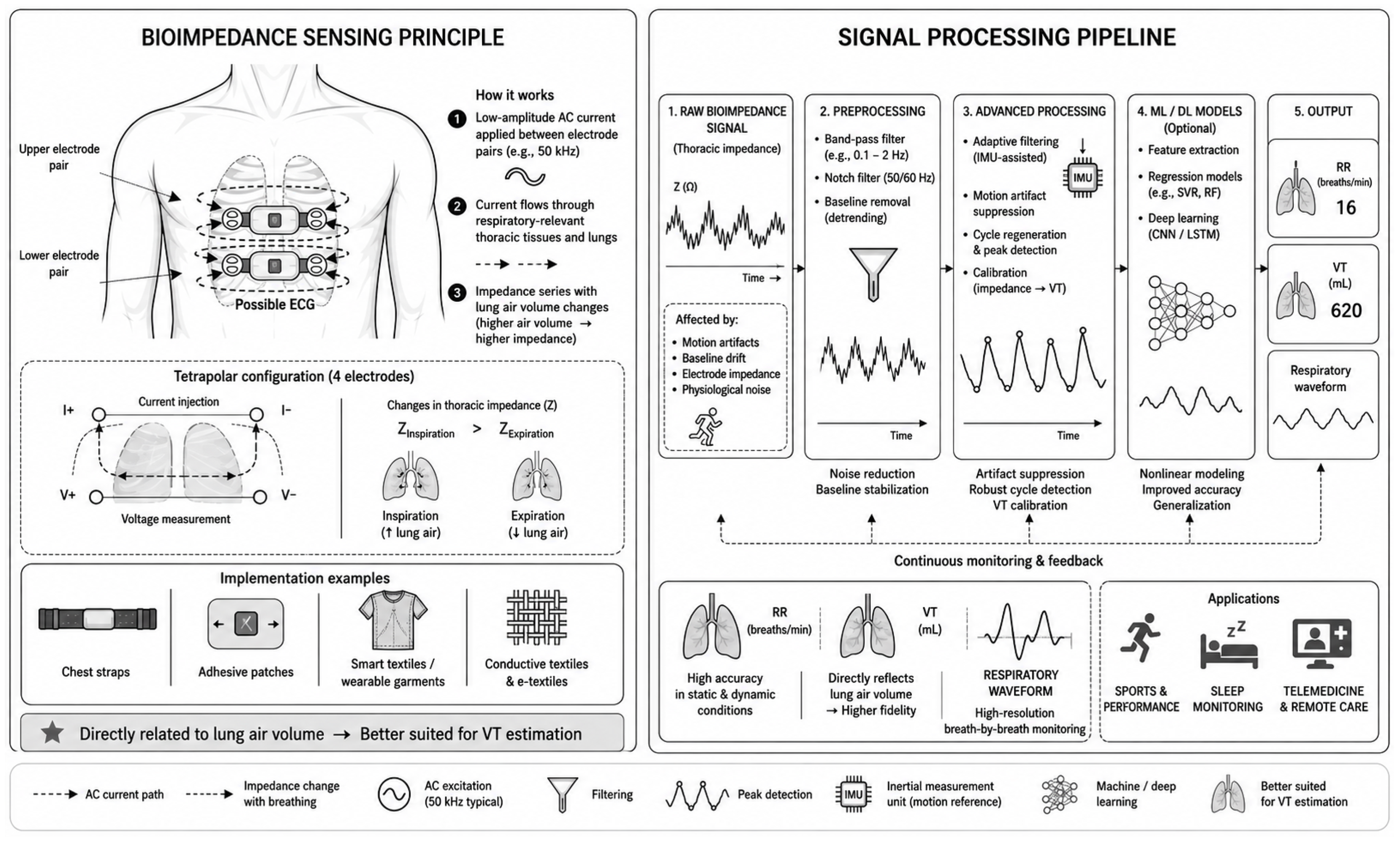
Bioimpedance-based respiratory monitoring, often also referred to as electrical impedance plethysmography (EIP) (Figure 3) (Table 2), measures cyclic changes in thoracic impedance associated with changes in lung air volume. Since air has a significantly higher electrical resistance than surrounding tissues, inspiration and expiration induce measurable fluctuations that are directly related to RR and VT. Bioimpedance is typically measured using surface electrodes arranged in a bipolar or tetrapolar configuration and integrated into chest straps, adhesive patches, or textile platforms. Groenendaal et al. [99] published a comprehensive review of wearable bioimpedance systems. The authors revealed the transition from hospital-based systems to unobtrusive home monitoring. Importantly, this review highlights ongoing challenges, including power consumption and the long-term stability of the electrode–skin interface and motion-induced artifacts.

2.2.1. RR Estimation

Several studies confirmed that bioimpedance-based sensing can provide accurate RR estimation, especially in controlled conditions. The early validation reported by John et al. [100] demonstrated the efficacy of the PhysioPatch against a respiratory chest belt across different breathing rates. Similarly, Heydari et al. [101] evaluated a chest-based system for simultaneous HR and RR monitoring using a TCO2 sensor as the reference and reported low RR errors across diverse breathing patterns. Piuzzi et al. [102] further extended this concept by introducing a textile-based thoracic belt that injects a 50 kHz current, enabling simultaneous ECG and respiratory monitoring with high RR accuracy during both quiet breathing and tachypnoea.
More recent developments have focused on patch-based wearable systems suitable for continuous and remote monitoring. Qiu et al. [103] proposed a chest patch capable of accurate RR monitoring across walking, running, and cycling, while also integrating Bluetooth and LoRa communication for telemedicine applications. A similarly designed real-time bioimpedance patch was presented in [35], where RR estimation remained highly accurate in both static and dynamic conditions. In contrast, not all systems retained such performance under physical load. For example, Wei et al. [44] evaluated a “Health Patch” integrating bioimpedance against the metabolic reference system Cosmed K5 [104] and observed only moderate agreement during exercise, highlighting the sensitivity of bioimpedance measurements to motion, contact variability, and physiological perturbations.

2.2.2. VT Estimation

In addition to RR estimation, several studies have focused on VT estimation, where this modality often offers greater physiological relevance than purely motion-based sensing. Early evidence was provided by Berkebile et al. [105], who evaluated a compact multifrequency tetrapolar sternal patch under both static and dynamic conditions against a conventional chest electrode configuration and spirometry. Their results demonstrated a strong Pearson correlation coefficient (0.93 ± 0.05) with reference VT and very low MAPEs of 0.93% for the patch and 0.74% for the chest configuration, even during physical activity, confirming that localized thoracic bioimpedance can capture meaningful ventilatory changes.
A systematic work was presented by Blanco-Almazán et al. [106,107,108], who investigated VT monitoring across multiple perspectives, including electrode configuration, inspiratory loading, and ambulatory monitoring. Across these studies, the authors demonstrated a strong linear relationship between impedance variation and VT, high respiration phase detection accuracy in dynamic conditions, and sensitivity to breathing variability during walking. In a complementary mechanistic analysis [109], combining bioimpedance with airflow and accelerometry, they further showed that signal composition changes with inspiratory muscle effort and that, under high inspiratory load, the bioimpedance signal is increasingly influenced by mechanical chest motion. By incorporating both volume and motion-related information into neural-network models, they achieved highly accurate VT estimation (MAPE < 4.29%), indicating that hybrid modeling may be particularly beneficial in dynamic conditions.
More advanced volumetric approaches have moved toward multichannel spatial bioimpedance acquisition. Khan et al. [110] introduced the concept of “virtual spirometry”, using a 10-channel bioimpedance vest and a segregated envelope and carrier (SEC) algorithm, enabling regression-based reconstruction of the respiratory waveform in a form analogous to spirometry. Similarly, Frerichs et al. [111] extended wearable bioimpedance toward electrical impedance tomography (EIT) by integrating 21 replaceable sensors into a textile vest, thereby increasing spatial resolution and the potential to capture regional ventilation patterns.

2.2.3. Algorithm Implementation

Beyond hardware design and electrode placement, the performance of wearable bioimpedance systems depends strongly on the applied signal processing and artifact suppression strategies, so algorithmic robustness has become a key determinant of practical usability in dynamic conditions.
One of the earlier dedicated solutions was proposed by Järvelä et al. [112], who introduced a three-electrode wearable system employing a “dual vector” algorithm to suppress motion artifacts. The signals were processed locally and transmitted wirelessly to a central monitoring station. In a clinical validation, the system achieved a mean RR difference of −0.6 ± 2.5 rpm compared with capnography, demonstrating that even relatively simple algorithmic compensation can substantially improve performance.
As wearable applications moved toward more realistic and less controlled environments, automated signal quality evaluation became increasingly important. Albaba et al. [113] addressed this need by developing a quality classification framework for capacitively coupled bioimpedance signals, designed to distinguish between high-quality and corrupted segments using statistical and spectral features. Their method showed strong robustness, achieving an accuracy of 91% on the primary test set, with sensitivity reaching up to 98%, while a fine Gaussian support vector machine (SVM) classifier achieved balanced accuracy up to 94% using 13 selected features out of 52. A similar trend toward data-driven artifact handling was further demonstrated by Moeyersons et al. [114], who investigated the use of ML methods for separating clean from noisy bioimpedance recordings in 47 chronic obstructive pulmonary disease (COPD) patients. They compared heuristic classification with SVM and CNN approaches, showing that both ML-based methods outperformed the heuristic baseline. Specifically, the SVM achieved an accuracy of 87.77 ± 2.64%, the CNN reached 87.20 ± 2.78%, and both yielded area under the curve (AUC) values above 92.5%, confirming the practical value of learned signal quality assessment for wearable respiratory monitoring.

2.2.4. Non-Chest Sensor Locations

Although chest placement logically provides the highest sensitivity, alternative locations to improve wearability and user comfort of long-term monitoring are also being explored. Goyal et al. [115] evaluated the long-term feasibility of a non-standard thigh-to-thigh placement, focusing on day-to-day variability. In their study, they achieved a high correlation of VT with spirometry, slightly outperforming the thoracic placement. The day-to-day variability in the thighs was also significantly lower compared to the thoracic placement, suggesting improved longitudinal stability despite sensitivity to physiological factors, like food and fluid intake. Sel et al. [116] analyzed signal attenuation by comparing standard thoracic measurements against distal configurations. The study demonstrated a markedly reduced impedance modulation and signal–noise ratio (SNR) at wrist locations but confirmed RR extractability using optimized frequency bands. In a subsequent study [117], gold e-tattoos then enabled wrist-based RR detection. Mathews [118,119] confirmed the same results by systematic evaluation of chest, forearm, wrist-to-wrist, and wrist-to-finger configurations using complex impedance spectroscopy. While thoracic placement yielded the highest modulation of 17% at 64 kHz, distal wrist-to-wrist measurements showed only 0.28% change at 256 kHz. Nevertheless, with appropriate filtering and extraction algorithms, usable respiratory signals were demonstrated, providing a methodological basis for smartwatch-integrated bioimpedance systems.

2.2.5. Integrated Circuits

Recent advances in bioimpedance measurement technology have allowed respiratory monitoring to become a standard feature of modern analogue front-end (AFE) integrated circuits. Typical implementations use low-amplitude AC excitation currents of approximately 8–32 µA at frequencies of 32–64 kHz, allowing for safe and low-noise measurements of thoracic impedance suitable for continuous monitoring by wearable electronics. Among early integrated solutions, the ADS129xR series (Texas Instruments, Dallas, TX, USA) [120] has been widely used. This chip combines 8-channel, 24-bit ECG acquisition with integrated respiration impedance, offering programmable gain, internal references, and flexible sampling for synchronized cardiorespiratory monitoring. Newer devices, such as AFE4960 [121] and AFE4500 [122], provide up to 22-bit bioimpedance resolution, configurable current sources, and targeting the use in compact patch-based and battery-powered designs. Analog Devices also offers comparable integrated solutions. The ADAS1000 (Analog Devices, Wilmington, MA, USA) [123] integrates a 5-channel ECG with dedicated thoracic impedance circuitry, supporting simultaneous ECG and respiration acquisition. For ultra-low-power applications, the MAX30001 [124] incorporates a single-channel bioimpedance function with sub-milliwatt consumption. More recent front-end solutions are specifically optimized for wearable bioimpedance systems. The MAX30002 series [125] provides ultra-low-power single-channel impedance with improved motion tolerance, while the MAX30009 [126] enhances energy efficiency and miniaturization even further. Very promising and used in our last research are multimodal AFEs, such as MAX86178 [127] that integrate bioimpedance, ECG, and PPG within a single chip, reducing board area and enabling synchronized multimodal monitoring. Similarly, AS7058 (ams OSRAM, Munich, Germany) [128] integrates dual PPG and a configurable ECG or bioimpedance, supporting compact multimodal wearable architectures.
Compared to discrete implementations where current sources, amplifiers, demodulation, and ADC stages are implemented separately, fully integrated AFEs significantly decrease system size, power consumption, and complexity. However, they have limited flexibility in choosing the excitation frequency, electrode configuration, and optimization of the dynamic range.
Table 2. Bioimpedance-based respiratory monitoring.
Sensor TypeApplicationSensing ElementKey ParametersRef.
Chest patchRR 1BioZ 2 system
(PhysioPatch)
Different respiratory rates experiment, 10 subjects, validation vs. chest belt, MAPE 3 4.12%,
Bland–Altman bias 0.27 ± 0.47 rpm
[100]
Shoulders electrodes RR, HR 4BioZ electrodesAFE 5 AD5933, sampling rate 500 Hz, BLE 6, seated and different respiration speeds, validation vs. TCO2 sensor, 10 subjects, RR error < 1 rpm, [101]
Chest beltRR, ECG 7Textile BioZ
electrodes
BioZ (50 kHz), MSP430 µ-controller, AFE AD8220, CC2500 wireless transceiver, sitting and standing position, resolution 16-bit, 10 subjects, average relative error 1.7%, maximum error 4%, time window 30 s[102]
Chest patchRR,
temperature
BioZ patch, IMU 8, Temp 9AFE AD5933, temperature MLX90632, IMU Bosh BMI160, validation across walking, running and cycling,
RR accuracy > 97.8% (static), >98.5% (dynamic), BLE + LoRa 10, 150 mAh, 4 h operating time, sampling rate 100 Hz
[103]
Chest patchRR, HRDry BioZ
electrodes
Validation vs. Cosmed K5 during exercise, 25 subjects, moderate agreement under physical load with LCCC 11 = 0.56, MAE 12 1.2–4.5 rpm[44]
Sternal chest patchRR, VT 13Multifrequency tetrapolar BioZ electrodes5.1 × 5.1 cm patch, AFE AD5940, patch vs. chest
electrode layout, validation vs. spirometer, 14 subjects, VT Pearson correlation coefficient 0.93 ± 0.05 (patch), 0.95 ± 0.05 (chest), RMSE 14: 177 mL (patch), 129 mL (chest), RR MAPE from 30 s segment: 0.93% (patch), 0.74% (chest
electrode layout)
[105]
Chest BioZ systemVT, phase
detection, COPD 15
BioZ +
Accelerometer
Evaluation of electrode placement, ambulatory and
dynamic conditions, 10 subjects, strong linearity of BioZ and VT (r > 0.965), MAPE < 11%, phase detection accuracy 96%, neural network combining VT and motion: MAPE < 4.29%
[106,107,108,109]
Chest
electrodes
RR, VTBioZ system10-channel BioZ, AFE AD5933, SEC 16 algorithm
modeling, 19 subjects, 5 distinct physical activities, SVM 17-based regression for reconstruction, dynamic
conditions: average RR error 5.81 ± 3.34 rpm (segregated envelope and carrier with wavelet-based)
[110]
Textile vestEIT 1821× replaceable BioZ electrodesWearable EIT, 50 subjects with >125,000 EIT images,
good-to-excellent ventilation imaging in 34 participants
[111]
3× chest
electrodes
RR,
tachypnea
BioZ systemLocal dual-vector preprocessing to suppress motion,
wireless transmission, validation vs. capnography,
40 subjects, mean RR difference −0.6 ± 2.5 rpm
[112]
Capacitively coupled chest
electrodes
Respiratory waveformBioZ electrodesQuality classification framework distinguishing
high-quality vs corrupted segments, statistical and spectral feature extraction, accuracy 91%, sensitivity 98%,
balanced accuracy 94%, fine Gaussian SVM with 13 out of 52 selected features
[113]
Chest BioZ systemArtifact
detection in respiratory signals
BioZ device
(ROBIN imec)
Separation of clean vs. noisy signals, heuristic, SVM, and CNN 19 approaches, validation vs. TSD107 Biopac, 47
subjects, accuracy: 84.69 ± 2.32% (heuristic), 87.77 ± 2.64% (SVM), 87.20 ± 2.78% (CNN), AUC 20 > 92.5% (SVM, CNN)
[114]
Thigh-to-thigh
system
RR, VTDry BioZ
electrodes on the seat
Non-standard placement for improved comfort, AFE MAX30001, 80 kHz signal, Validation vs. spirometry,
5 subjects, VT correlation: 0.94 ± 0.03 (thighs), 0.92 ± 0.07 (chest), Day-to-day variability: 14% (thighs), 40% (chest)
[115]
Distal
BioZ sensors/e-tattoos
RRBioZ electrodes, 35 × 5 mm gold
e-tattoos
Distal vs. thoracic placement, reduced BioZ modulation and SNR at wrist, RR using optimized frequency bands, RMSE < 13% and MAE 0.3% for wrist-based e-tattoo[116,117]
Body
electrodes
RRThoracic and distal BioZ
electrodes
Electrodes configurations: chest, forearm, wrist-to-wrist, wrist-to-finger, TI AFE4300 and MAX30009, complex BioZ spectroscopy: 64–256 kHz, thoracic placement modulation 17% at 64 kHz, wrist-to-wrist 0.28% at 256 kHz, filtering enables detection even in low SNR 21[118,119]
Integrated
circuit
Respiration, ECG, EEG 22 BioZ AFE
ADS129xR
8-channels, 24-bit AFE, sampling rate 250 Hz–32 kHz,
−115 dB CMRR 23, internal oscillator
[120]
Integrated
circuit
Respiration, ECGBioZ AFE
AFE4960
2-channels, 22-bit, single ADC 24, SPI 25 and IC 26
interface, sine wave or square wave excitation
[121]
Integrated
circuit
Respiration, ECG, HRBioZ AFE
AFE4500
4-channel, 22-bit, single ADC, SPI and IC
interface
[122]
Integrated
circuit
Respiration, ECGBioZ AFE
ADAS1000
5-channels and one driven lead, serial interface SPI/QSPI 27, AC 28 and DC 29 lead-off detection[123]
Integrated
circuit
Respiration, ECGBioZ AFE
MAX30001
High input impedance (>1 GΩ), High-Speed SPI interface, 32-Word ECG and 8-Word BioZ FIFOs 30, EMI 31 filtering, ESD 32 protection, DC leads-off detection[124]
Integrated
circuit
RespirationBioZ AFE
MAX30002
Ultra-low-power 158 mW at 1.1 V, 20-bit ADC, 17-bit effective resolution, sampling rate 25–64 Hz, SPI interface[125]
Integrated
circuit
RespirationBioZ AFE
MAX30009
2 and 4 electrode configurations, ultra-low power 250 mW at 1.8 V, 20-bit ADC, 17 bits effective resolution, sampling rate 16 Hz–4 kHz, SPI and IC interface[126]
Integrated
circuit
PPG 33, ECG, respirationBioZ AFE
MAX86178
PPG up to 6× LEDs and 4 photodiodes, 8-bit LED drivers, 20-bit ADC, ECG (0.05–40 Hz), low-noise 17-bits,
Stimulus 16 Hz–500 kHz, ultra-low power, 115 dB SNR
[127]
Integrated
circuit
PPG, ECG, respiration, EDA 34BioZ AFE
AS7058
2× ADC (20-bit) for PPG acquisition, 1× ADC (20-bit) for ECG/BioZ acquisition, SPI and IC interface[128]
1 Respiration rate, 2 bioimpedance, 3 mean absolute percentage error, 4 heart rate, 5 analog front-end, 6 Bluetooth low energy, 7 electrocardiography, 8 inertial measurement unit, 9 temperature, 10 long Range, 11 Lin’s concordance correlation coefficient, 12 mean absolute error, 13 tidal volume, 14 root mean square error, 15 chronic obstructive pulmonary disease, 16 segregated envelope and carrier, 17 support vector machine, 18 electrical impedance tomography, 19 convolutional neural network, 20 area under the curve, 21 signal-to-noise ratio, 22 electroencephalography, 23 common-mode rejection ratio, 24 analog-to-digital converter, 25 serial peripheral interface, 26 inter-integrated circuit, 27 quad serial peripheral interface, 28 alternating current, 29 direct current, 30 first-in, first-out, 31 electromagnetic interference, 32 electrostatic discharge, 33 photoplethysmography, 34 electrodermal activity.

2.3. Inertial Measurement Units and Seismocardiography

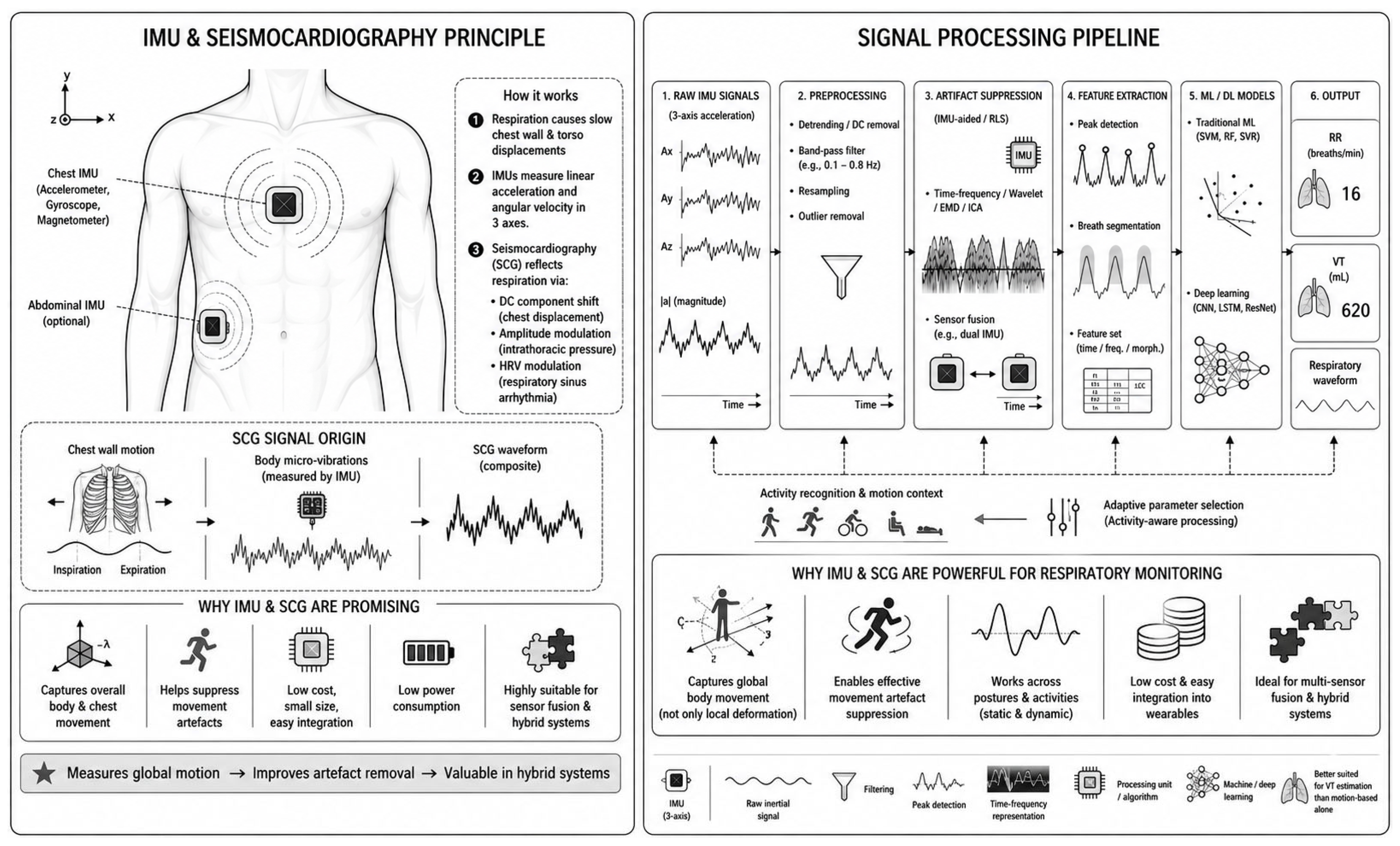
Inertial measurement units (IMUs) (Figure 4) (Table 3), which include accelerometers, gyroscopes, and magnetometers, are widely used in wearable devices due to their easy integration [129]. Respiratory activity in inertial signals is primarily manifested as slow, quasi-periodic displacements of the chest wall, which can be separated from faster motion components using bandpass filters, wavelet transforms, empirical mode decomposition, or independent component analysis [130,131]. The scientific discipline that uses these processes is called seismocardiography (SCG) [132]. Respiration affects the measured waveform through several mechanisms: displacements of the DC component caused by chest movement, amplitude modulation related to changes in intrathoracic pressure, and indirectly by modulation via respiratory sinus arrhythmia, which manifests itself in heart rate variability (HRV) [133]. Separating respiratory components starts to become difficult under dynamic conditions, where gross body motion often exceeds the amplitude of respiratory-induced micromotions. To address this problem, recent studies have proposed adaptive and activity-aware processing techniques, including recursive least-squares filtering [40], time–frequency distribution analysis [134], and quantitative modeling of motion artifacts [135]. Sensor fusion strategies, such as using two cooperating accelerometers placed at different locations, can also improve robustness [136].

2.3.1. Hardware-Oriented Research

An excellent starting point for analyzing the current state of hardware-oriented research is the early work of Tadi et al. [137]. The study demonstrated the feasibility of extracting both cardiac and respiratory activity from SCG using an MMA8451Q (Freescale Semiconductor, Austin, TX, USA) accelerometer. A major strength is its rigorous validation strategy, which correlated SCG-derived parameters not only with ECG and chest belts, but also with computed tomography (CT) imaging to link signal morphology to anatomical displacement of the heart. The results confirmed that accelerometer-based SCG can provide highly accurate information on HR and RR variability. Further progress in SCG hardware was presented by Andreozzi et al. [138], who introduced a dome-shaped force-sensing resistor (FSR03CE, Ohmite Mfg Co., Warrenville, IL, USA) for SCG acquisition. Validated against ECG-derived respiration and resistive bands, the system proved to be suitable for real-time biofeedback applications. Despite its hardware simplicity and user-oriented design, the device demonstrated reliable respiratory tracking.
Several studies have also explored the broader use of inertial sensing for unobtrusive respiratory monitoring. Valdés Tirado et al. [139] presented a custom-designed wearable IMU optimized for cardiorespiratory monitoring, detailing hardware characterization and parameter tuning required for high-precision sensing in sports and rehabilitation contexts. Similarly, Ikarashi et al. [140] addressed the limitations of conventional respiration measurement techniques, such as thermistor and impedance methods, by investigating clothing-attached, non-contact sensing with a 6-axis IMU. Frequency-domain analysis confirmed accurate RR estimation even without direct skin contact, supporting the feasibility of low-burden monitoring in everyday environments.
A more integrated wearable concept was proposed by Rahman et al. [141] through the “CardioResp Device”, which integrated inkjet-printed ECG electrodes with a 6-axis IMU. Robustness across static and dynamic postures was achieved using a quaternion-based update algorithm together with multi-stage filtering. Validation against a Vernier Go Direct chest belt demonstrated overall accuracies of 99.3% in static and 98.6% in dynamic conditions.
Significant contributions to IMU-based dynamic monitoring were made by the research group led by Angelucci. Their Wireless Body Sensor Network (WBSN) [29] utilized three chest-mounted IMUs for RR detection and a wrist-worn unit with a PPG sensor for HR monitoring. In the wrist unit, RR is derived directly using the embedded algorithm of the MAX32664C sensor. Data from all units are transmitted via ANT protocol to a smartphone for storage and analysis. The system achieved an RMSE of 3.77 rpm during cycling. In their most recent study [30], they extended the architecture to simultaneous RR estimation and human activity recognition using 9-axis quaternion computation on an nRF52832 microcontroller, implementing the Madgwick gradient descent algorithm [142]. The sensor network consisted of three IMUs: two placed on the chest wall (thorax and abdomen), and a third positioned on the lower back. Their subsequent work [143] introduced a dual-IMU chest–back differential configuration for RR and VT estimation.

2.3.2. Software-Oriented Research

Beyond hardware innovations, a substantial portion of recent studies has focused on improving algorithmic evaluation. Early algorithmic approaches primarily relied on frequency-domain and morphology-based analysis. For example, Pandia et al. [144] performed a detailed spectral investigation of SCG signals acquired using a MEMS LIS3L02AL accelerometer (STMicroelectronics, Geneva, Switzerland). By systematically analyzing the 0–100 Hz band and dividing it into 5 and 10 Hz sub-bands, they identified significant respiratory-related spectral differences in the 10–40 Hz range, thereby establishing a basis for frequency-selective feature extraction. Along similar lines, Dhar et al. [145] investigated SCG morphological changes induced by respiration and exercise, further supporting the physiological sensitivity of SCG to respiratory modulation.
Building on such signal-level observations, several studies introduced increasingly structured feature extraction and ML frameworks. Sadat-Mohammadi et al. [146] combined low-cost accelerometry with 4 different ML approaches to identify physical demand from the respiratory pattern. Likewise, Sandler et al. [147] employed supervised SVM classification for inspiratory/expiratory phase detection. Their methodology was based on ECG-guided segmentation of SCG events and feature extraction from median waveform amplitudes within contiguous 4 ms windows. In a more computationally efficient direction, Ku et al. [148] developed an RR estimation algorithm combining Gaussian averaging filtering with a complex Morlet wavelet scalogram. Bhongade et al. [149] proposed “ResPara-Net,” a system combining a single IMU with a deep convolutional neural network during daily activities. The model achieved low RMSE values of 0.14, 0.12, and 0.13 for normal, fast, and slow breathing, respectively, while normalized MAE remained below 4% across all subjects. Correlation coefficients ranged from 64.47% to 71.53%.
As the field progressed, attention increasingly shifted toward more adaptive and data-driven architectures capable of handling dynamic and noisy real-world conditions. Steinmetzer and Michel [150] proposed a 1D convolutional recurrent neural network (1D-CRNN) trained on IMU data acquired from a smart e-textile. By combining convolutional layers for local feature extraction with recurrent layers for temporal context modeling, the architecture enabled robust segmentation of breathing-related activity from noise. Similarly, Hung et al. [151] introduced an alternative waistband configuration using dual IMUs together with a ResNet-based deep learning (DL) model. Their system effectively separated respiration from stride-induced motion artifacts during running. In a related effort targeting volumetric estimation, Ba et al. [152] employed the Xsens DOT sensor platform and developed a DL framework for VT estimation, integrating a nonlinear high-gain observer with a CNN-LSTM network. Their approach demonstrated substantial robustness even under repeated sensor removal and re-wearing.
Another important research direction has focused on improving robustness through signal decomposition, posture-aware modeling, and adaptive signal quality handling. Azad et al. [153] examined postural and longitudinal SCG variability over a five-month period, showing that while SCG patterns remain relatively stable over time, they are strongly dependent on posture. To mitigate this influence, the authors applied unsupervised ML to group signals into two clusters with reduced waveform heterogeneity. Shipper et al. [154] addressed the problem from a complementary perspective by combining recursive and constrained principal component analysis (PCA) with an SQI for RR estimation, achieving LoAs below 1.45 rpm with at least 80% temporal coverage across variable postures. Similarly, Cheng et al. [155] optimized a dual-IMU chest/back configuration using a combination of PCA, discrete Fourier transform (DFT), empirical mode decomposition, Savitzky–Golay filtering, and Butterworth bandpass filtering. Validation using Xsens DOT sensors [156] against a TI ADS1298R reference demonstrated an RMSE < 0.8 rpm and a correlation coefficient > 0.7 across dynamic scenarios, including standing, sitting, walking, and squatting.

2.3.3. Sensor Location Optimization

Sensor placement critically influences SCG morphology, and therefore, a lot of research has been devoted to optimizing sensor position and orientation. Romano et al. [157] investigated optimal locations for skin-interfaced IMU sensors by collecting accelerometric data from subjects during rest and walking. Their results identified the mitral valve level as the most promising placement, with the dorsoventral axis providing the most informative signal for respiration monitoring. In a related study, the same authors also provided a comprehensive comparison of chest-worn accelerometers and gyroscopes for simultaneous HR and RR monitoring [158]. By evaluating both sensing modalities across multiple positions and analysis window lengths, they showed that accelerometers consistently outperformed gyroscopes in estimating both HR and RR, while improvements became marginal beyond 25 s analysis windows.
Further evidence for the importance of sensor localization was provided by Demirsoy et al. [159], who quantified SCG variability across 16 torso locations. Their findings highlighted the need to minimize sensor drift and to account for axis-specific variability when developing generalized monitoring models. Similarly, Utama et al. [160] investigated optimal gyro-accelerometer placement and reported their best performance when the sensors were positioned on the stomach and chest, with the highest recorded error remaining as low as 2.06%. A comparable multi-location validation was performed by Centracchio et al. [161], who simultaneously evaluated 16 accelerometer positions in nine subjects using a respiratory belt as the reference.
Table 3. IMU/SCG-based respiratory monitoring.
Sensor TypeApplicationSensing ElementKey ParametersRef.
Chest
IMU 1
RR 2, HR 33D MEMS 4
accelerometer (MMA8451Q)
Sampling rate up to 800 Hz, validation vs. ECG 5, chest belt, and CT 6 imaging, Pearson correlation coefficient 0.995, 0.998, and 0.994, standard deviation 1.7, 1.8, 8.9 rpm 7 for normal (11.1 rpm), slow (6.7 rpm), and fast breathing (23.3 rpm)[137]
Body
attachment
SCG 8
RRDome-shaped force-sensing
resistor (FSR03CE)
Validated vs. EDR 9 and chest belt, 7 subjects, NI-USB6009 DAQ board, 13-bit, sampling rate 5 kHz, RR accuracy 0.98 (slope 0.99, intercept 0.026 s), LoAs 10 ± 0.61 s, respiratory acts detection sensitivity 100%, PPV 11 98.9%[138]
Custom wearable IMURR, HR6-axis IMU (LSM6DSL)Sampling rate up to 4 kHz, range ± 4 g, ±250 dps 12,
processing cycle 220 µs, power consumption 8.5 mA,
error characterized via Allan deviation and PSD 13
[139]
Clothing
attached
RR6-axis IMU
(MPU-6050)
Validated vs. BioZ 14, 5 subjects, sampling rate 100 Hz, non-contact measurement, frequency-domain analysis[140]
Integrated patch
(CardioResp)
RR, ECG6-axis IMU + Inkjet-printed ECG electrodesValidated vs. Vernier Go Direct chest belt, 10 subjects, BLE 15, quaternion-based update algorithm, multi-stage filtering, accuracy 99.3% (static), 99.2% (walking), 98% (running), 98.6% (cycling), MAE 16 0.13 rpm (static), 0.17 rpm (walking), 0.36 rpm (running), 0.23 (cycling)[141]
Chest and wrist IMUsRR, HR3× IMU (ICM-20948) + PPG 17 (MAXM86161)Wireless Body Sensor Network, ANT 18 protocol transmission, IMU (10 Hz), HR (1 Hz), embedded HR algorithm 30 subjects, RR RMSE 19 3.77 rpm (cycling)[29]
Thorax,
abdomen, lower back sensors
RR, HAR 203× 9-axis IMU Wearable sensor network, nRF52832 µprocessor,
sampling rate 40 Hz, 20 subjects, Madgwick gradient
descent algorithm, ANT protocol, AI 21 method: accuracy of HAR 97%
[30]
Dual-IMU wearable bandRR, VT 222× IMU
(MPU-6050)
Dual-IMU chest–back differential configuration, SAMD21G18A microcontroller, IC 23, BLE, 15 mAh battery, RR correlation r = 0.92, mean difference −0.27 rpm, LoAs +1.16/−1.75 rpm, RR MAE 1.15%, VT MAE < 5%.[143]
SCG patchRRMEMS
accelerometer (LIS3L02AL)
18 subjects, frequency-domain analysis of inspiration,
expiration, and apnea, significant spectral differences identified in the 10–40 Hz range
[144]
Chest beltRespiratory waveformAccelerometer +
RIP 24
Sample rate 1 kHz, resolution 16-bit, 15 subjects, during physically demanding tasks, different ML 25 algorithm for physical demand classification: mean accuracy 90.5% (SVM 26), 91.3% (KNN 27), 93.4% (RF 28), 90.2% (ANN 29)[146]
Chest beltRR, phase detectionSCGRespiratory phase detection, 15 subjects, validated vs spirometry, SVM model, accuracy 90.2 ± 6.5%[147]
DatasetsRRSCG and PPGCEBS 30 (PhysioNet) datasets, paced and spontaneous respiration, 20 subjects, STMicroelectronics LIS344ALH IMU, complex Morlet wavelet scalogram, Gaussian averaging filter, validated vs. magnetic field-based sensor during 15 activities, 16 subjects, LoAs 95%[148]
Chest wornRR,
Respiratory waveform
IMUResPara-Net DCNN 31 algorithm, RMSE: 0.14 rpm
(normal), 0.12 rpm (fast), 0.13 (slow breathing),
correlation coefficient 64.47–71.53%, MAE: <4%
[149]
Smart
e-textile
RR2× IMU
(Adafruit BNO085)
Abdomen and spine IMU, sampling rate 330 Hz, 1D-CRNN 32 architecture, 59 subjects, 2000-sample window, mean accuracy 0.88, F1-score 0.92, best case accuracy 99.5%, near-real-time processing[150]
Dual IMU waistbandRR2× IMUResNet-based DL 33 model, 20 subjects, sampling rate 10 Hz, 32 s windows, separation of respiration from stride-induced motion artifacts, outperformed PCA 34 and relative angle baselines during running, MAPE 9.1% (sit), 8.9% (stand), 20% (walk), 9.9% (run)[151]
Chest and abdominal IMUsVT4× IMU
(Xsens DOT)
High-gain observer combined with CNN 35-LSTM 36,
6 subjects, averaged RMSE 40.38 mL, robust to sensor drift and repeated re-wearing
[152]
Chest wornRRAccelerometer
(ADXL355)
Sampling rate 250 Hz, recursive and constrained PCA, signal quality index, 20 subjects, variable postures,
LoA < 1.45 rpm, ≥80% temporal coverage,
[154]
Chest/back IMUsRR2× IMU
(Xsens DOT)
on chest/back
PCA, DFT 37, empirical mode decomposition, Savitzky–Golay + Butterworth filtering, validated vs. TI ADS1298R (dynamic scenarios), RMSE < 0.8 rpm,
correlation coefficient > 0.7
[155]
Body
attached IMU
RR, HRIMU (Xsens DOT)Sampling rate 120 Hz, 15 subjects (rest and walking),
optimal location—mitral valve level, most informative-dorsoventral axis, HR MAE < 1.5 bpm, RR MAE < 4 rpm, accelerometer outperformed gyroscope in accuracy,
diminishing returns beyond 25 s analysis windows
[157,158]
16× SCG
on torso
Respiratory phaseAccelerometers (ADXL355)Sampling rate 500 Hz, evaluated across 16 torso locations, accuracy 92% (location), 90% (respiratory phase)[159]
Body
attached
RRGyroscope +
Accelerometer
GY-521 MPU6050 IMU, paced breathing,
highest error 2.06% at 25 rpm
[160]
Multi-accelerometer setupRespiratoryAccelerometers
(ADXL355)
Sampling rate 500 Hz, 16 simultaneous body positions, 9 subjects, validated vs. chest belt, average sensitivity and PPV 95.8%/95.5% (chest inclination), 85.9%/84.4% (AM 38), 94.3%/95.7% (morphological similarity index)[161]
1 Inertial measurement unit, 2 respiration rate, 3 heart rate, 4 microelectromechanical systems, 5 electrocardiography, 6 computed tomography, 7 respirations per minute, 8 seismocardiography, 9 ECG-derived respiration, 10 limits of agreement, 11 positive predictive value, 12 degrees per second, 13 power spectral density, 14 bioimpedance, 15 Bluetooth low energy, 16 mean absolute error, 17 photoplethysmography, 18 advanced and adaptive network technology, 19 root mean square error, 20 human activity recognition, 21 artificial intelligence, 22 tidal volume, 23 inter-integrated circuit, 24 respiratory induction plethysmography, 25 machine learning, 26 support vector machine, 27 k-nearest neighbors, 28 random forest, 29 artificial neural network, 30 combined measurement of ECG, breathing and seismocardiograms dataset, 31 dense neural network, 32 one-dimensional convolutional recurrent neural network, 33 deep learning, 34 principal component analysis, 35 convolutional neural network, 36 long short-term memory, 37 discrete Fourier transform, 38 amplitude modulation.

2.4. Other Methods

Beyond chest belts, bioimpedance, and IMU sensing, several alternative sensing principles have also been explored, including pressure-based airflow sensing, electromyography (EMG), acoustic analysis, and radiofrequency resonant methods (Table 4).
Massaroni et al. [162] developed a device measuring breath-induced pressure drops at the nostril level during physical exercise. The system demonstrated high robustness under dynamic conditions. This approach offers direct quantification of an airflow while maintaining compatibility with dynamic scenarios, although nasal interfaces may limit long-term comfort compared to completely unobtrusive modalities.
Surface EMG has also become one of the surrogate markers of respiratory effort. Gronska et al. [163] evaluated diaphragmatic EMG features across time, frequency, and statistical domains. Correlation with gold-standard esophageal pressure measurements showed that time-domain features, particularly filtered envelope, RMS, and waveform length, achieved moderately strong correlations with respiratory effort. Notably, waveform length and slope sign change remained robust even in low-quality signals, highlighting the resilience of selected EMG-derived methods. Extending this concept, George et al. [42] proposed a multimodal configuration combining diaphragmatic and intercostal EMG with a piezoelectric microphone. This dual-sensor configuration enables a more comprehensive assessment of respiratory mechanics and sounds, offering a robust solution. In EMG-based respiratory monitoring, advanced DL frameworks are increasingly utilized for robust signal reconstruction and modality mapping. Huang et al. [164] developed a cascaded CNN–LSTM architecture for diaphragm EMG that effectively suppresses ECG interference and quantifies nonlinear motion. This model achieved a Pearson correlation of 0.95 ± 0.03 without requiring additional post-processing. Addressing the complex mapping between different physiological sequences, Chen et al. [165] introduced a Multi-Scale Patch Transformer. By incorporating an Attention-based modality transition module for cross-sequence EMG-to-respiration forecasting, this framework outperformed conventional state-of-the-art models.
As discussed above, acoustic sensing may be another way forward. Liu et al. [166] presented the “EarMeter”, which is an in-ear system embedded in conventional earbuds for VT estimation from internally propagated breathing sounds. To address weak acoustic coupling and motion interference, the authors implemented a deep learning framework using transfer learning from high-quality nasal sound and physiological cardiorespiratory coupling. In LOSO validation, the system demonstrates the feasibility of continuous monitoring at the consumer level. Clinical-grade acoustic validation was provided by Abdulsadig et al. [167] in their assessment of the AcuPebble RE100 (Acurable, London, UK). Compared with capnography and polygraphy, the wearable sensor meets performance levels compatible with medical device standards. Together, these studies indicate that on-body acoustic monitoring supported by robust algorithms can achieve clinically meaningful RR accuracy.
Non-contact and textile-integrated electromagnetic approaches represent an alternative. Abounasr et al. [41] proposed an electromagnetic coupling system based on a loop antenna and a flexible split-ring resonator tag. The 50 × 50 mm conformal sensor, fabricated by inkjet and extrusion printing on polyimide and polyethylene terephthalate (PET) substrates, detects chest wall displacement via resonant frequency shifts, achieving a sensitivity of 1.7 MHz/mm and strong correlation with the BIOPAC respiratory belt. Similarly, Gharbi et al. [168] developed a textile-integrated embroidered loop antenna embedded in an abdominal belt, coupled with a compact Bluetooth transmitter. Respiratory motion modulates antenna resonance through mechanical stretching, enabling wireless respiration tracking within garment-integrated architectures.
Table 4. Other methods for respiratory monitoring.
Sensor TypeApplicationSensing ElementKey ParametersRef.
Nasal deviceRR 1, VT 2Pressure sensor
(SDP610,
Sensirion)
Breath-induced pressure drops at nostril level, evaluated during physical exercise, RR percentage error 4.03%,
30 s window averaging error 2.38%,
HIIT 3 test LoA 4 ±1.6 rpm
[162]
Surface EMG 5Respiratory effort, OSA 6EMG electrodesEvaluated diaphragmatic EMG features, validated vs.
esophageal pressure, 10 subjects, time-domain (filtered envelope, RMS 7, waveform length), moderately strong
correlation R > 0.6, robust in low-quality signals R > 0.5
[163]
Surface EMG + acousticRR, VT, soundsEMG electrodes + piezoelectric
microphone
Combined diaphragmatic and intercostal EMG with
microphone, 2 subjects, mean AUC 8 0.4–1.23 × 10⁸ for VT (500–1000 mL)
[42]
Surface EMGRespiratory waveformEMG electrodes on diaphragmCNN 9–LSTM 10 + multi-scale CNN, 49 subjects, 0.95 ± 0.03 correlation coefficient, ECG 11 artifact suppression without post-processing, real-time monitoring[164]
In-ear
acoustic
system
VTMicrophoneDL 12 framework with transfer learning, internally
propagated breathing sounds, LOSO 13 validation, validated vs. VO2Master, average MAPE 14 18.19%
[166]
Acoustic
sensor
RRAcoustic sensor AcuPebble RE100Validated vs. capnography and polygraphy,
RMS deviation < 3 rpm, MAE 15 1.83 rpm 16
[167]
Flexible electromagnetic tagRRLoop antenna + split-ring
resonator
50 × 50 mm conformal sensor, inkjet and extrusion printing on polyimide and PET 17 substrate, sensitivity 1.7 MHz/mm, validated vs. BIOPAC belt, 1 subject, depth correlation (0.991–0.996), RR correlation 0.993[41]
Textile-
integrated antenna
RREmbroidered loop antenna17 × 11 mm compact 2.4 GHz BL 18 transmitter,
antenna resonance modulated by mechanical stretching, sensitivity 96.7%, validated vs Biopac MP36, LoAs −7.3–10.6 rpm, RMSE 4.7 rpm
[168]
1 Respiration rate, 2 tidal volume, 3 high intensity intermittent training, 4 Limit of agreement, 5 electromyography, 6 obstructive sleep apnea, 7 root mean square, 8 area under the curve, 9 convolutional neural network, 10 long short-term memory, 11 electrocardiography, 12 deep learning, 13 Leave-One-Subject-Out, 14 mean absolute percentage error, 15 mean absolute error, 16 respiration per minute, 17 polyethylene terephthalate, 18 Bluetooth.

2.5. Conclusions

Overall, chest motion sensors, bioimpedance systems, and IMU/SCG-based approaches represent the three dominant wearable strategies for respiratory monitoring in dynamic conditions, each offering different compromises between physiological relevance, robustness, wearability, and computational complexity. Chest belts and patch-based systems remain among the most physiologically intuitive and accurate approaches because they provide direct access to the respiratory waveform. In their simplest single-band form, they are primarily suited for RR estimation, whereas reliable VT assessment usually requires dual-band or multi-point measurements capturing both thoracic and abdominal motion [92]. Although respiration belts are highly effective for waveform tracking [50], disagreement with airflow-based reference methods arises from the fact that they measure only regional surface displacement rather than the full respiratory volume. Consequently, VT estimation strongly depends on sensor placement, sensing area, number of sensing elements, and subject-specific calibration [54,95]. The growing importance of textile-integrated respiratory sensing is reflected by a systematic review [169], which highlighted the rapid expansion of smart textile research, particularly in piezoresistive, capacitive, and fiber-based sensing technologies for continuous breathing monitoring. Across the reviewed studies, the sensing principle itself often appeared less critical than factors such as mechanical coupling, electrode or sensor positioning, stability of body contact, and signal-processing methodology [57]. This is particularly evident under unconstrained conditions, where motion artifacts, posture transitions, irregular breathing, speech, and upper-body movement substantially increase variability compared with controlled or paced-breathing scenarios [51,90,95,98].
Compared with purely mechanical sensing, bioimpedance provides a more direct physiological representation of respiratory-related lung volume changes and therefore appears particularly attractive for VT monitoring [107]. Measurement quality, however, strongly depends on electrode configuration and on how effectively the injected electrical field traverses lung-representative thoracic regions [106,108]. Larger electrode spacing and multichannel configurations generally improve physiological sensitivity [110], while several studies suggest that more distant placements may improve long-term repeatability [115,116,119]. In practical ambulatory conditions, bioimpedance remains sensitive to motion artifacts, variability in electrode–skin contact, skin impedance changes, and EMG interference, requiring advanced filtering, adaptive modeling, and increasingly also ML-based signal-quality evaluation and artifact suppression [112,113,114]. An important practical advantage is that bioimpedance can often share electrode configurations with ECG acquisition, making it particularly suitable for multimodal wearable systems aimed at simultaneous cardiorespiratory monitoring [120,121,122,123,124,127,128].
IMU and SCG approaches generally achieve lower standalone accuracy for RR and VT estimation than chest belts or bioimpedance systems, mainly because respiratory micromotions are easily masked by locomotor activity. Their major advantage, however, lies in the direct capture of posture and body movement, which makes them highly valuable as complementary modalities for motion compensation and contextual awareness in hybrid wearable systems. Recent studies show that advanced signal-processing pipelines incorporating quaternion modeling, adaptive filtering, ML, DL [149], and AI-based approaches [170,171] can substantially improve robustness, particularly when longer analysis windows are available. Owing to their low cost, small dimensions, low power consumption, and straightforward electronic integration, IMU sensors remain highly attractive for telemedicine and multimodal wearable monitoring, as discussed further in Section 4. Nevertheless, increasingly sophisticated algorithms also raise computational demands, which may complicate real-time implementation in low-power wearable devices.

3. Indirect (Derived) Methods

Indirect respiratory monitoring methods leverage physiological modulations embedded in routinely acquired cardiovascular signals, enabling respiration estimation without dedicated sensors. This paradigm is particularly attractive for wearable systems, where minimizing sensor complexity, power consumption, and user discomfort is critical. In this context, Charlton et al. [172] and Ponsiglione et al. [173] compared ECG-derived and PPG-derived respiratory estimation approaches and reached a consistent conclusion: ECG-derived respiration is generally more accurate and robust than PPG-based estimation. In the best dynamic scenario, the first study indicated that PPG-based estimation was roughly 30% less accurate than ECG-derived respiration, whereas the second reported an even more pronounced degradation, with PPG performance being approximately five times worse. Together, these studies highlight that although both modalities can support indirect respiratory monitoring, ECG-derived methods currently provide higher reliability, while PPG remains attractive mainly because of its superior practicality and widespread integration in wearable devices.

3.1. ECG-Derived Respiration

Respiration induces systematic modulations in the ECG. These modulations result from a combination of physiological and mechanical mechanisms, including changes in thoracic impedance associated with respiration, cyclic displacement of ECG electrodes relative to the heart, and autonomic modulation of heart rate. Together, these effects form the basis of ECG-derived (EDR) approaches to respiration (Figure 5) (Table 5) [174,175,176]. Most EDR methods, especially temporal approaches, extract respiration-related features contained in the ECG signal, such as baseline wandering, QRS complex amplitude modulation, and HRV. Frequency-domain methods isolate low-frequency components corresponding to typical respiratory frequencies and estimate the RR from dominant spectral peaks [177,178]. However, EDR performance degrades during physical activity due to motion artifacts, electrode drift, and exercise-induced changes in heart rate dynamics [16]. Several algorithms have been proposed to address these limitations, combining multiple EDR features or using advanced signal processing techniques [179,180,181,182,183].

3.1.1. RR Estimation

The most common ECG-derived approach for RR estimation is based on respiratory sinus arrhythmia (RSA), which exploits respiration-related modulations in cardiac rhythm. Several studies have shown that this principle can provide reasonably accurate RR estimates even during physical activity, although performance generally declines as motion intensity increases. For example, treadmill-based validation on runners [184] showed that ECG-derived RR can be estimated with acceptable error when appropriate preprocessing and frequency-tracking methods are applied. They specifically applied bandpass filtering with short-time Fourier transform (STFT), and relative RR interval transformation with harmonic frequency tracking. Similarly, Gronwald et al. [185] evaluated EDR based on HRV and R-wave amplitude variability during treadmill running and reported good agreement with the reference under resting conditions, whereas accuracy deteriorated during exercise. These findings confirm that ECG-derived RR is feasible in motion, but remains sensitive to changes in exercise intensity, signal quality, and breathing irregularity.
To improve robustness, a wide range of EDR signal extraction strategies has been proposed. Lenis et al. [186] proposed that optimal linear combinations of EDR methods outperform individual approaches. Comparative studies have analyzed a wide range of EDR characteristics derived from QRS complex morphology [187,188], T-wave characteristics, HRV, and low-frequency spectral components, consistently identifying QRS complex-based methods, such as principal component analysis (PCA), R-peak amplitude, QRS complex integral, and RS complex amplitude, as among the most reliable. Langley et al. [189] introduced PCA as a superior approach for deriving RR from ECG amplitude variations, showing improved robustness in tracking beat-to-beat morphological changes compared with traditional RSA-based methods. This line of work was extended by Varon et al. [190], who performed a comprehensive comparative analysis of ten EDR methods using single-lead ambulatory ECG recordings. Their results showed that methods based on QRS complex slope, particularly slope range, provided the highest stability and accuracy, outperforming amplitude-based approaches in noisy and motion-corrupted conditions. In a similar effort to improve real-time applicability, Krishnapriya et al. [191] proposed a computationally efficient time-domain algorithm based on mean, prominence, and distance (MPD) parameters for respiratory peak detection in EDR signals. When evaluated on benchmark datasets and real-time recordings from subjects performing dynamic activities, the method outperformed several conventional time–domain approaches. An important practical advantage of such time–domain methods is that they process signals directly, making them attractive for low-power and resource-constrained wearable implementations [16]. More recently, EDR research has also moved toward AI-driven architectures. Qi Zhao et al. [192] proposed an improved transformer-based model for RR prediction from ECG and PPG signals, using initial feature extraction blocks followed by deep temporal modeling. Trained and evaluated using subject-level ten-fold cross-validation on the combined BIDMC and CapnoBase datasets, the model outperformed five commonly used deep-learning baselines by reducing MAE and improving correlation with the reference. Addressing the susceptibility of EDR respiration to ambulatory noise, Saha et al. [193] proposed a lightweight frequency demodulation framework integrating signal quality assessment. The method extracts respiratory-induced frequency variations and estimates RR via Fourier analysis. Validated on the CapnoBase and BIDMC datasets (achieving MAEs of 5.01 and 5.37 rpm, respectively), the integrated signal quality-aware (SQA) crucially reduced clinical false alarm rates by 84.85%. Due to its low computational complexity and simplified pipeline, the framework is highly optimized for real-time wearable monitoring.
This translational direction is also reflected in recent hardware-integrated EDR systems. The Frontier X2 chest strap (Fourth Frontier Technologies Ltd., London, UK) [194] combines continuous ECG waveform acquisition with embedded EDR-based RR monitoring, enabling real-time respiratory tracking together with cardiac workload assessment during both exercise and sleep. In parallel, Fan et al. [195] designed and fabricated a dedicated ultra-low-power processor for EDR estimation in wearable applications. Using 55 nm technology, they implemented QRS detection together with adaptive threshold-based EDR extraction, achieving estimation errors of 0.73 on the CEBS database and 1.2 on the MIT-BIH polysomnography database.

3.1.2. VT Estimation

All research so far has predominantly focused on RR estimation. However, some researchers have also explored the more challenging task of VT estimation. Lazaro et al. [196] validated a wrist-worn device capable of deriving not only RR but also VT from ECG-related features, including QRS slope range, R-wave angle, and R–S amplitude. In static conditions, the proposed approach demonstrated a strong linear relationship with reference spirometry. Yang et al. [197] investigated the feasibility of VT estimation using clinical ICU data. EDR waveforms were compared with impedance-based respiration references using both linear regression and DL approaches. While short-term correlations between VT and respiration waveforms were relatively strong (r = 0.78–0.96), performance substantially deteriorated over longer recordings due to noisy ECG conditions. Population-level VT prediction showed limited performance (R2 = 0.17), whereas constrained subject-specific analyses achieved considerably higher accuracy (R2 = 0.84–0.94). This line of research was later extended to dynamic conditions by Milagro et al. [198], who investigated VT estimation during treadmill exercise. Their approach exploited several ECG-derived features previously linked to VT, including EDR, HRV, and RR, which were combined in a subject-specific linear model. The model was calibrated using maximal treadmill-test data and subsequently applied to submaximal exercise.

3.1.3. Location Optimization

Klum et al. [199] described optimal sensor placement using a chest ECG sensor. By implementing three EDR algorithms, derived from HRV, QRS amplitude, and linear PCA, they again found that linear PCA significantly outperformed the other methods, particularly in maintaining signal integrity across various postures. Their study also confirmed that specific electrode positions yield significantly higher signal correlations, directly supporting the findings of our previous research [200], where we optimized electrode placement for miniaturized sensors and demonstrated that, despite low inter-electrode separation, reliable EDR signals can be obtained by leveraging respiration-induced changes in thoracic impedance distribution that modulate QRS complex amplitude.
Table 5. ECG-derived respiratory monitoring.
Sensor TypeApplicationSensing Element/AlgorithmKey ParametersRef.
ECG 1 datasetRR 2VORTAL datasetValidated vs. oral–nasal pressure, 39 subjects, supine and exercise, sampling rate 500 Hz, AM 3, FM 4 and BWM 5 method for signal extraction, SQI 6 + fusion technique, TD 7 based RR MAE 8 6.4 rpm 9 (zero crossing method), 4.7 rpm (Count-Orig approach), bias 0 rpm[172]
ECG
datasets
HR 10, RREDR 11ECG-based (R-peak, QRS area, up-slope, down-slope), 30 s-time window: iAMwell dataset (running) MAE 0.99–1.04 rpm, Capnobase dataset MAE 3.07–3.74 rpm[173]
ECG
dataset
ECG, RRFrequency EDRCapnoBase dataset, extract QRS + compute PBA 12 + filter (0.07–0.5 Hz), MAE 0.5 rpm, TD analysis, MAE 6 rpm[174]
ECG
+ dataset
RR, HREDR, PAV 13
Biopac MP45
Fantasia database (n = 20) + real-time ECG (n = 10),
sampling rate 1 kHz, validated vs. chest belt, EDR vs. PAV method, MAE ± 0.57 rpm (EDR), ±0.7 rpm (PAV)
[178]
ECG
dataset
RR, HR,
respiration waveform
EDR, EMD 14MATLAB R2026a algorithms, Fantasia database, validate vs.
respiratory stretch sensor, 40 subjects,
MAE (0.89–1.07 rpm), percentage error 4.78–6.60%
[180]
Chest beltRR, HREDR, RSA 1531 subjects, running on a treadmill with a gradual increase in power until exhaustion, HR from a Polar H10A, validated vs. Cosmed Quark CPET system, 18 methods, best results: Bandpass in combination with STFT 16 (MAPE 17 5.5%) and relative transformation of RR intervals with harmonic frequency tracking (MAPE 7.6%)[184]
Chest beltRREDR, HRV 18 + R-wave amplitude variabilityMovesense Medical sensor, 15 subjects during treadmill running, validated vs. metabolic cart, correlation 0.80, ICC 19 = 0.87, mean difference: −0.5 ± 2.4 rpm (rest), 1.8 ± 4.4 rpm (exercise), LoAs 20: −5.2–4.2 rpm (rest), −6.9–10.4 rpm (exercise), MAE 1.6 ± 1.8 rpm (rest), 3.1 ± 3.6 rpm (exercise)[185]
ECG
datasets
RREDR, RSAFantasia (n = 40), MIT-BIH Polysomnographic dataset (n = 18), sampling rate 250 Hz, optimal linear combination of EDR methods (PCA 21, R peak, QRS integral, RS
amplitude T peak, T integral), time window 20 s, fixed coefficient vector MCCC 22 0.8 (Fantasia), 0.9 (MIT-BIH)
[186]
ECGRRECG amplitudes, PCAECG (500 Hz), paced and normal breathing, validated vs. magnetic displacement
sensor, 20 subjects, PCA-based algorithm,
coherence < 0.05, correlation < 0.0001
[189]
ECG
datasets
RR,
respiratory waveform
EDR3× datasets: Fantasia (n = 40), drivers (n = 16) and PSG 23 (n = 100), validated vs. chest belts and airflow, RS complex + QRS slope best for respiratory waveform reconstruction[190]
ECG +
dataset
RREDR
(AFE 24 AD8232),
TDA 25
MPD 26 algorithm optimized on Physio Net dataset, MPD vs. count origin method, MAE 3.66 rpm (MPD), 5.09 rpm (Count-Orig), MAPE 23.69% (MPD), 32.76% (Count-Orig), MPD in dynamic activities MAE 1.53 rpm, MAPE 7.25%[191]
ECG
dataset
RRECG + PPG 27, transformer-based modelTransformer-based model, ECG + PPG fusion, CapnoBase (n = 42) BIDMC (n = 53) datasets, sampling rate 125 Hz, BIIRF 28 extract RR, LoA 95%, MAE: 1.33 rpm (BIDMC), 0.96 rpm (Capnobase), 1.20 rpm (combined training), LoA: −3.46–3.71 rpm (BIDMC), −2.87–3.11 (Capnobase), −3.25–3.97 (combined), PCC 29 0.85[192]
ECG
datasets
RREDRSignal quality-aware frequency demodulation algorithm, MAE 5.01 rpm (CapnoBase), 5.37 rpm (BIDMC), signal quality assessment accuracy 85.25%[193]
Chest-worn ECG, HR, HRV, RR, VO2 max 30ECG electrodes, IMU 31Continual ECG, strain metrics, training load, recovery metrics, step cadence, activity tracking, Bluetooth, IP67, 14-day battery life[194]
EDR chip, datasetsRR, HREDR55 nm fabricated processor, refractory period, adaptive threshold EDR, QRS detection accuracy 99.18%, tested on 2 datasets, MAE 0.73 rpm (CEBS 32), 1.2 rpm (MIT-BIH)[195]
ArmbandRR, VT 33EDREDR: QRS slope, R-wave angle, R-S amplitude, PCA,
breathing exercise, validated vs. spirometry, VT and EDR amplitude correlation 0.045–0.85, MLR 34 model correlation 0.82–0.92
[196]
ECGVTEDRDL 35 + linear regression, 90 ICU 36 subjects, validated vs. impedance respiratory waveform, correlation 0.78–0.96, population-level performance 0.17,
subject-specific performance 0.84–0.94
[197]
Multi-lead ECGVTEDRTreadmill exercise, 25 subjects, validated vs. spirometry, sampling rate 1000 Hz, subject-specific linear model (EDR, HRV, RR), relative fitting error < 14%, VT relative error 10.23–22.72%[198]
Chest patch systemRR, HRVEDRECG at 27 differential chest positions, 3 EDR algorithms EDR, HRV, EDR amplitude, linear PCA, lowest RR mean error: 0.68 ± 0.33 rpm (F.III electrode position)[199]
1 Electrocardiography, 2 respiration rate, 3 amplitude modulation, 4 frequency modulation, 5 baseline wander modulation, 6 signal quality index, 7 time domain, 8 mean absolute error, 9 respirations per minute, 10 heart rate, 11 ECG-derived respiration, 12 peak-to-baseline amplitude, 13 peak-to-peak amplitude variation, 14 empirical mode decomposition, 15 respiratory sinus arrhythmia, 16 short-time Fourier transform, 17 mean absolute percentage error, 18 heart rate variability, 19 intraclass correlation coefficient, 20 limits of agreement, 21 principal component analysis, 22 multiple correlation coefficient, 23 polysomnography, 24 analog front-end, 25 time –domain analysis, 26 modified peak detection, 27 photoplethysmography, 28 band-limited instantaneous respiratory frequency, 29 Pearson correlation coefficient, 30 maximal oxygen uptake, 31 inertial measurement unit, 32 combined measurement of ECG, breathing and seismocardiograms dataset, 33 tidal volume, 34 multiple linear regression, 35 deep learning, 36 intensive care unit.

3.2. PPG-Derived Respiration

PPG is an optical measurement of changes in blood volume of peripheral tissues and is the most widely used physiological signal in current wearable devices like smartwatches, fitness trackers, and self-adhesive skin patches. Respiratory activity, like ECG, also systematically modulates the PPG signal through several mechanisms. This enables indirect estimation of respiratory parameters without special respiratory hardware (Figure 6) (Table 6). These effects arise, among others, from respiration-induced changes in venous return, intrathoracic pressure, and autonomic regulation, which together affect the pulse amplitude, baseline shift, and temporal characteristics of the PPG waveform [16,182,201].

3.2.1. RR Estimation

Signal processing approaches commonly use envelope extraction, Hilbert transform-based analysis, adaptive filtering, or time-frequency methods to reconstruct the respiratory waveform and estimate RR. Under controlled or low-motion conditions, these techniques provide reliable results [202,203]. In commercial wearables, the WHOOP algorithm [204] is often incorporated as a standard solution. However, under dynamic conditions, PPG signals are highly susceptible to motion artifacts, mainly caused by sensor displacement, tissue deformation, and variations in optical coupling. These artifacts often dominate respiratory-related modulations and substantially reduce estimation accuracy [205]. For example, Motin et al. [206] achieved an MAE of 3.05 rpm using ensemble empirical mode decomposition.
To improve robustness, several studies have focused on combining multiple signal processing strategies or incorporating signal quality assessment. A method-fusion framework integrating several beat-detection- and waveform-morphology-based RR estimation approaches was proposed by Koumpouzi et al. [207], evaluated on the CapnoBase benchmark respiration database, and the fused PPG approach outperformed the individual methods. Similarly, Pimentel et al. [208] proposed a robust fusion-based technique incorporating probabilistic estimation for clinical RR monitoring. Cernat et al. [209] estimated RR from both infrared and green PPG channels and developed a real-time fusion model combining five PPG features. Along similar lines, Suleman et al. [210] demonstrated the feasibility of estimating respiratory events from PPG across multiple body positions, supporting low-complexity and location-independent RR monitoring. Dai et al. [211] further advanced this concept with “RespWatch,” a smartwatch-based system that combines a signal-processing RR estimator optimized for low-noise conditions with a CNN-based estimator designed for severe motion. An adaptive hybrid estimator dynamically switched between the two according to an Estimation SQI.
Several studies also demonstrated that these approaches can be translated into practical wearable devices under demanding conditions. Muller et al. [212] used the CardioWatch 287-2 (Corsano Health, Den Haag, The Netherlands) during high-intensity cycling. Using an ECG patch (Vivalink) as the reference, the PPG-based algorithm demonstrated acceptable accuracy even during intense exercise. Similarly, Eisenkraft et al. [213] clinically validated a wearable RR monitoring device based on the BB-613P sensor platform (Biobeat Technologies Ltd., Petah Tikva, Israel) [214]. Across three independent studies, the system showed high correlations with the reference and excellent Bland–Altman agreement, with biases remaining below 0.1 rpm. Zhao et al. [215] proposed “BreathAnalyzer,” a system implemented on commercial smartwatches that addresses the common limitation of weakening RSA-related signatures at high RR. Rather than relying on a single spectral component, the method integrates features from frequency, time, and nonlinear Poincaré domains. To accommodate both motion artifacts and wearable computational constraints, the system employs a lightweight tree-based learning model. Extensive evaluation showed that this multidomain approach significantly outperformed several state-of-the-art techniques, particularly during activities associated with high RR. These findings indicate that, despite the known sensitivity of PPG to motion, appropriately designed wearable systems can still achieve clinically relevant RR estimation performance in real-world scenarios.
More recently, machine learning approaches have gained traction as a means of improving robustness in dynamic and motion-corrupted scenarios [216]. Stankoski et al. [217], using the XGB algorithm, achieved an MAE of 1.38 rpm and a Pearson’s correlation coefficient of 0.86. Chin et al. [218], employing the RR estimation toolbox together with convolutional and LSTM layers, reported an MAE of 2 rpm. Baker et al. [219] combined signal quality quantification with several neural networks and achieved an MAE of 0.638 rpm. Shuzan et al. [220] presented an ML-based RR estimation framework incorporating motion artifact correction and PPG features, where feature selection was used to reduce computational complexity and overfitting. Their best-performing Gaussian process regression model achieved an RMSE of 2.63 rpm, an MAE of 1.97 rpm, and a two-standard deviation of 5.25 rpm. Ganeshmurthy et al. [221] proposed an RR estimation method based on optimization of temporal segmentation windows and preprocessing, achieving strong performance on both the BIDMC and TMCH datasets. Lee et al. [222] further improved estimation by combining autocorrelation-based spectral features with nonparametric bootstrap feature generation and Gaussian process regression, while also providing uncertainty estimates.
Additional conventional algorithmic studies include Karlen et al. [223], who estimated RR using the Incremental-Merge Segmentation algorithm and fast Fourier transform (FFT), Schäfer and Kratky [177], who compared multiple approaches and reported the best-performing method, and Dubey et al. [224], who applied a spectral kurtosis-based method for RR estimation.
The most advanced recent studies have moved toward end-to-end deep learning and signal reconstruction. Ravichandran et al. [225] proposed a ResNet-based model that directly reconstructs the respiratory waveform from PPG signals, achieving high cross-correlation and low reconstruction error across two benchmark datasets. A similar ResNet-based strategy for RR estimation was later adopted by Bian et al. [226], who showed that training with augmented synthetic data improved performance by approximately 34%. Aqajari et al. [227] applied CycleGAN to reconstruct clean respiratory signals from PPG, while Zargari et al. [228] further demonstrated CycleGAN-based correction of motion-corrupted PPG without relying on accelerometer input, achieving substantial improvement in artifact suppression (9.5–times).
Recent advancements in PPG-based respiratory monitoring leverage sophisticated AI to overcome inherent sensor limitations, such as signal degradation, computational demands, and dataset imbalances. To address signal quality degradation under free-living conditions, Pham et al. [229] introduced “CP-PPG,” an adversarial generative model designed to correct waveforms distorted by variable skin–sensor contact pressure. By stabilizing morphology-related amplitude distortions, this framework improved RR estimation accuracy by approximately 6.85%. Tackling motion artifacts, Rajendran et al. [230] proposed a heuristic-aided ensemble learning framework that combines multilayer perceptron (MLP), AdaBoost, and attention-based LSTM (A-LSTM) architectures. Optimized by the Advanced Golden Tortoise Beetle Optimizer (AGTBO), this multimodal approach achieved up to 96% accuracy in simultaneous RR and SpO2 estimation, outperforming conventional MLP (90%), AdaBoost (92%), A-LSTM (92%), and hybrid MLP-ADA-A-LSTM (94%) approaches. Transitioning from manual feature engineering to end-to-end DL, Shuzan et al. [231] developed “PPG2RespNet,” a U-Net-inspired architecture with hierarchical skip connections for direct respiratory waveform reconstruction. Validated across multiple datasets (BIDMC, VORTAL, CapnoBase), it achieved high Pearson correlation coefficients (0.94–0.96) and extremely low MAEs of 0.11–0.69 rpm.
Miao et al. [232] introduced “RespDiff,” an end-to-end multi-scale recurrent neural network (RNN) diffusion model. Notably, this framework bypasses the need for handcrafted feature extraction or the exclusion of low-quality signal segments, significantly enhancing its viability for real-world wearable applications. By integrating multi-scale encoders with a bidirectional RNN and a specific spectral loss term, the model effectively captures temporal respiratory dynamics while maintaining high waveform fidelity. Validation on the BIDMC dataset demonstrated superior respiratory rate estimation with an MAE of 1.18 rpm, outperforming conventional methods with results between 1.66 and 2.15 rpm. For power-constrained wearables, Yang et al. [233] presented a highly energy-efficient alternative using spiking neural networks (SNNs). By directly converting PPG segments into sequential spike events, the model preserved temporal dynamics while substantially reducing computational overhead, maintaining competitive RR estimation accuracy (MAEs of 1.15–1.37 rpm). Finally, addressing the critical issue of imbalanced training data, Lee et al. [222] proposed an imbalanced power spectral generation (IPSG) framework combined with Gaussian Process Regression (GPR). By generating artificial spectral feature curves, the model improved learning for underrepresented abnormal respiratory conditions. Validated on the BIDMC dataset, it achieved MAEs between 0.79 and 1.47 rpm, while distinctively providing uncertainty estimates to quantify prediction reliability for clinical interpretation.

3.2.2. VT Estimation

Unlike RR estimation, PPG-based estimation of VT has been investigated only in a limited number of studies. Early physiological studies demonstrated that respiratory modulations in the PPG waveform are influenced by VT and respiration pattern, indicating the theoretical feasibility of VT tracking from PPG. More recent wearable work, such as wearable PPG systems, has extended this concept toward continuous monitoring of breathing phase and tidal volume, although this area remains much less mature than PPG-derived RR estimation [234]. More recently, Romero et al. [235] proposed OptiBreathe, a wearable PPG-based system for estimating multiple respiratory biomarkers, including RR, breathing phases, and VT. Their pipeline takes into consideration three modulations: respiratory-induced intensity variation, AM, and FM. In validation against spirometry, during static testing, the device achieved a best MAE of 1.96 rpm for RR and a best subject-averaged MAPE of 17% for VT, suggesting that PPG may also support volumetric respiratory monitoring. Unfortunately, we did not find tests in a dynamic environment.
Table 6. PPG-derived respiratory monitoring.
Sensor TypeApplicationSensing Element/AlgorithmKey ParametersRef.
PPG 1 datasetRR 2VORTAL datasetValidated vs. oral–nasal pressure, 39 subjects, supine and exercise, sampling rate 500 Hz, AM 3, FM 4 and BWM 5 method for signal extraction, SQI 6 + fusion technique, TD 7 based Count-Orig approach LoAs 8 −5.1–7.2 rpm 9, bias 1 rpm [172]
PPG
datasets
HR 10, RRCapnoBase,
iAMwell datasets
PPG-based on FM, AM, 30 s-time window: MAE 11 5.10–5.12 rpm (iAMwell dataset—running), 10.7–13.9 rpm (Capnobase dataset) [173]
PPG
dataset
RRVORTAL dataset,
intrinsic modes
39 subjects during rest (supine), Recursive Bayesian Tracking, intrinsic modes, time-frequency spectra, extraction of amplitude variability, VORTAL database, WSST 12 MAE 2.33 rpm, ME 13 1.15 rpm[202]
Laboratory setupRR, HRPPG
Nnormalized LMS 14
In vitro PPG, skin perfusion phantom, motion artifacts correlation, measuring via self-mixing interferometry,
artifact reduction −9.9 dB
[205]
PPG
sensors
RRPPG
EEMD 15
PPG BIOPAC MP150, 10 subjects, 5 activities, validated vs. chest belt, MAE: 3.16 rpm (sitting), 3.02 rpm (standing), 3.01 rpm (walking), 3.07 rpm (stairs climbing), 3.18 (running)[206]
PPG
dataset
RRCapnoBase
dataset
42 subjects, PPG beat segmentation, peak extraction, 60 s-time window, RMSE 16 3.4 rpm[207]
PPG
datasets
RRCapnoBase, MMIC II datasets
segmentation
PPG modulations exhibited, CapnoBase (300 Hz), MMIC II conventional BioZ 17 (125 Hz), AM + FM + BW 18 extraction, segmentation algorithm, Gaussian process, MAE 2.7 rpm (2.1–3.2 rpm)[208]
PPG sensorRRReal-time PPGIR 19 PPG signal, 12 features, determine PAV 20, PWV 21, shimmer sensor node, sample rate 102.4 Hz, simpler FFT 22, Error < 2 rpm (in range 6–30 rpm), RMSE: 0.77–1.41 rpm (low RR), 5.86–17.34 rpm (high RR)[209]
PPG
datasets
RRCapnoBase, BIDMC datasetsFFT analysis and peak detection, MAE 2.14 ± 5.59 rpm (CapnoBase), 1.59 ± 3.21 rpm (BIDMC)[210]
RespWatchRR, HRVPPG watch
CNN 23
model
Gen 4 Explorist watch, sampling rate 50 Hz, validated vs. Vernier belt, 32 subjects, RR during high activity,
new estimation quality index, MAE 0.9–2 rpm (based on motion intensity)
[211]
CardioWatch RR, HRPPGHIIT RR, CardioWatch, 35 subjects, validated vs. Vivalink ECG, during high activity, average RMSE 2.13 rpm (Rest: 1.5 rpm, moderate motion: 2.4 rpm), bias 0.09 rpm, LoAs 4.28–4.09 rpm[212]
Wrist and chest
monitor
RR, HRPPG
Biobeat BB-613WP
3 studies: 35 subjects, 18 ventilated, 92 COVID-19 patients, validated vs. ventilatory system, PPG-enhanced by skin tone and BMI 24, Pearson’s correlations ≤ 0.05, correlation 0.991, 0.884, 0.888, resp. p < 0.001, 95% LoA ± 2.3 rpm[213]
WatchRRPPGRSA 25, single spectrum or raw signal, 3× based learning model, RespBoost (BreathAnalyzer) model, high RRs improvement 35.37–80.42%, MAE during sport: 3.94 rpm (BreathAnalyzer), 13.3 rpm (HeartPy), 6 rpm (Respwatch), 8 rpm (WearBreathing) [215]
PPG
sensors
HR, RRPPGMachine-aided Signal quality assessment applied to PPG, 116 subjects, MAE for HR 3.06 bpm 26, for RR 1.36 rpm, Predicting hypertension +24%[216]
Head worn PPG sensorRRPPG, accelerometer, XGB 27VR headset, EmteqPRO biometric mask, controlled breathing, 37 subjects, XGB algorithm, MAE 1.38 rpm[217]
PPG finger probe
dataset
RRPPG,
CNN-LSTM 28 model
BIDMC datasets–ECG, PPG, BioZ, CapnoBase dataset, 42 subjects, resampled 125 Hz to 30 Hz, CNN-LSTM model MAE 2.02 rpm, CapnoBase MAE 1.24 rpm[218]
PPG finger probe
datasets
RRPPG, ECG BiLSTM 29
network
MIMIC-III database, BW, AM, FM, 3 RR segment lengths, signal quality index, respiratory quality index reduced MAE 36.89%[219]
Wrist
monitor
RR, HRVECG, PPGVORTAL dataset, different ML 30 (SVR 31, GPR 32), sampling rate 500 Hz, 758 PPG segments, MAE 1.91 rpm, RMSE (2SD 2.66 and 5.30 rpm)[220]
PPG
datasets
RRTMCH + BIDMC datasetsPreprocessing: Chebyshev filtering, signal quality index, 2× datasets: TMCH (n = 524), MAE 0.73 rpm, RMSE 0.93 rpm in 40s window, BIDMC (n = 53), MAE 2.07 rpm, RMSE 1.95 rpm in 120 s window[221]
PPG
sensor
RR, HRReal-time PPG
IMS 33 algorithm
Real-time frequency, intensity and amplitude extracted via IMS, respiratory-induced frequency variation obtained using FFT, 42 subjects, algorithm in a mobile phone, RMSE 3 ± 4.7 rpm[223]
Wrist bandRRPPGSmartphone processor, 556 nm LED 34, ELM 35 regression, spectral kurtosis-based method, CapnoBase,
RMSE 1.2  ±  0.3 rpm, BLE 36
[224]
PPG sensor
datasets
RRPPGResNet, Capnobase, and Vortal datasets,
mean square error 0.262, 0.145 rpm,
cross-correlation coefficient 0.933, 0.931 rpm
[225]
PPG sensor datasetsRRPPG
CNN ResNet
CapnoBase, BIDMC, AM, FM, BW, CNN ResNet,
sampling rate 30 Hz, real data MAE 3.8 ± 0.5 rpm,
synthetic data MAE 4.2 ± 0.5 rpm
[226]
PPG sensor
dataset
RRPPGBIDMC PPG and respiration dataset (MIMIC II), sampling rate 125 Hz, CycleGAN 37 for signal reconstruction, 5-fold cross validation, MAE 1.9 ± 0.3 rpm[227]
PPG + ECG datasetHR, RRPPGNon-accelerometer motion artifacts removal from PPG, CycleGAN, BIDMC, MIMIC II dataset, 9.5× improved
motion artifacts removal, improvement of RMSE 41×, PPE 38 58×,
[228]
Wrist PPGHR, HRV, RR, BP 39PPGCompensation of skin–sensor contact artifacts,
adversarial deep generative model, CP-PPG 40 framework, window length: 16 s, 5-fold subject-independent cross-validation, RR improvement +6.85%, signal fidelity
improvement 40% (MAE = 0.09 rpm)
[229]
PPG
dataset
RR, SpO2 41PPGMotion artifacts compensation, accuracy: 90% (MLP 42), 92% (A-LSTM 43, AdaBoost 44), 94% (MLP-AdaBoost-A-LSTM), 96% (AGTBO 45),[230]
PPG
datasets
RR,
Respiratory waveform
PPGBIDMC, VORTAL, CapnoBase, and PPG2RespNet datasets, (U-Net-inspired DL 46 model) algorithm, PCC 47 0.94 (BIDMC), 0.95 (VORTAL), 0.96 (CapnoBase), MAE: 0.69/0.58/0.11 rpm[231]
PPG
dataset
RR,
Respiratory waveform
PPGBIDMC dataset, “RespDiff” algorithm, Diffusion model + bidirectional RNN 48 AI type, multi-scale encoder + BiRNN 49 architecture, MAE 1.18 rpm [232]
PPG
dataset
RRPPGBIDMC dataset, SNN 50 AI, input windows: 16/32/64 s, MAE: 16 s: 1.37 ± 0.04 rpm, 32 s: 1.23 ± 0.03 rpm, 64 s: 1.15 ± 0.07 rpm, energy-efficient[233]
PPG
datasets
RRPPGDataset: BIDMC—53 subjects, 480 s record length, RRSYNTH–192 subjects, 210 s record length, Kaiser
window algorithm with cutoff frequency 35 Hz,
resampled to 5 Hz, IPSG 51 + GPR, uncertainty-aware ML/bootstrap augmentation AI, MAE: 0.79–1.47 rpm
[222]
PPG in earRR, VT 52PPGOptiBreathe, sampling rate 100 Hz, 11 subjects, validated vs. spirometry, static test, pipeline (respiratory induced intensity variation, AM, FM), 50–100 s time window, RR MAE 1.96 rpm, averaged VT MAPE 53 17%[235]
1 Photoplethysmography, 2 respiration rate, 3 amplitude modulation, 4 frequency modulation, 5 bandwidth modulation, 6 signal quality index, 7 time domain, 8 limits of agreement, 9 respiration per minute, 10 heart rate, 11 mean absolute error, 12 wavelet synchro-squeezed transform, 13 mean error, 14 normalized least mean squares, 15 ensemble empirical mode decomposition, 16 root mean square error, 17 bioimpedance, 18 bandwidth modulation, 19 infrared, 20 pulse amplitude variability, 21 pulse wave velocity, 22 fast Fourier transform, 23 convolutional neural network, 24 body mass index, 25 respiratory sinus arrhythmia, 26 beat per minute, 27 extreme gradient boosting, 28 convolutional neural network–long short-term memory, 29 bidirectional long short-term memory, 30 machine learning, 31 support vector regression, 32 Gaussian process regression, 33 iterative multi-scale spectrum, 34 light-emitting diode, 35 extreme learning machine, 36 Bluetooth low energy, 37 cycle-consistent generative adversarial network, 38 pulse rate estimation, 39 blood volume pressure, 40 contact pressure-distorted PPG, 41 peripheral oxygen saturation, 42 multilayer perceptron, 43 attention-based long short-term memory, 44 adaptive boosting, 45 advanced golden tortoise beetle optimizer, 46 deep learning, 47 Pearson correlation coefficients, 48 recurrent neural network, 49 bidirectional recurrent neural network, 50 spiking neural networks, 51 imbalanced power spectral generation, 52 tidal volume, 53 mean absolute percentage error.

3.3. Conclusions

ECG- and PPG-derived respiratory methods are used predominantly for RR estimation rather than for full reconstruction of the respiratory waveform. Their physiological basis lies in respiration-related modulation of the cardiovascular signal, most commonly through amplitude modulation (AM), frequency modulation (FM), and baseline wander (BW), which are linked to respiratory sinus arrhythmia, thoracic pressure changes, and respiration-dependent alterations in venous return and stroke volume. Because these signals reflect respiration only indirectly, they are generally more suitable for averaged RR estimation over longer analysis windows than for reliable breath-by-breath waveform reconstruction or robust VT estimation.
A wide spectrum of algorithms has been proposed, ranging from simple rule-based and spectral methods to adaptive filtering, machine learning, and deep-learning frameworks. In general, algorithmic complexity tends to improve robustness, particularly under noisy or motion-corrupted conditions, but at the cost of greater computational and energy demands. Simpler approaches often rely on only one or two modulated components, frequently FM alone, which is closely related to HRV, whereas more advanced methods combine multiple respiratory surrogates to improve stability and reduce susceptibility to signal-specific artifacts. In practice, performance therefore depends strongly on the trade-off between accuracy, latency, computational burden, and suitability for continuous wearable deployment.
From the reviewed studies, ECG-derived respiration generally achieves somewhat higher accuracy and physiological consistency than PPG-derived approaches, particularly in controlled conditions [172,173]. However, both modalities possess distinct performance ceilings in dynamic scenarios. ECG is fundamentally limited at extremely high HR, where fewer cardiac cycles per breath reduce the sampling resolution needed for respiratory extraction. Furthermore, during intense physical activity, the shifting of the heart within the chest cavity varies the distance to the electrodes, which can introduce severe artifacts that completely drown out the ECG signal. Conversely, the functional ceiling of PPG is dictated by optical limitations, making it highly susceptible to strong ambient light, poor peripheral perfusion, and signal attenuation from darker skin tones. Interestingly, while the literature emphasizes PPG’s vulnerability to extremity motion artifacts, our preliminary research indicates that chest-mounted PPG can marginally outperform ECG by directly capturing mechanical thoracic expansion while avoiding wrist-induced noise. Nevertheless, because PPG offers substantially better comfort and integration potential, already embedded in most contemporary smartwatches, it is becoming the most widely adopted indirect approach in consumer wearable monitoring, despite its inherently weaker relation to respiratory mechanics.
Overall, derived methods should be viewed primarily as practical and low-hardware-cost solutions for RR tracking, while their main limitations remain reduced reliability during intense motion, dependence on cardiovascular–respiratory coupling, and limited capability for accurate VT estimation [196,198,235].

4. Hybrid and Multisensor Approaches

Hybrid and multisensor approaches (Figure 7) (Table 7) have emerged in response to the limited robustness of single-modal systems. By combining complementary sensing principles, these systems attempt to compensate for modality-specific weaknesses and improve reliability [236]. A common strategy is the integration of physiological respiratory sensing with inertial measurements. In such configurations, accelerometers or gyroscopes provide contextual information about body movement and orientation, which can be used for artifact detection, adaptive filtering, or activity-aware weighting of respiratory features [17,237].

4.1. Chest Belts Enhanced with IMU

One of the simplest and most practical hybrid concepts combines a chest-mounted respiratory sensor with an IMU. In these systems, the chest belt provides the primary respiratory waveform, while inertial sensing helps to identify posture changes and suppress motion-induced distortions.
An early example was presented by Wu et al. [238], who implemented a digital RIP sensor within a wireless body sensor network. Their system incorporated a textile sensor positioned on the thorax or abdomen, together with a 3-axis accelerometer used to contextualize respiratory measurements with body posture. This fusion improved the robustness of RR estimation during ambulatory use. A similar concept was further developed by De Fazio et al. [239], who designed a low-power chest band integrating a custom piezoresistive textile sensor (EeonTex LTT-SLPA-20K) with an MPU-6050 IMU. The IMU was used to mitigate motion artifacts, while onboard processing enabled local filtering and respiratory parameter extraction. The system showed strong agreement with manual breath counting in seated conditions, although performance deteriorated during walking, highlighting the persistent challenge of motion contamination.
Rather than relying solely on chest motion sensing, some systems combine inertial data with more physiologically direct modalities. Fedotov et al. [240], for example, developed a hybrid system based on bioelectrical impedance plethysmography and a 3D accelerometer. Their artifact suppression strategy combined hardware bandpass filtering with adaptive software denoising, based on an RLS algorithm derived from the Wiener–Hopf approach. Especially during higher-intensity activity, this hybrid processing substantially improved SNR and enabled reliable real-time RR estimation where conventional filtering alone was insufficient.
Several studies also extended this architecture beyond simple RR detection toward fuller respiratory characterization. Whitlock et al. [241] introduced A-Spiro, a system combining a respiratory sensor, an IMU, and lung hysteresis modeling to estimate not only RR, but also respiratory flow, VT, and minute ventilation. Validation across six activities demonstrated 93% accuracy for flow estimation, 94.4% accuracy for minute ventilation, and a mean RR accuracy of 96%. The reported performance suggests that hybrid motion-compensated chest systems may be particularly valuable when more detailed respiratory outputs are required. A related multisensor strategy was proposed by Zabihi et al. [242], who developed a wearable patch that fuses data from an IMU (MPU6050, TDK InvenSense, San Jose, CA, USA) with a flexible resistive pressure sensor. Their signal fusion framework incorporated FFT, short-time Fourier transform (STFT), and inertial-signal-based filtering to remove non-respiratory motion components before respiratory feature extraction. Validation against spirometry during multiple breathing maneuvers demonstrated improved robustness compared with single-sensor sensing alone.

4.2. EDR Enhanced with IMU

The next group comprises systems that combine EDR methods, most commonly with IMUs. In these architectures, IMU signals are again primarily used to improve robustness by identifying body motion, supporting artifact suppression, or enabling activity-aware estimation.
A representative example was presented by Alam et al. [17], who proposed a modular and generalizable framework for estimating respiratory parameters from an ECG Holter Shimmer3 ECG (Shimmer, Dublin, Ireland) and wrist-worn motion signals collected by Shimmer3 IMU (Shimmer, Dublin, Ireland). Their pipeline combined activity classification with subsequent regression models, including generalized linear models, random forest, SVM, Gaussian process regression, and neighborhood component analysis. This multimodal strategy enabled accurate estimation of both RR and VT. A slightly different perspective was offered by Leube et al. [243]. Interestingly, their results showed that wrist acceleration-derived respiratory proxies achieved higher phase synchronization with the reference flow signal than ECG-derived proxies and enabled more precise RR estimation. However, this advantage was largely limited to periods of minimal physical activity, such as sleep or low-movement conditions, indicating that wrist-based inertial respiration remains highly context dependent.
Several studies have focused more specifically on signal-level fusion for artifact suppression. Alhaskir et al. [244] combined ECG-derived RSA features with accelerometer signals using adaptive line enhancement, least mean squares (LMS) filtering, and singular spectrum analysis. Their work highlights how inertial information can be used not only for motion detection, but also as a direct aid in separating respiratory-related oscillations from movement-induced interference. Inertial sensing can also be incorporated as a fully complementary respiratory modality, particularly through SCG. In this context, SDR captures respiration-related mechanical modulations of chest vibrations and can therefore complement conventional EDR. A deep-learning-based example was presented by Chan et al. [245], who developed a cascaded framework for RR estimation from ECG and SCG signals acquired using a chest-worn patch. EDR and SDR signals were computed, transformed into the spectrotemporal domain, and denoised using a 2D U-Net architecture prior to feature fusion. Experimental evaluation demonstrated that multimodal fusion outperformed single-signal approaches, achieving an MAE of 0.82 rpm. Soliman et al. [246] fused EDR and SDR signals and trained an ML model for VT estimation. They achieved an RMSE of 181.45 mL and a Pearson correlation coefficient of 0.61. Their results suggest that combining electrical and mechanical respiration surrogates may offer a promising route toward richer respiratory monitoring, particularly when direct airflow or volume sensing is impractical.

4.3. PPG Enhanced with IMU

Multimodal fusion has also been extensively explored in wrist-based and smartwatch platforms, where motion artifacts are most challenging.
A representative signal-processing approach was presented by Jarchi et al. [247], who used accelerometer signals as inputs to a normalized LMS adaptive filter to suppress motion-corrupted spectral components in the PPG waveform. The corrected signal was then reconstructed in the Hilbert domain, and RR was estimated using autoregressive spectral analysis. A related artifact-reduction strategy was proposed by Nabavi and Bhadra [248], who filtered motion-induced distortions in the PPG-derived respiratory signal using information extracted from the accelerometer spectrum combined with a band-stop filtering approach. Together, these studies illustrate the importance of IMU-assisted preprocessing as a first line of defense against motion corruption in wearable PPG systems.
More recently, the field has shifted toward data-driven smartwatch solutions. Kazemi et al. [249] used a DL framework for RR estimation from raw smartwatch PPG and accelerometer signals, combining dilated residual inception modules, multi-scale convolutions, and transfer learning from a pretrained foundation network. In a follow-up study, the same group further enhanced the framework by incorporating gyroscope data [237], demonstrating that multimodal inertial fusion can further improve RR estimation in free-living wearable scenarios.
A similar philosophy was adopted by Liaqat et al. [250], who developed WearBreathing, a smartwatch-based framework that prioritizes signal reliability over estimate density. Their method uses IMU data to reject motion-corrupted segments and applies CNN-based RR estimation only to sufficiently clean signal windows. This strategy yielded substantially lower errors than prior approaches and highlighted an important practical trade-off between estimation accuracy and temporal resolution.
Semiz [251] developed a compact multimodal patch designed for operation without conventional gel electrodes. The device integrated dual-wavelength PPG, SCG, and skin temperature sensing. The system demonstrated high accuracy for HR, HRV (MAE < 1%), and RR estimation (MAE = 1.6%). Importantly, the use of Teager–Kaiser energy operator-based SCG processing improved robustness of the respiratory extraction pipeline, supporting the feasibility of compact low-burden cardiorespiratory monitoring.
Exploring alternative anatomical sensor placements, Abdulsadig et al. [252] developed a neck-worn device integrating PPG and accelerometry. To mitigate the severe motion and postural artifacts inherent to the neck region, the framework utilizes recursive FFT-based dominance scoring combined with exponentially weighted moving average (EWMA) aggregation. Furthermore, the authors introduced rate-band estimation rather than relying solely on point estimates, thereby improving clinical interpretability. Validated under guided breathing protocols and varying oxygen conditions using an altitude generator, the system achieved RR accuracies of 88.4 ± 7.63% against reference instrumentation and 94.94 ± 3.56% relative to a visual metronome, alongside highly accurate HR extraction.

4.4. ECG and PPG Fusion

Fusion of ECG and PPG has also proven effective for robust RR estimation, as both modalities carry complementary respiratory information and can partially compensate for each other’s weaknesses under variable signal quality conditions. In general, such systems aim to improve estimation stability by combining multiple respiratory surrogates extracted from both waveforms.
Lin et al. [253] proposed a real-time temporal fusion framework. Their method derived six respiratory components, selected the most reliable ones using a respiratory quality index, and fused them into a single respiratory signal via component analysis. Validation on the CapnoBase and BIDMC datasets yielded MAEs of 1.39 and 3.29 rpm, respectively, corresponding to an average MAE reduction of 11.61% compared with state-of-the-art methods. A similar fusion concept was explored by John et al. [254], who developed a real-time RR estimation framework based on the discrete wavelet transform (DWT). In contrast to static fusion strategies, their method used instantaneous signal quality indices as adaptive fusion weights, allowing the system to dynamically prioritize the cleaner modality. Evaluation on the CapnoBase TBME RR dataset demonstrated excellent robustness, achieving an MAE of 0.34 rpm across a wide SNR range from −50 to 50 dB. Leet and Lee [255] introduced an uncertainty-aware framework for simultaneous RR and confidence interval (CI) estimation. The architecture integrates exact Gaussian process regression (EGPR) with multiple multilevel feature extraction (MMFE) and adaptive neighbor component analysis (ANCA) for optimized feature selection and fusion. Moving beyond conventional point predictions, the model explicitly quantifies prediction uncertainty to improve robustness under limited data conditions. Validation demonstrated high accuracy and reliable uncertainty metrics, with the PMF-EGPR configuration achieving an MAE of 0.98 rpm (CI: 4.85 rpm) and the EMF-EGPR setup yielding an MAE of 1.155 rpm (CI: 7.47 rpm).
A stronger emphasis on adaptive multimodal fusion under motion was introduced by Chan et al. [256], who addressed RR estimation during walking and exercise recovery using a chest-worn patch combining ECG, PPG, and SCG. Respiratory surrogate signals were first extracted independently from each modality, after which an adaptive channel selection strategy based on a spectral respiratory SQI identified the most reliable signal source in real time. A modality-attentive fusion framework was then used to combine spectral–temporal information, followed by a U-Net-based denoising model. This architecture achieved robust performance even under dynamic conditions and further improved after excluding low-quality segments. The fusion framework achieved an MAE of 2.21 rpm during walking, which was further reduced to 1.59 rpm after excluding low-quality segments.
A related DL perspective was proposed by Rathore et al. [257], who introduced “MRNet”, a multitasking framework for simultaneous RR estimation and respiratory waveform reconstruction from fused ECG, PPG, and IMU data. The model maintained reliable performance during walking (MAE = 2.93 rpm) and stair climbing (MAE = 3.32 rpm), and the authors explicitly emphasized the importance of architectural optimization for real-time deployment on wearable systems. Similarly, Kumar et al. [258] evaluated multiple DL architectures for multimodal RR prediction from ECG, PPG, and EMG, showing that attention-enhanced bidirectional LSTM models can provide very high accuracy (MAE = 0.24 ± 0.03 rpm), especially when longer temporal windows are available. Their findings also clearly demonstrated the trade-off between estimation accuracy and window length, which remains a key design consideration in practical wearable deployment.
An important direction within this field is represented by upper-arm multimodal wearables, which aim to balance signal quality, comfort, and unobtrusive long-term use. Branan et al. [43] developed a device integrating multi-wavelength PPG, single-sided ECG, bioimpedance, and IMU sensing. Their results showed that multimodal fusion improved the robustness and accuracy of RR and HR estimation, helping to overcome the classical accuracy–robustness trade-off associated with single-modality systems. Interestingly, their feature importance analysis revealed that bioimpedance-derived baseline wander was the dominant contributor to RR estimation, while ECG-derived features provided a smaller but complementary contribution. This is particularly notable because bioimpedance, despite not being the strongest standalone modality for all parameters, provided highly valuable respiration-specific information within the fused framework. This concept was further examined by Kurian [259] in a closely related upper-arm multimodal system using a similar sensing architecture but without IMU integration. Under controlled breathing conditions, the study systematically compared unimodal and multimodal configurations, consistently confirming the benefit of sensor fusion for RR estimation even in well-defined and relatively stable scenarios.
Pushing the boundaries of multimodal integration, recent frameworks emphasize both hardware and algorithmic fusion for robust ambulatory monitoring. Exemplifying advanced hardware integration, the chest-worn reSPIRE system [260] combines SCG, PPG, impedance pneumography, MMG, EMG, and ECG. Validated across static and dynamic conditions, including stationary cycling, the platform achieved highly accurate VT estimation (R2 = 0.91) and respiratory muscle force tracking (Spearman ρ = 0.87). Complementing hardware advancements with algorithmic fusion, Feli et al. [261] proposed a deep MTL framework utilizing smartwatch-derived PPG, ECG, and IMU signals. By simultaneously optimizing signal quality assessment, HR, and RR estimation, the MTL architecture exploited cardiovascular-respiratory interdependencies to outperform single-task models in free-living conditions, achieving an RR MAE of 1.98 rpm.

4.5. Acoustic Signal Incorporation

Some hybrid systems also incorporate acoustic signals, which provide complementary information related not only to respiration rate, but also to breathing events, airflow characteristics, and cardiopulmonary abnormalities.
For example, Moon and Lee [262] developed a compact skin-adhesive device integrating acoustic lung sounds and ECG for real-time respiratory event detection, demonstrating statistically significant detection of shallow breathing and coughing events. A different application was presented by Lee et al. [263], who combined IMU data with respiratory audio acquired from smart earbuds for exercise repetition counting, achieving higher accuracy than IMU-only models across 30 exercise types. More comprehensive multimodal integration was demonstrated by Qiu et al. [264], who presented a lightweight multimodal smart chest patch integrating flexible ECG, heart sound, and respiratory sensors with a multi-criterion multimodal fusion ML model. Evaluation of 5561 recordings from 475 subjects achieved 87% accuracy for cardiopulmonary anomaly detection and demonstrated stable signal acquisition during exercise.
Table 7. Hybrid respiration sensors.
Sensor TypeApplicationSensing ElementKey ParametersRef.
Textile belt
integrated in garment
RR 1Digital RIP 2
textile sensor +
3D accelerometer
Wireless body sensor network, sensor fusion with motion data improved robustness, microprocessor MSP430F14, wireless data transmission, 800 mAh
battery, 6 h battery life, 10 subjects,
dynamic experiments, reliable RR
[238]
Chest beltRR, flow ratePiezoresistive
textile sensor + IMU 3
IMU (MPU-6050), BLE 4, microcontroller SAMD21G18A for filtering, motion artifacts reduced with IMU, 6 subjects, walking: Pearson correlation coefficient 0.923, LoAs 5 −3.37 to +3.7 rpm (with IMU),
LoAs −3.72–4.32 rpm (without IMU),
onboard preprocessing and parameter extraction
[239]
Chest beltRRImpedance
electrodes
+ accelerometer
Hybrid artifact suppression (active bandpass filter + software adaptive RLS 6 algorithm via Wiener–Hopf), sampling rate 500 Hz, 15 subjects, evaluated during rest and dynamic conditions, relative error ~1.5% (rest), ~9.2% (dynamic), increased SNR 7[240]
Chest beltRR, VT 8Capacitance
sensors + IMU
(A-Spiro)
Lung hysteresis modeling, evaluated across 6
activities, 20 subjects, motion correction,
mean accuracy 93% (VT), 96% (RR)
[241]
Multisensor beltRespiration waveformFlexible resistive pressure sensor + IMU (MPU6050)Atmega328P processor, I2C 9, sensor data fusion (FFT 10, STFT 11, inertial filtering), eliminates non-breathing motion artifacts, validated vs. spirometry, 6 subjects,
RR error < 1 rpm
[242]
ECG 12 Holter + wrist IMURR, VT,
Activity
ECG + IMU (Shimmer3)Activity classification and regression (GLM 13, random forest, SVM 14, Gaussian process regression, NCA 15), 15 subjects, MAE 16 1.17 rpm (RR), 1.39 L/min (VT)[17]
ECG + wrist accelerometerRR,
respiratory waveform
ECG +
3D accelerometer
Reconstruction of respiratory waveform, PSG 17 data,
signal fusion, validated vs. airflow, 223 subjects, ECG baseline, amplitude, frequency,
MAE 0.72 rpm (wrist-motion), 1.08 rpm (chest-motion)
[243]
ECG +
accelerometer
RRECG +
accelerometer
Spectral fusion of EDR 18 RSA 19 features and
accelerometer, adaptive line enhancement based on LMS 20, singular spectrum analysis
[244]
Multimodal chest patchRRECG +
accelerometer
Cascaded framework for EDR and SDR 21, spectrotemporal domain transformation, 2D U-Net denoising, validated vs. COSMED K5, 21 subjects, walking, MAE 0.82 rpm, R2 0.89[245]
Multimodal chest patchVTECG +
accelerometer
Fusing EDR + SDR signals via ML 22 model, sampling rate 1 kHz, validated vs. COSMED K5, 18 subjects, during activity recovery, RMSE 23 181.45 mL, Pearson correlation coefficient 0.61[246]
Wrist deviceRRPPG 24 +
accelerometer
12 subjects on treadmill (walking and running), sampling rate 125 Hz, reconstructs of motion corrupted PPG signals in the Hilbert domain + autoregressive technique, MAE 5.53 rpm[247]
Finger devicesRRPPG +
accelerometer
MAX30102 PPG sensor, 8 subjects, sitting, validated vs. Vernier chest belt, fusion method, LMS adaptive filter, MAE increased from 3.1 to 1.1 rpm,
RR accuracy > 95%
[248]
Smartwatch datasetsRRPPG +
accelerometer
PPG + accelerometer, DL 25 method, dilated residual
inception modules with multi-scale convolutions, transfer learning, evaluated on PPG-DaLiA and WESAD datasets, MAE 2.29 rpm (PPG-DaLiA)/3.09 rpm (WESAD), RMSE 3.11 rpm (PPG-DaLiA)/3.79 rpm (WESAD)
[249]
SmartwatchRRPPG +
accelerometer + gyroscope
Samsung Gear Sport watch + Shimmer3 ECG device, DL method incorporating gyroscope data, ulti-scale residual CNN 26, evaluated on 1-day recordings,
36 subjects, MAE 1.85 rpm, RMSE 2.34 rpm
[237]
SmartwatchRRPPG +
accelerometer + gyroscope
LG Urbane watch, sampling rate 20 Hz, validated vs. Zephyr Bioharness 3.0, 14 subjects, IMU rejects
corrupted segments, CNN-based RR, tuneable
accuracy–latency trade-off, MAE 2.05 rpm (50 s)/1.09 rpm (5 min), 2.5–5.8× lower MAE than prior methods
[250]
Gel-free
multimodal chest patch
RR, HR 27, HRV 28PPG +
accelerometer + temperature
Accelerometer (ADXL355), (500 Hz), PPG (MAX30102), (200 Hz), Atmega328pb, I2C, Teager–
Kaiser energy operator based SCG 29 processing,
validated vs. BIOPAC, 12 subjects, daily-life,
MAE < 1% (HR), ~1.6% (RR)
[251]
Neck-worn wearableRR, HRPPG +
accelerometer
22 subjects, guided breathing, RR accuracy: 94.94 ± 3.56% (vs. metronome), 88.4 ± 7.63% (vs. Capnostream), HR accuracy 93.67 ± 7.64%[252]
DatasetsRRECG + PPG
fusion
Real-time fusion, 6 derived components filtered by quality index, component analysis, evaluated on
Capnobase (n = 42), BIDMC (n = 53), MAE 1.39 rpm (Capnobase)/3.29 rpm (BIDMC), 11.61% average MAE reduction vs. state-of-the-art
[253]
DatasetsRRECG + PPG fusion DWT 30, instantaneous signal quality indices used as adaptive fusion weights, evaluated on CapnoBase TBME RR dataset (n = 42), sampling rate 300 Hz, MAE 0.34 rpm, robust across SNR (−50 to 50 dB)[254]
DatasetRR, CI 31ECG + PPG fusionBIDMC (53 subjects), 400 s, sampling frequency 125 Hz, EGPR 32 algorithm, adaptive neighbor component analysis, PMF 33-EGPR setup MAE 0.98 rpm (CI 4.85 rpm), EMF 34-EGPR set. MAE 1.155 rpm (CI 7.47 rpm)[255]
Multimodal chest patchRRECG + PPG +
accelerometer
Respiratory surrogate signals extraction, adaptive channel selection via spectral respiratory quality index, modality-attentive fusion, U-Net-based DL
denoising, 17 subjects, MAE 2.21 rpm (walking), MAE reduced to 1.59 rpm (excluding low-quality segments)
[256]
Chest worn sensorsRR,
Respiratory waveform
ECG + PPG + IMUSampling rate 700 Hz, 15 subjects, evaluated during walking (MAE 2.93 rpm), stair climbing (MAE 3.32 rpm), Bland–Altman mean bias 0.89 rpm (95.2% within LoAs −6.14–7.90 rpm)[257]
DatasetsRRECG + PPG + EMG 35 fusion Capnobase, BIDMC datasets, evaluated LSTM 36, CNN–LSTM, and attention-based models, best
performance: bidirectional LSTM with attention, MAE 0.24 ± 0.03 rpm (ECG/PPG), MAE 0.51 ± 0.03 rpm (EMG), 64 s observation window
significantly improved accuracy vs. 32 s window
[258]
Upper-arm wearableRR, HRPPG + Single-sided ECG + BioZ 37 + IMUMicrocontroller NucleoWB55RG, sampling rate 100 Hz, 16 subjects, 6 tasks (sitting + controlled breathing + arm movement), multimodal fusion (3× regression model), AM 38 + FM 39 + BW 40 regression, MAE: 0.97 ± 0.62 rpm (Red diode PPG), 0.13 ± 0.27 rpm (BioZ), 0.66 ± 0.88 rpm (EDR), 14-channel regression MAE 0.22 ± 0.37 rpm, BioZ baseline dominated RR estimation (80–95% importance), EDR FM contributed 5–20%.[43]
Chest wornRR, VTECG + SCG + PPG + EMG + BioZ
fusion
18 subjects, cycling, VT coefficient of determination 0.91, agreement of respiratory muscle force indices vs. mouth pressure (Spearman ρ = 0.87, repeated measures 0.76)[260]
SmartwatchRR, HRPP + ECG + IMU fusionFree-living dataset, 46 subjects, multitask DL,
MAE 1.98 rpm, RMSE = 2.51 rpm, MAPE = 0.13%
[261]
Chest patchVTECG + acousticECG + lung sounds fusion, real-time respiration pattern analysis, 10 mm piezoelectric plate + ECG (RHD2116, Intan Tech Chip), 2.4 GHz communication, different breathing protocols, VT p-value 0.0018–0.052[262]
Chest patchHR, RRECG + acoustic + respiratory
sensors
Multi-criterion multimodal fusion ML model,
large-scale evaluation (5561 recordings, 475 subjects), 87% accuracy, during exercise, Weight 5.4 g
[264]
1 Respiration rate, 2 respiratory inductance plethysmography, 3 inertial measurement unit, 4 Bluetooth low energy, 5 limits of agreement, 6 recursive least squares, 7 signal-to-noise ratio, 8 tidal volume, 9 inter-integrated circuit, 10 fast Fourier transform, 11 short-time Fourier transform, 12 electrocardiography, 13 generalized linear model, 14 support vector machine, 15 neighborhood component analysis, 16 mean absolute error, 17 polysomnography, 18 ECG-derived respiration, 19 respiratory sinus arrhythmia, 20 least-mean-square, 21 SCG-derived respiration, 22 machine learning, 23 root mean square error, 24 photoplethysmography, 25 deep learning, 26 convolutional neural network, 27 heart rate, 28 heart rate variability, 29 seismocardiography, 30 discrete wavelet transform, 31 confidence interval, 32 exact Gaussian process regression, 33 positive matrix factorization, 34 expectation–maximization factorization, 35 electromyography, 36 long short-term memory, 37 bioimpedance, 38 amplitude modulation, 39 frequency modulation, 40 baseline wander.

4.6. Conclusions

Multimodal systems represent a key direction for achieving robust respiratory monitoring under real-world conditions. Their advantage lies not in replacing individual sensing modalities but in combining complementary physiological and motion-related information to overcome the limitations of single modalities.
Across the reviewed studies, inertial sensing plays a central role, primarily by providing motion context for artifact suppression and activity-aware adaptation, rather than serving as a standalone modality [238,239,244,247,250]. At the same time, in specific configurations, such as SCG, inertial sensors can also contribute directly to respiratory signal estimation [245,246]. This dual role makes IMUs particularly valuable in multimodal architectures.
The effectiveness of multimodal systems is strongly context dependent. Under stable conditions, relatively simple fusion strategies are often sufficient. However, in ambulatory or dynamic scenarios, performance increasingly depends on signal quality assessment, adaptive channel selection, and fusion strategies, frequently supported by ML. This is particularly evident in wrist-worn devices, where PPG–IMU integration has become essential for maintaining acceptable performance during daily activity [237,249,250].
An important trend is the transition toward fully integrated wearable platforms, including chest patches and textile-based systems. These platforms extend beyond RR estimation and enable broader physiological monitoring, including respiratory waveform reconstruction, VT estimation, and contextual interpretation of movement and posture [17,241,246,262]. However, these improvements come at the cost of increased algorithmic complexity and energy consumption [254,265]. Consequently, the optimal system design depends on the intended application and requires balancing robustness, accuracy, and resource constraints.
Overall, the evidence suggests that multimodal fusion, combined with context-aware processing, currently represents the most practical pathway toward reliable and wearable respiratory monitoring in uncontrolled environments, where single-modality systems remain insufficient.

5. Discussion and Conclusions

Modern wearable respiratory monitoring is steadily progressing from proof-of-concept systems toward more integrated, multimodal, and application-oriented solutions.

5.1. Review Articles

While the previous sections focused primarily on individual experimental studies and device implementations, several recent review articles provide a broader perspective on wearable respiratory monitoring technologies. These works summarize methodological developments, sensor modalities, and algorithmic approaches across a wider body of the literature, helping to contextualize the findings discussed above. For example, Hussain et al. [18] highlighted the difference between advanced prototypes based on promising materials and commercially available systems, such as Hexoskin and Zephyr, and proposed a frequency-based classification of wearable sensors and emphasized the need to move from laboratory innovations to regulatory-compliant medical products. Their work emphasizes that material innovation alone is not enough without manufacturability, reproducibility, and validation.
A broader perspective at the systems level is provided by Kim et al. [266] and Vicente et al. [267], who extended the discussion beyond RR to multimodal detection of complex biomarkers, IoT connectivity, and ML integration. These studies highlight the growing convergence of mechanical, environmental, and biochemical sensing, as well as the ethical and data governance implications of large-scale respiratory data collection in healthcare ecosystems. Jia et al. [268] and Karpiel et al. [269] further outlined ongoing challenges, including energy efficiency, ergonomic integration, signal stability, and privacy concerns. Non-contact approaches, such as millimeter-wave radars, hold promise for seamless monitoring, especially when combined with AI for predictive analysis. These developments suggest a shift from reactive monitoring to proactive care. Materials innovation remains a central driver of this transformation. Chen et al. [270] and Yin et al. [271] detailed advances in nanomaterials, conductive polymers, and mechanically soft substrates that improve biomechanical compliance and long-term durability. Xu et al. [272] expanded the knowledge on AI-driven soft bioelectronics for self-powered respiratory monitoring. Importantly, Yin et al. also highlighted the emerging frontier of chemical breath analysis, where detection of volatile organic compounds complements mechanical respiration monitoring.

5.2. Algorithmic Processing

The transition from raw sensor data to clinically actionable respiratory metrics relies fundamentally on the synergy between hardware design and algorithmic processing [13]. As highlighted throughout this review, hardware innovations alone are insufficient under dynamic conditions. The performance of wearable respiratory monitoring systems is determined not only by the sensing modality itself, but increasingly by the associated signal-processing and algorithmic pipeline. Across most wearable modalities, including chest belts, bioimpedance, IMU/SCG systems, and ECG/PPG-derived respiration, the raw signals are strongly affected by motion artifacts, posture changes, sensor displacement, environmental interference, and inter-subject physiological variability. Consequently, modern respiratory monitoring systems should be regarded as integrated sensor–algorithm platforms rather than isolated hardware solutions.
A fundamental processing stage common to most systems is filtering and signal conditioning. Typically, respiratory signals are bandpass filtered to isolate the breathing-related frequency range while suppressing low-frequency drift and high-frequency noise [16]. Low cutoff frequencies around 0.01 Hz are commonly used to remove slow baseline fluctuations, whereas upper cutoff frequencies are generally below 1 Hz for resting conditions and may extend to 2 Hz in sports or neonatal monitoring applications with elevated RR [7]. Butterworth filters are frequently preferred because of their flat passband characteristics, while Chebyshev Type I filters provide steeper roll-off properties [273]. Following filtering, respiratory estimation algorithms generally divide into time-domain and frequency-domain approaches. Time-domain methods typically rely on peak detection, where RR is derived from the respiratory period between consecutive waveform peaks. Frequency-domain methods based on FFT, Welch’s method for PSD, or periodogram analysis identify the fundamental respiratory frequency [144]. While these classical approaches are computationally lightweight, require minimal memory, and are perfectly suited for edge–device integration [274], their robustness decreases substantially under unconstrained dynamic environments. Simple bandpass filters cannot distinguish between physical movements and respiratory effort if both share the same frequency band, often leading to signal distortion or loss of data during coughs or strenuous exercise [275]. Consequently, wavelet-based denoising and adaptive filtering approaches have attracted increasing interest because they preserve transient respiratory dynamics more effectively.
The importance of sensor–algorithm co-design becomes particularly evident in motion-prone wearable systems. In accelerometer- and strain-based monitoring, motion artifacts often overlap spectrally with the respiratory component. Several studies, therefore, incorporated adaptive filtering driven by auxiliary IMU signals, PCA for dimensionality reduction, or quaternion-based orientation tracking to fuse multi-axis data [276,277]. Madgwick’s algorithm offers a highly favorable balance between computational efficiency, low-sampling-rate operation, and robustness against gyroscope drift, making it ideal for low-power wearables. In these approaches, hardware selection directly constrains the algorithmic possibilities. Adding gyroscopes substantially improves orientation tracking and posture compensation but simultaneously increases power consumption, sampling synchronization requirements, and computational complexity [13].
To address the limitations of classical filters, recent years have seen a substantial shift toward data-driven ML and DL architectures capable of adaptive artifact suppression and nonlinear modality mapping. Supervised models like SVM have been successfully employed for inspiratory/expiratory phase detection using contiguous 4 ms analysis windows [147], while unsupervised learning, such as K-means clustering, has proven critical in grouping morphologically varying SCG events caused by postural changes to reduce waveform heterogeneity prior to RR extraction [153,278]. DL models excel at extracting latent features directly from raw data. For example, recurrent neural networks and meta-learning approaches improve respiratory flow estimation from multimodal FBG and IMU systems, even with reduced sensor counts [279], and mitigate hysteresis and drift in flexible piezoresistive strain sensors, reducing the RMSE by up to 58% [13]. U-Net-based architecture applied to triaxial accelerometer signals significantly improves RR estimation by combining respiratory demodulation and denoising stages [245]. Architectures such as 1D convolutional recurrent neural networks (1D-CRNNs), CNN-LSTM hybrids, ResNet for stride–artifact removal, and transformer-based models increasingly dominate the recent literature because of their superior ability to separate respiratory motion from locomotion artifacts [150,151,152]. The “ResPara-Net” framework demonstrates the efficacy of CNNs in daily activities, achieving normalized MAE below 4% and low RMSE values (0.12–0.14) across various breathing regimes [149]. End-to-end DL models offer superior robustness and can synthesize respiratory waveforms from highly corrupted data, such as extracting RR from wrist-worn PPG during exercise [237,249,250].
Despite high estimation accuracy, DL models face significant translational challenges. Many studies report performance metrics without critically addressing dataset sizes, leading to a high risk of overfitting. A model trained exclusively on stationary subjects or specific postures often lacks generalization capability when deployed in free-living environments. Furthermore, the continuous matrix multiplications required for DL inference impose severe computational and memory demands, resulting in high latency and rapid battery depletion, which limits their real-time feasibility on microcontroller-based wearables [236,245,280]. To improve reliability, several studies have introduced SQI, adaptive channel selection, and context-aware processing pipelines. The concept of sensor–algorithm co-design emphasizes that algorithms should not merely act as post-processing filters but rather dictate how and when hardware operates. SQI algorithms critically evaluate the signal-to-noise ratio in real-time. If the SQI drops below a defined threshold due to severe motion, the system can dynamically power down the primary sensor, switch to an auxiliary modality, or flag the segment to prevent false clinical alarms. Shipper et al. [154] demonstrated this by combining recursive PCA with an SQI threshold, achieving an LoA below 1.45 rpm, with 80% temporal coverage across variable postures. Similarly, in bioimpedance monitoring, identifying and preserving physiologically meaningful segments algorithmically is often as critical as the hardware’s raw sensitivity. However, SQI algorithms must be carefully calibrated, as overly aggressive thresholds may reject valid physiological anomalies like dyspnea, while lenient thresholds allow motion artifacts to corrupt RR estimation.
A consistent conclusion across the reviewed literature is that algorithmic selection is a zero-sum game, balancing accuracy, robustness, and power consumption [236,245,274,280]. Since no single sense of modality performs optimally under all dynamic conditions, modern wearable systems increasingly combine complementary sensing principles. Systematic reviews highlight sensor fusion, such as combining ECG/PPG with IMU context, consistently outperforms complex single-sensor DL models, reducing error rates from 11.9% to 7.3% [280,281]. Fusion may be implemented at the raw-signal, feature, or decision level, each introducing different trade-offs between flexibility, interpretability, synchronization requirements, and computational cost [282,283]. Ultimately, the most effective wearable respiratory systems do not rely on the most complex AI in absolute terms. Instead, they utilize context-aware fusion logic deploying lightweight heuristic algorithms during rest and triggering computationally heavy artifact-suppression ML models only when accelerometers detect significant physical activity [282,283]. Future wearable respiratory systems will therefore likely evolve toward tightly integrated multimodal sensor–algorithm ecosystems optimized jointly for physiological relevance, robustness, energy efficiency, and real-time deployment.
Summary information about the algorithms is written in Table 8.

5.3. Limitations of the Validation Protocols and the Need for Metrological Standardization

Despite substantial technological progress in wearable respiratory monitoring, objective comparison across studies remains severely limited. Critical synthesis of the current literature reveals a fundamental metrological issue: studies frequently conflate statistical error with true measurement uncertainty and natural physiological variability. Consequently, the apparent accuracy of many systems often reflects the specific, highly controlled experimental design rather than robust real-world performance. The lack of standardized validation protocols differing in reference methods, breathing tasks, activity types, evaluation windows, and reported metrics significantly limits reproducibility and makes meta-analytical benchmarking or direct ranking practically impossible [284].
Current validation approaches generally fall into three categories: artificial prototypes, metronome-guided breathing, and validation against clinical reference devices. While mechanical prototypes, such as motorized artificial chests [285,286] or custom traction/compression machines for strain sensors [287], eliminate human physiological variability, they fail to replicate the complex morphological variations of human respiration. Conversely, metronome-guided validation introduces human compliance errors, as subjects rarely follow the acoustic pacing perfectly. Validation against reference devices (e.g., capnography or spirometry) is the most clinically relevant method, but it introduces a major, often unacknowledged bias: the reference devices themselves possess inherent measurement uncertainties and synchronization delays that are rarely quantified or factored into the final error evaluation.
A particularly important source of variability is the breathing pattern itself. Respiratory monitoring performs best under paced breathing, where the waveform is well defined and quasi-periodic. Under these stationary conditions, many algorithms naturally achieve excellent apparent accuracy. In contrast, during spontaneous, irregular, or conversational breathing, the signal becomes highly susceptible to interference from posture changes, speech, sensor displacement, and motion artifacts, which can significantly degrade sensor performance [288,289]. This likely explains why accuracy often appears very high at rest, deteriorates significantly during moderate unconstrained activity, and partially stabilizes again at higher exercise intensities where breathing mechanically returns to a rhythmically constrained pattern [284].
From a metrological perspective, a significant shortcoming in the existing literature is the lack of adherence to standardized frameworks, such as the ISO Guide to the Expression of Uncertainty in Measurement (GUM) [290]. Most studies report discrete error metrics, such as the absolute error, MAE, or RMSE, without defining measurement uncertainty, repeatability, or traceability to calibrated standards.
Furthermore, the reported metrics are highly inconsistent. While some authors report accuracy as a percentage of correctly identified patterns, others rely on linear regression, correlation coefficients, or purely descriptive statistics. Even when Bland–Altman analysis [291] is employed to assess agreement, studies frequently report only the mean bias, omitting the LoA, which is crucial for understanding the true variability and measurement envelope of the sensor under test. In fact, only a limited number of studies currently provide comprehensive statistical evaluations that combine LoAs with complementary metrics like MAE or correlation factors [292,293,294]. Without these standardized bounds, distinguishing whether a variation is caused by sensor inaccuracy, algorithmic limitation, or true physiological change is impossible.
Another critical but frequently underreported factor is the evaluation window length. Longer time windows inherently smooth out random variability and improve the stability of the estimated RR. Across the reviewed literature, reported accuracy often increases with window duration, frequently saturating around 30 s. Therefore, the choice of window length is not a minor technical detail, but a core design parameter that biases the reported accuracy. Furthermore, the current literature is heavily skewed toward short-term laboratory validation. The long-term behavior of these sensors, including the effects of temperature drift, material aging, prolonged wear, and continuous dynamic motion, remains largely unexplored, highlighting a significant gap in evaluating long-term reproducibility.
From a translational perspective, the field would benefit substantially from adopting strict metrological validation recommendations. Future studies should explicitly distinguish between systemic bias, random error, and physiological variability. To enable future quantitative meta-analyses, authors should standardize their reporting by:
-
Providing full Bland–Altman statistics (mean bias and 95% LoA) alongside MAE or RMSE.
-
Clearly defining calibration procedures, synchronization techniques, and the inherent measurement uncertainty of the chosen reference device.
-
Standardizing evaluation windows and testing protocols to include both paced rest and unconstrained dynamic activities.
-
Evaluating the agreement of the reconstructed respiratory waveform itself (or a standardized periodic surrogate), rather than solely comparing the extracted RR, to better reflect the physiological fidelity of the output. Obtaining an accurate estimate of deeper volumetric parameters remains a significant challenge, especially for wearable systems, with only a limited number of studies addressing it simultaneously.

5.4. Comparative Performance Analysis

Given the previously discussed limitations of direct statistical aggregation, we propose a synthesis based on approximate performance envelopes and a comparative methodological matrix. Instead of directly ranking technologies using often incompatible accuracy metrics, this framework emphasizes the practical trade-offs between motion robustness, wearability, computational complexity, and suitability for estimating specific respiratory parameters such as RR and VT.
Although exact error values vary depending on datasets and validation protocols, several general performance trends can still be identified. Under controlled resting conditions, differences between sensing modalities become relatively small. Both indirect approaches and direct methods, bioimpedance, and chest belts commonly achieve MAE between 0.5 and 2.0 rpm, while LoAs usually remain below 3 rpm. Under moderate and high-intensity dynamic conditions, however, the performance envelopes diverge considerably due to differing susceptibility to motion artifacts. Wrist-worn PPG systems using conventional processing show the largest degradation, where errors can increase to 4–6 rpm or more because rhythmic arm motion often overlaps with respiratory modulation. Chest-mounted ECG-derived respiration generally maintains lower errors, typically around 2–4 rpm, although severe baseline wander caused by electrode motion during exercise remains a major limitation. Direct thoracic sensing approaches, particularly chest belts and bioimpedance systems, provide more stable performance due to their stronger mechanical coupling with thoracic expansion, usually maintaining errors around 2–3 rpm. The best performance in dynamic scenarios is currently achieved by multimodal fusion systems integrating PPG or ECG with IMUs and AI-assisted processing. By combining sensor redundancy with adaptive artifact compensation, these systems can reduce errors to approximately 1–2.5 rpm, approaching resting-state performance.
As summarized in Table 9, wearable respiratory monitoring inherently involves a trade-off between wearability and signal robustness. Wrist-worn PPG devices offer the highest comfort and user compliance, requiring no additional hardware and relatively low energy consumption, but their robustness during movement remains limited, and VT estimation is generally not feasible. Conversely, chest belts and strain-based systems provide physiologically interpretable signals with higher robustness, although reduced comfort limits their suitability for long-term continuous monitoring.
Overall, the most promising compromise currently appears to be flexible multimodal chest patches combining bioimpedance, ECG, SCG, or chest belts with IMU sensors and adaptive ML algorithms. These systems achieve relatively high robustness while preserving acceptable wearability. Their main limitation shifts toward increased computational and energetic demands, algorithmic complexity, and the frequent need for subject-specific calibration.

Author Contributions

Conceptualization, E.V.; investigation, M.P., E.V. and J.N.; resources, M.P., E.V., D.V., H.K., J.N. and E.F.; writing—original draft preparation, M.P. and E.V.; writing—review and editing, M.P., E.V., D.V., H.K., J.N. and E.F.; visualization, E.V.; supervision, E.V.; funding acquisition, E.V. All authors have read and agreed to the published version of the manuscript.

Funding

This research received funding from the European Union NextGenerationEU through the Recovery and Resilience Plan for Slovakia under the project No. 09I01-03-V04-00113/2025/VA.

Institutional Review Board Statement

Not applicable.

Informed Consent Statement

Not applicable.

Data Availability Statement

No new data were created or analyzed in this study.

Acknowledgments

During the preparation of this manuscript, the authors used generative artificial intelligence tools, including ChatGPT (GPT-5.3; OpenAI, San Francisco, CA, USA) and Gemini (Gemini 3 Flash; Google, Mountain View, CA, USA), for language editing and text refinement. In addition, Gemini (Gemini 3 Flash Image; Google, Mountain View, CA, USA) and ChatGPT (GPT-4o; OpenAI, San Francisco, CA, USA) were used as an assistive tool in the iterative development of figures and the graphical abstract. The AI tools were used in a guided, author-driven workflow, in which all figure concepts, scientific content, and final visual designs were defined, generated, and critically revised by the authors. The process involved multiple refinement steps rather than single-prompt generation. The authors confirm that no AI-generated output was used without human interpretation, validation, and substantial modification, and that they take full responsibility for the content of this publication.

Conflicts of Interest

The authors declare no conflicts of interest.

Abbreviations

The following abbreviations are used in this manuscript:
1D-CRNNOne-dimensional convolutional recurrent neural network
AdaBoostAdaptive Boosting
ADCAnalog-to-digital converter
AFEAnalogue front-end
AGTBOAdvanced golden tortoise beetle optimizer
AIArtificial intelligence
A-LSTMAttention-based long short-term memory
ANCAdaptive noise cancellation
ANCAAdaptive neighbor component analysis
AUCArea under the curve
BioZBioimpedance
bpmBeat per minute
CAMCount advanced method
CMRRCommon-mode rejection ratio
CNNConvolutional neural network
CNTsCarbon nano tubes
COPDChronic obstructive pulmonary disease
CPETCardiopulmonary exercise test
CTComputed tomography
DFTDiscrete Fourier transformation
DNNDense neural network
DLDeep learning
DWTDiscrete wavelet transform
ECGElectrocardiography
EDRECG-derived respiration
EEGElectroencephalography
EGPRExact Gaussian process regression
EIPElectrical impedance plethysmography
EITElectrical impedance tomography
ELMExtreme learning machine
EMDEmpirical mode decomposition
EMGElectromyography
EMIElectromagnetic interference
ESDElectrostatic discharge
EWMAExponentially weighted moving average
FBGFiber Bragg gratings
FFTFast Fourier transform
FIFOFirst-in, first-out
FIRFinite impulse response
GANGenerative adversarial network
GPRGaussian process regression
GNSGraphene nanosheet
HIITHigh-intensity intermittent training
HRHeart rate
ICCIntraclass correlation coefficient
ICUIntensive care unit
IMUInertial measurement unit
IoTInternet of Things
IPSGImbalanced power spectral generation
LCCCLin’s concordance correlation coefficient
LMSsLeast mean squares
LoAsLimits of agreement
LOSOLeave one subject out
LSTMLong short-term memory
MAEMean absolute error
MAPEMean absolute percentage error
mCAFTModified Canadian aerobic fitness test
MEMSsMicroelectromechanical systems
MLMachine learning
MLPMultilayer perceptron
MMFEMultiple multilevel feature extraction
MMGMechanomyography
MSEMean squared error
OSAObstructive sleep apnea
PCAPrincipal component analysis
PCGPhonocardiogram
PDMSPolydimethylsiloxane
PETPolyethylene terephthalate
PPGPhotoplethysmography
PSGPolysomnography
PVDFPolyvinylidene fluoride
RIPRespiratory inductance plethysmography
RLSsRecursive least squares
RMSERoot mean square error
RNNRecurrent neural network
rpmRespiration per minute
RRRespiration rate
RSARespiratory sinus arrhythmia
SCGSeismocardiography
SDRSCG-derived respiration
SECSegregated envelope and carrier
SEMStandard error of measurement
SHAPShapley additive explanation
SNNsSpiking neural networks
SNRSignal-to-noise ratio
SQASignal quality-aware
SQISignal quality index
STFTShort-time Fourier transform
SVMSupport vector machine
TENGTriboelectric nanogenerator
VEMinute ventilation
VTTidal volume
WBSNWireless body sensor network

References

  1. Nicolò, A.; Marcora, S.M.; Bazzucchi, I.; Sacchetti, M. Differential Control of Respiratory Frequency and Tidal Volume during High-intensity Interval Training. Exp. Physiol. 2017, 102, 934–949. [Google Scholar] [CrossRef]
  2. Nicolò, A.; Massaroni, C.; Passfield, L. Respiratory Frequency during Exercise: The Neglected Physiological Measure. Front. Physiol. 2017, 8, 922. [Google Scholar] [CrossRef]
  3. Duffin, J. The Fast Exercise Drive to Breathe. J. Physiol. 2014, 592, 445–451. [Google Scholar] [CrossRef] [PubMed]
  4. Busse, M.W.; Maassen, N.; Konrad, H. Relation between Plasma K+ and Ventilation during Incremental Exercise after Glycogen Depletion and Repletion in Man. J. Physiol. 1991, 443, 469–476. [Google Scholar] [CrossRef] [PubMed]
  5. Voduc, N.; Webb, K.A.; D’Arsigny, C.; McBride, I.; O’Donnell, D.E. McArdle’s Disease Presenting as Unexplained Dyspnea in a Young Woman. Can. Respir. J. 2004, 11, 163–167. [Google Scholar] [CrossRef]
  6. Davies, R.C.; Rowlands, A.V.; Poole, D.C.; Jones, A.M.; Eston, R.G. Eccentric Exercise-Induced Muscle Damage Dissociates the Lactate and Gas Exchange Thresholds. J. Sports Sci. 2011, 29, 181–189. [Google Scholar] [CrossRef] [PubMed]
  7. Nicolò, A.; Massaroni, C.; Schena, E.; Sacchetti, M. The Importance of Respiratory Rate Monitoring: From Healthcare to Sport and Exercise. Sensors 2020, 20, 6396. [Google Scholar] [CrossRef]
  8. Nguyen, T.-V.; Ichiki, M. MEMS-Based Sensor for Simultaneous Measurement of Pulse Wave and Respiration Rate. Sensors 2019, 19, 4942. [Google Scholar] [CrossRef]
  9. Daiana Da Costa, T.; De Fatima, M.; Vara, F.; Santos Cristino, C.; Zanella, Z.; Nunes, G.; Neto, N.; Nohama, P. Breathing Monitoring and Pattern Recognition with Wearable Sensors. In Wearable Devices: The Big Wave of Innovation; Books on Demand: Hamburg, Germany, 2019. [Google Scholar]
  10. Dieffenderfer, J.; Goodell, H.; Mills, S.; McKnight, M.; Yao, S.; Lin, F.; Beppler, E.; Bent, B.; Lee, B.; Misra, V.; et al. Low-Power Wearable Systems for Continuous Monitoring of Environment and Health for Chronic Respiratory Disease. IEEE J. Biomed. Health Inform. 2016, 20, 1251–1264. [Google Scholar] [CrossRef]
  11. Fang, Y.; Jiang, Z.; Wang, H. A Novel Sleep Respiratory Rate Detection Method for Obstructive Sleep Apnea Based on Characteristic Moment Waveform. J. Healthc. Eng. 2018, 2018, 1–10. [Google Scholar] [CrossRef]
  12. Massaroni, C.; Nicolò, A.; Lo Presti, D.; Sacchetti, M.; Silvestri, S.; Schena, E. Contact-Based Methods for Measuring Respiratory Rate. Sensors 2019, 19, 908. [Google Scholar] [CrossRef] [PubMed]
  13. Massaroni, C.; Presti, D.L.; Schena, E. Wearable Sensors for Cardiorespiratory Monitoring Based on Chest Wall Motion and Their Applications. IEEE Sens. Rev. 2024, 1, 2–13. [Google Scholar] [CrossRef]
  14. Vitazkova, D.; Foltan, E.; Kosnacova, H.; Micjan, M.; Donoval, M.; Kuzma, A.; Kopani, M.; Vavrinsky, E. Advances in Respiratory Monitoring: A Comprehensive Review of Wearable and Remote Technologies. Biosensors 2024, 14, 90. [Google Scholar] [CrossRef]
  15. Monaco, V.; Stefanini, C. Assessing the Tidal Volume through Wearables: A Scoping Review. Sensors 2021, 21, 4124. [Google Scholar] [CrossRef]
  16. Charlton, P.H.; Birrenkott, D.A.; Bonnici, T.; Pimentel, M.A.F.; Johnson, A.E.W.; Alastruey, J.; Tarassenko, L.; Watkinson, P.J.; Beale, R.; Clifton, D.A. Breathing Rate Estimation From the Electrocardiogram and Photoplethysmogram: A Review. IEEE Rev. Biomed. Eng. 2018, 11, 2–20. [Google Scholar] [CrossRef]
  17. Alam, R.; Peden, D.B.; Lach, J.C. Wearable Respiration Monitoring: Interpretable Inference With Context and Sensor Biomarkers. IEEE J. Biomed. Health Inform. 2021, 25, 1938–1948. [Google Scholar] [CrossRef] [PubMed]
  18. Hussain, T.; Ullah, S.; Fernández-García, R.; Gil, I. Wearable Sensors for Respiration Monitoring: A Review. Sensors 2023, 23, 7518. [Google Scholar] [CrossRef] [PubMed]
  19. Stevenson, J.D.; Kilding, A.E.; Plews, D.J.; Maunder, E. Prolonged Exercise Shifts Ventilatory Parameters at the Moderate-to-Heavy Intensity Transition. Eur. J. Appl. Physiol. 2024, 124, 309–315. [Google Scholar] [CrossRef]
  20. Angelucci, A.; Aliverti, A. The Medical Internet of Things: Applications in Respiratory Medicine. In Digital Respiratory Healthcare; European Respiratory Society: Lausanne, Switzerland, 2023. [Google Scholar]
  21. Antonelli, A.; Guilizzoni, D.; Angelucci, A.; Melloni, G.; Mazza, F.; Stanzi, A.; Venturino, M.; Kuller, D.; Aliverti, A. Comparison between the AirgoTM Device and a Metabolic Cart during Rest and Exercise. Sensors 2020, 20, 3943. [Google Scholar] [CrossRef]
  22. Massaroni, C.; Di Tocco, J.; Lo Presti, D.; Longo, U.G.; Miccinilli, S.; Sterzi, S.; Formica, D.; Saccomandi, P.; Schena, E. Smart Textile Based on Piezoresistive Sensing Elements for Respiratory Monitoring. IEEE Sens. J. 2019, 19, 7718–7725. [Google Scholar] [CrossRef]
  23. Naranjo-Hernández, D.; Talaminos-Barroso, A.; Reina-Tosina, J.; Roa, L.M.; Barbarov-Rostan, G.; Cejudo-Ramos, P.; Márquez-Martín, E.; Ortega-Ruiz, F. Smart Vest for Respiratory Rate Monitoring of COPD Patients Based on Non-Contact Capacitive Sensing. Sensors 2018, 18, 2144. [Google Scholar] [CrossRef]
  24. Merritt, C.R.; Nagle, H.T.; Grant, E. Textile-Based Capacitive Sensors for Respiration Monitoring. IEEE Sens. J. 2009, 9, 71–78. [Google Scholar] [CrossRef]
  25. Sackner, M.A.; Watson, H.; Belsito, A.S.; Feinerman, D.; Suarez, M.; Gonzalez, G.; Bizousky, F.; Krieger, B. Calibration of Respiratory Inductive Plethysmograph during Natural Breathing. J. Appl. Physiol. 1989, 66, 410–420. [Google Scholar] [CrossRef]
  26. Clarenbach, C.F.; Senn, O.; Brack, T.; Kohler, M.; Bloch, K.E. Monitoring of Ventilation During Exercise by a Portable Respiratory Inductive Plethysmograph. Chest 2005, 128, 1282–1290. [Google Scholar] [CrossRef]
  27. De Jonckheere, J.; Jeanne, M.; Grillet, A.; Weber, S.; Chaud, P.; Logier, R.; Weber, J.L. OFSETH: Optical Fibre Embedded into Technical Textile for Healthcare, an Efficient Way to Monitor Patient under Magnetic Resonance Imaging. In Proceedings of the 2007 29th Annual International Conference of the IEEE Engineering in Medicine and Biology Society; IEEE: Piscataway, NJ, USA, 2007; pp. 3950–3953. [Google Scholar]
  28. Pennati, F.; Angelucci, A.; Morelli, L.; Bardini, S.; Barzanti, E.; Cavallini, F.; Conelli, A.; Di Federico, G.; Paganelli, C.; Aliverti, A. Electrical Impedance Tomography: From the Traditional Design to the Novel Frontier of Wearables. Sensors 2023, 23, 1182. [Google Scholar] [CrossRef] [PubMed]
  29. Angelucci, A.; Camuncoli, F.; Dotti, F.; Bertozzi, F.; Galli, M.; Tarabini, M.; Aliverti, A. Validation of a Body Sensor Network for Cardiorespiratory Monitoring during Dynamic Activities. Biocybern. Biomed. Eng. 2024, 44, 794–803. [Google Scholar] [CrossRef]
  30. Angelucci, A.; Aliverti, A. An IMU-Based Wearable System for Respiratory Rate Estimation in Static and Dynamic Conditions. Cardiovasc. Eng. Technol. 2023, 14, 351–363. [Google Scholar] [CrossRef] [PubMed]
  31. Roshan Fekr, A.; Radecka, K.; Zilic, Z. Tidal Volume Variability and Respiration Rate Estimation Using a Wearable Accelerometer Sensor. In Proceedings of the 4th International Conference on Wireless Mobile Communication and Healthcare—“Transforming Healthcare Through Innovations in Mobile and Wireless Technologies”; ICST: Budapest, Hungary, 2014. [Google Scholar]
  32. Karacocuk, G.; Hoflinger, F.; Zhang, R.; Reindl, L.M.; Laufer, B.; Moller, K.; Roell, M.; Zdzieblik, D. Inertial Sensor-Based Respiration Analysis. IEEE Trans. Instrum. Meas. 2019, 68, 4268–4275. [Google Scholar] [CrossRef]
  33. De Fazio, R.; Stabile, M.; De Vittorio, M.; Velázquez, R.; Visconti, P. An Overview of Wearable Piezoresistive and Inertial Sensors for Respiration Rate Monitoring. Electronics 2021, 10, 2178. [Google Scholar] [CrossRef]
  34. Angelucci, A.; Aliverti, A. Detrended Fluctuation Analysis of Day and Night Breathing Parameters from a Wearable Respiratory Holter. Comput. Biol. Med. 2025, 188, 109907. [Google Scholar] [CrossRef]
  35. Al-Halhouli, A.; Albagdady, A.; Rabadi, A.; Hamdan, M.; Abu-Khalaf, J.; Abu-Abeeleh, M. Screen-Printed Wearable Sensors for Continuous Respiratory Rate Monitoring: Fabrication, Clinical Evaluation, and Point-of-Care Potential. Mater. Adv. 2024, 5, 9586–9595. [Google Scholar] [CrossRef]
  36. Cay, G.; Solanki, D.; Al Rumon, M.A.; Ravichandran, V.; Fapohunda, K.O.; Mankodiya, K. SolunumWear: A Smart Textile System for Dynamic Respiration Monitoring across Various Postures. iScience 2024, 27, 110223. [Google Scholar] [CrossRef]
  37. Shi, Y.; Li, H.; Yang, L.; Wang, Y.; Sun, Z.; Zhang, C.; Fu, X.; Niu, Y.; Han, C.; Xie, F. Self-Powered Wearable Displacement Sensor for Continuous Respiratory Monitoring and Human-Machine Synchronous Control. Small Methods 2025, 9, 2401189. [Google Scholar] [CrossRef]
  38. Kim, J.; Kantharaju, P.; Yi, H.; Jacobson, M.; Jeong, H.; Kim, H.; Lee, J.; Matthews, J.; Zavanelli, N.; Kim, H.; et al. Soft Wearable Flexible Bioelectronics Integrated with an Ankle-Foot Exoskeleton for Estimation of Metabolic Costs and Physical Effort. npj Flex. Electron. 2023, 7, 3. [Google Scholar] [CrossRef]
  39. Yu, S.; Liu, S. A Novel Adaptive Recursive Least Squares Filter to Remove the Motion Artifact in Seismocardiography. Sensors 2020, 20, 1596. [Google Scholar] [CrossRef] [PubMed]
  40. Ryser, F.; Hanassab, S.; Lambercy, O.; Werth, E.; Gassert, R. Respiratory Analysis during Sleep Using a Chest-Worn Accelerometer: A Machine Learning Approach. Biomed. Signal Process. Control 2022, 78, 104014. [Google Scholar] [CrossRef]
  41. Abounasr, J.; El Gharbi, M.; Fernández-García, R.; Gil, I. Flexible Body-Integrated Breathing Monitoring System Based on near-Field Coupling Printed Sensor. Measurement 2026, 258, 119371. [Google Scholar] [CrossRef]
  42. George, U.Z.; Moon, K.S.; Lee, S.Q. Extraction and Analysis of Respiratory Motion Using a Comprehensive Wearable Health Monitoring System. Sensors 2021, 21, 1393. [Google Scholar] [CrossRef] [PubMed]
  43. Branan, K.L.; Kurian, R.; McMurray, J.P.; Erraguntla, M.; Gutierrez-Osuna, R.; Coté, G.L. Robust Prediction of Cardiorespiratory Signals from a Multimodal Physiological System on the Upper Arm. Biosensors 2025, 15, 493. [Google Scholar] [CrossRef]
  44. Wei, J.C.J.; van den Broek, T.J.; van Baardewijk, J.U.; van Stokkum, R.; Kamstra, R.J.M.; Rikken, L.; Gijsbertse, K.; Uzunbajakava, N.E.; van den Brink, W.J. Validation and User Experience of a Dry Electrode Based Health Patch for Heart Rate and Respiration Rate Monitoring. Sci. Rep. 2024, 14, 23098. [Google Scholar] [CrossRef]
  45. Dziuda, Ł.; Skibniewski, F.W.; Krej, M.; Baran, P.M. Fiber Bragg Grating-Based Sensor for Monitoring Respiration and Heart Activity during Magnetic Resonance Imaging Examinations. J. Biomed. Opt. 2013, 18, 057006. [Google Scholar] [CrossRef] [PubMed]
  46. Chu, M.; Nguyen, T.; Pandey, V.; Zhou, Y.; Pham, H.N.; Bar-Yoseph, R.; Radom-Aizik, S.; Jain, R.; Cooper, D.M.; Khine, M. Respiration Rate and Volume Measurements Using Wearable Strain Sensors. npj Digit. Med. 2019, 2, 8. [Google Scholar] [CrossRef]
  47. Vanegas, E.; Igual, R.; Plaza, I. Piezoresistive Breathing Sensing System with 3D Printed Wearable Casing. J. Sens. 2019, 2019, 1–19. [Google Scholar] [CrossRef]
  48. Vanegas, E.; Igual, R.; Plaza, I. The Effect of Measurement Trends in Belt Breathing Sensors. Eng. Proc. 2021, 6, 84. [Google Scholar]
  49. Loranca Gómez, S.; García Díaz, A.; Candia García, F.; Ambrosio Lázaro, R.C. Implementation of a Wearable Sensor for Breathing Monitoring. Comput. Sist. 2025, 29, 1067–1072. [Google Scholar] [CrossRef]
  50. Lin, Y.-A.; Noble, E.; Loh, C.-H.; Loh, K.J. Respiration Monitoring Using a Motion Tape Chest Band and Portable Wireless Sensing Node. J. Commer. Biotechnol. 2022, 27. [Google Scholar] [CrossRef]
  51. Di Paco, A.; Bonilla, D.A.; Perrotta, R.; Canonico, R.; Cione, E.; Cannataro, R. Validity and Reliability of a New Wearable Chest Strap to Estimate Respiratory Frequency in Elite Soccer Athletes. Sports 2024, 12, 277. [Google Scholar] [CrossRef] [PubMed]
  52. Solanki, D.; Cay, G.; Al Rumon, M.A.; Ravichandran, V.; Mankodiya, K. A Step Towards Design and Validation of a Wearable Multi-Sensory Smart-Textile System for Respiration Monitoring. In Proceedings of the 2022 IEEE Sensors; IEEE: Piscataway, NJ, USA, 2022; pp. 1–4. [Google Scholar]
  53. Egwu, K.; Heer, R.; Ender, F.; Kokkinis, G. Tiny Machine Learning Implementation for a Textile-Integrated Breath Rate Sensor. Electronics 2026, 15, 1646. [Google Scholar] [CrossRef]
  54. Laufer, B.; Jalal, N.A.; Docherty, P.D.; Busch, C.; Krueger-Ziolek, S.; Hoeflinger, F.; Chase, J.G.; Reindl, L.; Moeller, K. Wearable Tidal Volume Determination via One Circumferential Measurement and Three Strain Gauges—A Pilot Study. IFAC-Pap. 2023, 56, 7359–7364. [Google Scholar] [CrossRef]
  55. Tang, J.; Wu, Y.; Ma, S.; Zhang, Y.; Xu, R.; Yan, T.; Pan, Z. Fabricating a Smart Clothing System Based on Strain-Sensing Yarn and Novel Stitching Technology for Health Monitoring. Sci. China Technol. Sci. 2024, 67, 587–596. [Google Scholar] [CrossRef]
  56. Arslan-Catak, D.; Yildiz, K.; Ozden-Yenigun, E.; Cebeci, H. Textile Sensor Geometries for Wearable Respiration Systems Incorporated Carbon Nanotube-Based Conductive Inks. Appl. Mater. Today 2025, 45, 102824. [Google Scholar] [CrossRef]
  57. Romano, C.; Lo Presti, D.; Silvestri, S.; Schena, E.; Massaroni, C. Flexible Textile Sensors-Based Smart T-Shirt for Respiratory Monitoring: Design, Development, and Preliminary Validation. Sensors 2024, 24, 2018. [Google Scholar] [CrossRef]
  58. Di Tocco, J.; Raiano, L.; Sabbadini, R.; Massaroni, C.; Formica, D.; Schena, E. A Wearable System with Embedded Conductive Textiles and an IMU for Unobtrusive Cardio-Respiratory Monitoring. Sensors 2021, 21, 3018. [Google Scholar] [CrossRef]
  59. Di Tocco, J.; Sabbadini, R.; Raiano, L.; Fani, F.; Ripani, S.; Schena, E.; Formica, D.; Massaroni, C. Breath-Jockey: Development and Feasibility Assessment of a Wearable System for Respiratory Rate and Kinematic Parameter Estimation for Gallop Athletes. Sensors 2020, 21, 152. [Google Scholar] [CrossRef]
  60. Yuan, Y.; Chen, H.; Xu, H.; Jin, Y.; Chen, G.; Zheng, W.; Wang, W.; Wang, Y.; Gao, L. Highly Sensitive and Wearable Bionic Piezoelectric Sensor for Human Respiratory Monitoring. Sens. Actuators A Phys. 2022, 345, 113818. [Google Scholar] [CrossRef]
  61. Lei, K.-F.; Hsieh, Y.-Z.; Chiu, Y.-Y.; Wu, M.-H. The Structure Design of Piezoelectric Poly(Vinylidene Fluoride) (PVDF) Polymer-Based Sensor Patch for the Respiration Monitoring under Dynamic Walking Conditions. Sensors 2015, 15, 18801–18812. [Google Scholar] [CrossRef] [PubMed]
  62. Ji, X.; Rao, Z.; Zhang, W.; Liu, C.; Wang, Z.; Zhang, S.; Zhang, B.; Hu, M.; Servati, P.; Xiao, X. Airline Point-of-Care System on Seat Belt for Hybrid Physiological Signal Monitoring. Micromachines 2022, 13, 1880. [Google Scholar] [CrossRef] [PubMed]
  63. Carry, P.-Y.; Baconnier, P.; Eberhard, A.; Cotte, P.; Benchetrit, G. Evaluation of Respiratory Inductive Plethysmography. Chest 1997, 111, 910–915. [Google Scholar] [CrossRef]
  64. Watson, H.L.; Poole, D.A.; Sackner, M.A. Accuracy of Respiratory Inductive Plethysmographic Cross-Sectional Areas. J. Appl. Physiol. 1988, 65, 306–308. [Google Scholar] [CrossRef] [PubMed]
  65. Ratnagiri, M.V.; Ryan, L.; Strang, A.; Heinle, R.; Rahman, T.; Shaffer, T.H. Machine Learning for Automatic Identification of Thoracoabdominal Asynchrony in Children. Pediatr. Res. 2021, 89, 1232–1238. [Google Scholar] [CrossRef]
  66. Rahman, T.; Page, R.; Page, C.; Bonnefoy, J.-R.; Cox, T.; Shaffer, T.H. PneuRIPTM: A Novel Respiratory Inductance Plethysmography Monitor. J. Med. Device. 2017, 11, 011010. [Google Scholar] [CrossRef]
  67. Holm, B.; Borsky, M.; Arnardottir, E.S.; Serwatko, M.; Mallett, J.; Islind, A.S.; Óskarsdóttir, M. BreathFinder: A Method for Non-Invasive Isolation of Respiratory Cycles Utilizing the Thoracic Respiratory Inductance Plethysmography Signal. Nat. Sci. Sleep 2024, 16, 1253–1266. [Google Scholar]
  68. Finnsson, E.; Arnardóttir, E.; Montazeri, K.; Keenan, B.T.; Schwab, R.J.; Gislason, T.; Pack, A.I.; Wellman, A.; Islind, A.S.; Ágústsson, J.S.; et al. Respiratory Inductance Plethysmography to Quantify Changes in Ventilation in Obstructive Sleep Apnea. IEEE Trans. Biomed. Eng. 2025, 73, 1943–1952. [Google Scholar] [CrossRef] [PubMed]
  69. Kim, D.G.; Wang, C.; Ho, J.G.; Min, S.D.; Kim, Y.; Choi, M.-H. Development and Feasibility Test of a Capacitive Belt Sensor for Noninvasive Respiration Monitoring in Different Postures. Smart Health 2020, 16, 100106. [Google Scholar] [CrossRef]
  70. Enokibori, Y.; Suzuki, A.; Mizuno, H.; Shimakami, Y.; Kawabe, T.; Mase, K. An E-Textile-Based Wearable Spirometer and Its Adaptability for Context Changes Depending on Sweat and Meal. In Proceedings of the MHS2013; IEEE: Piscataway, NJ, USA, 2013; pp. 1–5. [Google Scholar]
  71. Vicente, B.A.; Sebastião, R.; Marques, A.; Sencadas, V. Respiratory Signal Processing and Analysis Using Flexible Capacitive Sensor Data. Adv. Mater. Technol. 2026, e02118. [Google Scholar] [CrossRef]
  72. Ali, A.; Wei, Y.; Elsaboni, Y.; Tyson, J.; Akerman, H.; Jackson, A.I.R.; Lane, R.; Spencer, D.; White, N.M. A Novel Wearable Sensor for Measuring Respiration Continuously and in Real Time. Sensors 2024, 24, 6513. [Google Scholar] [CrossRef] [PubMed]
  73. Park, S.W.; Das, P.S.; Chhetry, A.; Park, J.Y. A Flexible Capacitive Pressure Sensor for Wearable Respiration Monitoring System. IEEE Sens. J. 2017, 17, 6558–6564. [Google Scholar] [CrossRef]
  74. Kobayashi, T.; Goto, D.; Sakaue, Y.; Okada, S.; Shiozawa, N. Low Compression Smart Clothing for Respiratory Rate Monitoring Using a Bending Angle Sensor Based on Double-Layer Capacitance. Biomed. Eng. Lett. 2025, 15, 389–399. [Google Scholar] [CrossRef]
  75. Bernhart, S.; Harbour, E.; Kranzinger, S.; Jensen, U.; Finkenzeller, T. Wearable Chest Sensor for Stride and Respiration Detection during Running. Sports Eng. 2023, 26, 19. [Google Scholar] [CrossRef]
  76. Kim, J.; Kim, J. Optimization of Deep Learning Models for Enhanced Respiratory Signal Estimation Using Wearable Sensors. Processes 2025, 13, 747. [Google Scholar] [CrossRef]
  77. Krizan, D.; Stipal, J.; Nedoma, J.; Oliveira, S.; Fajkus, M.; Cubik, J.; Siska, P.; Schena, E.; Lo Presti, D.; Marques, C. Embedding FBG Sensors for Monitoring Vital Signs of the Human Body: Recent Progress over the Past Decade. APL Photonics 2024, 9, 081201. [Google Scholar] [CrossRef]
  78. Zha, B.; Wang, Z.; Li, L.; Hu, X.; Ortega, B.; Li, X.; Min, R. Wearable Cardiorespiratory Monitoring with Stretchable Elastomer Optical Fiber. Biomed. Opt. Express 2023, 14, 2260–2275. [Google Scholar] [CrossRef] [PubMed]
  79. Hou, Z.; Liu, J.; Liao, Y.; Gong, J.; Li, C.; Li, M.; Liu, H.; Huang, Q. Design and Application of Flexible Wearable Sensors Based on Optical Fibers. Talanta 2026, 297, 128576. [Google Scholar] [CrossRef] [PubMed]
  80. Li, C.; Xu, Z.; Xu, S.; Wang, T.; Zhou, S.; Sun, Z.; Wang, Z.L.; Tang, W. Miniaturized Retractable Thin-Film Sensor for Wearable Multifunctional Respiratory Monitoring. Nano Res. 2023, 16, 11846–11854. [Google Scholar] [CrossRef]
  81. Xu, H.; Han, W.; Yuce, M.R. A Wearable Device with Triboelectric Nanogenerator Sensing for Respiration and Spirometry Monitoring. ACS Sens. 2025, 10, 264–271. [Google Scholar] [CrossRef]
  82. Sharma, A.; Chakraborty, S.; Mathew, T.; Rusum, S.S.; Tejaswini, S.; Renjith, A.T.J.; Sreekantan, A.C. A GMR-Based Smart Respiration Belt for Noninvasive Real-Time Monitoring. IEEE Sens. Lett. 2026, 10, 2501404. [Google Scholar] [CrossRef]
  83. Zhao, J.; Pan, X.; Yuan, M.; Long, Y.; Niu, Y.; Sun, Y.; Wang, J.; Lin, T.; Gan, J.; Xu, F.; et al. Machine Learning-Enabled on-Mask Triboelectric Textile Electronic System for Real-Time Respiratory Dynamics Monitoring. Soft Sci. 2026, 6, 4. [Google Scholar] [CrossRef]
  84. AirgoTM—Airgo Respiratory Monitor. Available online: https://www.myairgo.com/airgo (accessed on 25 February 2026).
  85. BioHarness 3.0 User Manual. 2012. Available online: https://www.zephyranywhere.com/media/download/bioharness3-user-manual.pdf (accessed on 25 February 2026).
  86. Panni, L.; Cosoli, G.; Antognoli, L.; Scalise, L. Measurement of Respiratory Rate with Cardiac Belt: Metrological Characterization. Meas. Sens. 2024, 34, 101244. [Google Scholar] [CrossRef]
  87. Hailstone, J.; Kilding, A.E. Reliability and Validity of the ZephyrTM BioHarnessTM to Measure Respiratory Responses to Exercise. Meas. Phys. Educ. Exerc. Sci. 2011, 15, 293–300. [Google Scholar] [CrossRef]
  88. Kim, J.-H.; Roberge, R.; Powell, J.; Shafer, A.; Jon Williams, W. Measurement Accuracy of Heart Rate and Respiratory Rate during Graded Exercise and Sustained Exercise in the Heat Using the Zephyr BioHarnessTM. Int. J. Sports Med. 2012, 34, 497–501. [Google Scholar] [CrossRef]
  89. Nazari, G.; MacDermid, J.C. Reliability of Zephyr BioHarness Respiratory Rate at Rest, During the Modified Canadian Aerobic Fitness Test and Recovery. J. Strength Cond. Res. 2020, 34, 264–269. [Google Scholar] [CrossRef]
  90. Romano, C.; Innocenti, L.; Schena, E.; Sacchetti, M.; Nicolò, A.; Massaroni, C. A Signal Quality Index for Improving the Estimation of Breath-by-Breath Respiratory Rate During Sport and Exercise. IEEE Sens. J. 2023, 23, 31250–31258. [Google Scholar] [CrossRef]
  91. Respiration Sensor—SA9311M. Available online: https://thoughttechnology.com/respiration-sensor-sa9311m/ (accessed on 25 February 2026).
  92. Sabz, M.; MacLean, J.; Martin, A.R.; Rouhani, H. Characterization of Wearable Respiratory Sensors for Breathing Parameter Measurements. IEEE Sens. J. 2024, 24, 32283–32290. [Google Scholar] [CrossRef]
  93. Resmetrix|Wearable Respiratory Monitoring System. Available online: https://www.resmetrix-medical.com/ (accessed on 25 February 2026).
  94. Hexoskin Smart Shirts—Cardiac, Respiratory, Sleep & Activity Metrics. Available online: https://hexoskin.com/ (accessed on 25 February 2026).
  95. Smith, C.M.; Chillrud, S.N.; Jack, D.W.; Kinney, P.; Yang, Q.; Layton, A.M. Laboratory Validation of Hexoskin Biometric Shirt at Rest, Submaximal Exercise, and Maximal Exercise While Riding a Stationary Bicycle. J. Occup. Environ. Med. 2019, 61, e104–e111. [Google Scholar] [CrossRef]
  96. Villar, R.; Beltrame, T.; Hughson, R.L. Validation of the Hexoskin Wearable Vest during Lying, Sitting, Standing, and Walking Activities. Appl. Physiol. Nutr. Metab. 2015, 40, 1019–1024. [Google Scholar] [CrossRef]
  97. Jayasekera, S.; Hensel, E.; Robinson, R. Feasibility of Using the Hexoskin Smart Garment for Natural Environment Observation of Respiration Topography. Int. J. Environ. Res. Public Health 2021, 18, 7012. [Google Scholar] [CrossRef] [PubMed]
  98. Innocenti, L.; Romano, C.; Greco, G.; Nuccio, S.; Bellini, A.; Mari, F.; Silvestri, S.; Schena, E.; Sacchetti, M.; Massaroni, C.; et al. Breathing Monitoring in Soccer: Part I—Validity of Commercial Wearable Sensors. Sensors 2024, 24, 4571. [Google Scholar] [CrossRef]
  99. Groenendaal, W.; Lee, S.; van Hoof, C. Wearable Bioimpedance Monitoring: Viewpoint for Application in Chronic Conditions. JMIR Biomed. Eng. 2021, 6, e22911. [Google Scholar] [CrossRef]
  100. John, M.; Garcia Van Der Westen, R.; Yordanova, G.; Groenendaal, W.; Agell, C.; John, M. Validation of a Novel Wearable Monitoring Patch for Continuous Respiratory Monitoring. Eur. Respir. J. 2022, 60, 4391. [Google Scholar] [CrossRef]
  101. Heydari, F.; Ebrahim, M.P.; Yuce, M.R. Chest-Based Real-Time Pulse and Respiration Monitoring Based on Bio-Impedance. In Proceedings of the 2020 42nd Annual International Conference of the IEEE Engineering in Medicine & Biology Society (EMBC); IEEE: Piscataway, NJ, USA, 2020; pp. 4398–4401. [Google Scholar]
  102. Piuzzi, E.; Pisa, S.; Pittella, E.; Podestà, L.; Sangiovanni, S. Wearable Belt With Built-In Textile Electrodes for Cardio—Respiratory Monitoring. Sensors 2020, 20, 4500. [Google Scholar] [CrossRef]
  103. Qiu, C.; Wu, F.; Han, W.; Yuce, M.R. A Wearable Bioimpedance Chest Patch for Real-Time Ambulatory Respiratory Monitoring. IEEE Trans. Biomed. Eng. 2022, 69, 2970–2981. [Google Scholar] [CrossRef]
  104. COSMED—K5: Wearable Metabolic System for Both Laboratory and Field Testing. Available online: https://www.cosmed.com/en/products/cardio-pulmonary-exercise-test/k5 (accessed on 25 February 2026).
  105. Berkebile, J.A.; Mabrouk, S.A.; Ganti, V.G.; Srivatsa, A.V.; Sanchez-Perez, J.A.; Inan, O.T. Towards Estimation of Tidal Volume and Respiratory Timings via Wearable-Patch-Based Impedance Pneumography in Ambulatory Settings. IEEE Trans. Biomed. Eng. 2022, 69, 1909–1919. [Google Scholar] [CrossRef]
  106. Blanco-Almazan, D.; Groenendaal, W.; Catthoor, F.; Jane, R. Wearable Bioimpedance Measurement for Respiratory Monitoring During Inspiratory Loading. IEEE Access 2019, 7, 89487–89496. [Google Scholar] [CrossRef]
  107. Blanco-Almazan, D.; Groenendaal, W.; Catthoor, F.; Jane, R. Detection of Respiratory Phases to Estimate Breathing Pattern Parameters Using Wearable Bioimpendace. In Proceedings of the 2021 43rd Annual International Conference of the IEEE Engineering in Medicine & Biology Society (EMBC); IEEE: Piscataway, NJ, USA, 2021; pp. 5508–5511. [Google Scholar]
  108. Blanco-Almazan, D.; Groenendaal, W.; Lijnen, L.; Onder, R.; Smeets, C.; Ruttens, D.; Catthoor, F.; Jane, R. Breathing Pattern Estimation Using Wearable Bioimpedance for Assessing COPD Severity. IEEE J. Biomed. Health Inform. 2022, 26, 5983–5991. [Google Scholar] [CrossRef]
  109. Blanco-Almazán, D.; Groenendaal, W.; Catthoor, F.; Jané, R. Chest Movement and Respiratory Volume Both Contribute to Thoracic Bioimpedance during Loaded Breathing. Sci. Rep. 2019, 9, 20232. [Google Scholar] [CrossRef] [PubMed]
  110. Khan, H.A.; Gore, A.; Ashe, J.; Chakrabartty, S. Virtual Spirometry and Activity Monitoring Using Multichannel Electrical Impedance Plethysmographs in Ambulatory Settings. IEEE Trans. Biomed. Circuits Syst. 2017, 11, 832–848. [Google Scholar] [CrossRef] [PubMed]
  111. Frerichs, I.; Vogt, B.; Wacker, J.; Paradiso, R.; Braun, F.; Rapin, M.; Caldani, L.; Chételat, O.; Weiler, N. Wearable Electrical Impedance Tomography for Chest Monitoring. Eur. Respir. J. 2020, 56, 1355. [Google Scholar] [CrossRef]
  112. Järvelä, K.; Takala, P.; Michard, F.; Vikatmaa, L. Clinical Evaluation of a Wearable Sensor for Mobile Monitoring of Respiratory Rate on Hospital Wards. J. Clin. Monit. Comput. 2022, 36, 81–86. [Google Scholar] [CrossRef]
  113. Albaba, A.; Castro, I.; Borzée, P.; Buyse, B.; Testelmans, D.; Varon, C.; Van Huffel, S.; Torfs, T. Automatic Quality Assessment of Capacitively-Coupled Bioimpedance Signals for Respiratory Activity Monitoring. Biomed. Signal Process. Control 2021, 68, 102775. [Google Scholar] [CrossRef]
  114. Moeyersons, J.; Morales, J.; Seeuws, N.; Van Hoof, C.; Hermeling, E.; Groenendaal, W.; Willems, R.; Van Huffel, S.; Varon, C. Artefact Detection in Impedance Pneumography Signals: A Machine Learning Approach. Sensors 2021, 21, 2613. [Google Scholar] [CrossRef]
  115. Goyal, K.; Shah, D.; Day, S.W. Day-to-Day Variability in Measurements of Respiration Using Bioimpedance from a Non-Standard Location. Sensors 2024, 24, 4612. [Google Scholar] [CrossRef]
  116. Sel, K.; Osman, D.; Jafari, R. Non-Invasive Cardiac and Respiratory Activity Assessment From Various Human Body Locations Using Bioimpedance. IEEE Open J. Eng. Med. Biol. 2021, 2, 210–217. [Google Scholar] [CrossRef]
  117. Sel, K.; Brown, A.; Jang, H.; Krumholz, H.M.; Lu, N.; Jafari, R. A Wrist-Worn Respiration Monitoring Device Using Bio-Impedance. In Proceedings of the 2020 42nd Annual International Conference of the IEEE Engineering in Medicine & Biology Society (EMBC); IEEE: Piscataway, NJ, USA, 2020; pp. 3989–3993. [Google Scholar]
  118. Mathews, R.J. Bioimpedance-Based Real-Time Wearable Physiological Monitoring. Ph.D. Thesis, The University of Alabama in Huntsville, Huntsville, AL, USA, 2023. [Google Scholar]
  119. Mathews, R.J.; Jovanov, E. Enabling Complex Impedance Spectroscopy for Cardio-Respiratory Monitoring with Wearable Biosensors: A Case Study. Electrochem 2023, 4, 389–410. [Google Scholar] [CrossRef]
  120. Texas Instruments. ADS129x Low-Power, 8-Channel, 24-Bit Analog Front-End for Biopotential Measurements Datasheet (Rev. K)|Enhanced Reader. Available online: https://www.ti.com/lit/ds/symlink/ads1296r.pdf (accessed on 16 December 2024).
  121. Texas Instruments. AFE4960 Data Sheet, Product Information and Support|TI.Com. Available online: https://www.ti.com/product/AFE4960 (accessed on 16 December 2024).
  122. Texas Instruments. AFE4500 Analog Front End (AFE)—TI|Mouser. Available online: https://www.mouser.sk/new/texas-instruments/ti-afe4500-afe/ (accessed on 16 December 2024).
  123. Analog Devices. ADAS1000 Datasheet and Product Info|Analog Devices. Available online: https://www.analog.com/en/products/adas1000.html#product-overview (accessed on 16 December 2024).
  124. Analog Devices. MAX30001 Datasheet and Product Info|Analog Devices. Available online: https://www.analog.com/en/products/max30001.html#product-overview (accessed on 16 December 2024).
  125. MAX30002 Datasheet and Product Info|Analog Devices. Available online: https://www.analog.com/en/products/max30002.html (accessed on 25 February 2026).
  126. MAX30009 Datasheet and Product Info|Analog Devices. Available online: https://www.analog.com/en/products/max30009.html (accessed on 25 February 2026).
  127. MAX86178 Datasheet and Product Info|Analog Devices. Available online: https://www.analog.com/en/products/max86178.html (accessed on 25 February 2026).
  128. Ams AS7058 High Performance Vital Sign—Analog Frontend Analog Frontend|Ams OSRAM. Available online: https://ams-osram.com/products/sensor-solutions/analog-frontend/ams-as7058-high-performance-vital-sign-analog-frontend (accessed on 25 February 2026).
  129. Vavrinsky, E.; Subjak, J.; Donoval, M.; Wagner, A.; Zavodnik, T.; Svobodova, H. Application of Modern Multi-Sensor Holter in Diagnosis and Treatment. Sensors 2020, 20, 2663. [Google Scholar] [CrossRef]
  130. Han, D.K.; Hong, J.H.; Shin, J.Y.; Lee, T.S. Accelerometer Based Motion Noise Analysis of ECG Signal. In Proceedings of the World Congress on Medical Physics and Biomedical Engineering, Munich, Germany, 7–12 September 2009; pp. 198–201. [Google Scholar]
  131. Pandia, K.; Inan, O.T.; Kovacs, G.T.A.; Giovangrandi, L. Extracting Respiratory Information from Seismocardiogram Signals Acquired on the Chest Using a Miniature Accelerometer. Physiol. Meas. 2012, 33, 1643–1660. [Google Scholar] [CrossRef] [PubMed]
  132. Balali, P.; Rabineau, J.; Hossein, A.; Tordeur, C.; Debeir, O.; van de Borne, P. Investigating Cardiorespiratory Interaction Using Ballistocardiography and Seismocardiography—A Narrative Review. Sensors 2022, 22, 9565. [Google Scholar] [CrossRef]
  133. Korsgaard, E.; Agam, A.; Søgaard, P.; Emerek, K.J.G.; Sørensen, K.; Helge, J.W.; Struijk, J.J.; Schmidt, S.E. Deep Learning-Based Beat-to-Beat Delineation of Heart Sounds and Fiducial Points in Seismocardiography. Front. Digit. Health 2025, 7, 1699611. [Google Scholar] [CrossRef] [PubMed]
  134. Taebi, A.; Mansy, H. Time-Frequency Distribution of Seismocardiographic Signals: A Comparative Study. Bioengineering 2017, 4, 32. [Google Scholar] [CrossRef] [PubMed]
  135. Marcelli, E.; Capucci, A.; Minardi, G.; Cercenelli, L. Multi-Sense CardioPatch: A Wearable Patch for Remote Monitoring of Electro-Mechanical Cardiac Activity. ASAIO J. 2017, 63, 73–79. [Google Scholar] [CrossRef]
  136. Luu, L.; Dinh, A. Artifact Noise Removal Techniques on Seismocardiogram Using Two Tri-Axial Accelerometers. Sensors 2018, 18, 1067. [Google Scholar] [CrossRef]
  137. Jafari Tadi, M.; Koivisto, T.; Pänkäälä, M.; Paasio, A. Accelerometer-Based Method for Extracting Respiratory and Cardiac Gating Information for Dual Gating during Nuclear Medicine Imaging. Int. J. Biomed. Imaging 2014, 2014, 1–11. [Google Scholar] [CrossRef] [PubMed]
  138. Andreozzi, E.; Centracchio, J.; Punzo, V.; Esposito, D.; Polley, C.; Gargiulo, G.D.; Bifulco, P. Respiration Monitoring via Forcecardiography Sensors. Sensors 2021, 21, 3996. [Google Scholar] [CrossRef] [PubMed]
  139. Tirado, D.V.; Carro, G.G.; Alvarez, J.C.; López, A.M.; Álvarez, D. Design and Characterization of a Wearable Inertial Measurement Unit. Sensors 2024, 24, 5388. [Google Scholar] [CrossRef]
  140. Ikarashi, A. Development of Respiration Measurement Methods Using Wearable Gyroscope and Acceleration Sensors. Sens. Mater. 2025, 37, 643–648. [Google Scholar] [CrossRef]
  141. Rahman, M.; Morshed, B.I. CardioResp Device: Hardware and Firmware of an Embedded Wearable for Real-Time ECG and Respiration in Dynamic Settings. Electronics 2025, 14, 4276. [Google Scholar] [CrossRef]
  142. Madgwick, S.O.H.; Harrison, A.J.L.; Vaidyanathan, R. Estimation of IMU and MARG Orientation Using a Gradient Descent Algorithm. In Proceedings of the 2011 IEEE International Conference on Rehabilitation Robotics; IEEE: Piscataway, NJ, USA, 2011; pp. 1–7. [Google Scholar]
  143. De Fazio, R.; Greco, M.R.; De Vittorio, M.; Visconti, P. A Differential Inertial Wearable Device for Breathing Parameter Detection: Hardware and Firmware Development, Experimental Characterization. Sensors 2022, 22, 9953. [Google Scholar] [CrossRef] [PubMed]
  144. Pandia, K.; Inan, O.T.; Kovacs, G.T.A. A Frequency Domain Analysis of Respiratory Variations in the Seismocardiogram Signal. In Proceedings of the 2013 35th Annual International Conference of the IEEE Engineering in Medicine and Biology Society (EMBC); IEEE: Piscataway, NJ, USA, 2013; pp. 6881–6884. [Google Scholar]
  145. Dhar, R.; Darwish, S.E.; Darwish, S.A.; Sandler, R.H.; Mansy, H.A. Effect of Respiration and Exercise on Seismocardiographic Signals. Comput. Biol. Med. 2025, 185, 109600. [Google Scholar] [CrossRef]
  146. Sadat-Mohammadi, M.; Shakerian, S.; Liu, Y.; Asadi, S.; Jebelli, H. Non-Invasive Physical Demand Assessment Using Wearable Respiration Sensor and Random Forest Classifier. J. Build. Eng. 2021, 44, 103279. [Google Scholar] [CrossRef]
  147. Sandler, R.H.; Hassan, T.; Azad, M.K.; Rahman, B.; Raval, N.; Mentz, R.; Mansy, H. Respiratory Phase Detection From Seismocardiographic Signals Using Machine Learning. J. Card. Fail. 2022, 28, S75–S76. [Google Scholar] [CrossRef]
  148. Ku, T.; Lin, Y.-D. A Computationally Efficient Algorithm for Estimating Respiratory Rate from Seismocardiogram. Biomed. Signal Process. Control 2025, 109, 108030. [Google Scholar] [CrossRef]
  149. Bhongade, A.; Gupta, R.; Prathosh, A.P.; Gandhi, T.K. ResPara-Net: Respiration Parameter Estimation Using Wearable Single Inertial Measurement Unit Sensor and Deep Learning. IEEE Sens. J. 2024, 24, 24931–24944. [Google Scholar] [CrossRef]
  150. Steinmetzer, T.; Michel, S. Towards Wearable Respiration Monitoring: 1D-CRNN-Based Breathing Detection in Smart Textiles. Sensors 2025, 25, 6832. [Google Scholar] [CrossRef] [PubMed]
  151. Hung, C.-C.; Yeh, Y.-Y.; Roger Jang, J.-S. Respiratory Rate Estimation Using Dual-IMU Signals and Deep Learning: A Spectrogram-Based Framework Toward Feasible Wearable Deployment. IEEE Access 2025, 13, 209838–209855. [Google Scholar] [CrossRef]
  152. Ba, M.; Pianosi, P.; Rajamani, R. Non-Invasive Tidal Volume Estimation with Wearable Sensors Using a High-Gain Observer and Deep Learning. Comput. Biol. Med. 2025, 198, 111114. [Google Scholar] [CrossRef]
  153. Azad, M.K.; Gamage, P.T.; Dhar, R.; Sandler, R.H.; Mansy, H.A. Postural and Longitudinal Variability in Seismocardiographic Signals. Physiol. Meas. 2023, 44, 025001. [Google Scholar] [CrossRef] [PubMed]
  154. Schipper, F.; van Sloun, R.J.G.; Grassi, A.; Derkx, R.; Overeem, S.; Fonseca, P. Estimation of Respiratory Rate and Effort from a Chest-Worn Accelerometer Using Constrained and Recursive Principal Component Analysis. Physiol. Meas. 2021, 42, 045004. [Google Scholar] [CrossRef]
  155. Cheng, H.; Jiang, H.; Duan, M.; Li, S.; Tong, Y.; Wan, T.; Liu, L. Design of a Wearable Dynamic Respiratory Monitoring System Based on a Distributed Inertial Measurement Unit. IEEE Sens. J. 2025, 25, 3295–3308. [Google Scholar] [CrossRef]
  156. Xsens DOT|Movella.Com. Available online: https://www.xsens.com/wearables/xsens-dot (accessed on 25 February 2026).
  157. Romano, C.; Formica, D.; Schena, E.; Massaroni, C. Investigation of Body Locations for Cardiac and Respiratory Monitoring with Skin-Interfaced Inertial Measurement Unit Sensors. IEEE Sens. J. 2023, 23, 7806–7815. [Google Scholar] [CrossRef]
  158. Romano, C.; Schena, E.; Formica, D.; Massaroni, C. Comparison between Chest-Worn Accelerometer and Gyroscope Performance for Heart Rate and Respiratory Rate Monitoring. Biosensors 2022, 12, 834. [Google Scholar] [CrossRef]
  159. Demirsoy, E.; Semiz, B. Investigating the Effect of Intra-Subject Variability in Seismocardiogram Analysis. In Proceedings of the 2024 32nd European Signal Processing Conference (EUSIPCO); IEEE: Piscataway, NJ, USA, 2024; pp. 1741–1745. [Google Scholar]
  160. Utama, E.G.; Triwiyanto, T.; Rahmawati, T.; Abdulhamid, M.; Abdullayev, V. Implementation of Gyro Accelerometer Sensor for Measuring Respiration Based on Inhale and Exhale with Delphi Interface. J. Teknokes 2025, 16, 103–109. [Google Scholar] [CrossRef]
  161. Centracchio, J.; Parlato, S.; Schmidt, S.E.; Bifulco, P.; Esposito, D.; Andreozzi, E. Monitoring of Respiration and Cardiorespiratory Interactions from Multichannel Seismocardiography Signals. Phys. Eng. Sci. Med. 2025, 49, 145–158. [Google Scholar] [CrossRef]
  162. Massaroni, C.; Nicolo, A.; Girardi, M.; La Camera, A.; Schena, E.; Sacchetti, M.; Silvestri, S.; Taffoni, F. Validation of a Wearable Device and an Algorithm for Respiratory Monitoring During Exercise. IEEE Sens. J. 2019, 19, 4652–4659. [Google Scholar] [CrossRef]
  163. Grońska, G.; Peri, E.; Long, X.; Overeem, S.; van Dijk, J.; Mischi, M. Estimation of Respiratory Effort Through Diaphragmatic Electromyography Features. Sensors 2025, 25, 5463. [Google Scholar] [CrossRef] [PubMed]
  164. Huang, Y.; Zhang, X.; Yan, D.; Ye, H.; Chan, C.; Jiang, N.; Zhong, R. A Cascaded CNN-LSTM Framework for Quantifying Respiratory Motion from Surface Electromyographic Signals. Phys. Med. Biol. 2026, 71, 045016. [Google Scholar] [CrossRef] [PubMed]
  165. Chen, Z.; Zhang, L.; Chen, J.; Li, L.; Zhang, J. A Multi-Scale Patch Transformer for Cross-Sequence Forecasting: Application to EMG-Respiration Prediction. Neural Netw. 2026, 202, 109029. [Google Scholar] [CrossRef] [PubMed]
  166. Liu, Y.; Yang, Q.; Butkow, K.-J.; Stuchbury-Wass, J.; Ma, D.; Mascolo, C. EarMeter: Continuous Respiration Volume Monitoring with Earables. Proc. ACM Interact. Mob. Wearable Ubiquitous Technol. 2025, 9, 1–29. [Google Scholar] [CrossRef] [PubMed]
  167. Abdulsadig, R.S.; Devani, N.; Singh, S.; Patel, Z.; Pramono, R.X.A.; Mandal, S.; Rodriguez-Villegas, E. Clinical Validation of Respiratory Rate Estimation Using Acoustic Signals from a Wearable Device. J. Clin. Med. 2024, 13, 7199. [Google Scholar] [CrossRef]
  168. El Gharbi, M.; Abounasr, J.; Fernández-García, R.; Gil, I. A Smart Belt With Embroidered Antenna-Based Sensor for Real-Time Respiratory Monitoring. IEEE Sens. J. 2025, 25, 37327–37338. [Google Scholar] [CrossRef]
  169. Wong, L.-C.; Lee, K.-P.; Liang, R.; Cheng, M.P.S.; Yip, J. Textile-Based Sensors for Human Breathing Monitoring: A Systematic Review and Bibliometric Analysis of Smart Material Trajectories. J. Ind. Text. 2026, 56, 15280837261442766. [Google Scholar] [CrossRef]
  170. Khan, S.; Alzaabi, A.; Ratnarajah, T.; Arslan, T. Novel Statistical Time Series Data Augmentation and Machine Learning Based Classification of Unobtrusive Respiration Data for Respiration Digital Twin Model. Comput. Biol. Med. 2024, 168, 107825. [Google Scholar] [CrossRef]
  171. Wang, K.; Ghafurian, M.; Chumachenko, D.; Cao, S.; Butt, Z.A.; Salim, S.; Abhari, S.; Morita, P.P. Application of Artificial Intelligence in Active Assisted Living for Aging Population in Real-World Setting with Commercial Devices—A Scoping Review. Comput. Biol. Med. 2024, 173, 108340. [Google Scholar] [CrossRef]
  172. Charlton, P.H.; Bonnici, T.; Tarassenko, L.; Clifton, D.A.; Beale, R.; Watkinson, P.J. An Assessment of Algorithms to Estimate Respiratory Rate from the Electrocardiogram and Photoplethysmogram. Physiol. Meas. 2016, 37, 610–626. [Google Scholar] [CrossRef] [PubMed]
  173. Ponsiglione, A.M.; Russo, M.; Petrellese, M.G.; Letizia, A.; Tufano, V.; Ricciardi, C.; Tedesco, A.; Amato, F.; Romano, M. Comparison of Techniques for Respiratory Rate Extraction from Electrocardiogram and Photoplethysmogram. Sensors 2025, 25, 5136. [Google Scholar] [CrossRef]
  174. Kozia, C.; Herzallah, R.; Lowe, D. ECG-Derived Respiration Using a Real-Time QRS Detector Based on Empirical Mode Decomposition. In Proceedings of the 2018 12th International Conference on Signal Processing and Communication Systems (ICSPCS); IEEE: Piscataway, NJ, USA, 2018; pp. 1–8. [Google Scholar]
  175. Pambianco, B.; Sbrollini, A.; Marcantoni, I.; Morettini, M.; Fioretti, S.; Burattini, L. Electrocardiogram Derived Respiratory Signal through the Segmented-Beat Modulation Method. In Proceedings of the 2018 40th Annual International Conference of the IEEE Engineering in Medicine and Biology Society (EMBC); IEEE: Piscataway, NJ, USA, 2018; pp. 5681–5684. [Google Scholar]
  176. Sadr, N.; de Chazal, P. A Fast Principal Component Analysis Method For Calculating The ECG Derived Respiration. In Proceedings of the 2018 40th Annual International Conference of the IEEE Engineering in Medicine and Biology Society (EMBC); IEEE: Piscataway, NJ, USA, 2018; pp. 5294–5297. [Google Scholar]
  177. Schäfer, A.; Kratky, K.W. Estimation of Breathing Rate from Respiratory Sinus Arrhythmia: Comparison of Various Methods. Ann. Biomed. Eng. 2008, 36, 476–485. [Google Scholar] [CrossRef]
  178. Sarkar, S.; Bhattacherjee, S.; Pal, S. Extraction of Respiration Signal from ECG for Respiratory Rate Estimation. In Proceedings of the Michael Faraday IET International Summit 2015; Institution of Engineering and Technology: London, UK, 2015; pp. 336–340. [Google Scholar]
  179. Kim, J.M.; Hong, J.H.; Kim, N.J.; Cha, E.J.; Lee, T.-S. Two Algorithms for Detecting Respiratory Rate from ECG Signal. In World Congress on Medical Physics and Biomedical Engineering 2006; Springer: Berlin/Heidelberg, Germany, 2007; pp. 4069–4071. [Google Scholar]
  180. Brandwood, B.M.; Naik, G.R.; Gunawardana, U.; Gargiulo, G.D. Combined Cardiac and Respiratory Monitoring from a Single Signal: A Case Study Employing the Fantasia Database. Sensors 2023, 23, 7401. [Google Scholar] [CrossRef]
  181. Dong, K.; Zhao, L.; Cai, Z.; Li, Y.; Li, J.; Liu, C. An Integrated Framework for Evaluation on Typical ECG-Derived Respiration Waveform Extraction and Respiration. Comput. Biol. Med. 2021, 135, 104593. [Google Scholar] [CrossRef] [PubMed]
  182. Meredith, D.J.; Clifton, D.; Charlton, P.; Brooks, J.; Pugh, C.W.; Tarassenko, L. Photoplethysmographic Derivation of Respiratory Rate: A Review of Relevant Physiology. J. Med. Eng. Technol. 2012, 36, 1–7. [Google Scholar] [CrossRef]
  183. Bailón, R.; Sörnmo, L.; Laguna, P. ECG-Derived Respiratory Frequency Estimation. In Advanced Methods and Tools for ECG Data Analysis 2006; Artech House: Boston, MA, USA, 2006; pp. 215–244. [Google Scholar]
  184. Prigent, G.; Aminian, K.; Rodrigues, T.; Vesin, J.-M.; Millet, G.P.; Falbriard, M.; Meyer, F.; Paraschiv-Ionescu, A. Indirect Estimation of Breathing Rate from Heart Rate Monitoring System during Running. Sensors 2021, 21, 5651. [Google Scholar] [CrossRef]
  185. Gronwald, T.; Schaffarczyk, M.; Fohrmann, D.; Hoos, O.; Hollander, K. Correlation Properties and Respiratory Frequency of ECG-derived Heart Rate Variability during Multiple Race-pace Running Intervals in Female and Male Long-distance Runners. Physiol. Rep. 2025, 13, e70177. [Google Scholar] [CrossRef] [PubMed]
  186. Lenis, G.; Conz, F.; Dössel, O. Combining Different ECG Derived Respiration Tracking Methods to Create an Optimal Reconstruction of the Breathing Pattern. Curr. Dir. Biomed. Eng. 2015, 1, 54–57. [Google Scholar] [CrossRef]
  187. Lázaro, J.; Alcaine, A.; Romero, D.; Gil, E.; Laguna, P.; Pueyo, E.; Bailón, R. Electrocardiogram Derived Respiratory Rate from QRS Slopes and R-Wave Angle. Ann. Biomed. Eng. 2014, 42, 2072–2083. [Google Scholar] [CrossRef]
  188. Kontaxis, S.; Lazaro, J.; Corino, V.D.A.; Sandberg, F.; Bailon, R.; Laguna, P.; Sornmo, L. ECG-Derived Respiratory Rate in Atrial Fibrillation. IEEE Trans. Biomed. Eng. 2020, 67, 905–914. [Google Scholar] [CrossRef] [PubMed]
  189. Langley, P.; Bowers, E.J.; Murray, A. Principal Component Analysis as a Tool for Analyzing Beat-to-Beat Changes in ECG Features: Application to ECG-Derived Respiration. IEEE Trans. Biomed. Eng. 2010, 57, 821–829. [Google Scholar] [CrossRef]
  190. Varon, C.; Morales, J.; Lázaro, J.; Orini, M.; Deviaene, M.; Kontaxis, S.; Testelmans, D.; Buyse, B.; Borzée, P.; Sörnmo, L.; et al. A Comparative Study of ECG-Derived Respiration in Ambulatory Monitoring Using the Single-Lead ECG. Sci. Rep. 2020, 10, 5704. [Google Scholar] [CrossRef] [PubMed]
  191. Krishnapriya, G.B.; Ponnalagu, R.N.; Goel, S. A Resource-Efficient Time-Domain-Based Algorithm to Estimate Respiration Rate From Single-Lead ECG Signal. IEEE Open J. Instrum. Meas. 2025, 4, 4000109. [Google Scholar] [CrossRef]
  192. Zhao, Q.; Liu, F.; Song, Y.; Fan, X.; Wang, Y.; Yao, Y.; Mao, Q.; Zhao, Z. Predicting Respiratory Rate from Electrocardiogram and Photoplethysmogram Using a Transformer-Based Model. Bioengineering 2023, 10, 1024. [Google Scholar] [CrossRef]
  193. Nalwaya, A.; Manikandan, M.S.; Pachori, R.B. Signal Quality-Aware Frequency Demodulation-Based ECG-Derived Respiration Rate Estimation Method With Reduced False Alarms. IEEE Sens. Lett. 2024, 8, 7004604. [Google Scholar] [CrossRef]
  194. Frontier X2 Smart Heart ECG Monitor|Real Time Wearable Heart Rate Monitoring|ECG Chest Strap Heart Tracker. Available online: https://uk.fourthfrontier.com/products/frontier-x (accessed on 2 March 2026).
  195. Fan, J.; Yang, S.; Liu, J.; Zhu, Z.; Xiao, J.; Chang, L.; Lin, S.; Zhou, J. A High Accuracy & Ultra-Low Power ECG-Derived Respiration Estimation Processor for Wearable Respiration Monitoring Sensor. Biosensors 2022, 12, 665. [Google Scholar] [CrossRef]
  196. Lazaro, J.; Reljin, N.; Bailon, R.; Gil, E.; Noh, Y.; Laguna, P.; Chon, K.H. Electrocardiogram Derived Respiration for Tracking Changes in Tidal Volume from a Wearable Armband. In Proceedings of the 2020 42nd Annual International Conference of the IEEE Engineering in Medicine & Biology Society (EMBC); IEEE: Piscataway, NJ, USA, 2020; pp. 596–599. [Google Scholar]
  197. Yang, H.-L.; Park, S.-A.; Lee, H.Y.; Lee, H.; Ryu, H.-G. Feasibility of Estimating Tidal Volume from Electrocardiograph-Derived Respiration Signal and Respiration Waveform. J. Crit. Care 2025, 85, 154920. [Google Scholar] [CrossRef]
  198. Milagro, J.; Hernando, D.; Lazaro, J.; Casajus, J.A.; Garatachea, N.; Gil, E.; Bailon, R. Electrocardiogram-Derived Tidal Volume During Treadmill Stress Test. IEEE Trans. Biomed. Eng. 2020, 67, 193–202. [Google Scholar] [CrossRef]
  199. Klum, M.; Minn, T.; Tigges, T.; Pielmus, A.-G.; Orglmeister, R. Minimally Spaced Electrode Positions for Multi-Functional Chest Sensors: ECG and Respiratory Signal Estimation. Curr. Dir. Biomed. Eng. 2016, 2, 695–699. [Google Scholar] [CrossRef]
  200. Svobodova, H.; Vavrinsky, E.; Turonova, D.; Donoval, M.; Daricek, M.; Telek, P.; Kopani, M. Optimization of the Position of Single-Lead Wireless Sensor with Low Electrodes Separation Distance for ECG-Derived Respiration. Adv. Electr. Electron. Eng. 2018, 16, 528–537. [Google Scholar] [CrossRef]
  201. Liu, H.; Allen, J.; Zheng, D.; Chen, F. Recent Development of Respiratory Rate Measurement Technologies. Physiol. Meas. 2019, 40, 07TR01. [Google Scholar] [CrossRef] [PubMed]
  202. Pirhonen, M.; Peltokangas, M.; Vehkaoja, A. Acquiring Respiration Rate from Photoplethysmographic Signal by Recursive Bayesian Tracking of Intrinsic Modes in Time-Frequency Spectra. Sensors 2018, 18, 1693. [Google Scholar] [CrossRef] [PubMed]
  203. Kim, H.; Kim, J.-Y.; Im, C.-H. Fast and Robust Real-Time Estimation of Respiratory Rate from Photoplethysmography. Sensors 2016, 16, 1494. [Google Scholar] [CrossRef]
  204. Berryhill, S.; Morton, C.J.; Dean, A.; Berryhill, A.; Provencio-Dean, N.; Patel, S.I.; Estep, L.; Combs, D.; Mashaqi, S.; Gerald, L.B.; et al. Effect of Wearables on Sleep in Healthy Individuals: A Randomized Crossover Trial and Validation Study. J. Clin. Sleep Med. 2020, 16, 775–783. [Google Scholar] [CrossRef]
  205. Wijshoff, R.W.C.G.R.; Mischi, M.; Veen, J.; van der Lee, A.M.; Aarts, R.M. Reducing Motion Artifacts in Photoplethysmograms by Using Relative Sensor Motion: Phantom Study. J. Biomed. Opt. 2012, 17, 117007. [Google Scholar] [CrossRef]
  206. Motin, M.A.; Kumar Karmakar, C.; Kumar, D.K.; Palaniswami, M. PPG Derived Respiratory Rate Estimation in Daily Living Conditions. In Proceedings of the 2020 42nd Annual International Conference of the IEEE Engineering in Medicine & Biology Society (EMBC); IEEE: Piscataway, NJ, USA, 2020; pp. 2736–2739. [Google Scholar]
  207. Koumpouzi, C.; Pediaditis, M.; Sakkalis, V. PPG-Based Respiration Rate Estimation with Beat Detection and Method Fusion. In Proceedings of the 2025 IEEE 25th International Conference on Bioinformatics and Bioengineering (BIBE); IEEE: Piscataway, NJ, USA, 2025; pp. 116–120. [Google Scholar]
  208. Pimentel, M.A.F.; Charlton, P.H.; Clifton, D.A. Probabilistic Estimation of Respiratory Rate from Wearable Sensors. In Wearable Electronics Sensors; Springer: Berlin/Heidelberg, Germany, 2015; pp. 241–262. [Google Scholar]
  209. Cernat, R.A.; Ungureanu, C.; Ungureanu, G.M.; Aarts, R.; Arends, J. Real-Time Extraction of the Respiratory Rate from Photoplethysmographic Signals Using Wearable Devices. In Proceedings of the European Conference on Ambient Intelligence, Eindhoven, The Netherlands, 11–13 November 2014. [Google Scholar]
  210. Suleman, M.; Motaman, K.; Hasanpoor, Y.; Ghamari, M.; Alipour, K.; Zadeh, M. Respiratory Events Estimation From PPG Signals Using a Simple Peak Detection Algorithm. In Proceedings of the 2022 29th National and 7th International Iranian Conference on Biomedical Engineering (ICBME); IEEE: Piscataway, NJ, USA, 2022; pp. 119–123. [Google Scholar]
  211. Dai, R.; Lu, C.; Avidan, M.; Kannampallil, T. RespWatch: Robust Measurement of Respiratory Rate on Smartwatches with Photoplethysmography. In Proceedings of the International Conference on Internet-of-Things Design and Implementation; ACM: New York, NY, USA, 2021; pp. 208–220. [Google Scholar]
  212. Muller, M.; Ebrahimkheil, K.; Vijgeboom, T.; van Eijck, C.; Ronner, E. Evaluation of Photoplethysmography-Based Monitoring of Respiration Rate During High-Intensity Interval Training: Implications for Healthcare Monitoring. Biosensors 2024, 14, 631. [Google Scholar] [CrossRef]
  213. Eisenkraft, A.; Goldstein, N.; Ben Ishay, A.; Fons, M.; Tabi, M.; Sherman, A.D.; Merin, R.; Nachman, D. Clinical Validation of a Wearable Respiratory Rate Device: A Brief Report. Chron. Respir. Dis. 2023, 20, 14799731231198865. [Google Scholar] [CrossRef]
  214. Biobeat BB-613WP (Chest-Monitor)—Medaval. Available online: https://www.medaval.ie/resources/EN/devices/Biobeat-BB-613WP-Chest-monitor.html (accessed on 2 March 2026).
  215. Zhao, L.; Zhang, F.; Zhang, H.; Liang, Y.; Zhou, A.; Ma, H. Robust Respiratory Rate Monitoring Using Smartwatch Photoplethysmography. IEEE Internet Things J. 2023, 10, 4830–4844. [Google Scholar] [CrossRef]
  216. Beh, W.-K.; Yang, Y.-C.; Lo, Y.-C.; Lee, Y.-C.; Wu, A.-Y. Machine-Aided PPG Signal Quality Assessment (SQA) for Multi-Mode Physiological Signal Monitoring. ACM Trans. Comput. Healthc. 2023, 4, 1–20. [Google Scholar] [CrossRef]
  217. Stankoski, S.; Kiprijanovska, I.; Mavridou, I.; Nduka, C.; Gjoreski, H.; Gjoreski, M. Breathing Rate Estimation from Head-Worn Photoplethysmography Sensor Data Using Machine Learning. Sensors 2022, 22, 2079. [Google Scholar] [CrossRef]
  218. Chin, W.J.; Kwan, B.-H.; Lim, W.Y.; Tee, Y.K.; Darmaraju, S.; Liu, H.; Goh, C.-H. A Novel Respiratory Rate Estimation Algorithm from Photoplethysmogram Using Deep Learning Model. Diagnostics 2024, 14, 284. [Google Scholar] [CrossRef]
  219. Baker, S.; Xiang, W.; Atkinson, I. Determining Respiratory Rate from Photoplethysmogram and Electrocardiogram Signals Using Respiratory Quality Indices and Neural Networks. PLoS ONE 2021, 16, e0249843. [Google Scholar] [CrossRef]
  220. Shuzan, M.N.I.; Chowdhury, M.H.; Hossain, M.S.; Chowdhury, M.E.H.; Reaz, M.B.I.; Uddin, M.M.; Khandakar, A.; Mahbub, Z.B.; Ali, S.H.M. A Novel Non-Invasive Estimation of Respiration Rate From Motion Corrupted Photoplethysmograph Signal Using Machine Learning Model. IEEE Access 2021, 9, 96775–96790. [Google Scholar] [CrossRef]
  221. Ganeshmurthy, M.S.; Periyasamy, R.; Joshi, D. Toward Accurate Estimation of Respiratory Rate from the Photoplethysmogram: Effect of Different Window Period of PPG Signals. Phys. Eng. Sci. Med. 2025, 48, 1249–1263. [Google Scholar] [CrossRef]
  222. Lee, S.; Al-Antari, M.A.; Joshi, G.P.; Gu, Y.H. Imbalanced Power Spectral Generation for Respiratory Rate and Uncertainty Estimations Based on Photoplethysmography Signal. Sensors 2025, 25, 1437. [Google Scholar] [CrossRef] [PubMed]
  223. Karlen, W.; Raman, S.; Ansermino, J.M.; Dumont, G.A. Multiparameter Respiratory Rate Estimation From the Photoplethysmogram. IEEE Trans. Biomed. Eng. 2013, 60, 1946–1953. [Google Scholar] [CrossRef] [PubMed]
  224. Dubey, H.; Constant, N.; Mankodiya, K. RESPIRE: A Spectral Kurtosis-Based Method to Extract Respiration Rate from Wearable PPG Signals. In Proceedings of the 2017 IEEE/ACM International Conference on Connected Health: Applications, Systems and Engineering Technologies (CHASE); IEEE: Piscataway, NJ, USA, 2017; pp. 84–89. [Google Scholar]
  225. Ravichandran, V.; Murugesan, B.; Balakarthikeyan, V.; Ram, K.; Preejith, S.P.; Joseph, J.; Sivaprakasam, M. RespNet: A Deep Learning Model for Extraction of Respiration from Photoplethysmogram. In Proceedings of the 2019 41st Annual International Conference of the IEEE Engineering in Medicine and Biology Society (EMBC); IEEE: Piscataway, NJ, USA, 2019; pp. 5556–5559. [Google Scholar]
  226. Bian, D.; Mehta, P.; Selvaraj, N. Respiratory Rate Estimation Using PPG: A Deep Learning Approach. In Proceedings of the 2020 42nd Annual International Conference of the IEEE Engineering in Medicine & Biology Society (EMBC); IEEE: Piscataway, NJ, USA, 2020; pp. 5948–5952. [Google Scholar]
  227. Aqajari, S.A.H.; Cao, R.; Zargari, A.H.A.; Rahmani, A.M. An End-to-End and Accurate PPG-Based Respiratory Rate Estimation Approach Using Cycle Generative Adversarial Networks. In Proceedings of the 2021 43rd Annual International Conference of the IEEE Engineering in Medicine & Biology Society (EMBC); IEEE: Piscataway, NJ, USA, 2021; pp. 744–747. [Google Scholar]
  228. Afandizadeh Zargari, A.H.; Aqajari, S.A.H.; Khodabandeh, H.; Rahmani, A.; Kurdahi, F. An Accurate Non-Accelerometer-Based PPG Motion Artifact Removal Technique Using CycleGAN. ACM Trans. Comput. Healthc. 2023, 4, 1–14. [Google Scholar] [CrossRef]
  229. Pham, H.M.; Ho, M.Y.; Zhang, Y.; Spathis, D.; Saeed, A.; Ma, D. Reliable Wrist PPG Monitoring by Mitigating Poor Skin Sensor Contact. Sci. Rep. 2025, 15, 45046. [Google Scholar] [CrossRef]
  230. Bondala, V.R.; Komalla, A.R. An Efficient Model for Extracting Respiratory and Blood Oxygen Saturation Data from Photoplethysmogram Signals by Removing Motion Artifacts Using Heuristic-Aided Ensemble Learning Model. Comput. Biol. Med. 2024, 180, 108911. [Google Scholar] [CrossRef]
  231. Shuzan, M.N.I.; Chowdhury, M.H.; Alam, S.B.; Reaz, M.B.I.; Khan, M.S.; Murugappan, M.; Chowdhury, M.E.H. PPG2RespNet: A Deep Learning Model for Respirational Signal Synthesis and Monitoring from Photoplethysmography (PPG) Signal. Phys. Eng. Sci. Med. 2024, 47, 1705–1722. [Google Scholar] [CrossRef]
  232. Miao, Y.; Chen, Z.; Li, C.; Mandic, D.P. RespDiff: An End-to-End Multi-Scale RNN Diffusion Model for Respiratory Waveform Estimation from PPG Signals. In Proceedings of the ICASSP 2025—2025 IEEE International Conference on Acoustics, Speech and Signal Processing (ICASSP); IEEE: Piscataway, NJ, USA, 2025; pp. 1–5. [Google Scholar]
  233. Yang, G.; Kang, Y.; Charlton, P.H.; Kyriacou, P.A.; Kim, K.K.; Li, L.; Park, C. Energy-Efficient PPG-Based Respiratory Rate Estimation Using Spiking Neural Networks. Sensors 2024, 24, 3980. [Google Scholar] [CrossRef]
  234. Charlton, P.H.; Allen, J.; Bailón, R.; Baker, S.; Behar, J.A.; Chen, F.; Clifford, G.D.; Clifton, D.A.; Davies, H.J.; Ding, C.; et al. The 2023 Wearable Photoplethysmography Roadmap. Physiol. Meas. 2023, 44, 111001. [Google Scholar] [CrossRef]
  235. Romero, J.; Ferlini, A.; Spathis, D.; Dang, T.; Farrahi, K.; Kawsar, F.; Montanari, A. OptiBreathe: An Earable-Based PPG System for Continuous Respiration Rate, Breathing Phase, and Tidal Volume Monitoring. In Proceedings of the 25th International Workshop on Mobile Computing Systems and Applications; ACM: New York, NY, USA, 2024; pp. 99–106. [Google Scholar]
  236. John, A.; Cardiff, B.; John, D. A Review on Multisensor Data Fusion for Wearable Health Monitoring. arXiv 2024, arXiv:2412.05895. [Google Scholar] [CrossRef]
  237. Kazemi, K.; Azimi, I.; Liljeberg, P.; Rahmani, A.M. Robust CNN-Based Respiration Rate Estimation for Smartwatch PPG and IMU. In Proceedings of the 2023 10th International Conference on Bioinformatics Research and Applications, Barcelona, Spain, 22–24 September 2023. [Google Scholar]
  238. Wu, D.; Wang, L.; Zhang, Y.-T.; Huang, B.-Y.; Wang, B.; Lin, S.-J.; Xu, X.-W. A Wearable Respiration Monitoring System Based on Digital Respiratory Inductive Plethysmography. In Proceedings of the 2009 Annual International Conference of the IEEE Engineering in Medicine and Biology Society; IEEE: Piscataway, NJ, USA, 2009; pp. 4844–4847. [Google Scholar]
  239. De Fazio, R.; De Vittorio, M.; Visconti, P. A BLE-Connected Piezoresistive and Inertial Chest Band for Remote Monitoring of the Respiratory Activity by an Android Application: Hardware Design and Software Optimization. Future Internet 2022, 14, 183. [Google Scholar] [CrossRef]
  240. Fedotov, A.A.; Akulov, S.A.; Akulova, A.S. Motion Artifacts Reduction in Wearable Respiratory Monitoring Device. In Proceedings of the EMBEC & NBC 2017, Tampere, Finland, 11–15 June 2017; pp. 1121–1124. [Google Scholar]
  241. Whitlock, J.; Sill, J.; Jain, S. A-Spiro: Towards Continuous Respiration Monitoring. Smart Health 2020, 15, 100105. [Google Scholar] [CrossRef]
  242. Zabihi, M.; Bhawya; Pandya, P.; Shepley, B.R.; Lester, N.J.; Anees, S.; Bain, A.R.; Rondeau-Gagné, S.; Ahamed, M.J. Inertial and Flexible Resistive Sensor Data Fusion for Wearable Breath Recognition. Appl. Sci. 2024, 14, 2842. [Google Scholar] [CrossRef]
  243. Leube, J.; Zschocke, J.; Kluge, M.; Pelikan, L.; Graf, A.; Glos, M.; Müller, A.; Bartsch, R.P.; Penzel, T.; Kantelhardt, J.W. Reconstruction of the Respiratory Signal through ECG and Wrist Accelerometer Data. Sci. Rep. 2020, 10, 14530. [Google Scholar] [CrossRef]
  244. Alhaskir, M.; Bauer, J.; Linke, F.; Schriewer, E.; Weber, Y.; Wolking, S.; Röhrig, R.; Rothermel, M.; Koch, H.; Kutafina, E. Spectral Fusion of Heartbeat and Accelerometer Data for Estimation of Breathing Rate in Wearable Patches. Stud. Health Technol. Inform. 2023, 302, 1025–1026. [Google Scholar] [PubMed]
  245. Chan, M.; Ganti, V.G.; Inan, O.T. Respiratory Rate Estimation Using U-Net-Based Cascaded Framework From Electrocardiogram and Seismocardiogram Signals. IEEE J. Biomed. Health Inform. 2022, 26, 2481–2492. [Google Scholar] [CrossRef]
  246. Soliman, M.M.; Ganti, V.G.; Inan, O.T. Toward Wearable Estimation of Tidal Volume via Electrocardiogram and Seismocardiogram Signals. IEEE Sens. J. 2022, 22, 18093–18103. [Google Scholar] [CrossRef]
  247. Jarchi, D.; Charlton, P.; Pimentel, M.; Casson, A.; Tarassenko, L.; Clifton, D.A. Estimation of Respiratory Rate from Motion Contaminated Photoplethysmography Signals Incorporating Accelerometry. Healthc. Technol. Lett. 2019, 6, 19–26. [Google Scholar] [CrossRef] [PubMed]
  248. Nabavi, S.; Bhadra, S. A Robust Fusion Method for Motion Artifacts Reduction in Photoplethysmography Signal. IEEE Trans. Instrum. Meas. 2020, 69, 9599–9608. [Google Scholar] [CrossRef]
  249. Kazemi, K.; Azimi, I.; Liljeberg, P.; Rahmani, A.M. Respiration Rate Estimation via Smartwatch-Based Photoplethysmography and Accelerometer Data: A Transfer Learning Approach. In Proceedings of the ACM on Interactive, Mobile, Wearable and Ubiquitous Technologies, Espoo, Finland, 12–16 October 2025; Volume 9, pp. 1–24. [Google Scholar] [CrossRef]
  250. Liaqat, D.; Abdalla, M.; Abed-Esfahani, P.; Gabel, M.; Son, T.; Wu, R.; Gershon, A.; Rudzicz, F.; De Lara, E. WearBreathing: Real World Respiratory Rate Monitoring Using Smartwatches. In Proceedings of the ACM on Interactive, Mobile, Wearable and Ubiquitous Technologies, London, UK, 9–13 September 2019; Volume 3, pp. 1–22. [Google Scholar] [CrossRef]
  251. Semiz, B. A Compact Multimodal Patch for Continuous and Electrode-Free Cardiopulmonary Monitoring. Gazi Univ. J. Sci. Part A Eng. Innov. 2025, 12, 873–893. [Google Scholar] [CrossRef]
  252. Abdulsadig, R.S.; Rodriguez-Villegas, E. A Novel Computational Signal Processing Framework towards Multimodal Vital Signs Extraction Using Neck-Worn Wearable Devices. Sci. Rep. 2024, 14, 22368. [Google Scholar] [CrossRef] [PubMed]
  253. Lin, Y.; Song, X.; Zhao, Y.; Zhang, C.; Ding, X. Continuous Respiratory Rate Monitoring through Temporal Fusion of ECG and PPG Signals. PLoS ONE 2025, 20, e0325307. [Google Scholar] [CrossRef]
  254. John, A.; Wang, H.; Cardiff, B.; Parhi, K.K.; John, D. Multimodal Fusion for Robust Respiratory Rate Estimation in Wearable Sensing. Inf. Fusion 2025, 123, 103253. [Google Scholar] [CrossRef]
  255. Lee, S.; Lee, G. Confidence Interval and Respiratory Rate Estimations Using Smart Feature Fusion Based on Exact Gaussian Process. IEEE Sens. J. 2024, 24, 41159–41173. [Google Scholar] [CrossRef]
  256. Chan, M.; Gazi, A.H.; Aydemir, V.B.; Soliman, M.; Ozmen, G.C.; Richardson, K.L.; Abdallah, C.A.; Nikbakht, M.; Nichols, C.; Inan, O.T. Respiratory Rate Estimation During Walking Using a Wearable Patch With Modality Attentive Fusion. IEEE Sens. J. 2023, 23, 29831–29843. [Google Scholar] [CrossRef]
  257. Rathore, K.S.; Sricharan, V.; Preejith, S.; Sivaprakasam, M. MRNet—A Deep Learning Based Multitasking Model for Respiration Rate Estimation in Practical Settings. In Proceedings of the 2022 IEEE 10th International Conference on Serious Games and Applications for Health(SeGAH); IEEE: Piscataway, NJ, USA, 2022; pp. 1–6. [Google Scholar]
  258. Kumar, A.K.; Ritam, M.; Han, L.; Guo, S.; Chandra, R. Deep Learning for Predicting Respiratory Rate from Biosignals. Comput. Biol. Med. 2022, 144, 105338. [Google Scholar] [CrossRef] [PubMed]
  259. Kurian, R. Respiration Rate Predictions Using Unimodal and Multimodal Approaches; Texas A&M University: College Station, TX, USA, 2025. [Google Scholar]
  260. Berkebile, J.A.; Crane, H.T.; Sánchez-Pérez, J.A.; Swamy, K.; Rahman, F.N.; Inan, O.T. ReSPIRE: A Wearable Multimodal Sensing System for Monitoring Ventilation, Cardiovascular Dynamics, and Respiratory Muscle Activity. Biosens. Bioelectron. 2026, 301, 118460. [Google Scholar] [CrossRef]
  261. Feli, M.; Kazemi, K.; Azimi, I.; Liljeberg, P.; Rahmani, A.M. Multitask Learning Approach for PPG Applications: Case Studies on Signal Quality Assessment and Physiological Parameters Estimation. Comput. Biol. Med. 2025, 188, 109798. [Google Scholar] [CrossRef]
  262. Moon, K.S.; Lee, S.Q. A Wearable Multimodal Wireless Sensing System for Respiratory Monitoring and Analysis. Sensors 2023, 23, 6790. [Google Scholar] [CrossRef]
  263. Lee, S.; Lim, Y.; Lim, K. Multimodal Sensor Fusion Models for Real-Time Exercise Repetition Counting with IMU Sensors and Respiration Data. Inf. Fusion 2024, 104, 102153. [Google Scholar] [CrossRef]
  264. Qiu, S.; Xiao, T.; Li, Y.; Yu, X.; Wu, S.; Zhang, Y.; Lin, Y.; Zhao, N. A Multi-Modal Smart Chest Patch for Real-Time Cardiopulmonary Monitoring and Anomaly Detection. Sci. China Mater. 2025, 68, 4413–4422. [Google Scholar] [CrossRef]
  265. Liu, C.; Fan, H.; Kim, M.; Zhou, T.; Yang, P.; Zhao, L.; Wang, Y.; Che, Z.; Liu, C.; Li, B.; et al. Multimodal Wearable Biosensing Meets Multidomain AI: A Pathway to Decentralized Healthcare. Adv. Sci. 2026, e22900. [Google Scholar] [CrossRef] [PubMed]
  266. Kim, D.; Lee, J.; Park, M.K.; Ko, S.H. Recent Developments in Wearable Breath Sensors for Healthcare Monitoring. Commun. Mater. 2024, 5, 41. [Google Scholar] [CrossRef]
  267. Vicente, B.A.; Sebastião, R.; Sencadas, V. Wearable Devices for Respiratory Monitoring. Adv. Funct. Mater. 2024, 34, 2404348. [Google Scholar] [CrossRef]
  268. Jia, Z.; Huth, H.; Teoh, W.Q.; Xu, S.; Wood, B.; Tse, Z.T.H. State of the Art Review of Wearable Devices for Respiratory Monitoring. IEEE Access 2025, 13, 18178–18190. [Google Scholar] [CrossRef]
  269. Karpiel, I.; Mysiński, M.; Olesz, K.; Czerw, M. Overview of Respiratory Sensor Solutions to Support Patient Diagnosis and Monitoring. Sensors 2025, 25, 1078. [Google Scholar] [CrossRef]
  270. Chen, J.; Yang, B.; Peng, C.; Yang, B.; Zhou, L.; Jiang, Z.; Liu, Z.; Liu, Y.; Tang, L. Recent Advances of Non-invasive Sensors for Smart Wearable Respiratory Monitoring. VIEW 2025, 7, 20250162. [Google Scholar] [CrossRef]
  271. Yin, Z.; Yang, Y.; Hu, C.; Li, J.; Qin, B.; Yang, X. Wearable Respiratory Sensors for Health Monitoring. NPG Asia Mater. 2024, 16, 8. [Google Scholar] [CrossRef]
  272. Xu, X.; Xiao, X.; Guo, R.; Chen, J. Artificial Intelligence-Driven Soft Bioelectronics for Self-Powered Respiration Monitoring. Adv. Sci. 2026, 13, e19271. [Google Scholar] [CrossRef]
  273. Podder, P.; Mehedi Hasan, M.; Rafiqul Islam, M.; Sayeed, M. Design and Implementation of Butterworth, Chebyshev-I and Elliptic Filter for Speech Signal Analysis. Int. J. Comput. Appl. 2014, 98, 12–18. [Google Scholar] [CrossRef]
  274. Charlton, P.H.; Bonnici, T.; Tarassenko, L.; Alastruey, J.; Clifton, D.A.; Beale, R.; Watkinson, P.J. Extraction of Respiratory Signals from the Electrocardiogram and Photoplethysmogram: Technical and Physiological Determinants. Physiol. Meas. 2017, 38, 669–690. [Google Scholar] [CrossRef]
  275. Keenan, D.B.; Wilhelm, F.H. Adaptive and Wavelet Filtering Methods for Improving Accuracy of Respiratory Measurement. Biomed. Sci. Instrum. 2005, 41, 37–42. [Google Scholar]
  276. Valenti, R.; Dryanovski, I.; Xiao, J. Keeping a Good Attitude: A Quaternion-Based Orientation Filter for IMUs and MARGs. Sensors 2015, 15, 19302–19330. [Google Scholar] [CrossRef]
  277. Lee, H.; Lee, H.; Whang, M. An Enhanced Method to Estimate Heart Rate from Seismocardiography via Ensemble Averaging of Body Movements at Six Degrees of Freedom. Sensors 2018, 18, 238. [Google Scholar] [CrossRef] [PubMed]
  278. Gamage, P.T.; Khurshidul.Azad, M.; Taebi, A.; Sandler, R.H.; Mansy, H.A. Clustering Seismocardiographic Events Using Unsupervised Machine Learning. In Proceedings of the 2018 IEEE Signal Processing in Medicine and Biology Symposium (SPMB); IEEE: Piscataway, NJ, USA, 2018; pp. 1–5. [Google Scholar]
  279. Filosa, M.; Massari, L.; Ferraro, D.; D’Alesio, G.; D’Abbraccio, J.; Aliperta, A.; Presti, D.L.; Di Tocco, J.; Zaltieri, M.; Massaroni, C.; et al. A Meta-Learning Algorithm for Respiratory Flow Prediction from FBG-Based Wearables in Unrestrained Conditions. Artif. Intell. Med. 2022, 130, 102328. [Google Scholar] [CrossRef] [PubMed]
  280. Pagotto, S.M.; Tognoni, F.; Rossi, M.; Bovio, D.; Salito, C.; Mainardi, L.; Cerveri, P. Finger-to-Chest Style Transfer-Assisted Deep Learning Method for Photoplethysmogram Waveform Restoration With Timing Preservation. IEEE Trans. Instrum. Meas. 2025, 74, 4014914. [Google Scholar] [CrossRef]
  281. Yoon, J.-W.; Noh, Y.-S.; Kwon, Y.-S.; Kim, W.-K.; Yoon, H.-R. Improvement of Dynamic Respiration Monitoring Through Sensor Fusion of Accelerometer and Gyro-Sensor. J. Electr. Eng. Technol. 2014, 9, 334–343. [Google Scholar] [CrossRef]
  282. Peng, F.; Zhang, Z.; Gou, X.; Liu, H.; Wang, W. Motion Artifact Removal from Photoplethysmographic Signals by Combining Temporally Constrained Independent Component Analysis and Adaptive Filter. Biomed. Eng. Online 2014, 13, 50. [Google Scholar] [CrossRef]
  283. Hoog Antink, C.; Schulz, F.; Leonhardt, S.; Walter, M. Motion Artifact Quantification and Sensor Fusion for Unobtrusive Health Monitoring. Sensors 2017, 18, 38. [Google Scholar] [CrossRef]
  284. Vanegas, E.; Igual, R.; Plaza, I. Sensing Systems for Respiration Monitoring: A Technical Systematic Review. Sensors 2020, 20, 5446. [Google Scholar] [CrossRef]
  285. Zieba, J.; Frydrysiak, M.; Blaszczyk, J. Textronic Clothing with Resistance Textile Sensor to Monitoring Frequency of Human Breathing. In Proceedings of the 2012 IEEE International Symposium on Medical Measurements and Applications Proceedings; IEEE: Piscataway, NJ, USA, 2012; pp. 1–6. [Google Scholar]
  286. Padasdao, B.; Shahhaidar, E.; Stickley, C.; Boric-Lubecke, O. Electromagnetic Biosensing of Respiratory Rate. IEEE Sens. J. 2013, 13, 4204–4211. [Google Scholar] [CrossRef]
  287. Zhang, H.; Zhang, J.; Hu, Z.; Quan, L.; Shi, L.; Chen, J.; Xuan, W.; Zhang, Z.; Dong, S.; Luo, J. Waist-Wearable Wireless Respiration Sensor Based on Triboelectric Effect. Nano Energy 2019, 59, 75–83. [Google Scholar] [CrossRef]
  288. Wang, C.-W.; Hunter, A.; Gravill, N.; Matusiewicz, S. Unconstrained Video Monitoring of Breathing Behavior and Application to Diagnosis of Sleep Apnea. IEEE Trans. Biomed. Eng. 2014, 61, 396–404. [Google Scholar] [CrossRef] [PubMed]
  289. Ramos-Garcia, R.I.; Da Silva, F.; Kondi, Y.; Sazonov, E.; Dunne, L.E. Analysis of a Coverstitched Stretch Sensor for Monitoring of Breathing. In Proceedings of the 2016 10th International Conference on Sensing Technology (ICST); IEEE: Piscataway, NJ, USA, 2016; pp. 1–6. [Google Scholar]
  290. ISO/IEC Guide 98-1:2024(En); Guide to the Expression of Uncertainty in Measurement—Part 1: Introduction. ISO: Geneva, Switzerland, 2024. Available online: https://www.iso.org/obp/ui/en/#iso:std:iso-iec:guide:98:-1:ed-2:v1:en (accessed on 15 May 2026).
  291. Giavarina, D. Understanding Bland Altman Analysis. Biochem. Med. 2015, 25, 141–151. [Google Scholar] [CrossRef]
  292. Jayarathna, T.; Gargiulo, G.D.; Breen, P.P. Polymer Sensor Embedded, IOT Enabled t-Shirt for Long-Term Monitoring of Sleep Disordered Breathing. In Proceedings of the 2019 IEEE 5th World Forum on Internet of Things (WF-IoT); IEEE: Piscataway, NJ, USA, 2019; pp. 139–143. [Google Scholar]
  293. Estrada, L.; Torres, A.; Sarlabous, L.; Jane, R. Respiratory Signal Derived from the Smartphone Built-in Accelerometer during a Respiratory Load Protocol. In Proceedings of the 2015 37th Annual International Conference of the IEEE Engineering in Medicine and Biology Society (EMBC); IEEE: Piscataway, NJ, USA, 2015; pp. 6768–6771. [Google Scholar]
  294. Prathosh, A.P.; Praveena, P.; Mestha, L.K.; Bharadwaj, S. Estimation of Respiratory Pattern From Video Using Selective Ensemble Aggregation. IEEE Trans. Signal Process. 2017, 65, 2902–2916. [Google Scholar] [CrossRef]
Figure 1. Respiratory monitoring in motion: (a) screen-printed resistive chest belt. Reprinted from ref. [35]; (b) SolunumWear smart textile system with 6 sensor pads. Reprinted from ref. [36]; (c) TENG self-powered chest belt. Reprinted from ref. [37]; (d) soft wearable flexible bioimpedance patch using TI ADS1292R. Reprinted from ref. [38]; (e) SCG sensor based on 3D accelerometer InvenSense ICM-20602 attached to chest. Reprinted from ref. [39]; (f) 9-axis IMU sensor ZurichMOVE. Reprinted from ref. [40]; (g) flexible body-integrated antenna system based on near-field coupling printed sensor. Reprinted from ref. [41]; (h) EMG-based respiratory device with integrated sound and ECG electrodes. Reprinted from ref. [42]; (i) ECG-derived device with minimized electrode distance, TI ADS1292R AFE and ZigBee communications (own design); (j) PPG-derived respiratory device with Analog Devices MAX86141 acquisition unit and 15x Osram SFH7016 LEDs (own design); (k) multimodal device combining 4-electrode bioimpedance, ECG, PPG, and IMU sensors. Reprinted from ref. [43]; and (l) Health Patch—hybrid device combining bioimpedance and ECG with dry electrodes and accelerometric sensor. Reprinted from ref. [44].
Biosensors 16 00306 g001
Figure 2. The principle of respiratory monitoring using chest and abdominal belts. The figure was developed through an iterative process combining the author’s manual drawing and generative AI tools, including ChatGPT (GPT-4o; OpenAI, San Francisco, CA, USA) and Gemini (Gemini 3 Flash Image; Google, Mountain View, CA, USA).
Biosensors 16 00306 g002
Figure 3. The principle of bioimpedance respiratory monitoring. The figure was developed through an iterative process combining the author’s manual drawing and AI generative tools, including ChatGPT (GPT-4o; OpenAI, San Francisco, CA, USA) and Gemini (Gemini 3 Flash Image; Google, Mountain View, CA, USA).
Biosensors 16 00306 g003
Figure 4. The principle of IMU and SCG respiratory monitoring. The figure was developed through an iterative process combining the author’s manual drawing and generative AI tools, including ChatGPT (GPT-4o; OpenAI, San Francisco, CA, USA) and Gemini (Gemini 3 Flash Image; Google, Mountain View, CA, USA).
Biosensors 16 00306 g004
Figure 5. The principle of ECG-derived respiratory monitoring. The figure was developed through an iterative process combining the author’s manual drawing and AI generative tools, including ChatGPT (GPT-4o; OpenAI, San Francisco, CA, USA) and Gemini (Gemini 3 Flash Image; Google, Mountain View, CA, USA).
Biosensors 16 00306 g005
Figure 6. The principle of PPG-derived respiratory monitoring. The figure was developed through an iterative process combining the author’s manual drawing and generative AI tools, including ChatGPT (GPT-4o; OpenAI, San Francisco, CA, USA) and Gemini (Gemini 3 Flash Image; Google, Mountain View, CA, USA).
Biosensors 16 00306 g006
Figure 7. The principle of hybrid bad multisensor respiratory monitoring. The figure was developed through an iterative process combining the author’s manual drawing and generative AI tools, including ChatGPT (GPT-4o; OpenAI, San Francisco, CA, USA) and Gemini (Gemini 3 Flash Image; Google, Mountain View, CA, USA).
Biosensors 16 00306 g007
Table 8. Summary and comparative analysis of algorithmic frameworks.
Algorithm CategoryTypical
Methods
Estimation
Accuracy
Comput. Complex.Power
Consum.
Real-Time
Feasibility
Key Limitations
Classical signal
processing
Digital filters,
zero-crossing,
peak detection
Moderate (static)/poor (dynamic)LowLowYes
(basic
µcontrollers)
Susceptible to artifacts,
unable to separate overlapping motion/breathing
frequencies
Adaptive filtering & decompositionPCA 1, wavelet transforms, Madgwick
algorithm, SQI 2
Good
(improved artifact handling and
posture stability)
Low to mediumLow to mediumYes
(edge devices)
Sensitive to posture changes, requires rigorous heuristic parameter tuning
Machine learningSVM 3, K-means
clustering,
Gaussian process regression
High
(in constrained scenarios)
MediumMediumEdge AIGeneralization challenges, risk of overfitting specific training datasets or
postures
Deep learningU-Net, ResNet 4, 1D-CRNN 5,
CNN-LSTM 6,
diffusion models
Very high
(low MAE 7)
HighHighEdge TPU 8 (cloud
required)
High latency, extensive memory constraints, rapid battery depletion, “black-box” interpretability
Multimodal
fusion
MTL 9, adaptive SQI-gating,
cross-sequence mapping
Excellent
(robust in
dynamic)
HighMedium to highEdge AI
(context-aware
execution)
High integration
complexity, requires
sensor synchronization and calibration
1 Principal component analysis, 2 signal quality indices, 3 support vector machines, 4 residual network, 5 one-dimensional convolutional recurrent neural network, 6 convolutional neural network long short-term memory, 7 mean absolute error, 8 tensor processing unit, 9 multitask learning.
Table 9. Comparative methodological matrix of wearable respiratory monitoring approaches.
Principle +
Algorithm
Typical ParameterMotion Robust 1ComfortEnergyComp. compl 2Pers 3AdvantagesLimitations
Chest belt + classical
filtering
RR 4,
limited VT 5
MidMidLowLowLowSimple implementation, high physio interpretabilityMotion artifacts,
discomfort, limited long-term compliance
Flexible, patches + classical
filtering
RR,
limited VT
Mid-
high
Mid-
high
LowLowLowSimple implementation, high physio interpretability,
increased comfort
Durability, calibration to individual body type
Chest belt + adaptive/ML 6RR,
improved VT
Mid-
high
MidMidMidMid-highImproved drift
correction, better robustness
Increased energy and computational load, limited long-term compliance
BioZ 7 + adaptive/MLRR, VTMid-
high
MidMidMidMid-highDirect link to lung volume—VT,
ECG 8 compatible
Sensitive to skin–
electrode impedance, requires calibration, EMG 9 artifacts
EDR 10 +
regression/ML
RR, HR 11Low-
mid
Mid (patch)MidMidMid-highNo extra hardware, interpretable
features
Sensitive to electrode placement and motion
PPG 12 + classical signal
processing
RR, HRLowHighLow-
mid
LowLowEasily embedded, low hardware complexitySensitive to motion, limited VT estimation capability
PPG + ML (IMU 13
fusion)
RR, HR, limited VTMid-
high
HighMid-
high
Mid-
high
HighImproved artifact compensation, nonlinear
modeling
Higher computational cost, model
generalization
challenges
SCG 14/IMU + signal
decomp 15
RR, HR, limited VTMidMid-high (patch)Low-MidMidMidSimultaneous
cardiorespiratory mechanics
Requires signal
separation,
posture sensitivity
Multimodal fusion + ML/DL 16RR, HR, improved VTHighMidHighHighHighRedundancy,
artifact compensation, robustness
Hardware complexity, higher power, needs calibration
1 Motion robustness, 2 computational complexity, 3 personalization, 4 respiration rate, 5 tidal volume, 6 machine learning, 7 bioimpedance, 8 electrocardiography, 9 electromyography, 10 ECG-derived respiration, 11 heart rate, 12 photoplethysmography, 13 inertial measurement unit, 14 seismocardiography, 15 decomposition, 16 deep learning.
Disclaimer/Publisher’s Note: The statements, opinions and data contained in all publications are solely those of the individual author(s) and contributor(s) and not of MDPI and/or the editor(s). MDPI and/or the editor(s) disclaim responsibility for any injury to people or property resulting from any ideas, methods, instructions or products referred to in the content.